in vivo imaging and noninvasive ablation of pyramidal neurons in
TRANSCRIPT

In Vivo Imaging and Noninvasive Ablation of Pyramidal Neurons in Adult NEX-CreERT2Mice
Amit Agarwal1,2, Payam Dibaj1, Celia M. Kassmann1, Sandra Goebbels1, Klaus-Armin Nave1 and Markus H. Schwab1
1Department of Neurogenetics, Max Planck Institute of Experimental Medicine, 37075 Goettingen, Germany
2Current address: The Solomon H. Snyder Department of Neuroscience, Johns Hopkins University, Baltimore, Maryland 21205, USA
Address correspondence to Dr Klaus-Armin Nave, Department of Neurogenetics, Max Planck Institute of Experimental Medicine, Hermann-Rein-
Strasse 3, 37075 Goettingen, Germany. Email: [email protected]
To study the function of individual neurons that are embedded ina complex neural network is difficult in mice. Conditionalmutagenesis permits the spatiotemporal control of gene expressionincluding the ablation of cells by toxins. To direct expression ofa tamoxifen-inducible variant of Cre recombinase (CreERT2)selectively to cortical neurons, we replaced the coding region ofthe murine Nex1 gene by CreERT2 cDNA via homologousrecombination in embryonic stem cells. When injected withtamoxifen, adult NEX-CreERT2 mice induced reporter geneexpression exclusively in projection neurons of the neocortex andhippocampus. By titrating the tamoxifen dosage, we achievedrecombination in single cells, which allowed multiphoton imagingof neocortical neurons in live mice. When hippocampal projectionneurons were genetically ablated by induced expression ofdiphteria toxin, within 20 days the inflammatory response includedthe infiltration of CD31 T cells. This marks a striking differencefrom similar studies, in which dying oligodendrocytes failed torecruit cells of the adaptive immune system.
Keywords: connectivity, Cre knock-in, hippocampus, pyramidal cells,tamoxifen
Introduction
Glutamatergic pyramidal neurons constitute the majority of
neurons in the mammalian hippocampus and neocortex
(Nieuwenhuys 1994), participating in large neuronal networks
that are the basis for higher brain functions. The perturbation
or physical degeneration of cortical pyramidal neurons and
their processes is associated with severe neurological and
psychiatric disorders, including Alzheimer’s disease (Morrison
and Hof 1997). While the morphological features of cortical
projection neurons are known for more than 100 years
(reviewed in DeFelipe and Farinas 1992; Spruston 2008) and
also transgenic and viral labeling techniques have been used to
trace the neuronal morphology (Dittgen et al. 2004), it remains
difficult to combine the morphology and function of mutant
neurons in vivo and in the context of wild-type cells.
Most genetic approaches to mouse brain function have
utilized either null mutations or targeted gene inactivation. The
latter is most often achieved with ‘‘floxed’’ genes and their site-
specific recombination by Cre recombinase expressed under
control of neuron or glial-specific promoters. However, the
activity of neuronal regulatory elements in Cre transgenic mice
is often not restricted to specific neuronal subtypes, such as in
Thy1.2-Cre, PrP-Cre, or NSE-Cre mice (Weber et al. 2001;
Campsall et al. 2002; Kwon et al. 2006). Other Cre transgenes
express already in multipotent neural precursor cells, for
example, in Nestin-Cre and Emx1-Cre mice (Tronche et al.
1999; Gorski et al. 2002) or include subcortical projection
neurons, such as in CaMKIIa mice (Minichiello et al. 2002).
Recently, we generated transgenic mice for the selective gene
targeting of hippocampal and neocortical pyramidal neurons,
by placing Cre expression under control of the endogenous
regulatory sequences of the Nex1 (Neurod6) gene (Goebbels
et al. 2006). NEX is a neuronal bHLH protein that is specific to
newly generated glutamatergic pyramidal neurons of the
cortex (Bartholoma and Nave 1994; Schwab et al. 1998) and
dispensable for brain development in homozygous Nex1mouse
mutants (Schwab et al. 1998, 2000).
A drawback of constitutive neuronal Cre expression is the
recombination of a large number of densely packed cells, often
preventing the analysis of gene functions at the single-cell level
or at later stages of development. This limitation can be solved
by inducible Cre recombination, experimentally controlled
by administration of tamoxifen. Here, a modified version of
Cre recombinase (termed CreERT2), resulting from the
N-terminal fusion of Cre to a mutated human estrogen receptor
(ER) ligand-binding domain, allows the temporal control of
Cre-mediated recombination (Feil et al. 1997; Metzger and
Chambon 2001). Peripheral administration of tamoxifen (a
synthetic ER ligand) to mice induces the dissociation of the
cytosolic fusion protein from HSP90, the nuclear import of
CreERT2, and the site-specific recombination of loxP-flanked
target genes. For some cell types in the nervous system, this
approach has been successfully used to induce Cre-mediated
genomic recombination, such as for neural progenitor cells
(Burns et al. 2007), oligodendrocytes (Leone et al. 2003; Traka
et al. 2010; Pohl et al. 2011), astrocytes (Hirrlinger et al. 2006),
and serotonergic neurons of the brain stem (Mori et al. 2006;
Weber et al. 2009). For a diverse group of neurons, placing
CreERT2 expression under control of the cloned Thy1.2
promoter (Young et al. 2008) or the CamKIIa promoter
(Erdmann et al. 2007) resulted similarly in inducible Cre
activity. However, the cell type specificity of these transgenes
was not well defined, and reporter gene activation could be
found throughout the CNS, including vital subcortical areas
(Erdmann et al. 2007, 2008; Young et al. 2008).
Recent work in mouse mutants has established that
oligodendrocytes support axonal integrity, independent of
myelination itself (Griffiths et al. 1998; Lappe-Siefke et al.
2003; Yin et al. 2006). The molecular mechanisms of such
support functions are not understood but may involve
metabolic and anti-inflammatory processes (Nave 2010). Mice
with a specific ablation of peroxisomal function in
� The Author 2011. Published by Oxford University Press. All rights reserved.
For permissions, please e-mail: [email protected]
Cerebral Cortex July 2012;22:1473– 1486
doi:10.1093/cercor/bhr214
Advance Access publication August 31, 2011
Downloaded from https://academic.oup.com/cercor/article-abstract/22/7/1473/290144by gueston 26 March 2018
oligodendrocytes develop progressive neurodegeneration with
severe demyelination and axonal loss, whereas oligodendrocyte
survival is not compromised. Moreover, peroxisome dysfunc-
tion leads to a massive reactive gliosis that precedes the
infiltration of blood-derived B and T cells (Kassmann et al.
2007). Similarly, overexpression of proteolipid protein, the
major CNS myelin protein, in transgenic mice causes invasion
of inflammatory blood cells into the brain parenchyma (Ip et al.
2006). A common feature of these mouse lines are changes in
lipid metabolism that might promote inflammation (Kassmann
and Nave 2008). In contrast, physical ablation of oligodendro-
cytes by diphteria toxin expression failed to trigger a similar T
cell response (Traka et al. 2010; Pohl et al. 2011) suggesting
that live oligodendrocytes are responsible for the signals that
trigger inflammation.
Here, we report the inducible and cell type-specific targeting of
cortical and hippocampal pyramidal neurons in NEX-CreERT2
mice, generated as a knock-in of the CreERT2 gene into the Nex1
locus. We demonstrate the utility of NEX-CreERT2 mice for
multiphoton imaging of single neurons in vivo and for achieving
the ablation of adult pyramidal neurons in live mice by inducing
the expression of diphteria toxin. Using this approach, we show
that in contrast to dying oligodendrocytes, the ablation of pyr-
amidal neurons recruits immune cells into cortical gray matter,
which is of relevance for understanding neuroinflammatory pro-
cesses in neurodegenerative diseases.
Materials and Methods
Gene-Targeting VectorFor assembly of the targeting vector (pAA-NEX-CreERT2), we used
Nex1 genomic DNA fragments from the vector pNEX-Cre (Goebbels
et al. 2006) and CreERT2 cDNA fragments from plasmid pCreERT2
(Feil et al. 1997). The ‘‘short arm’’ of the targeting vector represents
a 1.47-kb fragment located immediately upstream of the NEX coding
region: the 5# part of this fragment was cloned as a KpnI/XhoI
fragment (824 bp) into pBluescript-KS (Stratagene) and the 3# part
was generated by polymerase chain reaction (PCR) using primers
KICreERNEX1-s: 5#-AGA CTT CCG TGG CTC TTA GAAC-3# and
KICreERNEX2-as: 5#-CAT GGT TCT TTA ACC TTA ATT TAC-3# and
pNEX-Cre as template DNA. The 5# part of the CreERT2 coding
sequence was generated by PCR (with pCreERT2 as template), using
primers KICreERNEX3-s: 5#-ATT AAG GTT AAA GAA CCA TG TCC
AAT TTA CTG ACC G -3# and KICreERNEX4-as: 5#-TTC GGA TCC GCC
GCA TAA CCAG -3#. Subsequently, the 3# part of the short arm was
fused to the 5# part of CreERT2 by ‘‘gene SOEing’’ PCR (Horton 1995,
1997) with primers KICreERNEX1-s and KICreERNEX4-as and
subcloned as an XhoI/BamHI fragment into the targeting vector.
The CreERT2 coding sequence was completed by subcloning a 1.8-kb
BamHI/SalI fragment (containing an SV40 polyadenylation signal)
from plasmid pCreERT2. Next, a neomycin resistance gene flanked by
FRT sites from pFRTNeo was amplified by PCR with primers SpeINeo-
s: 5#-GCG CGC CAC TAG TCT CGA GAC CGG T-3# and NdeINeo-as:
5#-GGG AAT TCC ATA TGG CGA TCG CGG CCG GCC AGA TCTC-3#and subcloned as a NdeI/SpeI fragment 3# to the CreERT2 coding
sequence. Finally, the 5.3 kb ‘‘long arm’’ harboring the 3# region of
exon 2 and downstream sequences of the Nex1 gene was subcloned
as a SpeI fragment. The final targeting vector (pAA-NEX-CreERT2) was
verified by restriction analysis, DNA sequencing, and Flp-mediated
recombination of the neomycine resistance gene in vitro.
Gene Targeting in Embryonic Stem Cells, Transgenic, andMutant MiceMurine embryonic stem (ES) cells (SV129/OLA) were electroporated
with the SacII linearized targeting vector pAA-NEX-CreERT2. A nested
PCR screening strategy was used to identify ES cell clones harboring the
correct genomic targeting event. Four correctly targeted ES cell clones
were used to generate chimeric mice by injection into C57Bl/6 derived
blastocysts. Germ line transmission was verified by breeding chimeric
founders to C57Bl/6 wild-type mice. Heterozygous offspring were
crossed to FLP deleter mice (Rodriguez et al. 2000) on a C57Bl/6
background to remove the neomycin selection cassette. Genomic DNA
from tail biopsies was prepared using Invisorb Spin tissue Mini Kit
(Invitek) according to manufacturer’s instructions. Routine genotyping
was performed by PCR using primers Exon1-s, Nex-ORF-as, and Cre-as
(5#-GAGTCCTGGAATCAGTCTTTTTC-3#, 5#-AGAATGTGGAGTAGGGT-GAC-3#, 5#-CCGCATAACCAGTGAAACAG-3#), respectively. Genotypingof Cre reporter lines R26R-lacZ (Soriano 1999), R26R-Yellow Fluores-
cent Protein (YFP) (Srinivas et al. 2001), R26R-td-tomato-mEGFP
(mTmG) (Muzumdar et al. 2007), CAG-CAT-EGFP (Nakamura et al.
2006), and R26R-DTA (Brockschnieder et al. 2004, 2006) has been
described. Detailed genotyping PCR protocols are available upon
request. All animal experiments were carried out in compliance with
approved animal policies of the Max Planck Institute of Experimental
Medicine.
Tamoxifen InjectionsTamoxifen solution (10 mg/mL) was freshly prepared by vigorous
shaking of tamoxifen freebase (T5748-56, Sigma) in corn oil (C8267,
Sigma) at room temperature (RT) for 45 min and stored at 4 �C. For‘‘prenatal’’ induction, a single dose of 5 mg tamoxifen (500 lL) was
administered to pregnant females by injection (intraperitoneally [i.p.])
at E19. For ‘‘perinatal’’ induction, lactating dams were injected with
tamoxifen at a dosage of 100 mg/kg body weight for 5 consecutive days
starting at P1. Finally, for adult induction (>3 weeks of age), tamoxifen
was injected at a dosage of 100 mg/kg body weight for 2--10
consecutive days.
X-gal HistochemistryMice were anesthetized with avertin and perfused with 4% para-
formaldehyde (PFA) in 0.1 M phosphate buffered saline (PBS). Brains
were postfixed at 4 �C in 4% PFA for 1 h. Free-floating vibratome
sections (40--50 lm) were incubated at 37 �C in X-gal solution (5 mM
K3[Fe(CN)6], 5 mM K4[Fe(CN)6], 2 mM MgCl2, 1.2 mg/mL 5-bromo-2-
chloro-3-indoyl-b-D-galactopyranoside in PBS) for 20 min to overnight
in the dark, rinsed in PBS (2--3 times), and mounted in Eukitt. Digital
images of stained sections were obtained using Axiophot microscope
(Zeiss, Germany). All images were processed with Photoshop CS3
software (Adobe).
Histology and ImmunostainingMice were anesthetized with avertin and perfused with 4% PFA in 0.1 M
phosphate buffer. Brains and spinal cords and were postfixed in 4% PFA
for 1 h to overnight at 4 �C. After postfixation, tissues were either
embedded in paraplast or stored in 1% PFA in 0.1 M PBS at 4 �C until
further processed. Free-floating vibratome sections (40--50 lm) were
incubated overnight with primary antibodies against Calbindin (mM;
1:300, Sigma), Calretinin (pRb; 1:1000, Chemicon), GAD67 (mM;
1:1000, Chemicon), GFAP (mM; 1:200, Novocastra), GFP (pGoat;
1:1000, Rockland), GFP (pRb; 1:1000, Abcam), NeuN (mM; 1:100,
Chemicon), Olig2 (pRb; 1:20,000, a gift of C. Stiles), and Parvalbumin
(pRb; 1:200, Swant).
Sections were further incubated with corresponding secondary
antibodies raised in either goat or donkey, Cy2 (1:10 000, Jackson
ImmunoResearch), Cy3 (1:10 000, Jackson ImmunoResearch), and Alex-
488 and Alexa 555 (1:2000, Invitrogen) for 1 h at RT. For the analysis of
neurodegenerative changes, 5--7 lm thick paraplast embedded brains
sections were used. Tissue sections were stained either with histological
stains such as hematoxylin--eosin (H&E, Merck); cresyl violet (Nissl); and
Fluoro-JadeC (Fluoro-JadeC [FJC], Histo-Chem Inc.), according to
manufacturer’s instructions or incubated with primary antibodies against
FNP7 (mM; 1:150, Zytomed Systems), GFAP (pRb; 1:200, DAKO), IbaI
(pRb; 1:1000, Wako), MAP2 (mM; 1:1000, Sigma), CD3 (T cell receptor,
1:150, Serotec), and CD45R (1:100, Santa Cruz) for DAB-based
immunostaining (Dako-LSAB2 kit was used according to manufacturer’s
Genetic Targeting of Adult Projection Neurons d Agarwal et al.1474Downloaded from https://academic.oup.com/cercor/article-abstract/22/7/1473/290144by gueston 26 March 2018
instructions). Digital images of stained sections were obtained using
Zeiss 510-meta LSM (Zeiss), Axiophot (Zeiss), DMRXA (Leica, Germany)
microscopes. All images were processed with Photoshop CS3, Illustrator
CS3 software (Adobe), ImageJ (NIH, Bethesda, USA; http://rsbweb.nih.-
gov/ij) and Fiji (Image processing package based on ImageJ).
Confocal Imaging of Neuronal MorphologyTwo- to three-month-old mice were perfused intracardially with freshly
prepared 4% PFA in 0.1 mM phosphate buffer. After perfusion brains
were postfixed in 4% PFA for 1--2 h at 4 �C. Coronal brain sections
(vibratome, 50--100 lm thick) were immunostained overnight at 4 �Cwith primary antibodies against GFP [either (pGoat; 1:1000, Rockland)
or (pRb; 1:1000, Abcam)]. Alexa-488-coupled anti-goat/rabbit second-
ary antibody (Invitrogen) was used for 2 h at RT for detecting
corresponding primary antibodies. After rinsing 3 times in 0.1 mM PBS
at 4 �C, sections were mounted on superfrost glass slides using Aqua
Polymount. A Zeiss laser scanning confocal microscope (Zeiss, 510
Meta) was used to acquire z-stacks of YFP-positive neurons at optical
slices of 0.53 lm with a 633 objective (1.4 numerical aperture). All
images were processed with ImageJ (NIH; http://rsbweb.nih.gov/ij)
and Fiji (Image processing package based on ImageJ). For 3D
visualization of confocal z-stacks as volumes, Java-based ‘‘ImageJ 3D
Viewer’’ plugin developed by Benjamin Schmid (Biozentrum, Wurzburg,
Germany) was used.
Two-Photon Laser Scanning Microscopy (2P-LSM) and ImageProcessingImaging was carried out with transgenic mice at 6--8 weeks of age
under general anesthesia using a gas mixture of O2:N2O (1:1) loaded
with 5% isoflurane in a closed box (flow rate: 1000 mL/min). After
initial sedation, anesthesia was maintained by a mask on a heated plate
and reduced flow rate (N2O: 100--200 mL/min; O2: 200--300 mL/min;
1.5--2% isoflurane). The respiration rate was kept below 2 per second
by adjusting the isoflurane dosage. The skull was attached to a custom-
made ring by cement to reduce respiratory-induced movements.
A cranial window through the parietal bone was produced inside the
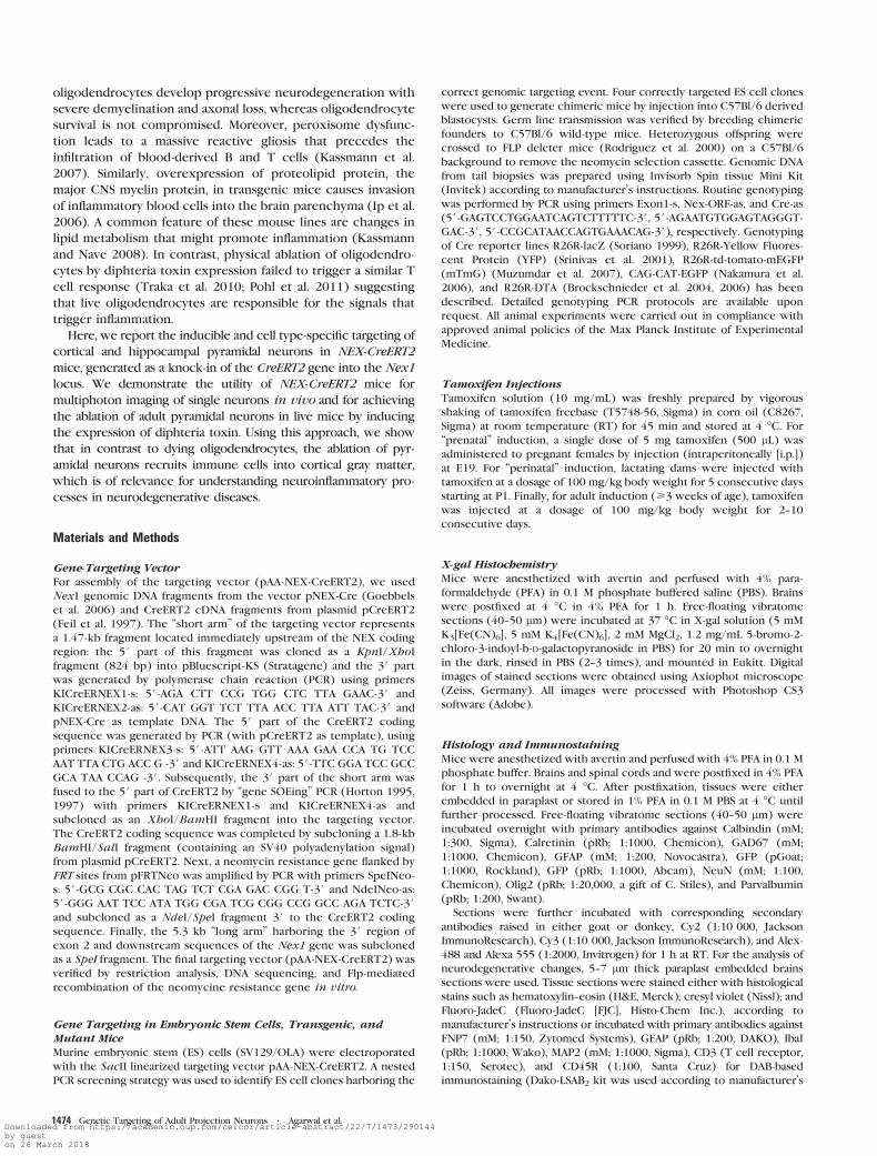
Figure 1. Strategy to ‘‘knock-in’’ CreERT2 into the mouse Nex1 gene. (A) 1) Genomic structure of the wild-type Nex1 allele. The locus comprises 2 exons (E1, E2; black boxes).The entire coding region (ORF; green box) is located on exon 2. 2) NEX-CreERT2 targeting vector. The construct harbors 5# and 3# homology arms (black), the CreERT2 cDNA (redbox) fused to the start codon of the Nex1 gene and a neomycin resistance cassette (NeoR; gray box) flanked by 2 FRT sites (blue triangles). 3) NEX-CreERT2 allele (NCER-Neo)after homologous recombination in murine ES cells. 4) NEX-CreERT2 allele after site-specific removal of the NeoR cassette (NCER) by breeding NCER-Neo mice to FLP deletermice. Arrows #1--5 indicate primer locations for PCR-based characterization of NCER-Neo and NCER mice. (B) FLP-mediated removal of the NeoR cassette is verified by PCRwith primers #4, 5 on genomic tail DNA from wt (no amplification), heterozygous NCER mice before (Neoþ; 1.6 kb), and after breeding to FLP deleter mice (Neo�; 400 bp).(C) PCR with primers #1--3 on genomic tail DNA from wild type (wt; 2.2 kb), NCER heterozygous (NCERþ/�; 2.2 and 2.8 kb), and NCER homozygous mice (NCER�/�; 2.8kb) confirms correct targeting of CreERT2 cDNA into the Nex1 locus. Note: primer #1 is located outside the targeting vector.
Cerebral Cortex July 2012, V 22 N 7 1475Downloaded from https://academic.oup.com/cercor/article-abstract/22/7/1473/290144by gueston 26 March 2018
ring close to the sagittal suture. The exposed cortex was covered by
a glass coverslip. Body temperature was kept constant (36--38 �C)throughout the experiment.
In vivo imaging was performed by a custom-made 2P-LSM equipped
with an fs-pulsed titanium-sapphire laser (Chameleon Ultra II, Co-
herent, Glasgow, UK) and a long-distance 203/1.0 NA water immersion
objective (Zeiss, Jena, Germany). For excitation, the laser was set at 925
± 5 nm. The fluorescence signal of Enhanced Green Fluorescent Protein
(EGFP)-positive pyramidal neurons was collected by a photomultiplier
tube (Hamamatsu, Japan) through a 510 ± 42 nm band-pass filter
(Semrock). Uniformly spaced (0.8--2 lm) planes of 125 3 125 to 500 3
500 lm2 regions of the cerebral cortex were recorded and processed
to obtain z-stacks of images (512 3 512 or 1024 3 1024 pixels in size).
Image processing was performed using Matlab (version 7, MathWorks,
Ismaning, Germany) and ImageJ (NIH; http://rsbweb.nih.gov/ij). Three-
dimensional visualization was performed as described before. For 3D
visualization of image stacks as volumes, Java-based ImageJ 3D Viewer
plugin developed by Benjamin Schmid (Biozentrum) was used.
Results
To achieve CreERT2 expression under control of regulatory
sequences from the endogenous Nex1 gene, we employed
a ‘‘knock-in’’ strategy based on homologous recombination in
mouse ES cells (Fig. 1). Using a targeting vector, in which the
NEX coding region in exon 2 was replaced by a CreERT2
expression cassette, we generated the mouse mutant NEX-
CreERT2-Neo that harbors the CreERT2 cassette adjacent to
a neomycin resistance (NeoR) gene driven by a thymidine
kinase promoter (placed in antisense orientation), as shown in
Figure 1A. Subsequently, we removed the NeoR gene in vivo by
breeding to FLP deleter mice (Rodriguez et al. 2000) and FLP-
mediated site-specific recombination (Fig. 1A). The resulting
mutant allele (in NEX-CreERT2 mice) carries a residual FLP site
3# to the CreERT2 cassette, thus providing a nearly ‘‘wild-type’’
genomic environment with respect to Nex1 gene expression.
Correct genomic targeting and removal of NeoR was verified by
a PCR-based analysis of genomic DNA (Fig. 1B,C and data not
shown).
Cre-Mediated Recombination in Mature PyramidalNeurons
For a detailed functional analysis of CreERT2 expression,
heterozygous mutants from mouse lines NEX-CreERT2 (in
subsequent text and figures referred to as NCER-Neo) and
NEX-CreERT2 (NCER) were crossbred with several transgenic
reporter lines that conditionally express b-galactosidase(Soriano 1999), YFP (Srinivas et al. 2001), membrane-tagged
EGFP (Muzumdar et al. 2007), and cytoplasmic EGFP
(Nakamura et al. 2006), following Cre-mediated deletion of
a floxed ‘‘stop-cassette.’’ At the age of 3--4 weeks, double
transgenic mice were injected i.p. with tamoxifen (100 mg/kg),
a treatment repeated daily for 10 consecutive days and analyzed
between 1 and 2 months after the last injection.
Overall, the recombination detected in young adult mice was
in the same subset of brain areas that we previously described
in NEX-Cre mice (Goebbels et al. 2006 and Table 1). By X-gal
histochemistry, serial coronal brain sections from NCER mice
with a LacZ reporter (a genotype termed NCER*R26R-floxLacZ
in the following) revealed prominent staining of the pyramidal
layer in the hippocampal CA1--3 region (Fig. 2C,D,H). Also the
majority of mossy cells in the hilar region (CA4) were labeled,
but no granule cells in the dentate gyrus (Fig. 2H). In the
neocortex, Cre-mediated recombination appeared restricted to
only a subset of pyramidal neurons, homogenously distributed
(Fig. 2A,B,G and Supplementary Fig. 1). When compared with
the neocortex, a modest increase in Cre recombination was
observed in the cingulate cortex, as well as the retrosplenial
granular and agranular cortices (Fig. 2A,B,C; Supplementary Fig.
1), which extend efferents into the anterior thalamic nuclei,
the lateral dorsal thalamic nucleus, and the hippocampal
anlage. This recombination pattern confirms a sparse NEX
mRNA expression in the adult mouse brain (Schwab et al. 1998;
see also Allen brain atlas, Supplementary Fig. 2; Table 1).
Cre recombination under control of the NEX promoter will
permanently mark transient NEX expression domains, such
that in conventional NEX-Cre mice (Goebbels et al. 2006), the
adult pattern of reporter gene expression most likely repre-
sents both transient and permanent NEX expression domains.
Applying a late recombination protocol (starting 3--4 weeks
after birth) to NCER mice, we identified transient NEX
promotor activity in neurons of the dentate gyrus, olfactory
bulb, thalamus, brain stem, and spinal cord (Fig. 2; Supplemen-
tary Fig. 1; and Table 1). In line with barely detectable
expression of NEX mRNA outside of neocortex and hippocam-
pus (e.g., in the cerebellum) (Schwab et al. 2000), cerebellar
granule cell labeling was minimal (Fig. 2E; Supplementary Fig.
1H; and Table 1). We also could not detect recombined
neurons in the absence of tamoxifen, thus demonstrating that
cytoplasmic CreERT2 retention is tight (data not shown).
Virtually identical results were obtained when we analyzed
mice from line NCER-Neo (Supplementary Fig. 3A) that still
harbor the neomycin resistance cassette (in antisense orienta-
tion) within the targeted Nex1 gene.
To further evaluate the identity of Cre recombinant cortical
cells in NCER mice, we utilized a second line of YFP reporter
mice (Srinivas et al. 2001) and induced CreERT2 activity by
Table 1Domains of Cre-mediated recombination in NEX-CreERT2/þ*R26R-LacZ mice (age 3 months) after tamoxifen induction at 4 weeks of age
Modest/complete recombination Scattered recombination (salt and pepper) No recombinationa
Hippocampus, CA1--3 Anterior olfactory nucleus Main olfactory bulbCingulate cortex Neocortex: layers II--VI ThalamusRetrosplenial agranular cortex Dentate gyrus (hilar cells in polymorphic layer) Dentate granule cellsb
Retrosplenial granular cortex Subiculum (including presubiculum and parasubiculum) HypothalamusInduseum griseum Tenia tecta Midbrain
Claustrum HindbrainPiriform cortex Medulla oblongataEntorhinal cortex PonsAmygdala Dorsal horn of spinal cordCerebellar granular cells
aUp to 10 recombined cells per brain slice in these structures.bPerinatal tamoxifen treatment leads to scattered recombination in dentate granule cells.
Genetic Targeting of Adult Projection Neurons d Agarwal et al.1476Downloaded from https://academic.oup.com/cercor/article-abstract/22/7/1473/290144by gueston 26 March 2018

injecting tamoxifen for 10 days in 4- to 5-week-old mice (as
described above). We then used immunolabeling at the age of
3--4 months and combined a fluorescent reporter (YFP) of Cre
activity with various cell type-specific neural markers. When
analyzed by confocal microscopy, all recombinant cells could
be identified as large NeuN-positive projection neurons (Fig.
3A--C; Supplementary Fig. 4A--C ). Neither interneurons nor
astrocytes or oligodendrocytes were recombined in
NCER*R26R-floxYFP mice, when using calbindin, calretinin,
parvalbumin, GAD67, GFAP, and Olig2 as respective cell type-
specific markers (Fig. 3D--F and Supplementary Fig. 4D--F ),
confirming that in the forebrain Cre recombination is restricted
to pyramidal neurons.
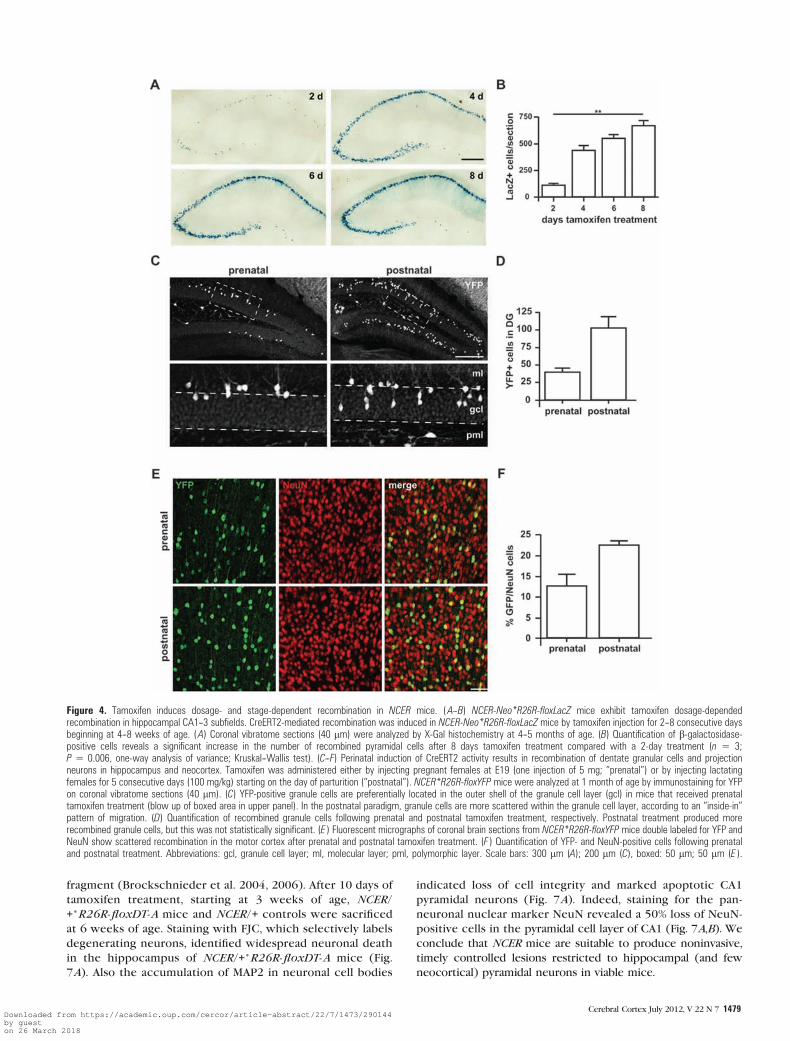
Tamoxifen Dosage-Dependent Gene Recombination
We next explored various protocols of tamoxifen administra-
tion at distinct developmental and adult stages. First, we
determined the impact of treatment duration (from 1 to 8
consecutive daily i.p. injections, starting at 3--4 weeks of age)
on the number of recombined cells in the hippocampus (Fig.
4A,B) and neocortex (Supplementary Fig. 5). While a single
tamoxifen injection was ineffective in inducing recombination
anywhere in the brain (data not shown), the number of
recombined cells gradually increased from 2 to 8 days of
treatment, both in hippocampus (Fig. 4A,B) and in neocortex
(Supplementary Fig. 5). Importantly, treatment between 2 and
5 days produced a quantity of recombined hippocampal
neurons that is suitable for single-cell analysis (Fig. 4A).
Tamoxifen treatment in 5- to 6-month-old mice produced
similar results (data not shown).
Tamoxifen administration at prenatal stages can induce
developmental defects and premature abortion (Jordan and
Murphy 1990; Danielian et al. 1998). Therefore, we tested the
efficacy of 2 ‘‘mild’’ application protocols, either a single
tamoxifen injection (5 mg) to the female at E19 during late
pregnancy (prenatal) or daily i.p. injections (100 mg/kg body
weight) to the female for 5 consecutive days starting
immediately after delivery (‘‘postnatal’’), such that pups receive
tamoxifen through the milk. Immunostaining for YFP at
postnatal day (P) 28 revealed that both administration
paradigms produced significant quantities of recombined cells
in neocortex (Fig. 4E,F) and the hippocampal CA1--3 region
(Supplementary Fig. 6A) of NCER*R26R-floxYFP mice. Both
paradigms also induced recombination in dentate granule cells
(Fig. 4C,D) providing further evidence that CreERT2 expres-
sion faithfully recapitulates transient NEX promoter activity in
dentate granule cells. As expected, the postnatal administration
protocol produced more recombined cells in dentate gyrus
(Fig. 4D) and cortex (Fig. 4F) when compared with the
prenatal paradigm. Longer duration of tamoxifen treatment in
the postnatal protocol is also reflected by the broader radial
distribution of recombined granular cells that settle in the
granule cell layer according an inside-in order (Fig. 4C). As for
adult treatment, recombination induced by perinatal tamoxifen
Figure 2. Inducible CreERT2-mediated recombination in adult brains of NCER mice. NCER*R26R-floxLacZ double transgenic mice at 3--4 weeks of age were injected withtamoxifen (100 mg/kg body weight) for 10 consecutive days. CreERT2-mediated recombination was analyzed by X-gal histochemistry on coronal vibratome sections (40 lm) at2--3 months of age. (A--F ) Overviews reveal b-galactosidase expression in neocortex, induseum griseum, and hippocampus, whereas only few cerebellar granular cells and nospinal cord neurons were recombined. (G) Blow up of boxed area in A. Scattered recombination in layers II--VI of motor cortex. (H) Virtually all pyramidal cells of the hippocampalCA1--3 region express b-galactosidase. While recombination is completely absent from dentate granule cells (arrows), a subset of cells in the dentate hilar region, most likelymossy cells, express b-galactosidase (inset). Abbreviations: cx, neocortex; DG, dentate gyrus; gcl, granule cell layer; hip, hippocampus; pml, polymorphic layer; I--VI, corticallayers. Scale bars: 500 lm (A--E ); 250 lm (F--H).
Cerebral Cortex July 2012, V 22 N 7 1477Downloaded from https://academic.oup.com/cercor/article-abstract/22/7/1473/290144by gueston 26 March 2018

administration was completely absent from interneurons
(Supplementary Fig. 6B,C) and glia cells (data not shown). In
general, we did not observe toxic effects of tamoxifen on the
pregnant (late stage) as well as lactating dams. However,
perinatal tamoxifen treatment caused a marked reduction in
body size (30--50%) of the offspring at 4 weeks of age. These
mice displayed improper gait, most likely as a result of ongoing
muscle degeneration in their hind limbs.
Single-Cell Imaging in Live Mice
NCER mice exhibit scattered recombination in a subset of
cortical projection neurons. To further evaluate these mice as
a suitable tool for ‘‘single-cell genetics,’’ we bred NCER mice to
a reporter mouse line that conditionally expresses a cytoplasmic
variant of EGFP (Nakamura et al. 2006). At 3--4 weeks of age,
NCER*CAG-CAT-EGFP mice were injected with tamoxifen for 2
or 10 consecutive days, and brains were analyzed 3--4 weeks later
by confocal microscopy. The 3D volumes of EGFP-positive cells
in different brain regions were calculated from confocal z-stacks
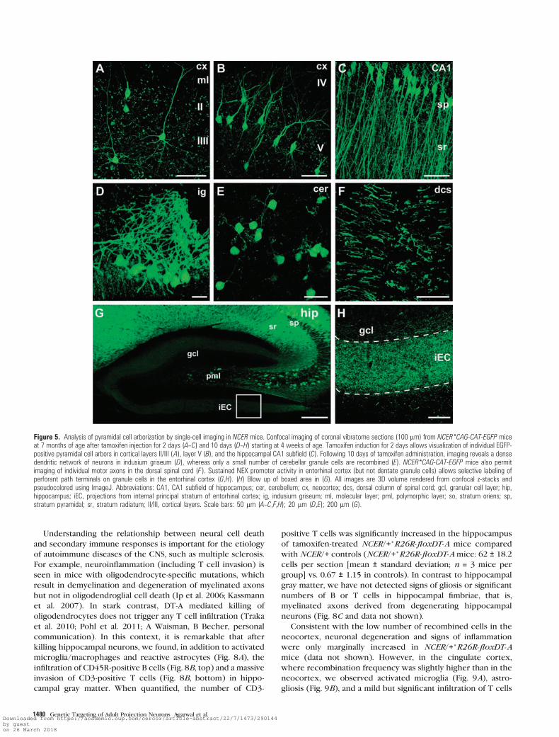
acquired from sections of 50--100 lm thickness (Fig. 5). Analysis
of mice that were tamoxifen injected for only 2 days revealed
a Golgi-like staining of pyramidal neurons that allowed the
tracing of single dendrites and axons in layers II/III and V of the
neocortex and in the CA1 region of the hippocampus (Fig. 5A--C;
Supplementary Video 1--3). In mice induced for 10 days, we were
able to reconstitute the complete arborization of neurons in the
indusium griseum (Fig. 5D) and cerebellar granule cells (Fig. 5E ).
As recombination was completely absent from the spinal cord,
single motor axons of the cortical spinal tract could be imaged at
the thoracal level (Fig. 5F; Supplementary Video 4). Similarly, in
the absence of recombination in dentate granule cells, we were
able to image terminals of the perforant path that innervates the
granule cell layer (Fig. 5G,H). Finally, by breeding NCER*R26R-
floxYFP mice to GFAP-CreERT2 mice, which express CreERT2 in
cortical astrocytes (Hirrlinger et al. 2006), we demonstrate the
feasibility of this approach to image interactions between
different cortical cell types (Supplementary Fig. 7).
The fluorescent labeling of single neurons in a ‘‘Golgi-like’’
fashion also permits the imaging of neuronal dynamics in
living mice (Kasthuri and Lichtman 2007). As proof of
principle for such a single-cell genetics approach, we imaged
the arborization of individual pyramidal cells in the cortex of
NCER*CAG-CAT-EGFP mice by in vivo multiphoton imaging
(Fig. 6A,B). After 2-day induction, we captured the complete
arborization of individual pyramidal neurons in layers II/III of
the motor cortex (Fig. 6A). Tamoxifen administration for 10
days allowed in vivo imaging of pyramidal neurons along the
entire width of the motor cortex (Fig. 6B; Supplementary
Video 5).
Nonnvasive Ablation of Pyramidal Neurons Triggers TCell Infiltration
Most animal models of neurodegeneration rely on invasive
interventions, are prone to inflammatory side effects, and do
not allow targeting of specific neural cell types. We therefore
took advantage of NCER mice to specifically ablate pyramidal
neurons in the adult brain by breeding to a mouse line that
conditionally expresses the diphtheria toxin-A (DT-A)
Figure 3. Nex promoter activity in neocortex is restricted to principal neurons. Confocal images of fluorescent immunostainings for a CreERT2-induced YFP reporter and the pan-neuronal marker NeuN (A--C) or neural cell type-specific markers (D--F) on coronal vibratome sections (40 lm) from 2- to 3-month-old NCER*R26R-floxYFP mice after tamoxifeninjections for 10 consecutive days starting at 3--4 weeks of age. (A) Individual YFP-positive neuron (arrowhead) in layers II/III of motor cortex. (B) Mossy cells in the polymorphiclayer (pml) of the hilar region express YFP (arrowheads), whereas dentate granule cells (gcl) completely lack NEX promoter activity at this age. (C) In CA1, NeuN-positive cells inthe pyramidal cell layer (sp) coexpress YFP, the remaining NeuN-positive cells are mainly interneurons. (D--F) NEX promoter activity is completely absent from interneurons andglial cells as demonstrated by immunostaining for YFP and antibodies directed against pan-interneuronal marker GAD67 (D), Olig2 (oligodendrocytes; E ) and GFAP (astrocytes, F ).All images are 3D volume rendered from confocal z-stacks and pseudocolored using ImageJ. Abbreviations: gcl, granule cell layer; ml, molecular layer; pml, polymorphic layer; so,stratum oriens; sp, stratum pyramidale; sr, stratum radiatum; II/III, cortical layers. Scale bars: 50 lm (A--C); 20 lm (D--F).
Genetic Targeting of Adult Projection Neurons d Agarwal et al.1478Downloaded from https://academic.oup.com/cercor/article-abstract/22/7/1473/290144by gueston 26 March 2018
fragment (Brockschnieder et al. 2004, 2006). After 10 days of
tamoxifen treatment, starting at 3 weeks of age, NCER/
+*R26R-floxDT-A mice and NCER/+ controls were sacrificed
at 6 weeks of age. Staining with FJC, which selectively labels
degenerating neurons, identified widespread neuronal death
in the hippocampus of NCER/+*R26R-floxDT-A mice (Fig.
7A). Also the accumulation of MAP2 in neuronal cell bodies
indicated loss of cell integrity and marked apoptotic CA1
pyramidal neurons (Fig. 7A). Indeed, staining for the pan-
neuronal nuclear marker NeuN revealed a 50% loss of NeuN-
positive cells in the pyramidal cell layer of CA1 (Fig. 7A,B). We
conclude that NCER mice are suitable to produce noninvasive,
timely controlled lesions restricted to hippocampal (and few
neocortical) pyramidal neurons in viable mice.
Figure 4. Tamoxifen induces dosage- and stage-dependent recombination in NCER mice. (A--B) NCER-Neo*R26R-floxLacZ mice exhibit tamoxifen dosage-dependedrecombination in hippocampal CA1--3 subfields. CreERT2-mediated recombination was induced in NCER-Neo*R26R-floxLacZ mice by tamoxifen injection for 2--8 consecutive daysbeginning at 4--8 weeks of age. (A) Coronal vibratome sections (40 lm) were analyzed by X-Gal histochemistry at 4--5 months of age. (B) Quantification of b-galactosidase-positive cells reveals a significant increase in the number of recombined pyramidal cells after 8 days tamoxifen treatment compared with a 2-day treatment (n 5 3;P 5 0.006, one-way analysis of variance; Kruskal--Wallis test). (C--F) Perinatal induction of CreERT2 activity results in recombination of dentate granular cells and projectionneurons in hippocampus and neocortex. Tamoxifen was administered either by injecting pregnant females at E19 (one injection of 5 mg; ‘‘prenatal’’) or by injecting lactatingfemales for 5 consecutive days (100 mg/kg) starting on the day of parturition (‘‘postnatal’’). NCER*R26R-floxYFP mice were analyzed at 1 month of age by immunostaining for YFPon coronal vibratome sections (40 lm). (C) YFP-positive granule cells are preferentially located in the outer shell of the granule cell layer (gcl) in mice that received prenataltamoxifen treatment (blow up of boxed area in upper panel). In the postnatal paradigm, granule cells are more scattered within the granule cell layer, according to an ‘‘inside-in’’pattern of migration. (D) Quantification of recombined granule cells following prenatal and postnatal tamoxifen treatment, respectively. Postnatal treatment produced morerecombined granule cells, but this was not statistically significant. (E ) Fluorescent micrographs of coronal brain sections from NCER*R26R-floxYFP mice double labeled for YFP andNeuN show scattered recombination in the motor cortex after prenatal and postnatal tamoxifen treatment. (F ) Quantification of YFP- and NeuN-positive cells following prenataland postnatal treatment. Abbreviations: gcl, granule cell layer; ml, molecular layer; pml, polymorphic layer. Scale bars: 300 lm (A); 200 lm (C), boxed: 50 lm; 50 lm (E ).
Cerebral Cortex July 2012, V 22 N 7 1479Downloaded from https://academic.oup.com/cercor/article-abstract/22/7/1473/290144by gueston 26 March 2018
Understanding the relationship between neural cell death
and secondary immune responses is important for the etiology
of autoimmune diseases of the CNS, such as multiple sclerosis.
For example, neuroinflammation (including T cell invasion) is
seen in mice with oligodendrocyte-specific mutations, which
result in demyelination and degeneration of myelinated axons
but not in oligodendroglial cell death (Ip et al. 2006; Kassmann
et al. 2007). In stark contrast, DT-A mediated killing of
oligodendrocytes does not trigger any T cell infiltration (Traka
et al. 2010; Pohl et al. 2011; A Waisman, B Becher, personal
communication). In this context, it is remarkable that after
killing hippocampal neurons, we found, in addition to activated
microglia/macrophages and reactive astrocytes (Fig. 8A), the
infiltration of CD45R-positive B cells (Fig. 8B, top) and a massive
invasion of CD3-positive T cells (Fig. 8B, bottom) in hippo-
campal gray matter. When quantified, the number of CD3-
positive T cells was significantly increased in the hippocampus
of tamoxifen-treated NCER/+*R26R-floxDT-A mice compared
with NCER/+ controls (NCER/+*R26R-floxDT-A mice: 62 ± 18.2
cells per section [mean ± standard deviation; n = 3 mice per
group] vs. 0.67 ± 1.15 in controls). In contrast to hippocampal
gray matter, we have not detected signs of gliosis or significant
numbers of B or T cells in hippocampal fimbriae, that is,
myelinated axons derived from degenerating hippocampal
neurons (Fig. 8C and data not shown).
Consistent with the low number of recombined cells in the
neocortex, neuronal degeneration and signs of inflammation
were only marginally increased in NCER/+*R26R-floxDT-Amice (data not shown). However, in the cingulate cortex,
where recombination frequency was slightly higher than in the
neocortex, we observed activated microglia (Fig. 9A), astro-
gliosis (Fig. 9B), and a mild but significant infiltration of T cells
Figure 5. Analysis of pyramidal cell arborization by single-cell imaging in NCER mice. Confocal imaging of coronal vibratome sections (100 lm) from NCER*CAG-CAT-EGFP miceat 7 months of age after tamoxifen injection for 2 days (A--C) and 10 days (D--H) starting at 4 weeks of age. Tamoxifen induction for 2 days allows visualization of individual EGFP-positive pyramidal cell arbors in cortical layers II/III (A), layer V (B), and the hippocampal CA1 subfield (C). Following 10 days of tamoxifen administration, imaging reveals a densedendritic network of neurons in indusium griseum (D), whereas only a small number of cerebellar granule cells are recombined (E). NCER*CAG-CAT-EGFP mice also permitimaging of individual motor axons in the dorsal spinal cord (F ). Sustained NEX promoter activity in entorhinal cortex (but not dentate granule cells) allows selective labeling ofperforant path terminals on granule cells in the entorhinal cortex (G,H). (H) Blow up of boxed area in (G). All images are 3D volume rendered from confocal z-stacks andpseudocolored using ImageJ. Abbreviations: CA1, CA1 subfield of hippocampus; cer, cerebellum; cx, neocortex; dcs, dorsal column of spinal cord; gcl, granular cell layer; hip,hippocampus; iEC, projections from internal principal stratum of entorhinal cortex; ig, indusium griseum; ml, molecular layer; pml, polymorphic layer; so, stratum oriens; sp,stratum pyramidal; sr, stratum radiatum; II/III, cortical layers. Scale bars: 50 lm (A--C,F,H); 20 lm (D,E); 200 lm (G).
Genetic Targeting of Adult Projection Neurons Agarwal et al.d1480Downloaded from https://academic.oup.com/cercor/article-abstract/22/7/1473/290144by gueston 26 March 2018

(Fig. 9C). We conclude that a minimal number of degenerating
neurons are required to induce T cell invasion into gray matter
areas. Whether these cells also contribute to the overall pattern
of neurodegeneration awaits the generation of T cell-deficient
double mutant mice.
Discussion
We have described a novel knock-in mouse line that expresses
CreERT2 under control of regulatory sequences of the Nex1
gene. Due to the very restricted expression pattern, NEX-
CreERT2 mice allow inducible, highly selective genetic
manipulations of neocortical and hippocampal projection
neurons in the adult brain of viable mice.
Cre-Mediated Recombination in Mature PyramidalNeurons
Tamoxifen-induced reporter expression in the adult brain of
NEX-CreERT2 mice revealed expression of the Nex1 locus in
a subset of brain areas that we previously defined in
conventional NEX-Cre mice (Goebbels et al. 2006). The adult
pattern of Nex1 promoter activity faithfully reproduces NEX
mRNA expression (Schwab et al. 1998; see also Allan brain atlas
at http://www.brain-map.org). Importantly, CreERT2-mediated
recombination is completely absent from interneurons, oligo-
dendrocytes, and astrocytes as well as non-neural cells. Adult
tamoxifen administration also allowed us to distinguish brain
regions with sustained NEX expression from those with
transient NEX promoter activity, such as spinal cord and deep
cerebellar nuclei. We also confirm a transient NEX expression
in dentate granule cells (Schwab et al. 2000; Goebbels et al.
2006) that ceases by 3 weeks of age. Transient promoter
activity indicates a role for NEX during the initial differentiation
of these brain regions but not for their maintenance in the
mature brain. Areas with sustained Nex1 promoter activity
include the hippocampal CA1--3 region and dentate hilus,
induseum griseum, and the cingulate cortex (reviewed in Table
1). While virtually all projection neurons express NEX during
cortical development (Goebbels et al. 2006), only a small subset
of projection neurons in cortical layers II--V maintain Nex1
promoter activity in the adult brain.
NEX null mutants display impaired spatial learning (O Ucar
and M.H.S., in preparation) and NEX cooperates with NeuroD2,
a closely related neuronal bHLH protein (Kume et al. 1996;
Yasunami et al. 1996), to promote long-range axogenesis
Figure 6. In vivo 2-photon imaging of individual pyramidal neurons in NCER mice.2-photon imaging of neocortical projection neurons was carried out through a cranialwindow in the motor cortex sealed with a cover glass. Fluorescent image stacks wererecorded in NCER*CAG-CAT-EGFP mice at 7--8 weeks of age following tamoxifeninduction (100 mg/kg body weight) for 2 (A) and 10 days (B) starting at 3 weeks ofage. Data were acquired with a pumped titanium:sapphire oscillator using a 203water immersion lens with NA 1.0 (Zeiss). Note different scales of imaging depthin A and B. Tamoxifen induction for 10 days permits imaging of projection neuronsthroughout the entire depth of the neocortex (~800 lm). Dimensions of corticallayers (L) are indicated. Images are presented as a 3D volume rendered fromfluorescent image stacks. Scale bars: A,B: 50 lm (horizontal); 100 lm (vertical).
Figure 7. Selective ablation of adult hippocampal pyramidal neurons by NCER-mediated diphteria toxin expression. (A) Expression of DT-A in NCER/þ*R26R-floxDT-A micecauses widespread death of hippocampal pyramidal neurons (arrowheads) as demonstrated by FJC staining (left panels). Accumulation of the dendritic marker Map2 in neuronalcell bodies (arrowheads; middle panels) supports neuronal degeneration. Immunostaining for NeuN reveals thinning of the pyramidal cell layer and loss of pyramidal cell somata inNCER/þ*R26R-floxDT-A mice (right panels). Heterozygous NCER controls (NCER/þ) and mice additionally harboring the diptheria toxin transgene (NCER/þ*R26R-floxDT-A) wereinjected with tamoxifen (100 mg/kg body weight) for 10 days starting at 3 weeks of age and analyzed at 7 weeks of age. (B) When quantified, the number of NeuN-positive cellsin the CA1 subfield of NCER/þ*R26R-floxDT-A mice is significantly reduced by 50% when compared with controls (n 5 3, P\ 0.001, unpaired, 2 tailed t-test with Welch’scorrection). Scale bars: 20 lm (FJC); 50 lm (Map2, NeuN).
Cerebral Cortex July 2012, V 22 N 7 1481Downloaded from https://academic.oup.com/cercor/article-abstract/22/7/1473/290144by gueston 26 March 2018
Figure 8. Genetic ablation of hippocampal projection neurons induces reactive gliosis and lymphocyte infiltration. (A) Pyramidal cell death in the hippocampal CA1--3 region oftamoxifen-treated NCER/þ*R26R-floxDT-A mice is associated with severe microgliosis and astrogliosis as demonstrated by immunostaining for microglia (Iba1, upper panels) andastrocytes (GFAP; lower panels). NCER/þ controls and NCER*R26R-floxDT-A mice were injected with tamoxifen for 10 days starting at 4 weeks of age and harvested at 6.5weeks of age. (B) Lymphocyte infiltration into the hippocampal region of mutant brains shown by immunostaining for B cell-specific phosphatase CD45R (upper panels; magnifiedin inset) and T cell receptor CD3 (arrows in lower right; magnified in inset). B cells were never observed in brains of control mice. (C) Immunostaining for CD3 demonstratesabsence of significant T cell infiltration in hippocampal fimbriae (i.e., myelinated axons derived from degenerating hippocampal neurons) of tamoxifen-treated NCER/þ*R26R-floxDT-A mice. Arrowhead marks rare example of a T cell infiltrate. Abbreviations: LV, lateral ventricle. Scale bars: 200 lm (A,B); 20 lm (insets); 100 lm (C).
Genetic Targeting of Adult Projection Neurons d Agarwal et al.1482Downloaded from https://academic.oup.com/cercor/article-abstract/22/7/1473/290144by gueston 26 March 2018

(Bormuth and M.H.S. unpublished data). These functions
suggest that NEX-positive projection neurons are recruited
for distinct plasticity processes in the adult brain, and we
speculate that external stimuli dynamically regulate the
compartment of NEX-positive neurons in the neocortex. This
hypothesis can be further addressed in NEX-CreERT2 null
mutants by combining environmental enrichment paradigms
with single-cell trancriptomics, in vivo multiphoton imaging,
and patch clamp recording.
Classical methods to study individual mammalian neurons,
such as dye filling or viral infection, are invasive interventions
(Dittgen et al. 2004; Young and Feng 2004). Therefore,
strategies for the noninvasive genetic labeling of single neurons
have been developed. For example, mosaic analysis with double
markers (MADM) makes use of Cre-dependent interchromo-
somal mitotic recombination to produce small numbers of
targeted cells, however, this method is not applicable to
postmitotic neurons of the adult brain (Zong et al. 2005). In
‘‘SLICK’’ mice, CreERT2 and EYFP are coexpressed from the
same Thy1.2 promoter (Young et al. 2008). Thus, EYFP
expression and CreERT2-mediated recombination are not
strictly coupled. Sparse cell labeling was achieved by the
insertion of CreERT2 coupled to an internal ribosomal entry
site into the 3# region of neurally expressed genes, at least
when combined with a reporter locus of low recombination
efficiency (Rotolo et al. 2008). Placing CreERT2 under control
of regulatory sequences from the CaMKIIa gene results in
highly efficient recombination in forebrain projection neurons
(Erdmann et al. 2007), but if sparse recombination can be
achieved was not addressed.
Due to the temporal and spatial expression pattern of the
Nex1 locus, NEX-CreERT2 mice present a unique tool to
perform single-cell genetics in individual cortical projection
neurons. Limiting tamoxifen treatment to 2 days allows
visualization of complex neuronal arbors, for example, by
breeding to a human placental alkaline phosphatase reporter
(Lobe et al. 1999). Short-term tamoxifen treatment also
produces sparse recombination in CA1--3 pyramidal cells,
which can be tested singly as well. Furthermore, NEX-CreERT2
mice provide the opportunity to address reciprocal interac-
tions between individual projection neurons and other neural
cells by breeding to novel CreERT2-dependent fluorescent
reporter mice (Madisen et al. 2010) and a variety of other
transgenic mouse lines, such as BAC transgenic lines from the
GENSAT collection (Gong et al. 2003), lines that express
calcium, chloride, and zinc sensors (Kuner and Augustine 2000;
Griesbeck 2004; Vinkenborg et al. 2009) or the transsynaptic
marker wheat germ agglutinin (Yoshihara et al. 1999).
NEX-CreERT2 mice are also unique with respect to CreERT2
expression in cells of the hippocampal hilar region (most likely
glutamatergic mossy cells; Henze and Buzsaki 2007) and
indusium griseum, a dorsal portion of the hippocampus, which
Figure 9. Gliosis and mild T cell infiltration in the cingulate cortex of tamoxifen-treated NCER/þ*R26R-floxDT-A mice. (A) Immunostaining for Iba1 demonstrates activatedmicroglia as a consequence of DT-A-induced death of pyramidal neurons in the cingulate cortex of tamoxifen-treated NCER/þ*R26R-floxDT-A mice. (B) Accumulation of reactiveastroglia in the cingulate cortex of NCER/þ*R26R-floxDT-A mice revealed by immunostaining for GFAP. (C) Occasional T cell infiltration in the cingulate cortex of NCER/þ*R26R-floxDT-A mice as demonstrated by immunostaining for CD3. Note that CD3þ T cells were not observed in the cingulate cortex of tamoxifen-treated NCER/þ controls.Abbreviations: ml, molecular layer; pcl, pyramidal cell layer. Scale bars: 50 lm.
Cerebral Cortex July 2012, V 22 N 7 1483Downloaded from https://academic.oup.com/cercor/article-abstract/22/7/1473/290144by gueston 26 March 2018
contains granule cells, pyramidal cells, and associated white
matter (Wyss and Sripanidkulchai 1983; Adamek et al. 1984).
Mossy cells and hilar interneurons are connected through
reciprocal local circuit networks (Larimer and Strowbridge
2008), and computational modeling suggests a role for hilar cells
in pattern separation in the dentate gyrus (Myers and Scharfman
2009), but little is known about the functions of these cells in
vivo. As adult-born granule cells establish new glutamatergic
synapses with hilar interneurons and mossy cells (Toni et al.
2008), genetic manipulation of mossy cells in NEX-CreERT2
mice provides a novel means to study synaptogenesis in the
adult brain. Similarly, the exact function of the indusium griseum
has not been addressed in vivo. The indusium griseum has been
shown to be resistant to neuronal cell loss during aging
(Sturrock 1986) and histopathologic hallmarks of Alzheimer’s
disease are largely absent from the indusium griseum in
postmortem brains of patients suffering from Alzheimer’s disease
(Lippa et al. 1990; Lippa and Smith 1992). Thus, NEX-CreERT2
mice provide the opportunity to study molecular mechanisms of
neuronal survival under disease conditions.
A Novel Mouse Model of Adult Neurodegeneration
Most animal models of neurodegeneration rely on invasive
interventions, for example, brain matter aspiration, mechanical
disruption of the blood-brain barrier or stereotactic injections
of toxins (Jarrard 2002; Siren et al. 2006; Tseng et al. 2009).
These techniques result in widespread damage, are prone to
inflammatory side effects, and cannot target specific cell types.
Although neurodegenerative diseases can produce massive
degeneration, they typically affect selective neuronal popula-
tions. For example, in brains of Alzheimer’s disease patients,
projection neurons in hippocampus and entorhinal cortex are
vulnerable to degeneration, whereas interneurons are largely
spared (Morrison and Hof 1997). To mimic some of these
features, we ablated pyramidal neurons by breeding NEX-
CreERT2 mice to transgenic mice that conditionally express
the DT-A fragment (Brockschnieder et al. 2004, 2006), which
kills cells by inhibiting protein synthesis (Collier 1975;
Pappenheimer 1977). By tamoxifen treatment of adult NCER/
+*R26R-floxDT-A double transgenic mice, we were able to
specifically ablate hippocampal pyramidal neurons (and in-
dividual projection neurons in the neocortex) without
affecting interneurons or glial cells. As tamoxifen treatment is
compatible with behavioral phenotyping (Vogt et al. 2008),
such an experimental paradigm will also allow to evaluate the
role of these cells in cognitive functions before and after
pyramidal neuron-restricted toxin expression.
Noninvasive Ablation of Neurons Causes T Cell Invasion
An unexpected finding, following the ablation of hippocampal
neurons by tamoxifen-induced DT-A expression, was the
invasion of T cell receptor (CD3)-positive lymphocytes into
the brain parenchyma. We note that 2 conceptually related
studies, reporting the ablation of oligodendrocytes by expres-
sion of the same toxin, failed to trigger any T cell response,
despite widespread oligodendrocyte death (Traka et al. 2010;
Pohl et al. 2011), even following additional proinflammatory
manipulations (A Waisman, B Becher, personal communica-
tion). These negative findings contrast with the infiltration of
CD8+ T cells in mouse mutants carrying defects that perturb
but do not kill oligodendrocytes (Ip et al. 2006; Kassmann et al.
2007). This suggests that T cell infiltration is triggered by
injured and live (but not dead) oligodendrocytes. One possible
explanation for the massive lymphocyte invasion in our study is
a higher level of inflammatory signals originating from ablated
neurons compared with ablated oligodendrocytes. Alterna-
tively, gray matter areas might lack anti-inflammatory mecha-
nisms that protect white matter from immune cell invasion.
The latter could be relevant for diseases, such as multiple
sclerosis, in which the protection from immune invasion fails.
We cannot exclude the theoretical possibility that neuronal
ablation causes an unspecific opening of the blood-brain
barrier. However, the very restricted localization of blood-
derived lymphocytes to sites of highest recombination fre-
quency argues against a general breakdown of blood-brain
barrier functions.
In conclusion, NEX-CreERT2 mice will be a valuable tool for
studying genetic functions in mature cortical and hippocampal
neurons in vivo, for neuroimaging at the single-cell level, and for
ablating single cortical and hippocampal neurons to study the
consequences of acute neurodegeneration in the adult brain.
Supplementary Material
Supplementary material can be found at: http://www.cercor.
oxfordjournals.org/
Funding
A.A. is supported by a Postdoctoral Fellowship from the
National Multiple Sclerosis Society. M.H.S. and K.A.N. acknowl-
edge grant support from the Deutsche Forschungsgemein-
schaft (DFG Research Center Molecular Physiology of the
Brain, CMPB). K.A.N. holds an ERC Advanced Investigator Grant.
Notes
We thank U. Bode for help with ES cell culture, M. Schindler for
blastocyst injections, and C. Casper, D. Flemming, and I. Malade for help
with animal husbandry. We also thank A. Fahrenholz and M. Floerl for
help with histology. We like to thank D. Riethmacher for providing
floxDT-A mice, J. Robbins for CAG-CAT*EGFP mice, and F. Kirchhoff for
GFAP-CreERT2 mice. We thank I. Bormuth, A. Saab, and members of the
Department of Neurogenetics for helpful discussions. Authors’ contri-
bution: A.A., M.H.S., and K.A.N. designed the study, drafted, and wrote
the manuscript. A.A. cloned gene-targeting constructs, generated NEX-
CreERT2 mice, and planned all the experiments. A.A. carried out
histology, imaging, and noninvasive cell ablation experiments as well as
analyzed data. P.D. helped with in vivo multiphoton recordings. C.M.K.
contributed in histological staining. S.G. provided Nex1 gene fragments
to clone targeting construct. All authors read and approved the final
manuscript. Conflict of Interest : None declared.
References
Adamek GD, Shipley MT, Sanders MS. 1984. The indusium griseum in
the mouse: architecture, Timm’s histochemistry and some afferent
connections. Brain Res Bull. 12:657--668.
Bartholoma A, Nave KA. 1994. NEX-1: a novel brain-specific helix-loop-
helix protein with autoregulation and sustained expression in
mature cortical neurons. Mech Dev. 48:217--228.
Brockschnieder D, Lappe-Siefke C, Goebbels S, Boesl MR, Nave KA,
Riethmacher D. 2004. Cell depletion due to diphtheria toxin
fragment A after Cre-mediated recombination. Mol Cell Biol.
24:7636--7642.
Brockschnieder D, Pechmann Y, Sonnenberg-Riethmacher E,
Riethmacher D. 2006. An improved mouse line for Cre-induced
cell ablation due to diphtheria toxin A, expressed from the Rosa26
locus. Genesis. 44:322--327.
Genetic Targeting of Adult Projection Neurons d Agarwal et al.1484Downloaded from https://academic.oup.com/cercor/article-abstract/22/7/1473/290144by gueston 26 March 2018
Burns KA, Ayoub AE, Breunig JJ, Adhami F, Weng WL, Colbert MC,
Rakic P, Kuan CY. 2007. Nestin-CreER mice reveal DNA synthesis by
nonapoptotic neurons following cerebral ischemia hypoxia. Cereb
Cortex. 17:2585--2592.
Campsall KD, Mazerolle CJ, De Repentingy Y, Kothary R, Wallace VA.
2002. Characterization of transgene expression and Cre recombi-
nase activity in a panel of Thy-1 promoter-Cre transgenic mice. Dev
Dyn. 224:135--143.
Collier RJ. 1975. Diphtheria toxin: mode of action and structure.
Bacteriol Rev. 39:54--85.
Danielian PS, Muccino D, Rowitch DH, Michael SK, McMahon AP. 1998.
Modification of gene activity in mouse embryos in utero by
a tamoxifen-inducible form of Cre recombinase. Curr Biol.
8:1323--1326.
DeFelipe J, Farinas I. 1992. The pyramidal neuron of the cerebral
cortex: morphological and chemical characteristics of the synaptic
inputs. Prog Neurobiol. 39:563--607.
Dittgen T, Nimmerjahn A, Komai S, Licznerski P, Waters J, Margrie TW,
Helmchen F, Denk W, Brecht M, Osten P. 2004. Lentivirus-based
genetic manipulations of cortical neurons and their optical and
electrophysiological monitoring in vivo. Proc Natl Acad Sci U S A.
101:18206--18211.
Erdmann G, Schutz G, Berger S. 2007. Inducible gene inactivation in
neurons of the adult mouse forebrain. BMC Neurosci. 8:63.
Erdmann G, Schutz G, Berger S. 2008. Loss of glucocorticoid receptor
function in the pituitary results in early postnatal lethality.
Endocrinology. 149:3446--3451.
Feil R, Wagner J, Metzger D, Chambon P. 1997. Regulation of Cre
recombinase activity by mutated estrogen receptor ligand-binding
domains. Biochem Biophys Res Commun. 237:752--757.
Goebbels S, Bormuth I, Bode U, Hermanson O, Schwab MH, Nave KA.
2006. Genetic targeting of principal neurons in neocortex and
hippocampus of NEX-Cre mice. Genesis. 44:611--621.
Gong S, Zheng C, Doughty ML, Losos K, Didkovsky N, Schambra UB,
Nowak NJ, Joyner A, Leblanc G, Hatten ME, et al. 2003. A gene
expression atlas of the central nervous system based on bacterial
artificial chromosomes. Nature. 425:917--925.
Gorski JA, Talley T, Qiu M, Puelles L, Rubenstein JL, Jones KR. 2002.
Cortical excitatory neurons and glia, but not GABAergic neurons,
are produced in the Emx1-expressing lineage. J Neurosci.
22:6309--6314.
Griesbeck O. 2004. Fluorescent proteins as sensors for cellular
functions. Curr Opin Neurobiol. 14:636--641.
Griffiths I, Klugmann M, Anderson T, Yool D, Thomson C, Schwab MH,
Schneider A, Zimmermann F, McCulloch M, Nadon N, et al. 1998.
Axonal swellings and degeneration in mice lacking the major
proteolipid of myelin. Science. 280:1610--1613.
Henze DA, Buzsaki G. 2007. Hilar mossy cells: functional identification
and activity in vivo. Prog Brain Res. 163:199--216.
Hirrlinger PG, Scheller A, Braun C, Hirrlinger J, Kirchhoff F. 2006.
Temporal control of gene recombination in astrocytes by transgenic
expression of the tamoxifen-inducible DNA recombinase variant
CreERT2. Glia. 54:11--20.
Horton RM. 1995. PCR-mediated recombination and mutagenesis.
SOEing together tailor-made genes. Mol Biotechnol. 3:93--99.
Horton RM. 1997. In vitro recombination and mutagenesis of DNA.
SOEing together tailor-made genes. Methods Mol Biol. 67:141--149.
Ip CW, Kroner A, Bendszus M, Leder C, Kobsar I, Fischer S, Wiendl H,
Nave KA, Martini R. 2006. Immune cells contribute to myelin
degeneration and axonopathic changes in mice overexpressing
proteolipid protein in oligodendrocytes. J Neurosci. 26:8206--8216.
Jarrard LE. 2002. Use of excitotoxins to lesion the hippocampus:
update. Hippocampus. 12:405--414.
Jordan VC, Murphy CS. 1990. Endocrine pharmacology of antiestrogens
as antitumor agents. Endocr Rev. 11:578--610.
Kassmann CM, Lappe-Siefke C, Baes M, Brugger B, Mildner A,
Werner HB, Natt O, Michaelis T, Prinz M, Frahm J, et al. 2007.
Axonal loss and neuroinflammation caused by peroxisome-deficient
oligodendrocytes. Nat Genet. 39:969--976.
Kassmann CM, Nave KA. 2008. Oligodendroglial impact on axonal
function and survival—a hypothesis. Curr Opin Neurol.
21:235--241.
Kasthuri N, Lichtman JW. 2007. The rise of the ‘projectome’. Nat
Methods. 4:307--308.
Kume H, Maruyama K, Tomita T, Iwatsubo T, Saido TC, Obata K. 1996.
Molecular cloning of a novel basic helix-loop-helix protein from the
rat brain. Biochem Biophys Res Commun. 219:526--530.
Kuner T, Augustine GJ. 2000. A genetically encoded ratiometric
indicator for chloride: capturing chloride transients in cultured
hippocampal neurons. Neuron. 27:447--459.
Kwon CH, Zhou J, Li Y, Kim KW, Hensley LL, Baker SJ, Parada LF. 2006.
Neuron-specific enolase-cre mouse line with cre activity in specific
neuronal populations. Genesis. 44:130--135.
Lappe-Siefke C, Goebbels S, Gravel M, Nicksch E, Lee J, Braun PE,
Griffiths IR, Nave KA. 2003. Disruption of Cnp1 uncouples
oligodendroglial functions in axonal support and myelination. Nat
Genet. 33:366--374.
Larimer P, Strowbridge BW. 2008. Nonrandom local circuits in the
dentate gyrus. J Neurosci. 28:12212--12223.
Leone DP, Genoud S, Atanasoski S, Grausenburger R, Berger P,
Metzger D, Macklin WB, Chambon P, Suter U. 2003. Tamoxifen-
inducible glia-specific Cre mice for somatic mutagenesis in
oligodendrocytes and Schwann cells. Mol Cell Neurosci.
22:430--440.
Lippa CF, Smith TW. 1992. The indusium griseum in Alzheimer’s
disease: an immunocytochemical study. J Neurol Sci. 111:39--45.
Lippa CF, Smith TW, Degirolami U, Drachman DA. 1990. The indusium
griseum: is it involved in Alzheimer’s disease? Neurobiol Aging.
11:551--554.
Lobe CG, Koop KE, Kreppner W, Lomeli H, Gertsenstein M, Nagy A.
1999. Z/AP, a double reporter for cre-mediated recombination. Dev
Biol. 208:281--292.
Madisen L, Zwingman TA, Sunkin SM, Oh SW, Zariwala HA, Gu H, Ng LL,
Palmiter RD, Hawrylycz MJ, Jones AR, et al. 2010. A robust and high-
throughput Cre reporting and characterization system for the
whole mouse brain. Nat Neurosci. 13:133--140.
Metzger D, Chambon P. 2001. Site- and time-specific gene targeting in
the mouse. Methods. 24:71--80.
Minichiello L, Calella AM, Medina DL, Bonhoeffer T, Klein R, Korte M.
2002. Mechanism of TrkB-mediated hippocampal long-term poten-
tiation. Neuron. 36:121--137.
Mori T, Tanaka K, Buffo A, Wurst W, Kuhn R, Gotz M. 2006. Inducible
gene deletion in astroglia and radial glia—a valuable tool for
functional and lineage analysis. Glia. 54:21--34.
Morrison JH, Hof PR. 1997. Life and death of neurons in the aging brain.
Science. 278:412--419.
Muzumdar MD, Tasic B, Miyamichi K, Li L, Luo L. 2007. A global double-
fluorescent Cre reporter mouse. Genesis. 45:593--605.
Myers CE, Scharfman HE. 2009. A role for hilar cells in pattern
separation in the dentate gyrus: a computational approach.
Hippocampus. 19:321--337.
Nakamura T, Colbert MC, Robbins J. 2006. Neural crest cells retain
multipotential characteristics in the developing valves and label the
cardiac conduction system. Circ Res. 98:1547--1554.
Nave KA. 2010. Myelination and support of axonal integrity by glia.
Nature. 468:244--252.
Nieuwenhuys R. 1994. The neocortex. An overview of its evolutionary
development, structural organization and synaptology. Anat Embryol
(Berl). 190:307--337.
Pappenheimer AM, Jr. 1977. Diphtheria toxin. Annu Rev Biochem.
46:69--94.
Pohl HB, Porcheri C, Mueggler T, Bachmann LC, Martino G,
Riethmacher D, Franklin RJ, Rudin M, Suter U. 2011. Genetically
induced adult oligodendrocyte cell death is associated with poor
myelin clearance, reduced remyelination, and axonal damage.
J Neurosci. 31:1069--1080.
Rodriguez CI, Buchholz F, Galloway J, Sequerra R, Kasper J, Ayala R,
Stewart AF, Dymecki SM. 2000. High-efficiency deleter mice show
that FLPe is an alternative to Cre-loxP. Nat Genet. 25:139--140.
Cerebral Cortex July 2012, V 22 N 7 1485Downloaded from https://academic.oup.com/cercor/article-abstract/22/7/1473/290144by gueston 26 March 2018

Rotolo T, Smallwood PM, Williams J, Nathans J. 2008. Genetically-
directed, cell type-specific sparse labeling for the analysis of
neuronal morphology. PLoS One. 3:e4099.
Schwab MH, Bartholomae A, Heimrich B, Feldmeyer D, Druffel-
Augustin S, Goebbels S, Naya FJ, Zhao S, Frotscher M, Tsai MJ, et
al. 2000. Neuronal basic helix-loop-helix proteins (NEX and BETA2/
Neuro D) regulate terminal granule cell differentiation in the
hippocampus. J Neurosci. 20:3714--3724.
Schwab MH, Druffel-Augustin S, Gass P, Jung M, Klugmann M,
Bartholomae A, Rossner MJ, Nave KA. 1998. Neuronal basic helix-
loop-helix proteins (NEX, neuroD, NDRF): spatiotemporal expres-
sion and targeted disruption of the NEX gene in transgenic mice.
J Neurosci. 18:1408--1418.
Siren AL, Radyushkin K, Boretius S, Kammer D, Riechers CC, Natt O,
Sargin D, Watanabe T, Sperling S, Michaelis T, et al. 2006. Global
brain atrophy after unilateral parietal lesion and its prevention by
erythropoietin. Brain. 129:480--489.
Soriano P. 1999. Generalized lacZ expression with the ROSA26 Cre
reporter strain. Nat Genet. 21:70--71.
Spruston N. 2008. Pyramidal neurons: dendritic structure and synaptic
integration. Nat Rev Neurosci. 9:206--221.
Srinivas S, Watanabe T, Lin CS, William CM, Tanabe Y, Jessell TM,
Costantini F. 2001. Cre reporter strains produced by targeted
insertion of EYFP and ECFP into the ROSA26 locus. BMC Dev Biol.
1:4.
Sturrock RR. 1986. A quantitative histological study of the indusium
griseum and neostriatum in elderly mice. J Anat. 149:195--203.
Toni N, Laplagne DA, Zhao C, Lombardi G, Ribak CE, Gage FH,
Schinder AF. 2008. Neurons born in the adult dentate gyrus form
functional synapses with target cells. Nat Neurosci. 11:901--907.
Traka M, Arasi K, Avila RL, Podojil JR, Christakos A, Miller SD, Soliven B,
Popko B. 2010. A genetic mouse model of adult-onset, pervasive
central nervous system demyelination with robust remyelination.
Brain. 133:3017--3029.
Tronche F, Kellendonk C, Kretz O, Gass P, Anlag K, Orban PC, Bock R,
Klein R, Schutz G. 1999. Disruption of the glucocorticoid receptor
gene in the nervous system results in reduced anxiety. Nat Genet.
23:99--103.
Tseng KY, Chambers RA, Lipska BK. 2009. The neonatal ventral
hippocampal lesion as a heuristic neurodevelopmental model of
schizophrenia. Behav Brain Res. 204:295--305.
Vinkenborg JL, Nicolson TJ, Bellomo EA, Koay MS, Rutter GA, Merkx M.
2009. Genetically encoded FRET sensors to monitor intracellular
Zn2+ homeostasis. Nat Methods. 6:737--740.
Vogt MA, Chourbaji S, Brandwein C, Dormann C, Sprengel R, Gass P.
2008. Suitability of tamoxifen-induced mutagenesis for behavioral
phenotyping. Exp Neurol. 211:25--33.
Weber P, Metzger D, Chambon P. 2001. Temporally controlled targeted
somatic mutagenesis in the mouse brain. Eur J Neurosci.
14:1777--1783.
Weber T, Bohm G, Hermann E, Schutz G, Schonig K, Bartsch D. 2009.
Inducible gene manipulations in serotonergic neurons. Front Mol
Neurosci. 2:24.
Wyss JM, Sripanidkulchai K. 1983. The indusium griseum and anterior
hippocampal continuation in the rat. J Comp Neurol. 219:251--272.
Yasunami M, Suzuki K, Maruyama H, Kawakami H, Nagai Y, Hagiwara M,
Ohkubo H. 1996. Molecular cloning and characterization of a cDNA
encoding a novel basic helix-loop-helix protein structurally related
to Neuro-D/BHF1. Biochem Biophys Res Commun. 220:754--758.
Yin X, Baek RC, Kirschner DA, Peterson A, Fujii Y, Nave KA,
Macklin WB, Trapp BD. 2006. Evolution of a neuroprotective
function of central nervous system myelin. J Cell Biol. 172:469--478.
Yoshihara Y, Mizuno T, Nakahira M, Kawasaki M, Watanabe Y,
Kagamiyama H, Jishage K, Ueda O, Suzuki H, Tabuchi K, et al.
1999. A genetic approach to visualization of multisynaptic neural
pathways using plant lectin transgene. Neuron. 22:33--41.
Young P, Feng G. 2004. Labeling neurons in vivo for morphological and
functional studies. Curr Opin Neurobiol. 14:642--646.
Young P, Qiu L, Wang D, Zhao S, Gross J, Feng G. 2008. Single-neuron
labeling with inducible Cre-mediated knockout in transgenic mice.
Nat Neurosci. 11:721--728.
Zong H, Espinosa JS, Su HH, Muzumdar MD, Luo L. 2005. Mosaic analysis
with double markers in mice. Cell. 121:479--492.
Genetic Targeting of Adult Projection Neurons d Agarwal et al.1486Downloaded from https://academic.oup.com/cercor/article-abstract/22/7/1473/290144by gueston 26 March 2018