in vivo proliferation of differentiated pancreatic islet
TRANSCRIPT

Abstract
Aims/hypothesis. It has previously been hypothesisedthat highly differentiated endocrine cells do not prolif-erate or regenerate. However, recent studies have re-vealed that cyclin-dependent kinase 4 (CDK4) is nec-essary for the proliferation of pancreatic islet beta cells.The aim of this study was to determine whether activa-tion of CDK4 can potentially be used as a radical treat-ment for diabetes without malignant transformation.Methods. We generated transgenic mice expressingmutant CDK4 under the control of the insulin pro-moter to examine the effect of activated CDK4 over-expression in the postnatal development of pancreaticislets.Results. In the transgenic mice, total CDK4 protein ex-pression was increased by up to 5-fold, with a concom-itant increase in CDK4 activity indicated by the detec-tion of phosphorylated Rb protein in pancreatic islets.Histopathologically, many cells tested positive for pro-liferating cell nuclear antigen, and pancreatic islets dis-played hyperplasia due to the extreme proliferation of
beta cells containing a large number of insulin gran-ules. Pancreatic islet alpha, delta and PP cells did notincrease. Over an 18-month observation period, thetransgenic mice did not develop insulinoma. Levels ofexpression of GLUT1 and c-myc were comparable tothose in the littermates of the transgenic mice. GLUT2expression was identified in the pancreatic islets oftransgenic mice. No significant differences in telom-erase activities were detected between transgenic miceand their littermates. Transgenic mice were superior totheir littermates in terms of glucose tolerance and insu-lin secretion in response to the intraperitoneal injectionof glucose, and hypoglycaemia was not observed.Conclusions/interpretation. Activated CDK4 stimu-lates postnatal pancreatic beta cell proliferation, dur-ing which the highly differentiated phenotypes of pan-creatic islet beta cells are preserved without malignanttransformation.
Keywords CDK4 · Cell differentiation · Cell proliferation · Pancreatic islet beta cell · Transgenicmice
Received: 16 April 2004 / Accepted: 29 June 2004Published online: 6 October 2004© Springer-Verlag 2004
M. Itakura (✉)Division of Genetic Information,Institute for Genome Research, The University of Tokushima, 3-18-15, Kuramoto-cho, Tokushima 770-8503, JapanE-mail: [email protected].: +81-88-6339454, Fax: +81-88-6339455
Diabetologia (2004) 47:1819–1830DOI 10.1007/s00125-004-1522-4
In vivo proliferation of differentiated pancreatic islet beta cells in transgenic mice expressing mutated cyclin-dependent kinase 4S. Hino1, 2 · T. Yamaoka1 · Y. Yamashita1 · T. Yamada3 · J. Hata3, 4 · M. Itakura1
1 Division of Genetic Information, Institute for Genome Research, The University of Tokushima, Tokushima, Japan2 Department of Oral and Maxillofacial Surgery, School of Medicine, Ehime University, Ehime, Japan3 Department of Pathology, School of Medicine, Keio University, Tokyo, Japan4 National Center for Child Health and Development, Tokyo, Japan
Introduction
The balance of growth-stimulatory and growth-inhibi-tory signals precisely controls proliferation, growtharrest and differentiation in mammalian cells. A seriesof kinase complexes govern the growth-stimulatory
Abbreviations: CDK, cyclin-dependent kinase · CDK4 R24C,Arg24 to Cys substituted CDK4 · DIG, digoxigenin · DTT, dithiothreitol · GST, glutathione S-transferase · H&E, haematoxylin and eosin · ipGTT, intraperitoneal glucose tolerance test · MEF, mouse embryonic fibroblast ·PCNA, proliferating cell nuclear antigen · PP, pancreatic polypeptide · Rb, retinoblastoma protein · Tg, transgenic
signals. Each kinase complex contains a regulatorysubunit (cyclin) and a catalytic subunit (cyclin-depen-dent kinase [CDK]), and the level of expression of cyclin and the extent of phosphorylation of CDK con-trol its enzymatic activity. In response to mitogenicsignals, cyclin D/CDK4, cyclin D/CDK6 and cyclinE/CDK2 complexes phosphorylate retinoblastoma(Rb) protein. Initially, Ser780 is phosphorylated bythe cyclin D/CDK4 complex, which is required for thetransition from G1 to S phase of the cell cycle [1, 2].The CDK inhibitors play a central role as growth-in-hibitory signals that bind to the cyclin/CDK complex-es and inhibit their catalytic activity, thus negativelyregulating cell cycle progression. The CDK inhibitorsare classified into two families based on functionaland structural homologies. The INK4 family of in-hibitors (p16INK4a, p15INK4b, p18INK4c and p19INK4d)specifically bind to CDK4 and CDK6 and preventtheir interaction with D-type cyclins. The Cip/Kipfamily is composed of p21Cip1/Waf1, p27Kip1 andp57Kip2 and has a broader inhibitory profile, blockingthe activities of cyclin D-, E- and A-dependent ki-nases [3].
It has previously been hypothesised that, like nervecells, highly differentiated endocrine cells do not pro-liferate or regenerate. Advances in developmental bi-ology and molecular biological techniques such as cellculture, transgenic (Tg) and knock-out and knock-inmice have allowed the molecular mechanisms of pan-creatic development to be gradually elucidated. Re-cent studies have revealed that islet cells can regener-ate even in adult pancreas [4]. This finding raises thepossibility that impaired pancreatic islets or a decreasein the number of islet cells in diabetic patients can berestored. The targeted homozygous disruption ofCDK4 (CDK4−/−) in mice results in a dramatic reduc-tion in the number of pancreatic beta cells and a dia-betic phenotype, including hyperglycaemia, ketosis,glucosuria and polydipsia [5, 6]. Furthermore, thesemice are small in size due to an overall reduction inthe size of major organs, and are infertile due to defective spermatogenesis and a reduced number of Leydig cells, or limited prolactin production [5, 6].Mouse embryonic fibroblasts (MEFs) derived fromCDK4−/− mice proliferate in a similar manner to wild-type MEFs. However, MEFs from CDK4−/− miceshow delayed re-entry into the S phase of the cell cycle following serum stimulation [5, 6]. Re-expres-sion of endogenous CDK4 in beta cells by crossingCDK4−/− mice with transgenic mice that express theCre recombinase under the control of the rat insulinpromoter restores cell proliferation and normogly-caemia [7]. These results indicate that CDK4 is indispensable for normal pancreatic development.Conversely, the CDK4R24C/R24C knock-in mice carry-ing the Arg24 to Cys substituted CDK4 (CDK4R24C) display pancreatic islet hyperplasia due to theexcessive proliferation of beta cells [6]. These mice
develop spontaneous multiple tumours with almostcomplete penetrance and increased susceptibility tochemical carcinogen-induced tumourigenesis [6, 8].The most common neoplasias are endocrine tumoursand haemangiosarcomas [8]. MEFs derived fromCDK4R24C/R24C mice are immortal and are insensitiveto contact inhibition of cell growth [8]. This mutationwas first identified in patients with hereditary melano-ma and causes CDK4 to lose its affinity for p16INK4a
without affecting its ability to bind cyclin D and forma functional kinase complex [9, 10]. In contrast, thismutation does not affect the interaction of CDK4 withp21Cip1/Waf1 or p27Kip1 [9].
Conventional diabetes therapies are partially effec-tive, but they are not necessarily directed toward thefundamental aetiology of the disease. Regenerativemedicine is expected to provide a greater degree ofphysiological control of blood glucose concentrations.However, there are concerns that these therapies couldcause the malignant conversion of targeted cells. Toinvestigate these possibilities, we generated transgenicmice expressing CDK4 R24C under the regulation ofthe insulin promoter in pancreatic islets (CDK4R24C-Tg mice). We examined whether CDK4 playsan important role in the fetal and postnatal develop-ment of pancreatic islets, and whether CDK4 can beused as a molecular target for the radical treatment ofdiabetes.
Materials and methods
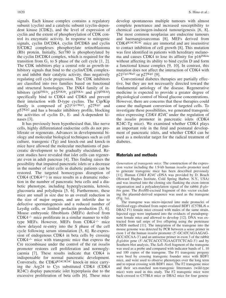
Generation of transgenic mice. The construction of the expres-sion vector including the 1.9-kb human insulin promoter usedto generate transgenic mice has been described previously[11]. Human CDK4 R24C cDNA was provided by D. Beach(Howard Hughes Institute, Cold Spring Harbor, N.Y., USA)and was inserted into the cloning site flanking the exon–intronorganisation and a polyadenylation signal of the rabbit β-glo-bin gene. The BssHII-excised fragment of this vector exclud-ing the plasmid-derived sequence was used as the transgene(Fig. 1a).
The transgene was micro-injected into male pronuclei offertilised eggs obtained from super-ovulated BDF1 (C57BL/6 ×DBA2 F1) female mice crossed with males of the same strain.Injected eggs were implanted into the oviducts of pseudopreg-nant female mice and allowed to develop [12]. DNA was ex-tracted from tail snips of live offspring using the proteinaseK/SDS method [11]. The integration of the transgene into themouse genome was detected by PCR between a sense primer inexon 1 of the human insulin promoter (5′-GCATCAGAAGAG-GCCATCAA-3′) and an antisense primer in exon 3 of the rabbitβ-globin gene (5′-ACTCACCCTGAAGTTCTCAG-3′) and bySouthern blot analysis. The SalI–NotI fragment of the transgenewas used as a probe and compared with indicator bands of 1, 10and 100 copies of the transgene. The F1 transgenic progenywere bred by crossing transgenic founder mice with BDF1mice, and were used to observe phenotypes over the long termand to repeat crossing with BDF1 mice. The F2 transgenic miceand their sex-matched non-transgenic littermates (wild-typemice) were used in this study. The F2 transgenic mice wereback-crossed to C57BL6 mice or DBA2 mice for four genera-
1820 S. Hino et al.:
tions. Body weight was determined weekly. All mice were sup-plied by CLEA Japan (Tokyo, Japan) and handled according tothe “Principles of Laboratory Animal Care” (NIH PublicationNo. 85–23, revised 1985).
Transgene expression in pancreatic islets. Pancreatic isletswere collected by the collagenase method [13, 14], total RNAwas extracted with ISOGEN (Wako Pure Chemical Industries,Osaka, Japan) and reverse-transcribed by SuperScript First-strand Synthesis System (Invitrogen, Carlsbad, Calif., USA) in20 µl of the reaction mixture. A 1-µl aliquot of the productswas subjected to PCR amplification. To determine whetherconditions were adequate for semi-quantitative RT-PCR, wereverse-transcribed 0.01, 0.1, 1 and 5 µg of total RNA and amplified the fragments under the same PCR conditions using15 to 30 cycles. We used 1 µg of total RNA as a template for
In vivo proliferation of differentiated pancreatic islet beta cells 1821
Fig. 1. Transgene construct and expression. a. The transgeneconstruct used in this study. Human CDK4 R24C transgenewas expressed in mouse pancreatic islets under the control ofthe human insulin promoter. b. In order to detect transgene-de-rived mRNA, a sense primer in exon 1 of the human insulinpromoter (left horizontal arrow) and an antisense primer inexon 3 of the rabbit β-globin gene (right horizontal arrow)were synthesised for RT-PCR. P, positive control; N, negativecontrol. c. The copy numbers of integrated transgene in lines 1to 3 of the transgenic mice were examined by Southern blot-ting. d. Bar graph of the body weights of the mice at 5, 15 and25 weeks of age. The results shown are the means ± SEM ofvalues for at least 13 mice. Black bars, littermates; white bars,CDK4 R24C-Tg mice

reverse transcription, and the cDNA fragment was amplifiedby using sense and antisense primers specific to human CDK4(5′-TACCTCTCGATATGAGCCAG-3′ and 5′-CACCAGGGT-TACCTTGATCT-3′). Sense and antisense primers to β-actinwere used as an internal standard (5′-GTGGGCCGCTCTA-GGCACCA-3′ and 5′-CGGTTGGCCTTAGGGTTCAGG-3′).After confirming the linear increase in the PCR product withincreasing amounts of total RNA, we chose the middle cyclenumber of 15 to 30 cycles in the phase of linear logarithmic in-crement for each gene to semi-quantitate the mRNA expres-sion in pancreatic islets. PCR products were separated by poly-acrylamide gel electrophoresis and visualised with ethidiumbromide. The expression of amylin (5′-TGTGCATCTCC-AAACTGCCA-3′ and 5′-GATTCCCTATTTGGATCCCC-3′),Pax4 (5′-ACACCAGGCAGCAGATTGTG-3′ and 5′-CCTGT-GCGGTAGTAGCGTCC-3′), c-myc (5′-CGTGAACTTCACC-AACAGGA-3′ and 5′-CTCTGCTGTTGCTGGTGATA-3′) andGLUT1 (5′-GAATCGTCGTTGGCATCCTT-3′ and 5′-AGAT-GACACTGAGCAGCAGA-3′) was examined similarly.
Immunoprecipitation, immunoblotting and the CDK4 kinaseassay. Pancreatic islets collected by the collagenase methodwere washed three times with ice-cold PBS and lysed in lysisbuffer containing 50 mmol/l HEPES (pH 7.5), 0.1% Tween 20,150 mmol/l NaCl, 2.5 mmol/l EGTA, 1 mmol/l dithiothreitol(DTT), 10% glycerol, complete protease inhibitor cocktail(Roche, Mannheim, Germany), 10 mmol/l β-glycerophosphate,1 mmol/l NaF, and 0.1 mmol/l sodium orthovanadate (all phos-phatase inhibitors were from Sigma, St. Louis, Mo., USA).Sonicated extracts were clarified by centrifugation and the su-pernatants were used as whole cell lysates [15]. The proteinconcentrations of the supernatants were determined by theBradford method (Bio-Rad, Hercules, Calif., USA). The super-natants were precipitated at 4 °C for 6 h with the indicated an-tibody, and immunoprecipitates were isolated using protein G-agarose beads (Roche) at 4 °C for 1 h. The beads were sub-sequently washed four times with lysis buffer. For immuno-blotting, boiling in SDS-PAGE sample buffer denatured wholecell lysates or immunoprecipitated beads. Denatured proteinsin the supernatants were separated by SDS-PAGE and trans-ferred onto Immobilon-P membranes (Millipore, Bedford,Mass., USA). Membranes were probed with antibodies toCDK4, Rb, β-tubulin (Pharmingen, San Diego, Calif., USA) or Rb phosphorylated at Ser780 (Cell Signaling Technology,Beverly, Mass., USA). Horseradish peroxidase-conjugated secondary antibody (Amersham Pharmacia Biotech, LittleChalfont, UK) and the Enhanced Chemiluminescence Detec-tion System (Amersham Pharmacia Biotech) were used for signal detection.
The assay used to assess CDK4 kinase activity was as de-scribed previously [16]. In brief, glutathione S-transferase(GST)-tagged Rb proteins derived from pGEX.3X.mRb.C′-tervector (a kind gift by Dr. H. Matsushime, Yamanouchi Phar-maceutical Company, Tokyo, Japan) were expressed in Escher-ichia coli BL21 (pLysS) and affinity purified using glutathi-one-Sepharose CL-4B (Amersham Pharmacia Biotech), ac-cording to the manufacturer’s instructions. Whole cell lysatewas incubated with an antibody to CDK4 at 4 °C for 6 h. Antigen–antibody complexes were precipitated with protein G-agarose beads at 4 °C for 1 h, which were then washed four times with lysis buffer. Recovered CDK4–cyclin D com-plexes were incubated at 30 °C for 30 min in 20 µl of kinasebuffer (50 mmol/l HEPES [pH 8.0], 10 mmol/l MgCl2,1 mmol/l DTT, 2.5 mmol/l EGTA, 10 mmol/l β-glycerophos-phate, 1 mmol/l NaF, 0.1 mmol/l sodium orthovanadate) sup-plemented with 20 µmol/l ATP, 3.7 × 10⁵ Bq of [γ-32P]ATP(Amersham Pharmacia Biotech) and 0.5 µg of purified GST-Rb
substrate. Reactions were stopped by boiling in sample bufferand the denatured proteins were separated by electrophoresisthrough a 10% polyacrylamide gel. Phosphorylated proteinswere visualised by autoradiography.
Examination of proliferation and differentiation markers ofpancreatic islets. For the histopathological examination, mousepancreas was fixed with 20% formalin in PBS, embedded inparaffin, sectioned and stained with haematoxylin and eosin(H&E). For immunohistochemistry, guinea pig polyclonal anti-bodies for porcine insulin, glucagon, somatostatin and pancre-atic polypeptide were used (DAKO Japan, Kyoto, Japan). Tu-mourigenesis of pancreatic islets was examined by insulin con-tent, expression of GLUT1, GLUT2, Pax4, c-myc and amylin,with an SV-40-transformed pancreatic islet beta cell line ofMIN6 used as a transformed insulinoma cell line. Fasting insu-lin concentration was used as an indicator of insulinoma.Mouse monoclonal antibodies were used to identify proliferat-ing cell nuclear antigen (PCNA) (DAKO Japan) in proliferat-ing cells and GLUT 2 in well-differentiated beta cells (Chemi-con International, Temecula, Calif., USA). Quantitative analy-sis of the islet cell area was carried out as previously described[11].
Glucose tolerance test, HbA1c measurement, plasma insulinconcentrations and insulin content in the whole pancreas. Af-ter an overnight fast, the intraperitoneal glucose tolerance test(ipGTT) was performed by intraperitoneally injecting 2 mg/gbody weight of glucose in physiological saline. At 0, 30, 60and 120 min after the injection, blood glucose concentrationswere determined by the glucose oxidase method with a Diasen-sor (Kyoto Daiichi Kagaku, Kyoto, Japan). We measuredHbA1c by ion exchange chromatography (Nippon Chemiphar,Tokyo, Japan) according to the manufacturer’s protocol. Plas-ma insulin concentrations were assayed using an insulinELISA kit with a mouse insulin standard (Seikagaku Kogyo,Tokyo, Japan). To determine insulin content in the whole pan-creas, each pancreas was homogenised in 4 ml of ice-coldacid-ethanol solution and insulin was extracted overnight at4 °C. After centrifugation, the supernatant was neutralised anddiluted with PBS. Insulin concentrations were assayed in sam-ples diluted 1000- or 10,000-fold [11, 17].
Determination of telomerase activities in pancreatic islets.Telomerase activities in pancreatic islets were measured usinga telomerase PCR ELISA kit (Roche). Pancreatic islets werelysed in ice-cold lysis buffer for 30 min, and 10 µg of cell ex-tract was analysed using a modified telomeric repeat amplifi-cation protocol (TRAP) [18]. Briefly, telomeric repeats areadded to a biotin-labelled primer during the first reaction, thenelongation products are amplified by PCR. An aliquot of thePCR product is denatured, hybridised to a digoxigenin (DIG)-labeled telomeric repeat-specific probe and bound to a strepta-vidin-coated 96-well plate. The immobilised PCR product isdetected with an anti-DIG-POD (peroxidase) antibody, visual-ised by a coloured reaction product generated using the sub-strate tetramethylbenzidine (TMB), and semiquantified photo-metrically at A450 nm/A690 nm.
Statistical analysis. All data are presented as means ± SEM.For comparison of two means, the Student’s unpaired t test was used. For comparison of two ratios (Q1, Q2), a normal distribution curve with a mean of Q1−Q2 and a variance ofQ1(1−Q1)/n1+Q2(1−Q2)/n2 was used. A p value less than 0.05was considered statistically significant.
1822 S. Hino et al.:

Results
Generation of CDK4 R24C transgenic mice. Thetransgene construct used in this study is shown in Figure 1a. Three CDK4 R24C-Tg founder mice wereobtained (Fig. 1b). The transgene copy numbers ofCDK4 R24C-Tg mice in lines 1 to 3 were 30, 70 and30 respectively (Fig. 1c). The percentages of F2 trans-genic mice at 5 weeks of age were 56% (63 of 112) inline 1, 54% (41 of 76) in line 2 and 47% (30 in 66) inline 3, which is proportional to Mendel’s law. Bodyweights at the age of 5, 15 and 25 weeks are shown inFigure 1d; there were no significant differences be-tween CDK4 R24C-Tg mice and their littermates.These results indicate that overexpressed CDK4 R24Cdoes not affect embryogenesis and survival of themice.
The expression of the transgene in the pancreaticislets of transgenic mice was confirmed by RT-PCR.Its expression was detected in lines 1 to 3 of CDK4R24C-Tg mice, but was not detected in their litter-mates (Fig. 2a). The levels of expression of the trans-gene in the three lines were not always correlated withtheir copy numbers (Figs. 1c, 2a).
The expression of CDK4 R24C and endogenousCDK4 was examined by immunoblotting with anti-CDK4 antibody. Levels of total CDK4 expression inthe pancreatic islets of CDK4 R24C-Tg mice were upto five times higher than in their littermates (Fig. 2b).Thus, the level of expression of CDK4 R24C drivenby the human insulin promoter was approximatelyfour times higher than that of the endogenous wild-type CDK4 (Fig. 2b). CDK4 kinase activity againstrecombinant GST-Rb protein substrate was clearly detected in the pancreatic islets of CDK4 R24C-Tgmice, but not in those of their littermates (Fig. 2c). We also examined the phosphorylation status of Rb by immunoblotting with antibody directed againstSer780-phosphorylated Rb. Levels of Rb expressionwere almost the same in the two groups of mice, butphosphorylation at Ser780 was only detected in thepancreatic islets of CDK4 R24C-Tg mice (Fig. 2d).These results suggest that the mitogenic signal is nottransduced in normal pancreatic islets even in thepresence of a sufficient supply of oxygen and nutri-ents.
Hyperplasia of pancreatic islets in CDK4 R24C trans-genic mice. Haematoxylin and eosin staining of pan-creases from 25-week-old mice revealed that islet areawas increased in CDK4 R24C-Tg mice relative to thatin their littermates (Figs. 3a, b). Islet area expressed asa percentage of the area of the whole pancreas was10.2 times higher in the CDK4 R24C-Tg mice than intheir littermates (Fig. 3e). Immunohistochemical anal-ysis for insulin revealed that the enlargement of isletarea in CDK4 R24C-Tg mice was due to the prolifera-tion of beta cells (Figs. 3c, d). Beta cell area expressed
as a percentage of the area of the whole pancreas was14.3 times higher in CDK4 R24C-Tg mice than intheir littermates (Fig. 3f). The areas of other pancre-atic hormonal cells, including alpha, delta and pancre-atic polypeptide (PP) cells, relative to the area of thewhole pancreas were similar in the two groups ofmice (Figs. 4a–m).
In order to define the initiation of beta cell prolifer-ation in CDK4 R24C-Tg mice, we examined the isletarea in neonatal mice (Day 0) and 5-week-old mice by immunohistochemical staining for insulin. At 5weeks, islet area was increased in CDK4 R24C-Tgmice due to the proliferation of beta cells, whereas no significant differences were observed betweenCDK4 R24C-Tg mice and their littermates at Day 0(Figs. 5a–f). At 5 weeks, islet area expressed as a
In vivo proliferation of differentiated pancreatic islet beta cells 1823
Fig. 2. CDK4 expression and its activity in CDK4 R24C-Tgmice islets. a. Total RNA was extracted from pancreatic islets(collected by the collagenase method) and applied to RT-PCR.In all lines of transgenic mice, RT-PCR products were ampli-fied. b. Whole cell lysates were prepared from pancreatic isletsas above and subjected to immunoblotting with anti-CDK4 an-tibody. This antibody recognises both wild-type CDK4 andCDK4 R24C. Expression of β-tubulin was measured as an in-ternal control. c. CDK4 activity in pancreatic islets assayed us-ing GST-Rb as a substrate after immunoprecipitation of CDK4.d. Antibody to Rb was used for immunoprecipitation, and pre-cipitated Rb was analysed by immunoblotting with antibodyspecific for Ser780-phosphorylated Rb. After removing priorantibody, the total amount of precipitated Rb was determinedby immunoblotting with antibody to Rb. LM, littermates; Tg,CDK4 R24C-Tg mice

percentage of the area of the whole pancreas was 3.9 times higher in CDK4 R24C-Tg mice than in theirlittermates (Fig. 5f). There were no significant differ-ences in the areas of other pancreatic islet endocrinecells between CDK4 R24C-Tg mice and their litter-mates at 5 weeks and 25 weeks (data not shown).
Glucose tolerance. In spite of the remarkable enlarge-ment of pancreatic islets, hypoglycaemia was not ob-served in CDK4 R24C-Tg mice. Levels of HbA1c inCDK4 R24C-Tg mice (1.22±0.06%, n=30) were simi-lar to those in their littermates (1.29±0.06%, n=31).Blood glucose concentrations examined at 0, 30, 60and 120 min after glucose injection at 5, 15 and 25
weeks of age were significantly lower in CDK4R24C-Tg mice than in their littermates at all timepoints (Figs. 6a–c). Because body weight increaseswith age, the glucose tolerance of the littermatesworsened, as indicated by the sustained high glucoselevels at 60 and 120 min after glucose injection in the15- and 25-week-old mice (Figs. 6b, c). In contrast,the glucose tolerance of the CDK4 R24C-Tg mice didnot alter with age.
Insulin secretion and storage. Plasma insulin concen-trations were determined at 0, 30, 60 and 120 min after glucose injection. Insulin secretion in CDK4R24C-Tg mice at 25 weeks of age was statisticallyhigher than that in their littermates at all time points(Fig. 6d). The insulin content of pancreases fromCDK4 R24C-Tg mice (108±30 µg/pancreas g) was 2.8times higher than that in the pancreases of their litter-mates (39±7 µg/pancreas g) (p<0.05, n=10 in bothgroups). The whole pancreatic weight was similar inboth mice (data not shown).
Proliferation and differentiation of pancreatic isletbeta cells. The proliferation of beta cells was con-
1824 S. Hino et al.:
Fig. 3. Histopathological examination of pancreatic islets. At25 weeks, sections of pancreas from littermates (a, c) andCDK4 R24C-Tg mice (b, d) were evaluated by H&E staining(a, b) and anti-insulin immunohistochemistry (c, d). Islet area(e) and beta cell area (f) relative to the area of whole pancreas(n=7 each). Both areas were significantly increased in CDK4R24C-Tg mice (* p<0.01 vs LM). LM, littermates; Tg, CDK4R24C-Tg mice

firmed by positive staining against PCNA in CDK4R24C-Tg mice (Fig. 7b). Conversely, hardly any pro-liferating beta cells were detected in their littermates(Fig. 7a). In order to investigate the degree of beta celldifferentiation in CDK4 R24C-Tg mice, the expres-sion of differentiation marker molecules for pancreaticislet beta cells was examined by RT-PCR and immu-nohistochemical analysis. Pancreatic islet beta cells inCDK4 R24C-Tg mice contained abundant insulingranules and immunohistochemical analysis for insu-lin showed them to be strongly stained (Figs. 3d, 4d).The level of expression of GLUT2, a marker of highlydifferentiated beta cells, was similar in the two groupsof mice (Figs. 7c, d). Pax4 is expressed only in poorly
differentiated beta cells, GLUT1 is specifically ex-pressed in beta cells transformed into insulinomacells, and the expression of c-myc is increased in betacells transformed into insulinoma cells. In the presentstudy, this pattern of expression was observed in aninsulinoma cell line of MIN6 cells, but not in the pan-creatic islets of CDK4 R24C-Tg mice or their litter-mates (Fig. 7e). The results showed that amylin is expressed in pancreatic islet beta cells regardless oftheir degree of differentiation (Fig. 7e). Moreover, insulinoma did not develop in CDK4 R24C-Tg miceover an 18-month observation period, as confirmed byhistopathological analysis (data not shown).
Determination of telomerase activities. As shown inFigure 8, telomerase activities increased in the pancre-atic islets of both groups of mice in an age-dependentmanner. Telomerase activities in the pancreatic isletsof CDK4 R24C-Tg mice were higher than in those oftheir littermates, but no significant differences weredetected. These results suggest that the pancreatic is-lets did not acquire the malignant phenotype by CDK4R24C overexpression.
Effect of genetic background on islet area and glucosetolerance. Islet area in the N4 generation of CDK4R24C-Tg mice, back-crossed to C57BL/6 and DBA2mice, was significantly increased in both strains incomparison with their littermates (p<0.05 for both). Intheir littermates, the islet area in C57BL/6 mice wassignificantly smaller than in DBA2 mice (p<0.01). Inboth strains, glucose concentrations measured at dif-ferent time points during the ipGTT were significantlylower in the CDK4 R24C-Tg mice than in their litter-mates (p<0.05 for both). Among the littermates, glu-cose concentrations were significantly lower in DBA2mice than in C57BL/6 mice (p<0.01).
Discussion
In three lines of CDK4 R24C-Tg mice, we observed ahigh number of copies of the transgene and generationrates of transgenic F2 mice according to Mendel’slaw. In other words,we have demonstrated that the expression of CDK4 R24C in pancreatic beta cellsdoes not produce any concomitant changes in the de-velopment and growth of mice.
The over-proliferation of pancreatic islet beta cellswas observed in 5-week-old CDK4 R24C-Tg mice,but not in neonatal CDK4 R24C-Tg mice. Even at 25 weeks, the beta cells of CDK4 R24C-Tg mice expressed PCNA protein, indicating that the prolifera-tion of beta cells continues at least up to 25 weeks ofage. The lack of proliferation of the beta cells of theneonatal CDK4 R24C-Tg mice may be explained byinsufficient insulin promoter activity of the fetus. Thepancreatic islets in fetal mice do not produce or
In vivo proliferation of differentiated pancreatic islet beta cells 1825
Fig. 4. Immunohistochemical analysis of pancreatic islets.Pancreata of littermates (a, c, e, g, i) and CDK4 R24C-Tg mice(b, d, f, h, j) at 25 weeks of age were examined by H&E stain-ing (a, b) and immunohistochemistry against insulin (c, d),glucagon (e, f), somatostatin (g, h) and PP (i, j). The areas ofalpha, delta and PP cells (k, l, m) relative to the area of thewhole pancreas were similar in both groups of mice (n=7each). LM, littermates; Tg, CDK4 R24C transgenic mice

secrete insulin in response to glucose stimulation, andblood glucose concentrations in fetal mice are mainlymaintained by maternal insulin [19, 20, 21, 22]. Underthe limited activity of the insulin promoter in fetalpancreatic islets, CDK4 R24C expression was nothigh enough to proliferate beta cells in fetal mice.
After birth, the differentiation and maturation ofbeta cells are induced by the demand for them to se-crete insulin; thus, activation of the insulin promoterwould be required to increase the expression of theCDK4 R24C transgene in order to induce the prolifer-ation of beta cells. Beta cell areas in CDK4−/− andCDK4R24C/R24C mice at Day 15.5 of gestation and Day5 of age have been demonstrated to be comparable tothose in their littermates [7]. Furthermore, the nucleartranslocation of CDK4 is developmentally regulated
to coincide with the initiation of beta cell prolifera-tion. Based on these results, another possibility is thatCDK4 itself is not essential for beta cell neogenesis inthe mouse embryo.
Over an 18-month observation period, insulinomadid not develop in CDK4 R24C-Tg mice as confirmedby histopathological examination. Hyperplasia of pancreatic islet beta cells was diagnosed in CDK4R24C-Tg mice based on the following findings. Pro-liferation of beta cells occurred with the concomitantpolyclonal enlargement of many islets. The ratio ofnuclear : cytoplasmic area did not increase in theCDK4 R24C-Tg mice. The size and number of nucle-oli were also similar in the two groups. In addition tothe proliferating beta cells, a comparable number ofalpha, delta, and PP cells were detected in pancreaticislets of CDK4 R24C-Tg mice and their littermates.Furthermore, the expression of GLUT2 (a molecularmarker of highly differentiated beta cells) [23], thelack of expression of Pax4 [24] and GLUT1 [23] andthe lack of an increase in c-myc expression [25, 26]indicated the absence of insulinoma development. Nosignificant increase in telomerase activity was detect-ed as a result of the overexpression of CDK4 R24C.In a previous study, the expression of CDK4 R24Calone had little effect on the life span of primary human keratinocytes, but simultaneous expression of
1826 S. Hino et al.:
Fig. 5. Immunohistochemical analysis of pancreatic islets inneonatal mice and 5-week-old mice. Pancreata from littermates(LM) (a, c) and CDK4 R24C-Tg (Tg) (b, d) at neonatal Day 0(a, b) and 5 weeks (c, d) were examined by immunohisto-chemistry for insulin. The beta cell area relative to the wholearea of pancreas was determined (n=7 each). The beta cell areas were similar in the two groups of neonatal Day 0 mice(e), but were significantly increased in CDK4 R24C-Tg miceat 5 weeks of age (f) (* p<0.05 vs littermates)

dominant negative p53 permitted cells to divide be-yond their normal limits [27]. In our system, the p53pathway would suppress tumour formation by inhibit-ing the increase in telomerase activity and promotingthe apoptosis of de-differentiated beta cells.
The results of the ipGTT confirmed that the betacells in CDK4 R24C-Tg mice were highly differenti-ated. Although the early morning fasting blood glu-cose concentrations of CDK4 R24C-Tg mice wereslightly lower than those of their littermates, theywere within the normal range and were not hypogly-caemic. Fasting plasma insulin concentrations weresimilar in the two groups of mice. One of the diagnos-tic criteria for insulinoma is the presence of increasingplasma insulin concentrations even at fasting [28].This criterion was not satisfied in CDK4 R24C-Tgmice. Furthermore, in line with the normal glucoseconcentrations observed in CDK4 R24C-Tg mice, in-sulin secretion was adequate and prompt in these miceafter glucose loading. It has previously been shownthat the precise regulation of insulin secretion is de-fective in the majority of patients with insulinoma
In vivo proliferation of differentiated pancreatic islet beta cells 1827
Fig. 6. Glucose levels and insulin secretion during the ipGTT.After 16 h of fasting, 2 mg/g body weight of glucose in physi-ological saline was intraperitoneally injected. Blood glucoseconcentrations were examined at 0, 30, 60 and 120 min afterglucose injection at 5 (a), 15 (b) and 25 (c) weeks. d. Plasmainsulin concentrations were measured at the same time points.* p<0.05 vs littermates; ** p<0.01 vs littermates. Empty cir-cles, CDK4 R24C-Tg mice; filled circles, littermates
Fig. 7. Analysis of gene expression in pancreatic islets. Pan-creata from littermates (a, c) and CDK4 R24C-Tg mice (b, d)at 25 weeks of age were examined by immunohistochemistryfor PCNA (a, b) or GLUT2 (c, d). e. Islets obtained from fivemice in the same line aged 20 to 25 weeks were used togetherfor RNA extraction. Total RNA was subjected to semi-quanti-tative RT-PCR with specific primers for the indicated genesand for β-actin (internal control). MIN6 cells were used as arepresentative insulinoma cell line. LM, littermates; Tg, CDK4R24C-Tg mice

[28]. Based on these observations, we concluded thatinsulinoma did not develop in the CDK4 R24C-Tgmice.
At 60 and 120 min after the intraperitoneal glucoseinjection, blood glucose concentrations increased withage in littermates. Because of an abundant food supply and hypomotility in the narrow cage, age-dependent obese mice acquired insulin resistance. Although a similar increase in body weight was observed in CDK4 R24C-Tg, their blood glucose con-centrations did not increase at 60 and 120 min withage. The CDK4 R24C-Tg mice possess the ability tosecrete an adequate amount of insulin in response toincreased insulin demand. At 25 weeks of age, theplasma insulin concentration curve of littermates afterglucose injection showed one peak at 30 min, and theconcentration of insulin at 120 min was not highenough to suppress the blood glucose concentrations.Conversely, the insulin curve of CDK4 R24C-Tg miceshowed two peaks, and a sufficient amount of insulinwas secreted to appropriately control the blood glu-cose concentrations for 120 min.
Although CDK4 R24C-Tg mice demonstrated thebetter glucose tolerance, HbA1c levels were not statis-tically different between CDK4 R24C-Tg mice andtheir littermates. This finding ruled out the possibilityof chronic hypoglycaemia due to the constitutive ex-cessive insulin secretion in CDK4 R24C-Tg mice. Thedifferences in the capacity for insulin secretion couldonly be detected when both mice were intraperitoneal-ly injected with 2 mg/g body weight of glucose. Bloodglucose homeostasis is highly differentiated in CDK4
R24C-Tg mice such that dispensable insulin is not se-creted except when glucose is loaded and a largeamount of insulin is needed. It has recently been reported that levels of preproinsulin I and II mRNAsare not significantly increased in CDK4R24C/R24C mice,whereas there is a dramatic reduction in preproinsulinlevels in CDK4−/− mice [29]. These results suggestthat CDK4 may play a critical role not only in postna-tal pancreatic development, but also in the functionalmaturation of insulin secretion.
Even after 18 months, no insulinoma developed inthe CDK4 R24C-Tg mice. Conversely, homozygousCDK4R24C/R24C knock-in mice have been shown to de-velop multiple tumours, including endocrine tumoursand haemangiosarcomas, at the age of 1 year [6]. Het-erozygous CDK4WT/R24C knock-in mice have also beenobserved to develop multiple tumours [8]. Given thepowerful activity of the insulin promoter and the manycopies of the CDK4 R24C transgene inserted into thegenome of the CDK4 R24C-Tg mice, it is unlikely thatthe level of transgene expression in the CDK4 R24C-Tg mice used in this study was lower than that in theknock-in mice used in previous studies. The level ofCDK4 R24C expression driven by the insulin promoterin CDK4 R24C-Tg mice was four times higher thanthe level of CDK4 expression driven by the CDK4 pro-moter. These data rule out the existence of a thresholdof CDK4 activity for tumourigenesis and indicate thattumour development is not determined simply by thelevel of CDK4 R24C expression.
Studies on transgenic mice expressing CDK4 inepidermis or astrocytes have reported that CDK4overexpression alone is not sufficient to cause tumourformation [30, 31]. The D-type cyclins control cyclinD/CDK4 activity by binding to and activating CDK4[1]. However, it has been demonstrated that the over-expression of both cyclin D1 and CDK4 does not havea combined effect on tumour development [32, 33].CDK4 may have other cyclin partners and additionalroles to the phosphorylation of Rb. It is possible that CDK4 is regulated differently to CDK4 R24Csince the amplification and overexpression of CDK4has been observed in a wide spectrum of human tumours [34], whereas CDK4 R24C has been shownto be overexpressed only in sporadic and familialmelanomas [9, 10].
The presence and the absence of neoplasms inCDK4R24C/R24C knock-in mice and CDK4 R24C-Tgmice respectively, suggests that the onset of CDK4R24C expression may well govern neoplasm develop-ment. In knock-in mice with the germ line mutation,CDK4 R24C was expressed even in poorly differenti-ated beta cells, which could have caused beta celltransformation. The lack of malignant conversion inCDR4 R24C-Tg mice may be due to the fact thatCDK4 R24C was only expressed in terminally differ-entiated beta cells under the control of the insulin pro-moter.
1828 S. Hino et al.:
Fig. 8. Telomerase activites in pancreatic islets. Pancreatic is-lets were lysed in ice-cold lysis buffer and 10 µg of protein ex-tract was subjected to the telomerase PCR ELISA assay (n=10each). Absorbance was measured at 450 nm, with 690 nm usedas a reference. MIN6 cell extract (P) and lysis buffer (N) wereanalysed as controls. LM, littermates; Tg, CDK4 R24C-Tgmice
CDK4 R24C-Tg mice in an N4 backcross toC57BL/6 or DBA2 showed a significant increase inislet area compared with their littermates, and this wassimilar to that observed in the original CDK4 R24C-Tg mice in BDF1 background. The poor glucose toler-ance of C57BL/6 mice compared with DBA2 wild-type mice was shown to be due to the genetic back-ground of these littermates. In CDK4 R24C-Tg micein an N4 backcross to C57BL/6 or DBA2, glucoseregulation was intact and no hypoglycaemia was ob-served. These results indicate that the increase in isletarea and the highly differentiated beta cell phenotypeinduced by CDK4 R24C overexpression are indepen-dent of the genetic background.
The proliferation and differentiation of beta cellsare not induced concurrently, and the ability of highlydifferentiated beta cells to proliferate is thought to beextremely restricted. Using a genetic-lineage-markingapproach, it has previously been shown that the main-tenance of adult pancreatic beta cell mass in mice isnot dependent on the stem cells, but on the self-dupli-cation of pre-existing beta cells [35]. Beta cell area isdetermined by the balance between the rate of neogen-esis or proliferation and the rate of apoptosis [36].Levels of beta cell proliferation as assessed by 5-bromo-2′-deoxyuridine (BrdU) incorporation were2.5 times higher in the islets of CDK4R24C/R24C micethan in the islets of their littermates at the age of 10 days [7]. The remarkable proliferation of beta cells observed in CDK4 R24C-Tg mice in our studydemonstrated that beta cells could proliferate even at25 weeks of age (i.e. at the highly differentiated stage)when the insulin promoter is functioning.
In this paper we have demonstrated in our trans-genic mouse model that pancreatic islet beta cells pro-liferate significantly, with the preservation of highlydifferentiated phenotypes, irrespective of the geneticbackground. Based on this evidence, the postnatal ac-tivation of CDK4 in pancreatic islets may potentiallybe used for the therapeutic stimulation of proliferationof differentiated pancreatic islet beta cells. Humanpancreatic islets transduced with CDK4 R24C by len-tivirus show proliferative potential in response to glu-cose stimulation in vitro [37]. The postnatal activationof CDK4 through the use of new techniques (includ-ing the administration of vectors expressing CDK4R24C under the control of the insulin promoter orCDK4-activating drugs) could potentially lead to thepostnatal proliferation of pancreatic islet beta cellswithout neoplasm formation, thus providing a radicalnew treatment for diabetes.
Acknowledgements. This study was supported by a Grant-in-Aid for Scientific Research from Japanese Society for the Pro-motion of Science and a Cooperative Link of Unique Scienceand Technology for Economic Revitalization (CLUSTER).The authors are not aware of any conflicts of interest.
References
1. Sherr CJ (1996) Cancer cell cycle. Science 274:1672–16772. Kitagawa M, Higashi H, Jung HK et al. (1996) The con-
sensus motif for phosphorylation by cyclin D1-Cdk4 is dif-ferent from that for phosphorylation by cyclin A/E-Cdk2.EMBO J 15:7060–7069
3. Sherr CJ, Roberts JM (1999) CDK inhibitors: positive andnegative regulators of G1-phase progression. Genes Dev13:1501–1512
4. Yamaoka T, Itakura M (1999) Development of pancreaticislets. Int J Mol Med 3:247–261
5. Tsutsui T, Hesabi B, Moons DS et al. (1999) Targeted dis-ruption of CDK4 delays cell entry with enhanced p27Kip1
activity. Mol Cell Biol 10:7011–70196. Rane SG, Dubus P, Mettus RV et al. (1999) Loss of CDK4
expression causes insulin-deficient diabetes and Cdk4 acti-vation results in beta-islet cell hyperplasia. Nat Genet22:44–52
7. Martin J, Hunt SL, Dudus P et al. (2003) Genetic rescue ofCdk4 null mice restores pancreatic beta-cell proliferationbut not homeostatic cell number. Oncogene 22:5261–5269
8. Sotillo R, Dubus P, Martin J et al. (2001) Wide spectrum oftumors in knock-in mice carrying a Cdk4 protein insensi-tive to INK4 inhibitors. EMBO J 20:6637–6647
9. Wolfel T, Hauer M, Schneider J et al. (1995) A p16INK4a-insensitive CDK4 mutant targeted by cytolytic T lympho-cytes in a human melanoma. Science 269:1281–1284
10. Zou L, Weger J, Yang Q et al. (1996) Germline mutationsin the p16INK4a binding domain of CDK4 in familial mela-noma. Nat Genet 12:97–99
11. Yamaoka T, Idehara C, Yano M et al. (1998) Hypoplasia of pancreatic islets in transgenic mice expressing activinreceptor mutants. J Clin Invest 102:294–301
12. Hogan B, Beddington R, Costantini F, Lacy E (1994) Manipulating the mouse embryo. A laboratory manual, 2ndedition. Cold Spring Harbor Laboratory Press, New York
13. Moritani M, Yoshimoto K, Ii S et al. (1996) Prevention of adoptively transferred diabetes in nonobese diabeticmice with IL-10-transduced islet-specific Th1 lymphocytes. J Clin Invest 98:1851–1859
14. Gotoh M, Maki T, Kiyoizumi T, Satomo S, Monaco AP(1985) An improved method for isolation of mouse pancre-atic islets. Transplantation 40:437–438
15. Kato A, Takahashi H, Takahashi Y, Matsushime H (1997)Inactivation of the cyclin D-dependent kinase in the rat fibroblast cell line, 3Y1, induced by contact inhibition. J Biol Chem 272:8065–8070
16. Matsushime H, Ewen ME, Strom DK et al. (1992) Identifi-cation and properties of an atypical catalytic subunit(p34PSK−J3/cdk4) for mammalian D type G1 cyclins. Cell71:323–334
17. Yamada T, Brunstedt J, Solomon T (1983) Chronic effectsof caerulein and secretion on endocrine of the rat. Am JPhysiol 24:G541–G545
18. Kim NW, Piatyszek MA, Prowse KR et al. (1994) Specificassociation of human telomerase activity with immortalcells and cancer. Science 266:2011–2015
19. Asplund K (1973) Dynamics of insulin release from the fetal and neonatal rat pancreas. Eur J Clin Invest 3:338–344
20. Hole RL, Pian-Smith MC, Sharp GW (1988) Developmentof the biphasic response to glucose in fetal and neonatal ratpancreas. Am J Physiol 254:E167–E174
21. Otonkoski T, Anderson S, Knip M, Simell O (1988) Matu-ration of insulin response to glucose during human fetaland neonatal development. Diabetes 37:286–291
In vivo proliferation of differentiated pancreatic islet beta cells 1829
22. Weinhaus AF, Poronnik P, Cook DI, Tuch BE (1995) Insu-lin secretagogues, but not glucose, stimulate an increase in[Ca++] in the fetal rat beta cell. Diabetes 44:118–124
23. Boden G, Murer E, Mozzoli M (1994) Glucose transporterproteins in human insulinoma. Ann Intern Med 121:109–112
24. Miyamoto T, Kakizawa T, Ichikawa K, Nishio, S, Kajikawa S, Hashizume K (2001) Expression of dominantnegative form of PAX4 in human insulinoma. BiochemBiophys Res Commun 282:34–40
25. Pavelic K, Hrascan R, Kapitanovic S et al. (1995) Multiplegenetic alterations in malignant metastatic insulinomas. J Pathol 177:395–400
26. Pavelic K, Hrascan R, Kapitanovic S et al. (1996) Molecular genetics of malignant insulinoma. AnticancerRes 16:1707–1717
27. Rheinwald JG, Hahn WC, Ramsey MR et al. (2002) A two-stage, p16INK4a- and p53-dependent keratinocyte senescence mechanism that limits replicative potential in-dependent of telomere status. Mol Cell Biol 22:5157–5172
28. Fowler DL, Wood WG, Koontz PG Jr (1980) Endogenoushyperinsulinism. Am J Gastroenterol 74:321–327
29. Mettus RV, Rane SG (2003) Characterization of the abnor-mal pancreatic development, reduced growth and infertilityin Cdk4 mutant mice. Oncogene 22:8413–8421
30. Miliani de Marval PL, Gimenez-Conti IB, LaCave M, Martinez LA, Conti CJ, Rodriguez-Puebla ML (2001)Transgenic expression of cyclin-dependent kinase 4 results
in epidermal hyperplasia, hypertrophy, and severe dermalfibrosis. Am J Pathol 159:369–379
31. Huang ZY, Baldwin RL, Hedrick NM, Gutmann DH(2002) Astrocyte-specific expression of CDK4 is not suffi-cient for tumor formation, but cooperates with p53 hetero-zygosity to provide a growth advantage for astrocytes invivo. Oncogene 21:1325–1334
32. Miliani de Marval PL, Macias E, Conti CJ, Rodriguez-Puebla ML (2004) Enhanced malignant tumorigenesis inCdk4 transgenic mice. Oncogene 23:1863–1873
33. Lazarov M, Kubo Y, Cai T et al. (2002) CDK4 coexpres-sion with Ras generates malignant human epidermal tu-morigenesis. Nat Med 8:1105–1114
34. Ortega S, Malumbres M, Barbacid M (2002) Cyclin D-dependent kinases, INK4 inhibitors and cancer. BiochimBiophys Acta 1602:73–87
35. Dor Y, Brown J, Martinez OI, Melton DA (2004) Adultpancreatic beta-cells are formed by self-duplication ratherthan stem-cell differentiation. Nature 429:41–46
36. Bonner-Weir S (2000) Life and death of the pancreatic betacells. Trends Endocrinol Metab 11:375–78
37. Marzo N, Mora C, Fabregat ME et al. (2004) Pancreatic islets from cyclin-dependent kinase 4/R24C (Cdk4)knockin mice have significantly increased beta cell massand are physiologically functional, indicating that Cdk4 isa potential target for pancreatic beta cell mass regenerationin Type 1 diabetes. Diabetologia 47:686–694
1830 S. Hino et al.: In vivo proliferation of differentiated pancreatic islet beta cells