in vivo reactivation of herpes simplex in rabbit trigeminal...
TRANSCRIPT

INFECTION AND IMMUNITY, Oct. 1981, p. 69-740019-9567/81/100069-06$02.00/0
Vol. 34, No. 1
In Vivo Reactivation of Herpes Simplex Virus in RabbitTrigeminal Ganglia: Electrode Model
MARY T. GREEN,`* JOHN P. ROSBOROUGH,2 AND EDMUND C. DUNKEL'
Cullen Eye Institute, Department of Ophthalmology,' and the Department of Physiology,2 Baylor College ofMedicine, Texas Medical Center, Houston, Texas 77030
Received 2 February 1981/Accepted 26 May 1981
The rabbit provides an excellent model for the study of ocular herpes becauseherpetic keratitis in the rabbit eye resembles human disease in its clinical featuresand in its propensity for spontaneous recurrence. This paper presents a methodfor the electrical induction of multiple episodes of in vivo reactivation of latentHSV-1 infection with peripheral shedding of virus. Physiological levels of currentdelivered via an electrode implanted over the trigeminal ganglion of latentlyinfected animals has enabled us to modify and synchronize virus shedding inpreocular tear film and to cause multiple episodes of reactivation in a singleanimal. For this reason, the model is well suited for antiviral efficacy testing andprovides an excellent opportunity for investigation of virus-host cell interactionsin latent and -recurring herpetic disease.
Herpes simplex keratitis is the most prevalentsevere ocular infection in this country. The pro-pensity of this infection to recur throughout lifeand to produce irreversible structural alterationsof the cornea and intraocular structures resultsin considerable visual morbidity, medical ex-pense, and loss of productivity of otherwisehealthy individuals. Interruption of the recur-rent herpes cycle depends on knowledge of thesequence by which latent infection of the ner-vous system is translated into active disease inthe peripheral tissues. For this reason, a reliableand reproducible means of triggering recurrentocular herpes has long been sought.
Historically, the rabbit has provided an excel-lent model for study of ocular herpes becauseherpetic keratitis in the rabbit eye resembleshuman disease in clinical features as well as inits propensity for spontaneous recurrence (5, 6,8). Attempts to induce viral reactivation in otheranimals models have been only partially suc-cessful. The apparent problem has been theinability to detect the propagation of virus toperipheral tissues (1, 3, 8, 12-14). The mostdependable animal model to date has been themanual manipulation of the rabbit trigeminalnerve (7). Although this model has providedinsight as to the relationship between trigeminalnerve and recurrent ocular herpes in rabbits, itsapplication is limited because animals rarelysurvive longer than 72 h. We recently deter-mined that passage of current along the trigem-inal nerve of latently infected animals via a smallelectrode implant elicits rapid release of virus at
the eye (Green and Dunkel, Arvo abstr., p. 156,1980). This paper presents a new method forinduction of multiple episodes of in vivo reacti-vation of herpes simplex virus type 1 (HSV-1)infection with peripheral shedding of virus inlatently infected aniimals.
MATERIALS AND METHODS
Electrode implantation. New Zealand albino rab-bits (2 to 3 kg) were anesthetized with acepromazinemaleate (5 mg/kg) and ketamine HCI (33 mg/kg),administered separately. Animals were then placed ina stereotaxic device (David Koff, Inc., Tujunga, Calif.).Both stereotaxic coordinates and morphological land-marks (the squamous portion of the temporal bone atthe attachment of the zygomatic arch) were utilized tointroduce a neurological electrode via a 5-mm crani-otomy burr hole onto the surface of the tentoriumoverlying either the right or left trigeminal nerveganglion. Each electrode consisted of a 22-mm, 22-gauge, epoxylite-coated, stainless-steel wire (SmallParts, Inc., Miami, Fla.) with a 0.5-mm exposed tip.Proper placement and function of the electrodes weredemonstrated by reflex twitching of the eyelids andextraocular muscles on the implanted side in responseto a continuous series of pulses of 1 ms duration at 10Hz with voltages ranging from 0.5 to 4 V. Beforeremoval from the stereotaxic device, electrodes wereattached to the cranium with skull screws (1/8-inch[ca. 0.32-cm] furled screws) and dental adhesive (GetzTru-Cure Dental Adhesive and Formix DentureResin). The incision was closed with 4.0 siLk sutures sothat only the contact tip of the electrode remainedexposed (Fig. 1A, B, and C).
Inoculation. Rabbits were anesthetized with 33 mgof ketamine HCl per kg. A 21-gauge needle was used
69
on May 14, 2018 by guest
http://iai.asm.org/
Dow
nloaded from

70 GREEN, ROSBOROUGH, AND DUNKEL
to administer four vertical and four horizontal lacera-tions to the corneal epithelium under direct vision,utilizing an operating microscope. A portion (50 pl)containing 105 plaque-forming units of McKrae strainHSV-1 was dropped onto each cornea. Eyes wereclosed and globes were massaged for 15 s (9).
Ocular culture. Virus shedding was monitored bythe culture of the preocular tear film. Sterile cotton-tipped applicators (Schering Medical Scientific, Inc.,Carson, Calif.) were passed over the upper and lower
INFECT. IMMUN.
conjunctival cul-de-sac, lightly rolled over the cornea,and retained in the nasal fornix for 5 to 10 s formaximum tear film absorption (11). Swabs were elutedin 0.5 ml of Hanks balanced salt solution (Gibco Lab-oratories, Grand Island, N.Y.) for 15 s with agitation.Portions (50 pl) of the eluate were absorbed ontoconfluent 16-mm Vero cell monolayers (CCL 81, FlowLaboratories Inc., Rockville, Md.) for 10 min at 37°Cin 5% CO2. Cultures were reconstituted with 0.5 ml ofminimal essential medium (GIBCO) plus 5% fetal bo-
CFIG. 1. (A) Rabbit placement in stereotaxic apparatus. (B) Postmortem autopsy of rabbit demonstrating
electrode placement over the left trigeminal ganglion. Tentorium intact during experimental period butdisrupted during dissection; tg, tentorium overlying trigeminal nerve ganglion: tn, tentorium overlyingtrigeminal nerve. (C) Diagrammatic representation of electrode placement: e, electrode; sa, adjustable sidearm; s, stereotaxic apparatus; cr, exposed cranium; o, optic nerve and chiasm.
A
tq11-
)i1-i :!!.-
1.tI; I I .-;i, N ieV. N.
on May 14, 2018 by guest
http://iai.asm.org/
Dow
nloaded from

ELECTRODE MODEL OF HSV REACTIVATION IN VIVO
vine serum (GIBCO). Cultures were monitored dailyfor 10 days for the development of cytopathologyconsistent with HSV-1 infection.Trigeminal nerve ganglion stimulation via
electrode implant. All rabbits were stimulated usinga Grass model S9 stimulator. Stimulation consisted ofa continuous series of pulses of 1 ms duration at 10Hz, with voltages ranging from 0.5 to 4 V for 15-sintervals. The physiological endpoint of extraocularmuscle and lid twitch was used to delineate the deliv-ery of appropriate amounts of electrical stimulation tothe trigeminal nerve.Experimental design. (i) Part A. Study A was
designed to determine whether there is a positivecorrelation between passage of low-dose electrical cur-rent along the trigeminal nerve of latently infectedanimals and release of virus in preocular tear film.Monopolar electrodes were implanted over the lefttrigeminal ganglion of 19 rabbits. Five to ten days afterimplantation, animals were stimulated and monitoredby ocular swab culture to assess probe function andplacement and to insure the absence of innate virus.After a negative "control stimulation," animals werebilaterally infected with 105 plaque-forming units ofMcKrae strain HSV-1. After resolution of active infec-tion and after a minimum of 4 days of negative bilat-eral ocular cultures, animals were "test stimulated"and monitored by daily ocular swab cultures for theshedding of virus. After several days of negative bilat-eral ocular cultures, animals were again stimulatedand monitored. To determine the relative quantity ofvirus shed in preocular tear film in response to elec-trical current stimulation, eyes of a separate group of10 rabbits were cultured after known quantities ofvirus were dropped into the cul-de-sacs. Each concen-tration (104, 103, 102, 10, and 5 plaque-forming units)of HSV-1 in 50 pl of Hanks balanced salt solution wasdropped into the cul-de-sac of four eyes. The eyeswere cultured as previously described after 5 min, toallow for some interaction between virus and hostfactors in the tear film, while minimizing the loss ofinfectious virus particles due to uptake by host cells.Cell monolayers were observed for 5 days for appear-ance of cytopathology.
(ii) Part B. Study B was designed (i) to confirm therelationship between administration of low-dose elec-trical current and the elicitation of virus shedding inlatently infected animals; (ii) to determine whetherthe acute stage of infection could be prolonged asevidenced by continuous shedding of virus for >14days; (iii) to determine whether a shift in the patternof induced virus shedding would occur with time; and(iv) to determine whether the ability to shed viruscould be exhausted with continuous long-term stimu-lation. Six test and six control animals were stimulatedevery other day for 60 days beginning on day 7 post-infection. Stimulus consisted of a series of pulses of 1ms duration at 10 Hz ranging from 0.1 to 0.5 mA. Fourof the test group animals were stimulated every thirdday for an additional 140 days and monitored viaocular culture.
(iii) Part C. Study C was designed to determine theoptimal interval of stimulation with respect to induc-tion of virus shedding in preocular tear film. Fiveanimals were stimulated daily for 60 days beginning 3
days postinoculation; five animals were stimulatedevery other day for 60 days beginning 7 days postin-fection; and five animals were stimulated every fifthday for 60 days beginning 7 days postinfection. Dataon stimulation every third day beginning day 7 post-inoculation was obtained from study B.
RESULTSStudy A. Twenty-three biologically definitive
unilateral stimuli resulted in 19 ipsilateral virusrecoveries in 15 animals (rabbit no. 1-4, 6-8, 11,.14-19) (Fig. 2). Three animals responded aftereach of two separate stimulations (rabbit no. 2,4, and 7). Virus shedding limited to the contra-lateral side was elicited in five animals afterstimulation (rabbit no. 1, 12, 14, 15, 18). Virusshedding occurred on both the ipsilateral andcontralateral side in 13 animals (rabbit no. 5-14,16, 18, 19). Increased voltage (7 to 10 V) wasadministered to all animals exhibiting contralat-eral shedding resulting in a bilateral biologicalresponse (lip and eye twitch on the contralateralas well as the ipsilateral side). Seven unilateralstimuli failed to induce virus shedding on eitherthe ipsilateral or contralateral side. All ipsilat-eral viral responses were initiated within 48 h ofstimulation. A single contralateral response wasinitiated 72 h poststimulation.
In a separate study, known virus concentra-tions were instilled into the conjunctival cul-de-sac of 20 eyes. Cytopathology was evident in 20of 28 total positive cultures within 24 h of swabinoculation. Because only those eyes receivingknown portions _103 plaque-forming units pro-duced positive cultures within 24 h of inocula-tion, it was inferred that 71% of the positivecultures represented shedding of a minimum of103 plaque-forming units in the preocular tearfilm.Study B. All inoculated animals developed
acute herpetic keratitis as evidenced by the pres-ence of corneal dendritic figures (by day 3 post-inoculation) and virus shedding in preocular tearfilm for a Iinimum of 12 to 14 consecutive days.During the period of acute infection, there wasno significant difference in virus shedding inpreocular tear film between test and controlanimals. By day 18 post-inoculation, acute infec-tion had cleared as evidenced by methylene bluestaining of intact corneal epithelium and nega-tive tear film cultures. After establishment oflatency (18 days postinfection), a significant dif-ference in tear film virus shedding was observedbetween stimulated and nonstimulated animals(Table 1). Virus shedding typical of acute diseasewas not significantly prolonged by consistentelectrode stimulation at 3-day intervals. Testanimals became refractile to induction of virusshedding in preocular tear film for periods of 5
VOL. 34, 1981 71
on May 14, 2018 by guest
http://iai.asm.org/
Dow
nloaded from
72 GREEN, ROSBOROUGH, AND DUNKEL10 14 IS 22 26 30 34 38 42 46 96 100 104 108.,,,,.,, ,,,,,E.
INFECT. IMMUN.
2 OSr oooo0oOD 000o0000oosC ooJoo1°°o°o °OsC 001000100
2 OD 00000000000
Os C 0.00l0 0 0 0
3 OD F -00000000000051O0 OO O
4 OD [ooF ooo-*oOsI 000000:.0060 L ooooooooooo
e8sFr oAololo::.o0Os~ L :ooooOS0000000:
7 OD FL 000 00 00:*0IE.. 0r. 000 .OO00 0Ao
8 OD oooFoooooooooooE 1O0I 0001 OD 00 0000
14OI
Osr 00oooolo00000:15 OD F 0 0 00 0000000 0
Os01 0 1000000000000E 12 D 0.**.00000,000000,
Osr ol*oo
1 OD F 0o oooooooooo-OSI ~~~~0000018I
Osr 0:0°'°000:0000
14 OD F__ __ __ -_ _ __ __ __ _
F ~~~~~~I16 0 14 0000000M4015ooF -00~~~0000A. 00000001isODF 00000000000
1ODF 000000000isODF ~~~~~~~000000000000000
10 14 IS 22 26 30 34 38 42 46 96 100 104 108
Days Post Inoculation
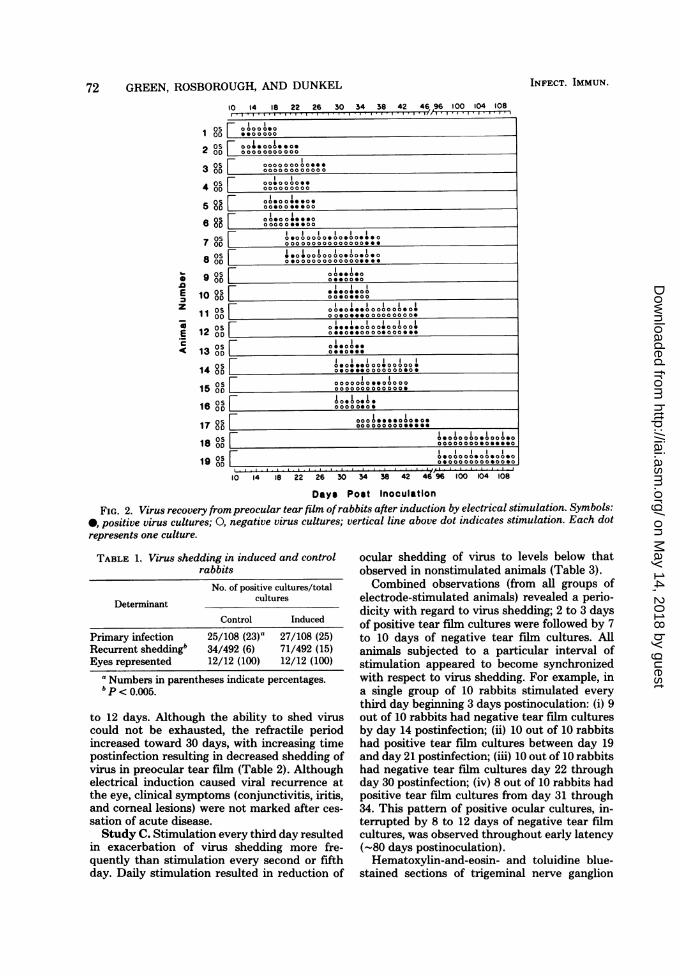
FIG. 2. Virus recovery frompreocular tearfilm ofrabbits after induction by electrical stimulation. Symbols:0, positive virus cultures; 0, negative virus cultures; vertical line above dot indicates stimulation. Each dotrepresents one culture.
TABLE 1. Virus shedding in induced and controlrabbits
No. of positive cultures/total
Determinant cultures
Control Induced
Primary infection 25/108 (23)a 27/108 (25)Recurrent sheddingb 34/492 (6) 71/492 (15)Eyes represented 12/12 (100) 12/12 (100)
a Numbers in parentheses indicate percentages.P < 0.005.
to 12 days. Although the ability to shed viruscould not be exhausted, the refractile periodincreased toward 30 days, with increasing timepostinfection resulting in decreased shedding ofvirus in preocular tear film (Table 2). Althoughelectrical induction caused viral recurrence atthe eye, clinical symptoms (conjunctivitis, iritis,and corneal lesions) were not marked after ces-sation of acute disease.Study C. Stimulation every third day resulted
in exacerbation of virus shedding more fre-quently than stimulation every second or fifthday. Daily stimulation resulted in reduction of
ocular shedding of virus to levels below thatobserved in nonstimulated animals (Table 3).Combined observations (from all groups of
electrode-stimulated animals) revealed a perio-dicity with regard to virus shedding; 2 to 3 daysof positive tear film cultures were followed by 7to 10 days of negative tear fih cultures. Allanimals subjected to a particular interval ofstimulation appeared to become synchronizedwith respect to virus shedding. For example, ina single group of 10 rabbits stimulated everythird day beginning 3 days postinoculation: (i) 9out of 10 rabbits had negative tear film culturesby day 14 postinfection; (ii) 10 out of 10 rabbitshad positive tear film cultures between day 19and day 21 postinfection; (iii) 10 out of 10 rabbitshad negative tear film cultures day 22 throughday 30 postinfection; (iv) 8 out of 10 rabbits hadpositive tear film cultures from day 31 through34. This pattern of positive ocular cultures, in-terrupted by 8 to 12 days of negative tear filmcultures, was observed throughout early latency(-80 days postinoculation).Hematoxylin-and-eosin- and toluidine blue-
stained sections of trigeminal nerve ganglion
on May 14, 2018 by guest
http://iai.asm.org/
Dow
nloaded from

ELECTRODE MODEL OF HSV REACTIVATION IN VIVO
TABLE 2. Effect of long-term induction on virus shedding
Positive ocular cultures/total no. of cultures on days postinfectionAnimals
20-59 60-119 120-179 180-239 240-300
Stimulated 81/432 (19)a 20/264 (8) 26/612 (4) 33/480 (7) 28/480 (6)Control 18/240 (7) 43/720 (5) 49/600 (8) 20/360 (5) 23/360 (6)
a Numbers in parentheses indicate percentages.
TABLE 3. Optimal pattern of stimulation: effect ofinduced virus shedding
Periodicity of Episodes of virus sheddingestimulation
Daily.10 episodes/10 eyes/60 daysbcEvery 2nd day.36 episodes/10 eyes/60 daysEvery 3rd day.64 episodes/10 eyes/60 daysEvery 5th day.47 episodes/10 eyes/60 daysNonstimulated.23 episodes/10 eyes/60 days
a An episode of virus shedding consisted of an isolatedseries of positive ocular cultures obtained on two or moreconsecutive days.
'The 60-day time period for both test and control animalsbegan on day 20 postinoculation (after 4 consecutive days ofnegative bilateral tear film cultures) and continued throughday 80 postinoculation.
' 600 cultures were obtained from each group during the 60-day test period.
from animals with long-term electrode implantsshowed no histological evidence of cellular dam-age. Corneal sensation was retained after multi-ple episodes of virus shedding in preocular tearfilm.
DISCUSSIONPhysiological levels of current delivered via an
electrode implanted over the trigeminal ganglionof latently infected animals has enabled us tomodify virus shedding in preocular tear film andto cause multiple episodes of reactivation in asingle animal. Although this method of electricalinduction does not prevent the natural progres-sion from active infection to latency, it has al-lowed us to accelerate and synchronize recurrentepisodes of virus shedding after resolution ofacute disease. The number of episodes of spon-taneous recurrence normally occurring in a 12-month period can be observed in 3 months underexperimental conditions. Because it is efficientand can be standardized (induction of multipleepisodes of virus shedding in the same eye), theelectrode model is well suited for antiviral effi-cacy testing. In addition, this model provides anexcellent opportunity for investigation of alter-ations in virus host cell interactions in latent andin recurring herpetic disease.
In this report, latency is defined as the stageof HSV infection when infectious virus can nolonger be isolated from cell-free ganglion ho-mogenates (10), although reactivation can be
induced by organ cocultivation. Reactivation ismeasured by shedding of infectious virus in pre-ocular tear film. All latently infected animals inthe present study (39 total test animals) havebeen induced to shed virus. Virus reactivation ismore consistently accomplished with a singleinduction stimulus within the first 40 days post-inoculation; we have not been able to exhaustthe potential for reactivation after multiple in-duction stimuli as late as 300 days postinfection.It is not known whether reactivation and sub-sequent infection with endogenous virus (viacentripetal and centrifugal intraaxonal trans-port) play a role in perpetuation of latency.
Refractile periods are' time intervals duringwhich virus is not released in preocular tear filmin the presence of inducing stimuli. Of interestis the fact that these refractile periods increasein duration with increasing time after primaryinfection. The decrease in ocular shedding ofinfectious virus observed in animals undergoinglong-term (285 days) maximal induction (stim-ulus interval every third day) is consistent withclinical observations of the natural history ofspontaneous reactivation of herpetic disease inhumans. The mechanism for this decrease inactivity is puzzling, since multiple recurrent ep-isodes do not lead to anesthesia in humans or inthe experimental rabbit.Use of physiological levels of electrical current
has allowed us to either induce or prevent HSV-1 reactivation in latently infected animals. Theoutcome of stimulation appears to depend onthe interval between stimuli. According to class-ical physiological concepts, application of cur-rent to nerve cell membranes causes permeabil-ity alterations resulting in reversal of cellularpotential and generation of an action potential(2, 4). Although the mechanism of viral reacti-vation in response to passage of current throughthe trigeminal nerve and ganglion is unknown,it is possible that slight changes in membranepermeability in response to an electrical currentmight initiate a chain of events culminatingeither in viral reactivation or the prevention ofreactivation.
ACKNOWLEDGMENTThis work was supported by Public Health Service grant
EY02724 from the National Eye Institute.
73VOL. 34, 1981
on May 14, 2018 by guest
http://iai.asm.org/
Dow
nloaded from
74 GREEN, ROSBOROUGH, AND DUNKEL
LITERATURE CITED
1. Hill, T. J., H. J. Field, and W. A. Blyth. 1975. Acuteand recurrent infection with herpes simplex virus in themouse: a model for studying latency and recurrentdisease. J. Gen. Virol. 28:341-353.
2. Hodgkin, A. L. 1964. The ionic basis of nerve conduction.Science 145:1148-1154.
3. Hough, V., and T. W. E. Robinson. 1975. Exacerbationand reactivation of herpes virus hominis infection inmice by cyclophosphamide. Arch. Virol. 48:75-83.
4. Huxley, A. F. 1964. Excitation and conduction in nerve:quantitative analysis. Science 145:1154-1159.
5. Kaufnan, H. E. 1965. In vivo studies with antiviralagents. Ann. N. Y. Acad. Sci. 130:168-180.
6. Kaufman, H. E. 1976. Ocular antiviral therapy in per-spective. J. Infect. Dis. 133:A96-A100.
7. Nesburn, A. B., R. Dickinson, M. Radnoti, and M. T.Green. 1976. Experimental reactivation of ocularherpes simplex in rabbits. Surv. Ophthalmol. 21:185-190.
8. Nesburn, A. B., J. H. Elliott, and H. M. Leibowitz.1967. Spontaneous reactivation of experimental herpessimplex keratitis in rabbits. Arch. Ophthalmol. 78:523-
INFECT. IMMUN.
529.9. Nesburn, A. B., and M. T. Green. 1976. Recurrence of
ocular herpes simplex infection. Invest. Ophthalmol.15:515-518.
10. Openshaw, H., A. Puga, and A. L. Notkins. 1979.Herpes simplex virus infection in sensory ganglia: im-mune control, latency and reactivation. Fed. Proc. 38:2660-2664.
11. Pavan-Langston, D., and A. B. Nesburn. 1968. Thechronology of primary herpes simplex infection of theeye and adnexal glands. Arch. Ophthalmol. 80:258-264.
12. Scriba, M. 1975. Herpes simplex virus infection in guineapigs: an animal model for studying latent and recurrentherpes simplex virus infection. Infect. Immun. 12:162-165.
13. Stevens, J. G., M. L. Cook, and M. C. Jordan. 1975.Reactivating latent herpes simplex virus after pneu-mococcal pneumonia in mice. Infect. Immun. 11:635-639.
14. Waltz, M. A., R. W. Price, and A. L. Notkins. 1974.Latent infection with herpes simplex virus types 1 and2: viral reactivation in vivo after neurectomy. Science184:1185-1187.
on May 14, 2018 by guest
http://iai.asm.org/
Dow
nloaded from