inactivation of beef brain a-ketoglutarate dehydrogenase ... · tablei. purification...
TRANSCRIPT

Inactivation of Beef Brain a-Ketoglutarate Dehydrogenase Complexby Vaiproic Acid and Valproic Acid MetabolitesPossible Mechanism of Anticonvulsant and Toxic Actions
Anthony S. Luder,* Janice K. Parks,* F. Frerman,* and William Davis Parker, Jr.tThe B. F. Stolinsky Research Laboratories, Section of Genetics,* Section of Pediatric Neurology,1 Department of Pediatrics, andDepartment of Neurology,* University of Colorado School of Medicine, Denver, Colorado 80262
Abstract
The anticonvulsant valproic acid (VPA, 2-n-propylpentanoicacid) causes inhibition of the citric acid cycle and elevations ofcentral nervous system (CNS) "y-aminobutyric acid (GABA)levels, which correlates with anticonvulsant action. No unify-ing mechanism for these actions of VPAhas won general ac-ceptance. a-Ketoglutarate dehydrogenase complex (KDHC) isa critical control enzyme in the CNS. Wehypothesized thatVPA may be an inhibitor of this enzyme since decreasedKDHCactivity would reduce substrate flux through the citricacid cycle and may increase flux into GABAsynthesis. To testthis hypothesis, inhibition of purified beef brain KDHCbyVPA and its metabolites 2-n-propylpent-2-enoic acid (A2,3VPE) and their coenzyme A (CoA) derivatives were studied.Preincubation of the NADH-reduced enzyme with A2,3 VPE,VPA-CoA, and A2,3 VPE-CoA caused time-dependent inacti-vation, reversible by addition of CoA. Under steady-state con-ditions, A2,3 VPEand VPA-CoA were competitive inhibitorsof KDHCand A2,3 VPE-CoA was a mixed inhibitor. Theseobservations have implications for the molecular mechanismsof VPAaction. VPAderivatives cause inactivation and inhibi-tion of KDHC, which may explain the anticonvulsant and sometoxic actions of VPA. (J. Clin. Invest. 1990 86:1574-1581.)Key words: valproic acid * Reye syndrome - gamma-aminobu-tyric acid - alpha-ketoglutarate dehydrogenase complex * anti-convulsant
Introduction
Valproic acid (VPA1; 2-n-propylpentanoic acid) is a branchchain fatty acid with anticonvulsant properties in humans andanimals (1). VPA is an effective drug for a variety of seizure
Dr. Luder's present address is Department of Pediatrics, Carmell Hos-pital, Haifa, Israel.
Address reprint requests to Dr. Parker, Department of Neurology,Box C233, University of Colorado Health Sciences Center, 4200 East9th Avenue, Denver, CO80262.
Received for publication 10 March 1989 and in revised form22 June 1990.
1. Abbreviations used in this paper: GABA, y-aminobutyric acid;KDHC, a-ketoglutarate dehydrogenase complex; MOPS, 3-[N-mor-pholinoipropane sulfonic acid; PDHC, pyruvate dehydrogenase com-plex; TPP, thiamine pyrophosphate; VPA, valproic acid; A2,3 VPE,A2,3 valproenic acid.
disorders, but its use is limited by its toxicity, which may besevere or occasionally fatal (2). Better understanding of themechanisms of anticonvulsant and toxic actions of this drugmight enable them to be separated and improved drugs de-signed. Since the biochemical toxicity of VPA resembles thatseen in Reye's syndrome and certain inborn errors of metabo-lism, better understanding of VPA biochemistry might alsoprovide some general insights into the mitochondrial dysfunc-tion common to these conditions.
The anticonvulsant action of VPA is correlated with in-creased y-aminobutyric acid (GABA) levels in the central ner-vous system (CNS) of VPA-treated rats (3). The mechanism ofthis GABA-elevating action is incompletely understood (4).VPA administration leads to increased incorporation of '4C-labeled glucose into GABAvia the citric acid cycle (5) but theenzymatic site at which this occurs is not known. A clue to thepossible site of action comes from the observations of thatVPA is also an inhibitor of the mitochondrial citric acid cycle(6-9). A candidate single site for both of these VPAactions isa-ketoglutarate dehydrogenase complex (KDHC) (EC 1.2.4.2,2.3.1.61, 1.6.4.3). The activity of this multienzyme complex isprobably rate-limiting for citric acid cycle activity in CNS(10)and its substrate, a-ketoglutarate, can be utilized directly forGABAsynthesis through glutamate (i 1). Wehypothesizedthat VPA or VPA derivatives may be inhibitors of KDHC,since KDHCactivity would lead to the observed inhibition ofthe citric acid cycle and could account for increased shuntingof carbon skeletons into glutamate and GABA. Previouslypublished data showing that oxygen utilization by rat livermitochondria is inhibited by VPA if glutamate is a substrate(12), but not if GABAor succinate are substrates (13), suggeststhat the locus of VPA-mediated inhibition of the citric acidcycle is proximal to succinyl-CoA (12, 13) and is also consis-tent with our hypothesis.
In order to test the proposed inhibition of KDHC, we de-cided to examine VPA and pertinent metabolites in kineticstudies with the purified beef brain enzyme. Because there isevidence that tissue isoenzymes of KDHCmay exist (14), wechose to study the brain enzyme, although KDHCfrom othertissues has been extensively characterized (15). A new methodfor purifying this enzyme was required because KDHCis pres-ent in relatively low concentration in the CNS (10) and thehigh lipid content of brain may impair its stability. Inhibitionstudies were performed with VPAand its major monounsatu-rated metabolite A2,3 valproenic acid (2-n-propylpent-2-enoicacid, A2,3 VPE) and their coenzyme A (CoA) esters. Ito et al.(16) have shown that VPA-CoA, which is present in rat liverduring administration of VPA, is oxidized to A2,3 VPE-CoAby a-methylbutyryl-CoA dehydrogenase. A2,3 VPEwas stud-ied because anticonvulsant activity is better correlated with itsplasma and tissue levels than the parent molecule (17, 18).
1574 Luder et al.
J. Clin. Invest.© The American Society for Clinical Investigation, Inc.0021-9738/90/11/1574/08 $2.00Volume 86, November 1990, 1574-1581
Methods
Reagents. All reagents were of analytic grade. Sepharose CL-2B andagarose-NAD' (AgNAD type 3) were obtained from Pharmacia Inc.,Piscataway, NJ. Bio-Beads were obtained from Bio-Rad Laboratories,Richmond, CA. VPAand A2,3 VPEwere supplied by Saber Laborato-ries, Morton Grove, IL. Tetrahydrofuran and triethylamine were ob-tained from Fisher Scientific Co., Fair Lawn, NJ. Ethyl chloroformatewas supplied by Aldrich Chemical Co., Milwaukee, WI. Acetonitrilewas obtained from J. T. Baker Chemical Co., Phillipsburg, NJ. Allother reagents were supplied by Sigma Chemical Co., St. Louis, MO.
Enzyme assays. KDHCwas assayed according to the method ofJackson and Singer (19). Pyruvate dehydrogenase complex (PDHC)was assayed by the same method except that sodium pyruvate (4 mM)was substituted for alpha-ketoglutarate. Maximum activity of KDHCand PDHCwhen assayed in crude tissue was obtained by presolubili-zation for 5 min with 0.1% Luberol (WX) and addition of 2 qMrotenone to inhibit NADHoxidoreductase activity. A diode array
spectrophotometer (model 8452, Hewlett-Packard Co., Palo Alto, CA)was used to follow the reduction of NAD' at 340 nm, and Fe(CN)6-3 at420 nm. Specific enzyme activity is expressed as micromoles of NAD'reduced per minute per milligram of protein at 250C. Kinetic con-
stants and standard errors were calculated according to Wilkinson (20).Protein was determined by the method of Lowry et al. (21). Sodium
dodecyl sulphate (SDS) 0.1% (3.47 mM)was added to prevent interfer-ence from Triton X-100 which forms a precipitate with the Folinreagent.
Gels. Analytical SDS/polyacrylamide gel electrophoresis was car-
ried out as described by Laemmli (22) on 10-13% gradient gels. Sam-ples of protein for loading onto gels were prepared as described byPerham and Thomas (23).
Fast-atom bombardment mass-spectrophotometry. Fast-atombombardment mass-spectrometry of CoA esters was carried out on
aqueous solutions with a mass spectrometer (model 7070E, V. G.Analytical, Manchester, UK).
Statistical analysis and reproducibility. Statistical evaluation was
carried out using the Crunch statistical program (Crunch Software,Oakland, CA). Information concerning reproducibility is contained inthe legends of tables and figures. Kinetic constants are calculated fromthe results of three experiments, done in duplicate, in each case.
Preparation of KDHC. All procedures were carried out at 4°Cunless otherwise stated. Buffer pH values were adjusted at 20°C.
Preparation of beef brain mitochondria. Two beef brains were
chilled immediately after slaughter and trimming. The brains were
diced in chilled isotonic buffer (50 mMTris, 0.25 Msucrose, 2.7 mMEDTA, pH 7.4) and homogenized by six passes in a 500-ml glass-teflonhomogenizer. The final volume was about 1.5 liter. The homogenatewas centrifuged for 5 min at 3,000 g and the supernatant was retained.The pellet was rehomogenized and centrifuged as above. The com-
bined supernatants were centrifuged at 17,000 g for 10 min. To rupturesynaptosomes, the pellet was suspended in hypotonic buffer containing6 mMTris, 2.7 mMEDTA, pH 7.4, and quickly hand homogenized ina final volume of 250 ml. The preparation was centrifuged for 10 minat 17,000 g, the supernatant was discarded, and this step was repeatedon the pellet. To remove excess lipid, the sedimented mitochondriawere taken up in 100 ml of the isotonic buffer and carefully layered inaliquots on Ficoll 7% (wt/wt, in isotonic buffer). The aliquots were
centrifuged at 1 1,500 g for 30 min. The resulting pellet, which now hada pronounced brown color, was used as the source of mitochondria.
Brain mitochondria were taken up in 400 ml of buffer containing50 mM3-[N-morpholino]propanesulfonic acid (MOPS), 3% TritonX-100 (vol/vol), 2.7 mMEDTA, 1 mMbenzamidine, 1 mMPMSF,0.1 mMthiamine pyrophosphate (TPP), 0.1 mMDTT, pH 7.4. Thesuspension was stirred for 30 min and centrifuged at 25,000 g for 10min. The golden supernatant was brought to 10 mMMg2" by the slowaddition of MgCl2 from a 1 Mstock solution and the pH reduced to6.45 by dropwise addition of 10% acetic acid. Polyethylene glycol(PEG) 35% was quickly added from a 35% stock solution to a final
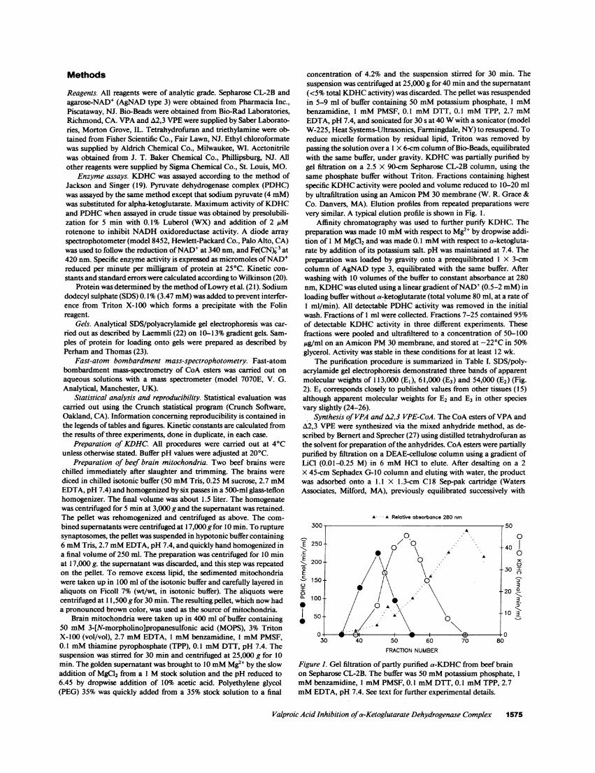
concentration of 4.2% and the suspension stirred for 30 min. Thesuspension was centrifuged at 25,000 g for 40 min and the supernatant(<5% total KDHCactivity) was discarded. The pellet was resuspendedin 5-9 ml of buffer containing 50 mMpotassium phosphate, 1 mMbenzamidine, 1 mMPMSF, 0.1 mMDTT, 0.1 mMTPP, 2.7 mMEDTA, pH 7.4, and sonicated for 30 s at 40 Wwith a sonicator (modelW-225, Heat Systems-Ultrasonics, Farmingdale, NY) to resuspend. Toreduce micelle formation by residual lipid, Triton was removed bypassing the solution over a I X 6-cm column of Bio-Beads, equilibratedwith the same buffer, under gravity. KDHCwas partially purified bygel filtration on a 2.5 X 90-cm Sepharose CL-2B column, using thesame phosphate buffer without Triton. Fractions containing highestspecific KDHCactivity were pooled and volume reduced to 10-20 mlby ultrafiltration using an Amicon PM30 membrane (W. R. Grace &Co. Danvers, MA). Elution profiles from repeated preparations werevery similar. A typical elution profile is shown in Fig. 1.
Affinity chromatography was used to further purify KDHC. Thepreparation was made 10 mMwith respect to Mg2" by dropwise addi-tion of 1 MMgCI2 and was made 0.1 mMwith respect to a-ketogluta-rate by addition of its potassium salt. pH was maintained at 7.4. Thepreparation was loaded by gravity onto a preequilibrated 1 X 3-cmcolumn of AgNADtype 3, equilibrated with the same buffer. Afterwashing with 10 volumes of the buffer to constant absorbance at 280nm, KDHCwas eluted using a linear gradient of NAD' (0.5-2 mM)inloading buffer without a-ketoglutarate (total volume 80 ml, at a rate of1 ml/min). All detectable PDHCactivity was removed in the initialwash. Fractions of 1 ml were collected. Fractions 7-25 contained 95%of detectable KDHCactivity in three different experiments. Thesefractions were pooled and ultrafiltered to a concentration of 50-100ug/ml on an Amicon PM30 membrane, and stored at -22°C in 50%glycerol. Activity was stable in these conditions for at least 12 wk.
The purification procedure is summarized in Table I. SDS/poly-acrylamide gel electrophoresis demonstrated three bands of apparentmolecular weights of 113,000 (EI), 61,000 (E3) and 54,000 (E2) (Fig.2). E, corresponds closely to published values from other tissues (15)although apparent molecular weights for E2 and E3 in other speciesvary slightly (24-26).
Synthesis of VPA and A2,3 VPE-CoA. The CoA esters of VPAandA2,3 VPE were synthesized via the mixed anhydride method, as de-scribed by Bernert and Sprecher (27) using distilled tetrahydrofuran asthe solvent for preparation of the anhydrides. CoAesters were partiallypurified by filtration on a DEAE-cellulose column using a gradient ofLiCl (0.01-0.25 M) in 6 mMHCI to elute. After desalting on a 2X 45-cm Sephadex G-10 column and eluting with water, the productwas adsorbed onto a 1.1 X 1.3-cm C18 Sep-pak cartridge (WatersAssociates, Milford, MA), previously equilibrated successively with
A A Relative absorbance 280 nm
300 50
E250 600~~~~~~0FigureA.Gel filtration of partly purified a fE
E~~~~~~~ :30'0E \1m,
0 m T-150 I.o ~~~~~~~~~~~~~~~~~3
W~~ ~ ~ ~ ~~~~_202.0100 * .
50 A 10 30~~~~~~~
0 ED030 40 50 60 70 80
FRACTION NUMBER
Figure 1. Gel filtration of partly purified a-KDHC from beef brainon Sepharose CL-2B. The buffer was 50 mMpotassium phosphate, 1mnMbenzamidine, 1 mMPMSF, 0.1I mMDTT, 0.1 mMTPP, 2.7mMEDTA, pH 7.4. See text for further experimental details.
Valproic Acid Inhibition of a-Ketoglutarate Dehydrogenase Complex 1575

Table I. Purification of a-KDHCfrom Beef Brain
Step Final volume Protein Enzyme activity Specific activity Yield
ml mg nmol/min mmol/mg per min %
Crude Triton extract 400 4854 52,190 0.0109 100*Poly(ethylene glycol) precipitation 9 1320 52,000 0.0394 99Gel filtration on Sepharose CL-2B 20 376 16,930 0.442 32Affinity gel filtration on AgNADtype 3 20 3.9 13,756 3.5 26
All values refer to a preparation from about 350 g of beef brain. * Accurate values for a-KDHC were difficult to obtain before Triton extrac-tion because of NADHoxidases.
HPLC-grade acetonitrile, water, and 50 mMammonium acetate, pH5.3. The product was eluted with a discontinuous gradient of 15%acetonitrile in 50 mMammonium acetate (pH 5.3) followed by 30%acetonitrile in 50 mMammonium acetate. The material eluted in the30% fraction was lyophilized and stored at -220C.
Identification and purity of CoA esters were assessed as follows.HPLC using the methanol/phosphate/acetonitrile buffer gradient as
described by Causey et al. (28) revealed a single peak. FAB-MSshowedmajor ion peaks at 894 and 892 for VPA-CoA and A2,3 VPE-CoA,respectively. These correspond to the calculated molecular weights ofthese two compounds. Absorption spectra showed a ratio at 232nm/260 nmof 0.42 for VPA-CoA and 0.56 for A2,3 VPE-CoA (Fig. 3).Thin-layer chromatography on cellulose thin layer (Eastman Kodak13181, Rochester, NY) by the method of Myers and Utter (29) showedsingle spots which migrated slightly ahead of a tiglyl-CoA standardwhen visualized by UV light. After alkaline hydrolysis, these spotsdisappeared and new spots running with CoA standard appeared.
tration isolated from beef heart may show extremely low ratesof NAD' reduction when branch-chain amino acids are sub-strates (0.04-0.15% compared to a-ketoglutarate [19]) but thatsuch activity was not detected with KDHCfrom pig heart (30,31). Dehydrogenase activity showed partial dependency on
cations. At saturating substrate concentrations, total activitywas 28% of maximum in the absence of Ca2+ or Mg2", both ofwhich were effective activators. EDTAdid not further reduceactivity in the absence of cations. Mn2" and Ba2" were alsoeffective activators but Zn2` and Cu2" irreversibly inactivatedthe enzyme. Exogenous TPP was required for full activity but70% activity was seen in its absence, presumably owing toenzyme bound TPP.
The steady-state kinetics of KDHCare complex (32-34);substrate concentrations less than 10 X Km can substantiallyreduce the apparent Vma.. Wechose to examine some kinetic
Results
Enzymatic properties of beef brain KDHC. Cofactor depen-dency and substrate specificity were determined for beef brainKDHC. Under steady-state conditions, assays of NAD' re-
duction showed marked substrate specificity. The followingsubstrates were tested at 4 mM(the order of magnitude seen ininherited metabolic disease): pyruvate, a-keto-3-methylvaler-ate, a-ketoisocaproate, a-ketobutyrate, and oxaloacetate. VPAand A2,3 VPEwere tested at 20 mM, concentrations possibleduring therapy or overdose. No activity was seen with any ofthese substrates. Compared with a-ketoglutarate (=100% ac-
tivity), activity with a-ketoisovalerate was 1.7%, with acetoace-tate 2.3%, and 16.6% with a-ketoadipate (all tested at 4 mM).These results are similar to those reported with KDHCfrompig heart (30). It should be noted that KDHCin high concen-
k s
Figure 2. SDS/polyacrylamide gel(10-13% wt/vol) electrophoresis of a-
KDHCfrom beef brain (K) and a stan-dard polypeptide mixture (S) (SDS 7,Sigma Chemical Co.).
Figure 3. (a) Absorption spectra of VPA-CoA and (b) 62,3 VPE-CoA200-300 nm. Spectra were recorded on a diode array spectropho-tometer (model 990, Waters Associates).
1576 Luder et al.
. . .. a .
220 a 4 . ... ..20........
-.1
__
2im1 2iO ^4@ 2iOWaveln#Sth 20 - - 2?9.4 no
2if

constants of brain KDHCfor its physiologic substrates. Foreach constant, one substrate was tested at varying concentra-tions and other substrates were at saturating concentrations(100 X Ki). Kinetic constants for CoA and a-ketoglutarateand inhibition by succinyl CoA with varied concentrations ofCoA are shown in Table II. In these conditions, the enzymaticproperties of the preparation from beef brain are similar tothose previously reported in other tissues (19, 34-37).
Inactivation of KDHCby VPA metabolites. Preincubationof KDHC(0.1 mg/ml) with 2 mMA2,3 VPE at 250C causedsignificant time-dependent loss of catalytic activity (P < 0.005)(Fig. 4), but VPA up to 20 mMshowed no inactivation. 0.1mg/ml KDHCwas used since more dilute solutions were lessstable and activity too low for accurate assay. Inhibitor con-centrations were 10-100-fold their steady-state Km to ensuresaturating conditions. Attempts to reactivate the enzyme byextensive dialysis at 4VC failed, whereas control activity wasunaffected by dialysis. Preincubation of NADH-reducedKDHCwith both VPA-CoA and A2,3 VPE-CoA caused simi-lar significant inactivation when incubated for up to 120 min(P < 0.001) (Fig. 5), although steady-state kinetic studiesshowed different patterns of inhibition (see below). "Rescue"experiments were performed, in which enzyme inactivated byA2,3 VPE-CoA as described above was further incubated witheither CoA, NAD', or a-ketoglutarate. 95-100% activity wasrestored by treatment with CoA (Fig. 5), but not with NAD' ora-ketoglutarate (data not shown).
Steady-state kinetics of KDHCinhibition. Initial velocityassays of KDHCwere done at different concentrations of VPAand A2,3 VPE at saturating NAD' and CoA concentrations.Reactions were initiated with enzyme to accurately estimateinitial rates, before inactivation of enzyme occurred. Assay ofE, subunit-mediated reduction of Fe(CN)-3 showed no inhibi-tion by VPAor A2,3 VPE. In contrast, A2,3 VPE inhibited thereduction of NAD' by KDHCand was a competitive inhibitorwith respect to a-ketoglutarate (Fig. 6 A). Under these condi-tions, Ki. was 41±1.3 MM. The Hill coefficient (Fig. 6 B) was
Table II. SomeKinetic Constants for a-KDHCfrom Beef Brain and Other Tissues
Substrate Km Reported Km(tissue)
AM AM
a-Ketoglutarate 113±19 110 (pig heart) (34)13 (pig heart) (35)81 (bovine liver mitochondria) (19)
CoA 1.3±0.2 4.5 (pig heart) (36)<0.1 (pig heart) (35)
2.7 (rat liver) (32)
Inhibitor K,.
Succinyl CoA 3.9±0.4 6.9 (rat liver) (32)
Assays were performed by initiating the reaction with the test sub-strate in the presence of saturating concentrations (10-100 X K.) ofother substrates. Inhibition by succinyl-CoA was studied understeady-state conditions in saturating concentrations of NAD+and a-ketoglutarate. Results were calculated from the data collected fromthree repeat experiments in each case.
1 UU - - - - -
> 0j O-0 control
)90 T * -* inhibitor
> 80
M 70
zLUJo 60OfLULa-
500 30 60 90 1 20
INCUBATION (MIN)
Figure 4. Inactivation of purified beef brain a-KDHC by 62,3 VPE.The complex (0.1 mg/ml) was incubated at 25°C in 50 mMMOPS,2 mMMgCl2, 0.5 mMCaCl2, 2.6 mMcysteine, 0.1 mMTPP, pH7.6, either in the presence of 2 mMB2,3 VPE (inhibitor) or not (con-trol). Aliquots taken at intervals were assayed as described in thetext. Each data point represents the mean of duplicate determina-tions from three experiments, with error bars indicating I SD. Theordinate represents percentage of initial rate activity of an untreatedsample. At 120 min, activity in the preincubated samples was signifi-.cantly lower than control (P < 0.005).
0.5, suggesting negative cooperativity and half-maximal inhi-bition occurred at 2.04 mM(intercept at log [VO/(Vi -V)]= 0). VPAdid not inhibit at concentrations as high as 10 mM.
VPA-CoA and A2,3 VPE-CoA were both inhibitors ofbrain KDHCunder steady-state conditions. VPA-CoA was acompetitive inhibitor with respect to CoAwith an apparent Kisof 2.9±0.2 MM(Fig. 7 A). A2,3 VPE-CoA showed characteris-
11011 00
90<12 80t ± -O control
< 70 T601> 6011I O Oinhibitor
< 50t I aloneF 40-- A-A inhibitor experimentZ 30 + + CoA added at 60 min.LUj0 20-LUJ 1 0-b
~- 04 II0 20 40 60 80 100 120
INCUBATION TIME (MIN)
Figure 5. Inactivation of purified beef brain a-KDHC by 62,3 VPE-CoA. The complex (0.1 mg/ml) was incubated at 25°C in 50 mMMOPS, 2 mMMgCl2, 0.5 mMCaCl2, 0.1 mMcysteine, 0.1 mMTPP, pH 7.6, either in the presence of 1 mM62,3 VPE-CoA (inhibi-tor) and 1 mMNADHor in 1 mMNADHonly (control). Aliquotswere taken at intervals and assayed as described in the text. Eachdata point represents the mean of duplicate determinations fromthree experiments, with error bars indicating I SD. The ordinate rep-resents percentage of initial rate activity of an untreated sample. Ac-tivity at 90 and 120 min was significantly lower in the preincubatedsamples (P < 0.01 90 min, P < 0.001 120 min). No significant differ-ence in activity from control if 0.5 mMCoA was added to aliquotsat 60 min and assayed at 90 and 120 min. Incubation with VPA-CoA gave similar results.
Valproic Acid Inhibition of a-Ketoglutarate Dehydrogenase Complex 1577

0.200.
-W'50.150-0
> 0.100.
0.050-
0.000
ozoo
>0.100-
°2-o.ioo.
-0.300-
A
Ii5
o controlInhibitor 4OvM
AInhIItor4IO/ M
indbltor 4mM
-10 -5 0 10 15 20 251/[a-ketoglutarate (mM)]
B
-0.400-0.200 0.000 0.200 0.400 0.600 0.800 1.000 1200
log [1)
Figure 6. (A) Inhibition of NAD' reduction by purified beef brain a-KDHCwith 62,3 VPE. Inhibitor was added to the assay buffer witha-ketoglutarate to initiate the reaction. For other assay conditions seedetails in text. (B) Inhibition of brain a-KDHC presented as a Hillplot. Vi = initial velocity in the presence of inhibitor, VO = initial ve-locity under the same conditions in the absence of inhibitor. a-Keto-glutarate concentration was 113 gM (=Km). Other substrate concen-trations as described in the text.
tics of a mixed type inhibitor (Fig. 7 B). Kii was 1.0±0.03 AMand Ki, was 6.1±0.4 MM. These experiments were repeatedwith variable enzyme concentrations at fixed concentrationsof CoAat its Km(1.3 AM). The results are shown in Fig. 8. Thepattern of inhibition shown by VPA-CoA was typical of acompetitive inhibitor and that of A2,3 VPE-CoA of mixedtype inhibition. Hill plots of data from the inhibition ofKDHCby A2,3 VPE-CoA gave a Hill coefficient of 0.74 sug-gesting negative cooperativity. Half-maximal inhibition con-centration was 1.39 MM(Fig. 9).
The metabolic effects of VPA in vivo resemble those seenin some inherited disorders of metabolism. Experiments were
0.180
0.160
0.140
> 0.120
0 0.100
WLl 0.080
\ 0.060
0.040
0.020
1 /[COENZYME A (vM)]
therefore performed to compare kinetic properties of somenaturally occurring keto-acyl CoAs with those of the VPAmetabolites. These experiments are summarized in Table III.The esters examined all showed marked inhibition of KDHC.All showed characteristics of competitive inhibitors with re-spect to CoA, except isobutyryl-CoA which was a mixed inhib-itor (Fig. 1O) and showed kinetic properties similar to those ofA2,3 VPE-CoA. Isobutyryl-CoA is known to be a suicide in-hibitor which inactivates KDHC, presumably owing to cova-lent modification of the E2 subunit (1 9).
Discussion
The results reported here are consistent with a proposed mech-anism of VPAaction in which inhibition and/or inactivationof KDHCby VPA would reduce citric acid cycle flux andincrease flux into GABAsynthesis. We have demonstratedthat KDHCfrom mammalian brain is inactivated and inhib-ited by VPA metabolites. Chronic administration of VPA intherapeutic doses in dogs produced brain tissue levels of A2,3VPE of - 0.5-2 ,gM and VPAwas considerably higher (38).Furthermore, VPA and its metabolites have been shown toaccumulate 10-fold in mitochondria (39). Although data de-scribing steady-state intramitochondrial concentrations ofCoA metabolites of VPAare lacking, even a conservative esti-mate based upon this published data suggests that they arepresent in concentrations well above the measured apparent Kivalues as determined by us. In addition, inactivation of KDHCby unsaturated VPAderivatives could provide a theoretic basisfor the prolonged action of VPA (40-42) and be consistentwith correlation of clinical effect with serum levels of unsatu-rated metabolites. During therapy, inactivation of enzyme andits resynthesis or reactivation would be in a steady state, at alower KDHCconcentration than before treatment. After dis-continuation of therapy, previous KDHClevels would onlyreturn after new synthesis and clearing of protein bound inhib-itor. A precedent for inactivation by a similar molecule is iso-butyryl-CoA, which is a known suicide inhibitor of KDHC(19). This compound is elevated in disorders of propionate andmethylmalonic acid metabolism, conditions whose clinicaland biochemical complications are very similar to VPA tox-icity.
The results reported here demonstrating inactivation ofKDHCby VPAmetabolites have implications for the molecu-lar mechanism of such inactivation. A2,3 VPE inactivatedKDHCwhereas the saturated parent compound VPAdid not.
Figure 7. Inhibition of purifiedbeef brain a-KDHCby (A) VPA-CoA and (B) 62,3 VPE-CoA. In-hibitor was added with CoA to theassay buffer to initiate the reac-tion. For other assay conditionssee details in text.
1578 Luder et al.

60 90 0 30(a) ENZYMEQUANTITY (vL)
Figure 8. Variable enzyme plots ofinhibition of purified beef brain a-KDHCby (A) VPA-CoA (5 AM= 2X Kii) and (B) 62,3 VPE-CoA (5IAM = 5 X K,,). Assays were initi-ated by the addition of a-ketogluta-rate and inhibitor to the assaybuffer. CoA concentration was 1.3MM(=Km). Assays were carried outwith increasing volumes of enzyme(0.1 mg/ml). For details of assayconditions see text.
Since it is known that double bonds in A2,3 unsaturated acidsare susceptible to nucleophilic attack (for example, during thechemical synthesis of unsaturated fatty acyl-CoA esters [27]),it is possible that such a mechanism could explain the differ-ence in behavior of the two molecules, although alternativepossibilities such as allosteric effects are not excluded. Tight,probably covalent binding to the enzyme is supported by thefact that dialysis failed to reactivate it. Reduction of Fe(CN) 3,which is carried out by the El subunit, a-ketoglutarate decar-boxylase, was not inhibited by A2,3 VPE, whereas NAD' re-duction was inhibited. This observation suggests that bindingof A2,3 VPE to KDHCis not at the El subunit.
The mechanism of inactivation of KDHCby VPA-CoAand A2,3 VPE-CoA was investigated by testing the possibilitythat these compounds bind to the reduced E2 subunit via athioester bond, in a similar manner to its physiological prod-uct, succinyl-CoA:
E2 E2
+ RCOSCoA*-P-
S S S SH H Co
RH
0.400U
0.300-\
D 0.100- .
0.000'.
-0.100-.
-0.200 a |-0.400 -0.200 0.000 0.200
E2 is the lipoamide succinyltransferase subunit of KDHC, R isthe acyl group, and SH the free sulfhydryl groups (afterMcMinn and Ottaway [33]). Excess free CoA can competewith the E2-bound acyl group, if it is bound via a thioesterbond, to regenerate the acyl-CoA and free sulfhydryl groups, inthe reverse of reaction 1. Succinyl-CoA is bound to E2 rela-tively weakly and under physiologic conditions is displacedfrom E2 by free CoA (33). It is proposed that the binding ofVPA-CoA and A2,3 VPE-CoA is much tighter. The acylgroups would be displaced only slowly under physiologicalconditions in this model, but higher concentrations of CoAwould shift the equilibrium in favor of displacement. The find-ing that re-activation of the enzyme occurred when excess freeCoA (0.5 mM) was added to the VPA-CoA or A2,3 VPE-CoA-inactivated enzyme is in support of this mechanism. Theinactivation of KDHCby isobutyryl-CoA has been shown tooccur by this mechanism (19). Further support for this is thefact that neither dialysis nor addition of NAD' nor a-ketoglu-tarate, which do not have free sulfhydryl groups, were effectivein reactivation. Binding of acyl-CoA by thioester bonds is de-pendent on the presence of reduced sulfhydryl groups. In thisexperiment NADHwas present in order to reduce any oxi-dized lipoamide as follows:
E2 E2
+ 2NADH - + 2NAD
S .----S S S
H
0.400
log (1]
Figure 9. Inhibition of purified beef brain a-KDHC by 62,3 VPE-CoA. Results are presented as a Hill plot. Vi is the initial rate of reac-
tion in the presence of inhibitor and V. the rate under the same con-
ditions without inhibitor. CoA concentration was 1.3 MM(=Km).Other substrates were as described in the text.
H
Table III. Inhibition of Beef Brain a-KDHCby Keto-acyl CoA Esters
Inhibitor Ku Inhibition type
Isovaleryl-CoA 77.8±4.4 competitiveTiglyl-CoA 44.0±1.9 competitiven-Propionyl-CoA 49.2±1.8 competitiveIsobutyryl-CoA 18.6±0.4 (Kii) mixed
41.3±1.5 (Kis,)
Assays were carried out under steady state conditions and constantscalculated as described in the legend to Table II.
Valproic Acid Inhibition of a-Ketoglutarate Dehydrogenase Complex 1579
1>0o

A inhibitor 183p/4C. 0.120-0
>L 0.080-
0.040-
0.000 I I-0.600 -0.400 -0.200 0.000 0.200 0.400 0.600
1 /[COENZYME A (vM)]Figure 10. Inhibition of purified beef brain a-KDHC by isobutyryl-CoA. Assay conditions are as described in the legend of Fig. 7.
In addition to inactivation, we have also demonstrated thatVPA metabolites and some naturally occurring fatty acyl-CoAs are potent (reversible) inhibitors of KDHC. Inhibition ofKDHC, which occurs in isolated rat hepatocytes under condi-tions favoring an increased acyl-CoA/free CoA ratio, is asso-ciated with reduced citric acid cycle flux (43-45). An increasedacyl-CoA/free CoA ratio occurs in the presence of excess acylgroups, such as in patients with organic acidemias and otherinborn errors of metabolism, and VPAtherapy (46). Althoughour studies do not directly address the relative importance invivo of inactivation and inhibition, the similarity of the ki-netics of VPAmetabolites and natural acyl-CoA esters presentin excess in inborn errors supports the notion that inhibition ofthe citric acid cycle and KDHCby VPAmay be a significanteffect in vivo.
The work presented here provides a testable model forVPA action. Experiments to analyze GABAflux from thebrain cell body and synaptosomic mitochondria treated withVPA are currently under way to study this model further.
Acknowledgments
The excellent technical assistance of Young-Hwa Lim is gratefullyacknowledged. Dr. Paul Fennessy performed the mass spectrometrystudies.
This work was supported by grants NS-24872 and NS-26520 fromthe National Institutes of Health to Dr. Parker, a Mental RetardationResearch Center grant, and NIH Research Resource Grant RR-01 152.Dr. Parker is the recipient of a Clinical Investigator DevelopmentAward from NINDS.
References
1. Meunier, H., G. Carray, Y. Meunier, P. Eymard, and M. Aicard.1963. Pharmacology of 2-propyl valeric acid. Therapie (Paris).18:435-483.
2. Zimmerman, H. J., and K. G. Ishak. 1982. Valproate-inducedhepatic injury: analyses of 23 fatal cases. Hepatology (Baltimore).2:591-597.
3. Simler, S., L. Chesielski, M. Maitre, H. Randrhanarisoa, and P.Mandel. 1973. Effects of sodium-n-dipropylacetate on audiogenic sei-zures and brain gamma-aminobutyric acid levels. Biochem. Pharma-col. 22:1701-1708.
4. Chapman, A., P. E. Keane, B. S. Meldrum, J. Simiand, and J. C.Vernieres. 1982. Mechanism of anticonvulsant action of valproate.Prog. Neurobiol. 19:315-359.
5. Taberner, P. B. 1979. Effects of sodium valproate and ethanol-amine-o-sulphate on GABAmetabolism. Br. J. Pharmacol. 67:441.
6. Turnbull, D. M., A. J. Bone, K. Barlett, P. P. Koundakjian, andH. S. A. Sheratt. 1983. The effects of valproate on intermediary metab-olism in isolated rat hepatocytes and intact rats. Biochem. Pharmacol.32:1887-1892.
7. Becker, C.-M., and R. A. Harris. 1983. Influence of valproic acidon hepatic carbohydrate and lipid metabolism. Arch. Biochem.Biophys. 223:381-392.
8. Eymard, P., M. Broll, and J. P. Werbend. 1970. Valproate di-minishes oxygen utilization in rat hepatocytes. Bull. Soc. Sci. Vet.Med. Comp. Lyon. 72:303-325.
9. Kukino, K., and T. Deguchi. 1977. Effects of sodium dipropyl-acetate on GABAand biogenic amines in rat brain. Chem. Pharm.Bull. (Tokyo). 25:2257-2262.
I 10. Lai, J. C. K., and J. B. Clarke. 1979. Methods Enzymol. 55(PartF):51-60.
11. Williams, R. J. H., P. N. Patsalos, and R. Lowe. 1980. Changesin brain amino acid neurotransmitters induced by sodium valproateand their relevance to epilepsy and interactions with other anticonvul-sants. In The Place of Sodium Valproate in the Treatment of Epilepsy.Royal Society of Medicine International Congress and SymposiumNo. 30. Academic Press, and The Royal Society, London. 95-102.
12. Haas, R., D. A. Stumpf, J. Parks, and L. Eguren. 1981. Inhibi-tory effects of sodium valproate on oxidative phosphorylation. Neurol-ogy. (NY). 31:1473-1476.
13. Cunningham, J., D. D. Clark, and W. J. Nicklas. 1980. Oxida-tive metabolism of 4-aminobutryrate by rat brain mitochondria: inhi-bition by branched chain fatty acid. J. Neurochem. 34:197-202.
14. Lai, J. C. K., and A. J. L. Cooper. 1986. Brain alpha-ketogluta-rate dehydrogenase complex: kinetic properties, regional distributionand effects of inhibitors. J. Neurochem. 47:1376-1386.
15. Koike, M., and K. Koike. 1976. Structure, assembly and func-tion of mammalian alpha-keto acid dehydrogenase complexes. Adv.Biophys. 9:187-227.
16. Ito, M., Y. Ikeda, G. Finocchiaro, J. Arnez, and K. Tanaka.1986. The enzymatic basis for the metabolism and inhibitory action ofvalproate: dehydrogenation of VPA-CoA by 2-methyl-branched chainacyl-CoA dehydrogenase. Pediatr. Res. 20:343A. (Abstr.)
17. Nau, H., and W. Loescher. 1982. Valproic acid: brain andplasma levels of the drug and its metabolites, anticonvulsant effectsand GABAmetabolism in the mouse. J. Pharmacol. Exp. Ther.220:654-659.
18. Loescher, W., H. Nau, and M. Marescaux-Vergnes. 1984.Comparative evaluation of anticonvulsant and toxic potencies of val-proic acid and 2-en-valproic acid in different animal models of epi-lepsy. Eur. J. Pharmacol. 99:211-218.
19. Jackson, R. H., and T. P. Singer. 1983. Inactivation of the2-ketoglutarate and pyruvate dehydrogenase complexes of the beefheart by branched chain keto acids. J. Biol. Chem. 258:1857-1865.
20. Wilkinson, G. N. 1961. Statistical estimations in enzyme ki-netics. Biochem. J. 80:324-333.
21. Lowry, 0. H., N. J. Rosebrough, A. L. Farr, and R. J. Radials.1951. Protein measurement with the folin phenol reagent. J. Biol.Chem. 193:265-275.
22. Laemmli, U. K. 1970. Cleavage of structural proteins duringthe assembly of the head of bacteriophage T4. Nature (Lond.).227:680-685.
23. Perham, R. N., and J. 0. Thomas. 1971. The subunit molecularweights of the a-keto acid dehydrogenase multienzyme complexesfrom E. coli. FEBS(Fed. Eur. Biochem. Soc) Lett. 15:8-12.
24. Barrera, C. R., G. Namihara, L. Hamilton, P. Munk, M. H.Eley, T. C. Linn, and L. J. Reed. 1972. Alpha-ketoacid dehydrogenasecomplexes-XVI. Arch. Biochem. Biophys. 148:343-347.
25. Weber, K., and M. Osborn. 1969. The reliability of molecular
1580 Luder et al.

weight determinations by dodecyl sulfate-polyacrylamide gel electro-phoresis. J. Biol. Chem. 244:4406-4410.
26. Tanaka, N., K. Koike, K.-I. Otsuka, M. Hamada, K. Ogasha-hara, and M. Koike. 1972. Mammalian a-ketoacid dehydrogenasecomplexes. J. Biol. Chem. 249:191-198.
27. Bernert, J. T., and H. Sprecher. 1977. An analysis of partialreactions in the overall chain elongation of saturated and unsaturatedfatty acids by rat liver microsomes. J. Biol. Chem. 252:6736-6744.
28. Causey, A. G., B. Middleton, and K. Barlett. 1986. A study ofthe metabolism of [U-'4C]3-methyl-2-oxopentanoate by rat liver mi-tochondria using h.p.l.c. with continuous on-line monitoring of radio-active intact acyl-coenzyme A intermediates. Biochem. J. 235:343-350.
29. Myers, D. E., and M. F. Utter. 1981. The enzymatic synthesis ofsome potential photoaffinity analogs of benzoyl-coenzyme A. Anal.Biochem. 112:23-29.
30. Kanzaki, T., T. Hayakawa, M. Hamada, Y. Fukuyoshi, and M.Koike. 1969. Mammalian a-ketoacid dehydrogenase complexes-IV.J. Biol. Chem. 244:1183-1187.
31. Parker, P. J., and P. J. Randle. 1978. Partial purification andproperties of branched chain 2-oxo acid dehydrogenase of ox liver.Biochem. J. 171:751-757.
32. Smith, C. M., J. Bryla, and J. R. Williamson. 1974. Regulatoinof mitochondrial alpha-ketoglutarate metabolism by product inhibi-tion at alpha-ketoglutarate dehydrogenase. J. Bid. Chem. 249:1497-1505.
33. McMinn, C. L., and J. H. Ottaway. 1977. Studies on the mecha-nism and kinetics of the 2-oxoglutarate dehydrogenase system from pigheart. Biochem. J. 161:569-581.
34. Hirashima, M., T. Hayakawa, and M. Koike. 1967. Mamma-lian a-ketoacid dehydrogenase complexes-II. J. Biol. Chem.242:902-907.
35. Massey, V. 1960. The composition of the ketoglutarate dehy-drogenase complex. Biochem. Biophys. Acta. 38:447-460.
36. Ishikawa, E., R. M. Oliver, and L. J. Reed. 1966. Alpha-keto-acid dehydrogenase complexes, V. Proc. Nati. Acad. Sci. USA.56:534-541.
37. Martin-Requero, A., B. E. Corkey, S. Cerdan, E. Walajtys-Rode, R. L. Parrilla, and J. R. Williamson. 1983. Interactions betweena-ketoglutarate metabolism and the pathways of gluconeogenesis andurea synthesis in isolated hepatocytes. J. Biol. Chem. 258:3673-3681.
38. Loscher, W., and H. Nau. 1983. Distribution of valproic acidand its metabolites in various brain areas of dogs and rats after acuteand prolonged treatment. J. Pharmacol. Exp. Ther. 226:845-854.
39. Aly, M. I., and A. A. Abdel-Latif. 1980. Studies on the distri-bution and metabolism of valproate in rat brain, liver and kidney.Neurochem. Res. 5:1231-1242.
40. Harding, G. F. A., C. E. Herrick, and P. M. Jeavons. 1978. Acontrolled study of the effect of sodium valproate on photosensitiveepilepsy and its prognosis. Epilepsia. 19:555-565.
41. Johnston, D., and G. E. Slater. 1982. Valproate-mechanismsof action. In Antiepileptic Drugs. D. M. Woodbury, J. K. Penry, andC. E. Pippenger, editors. Raven Press, NewYork. 611-616.
42. Lockard, J. S., and R. H. Levy. 1976. Valproic acid: reversiblyacting drug? Epilepsia. 17:477-479.
- 43. Williamson, J. R., E. Walajtys-Rode, and K. E. Coll. 1979.Effects of branch chain alpha-ketoacids on the metabolism of isolatedrat liver cells-1. J. Biol. Chem. 254:11511-11520.
44. Walajtys-Rode, E., K. E. Coll, and J. R. Williamson. 1979.Effects of branch chain alpha-ketoacids on the metabolism of isolatedrat liver cells-2. J. Biol. Chem. 254:11521-11529.
45. Walajtys-Rode, E., and J. R. Williamson. 1980. Effects ofbranch chain alpha-ketoacids on the metabolism of isolated rat livercells-3. J. Biol. Chem. 255:413-418.
46. Turnbull, D. M., D. J. Dick, L. Wilson, H. S. A. Sheratt, andK. G. M. M. Alberti. 1986. Valproate causes metabolic disturbance innormal man. J. Neurol. Neurosurg. Psychiatry. 49:405-410.
Valproic Acid Inhibition of a-Ketoglutarate Dehydrogenase Complex 158.1