incidence of enhancement of the optic nerve/sheath complex ... · incidence of enhancement of the...
TRANSCRIPT
Journal of the Korean Radiological Society, 1995 ; 32( 4): 541 - 544
Incidence of Enhancement of the Optic Nerve/Sheath Complex in Fat-Suppression Orbit MRl1
Ho Kyu Lee, M.D. , Kwon-Ha Yoon, M.D. ,
Choong Gon Choi , M.D., DaeChul Suh, M.D.
Purpose: To elucidate the incidence of Gd-DTPA enhancement of the optic nerve/sheath complex (ONC) in patients with various ophthalmopathies using fat-suppression MR I.
Materials and Methods: Orbit MRI with fat-suppression technique (ChemSat) was performed in 58 patients with normal and various orbital lesions. The fat-suppression MR was done with and without Gd-DTPA injection in all cases. MR findings were reviewed retrospectively in a blind fashion with respect to presence or absence of contrast en hancement of the 0 NC.
Results: Contrast enhancement of the ONC was seen in 86% (6/기 of cavernous sinus lesions, 80% (8/10) of intraconallesions excluding the ONC, 57% (16/28) of ONC lesions, 38% (3/8) of ocular lesions, and 2% (1/55) of normal orbits. The ONC enhancement was the most common in optic nerve/sheath tumors (10/ 1 이, and pseudotumors (6/6), cavernous sinus dural arteriovenous malformations (3/3) and cavernous sinus thrombosis (2/2), and less frequently seen in optic neuritis (3/14).
Conclusion: Enhancement of the ONC may be seen in lesions of the cavernous sinus and orbit otherthan optic nerve/sheath lesion.
Index Words: Orbit, MR Magnetic resonance(MR), contrast enhancement Magnetic resonance(MR) , fat suppression
INTRODUCTION
Fat -suppression MR imaging with a paramagnetic agent is a requisite technique in the evaluation of the 。ptic nerve/sheath complex (ONC). Incidence of contrast enhancement of the ONC in optic neuritis and optic nerve sheath meningioma is known to be high (1 -3) But in our study , we found out higher incidence of the contrast enhancement of the ONC in other orbital lesions than primary ONC lesion. It could suggest that contrast enhancement of the ONC does not always mean a specific sign for primary ONC pathologies such as a tumor or inflammation. Therefore, we elucidatethe incidence of various disease including optic neuritis and meningioma.
'Oepartment of Radiology, Asan Medical Center , University ofUlsan Coll ege of Medicine Received January 26,1995 ; Accepted March 29, 1995
Address reprint requests to: Ho Kyu Lee , M.O. , Oepartment of Radiology, Asan Medical Center , University of Ulsan , 388.1 Poongnap-dong , Songpa-ku , Seoul ,
138-040 Kore a. Tel. 82- 2- 224- 4400 Fax. 82- 2- 476- 4719
MATERIALS and METHODS
From April 1992 to May 1994, orbit MRI was performed in 58 consecutive patients (116 sides of orbits). AII MR studies were performed on a 1.5 tesla GE Signa (Milwaukee , Wisconsin) with a quadrature head coil Fat -suppression was accomplished with a frequency selective presaturation p비 se (ChemSat) , which is to excite fat preferently (compared with water) with a narrow -band pulse in the absence of any gradients and dephase its signal with added grad ients , and then immediately continue with the choice of imaging sequence (4). AII images were obtained with both the pre - and postenhanced T1 weighted images with ChemSat using Gd -DTPA. Twenty - one patients were surgically proven and thirty - seven were diagnosed clinically.
On the assumption that the incidence of the contrast enhancement of the ONC is related with proximity of the lesion to the ONC , we evaluated its incidence on the base of orbital compartments.
- 541 -

Journ al of the Korean Radiologica l Society, 1995: 32( 4): 541 -544
Table 1. Incidence ofContrast Enhancement of ONC
NO.ofCE NO. oforbit (%)
Optic nerve/sheath lesions 16 28 57
。ptic neuriti s 3 14
optic nerve contusi on 2 3
optic nerve atrophy
optic nerve/sheath tumor 10 10
perioptic meningioma (5)
optic glioma (2) schwannoma (1 )
Iymphoma (2)
Other intraconallesions 8 10 80
pseudotumor 6 6
retrobulbar Iymphoma
cellulitis
cavernous hemangioma 0 2
Conal and extraconallesions o 8 0
Graves' disease 0
plexiform neurofibroma 0
lacrimal gland mass 0
c。미unctivallymphoma 0 3
dermoid 0 2
Ocular lesions 3 8 38
retinoblastoma 2
vitreous hemorrhage
congenital cataract
chorioretinitis 0
choroidal hemangioma 0 2
retinal artery occlusion 0
Cavernous sinus lesions 6 7 86
du ral AVM 3 3
cavernous sinus thrombosis 2 2
Tolosa-Hunt syndrome 2
Clini cally normal 55 2
Total 34 116 29
* Abbreviati on : ONC - optic nerve/sheath complex No. - number, CE - contrast enhancement
RESUlTS
The enhancement of the ONC was observed in 34 (29 %) in total116 sides ; 6 of 7 cavernous sinus lesions (86 %), 8 of 10 intraconal lesions excluding the ONC (80 %), 16 of 28 ONC lesions (57 %) , 3 of 8 ocular lesions (38 %), 1 of 55 normal orbits (2 %), and none of 8 conal and extraconal lesions (0 % ) (Table 1). Out of the ONC lesions, ONC enhancement was the most common in optic nerve/sheath tumors(10/1 이 , and it was less frequently seen in optic nerve contusion (2/3) and optic neuritis (3/14) (Fig. 1 a , b). Out of the cavernous sinus lesions (Fig. 2) , ONC enhancement was frequently seen in dural arteriovenous malformations (2/2) , cavernous sinus thrombosis (2/2) , and Tolosa - Hunt syndrome (2/3). Out of the intraconal lesions excluding ONC(Fig. 3) , ONC enhancement was frequently seen in pseudotumors (6/6) , retrobulbar Iymphomas (1/1) , and orbital cellulitis (1 11).
DISCUSSION
In the evaluation for orbital lesions, fat - suppression MR techniques have been widely used because they can depict improved anatomic detail , decrease chemical shift artifact, and increase gray scale width (4). Use of a paramagnetic contrast agent in combination with the fat - suppression technique has advanced contrast between pathologic lesions and normal structures. To detect ONC enhancement precisely, we obtained both the pre - and post - enhanced fat - suppression T1 weighted images comparing with other reports (1 , 2) using post - enhanced fat - suppression only.
Hendrix et a l. (2) initially reported usefullness of fat suppression technique using Gd - DTPA in the evaluation of the optic nerve meningioma and Tien et al. (1) applied it to optic neuritis. In addition , we noted enhancement of ONC in other pathologies such as optic nerve contusion , retinoblastoma, dural arteriovenous
Fig. 1. Optic neuritis on the both sides a. Relatively high signal intensity of the optic nerve/sheath complex on pre-enhanced coronal fat su ppressed T1 WI. Fat-suppression failure is noted on the cheek area. b. Bilateral ring enhancement of the optic nerve sheath (arrow heads) on post-enhanced fat suppressed T1 WI. Note intensi ty of the enhanced optic nerve/sheath complex similar to extraocular muscles
a b
m *
Ho Kyu Lee, et al: Incidence of Enhancement of the Optic Nerve/ Sheath Complex in Fat-Suppression Orbit MRI
a b
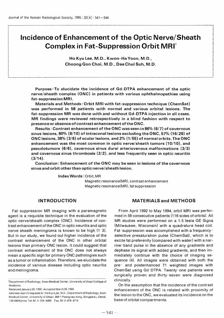
Fig. 2. A small dural arteriovenous mallormation 01 the right cavernous si nus a, b. Contrast-enhanced T1-weighted latsuppressed image shows perioptic enhancement (black arrow) and swelling 01 whole extraocular muscles on the right Note the dilated superior ophthalmic vein (open arrows) on the axial and coronal planes.
be detected by our current MR techniques. There were various orbitallesions showing ONC en
hancement. Higher rates of ONC enhancement were identified in cavernous sinus lesions and intraconal lesions excluding ONC. It is interesting that cavernous sinus lesions showed a highest rate. We thought it might be caused by not disruption of blood optic nerve barrier (7) , but hyperemic state of the optic nerve sheath
In conclusion , contrast enhanced MRI with fat - suppression technique is a powerf비 adjunct in MR evaluation of the optic nerve pathology. ONC enhancement in fat -suppression MRI is not specific for optic nerve lesions. It may be seen in various pathologies including
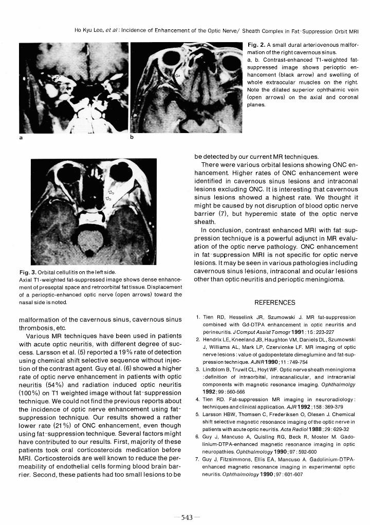
Fig. 3. Orbital cellulitison the leftside. cavernous sinus lesions, intraconal and ocular lesions Axial T1 -weighted lat-suppressed image shows dense enhance- otherthan optic neuritis and perioptic meningioma ment 01 preseptal space and retroorbital lat tissue. Displ acement 01 a perioptic-enhanced optic nerve (open arrows) toward the nasal side is noted
malformation of the cavernous sinus , cavernous sinus thrombosis , etc
Various MR techniques have been used in patients with acute optic neuritis, with different degree of success. Larsson et al. (5) reported a 19% rate of detection using chemical shift selective sequence without injection of the contrast agen t. Guy et a l. (6) showed a higher rate of optic nerve enhancement in patients with optic neuritis (54%) and radiation induced optic neuritis (100%) on T1 weighted image without fat - suppression technique. We could not find the previous reports about the incidence of optic nerve enhancement using fat suppression technique. Our results showed a rather lower rate (21 %) of ONC enhancement , even though using fat - suppression technique. Several factors might have contributed to our results. First, majority of these patients took oral corticosteroids medication before MRI. Corticosteroids are well known to reduce the permeability of endothelial cells forming blood brain barrier. Second , these patients had too smalllesions to be
REFERENCES
1. Tien RD, Hesselink JR , Szumowski J. MR lat-suppression combined with Gd-DTPA enhancement in optic neuritis and perineuritis. JComput AssistTomogr1991 ; 15 : 223-227
2. Hendrix LE. Kneeland JB . Haughton VM, Daniels DL. Szumowski J. Williams AL. Mark LP. Czervionke LF. MR imaging 01 optic nerve lesions: value 01 gadopentetate dimeglumine and lat-suppression technique. AJNR1990 ; 11 : 749-754
3. Lindblom B, Truwit CL. Hoyt WF. Optic nervesheath meningioma delin ition 01 intraorbital. intracanalicular. and intracranial
components with magnetic resonance imaging. Ophthalmolgy
1992 ;99 ‘ 560-566 4. Tien RD. Fat-suppression MR imaging in neuroradiology
techniques and clinical application. AJR 1992 ; 158 : 369-379 5. Larsson HBW. Thomsen C, Frederiksen O. Olesen J. Chemical
shilt selective magnetic resonance imaging 01 the optic nerve in patients with acuteoptic neuritis. Acta Radio/1988 ; 29 : 629-32
6. Guy J. Mancuso A. Quisling RG . Beck R. Moster M. Gadolinium-DTPA-enhanced magnetic resonance imaging in optic neuropathies. Ophthalmology 1990 ; 97 : 592-600
7. Guy J. Fitzsimmons. 티 lis EA. Mancuso A. Gadolinium-DTPAenhanced magnetic resonance imaging in experimental optic neuritis. Ophthalmology 1990 ; 97 : 601-607
m j

Journal of the Korean Radiological Society, 1995; 32( 4) : 541 - 544
대 한 방사선 의 학회 지 1995 ; 32( 4) : 541 - 544
지방조직 억제 MR을 이용한 시신경 ·신경초 복합체의 조영증강의 빈도1
1 울산대학교 의과대학 진 단방사선과학교실
이호규·윤권하·최충곤·서대철
목 적 :지방조직 억제 MR을 이용한 안와 MR 영상에서 시신경 • 신경초 복합체의 조영증강의 빈도를 알아보고자 하였다.
대상 및 방법 :총 58명의 환자에서 지방억제 안와 MR을 시행하였다. 지방억제기법은 frequency selective presaturation
pulse인 “ChemSat"을 이용하였다. 전예에서 Gd-DTPA 주입전 및 주입후 지방억제 MR영상을 모두 얻었다. 시신경 신경초 복
합체의 조영증강 여부를 후향적으로 분석하였다.
결 과:시신경 · 신경초 복합체의 조영증강의 빈도는 해면정맥동 병변 86% (6/7), 시신경 · 신경초 이외의 원추내
(intraconal) 병변 80% (8/ 10) , 시신경 · 신경초 병변 57% (16/28) , 안구병변 38% (3/8) , 정상안 2% (1 /55) 등의 순이었다.
시신경 · 신경초 복합체 조영증강은 시신경 · 신경초 종앙 ( 10/ 10) , 및 가성종앙 (6/6)에서 가장 많았고, 시신경 화상 (con
tusion) (2/ 3) , 시신경염 (3/ 14) 해면동정맥 기형 (3/3) , 해면동 혈전 (2/2) 등에서도 나타났다.
결 론:시신경 · 신경초 복합체의 조영증강은 단순한 신경염이나 신경초 수막종 등의 시신경 · 신경초 복합체 병변 이외
에도 해면정맥동 병변이나 원추내 병변 등에서도 나타날 수 있다.
544 -