incidental findings - nucradshare.com incidental findings guidelines.pdf · management of...
TRANSCRIPT
INCIDENTAL FINDINGSClick on the hyperlinks below:
CT or MRI Abdomen/Pelvis:Adrenal Lesions
Pancreatic Cystic LesionsRenal Lesions
Liver Lesions, no risk of HCCLiver Lesions, risk of HCC (LI-RADS)
Splenic LesionsLymph Node Findings
Adnexal LesionsGallbladder and Biliary Lesions *new*
Ultrasound:Cystic Adnexal LesionsOther Adnexal Lesions
Thyroid NodulesChest:
Solid Pulmonary NodulesSubsolid Pulmonary Nodules *new*
Vascular:Abdominal Aortic or Iliac Aneurysms
Penetrating Aortic UlcersSplenic or Renal Aneuryms
Other Abdominal Vascular Findings
v.5 Sept. 2014
The Latest Management Recommendations
Notes: This resource is intended to be a readily available, continuously updated document for both residents and attend-ings to regularly refer to when making recommendations and management decisions for common incidental findings. The goal of this resource is to decrease variability in the way we manage incidental findings by implementing the best and most recent research and expert opinion. The ACR White Paper guidelines (and non-ACR guidelines herein) are not to be confused with the ACR Practice Guidelines and Technical Standards, do not represent official ACR policy, and should not represent the legal standard of care. Please share your feedback ([email protected]) and suggestions.
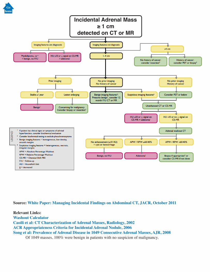
Source: White Paper: Managing Incidental Findings on Abdominal CT, JACR, October 2011
Relevant Links:Washout CalculatorCaoili et al: CT Characterization of Adrenal Masses, Radiology, 2002ACR Appropriateness Criteria for Incidental Adrenal Nodule, 2006Song et al: Prevalence of Adrenal Disease in 1049 Consecutive Adrenal Masses, AJR, 2008 Of 1049 masses, 100% were benign in patients with no suspicion of malignancy.
Incidental Adrenal Mass≥ 1 cm
detected on CT or MR
Gohome
Incidental Cystic Pancreatic MassIn an asymtomatic1 patient,
detected on CT, MRI (w/ or w/o contrast) or US.
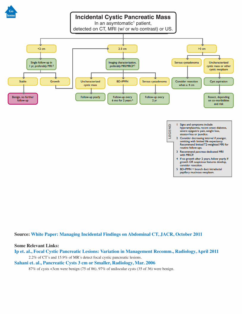
Source: White Paper: Managing Incidental Findings on Abdominal CT, JACR, October 2011
Some Relevant Links:Ip et. al., Focal Cystic Pancreatic Lesions: Variation in Management Recomm., Radiology, April 2011 2.2% of CT’s and 15.9% of MR’s detect focal cystic pancreatic lesions.Sahani et. al., Pancreatic Cysts 3 cm or Smaller, Radiology, Mar. 2006 87% of cysts <3cm were benign (75 of 86), 97% of unilocular cysts (35 of 36) were benign.
Gohome
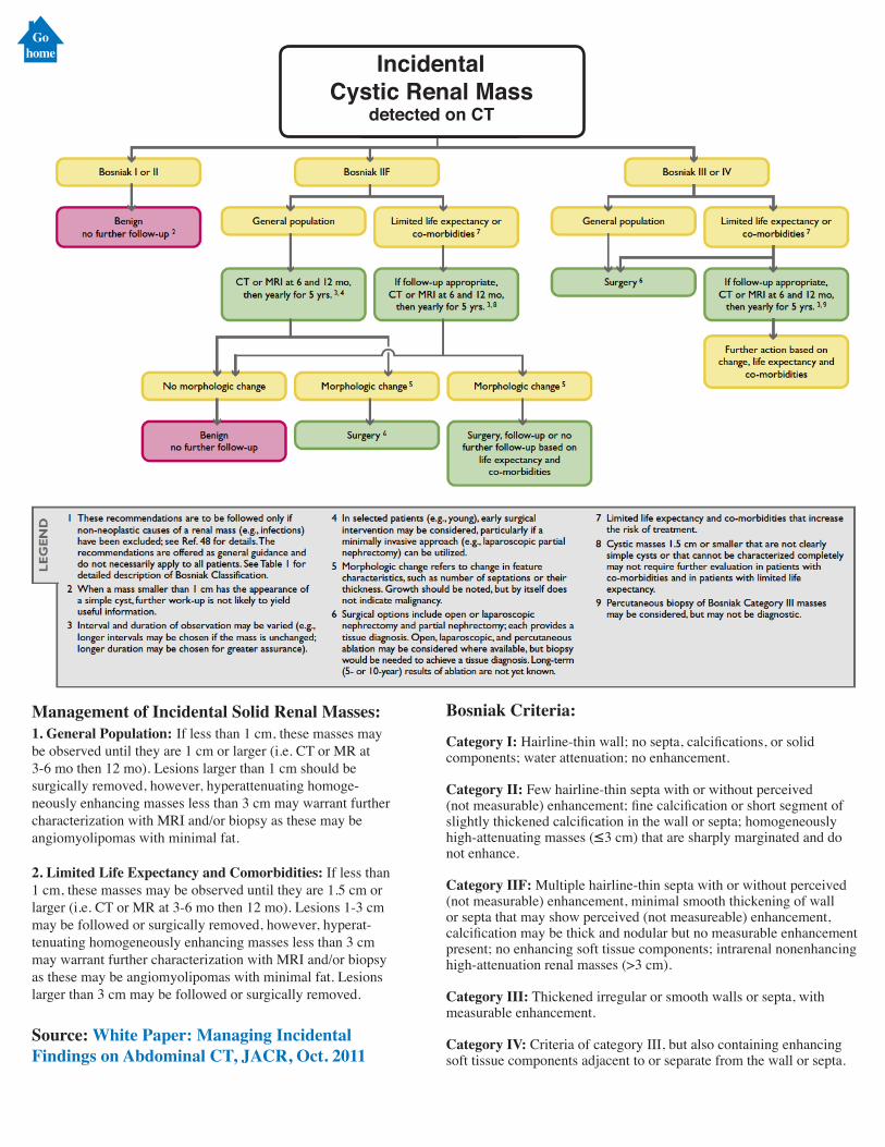
Management of Incidental Solid Renal Masses: 1. General Population: If less than 1 cm, these masses may be observed until they are 1 cm or larger (i.e. CT or MR at 3-6 mo then 12 mo). Lesions larger than 1 cm should be surgically removed, however, hyperattenuating homoge-neously enhancing masses less than 3 cm may warrant further characterization with MRI and/or biopsy as these may be angiomyolipomas with minimal fat.
2. Limited Life Expectancy and Comorbidities: If less than 1 cm, these masses may be observed until they are 1.5 cm or larger (i.e. CT or MR at 3-6 mo then 12 mo). Lesions 1-3 cm may be followed or surgically removed, however, hyperat-tenuating homogeneously enhancing masses less than 3 cm may warrant further characterization with MRI and/or biopsy as these may be angiomyolipomas with minimal fat. Lesions larger than 3 cm may be followed or surgically removed.
Source: White Paper: Managing Incidental Findings on Abdominal CT, JACR, Oct. 2011
Bosniak Criteria:
Category I: Hairline-thin wall; no septa, calcifications, or solidcomponents; water attenuation; no enhancement.
Category II: Few hairline-thin septa with or without perceived(not measurable) enhancement; fine calcification or short segment of slightly thickened calcification in the wall or septa; homogeneously high-attenuating masses (≤3 cm) that are sharply marginated and do not enhance.
Category IIF: Multiple hairline-thin septa with or without perceived (not measurable) enhancement, minimal smooth thickening of wall or septa that may show perceived (not measureable) enhancement, calcification may be thick and nodular but no measurable enhancement present; no enhancing soft tissue components; intrarenal nonenhancing high-attenuation renal masses (>3 cm).
Category III: Thickened irregular or smooth walls or septa, with measurable enhancement.
Category IV: Criteria of category III, but also containing enhancing soft tissue components adjacent to or separate from the wall or septa.
Incidental Cystic Renal Mass
detected on CT
Gohome
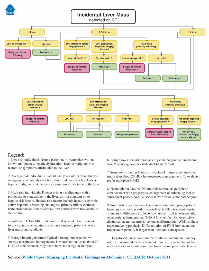
Incidental Liver Massdetected on CT
Source: White Paper: Managing Incidental Findings on Abdominal CT, JACR, October 2011
Legend:1. Low risk individuals: Young patient (≤ 40 years old), with no known malignancy, hepatic dysfunction, hepatic malignant risk factors, or symptoms attributable to the liver.
2. Average risk individuals: Patient >40 years old, with no known malignancy, hepatic dysfunction, abnormal liver function tests or hepatic malignant risk factors or symptoms attributable to the liver.
3. High risk individuals: Known primary malignancy with a propensity to metastasize to the liver, cirrhosis, and/or other hepatic risk factors. Hepatic risk factors include hepatitis, chronic active hepatitis, sclerosing cholangitis, primary biliary cirrhosis, hemochromatosis, hemosiderosis, oral contraceptive use, anabolic steroid use.
4. Follow-up CT or MRI in 6 months. May need more frequent follow-up in some situations, such as a cirrhotic patient who is a liver transplant candidate.
5. Benign imaging features: Typical hemangioma (see below), sharply marginated, homogeneous low attenuation (up to about 20 HU), no enhancement. May have sharp, but irregular margins.
6. Benign low attenuation masses: Cyst, hemangioma, hamartoma, Von Meyenberg complex (bile duct hamartomas).
7. Suspicious imaging features: Ill-defined margins, enhancement (more than about 20 HU), heterogeneous, enlargement. To evaluate, prefer multiphasic MRI.
8. Hemangioma features: Nodular discontinuous peripheral enhancement with progressive enlargement of enhancing foci on subsequent phases. Nodule isodense with vessels, not parenchyma.
9. Small robustly enhancing lesion in average risk, young patient: hemangioma, focal nodular hyperplasia (FNH), transient hepatic attenuation difference (THAD) flow artifact, and in average risk, older patient: hemangioma, THAD flow artifact. Other possible diagnoses: adenoma, arterio-venous malformation (AVM), nodular regenerative hyperplasia. Differentiation of FNH from adenoma important especially if larger than 4 cm and subcapsular.
10. Hepatocellular or common metastatic enhancing malignancy: islet cell, neuroendocrine, carcinoid, renal cell carcinoma, mela-noma, choriocarcinoma, sarcoma, breast, some pancreatic lesions.
Gohome
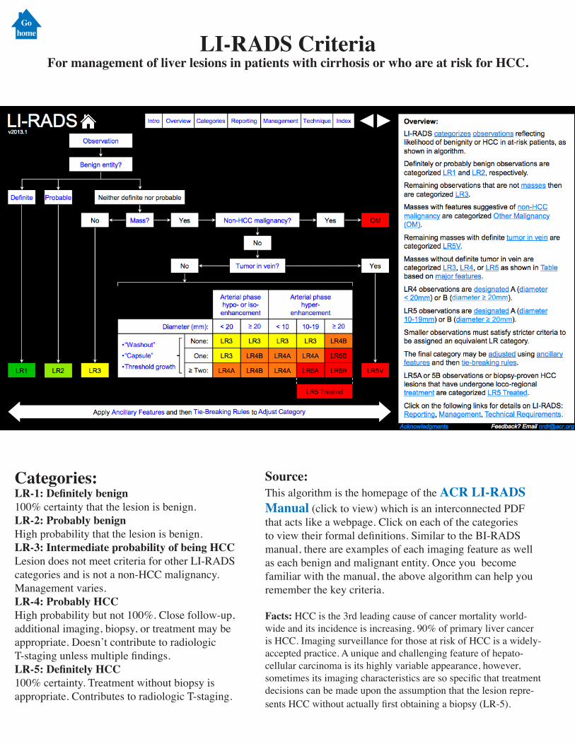
Source:This algorithm is the homepage of the ACR LI-RADS Manual (click to view) which is an interconnected PDF that acts like a webpage. Click on each of the categories to view their formal definitions. Similar to the BI-RADS manual, there are examples of each imaging feature as well as each benign and malignant entity. Once you become familiar with the manual, the above algorithm can help you remember the key criteria.
Facts: HCC is the 3rd leading cause of cancer mortality world-wide and its incidence is increasing. 90% of primary liver cancer is HCC. Imaging surveillance for those at risk of HCC is a widely-accepted practice. A unique and challenging feature of hepato-cellular carcinoma is its highly variable appearance, however, sometimes its imaging characteristics are so specific that treatment decisions can be made upon the assumption that the lesion repre-sents HCC without actually first obtaining a biopsy (LR-5).
Categories:LR-1: Definitely benign100% certainty that the lesion is benign.LR-2: Probably benignHigh probability that the lesion is benign.LR-3: Intermediate probability of being HCCLesion does not meet criteria for other LI-RADS categories and is not a non-HCC malignancy. Management varies.LR-4: Probably HCCHigh probability but not 100%. Close follow-up, additional imaging, biopsy, or treatment may be appropriate. Doesn’t contribute to radiologic T-staging unless multiple findings.LR-5: Definitely HCC100% certainty. Treatment without biopsy is appropriate. Contributes to radiologic T-staging.
LI-RADS CriteriaFor management of liver lesions in patients with cirrhosis or who are at risk for HCC.
Gohome
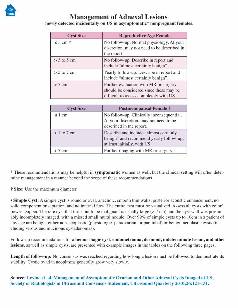
Management of Adnexal Lesions newly detected incidentally on US in asymptomatic* nonpregnant females.
Cyst Size Reproductive Age Female ≤ 3 cm † No follow-up. Normal physiology. At your
discretion, may not need to be described in the report.
> 3 to 5 cm No follow-up. Describe in report and include “almost certainly benign”.
> 5 to 7 cm Yearly follow-up. Describe in report and include “almost certainly benign”.
> 7 cm Further evaluation with MR or surgery should be considered since these may be difficult to assess completely with US.
Cyst Size Postmenopausal Female † ≤ 1 cm No follow-up. Clinically inconsequential.
At your discretion, may not need to be described in the report.
> 1 to 7 cm Describe and include “almost certainly benign” and recommend yearly follow-up, at least initially, with US.
> 7 cm Further imaging with MR or surgery.
* These recommendations may be helpful in symptomatic women as well, but the clinical setting will often deter-mine management in a manner beyond the scope of these recommendations.
† Size: Use the maximum diameter.
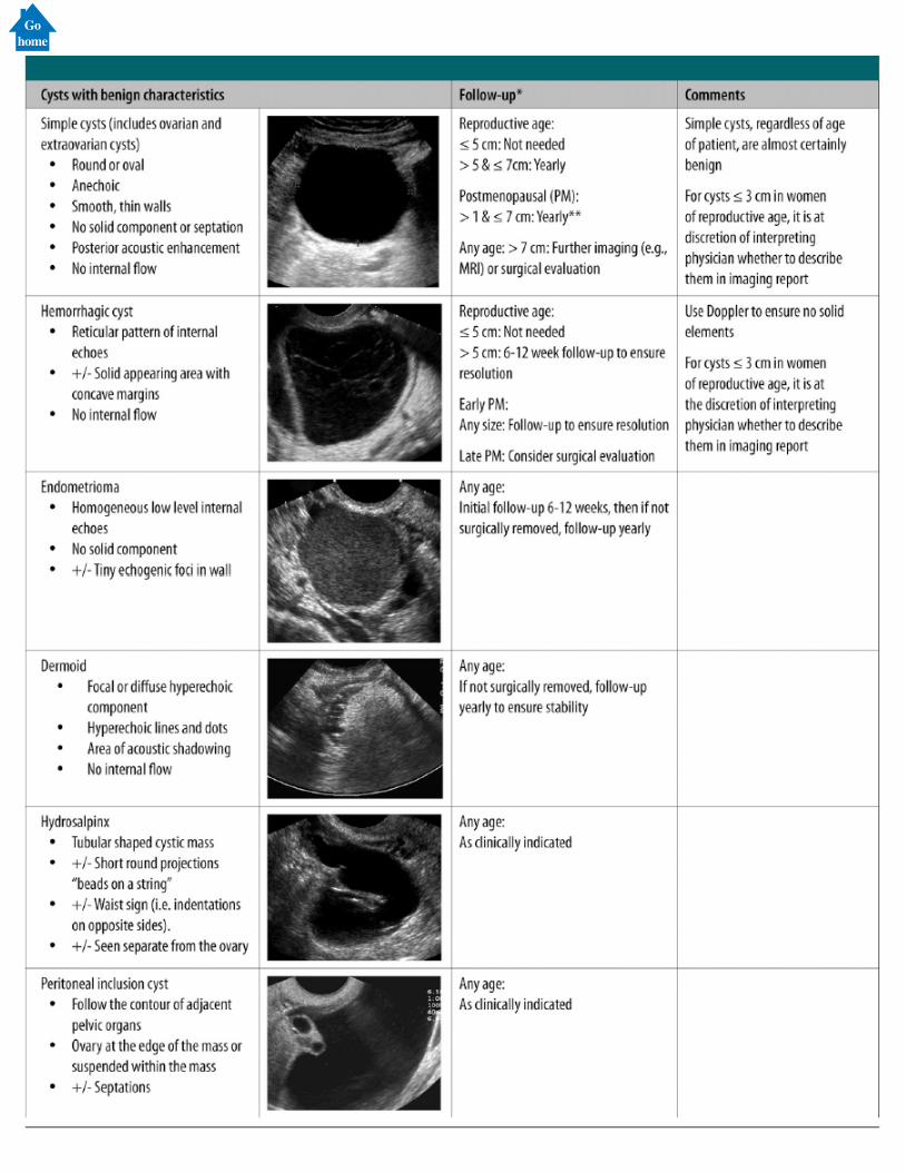
• Simple Cyst: A simple cyst is round or oval, anechoic, smooth thin walls, posterior acoustic enhancement, no solid component or septation, and no internal flow. The entire cyst must be visualized. Assess all cysts with color/power Dopper. The rare cyst that turns out to be malignant is usually large (> 7 cm) and the cyst wall was presum-ably incompletely imaged, with a missed small mural nodule. Over 99% of simple cysts up to 10cm in a patient of any age are benign, either non-neoplastic (physiologic, paraovarian, or paratubal) or benign neoplastic cysts (in-cluding serous and mucinous cystadenomas).
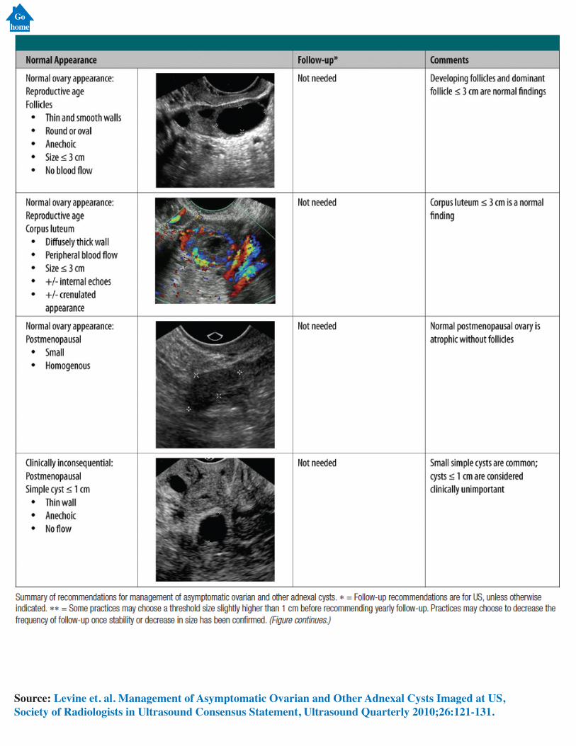
Follow-up recommendations for a hemorrhagic cyst, endometrioma, dermoid, indeterminate lesion, and other lesions, as well as simple cysts, are presented with example images in the tables on the following three pages.
Length of follow-up: No consensus was reached regarding how long a lesion must be followed to demonstrate its stability. Cystic ovarian neoplasms generally grow very slowly.
Source: Levine et. al. Management of Asymptomatic Ovarian and Other Adnexal Cysts Imaged at US, Society of Radiologists in Ultrasound Consensus Statement, Ultrasound Quarterly 2010;26:121-131.
Gohome
Source: Levine et. al. Management of Asymptomatic Ovarian and Other Adnexal Cysts Imaged at US, Society of Radiologists in Ultrasound Consensus Statement, Ultrasound Quarterly 2010;26:121-131.
Gohome
Gohome
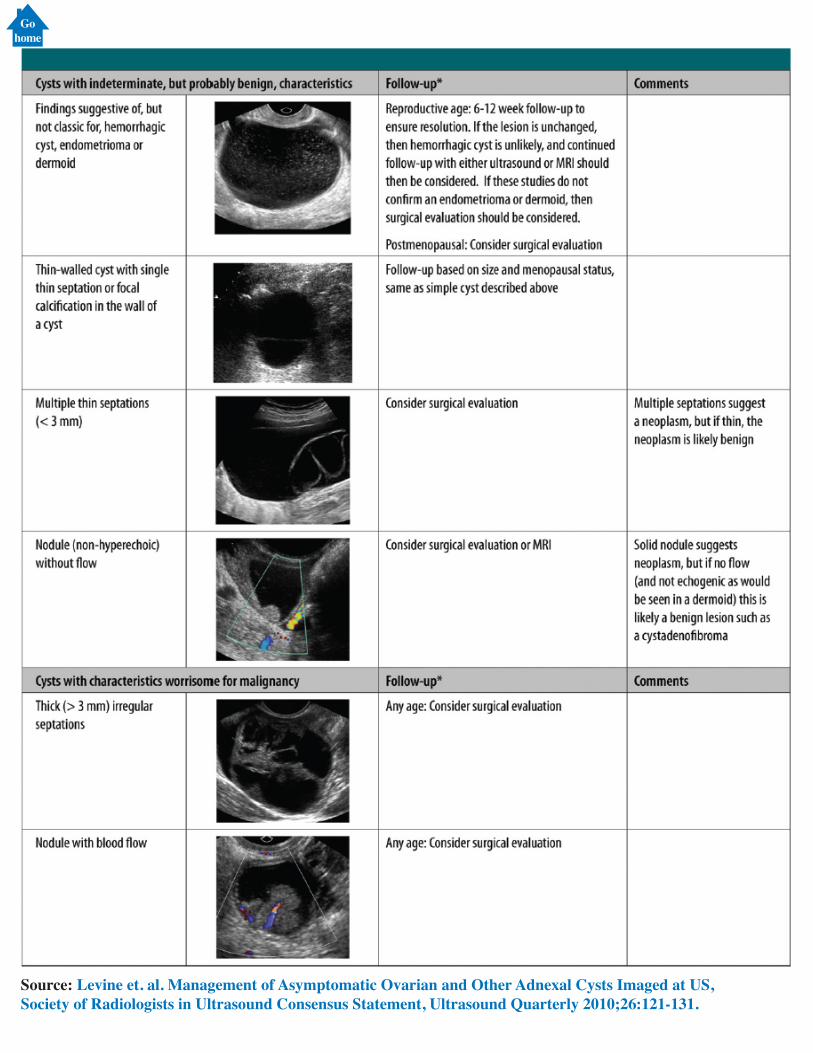
Source: Levine et. al. Management of Asymptomatic Ovarian and Other Adnexal Cysts Imaged at US, Society of Radiologists in Ultrasound Consensus Statement, Ultrasound Quarterly 2010;26:121-131.
Gohome
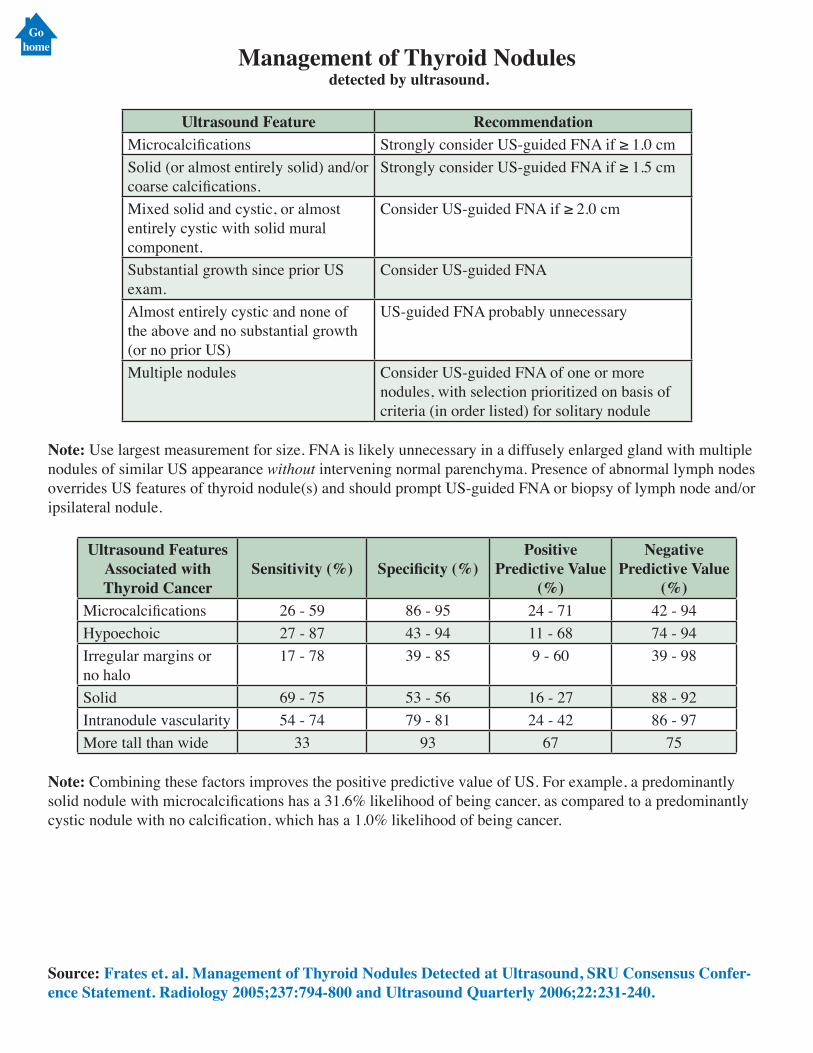
Management of Thyroid Nodules detected by ultrasound.
Ultrasound Feature RecommendationMicrocalcifications Strongly consider US-guided FNA if ≥ 1.0 cmSolid (or almost entirely solid) and/or coarse calcifications.
Strongly consider US-guided FNA if ≥ 1.5 cm
Mixed solid and cystic, or almost entirely cystic with solid mural component.
Consider US-guided FNA if ≥ 2.0 cm
Substantial growth since prior US exam.
Consider US-guided FNA
Almost entirely cystic and none of the above and no substantial growth (or no prior US)
US-guided FNA probably unnecessary
Multiple nodules Consider US-guided FNA of one or more nodules, with selection prioritized on basis of criteria (in order listed) for solitary nodule
Note: Use largest measurement for size. FNA is likely unnecessary in a diffusely enlarged gland with multiple nodules of similar US appearance without intervening normal parenchyma. Presence of abnormal lymph nodes overrides US features of thyroid nodule(s) and should prompt US-guided FNA or biopsy of lymph node and/or ipsilateral nodule.
Ultrasound Features Associated with Thyroid Cancer
Sensitivity (%) Specificity (%)Positive
Predictive Value (%)
Negative Predictive Value
(%)Microcalcifications 26 - 59 86 - 95 24 - 71 42 - 94Hypoechoic 27 - 87 43 - 94 11 - 68 74 - 94Irregular margins or no halo
17 - 78 39 - 85 9 - 60 39 - 98
Solid 69 - 75 53 - 56 16 - 27 88 - 92Intranodule vascularity 54 - 74 79 - 81 24 - 42 86 - 97More tall than wide 33 93 67 75
Note: Combining these factors improves the positive predictive value of US. For example, a predominantly solid nodule with microcalcifications has a 31.6% likelihood of being cancer, as compared to a predominantly cystic nodule with no calcification, which has a 1.0% likelihood of being cancer.
Source: Frates et. al. Management of Thyroid Nodules Detected at Ultrasound, SRU Consensus Confer-ence Statement. Radiology 2005;237:794-800 and Ultrasound Quarterly 2006;22:231-240.
Gohome
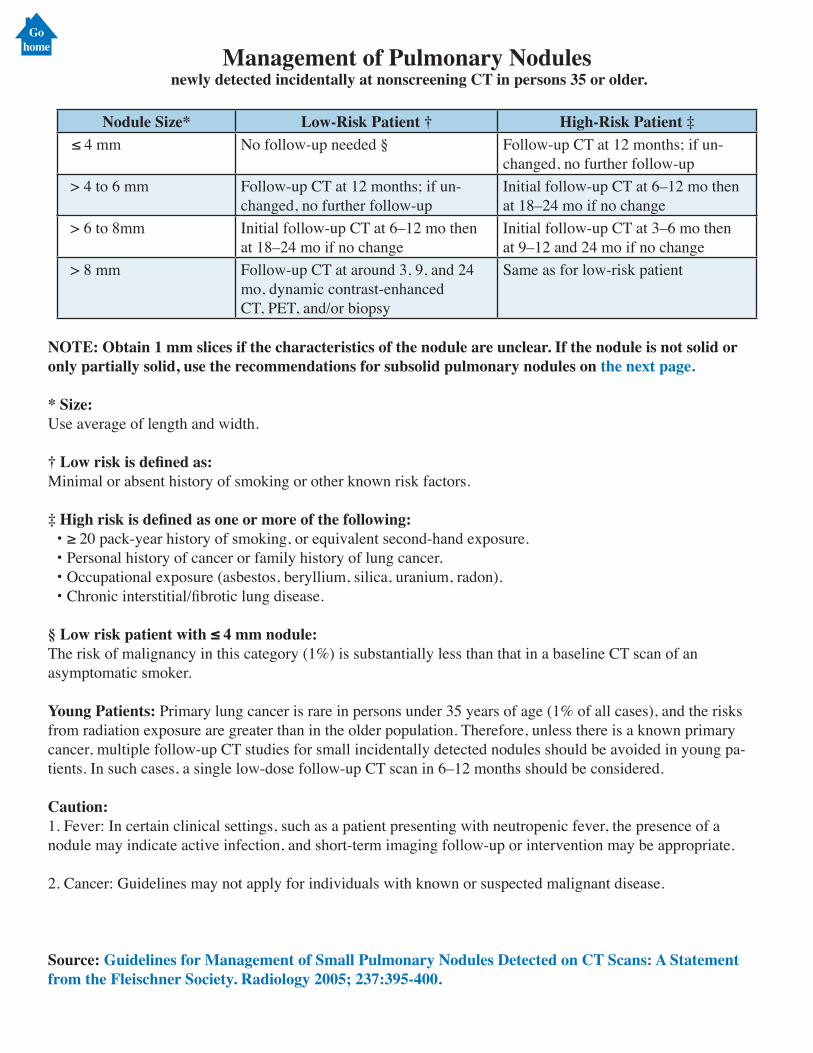
Management of Pulmonary Nodules newly detected incidentally at nonscreening CT in persons 35 or older.
Nodule Size* Low-Risk Patient † High-Risk Patient ‡ ≤ 4 mm No follow-up needed § Follow-up CT at 12 months; if un-
changed, no further follow-up > 4 to 6 mm Follow-up CT at 12 months; if un-
changed, no further follow-upInitial follow-up CT at 6–12 mo thenat 18–24 mo if no change
> 6 to 8mm Initial follow-up CT at 6–12 mo thenat 18–24 mo if no change
Initial follow-up CT at 3–6 mo thenat 9–12 and 24 mo if no change
> 8 mm Follow-up CT at around 3, 9, and 24mo, dynamic contrast-enhancedCT, PET, and/or biopsy
Same as for low-risk patient
NOTE: Obtain 1 mm slices if the characteristics of the nodule are unclear. If the nodule is not solid or only partially solid, use the recommendations for subsolid pulmonary nodules on the next page.
* Size:Use average of length and width.
† Low risk is defined as:Minimal or absent history of smoking or other known risk factors.
‡ High risk is defined as one or more of the following: • ≥ 20 pack-year history of smoking, or equivalent second-hand exposure. • Personal history of cancer or family history of lung cancer. • Occupational exposure (asbestos, beryllium, silica, uranium, radon). • Chronic interstitial/fibrotic lung disease.
§ Low risk patient with ≤ 4 mm nodule:The risk of malignancy in this category (1%) is substantially less than that in a baseline CT scan of an asymptomatic smoker.
Young Patients: Primary lung cancer is rare in persons under 35 years of age (1% of all cases), and the risks from radiation exposure are greater than in the older population. Therefore, unless there is a known primary cancer, multiple follow-up CT studies for small incidentally detected nodules should be avoided in young pa-tients. In such cases, a single low-dose follow-up CT scan in 6–12 months should be considered.
Caution:1. Fever: In certain clinical settings, such as a patient presenting with neutropenic fever, the presence of a nodule may indicate active infection, and short-term imaging follow-up or intervention may be appropriate.
2. Cancer: Guidelines may not apply for individuals with known or suspected malignant disease.
Source: Guidelines for Management of Small Pulmonary Nodules Detected on CT Scans: A Statement from the Fleischner Society. Radiology 2005; 237:395-400.
Gohome
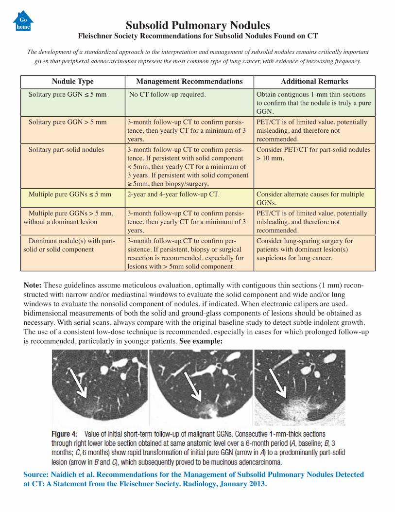
Gohome Subsolid Pulmonary Nodules
Fleischner Society Recommendations for Subsolid Nodules Found on CT
The development of a standardized approach to the interpretation and management of subsolid nodules remains critically important given that peripheral adenocarcinomas represent the most common type of lung cancer, with evidence of increasing frequency.
Nodule Type Management Recommendations Additional Remarks Solitary pure GGN ≤ 5 mm No CT follow-up required. Obtain contiguous 1-mm thin-sections
to confirm that the nodule is truly a pure GGN.
Solitary pure GGN > 5 mm 3-month follow-up CT to confirm persis-tence, then yearly CT for a minimum of 3 years.
PET/CT is of limited value, potentially misleading, and therefore not recommended.
Solitary part-solid nodules 3-month follow-up CT to confirm persis-tence. If persistent with solid component < 5mm, then yearly CT for a minimum of 3 years. If persistent with solid component ≥ 5mm, then biopsy/surgery.
Consider PET/CT for part-solid nodules > 10 mm.
Multiple pure GGNs ≤ 5 mm 2-year and 4-year follow-up CT. Consider alternate causes for multiple GGNs.
Multiple pure GGNs > 5 mm, without a dominant lesion
3-month follow-up CT to confirm persis-tence, then yearly CT for a minimum of 3 years.
PET/CT is of limited value, potentially misleading, and therefore not recommended.
Dominant nodule(s) with part-solid or solid component
3-month follow-up CT to confirm per-sistence. If persistent, biopsy or surgical resection is recommended, especially for lesions with > 5mm solid component.
Consider lung-sparing surgery for patients with dominant lesion(s) suspicious for lung cancer.
Note: These guidelines assume meticulous evaluation, optimally with contiguous thin sections (1 mm) recon-structed with narrow and/or mediastinal windows to evaluate the solid component and wide and/or lung windows to evaluate the nonsolid component of nodules, if indicated. When electronic calipers are used, bidimensional measurements of both the solid and ground-glass components of lesions should be obtained as necessary. With serial scans, always compare with the original baseline study to detect subtle indolent growth. The use of a consistent low-dose technique is recommended, especially in cases for which prolonged follow-up is recommended, particularly in younger patients. See example:
Source: Naidich et al. Recommendations for the Management of Subsolid Pulmonary Nodules Detected at CT: A Statement from the Fleischner Society. Radiology, January 2013.
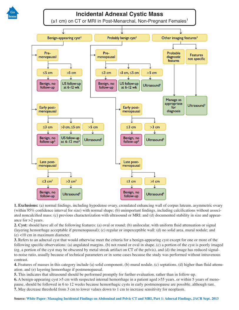
Incidental Adnexal Cystic Mass(≥1 cm) on CT or MRI in Post-Menarchal, Non-Pregnant Females1
1. Exclusions: (a) normal findings, including hypodense ovary, crenulated enhancing wall of corpus luteum, asymmetric ovary (within 95% confidence interval for size) with normal shape; (b) unimportant findings, including calcifications without associ-ated noncalcified mass; (c) previous characterization with ultrasound or MRI; and (d) documented stability in size and appear-ance for >2 years. 2. Cyst: should have all of the following features: (a) oval or round; (b) unilocular, with uniform fluid attenuation or signal (layering hemorrhage acceptable if premenopausal); (c) regular or imperceptible wall; (d) no solid area, mural nodule; and (e) <10 cm in maximum diameter. 3. Refers to an adnexal cyst that would otherwise meet the criteria for a benign-appearing cyst except for one or more of the following specific observations: (a) angulated margins, (b) not round or oval in shape, (c) a portion of the cyst is poorly imaged (eg, a portion of the cyst may be obscured by metal streak artifact on CT of the pelvis), and (d) the image has reduced signal-to-noise ratio, usually because of technical parameters or in some cases because the study was performed without intravenous contrast. 4. Features of masses in this category include (a) solid component, (b) mural nodule, (c) septations, (d) higher than fluid attenu-ation, and (e) layering hemorrhage if postmenopausal. 5. This indicates that ultrasound should be performed promptly for further evaluation, rather than in follow-up. 6. A benign-appearing cyst >5 cm with suspected internal hemorrhage in a patient aged >55 years, or within 5 years of meno-pause, should be followed in 6 to 12 weeks because hemorrhagic cysts in early postmenopause are possible, although rare. 7. May decrease threshold from 3 cm to lower values down to 1 cm to increase sensitivity for neoplasm.
Source: White Paper: Managing Incidental Findings on Abdominal and Pelvic CT and MRI, Part 1: Adnexal Findings, JACR Sept. 2013
Gohome
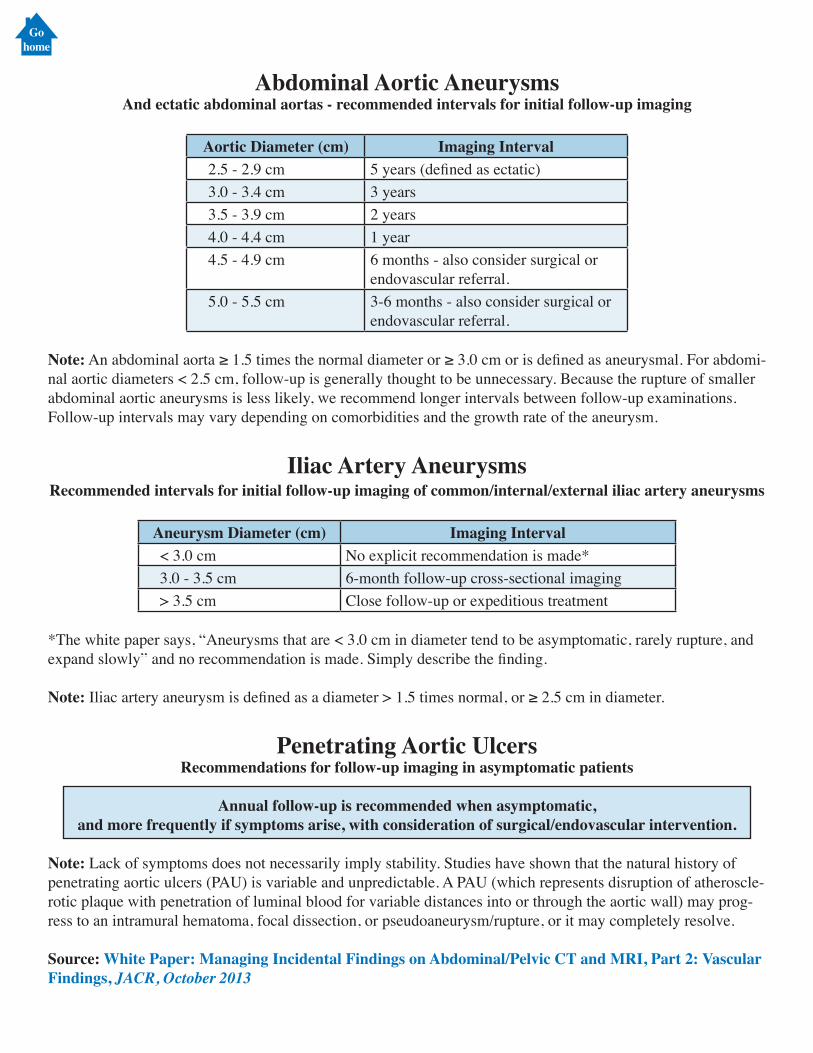
Abdominal Aortic AneurysmsAnd ectatic abdominal aortas - recommended intervals for initial follow-up imaging
Aortic Diameter (cm) Imaging Interval 2.5 - 2.9 cm 5 years (defined as ectatic) 3.0 - 3.4 cm 3 years 3.5 - 3.9 cm 2 years 4.0 - 4.4 cm 1 year 4.5 - 4.9 cm 6 months - also consider surgical or
endovascular referral. 5.0 - 5.5 cm 3-6 months - also consider surgical or
endovascular referral.
Note: An abdominal aorta ≥ 1.5 times the normal diameter or ≥ 3.0 cm or is defined as aneurysmal. For abdomi-nal aortic diameters < 2.5 cm, follow-up is generally thought to be unnecessary. Because the rupture of smaller abdominal aortic aneurysms is less likely, we recommend longer intervals between follow-up examinations. Follow-up intervals may vary depending on comorbidities and the growth rate of the aneurysm.
Iliac Artery AneurysmsRecommended intervals for initial follow-up imaging of common/internal/external iliac artery aneurysms
Aneurysm Diameter (cm) Imaging Interval < 3.0 cm No explicit recommendation is made* 3.0 - 3.5 cm 6-month follow-up cross-sectional imaging > 3.5 cm Close follow-up or expeditious treatment
*The white paper says, “Aneurysms that are < 3.0 cm in diameter tend to be asymptomatic, rarely rupture, and expand slowly” and no recommendation is made. Simply describe the finding.
Note: Iliac artery aneurysm is defined as a diameter > 1.5 times normal, or ≥ 2.5 cm in diameter.
Penetrating Aortic UlcersRecommendations for follow-up imaging in asymptomatic patients
Annual follow-up is recommended when asymptomatic, and more frequently if symptoms arise, with consideration of surgical/endovascular intervention.
Note: Lack of symptoms does not necessarily imply stability. Studies have shown that the natural history of penetrating aortic ulcers (PAU) is variable and unpredictable. A PAU (which represents disruption of atheroscle-rotic plaque with penetration of luminal blood for variable distances into or through the aortic wall) may prog-ress to an intramural hematoma, focal dissection, or pseudoaneurysm/rupture, or it may completely resolve.
Source: White Paper: Managing Incidental Findings on Abdominal/Pelvic CT and MRI, Part 2: Vascular Findings, JACR, October 2013
Gohome
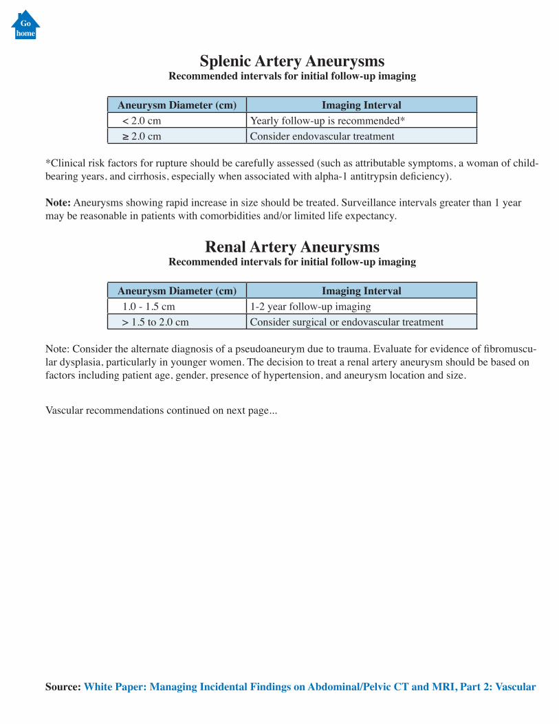
Splenic Artery AneurysmsRecommended intervals for initial follow-up imaging
Aneurysm Diameter (cm) Imaging Interval < 2.0 cm Yearly follow-up is recommended* ≥ 2.0 cm Consider endovascular treatment
*Clinical risk factors for rupture should be carefully assessed (such as attributable symptoms, a woman of child-bearing years, and cirrhosis, especially when associated with alpha-1 antitrypsin deficiency).
Note: Aneurysms showing rapid increase in size should be treated. Surveillance intervals greater than 1 year may be reasonable in patients with comorbidities and/or limited life expectancy.
Renal Artery AneurysmsRecommended intervals for initial follow-up imaging
Aneurysm Diameter (cm) Imaging Interval 1.0 - 1.5 cm 1-2 year follow-up imaging > 1.5 to 2.0 cm Consider surgical or endovascular treatment
Note: Consider the alternate diagnosis of a pseudoaneurym due to trauma. Evaluate for evidence of fibromuscu-lar dysplasia, particularly in younger women. The decision to treat a renal artery aneurysm should be based on factors including patient age, gender, presence of hypertension, and aneurysm location and size.
Vascular recommendations continued on next page...
Source: White Paper: Managing Incidental Findings on Abdominal/Pelvic CT and MRI, Part 2: Vascular
Gohome
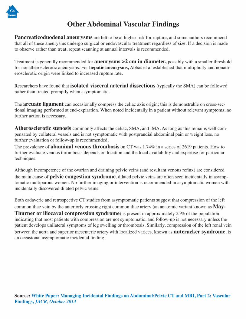
Other Abdominal Vascular Findings
Pancreaticoduodenal aneurysms are felt to be at higher risk for rupture, and some authors recommend that all of these aneurysms undergo surgical or endovascular treatment regardless of size. If a decision is made to observe rather than treat, repeat scanning at annual intervals is recommended.
Treatment is generally recommended for aneurysms >2 cm in diameter, possibly with a smaller threshold for nonatherosclerotic aneurysms. For hepatic aneurysms, Abbas et al established that multiplicity and nonath-erosclerotic origin were linked to increased rupture rate.
Researchers have found that isolated visceral arterial dissections (typically the SMA) can be followed rather than treated promptly when asymptomatic.
The arcuate ligament can occasionally compress the celiac axis origin; this is demonstrable on cross-sec-tional imaging performed at end-expiration. When noted incidentally in a patient without relevant symptoms, no further action is necessary.
Atherosclerotic stenosis commonly affects the celiac, SMA, and IMA. As long as this remains well com-pensated by collateral vessels and is not symptomatic with postprandial abdominal pain or weight loss, no further evaluation or follow-up is recommended.The prevalence of abominal venous thrombosis on CT was 1.74% in a series of 2619 patients. How to further evaluate venous thrombosis depends on location and the local availability and expertise for particular techniques.
Although incompetence of the ovarian and draining pelvic veins (and resultant venous reflux) are considered the main cause of pelvic congestion syndrome, dilated pelvic veins are often seen incidentally in asymp-tomatic multiparous women. No further imaging or intervention is recommended in asymptomatic women with incidentally discovered dilated pelvic veins.
Both cadaveric and retrospective CT studies from asymptomatic patients suggest that compression of the left common iliac vein by the anteriorly crossing right common iliac artery (an anatomic variant known as May-Thurner or iliocaval compression syndrome) is present in approximately 25% of the population, indicating that most patients with compression are not symptomatic, and follow-up is not necessary unless the patient develops unilateral symptoms of leg swelling or thrombosis. Similarly, compression of the left renal vein between the aorta and superior mesenteric artery with localized varices, known as nutcracker syndrome, is an occasional asymptomatic incidental finding.
Source: White Paper: Managing Incidental Findings on Abdominal/Pelvic CT and MRI, Part 2: Vascular Findings, JACR, October 2013
Gohome
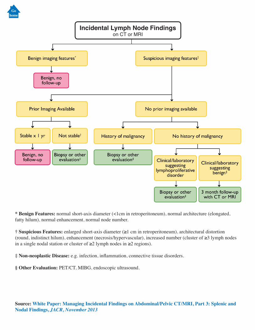
Incidental Lymph Node Findingson CT or MRI
* Benign Features: normal short-axis diameter (<1cm in retroperitoneum), normal architecture (elongated, fatty hilum), normal enhancement, normal node number.
† Suspicious Features: enlarged short-axis diameter (≥1 cm in retroperitoneum), architectural distortion (round, indistinct hilum), enhancement (necrosis/hypervascular), increased number (cluster of ≥3 lymph nodes in a single nodal station or cluster of ≥2 lymph nodes in ≥2 regions).
‡ Non-neoplastic Disease: e.g. infection, inflammation, connective tissue disorders.
§ Other Evaluation: PET/CT, MIBG, endoscopic ultrasound.
Source: White Paper: Managing Incidental Findings on Abdominal/Pelvic CT/MRI, Part 3: Splenic and Nodal Findings, JACR, November 2013
Gohome
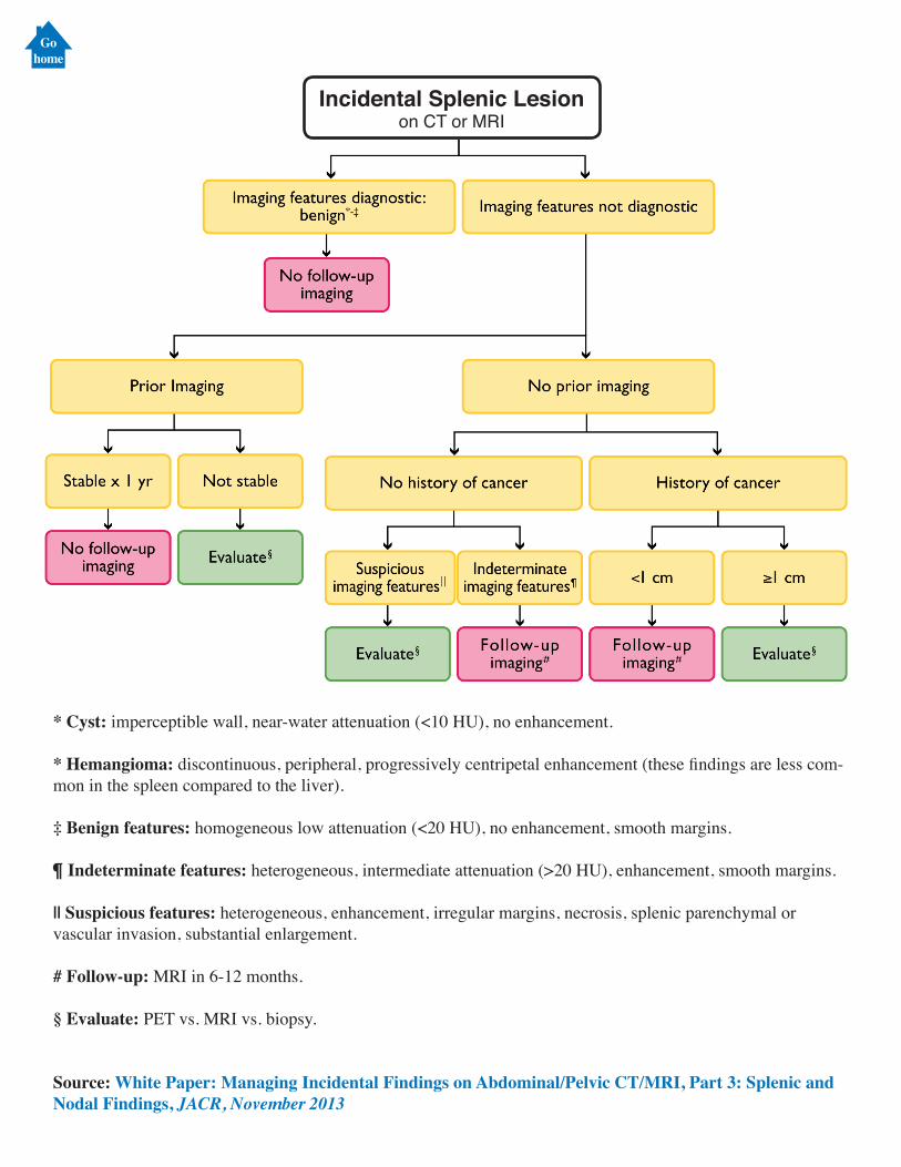
Incidental Splenic Lesionon CT or MRI
* Cyst: imperceptible wall, near-water attenuation (<10 HU), no enhancement.
* Hemangioma: discontinuous, peripheral, progressively centripetal enhancement (these findings are less com-mon in the spleen compared to the liver).
‡ Benign features: homogeneous low attenuation (<20 HU), no enhancement, smooth margins.
¶ Indeterminate features: heterogeneous, intermediate attenuation (>20 HU), enhancement, smooth margins.
|| Suspicious features: heterogeneous, enhancement, irregular margins, necrosis, splenic parenchymal or vascular invasion, substantial enlargement.
# Follow-up: MRI in 6-12 months.
§ Evaluate: PET vs. MRI vs. biopsy.
Source: White Paper: Managing Incidental Findings on Abdominal/Pelvic CT/MRI, Part 3: Splenic and Nodal Findings, JACR, November 2013
Gohome
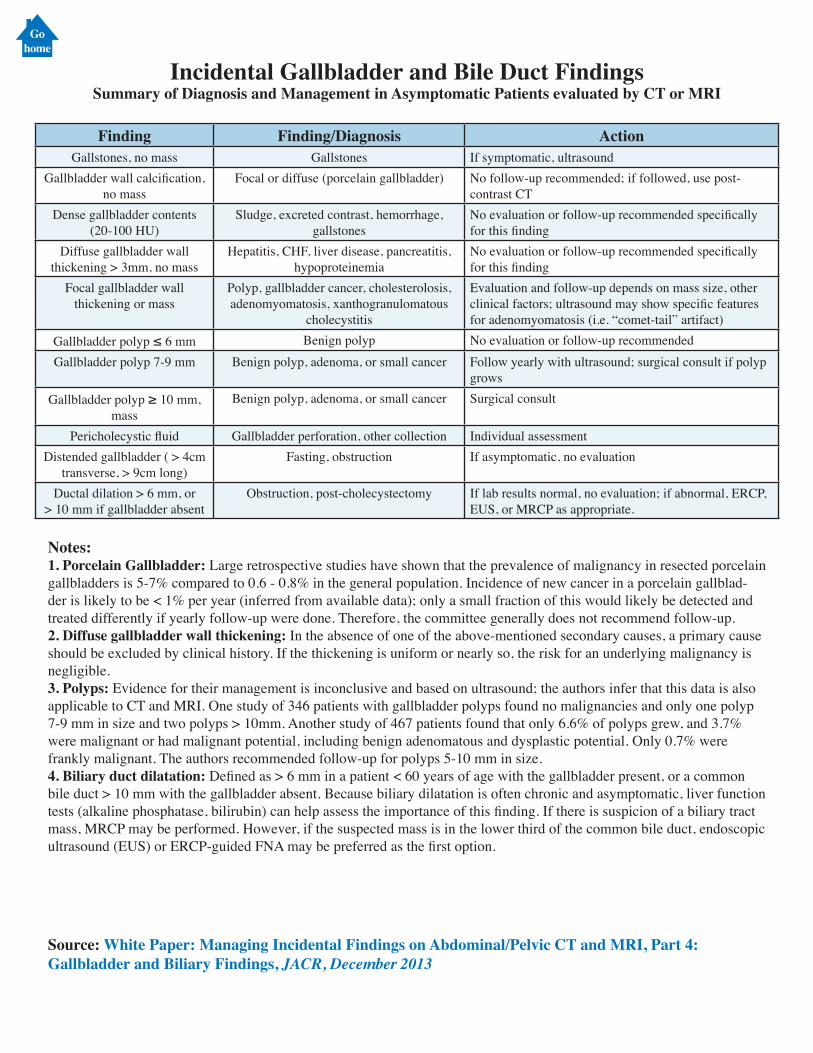
Incidental Gallbladder and Bile Duct FindingsSummary of Diagnosis and Management in Asymptomatic Patients evaluated by CT or MRI
Finding Finding/Diagnosis ActionGallstones, no mass Gallstones If symptomatic, ultrasound
Gallbladder wall calcification, no mass
Focal or diffuse (porcelain gallbladder) No follow-up recommended; if followed, use post-contrast CT
Dense gallbladder contents (20-100 HU)
Sludge, excreted contrast, hemorrhage, gallstones
No evaluation or follow-up recommended specifically for this finding
Diffuse gallbladder wall thickening > 3mm, no mass
Hepatitis, CHF, liver disease, pancreatitis, hypoproteinemia
No evaluation or follow-up recommended specifically for this finding
Focal gallbladder wall thickening or mass
Polyp, gallbladder cancer, cholesterolosis, adenomyomatosis, xanthogranulomatous
cholecystitis
Evaluation and follow-up depends on mass size, other clinical factors; ultrasound may show specific features for adenomyomatosis (i.e. “comet-tail” artifact)
Gallbladder polyp ≤ 6 mm Benign polyp No evaluation or follow-up recommendedGallbladder polyp 7-9 mm Benign polyp, adenoma, or small cancer Follow yearly with ultrasound; surgical consult if polyp
growsGallbladder polyp ≥ 10 mm,
massBenign polyp, adenoma, or small cancer Surgical consult
Pericholecystic fluid Gallbladder perforation, other collection Individual assessmentDistended gallbladder ( > 4cm
transverse, > 9cm long)Fasting, obstruction If asymptomatic, no evaluation
Ductal dilation > 6 mm, or > 10 mm if gallbladder absent
Obstruction, post-cholecystectomy If lab results normal, no evaluation; if abnormal, ERCP, EUS, or MRCP as appropriate.
Notes:1. Porcelain Gallbladder: Large retrospective studies have shown that the prevalence of malignancy in resected porcelain gallbladders is 5-7% compared to 0.6 - 0.8% in the general population. Incidence of new cancer in a porcelain gallblad-der is likely to be < 1% per year (inferred from available data); only a small fraction of this would likely be detected and treated differently if yearly follow-up were done. Therefore, the committee generally does not recommend follow-up.2. Diffuse gallbladder wall thickening: In the absence of one of the above-mentioned secondary causes, a primary cause should be excluded by clinical history. If the thickening is uniform or nearly so, the risk for an underlying malignancy is negligible.3. Polyps: Evidence for their management is inconclusive and based on ultrasound; the authors infer that this data is also applicable to CT and MRI. One study of 346 patients with gallbladder polyps found no malignancies and only one polyp 7-9 mm in size and two polyps > 10mm. Another study of 467 patients found that only 6.6% of polyps grew, and 3.7% were malignant or had malignant potential, including benign adenomatous and dysplastic potential. Only 0.7% were frankly malignant. The authors recommended follow-up for polyps 5-10 mm in size. 4. Biliary duct dilatation: Defined as > 6 mm in a patient < 60 years of age with the gallbladder present, or a common bile duct > 10 mm with the gallbladder absent. Because biliary dilatation is often chronic and asymptomatic, liver function tests (alkaline phosphatase, bilirubin) can help assess the importance of this finding. If there is suspicion of a biliary tract mass, MRCP may be performed. However, if the suspected mass is in the lower third of the common bile duct, endoscopic ultrasound (EUS) or ERCP-guided FNA may be preferred as the first option.
Source: White Paper: Managing Incidental Findings on Abdominal/Pelvic CT and MRI, Part 4: Gallbladder and Biliary Findings, JACR, December 2013
Gohome