incorporating magnification into dental practice · pdf filelearn about the advantages of...
TRANSCRIPT

Incorporating Magnification into your Dental Practice.
by Dr. Glenn A. van As
Learning Objectives
1. Understand the resolution of the native eye and how magnification can improve visual acuity.
2. Discover how presbyopia and aging of clinicians may not be the only reason for integrating magnification into routine daily practice.
3. Understand the relationship between magnification and resolution.4. Discover the concept of the magnification continuum and how it may impact clinicians
who begin using magnification.5. Learn about options available for increasing both magnification and illumination from
entry level loupes through to operating microscopes and heads up displays.6. Appreciate the advantages of telescopic loupes in improving ergonomics (posture)
and precision of treatment outcomes.7. Learn about the advantages of operating microscopes in improving ergonomics,
precision of treatment, documentation, and communication with patients, staff and colleagues.
8. Develop an appreciation for the challenges of incorporating magnification into routine daily practice.
Introduction
Dentistry has encountered a tremendous surge in technology over the last 25 years in nearly all areas including armamentarium, materials and techniques. (1-5) Patients now wish to maintain their their own dentition for a lifetime, and they are keenly aware of how cosmetic dentistry can bring about a beautiful smile. These technological advancements have given dentists a new opportunity to create clinical excellence as long as microscopic attention to detail in both the use of materials and the techniques necessary to provide excellence is observed. Clinicians must now focus on how to provide dentistry that is not only functional but esthetically exceptional, with the most minimal intervention possible or in some cases no intervention at all. (6-8). These factors have created a true challenge for many clinicians nowadays as demand by patients for “invisible white restorations” which are long lasting are expected. (9-11)
The increasing demand of patients for these esthetic, conservative and durable restorations requires that the clinician be able to visualize the operating field in greater detail. The field of medicine has long advocated the use of magnification for aiding in delicate procedures. Disciplines such as otolaryngology, neurology and more recently acute care have embraced magnification for surgery as well as delicate procedures

such as reanastomoses of severed appendages. Our medical colleagues have relied upon enhanced visual acuity to improve treatment outcomes for many years. (12-18)
The use of magnification in dentistry has been slower to be adopted by the dental profession. This concept that magnification was reserved for older “mature” practitioners whose eyesight were failing, was generated for many of us in dental school. (19) During our education, some of the instructors wore magnification of some sort, but the perception was that the inability to focus sharply on close objects (Presbyopia) that occurs after the age of 45 was responsible for the need to use those “magnifiers”. In order to see objects that are closer than 6m clearly, the ocular lens thickness must be changed and this is done by contraction or relaxation of the ciliary muscles of the eye. (20) It is interesting to note that many dental schools and dental hygiene schools are now requiring their students to purchase magnification as part of their armamentarium as the understanding of how ergonomics and treatment outcomes can be improved. (21-22) . Further, it has been understood that magnification can be a useful tool during the teaching process itself, (23) and although there are some issues in getting all faculty members to embrace the value of magnification, (24) a growing number of dental schools and dental hygiene programs are embracing magnification as being an important in the education process in dentistry. (25-26).
The growth in the usage of surgical telescopes (loupes) from a rarity to the norm in general practice increased dramatically from 1980 to 2001. In the authors home province of British Columbia, the percentage of clinicians using any form of magnification rose from 20 percent in 1986 to 75 % in 2000. (27-28). In the past decade, the growth of high levels of magnification (dental operating microscope) has continued to climb in endodontics. In 1999 one survey found 52% of endodontists had access to and used an operating microscope, and most of the time it was used for difficult treatments where high levels of magnification were required such as separated instruments, root-end preparations and root-end fillings (29) . In 2008, a similar survey found that now 90% of endodontists in the United States have access to and used the operating microscope in their practice in a similar fashion to the study from 1999, but with a greater number using it for locating canal orifices. (30). So it appears that there is a growing interest in all levels of magnification and this article will discuss reasons for incorporating magnification into your practice, the alternatives, benefits and challenges that exist in this journey.
Resolution of the Human Eye and Visual Information.
Carr, in his landmark article, (31) cited research from the medical literature which showed that the human eye, when unaided by magnification, has the ability to resolve or distinguish two distinct lines or objects that are separated by a minimum distance of 200 µm (0.2 mm). If the lines are closer together than 0.2 mm, the eye will see them only as a single line.
Magnification improves the ability of the eye to resolve these objects, and allows the clinician to see greater detail than is possible with the eye alone. For example, 2.5X

magnifiers such as telescopic loupes (Fig. 1) improve the resolution of the human eye to 80 µm (microns) , whereas 4.5 X (fig.2) telescopic loupes improve the resolution of the human eye to 44 µm, or 0.044 mm (Table 1).
Figure 1 shows 2.5X flip up loupes Fig. 2 shows 4.5 X TTL loupes + headlight
One study has shown that a skilled, trained clinician with a sharp, new explorer can determine marginal gaps in the range of 35 to 50 µm (32). It is therefore possible to conclude that at levels above 4-6x magnification, the dependance on tactile clues may be replaced with a greater reliance on visual cues. It is interesting to note that a dental operating microscope at 20X power will provide a resolution of 10 microns.( a human hair is around 50 microns in width).
Table 1 shows how the resolution of the human eye is improved with magnification.

Some clinicians are surprised as to how much more they “see” when they increase the amount of magnification they are using. The amount of visual information that is provided to the clinician is in fact the area of the surgical site ( X axis times Y axis) and is indicative of the power of increasingly higher levels of magnification. Table 2 and Figure 3 show the impact that higher levels of magnifiers have on the amount of visual information that is provided.
A jump from 2x loupes to 4x loupes will in fact not provide double what you can see, but in fact provides a FOUR fold increase in the amount of visual information. Correspondingly, when an operating microscope is used at 12X magnification then the amount of visual information provided is in fact 24X what is seen with 2x loupes. As the clinician begins to understand how seeing better may mean better treatment outcomes, routine use of magnification in daily practice becomes a necessity not a luxury.
Table 2 shows the non-linear relationship between increasing magnification and the amount of visual information that passes to the retina

Figure 3 shows graphically the relationship between magnification and the amount of visual information seen by the retina.
Magnification Alternatives in Dental Practice.
There are three basic magnification systems available in dentistry for surgical telescopes, and these include Single lens, Galilean loupes and Prismatic (Keplerian) loupes. (20)
Single lens loupes (Amsted) are the simplest form of magnification and are low powered clip on magnifying lenses that attach to glasses or a headband. They are less expensive but have limited magnification (up to about 3X), limited depth diameter of field of vision.
Galilean loupes are used for 2-2.5X magnification in that they use 2 or more lenses to focus the image. The systems are more expensive that single lens loupes and although the level of magnification is limited the depth and diameter of the field of view as well as the resolution of the image are improved. (Fig.1)

Finally, Prismatic (Keplerian) loupes have multiple lenses (around 5 as well as 2 prisms) to focus the image and give greater clarity. The price is more expensive to make these and the lenses are larger, heavier and result in a smaller depth of field as the magnification increases. (Fig.2)
When choosing loupes, a clinician should look at not only the magnification of the system and choose one that initially will give them the greatest chance of adaptation (i.e. 2-3X loupes is the most often suggested initial magnification), but also look at the field width, depth of field, as well as whether they would like to flip up the loupes for an unmagnified assessment of the field of view. These “flip up” loupes typically are bulkier and heavier than their TTL (through the lens) counterparts and can easily go out of adjustment which can affect not only the view that you have (double image) but also your posture. The ability of TTL loupes to be mounted directly into a prescription lens does necessitate repair anytime that the prescription changes. TTL loupes also can only be mounted at an angle up to around 45 degrees (called the declination angle ) from horizontal. The heavier the Keplerian loupes the greater the tendency for the loupes to want to ride down the nose and the greater the need for a strap that fits tight around the back of the head, to hold the loupes tight.
Clinicians should focus on proper working distance as many loupes systems can alter the focal distance. The working distance variation can be set for operators who like to lean closer to the operating field (Near focal distance) all the way up to those who like to sit upright and a further distance away from the surgical site. Under ideal circumstances the latter is better from an ergonomic standpoint as the operator will have less fatigue in both the lower (back) and upper spine (neck and shoulder) and be sitting in a more upright fashion. It is nearly impossible for any set of loupes to achieve a straight forward vision (as can be obtained with the operating microscope) and if neck and back pain are severe for a dentist or a hygienist an operating microscope may lessen the severity of the pain.
Magnification Continuum
In 2001, the author developed the term the “Magnification Continuum “ to try and quantify the growing number of magnification users.(33) I had found from personal experience and also from my lectures and discussions with other dentists a growing number of clinicians who were using magnification routinely in their dental practice. It was noted that many clinicians were just beginning their journey into enhanced visual acuity, while others had been using loupes for many years. In fact it was not uncommon to hear of dentists who had several different sets of magnifiers ranging from low level 2-2.5x loupes through to 6X loupes with a headlamp. In many instances with experienced clinicians, there seemed to be waves of progression forward to higher and higher levels of magnification with time. It seemed that as the appreciation for the value of magnification matched the level of expertise with the increased level of magnification being used, the clinician would often look to add a new magnification to their armamentarium. In addition, some clinicians were opting to use dental operating microscopes for their procedures which had multiple levels of magnification (2x-20x

power), and these clinicians were often focussing on disciplines such as endodontics or cosmetics where magnification and illumination could help either with discovery of pulpal anatomy or with details in preparation that could enhance the final treatment esthetically in the case of cosmetic dentistry. (figure 4).
Eight years after this initial idea surfaced, the author has revised his original concept to include two to three phases of magnification progression.(Figure 5).

Figure 5 shows the 2009 version of the Magnification Continuum.
Although, the entry level magnification of 2-2.5X magnification is what many dental hygienists and dental students are exposed to during their education now. It is also the suggested starting point for many more experienced clinicians looking for a solution for presbyopia, to improve clinical results or to aid with neck and back pain. During this initial phase the clinician will encounter a learning curve (A) that is often from a week to a month. During this period of time the clinician will at times fluctuate between using the loupes for endodontics, cosmetics, difficult extractions of root tips, crown preparations or other procedures but may also provide treatment without them. After 1 or 2 more periods of sustained use with shorter learning curves, (B) the clinician will use them full time for most procedures and at times question how they ever did dentistry without them!
It is with regular use and time that the clinician may eventually seek out higher powers of magnification (C) and at this point enter into their 2nd and eventually even 3rd set of loupes. Initially some practitioners may envision only using the new medium powered loupes (eg. 3.5X mag) for endodontics or during difficult to visualize procedures (D) and they may then fluctuate between the low and medium powers as the procedure dictates. WIth time, the dentist may then reach a period where they are seldom using the lower

power and using only the more recent purchase. (E). At times, clinicians will choose another higher magnification again, and in addition incorporate a headlamp (often needed above 3.5X mag) and now will have 2-3 different sets of loupes with a headlamp. (F).
If the clinician enjoys endodontics, wants multiple powers of magnification without changing loupes, coaxial shadow free illumination, and the ease of documentation of procedures a dental operating microscope (Global, Seiler or Zeiss) or heads up display system (Magna Vu, MoraVision 3D) offer the ultimate in higher levels of magnification.
These systems involve a new learning curve that requires the clinician to learn to reposition the patient, or the patients head and requires a greater dependence on indirect vision (intra oral mirrors) in both arches in order to optimize the view. This contrasts with positioning in loupes based dentistry where the operator frequently repositioning of themselves and the patient remains stationary.(G). This learning period can be anywhere from a month to a year depending on the dentist, but eventually the operator will learn to use all magnifications of the system from low to High power. After several phases of using the lower powers of the microscope only (H), the clinician will with time learn to operate throughout the ranges of magnification and be able to comfortably work at most of the magnifications available to them as dictated by the procedure, patient and challenges they create for the clinician. (I).
There is always a price to be paid for the increased amount of visual information provided by higher levels of magnification. Although the amount of visual information continues to increase, the value of this information clinically is maximized at around 20X magnification. The depth of field clinically and the difficulty of the finite motor movements necessary to work at this magnification make this figure the approximate upper level of clinically valuable magnification. Stabilization of the gross motor joints (elbows and shoulders) through the use of a microsurgeons chair allow for improved control of the micromotor muscles and joints (Fingers and wrists). Shanelec and Tibetts showed in working with high levels of magnification (20X power) that clinicians were able to learn out how to shrink their movements from 1-2 mm (1000-2000 micron) increments to 10-20 microns (10-20/1000th of a mm) movements at a time. It is useful then to appreciate that the limitation to precision of treatment is not in the hands but in the eyes. (34)
It is interesting to note that even though the magnification continuum for many clinicians may consist of three phases, some clinicians may never see the need to proceed past an entry set of loupes, while others will jump from low power to a microscope without using a medium level of magnification. On rare instances a practitioner will actually not be able to become proficient with low power loupes but will jump into a microscope right away. This can be because of the ergonomic value of the scope to sit upright relieving back and neck pain, or the stereoscope vision that in some clinicians is easier for their eyes to accommodate too then the convergent vision of loupes. (Fig.6) The Magnification Continuum is simply a concept to describe the process that many

clinicians progress through when they begin to grasp the value of the magnification for improving their work and if properly aligned for their ergonomic posture.
Figure 6. Loupes vision versus Microscope vision
The author has progressed through entry level loupes, 3.5, 4.5X loupes and for the last 11 years has completed almost all of his clinical work through the operating microscope (G6 from GLobal).( FIgures 7-8) All nine operatories in the authors current office have a microscope available for all procedures and the operatories have been designed to function ideally with the microscope as the centerpiece visually, ergonomically and functionally. The increasing ability to see with higher levels of magnification (fig.8) was correlated in an ability to sit upright as the operator had the loupes adjusted properly (Focal distance) and as the clinician switched from flip loupes to Through the Lens loupes (TTL loupes have a higher declination angle) to an operating microscope where true neutral and balanced posture was achieved.

Figure 7 showing authors progression from 2.5X flip up (Surgitel loupes) to 3.5X Flip up (Keeler), and eventually to the 4.5X extended field of view (Designs for Vision) TTL loupes.
Fig 8a. Global microscope with six magnifications including 2.1, 3.2, 5, 8, 13, and 19.2 X magnification. A simple one chip medical grade cube camera (black square on right) provides video so that patients can see in real time during exams and procedures.

Figure 8b Global G6 Operating Microscope with 3 chip digital Video camera on left and Digital SLR camera on Right side of microscope
Figure 9a. Microscope Centered Operatory.
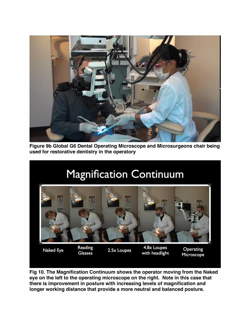
Figure 9b Global G6 Dental Operating Microscope and Microsurgeons chair being used for restorative dentistry in the operatory
Fig 10. The Magnification Continuum shows the operator moving from the Naked eye on the left to the operating microscope on the right. Note in this case that there is improvement in posture with increasing levels of magnification and longer working distance that provide a more neutral and balanced posture.

Advantages of using Surgical Telescopic Loupes in Dentistry
The two primary benefits that most clinicians cite for using loupes in dentistry are an improvement in ergonomics, and an improvement in precision, The perception is that magnification when properly fitted will encourage improvements in posture and in doing so reduce stress on the neck and back. It is a common refrain from the loupes companies that properly adjusted loupes lead to improvements in ergonomics which in turn will decrease the risk of debilitating injury to the clinician. (21,35-44). The concept is that with a forward head position of 20 degrees or more for greater than 70% of the time, that a dentist will have a higher risk of neck pain.(45) In dentistry most of us are working with a forward head position of 30 degrees or more for 85% of the time in the operatory. and this leads to neck pain in 70% of dentists and dental hygienists. (46-48) The need to see the operating field causes the clinician to lean forward and the neck vertebrae cannot support the spine, leading to rapid fatigue of the shoulder muscles that stabilize, and inclusion of other muscles to compensate for a job they are not designed to do. This can lead to Tension Neck Syndrome (TNS) with pain in the neck, shoulders and intercapsular muscles. Long term disc degeneration has been noted from a prolonged forward position of the head. It is important to know that improper adjustment or use of magnification aids can either increase the risk of injury or worsen existing pain.(49-51).
While none of the telescopic loupes systems mentioned previously (Single lens, Galilean, or Prismatic) can lead to a complete neutral and balanced posture, the use of well designed loupes can enable a working posture of 25 degrees forward (35). It is important to choose loupes with a good declination angle that will allow for as minimal forward head position as possible as the more forward the loupes push the head the more likely that strain on the neck muscles and discs will occur.(52-53).
If the working distance to the surgical site is chosen as too short the operator will likely be forced to either hunch or use excessive neck flexion in order to obtain a sharp image of the operating field.(54) It is important to realize that the working distance is related to operator height, is measured from the corner of the eye to the working surface. It can vary from 14 to over 20 inches and need to be tailored to the operator.
Finally the larger a frame the better the lower the glasses will sit on the cheek and this will all for a better declination angle as the TTL telescopes can be placed with a steeper declination angle at the bottom of the frame. As the level of magnification gets larger the need for accessory illumination increases. Today, many powerful LED diode lights offer tremendous illumination with reduced weight and extended battery life. The range in cost for loupes is typically between $700-2200 depending on the company, magnification chosen, and type of loupe chosen. A properly adjusted set of loupes, set properly for declination angle, working distance and frame size can significantly reduce the risk of debilitating occupationally induced neck and back discomfort. (Figure11-12)

Fig. 11 shows Dental Hygienist with improved posture using 2.5 x TTL lopues with headlight.

Fig.12 Shows Key ergonomic factors to consider when considering ergonomic principles with surgical Loupes.
The other advantage of using loupes has been cited as the improvement in precision due to the increased amount of visual information that magnification and illumination provide to the operator. Unfortunately, there are very few well designed, peer reviewed, scientific studies that have been published that show a correlation between enhanced visual acuity leading to an improvement in quality of care. (55-56). In fact although some studies have shown that dental students make less errors with loupes,(57-60) other research is not so definitive in its conclusion of a positive impact in quality with magnification. (61, 62). Although, literature doesnʼt at this point support the fact that quality of treatment, longevity of treatment or overall prognosis of treatment is affected by magnification, the general consensus amongst many clinicians is that their work was better upon beginning with surgical magnification than before and that subsequent increases in the amount of magnification used yielded improvements in the precision of their work. The list below is meant as an introduction to some of the manufacturers in North America, and this should not be seen as an exhaustive list, but a starting point for those with an interest in purchasing loupes or headlights.

Designs for Vision - http://www.designsforvision.com/ Orascoptic Loupes - http://www.orascoptic.com/index/orascoptic Surgitel Loupes - http://www.surgitel.com/web/index.asp Zeiss Loupes - http://www.meditec.zeiss.com/C1256CAB00599F5D/Contents-Frame/93210AA396A78DCC8825726C0000BFD5 Sheervision Loupes - http://www.sheervision.com/index.asp Perioptix Loupes - http://www.perioptix.com/
Advantages of Using Operating Microscopes in Dentistry.
As the clinician enters into the higher levels of magnification (4-6X power and above) the choices are limited to heavier and more expensive TTL loupes with headlamps, the dental operating microscope and a few heads up display systems. Mamoun (63) has cited the benefits of higher magnifications for all areas of dental practice.
The Dental Operating Microscope (D.O.M.) is different that loupes in that it offers true stereoscopic vision (as compared to the loupes with its convergent vision).(Fig. 6) The microscope can be mounted on a ceiling, wall or floor stand, and can also have a mobile base. The D.O.M. has coaxial (light pathway coincident with the visual pathway) illumination and this provides intense shadow free lighting. The microscope has multiple levels of magnification from low mag (2.1, 3.2X) to medium mag (5-8X) and high levels of mag (13-19X) that is easy to access with the turn of a turret. All ranges of the magnification spectrum are used depending on the procedural needs. (Fig. 13-14)
Fig. 13a - 2.1X mag Fig 13b - 8X mag Fib 14- 19X mag
The D.O.M. will provide clinicians with four main advantages. These include improvements in precision of treatment, improvements in ergonomics, communication with patients (though live video ) and an increased ease in documentation.
The application of the operating microscope in clinical dentistry can be traced to Apotheker in 1981. (64) He converted a medical operating microscope for use in endodontics. Although the scope itself was a rudimentary one level of magnification and required the operator to stand upright, it was the introduction of a concept that would revolutionize the discipline of endodontics. Others including Carr, Arens, Buchanan,

Kim, Ruddle, led to the routine use of the microscope in the 1990s for improvements in both surgical and standard endodontic therapy. (65-72) The usage of the operating microscope in endodontics has been shown to improve the ability to uncover more pulpal anatomy as compared to the use of little magnification or low power loupes. (73-80) (Fig.15-16)
Fig 15. MB1 and MB2 canal - 8X mag Fig.16. Pulp Chamber lower molar.
After the introduction of the microscope to endodontics, there was a spike of interest in the D.O.M. for periodontics, and it was found by Shanelec, Belcher and others, that routine usage of the D.O.M. could provide for more delicate surgical procedures, utilizing microsurgical armamentarium including smaller blades and 7-0 to 10-0 sutures. These delicate surgical procedures allowed for improvements in postoperative pain and quicker healing. (81-89)
During the 1990s, a small group of restorative dentists, many with an active interest in endodontics, started to incorporate the microscope as an important part of the armamentarium in general practice. The microscope has found value in all areas of dentistry. For these restorative dentists, the microscope became an integral part of all dental procedures, as they discovered that the dramatic improvement in visual information provided by the D.O.M allowed for a level of precision in both diagnosis and treatment outcomes that were not previously possible. (90-105)(Figures 12-22)
Fig 17 Early occlusal caries- 13X mag Figure 18 Cracked tooth- 13X mag

Fig 19 Veneer preps- 5X mag Fig. 20. Evaluating impressions - 13X
Fig. 21 Crown Prep - 13X mag Fig.22 Veneer cement removal - 19X
In addition, the operating microscope provides important ergonomic benefits. The clinician sits in a comfortable upright position, relying upon directed patient movements or movements of a mirror to visualize the surgical site (Fig.23). The head has the ability to look straight ahead, with little forward head rotation if properly adjusted. If the operator uses a microsurgeons chair to support the gross motor joints (shoulders and forearms) then precise micromotor movements can be completed. (Fig.9b) The balanced position of the clinician when using the microscope, may help reduce musculoskeletal injuries that are common among members of the dental profession. (44, 106-107) Future work needs to be completed to see whether the ergonomic benefits in using a microscope will be superior to that of well adjusted loupes but the author is familiar with several clinicians who have had severe neck pain, degenerative discs, or in one instance (Fig.24) spinal injury who were able to resume their practice with the introduction of the operating microscope to their practice.

Fig.24 - Dr. Rex Hawthorne (General Dentist, Vernon, British Columbia) was paralyzed from the waist down in a mountain bike accident but was able to return to work with a specially designed wheelchair, an operating microscope and a tremendous desire helped him accomplish a full return to private practice.
As well as the ergonomic benefits, dentists using the operating microscope have discovered that either a medical grade cube video camera (Fig 8a- 8b) or a slightly heavier High Definition digital camcorder (Sony HDR SR12 or similar) (Fig. 25) when attached to the microscope can be useful in providing both patients and the auxiliary staff with the ability to observe treatment in real time. The ability to have the assistant in the “micro” world is made possible when the video from the microscope is connected to an LCD monitor. This helps aid in instrument passages and in addition helps alleviate the assistant being inadvertently “blinded” from reflections of the microscope light off an intra-oral mirror.

Figure 25 shows the attachment of a Sony digital camcorder (Sony HDR HC7) to document procedures in video format and transfer it to monitors in the operatory.
Patients can benefit from being able to watch the video overhead via connection to a LCD monitor or television in that they are able to follow along live during treatment. A consequence of this is that patients tend to hold still, and are educated to the complexity of the procedure by being given an opportunity to observe treatment as it progresses. There are a multitude of benefits to the integration of video to the microscope. (108)
Mehrabian has shown that as much as 55% of the understanding that occurs in verbal communication is through visual cues, and that only 7% of the comprehension in communication comes from the words we use. (109) Stated differently, patients remember more of what they see than what they hear. Clinicians have found that the images from the operating microscope are a benefit in educating patients about their treatment needs. The ability to easily document a procedure using digital microphotography and micro-videography with cameras attached to the operating microscope opens up new possibilities for patient education, documentation for professional presentations, and medical/legal documentation. (110). Microscopes costs vary between 5000.00 USD to 50,000 USD depending on the manufacturer, number of

magnification steps, options and accessories chosen. (Global Microscopes - http://globalsurgical.com/dental/dental.asp , Seiler microscopes - http://www.seilerinst.com/micro/default_dental.asp , Zeiss Microscopes - http://www.meditec.zeiss.com/us/dentistry )
The newest technology in the arena of magnification is heads up display that involves a camera that is placed over the patient and projects the image to a monitor. The projection of the image can be 2D (Magna Vu - http://www.magnavu.com/home.php ) or 3D (MoraVision 3D. http://www.moramicro.com/ ) and may help reduce the learning curve that has been associated with the operating microscope.(19) The ergonomic benefits of these heads up displays have also been discussed as have the possible improvements in treatment outcome and communication through documentation with video and captured stills. (111) These systems are in many cases more expensive than the operating microscope ranging in price from 15,000 dollars 30,000 dollars with the 3D system being more expensive as well. (Fig.26)
Fig.26. The author tries out the 3D heads up display (Moravision 3d) where the operator works in 3D with glasses while looking at a monitor. There are 4 high

definition cameras that supply the image to the monitors for both clinician, and dental assistants.
Challenges of Incorporating Magnification into Daily Practice.
Anytime that a practitioner is interested in incorporating new technology into a practice there will be challenges and magnification is no different. Initially there is a cost to purchase any magnification. Typically, loupes are less expensive that an operating microscope or heads up display systems. The price of magnification can be sizable if one is equipping multiple operatories or purchasing more than one set of loupes. Most clinicians agree that the first set of loupes will likely not be your last, and those clinicians that fully integrate microscopes into their office often discover that one microscope is not enough if multiple operatories are used.
The learning curve varies with the degree of magnification and for that reason many clinicians would be wise to purchase their initial loupes in the 2-3X magnification range. This will make the learning curve from days to weeks, but of course this can vary with many factors such as the age of the practitioner, the speed of the office pace, the ability of the practitioner to handle the slow down that is necessary initially as more visual information is seen. There is also the magnification scotoma (blind zone) that makes peripheral vision difficult with loupes. This blind zone increase with the power of magnification, and with loupes can be best overcome by subtle repositioning of the head. In the case of the microscope, the operator must learn to move themselves less, and rely on moving the patient , their head or the mirrors more and this process will increase the learning curve in microscope integration to months before proficiency is met with all magnifications in all areas of the mouth.
Many clinicians are concerned with the perception that patients including children will find the magnification systems to be peculiar. However, even microscopes and heads-up displays have been integrated into pediatric practices (111-112).
Finally there is a concern that dependency will occur with the full integration of magnification in ones practice. There is no evidence in either the dental literature or the medical literature to suggest that consistent usage of loupes will create a weakness or deterioration of natural eyesight, and as mentioned the clinician who is in their 40s will naturally encounter difficulty with presbyopia which necessitates the usage of reading glasses for close up viewing.
Dentistry is both physically and mentally challenging on a daily basis, and the rapid influx of new equipment, techniques and materials has made dentistry even more demanding. The incorporation, proper setup, and regular use of magnification into regular daily practice can alleviate neck and back pain, aid in treatment outcomes and assist in documenting and communicating with patients. You cannot treat what you

cannot see, so donʼt hesitate to begin your journey along the magnification continuum or to continue to a higher level of magnification should you already have some form of magnifiers. Its a win-win situation for you and your patients.