increased capsaicin receptor trpv1-expressing sensory
TRANSCRIPT
Increased capsaicin receptor TRPV1-expressingsensory fibres in irritable bowel syndrome and theircorrelation with abdominal pain
A Akbar,1 Y Yiangou,2 P Facer,2 J R F Walters,1 P Anand,2 S Ghosh1
See Commentary, p 882
1 Department ofGastroenterology, ImperialCollege London, UK;2 Department of ClinicalNeuroscience, Imperial CollegeLondon, UK
Correspondence to:Professor S Ghosh, Departmentof Gastroenterology, ImperialCollege London, HammersmithHospital, Du Cane Road, LondonW12 0NN, UK; [email protected]
Revised 21 December 2007Accepted 22 January 2008Published Online First4 February 2008
This paper is freely availableonline under the BMJ Journalsunlocked scheme, see http://gut.bmj.com/info/unlocked.dtl
ABSTRACTObjective: The capsaicin receptor TRPV1 (transientreceptor potential vanilloid type-1) may play an importantrole in visceral pain and hypersensitivity states. In irritablebowel syndrome (IBS), abdominal pain is a common anddistressing symptom where the pathophysiology is stillnot clearly defined. TRPV1-immunoreactive nerve fibreswere investigated in colonic biopsies from patients withIBS, and this was related to abdominal pain.Methods: Rectosigmoid biopsies were collected from 23IBS patients fulfilling Rome II criteria, and from 22controls. Abdominal pain scores were recorded using avalidated questionnaire. TRPV1-, substance P- andneuronal marker protein gene product (PGP) 9.5-expres-sing nerve fibres, mast cells (c-kit) and lymphocytes (CD3and CD4) were quantified, following immunohistochem-istry with specific antibodies. The biopsy findings wererelated to the abdominal pain scores.Results: A significant 3.5-fold increase in mediannumbers of TRPV1-immunoreactive fibres was found inbiopsies from IBS patients compared with controls(p,0.0001). Substance P-immunoreactive fibres(p = 0.01), total nerve fibres (PGP9.5) (p = 0.002), mastcells (c-kit) (p = 0.02) and lymphocytes (CD3) (p = 0.03)were also significantly increased in the IBS group. Inmultivariate regression analysis, only TRPV1-immuno-reactive fibres (p = 0.005) and mast cells (p = 0.008)were significantly related to the abdominal pain score.Conclusions: Increased TRPV1 nerve fibres are observedin IBS, together with a low-grade inflammatory response.The increased TRPV1 nerve fibres may contribute tovisceral hypersensitivity and pain in IBS, and provide anovel therapeutic target.
Irritable bowel syndrome (IBS) is the most commondisorder presenting to gastroenterologists, with aprevalence of up to 20% in the UK and the USA.1 2
Patients commonly present with abdominal painassociated with altered bowel habit. Self-reportedabdominal pain is a very common symptom in thepopulation including healthy individuals, but pain ismore severe and frequent in patients with IBS.3
Untreated pain leads to a decrease in daily functioncapability, social stresses, loss of work and poorquality of life.4 Furthermore, a study carried out bySandler et al5 revealed that abdominal pain was thesymptom most likely to result in medical consulta-tion in IBS patients. Therapeutic options currentlyavailable are limited, and often disappointing inefficacy. Functional bowel disorders such as IBS arecharacterised by visceral hypersensitivity,6 whichmay manifest as pain associated with boweldisturbances.7 Although the pathogenesis of visceral
hypersensitivity is not fully understood, severalmechanisms have been proposed, including subtleinflammation, psychosocial factors and alteredsensorimotor function of the gut, a major compo-nent of which is believed to be peripheral and centralsensitisation of visceral afferent neuronal pathways.8
The molecular and cellular mechanisms of thepathophysiology of IBS are increasingly the subjectof study. The transient receptor potential vanilloidtype-1 (TRPV1 or VR1) has been shown to play arole in animal models of inflammatory hyperalge-sia. TRPV1 is expressed by sensory neurons andactivated by capsaicin,9 heat (.43uC), acid(pH,5.9) and inflammatory mediators, withdepolarisation leading to burning pain. TRPV1activation also leads to local release of sensoryneuropeptides including calcitonin gene-relatedpeptide (CGRP) and substance P (SP) which, inturn, activate their effector cell receptors andcontribute to the process of neurogenic inflamma-tion. TRPV1 is expressed throughout the gastro-intestinal (GI) tract in myenteric ganglia, muscularlayers and mucosa.10 Our previous studies haveshown changes of TRPV1 in upper and lower GIdisorders: in inflammatory bowel disease, wereported greatly increased TRPV1 immunoreactiv-ity in biopsies taken from patients with activepainful Crohn’s disease compared with controls(Yiangou et al10). Our studies of biopsies frompatients with Hirschprung disease11 and patientswith idiopathic rectal hypersensitivity with faecalurgency12 also revealed increased TRPV1-expressingnerve fibres. In the latter study, increased levels ofTRPV1-expressing nerve fibres were correlatedsignificantly with hypersensitivity to rectal disten-sion and mid-rectal heat stimulation (Chan et al12).
Studies which specifically address any involve-ment of TRPV1 in IBS are lacking. We have thereforeinvestigated the presence of TRPV1 nerve fibres incolonic biopsies of IBS patients and controls, andrelated these to the degree of abdominal pain. IBSand control subjects were further characterised byparameters known to be related to IBS such as theneuropeptide SP, markers of inflammation includingc-kit (for mast cells) and psychological assessmentsusing validated questionnaires.
MATERIALS AND METHODS
PatientsThe study was approved by HammersmithHospitals Research Ethics Committee and all sub-jects gave fully informed consent before taking part.Twenty-three unselected patients with IBS and 22controls participated in the study (see table 1). IBS
Neurogastroenterology
Gut 2008;57:923–929. doi:10.1136/gut.2007.138982 923
on March 21, 2022 by guest. P
rotected by copyright.http://gut.bm
j.com/
Gut: first published as 10.1136/gut.2007.138982 on 5 F
ebruary 2008. Dow
nloaded from
patients were undergoing either a flexible sigmoidoscopy (n = 7)or a colonoscopy (n = 16) at Hammersmith Hospital, London. IBSwas diagnosed according to the Rome II criteria and the subjectswere further subclassified according to Rome II criteria into eitherdiarrhoea-predominant (IBS-D), constipation-predominant (IBS-C) or IBS with alternating stool pattern (IBS-A) (see table 2).Controls were selected from patients who were undergoingcolonoscopy for other indications (such as polyp and cancersurveillance) and had a normal colon (see table 3). None of thepatients was taking anti-inflammatory drugs or immunosuppres-sants. IBS patients had previously been seen in clinic, and other GIdiseases had been excluded. Coeliac disease was excluded in all IBSpatients by checking coeliac serology (immunoglobulin A (IgA),antiendomysial and antitissue transglutaminase (TTG) antibo-dies). Patients under active psychiatric care were excluded.
Patients who were undergoing colonoscopy received standardbowel preparation in the form of 4 litres of Kleanprep. Forflexible sigmoidoscopy patients received a phosphate enema 1 hprior to the procedure. All patients had macroscopically normalbowel mucosa on examination. A total of four mucosal biopsyspecimens were taken using standard biopsy forceps from allsubjects from the rectosigmoid junction in order to standardisethe site of sampling. Of these, two biopsies were sent forroutine H&E histological analysis to exclude any evidence ofinflammation and two for immunohistochemical staining usingspecific antibodies.
Questionnaires
Pain severity, depression and anxietyPatients and controls were given a pain diary to complete for the7 days prior to commencing their bowel preparation forendoscopy. The validated Short Form McGill PainQuestionnaire (SF-MPQ) was used.13 This has a 0–10 cm visualanalogue scale for patients to record pain severity or ‘‘presentpain intensity-visual analogue scale’’ (PPI-VAS), and a descrip-tive table to characterise the type of pain and formulate asensory and affective pain score. The pain scores from thesediaries were collected and the maximum recorded PPI-VAS overthe 7 days, or VASmax, noted for each subject. As typically IBSpatients experience episodic pain which lasts 1–2 days, theVASmax was thought to be more representative of the impactof pain on the IBS patients than the mean pain score. The meanpain score, or VASav, was also calculated.
Depression was measured using the Beck DepressionInventory (BDI).14 This consists of a 21-item measure with a4-point intensity scale widely used as a self-report measure thatevaluates the presence and severity of depressive symptoms.Participants were asked to complete the BDI based on how theyhad been feeling over the past 2 weeks. A score of .13 indicatesa degree of depression.
The Hospital Anxiety and Depression Scale (HADS),15 whichis a 14-item self-report questionnaire with a 4-point intensityscale for each item to measure anxiety and depression severity ina medical context, was given to each subject to complete. Thishas two subscales (anxiety and depression) with each subscalegiving maximum scores of 21, and total scores range from 0 to42. A score of 8 or above on a subscale signifies a degree ofanxiety/depression.
Patients were asked to complete the questionnaires at homeso they had time to comprehend and complete them and thenreturn them via the post.
Immunohistochemistry and histologyThe two rectosigmoid biopsies were fixed in buffered 10%formalin and processed routinely for H&E histology. Thehistological sections were all evaluated by an experienced GIpathologist.
A further two biopsies were used for immunohistochemistryusing c-kit/CD117, CD3 and CD4 antibodies to excludeinflammation or other pathology, and antibodies for TRPV1,SP and the pan-neuronal structural nerve marker protein geneproduct (PGP) 9.5. The primary antibodies used in the study arelisted in table 4, and they were used as described previously.12
Tissues were snap-frozen and stored at 270uC or immersed infixative (4% (w/v) paraformaldehyde in phosphate-bufferedsaline (PBS: 0.1 M phosphate; 0.9% (w/v) saline; pH 7.3)) thenwashed in PBS containing 15% (w/v) sucrose and 0.05% (w/v)azide for 1 h before snap freezing in embedding medium(Tissue-Tek OCT compound, Sakura Finetek, Torrance,California, USA). Frozen tissue sections (15 mm) were collectedonto poly-L-lysine-coated (Sigma, Poole, Dorset, UK) glassslides. Unfixed tissue sections were postfixed in 4% (w/v)paraformaldehyde, whilst tissue sections from immersion fixedbiopsies were allowed to dry on the slide (for SP and PGP9.5antibodies only). Endogenous peroxidase was blocked byincubation in 0.3% (w/v) hydrogen peroxide in industrialmethylated spirit. After rehydration in PBS, sections wereincubated overnight with primary antibodies (table 4). Controlsincluded omission of primary antibodies, or their replacementwith preimmune serum. The specificity of TRPV1 immunos-taining was confirmed by preincubation of primary antibodieswith cognate peptide antigen as previously described by us inhuman intestine.12 Sites of antibody attachment were revealedusing the nickel-enhanced, immunoperoxidase method (avidin–biotin complex, ABC elite; Vector Laboratories, High Wycombe,Bucks, UK). Nuclei were counterstained with 0.1% (w/v)aqueous neutral red.
Analysis of immunoreactive fibresImmunoreactive nerve fibres and cell markers were quantifiedby computerised image analysis (Olympus Analysis Five DPSoft, UK). Analogue images were captured via video link to anOlympus BX50 microscope and converted into digital mono-chrome images by the computer. The grey-shade detectionthreshold was set at a constant level to allow detection ofpositive immunostaining, and the area of highlighted immunor-eactivity in the mucosa was obtained as a percentage (% area) ofthe field scanned. Five fields (640 objective magnification) pertissue section, chosen at random, were scanned, and the meanvalues of readings obtained by two independent blindedobservers were used for final analysis. For TRPV1 analysis, asthe immunostaining revealed fine fibres, the total numbers offibres were counted per section and results were expressed as
Table 1 Patient demographics
Number Age (years) Gender (M:F)
Control 22 64 (55–75) 7:15
IBS 23 53 (30–65) 3:20
IBS-D 8 34 (26–57) 1:7
IBS-C 8 59 (49–69) 2:6
IBS-A 7 65 (24–70) 0:7
Values are the median and interquartile range.F, female; IBS, irritable bowel syndrome; IBS-A, IBS with an alternating stool pattern;IBS-C constipation-predominant IBS; IBS-D diarrhoea-predominant IBS; M, male.
Neurogastroenterology
924 Gut 2008;57:923–929. doi:10.1136/gut.2007.138982
on March 21, 2022 by guest. P
rotected by copyright.http://gut.bm
j.com/
Gut: first published as 10.1136/gut.2007.138982 on 5 F
ebruary 2008. Dow
nloaded from
mean number of fibres/mm2. Further details of methods areprovided in our previous paper (Chan et al).12
Statistical analysisData were compared using the Mann–Whitney U test withWinstat for EXCEL software. p Values ,0.05 were considered asstatistically significant. Correlations between two parameterswere performed using Spearman rank correlation. Univariateand multivariate linear regression models were used to assessthe associations between primary antibody variables andVASmax. All data are reported as median values and inter-quartile range unless otherwise stated.
RESULTSAll biopsies were reported as normal for H&E histology.
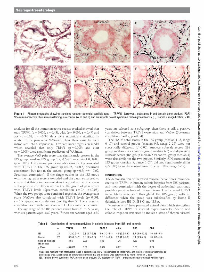
Neuronal markersTRPV1-immunoreactive fine fibres were seen scattered through-out the mucosa in all biopsies, but were more abundant in thosefrom IBS patients than controls (fig 1A,B). Quantitationrevealed that the median number of TRPV1 fibres wassignificantly (3.5-fold) higher in IBS patients compared withcontrols (p,0.0001, table 5). SP-immunoreactive fibres(fig 1C,D) were also significantly (3-fold) greater in IBS patientscompared with controls (p = 0.01, table 5), as were PGP9.5-immunoreactive fibres (p = 0.002, fig 1E,F, table 5).
When the IBS group was divided into symptom subgroups,there was no statistically significant difference in the numbersof TRPV1-immunoreactive fibres (IBS-D, n = 8, fibres/mm2,median 3.7, interquartile range 2.6–6.6; IBS-C, n = 7, 3.5, 2.1–4.9;and IBS-A, n = 8, 2.2, 2.0–5.2; ANOVA (analysis of variance)test, p = 0.23).
In order to exclude possible changes arising from differencesin bowel preparation, the IBS group was also analysed bywhether they had a flexible sigmoidoscopy (n = 7) or acolonoscopy (n = 16). TRPV1-immunoreactive fibres in theflexible sigmoidoscopy group (3.2; 2.1–4.5) and in the colono-scopy group (3.4; 2.2–5.6) were not significantly different(p = 0.6). Similarly PGP9.5-staining fibres (flexible sigmoido-scopy 5.0; 3.7–5.5 vs colonoscopy 5.0; 3.1–7.2; p = 0.8) and SP-staining fibres (flexible sigmoidoscopy 2.4; 1.2–2.8 vs colono-scopy 1.6; 0.7–3.4; p = 0.6) did not differ significantly betweenthe two groups.
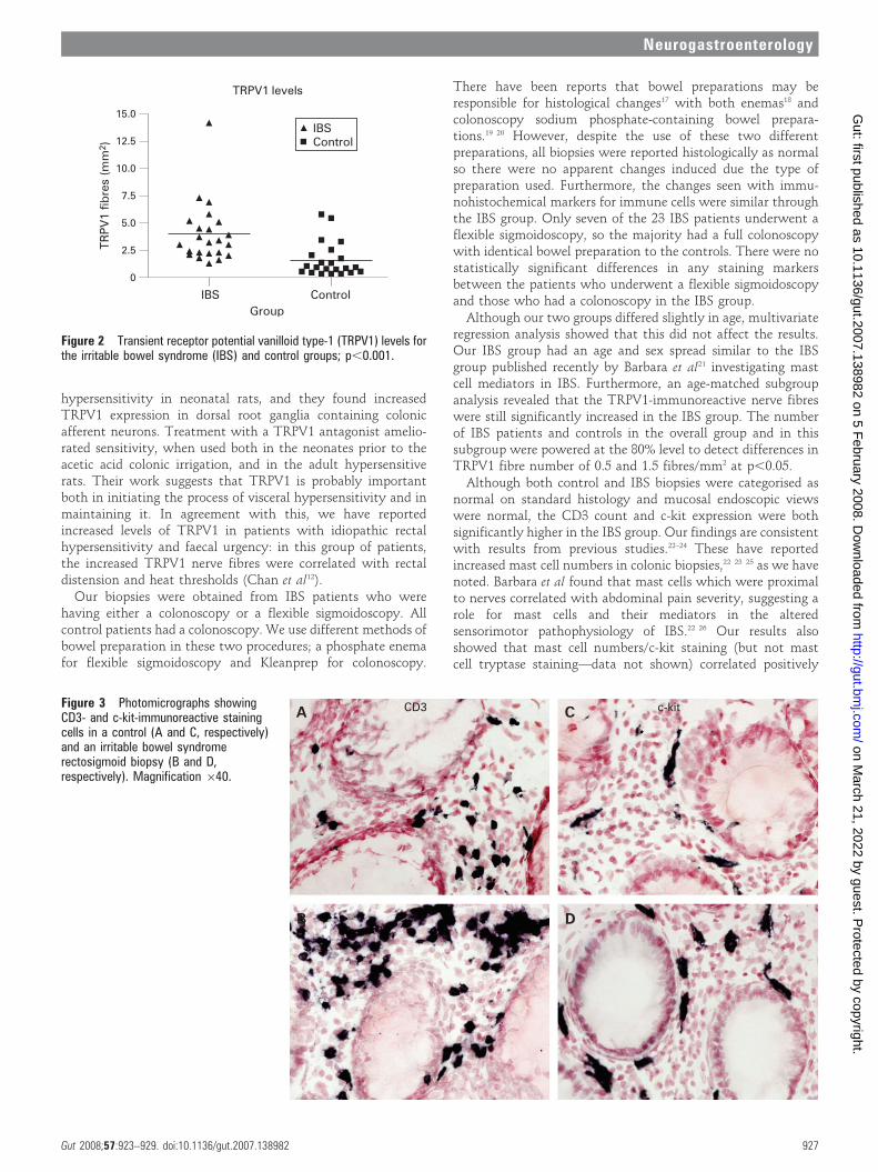
As there was a difference in the age structure of the IBS andcontrol groups, we also compared TRPV1 results in a subgroupof 13 subjects from the IBS and 13 subjects from the controlgroup, both with an age range 50–75 years. In these age-matched subgroups, TRPV1-immunoreactive fibres were sig-nificantly increased in the IBS group (3.0; 2.2–4.8) comparedwith the control group (1.0; 0.6–2.1; p = 0.003). Multivariatelinear regression analysis revealed that, unlike the presence ofIBS, gender, age and type of bowel preparation/procedure werenot significant independent predictors of TRPV1 levels (fig 2).
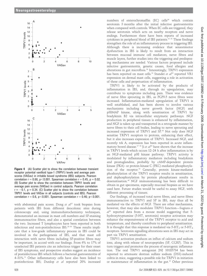
Inflammatory markersCD3+ T cells were seen scattered throughout the mucosa in allthe specimens (fig 3A,B), including controls. There was asignificantly greater percentage area of CD3+ cells in the IBSgroup compared with the controls (p = 0.03, table 4). Similarstaining but of fewer cells was seen using the CD4 T cellantibody in both groups, and this was not statisticallysignificant (p = 0.29, table 4). Mast cells immunoreactive to c-kit were also scattered throughout the mucosa (fig 3C,D), andwere significantly elevated in the IBS group compared withcontrols (p = 0.02, table 4). With subgroup analysis of theflexible sigmoidoscopy versus colonoscopy group to excludepossible effects of differing bowel preparation, c-kit staining(flexible sigmoidoscopy 5.5; 3.4–6 vs colonoscopy 3.9; 2.6–4.5)did not differ significantly (p = 0.2) between the two groups.
Pain severity, depression and anxietyAs expected, the median VASmax pain score was significantlyhigher in IBS patients than in controls (IBS 4.5; 1.4–6.5 vscontrol 0; 0–1.25; p = 0.002). Univariate linear regression
Table 2 Symptoms reported by patients with irritable bowel syndrome
IBS-type Stool frequency
Stool consistency/type (Bristol stoolform scale)
Frequency of abdominalpain in days/week
IBS-D 3–4/day 7 7
IBS-A Variable 2, 3, 7 7
IBS-C Once every 2–3 days 2, 3 6
IBS-A Variable 3, 6 2
IBS-A Variable 3, 6 7
IBS-A Variable 2, 3, 6 7
IBS-D 2–3/day 7 7
IBS-C Once every 1–2 days 3 2
IBS-C Once every 2 days 2 1
IBS-D 3–4/day 6, 7 7
IBS-A Variable 2, 6 2
IBS-C Once every 1–2 days 2 0 (pain every few weeks)
IBS-D 3/day 7 6
IBS-D 2/day 6 3
IBS-C Once every 2 days 2 3
IBS-C Once every 1–2 days 2 1
IBS-D 4–5/day 7 7
IBS-A Variable 2, 6 7
IBS-D 4/day 6 2
IBS-A Variable 2, 7 5
IBS-A Variable 3, 6 2
IBS-C Once every 1–3 days 2 3
IBS-D 4/day 6 2
IBS-A, irritable bowel syndrome with an alternating stool pattern; IBS-C constipation-predominant irritable bowel syndrome; IBS-D diarrhoea-predominant irritable bowelsyndrome.
Table 3 Indications for colonoscopy in the control group
Indication for colonoscopy n
Polyp follow-up 8
Polyp follow-up; family history of colorectal cancer 1
Iron deficiency 4
Per rectal bleeding 7
Family history of colorectal cancer 2
Table 4 Primary antibodies
Antibody Host Source: Reference Titre
TRPV1 Rabbit GSK/C22 1:10 000
Substance P Rabbit Chemicon UK 1:8000
PGP9.5 Rabbit Ultraclone 1/80 000
CD3 Mouse Dako Cytomation, Ely, UK Clone UCHTI 1:5000
CD4 Mouse Dako Cytomation, Ely, UK Clone MT310 1:5000
c-kit Mouse Novacastra, Newcastle upon Tyne, UK,clone 57A5D8
1:2000
PGP, protein gene product; TRPV1, transient receptor potential vanilloid type-1.
Neurogastroenterology
Gut 2008;57:923–929. doi:10.1136/gut.2007.138982 925
on March 21, 2022 by guest. P
rotected by copyright.http://gut.bm
j.com/
Gut: first published as 10.1136/gut.2007.138982 on 5 F
ebruary 2008. Dow
nloaded from
analyses for all the immunoreactive species studied showed thatonly TRPV1 (p = 0.005, r = 0.43), c-kit (p = 0.004, r = 0.47) andage (p = 0.02; r = 20.34) data were statistically significantlyrelated to the pain score VASmax. These three variables wereintroduced into a stepwise multivariate linear regression modelwhich revealed that only TRPV1 (p = 0.005) and c-kit(p = 0.008) were significant predictors of VASmax.
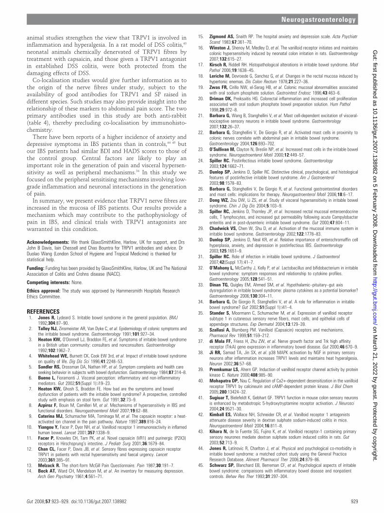
The average VAS pain score was significantly greater in theIBS group; median IBS group 1.7; 0.6–4.1 vs control 0; 0–0.5(p = 0.001). The average pain score also significantly correlatedwith TRPV1 in the IBS group (p = 0.02, r = 0.5, Spearmancorrelation) but not in the control group (p = 0.5; r = 20.02,Spearman correlation). If the single outlier in the IBS groupwith the high pain score is excluded and the data re-analysed toensure that this point does not skew the p value, then there wasstill a positive correlation within the IBS group of pain scoreswith TRPV1 levels (Spearman correlation r = 0.4; p,0.05).When the two groups were combined together, the average painscore (VASav) also correlated with TRPV1 levels (p,0.001,r = 0.5 Spearman correlation) (see fig 4A–C). There was nocorrelation seen with pain score and CD3 or mast cell counts.
The age range of the IBS patients varied from 21 to 77 years,with six patients aged (30 years. If these six patients aged (30
years are selected as a subgroup, then there is still a positivecorrelation between TRPV1 expression and VASav (Spearmancorrelation r = 0.7, p = 0.04).
The HADS total scores in the IBS group (median 11.5, range0–17) and control groups (median 9.5, range 2–25) were notstatistically different (p.0.05). Anxiety subscale scores (IBSgroup median 7.5 vs control group median 6.5) and depressionsubscale scores (IBS group median 5 vs control group median 4)were also similar in the two groups. Similarly, BDI scores in theIBS group (median 9, range 1–24) did not significantly differ(p.0.05) from the control group (median 10.5, range 1–19).
DISCUSSIONThe demonstration of increased mucosal nerve fibres immunor-eactive to TRPV1 in human colonic biopsies from IBS patients,and their correlation with the degree of abdominal pain, mayprovide a putative basis of IBS symptoms. The increased TRPV1nerve fibres were seen throughout the IBS group, with nodifference when the group was subclassified by Rome IIdefinitions into IBS-D, IBS-C and IBS-A.
Winston et al16 have presented animal data which strengthenthe role of TRPV1 in visceral hypersensitivity. Acetic acidcolonic irrigation was used to induce a state of chronic visceral
Figure 1 Photomicrographs showing transient receptor potential vanilloid type-1 (TRPV1)- (arrowed), substance P and protein gene product (PGP)9.5-immunoreactive fibre immunostaining in a control (A, C and E) and an irritable bowel syndrome rectosigmoid biopsy (B, D and F), magnification 640.
Table 5 Quantitation of immunoreactivities in colonic biopsies from IBS and controls
n TRPV1 SP PGP9.5 c-kit CD3 CD4
IBS 23 3.2 (2.2–5.1) 2.3 (0.7–3.1) 5.0 (3.2–6.1) 4.0 (2.9–5.6) 8.7 (5.9–12.1) 1.0 (0.5–3.8)
Control 22 0.9 (0.5–2.1) 0.8 (0.5–1.5) 2.7 (1.7–3.8) 2.9 (1.9–3.9) 5.4 (3.9–7.9) 1.0 (0.3–1.8)
Ratio of mediansIBS:control
– 3.56 2.96 1.86 1.36 1.60 0.98
p Value – ,0.0001 0.01 0.002 0.02 0.03 0.29
Values are medians with interquartile range in parentheses. TRPV1 is expressed as fibres/mm2, and the other immunoreactivities aspercentage area. Significance of differences between IBS and controls was determined by Mann–Whitney U test.IBS, irritable bowel syndrome; PGP, protein gene product; SP, substance P; TRPV1, transient receptor potential vanilloid type-1.
Neurogastroenterology
926 Gut 2008;57:923–929. doi:10.1136/gut.2007.138982
on March 21, 2022 by guest. P
rotected by copyright.http://gut.bm
j.com/
Gut: first published as 10.1136/gut.2007.138982 on 5 F
ebruary 2008. Dow
nloaded from
hypersensitivity in neonatal rats, and they found increasedTRPV1 expression in dorsal root ganglia containing colonicafferent neurons. Treatment with a TRPV1 antagonist amelio-rated sensitivity, when used both in the neonates prior to theacetic acid colonic irrigation, and in the adult hypersensitiverats. Their work suggests that TRPV1 is probably importantboth in initiating the process of visceral hypersensitivity and inmaintaining it. In agreement with this, we have reportedincreased levels of TRPV1 in patients with idiopathic rectalhypersensitivity and faecal urgency: in this group of patients,the increased TRPV1 nerve fibres were correlated with rectaldistension and heat thresholds (Chan et al12).
Our biopsies were obtained from IBS patients who werehaving either a colonoscopy or a flexible sigmoidoscopy. Allcontrol patients had a colonoscopy. We use different methods ofbowel preparation in these two procedures; a phosphate enemafor flexible sigmoidoscopy and Kleanprep for colonoscopy.
There have been reports that bowel preparations may beresponsible for histological changes17 with both enemas18 andcolonoscopy sodium phosphate-containing bowel prepara-tions.19 20 However, despite the use of these two differentpreparations, all biopsies were reported histologically as normalso there were no apparent changes induced due the type ofpreparation used. Furthermore, the changes seen with immu-nohistochemical markers for immune cells were similar throughthe IBS group. Only seven of the 23 IBS patients underwent aflexible sigmoidoscopy, so the majority had a full colonoscopywith identical bowel preparation to the controls. There were nostatistically significant differences in any staining markersbetween the patients who underwent a flexible sigmoidoscopyand those who had a colonoscopy in the IBS group.
Although our two groups differed slightly in age, multivariateregression analysis showed that this did not affect the results.Our IBS group had an age and sex spread similar to the IBSgroup published recently by Barbara et al21 investigating mastcell mediators in IBS. Furthermore, an age-matched subgroupanalysis revealed that the TRPV1-immunoreactive nerve fibreswere still significantly increased in the IBS group. The numberof IBS patients and controls in the overall group and in thissubgroup were powered at the 80% level to detect differences inTRPV1 fibre number of 0.5 and 1.5 fibres/mm2 at p,0.05.
Although both control and IBS biopsies were categorised asnormal on standard histology and mucosal endoscopic viewswere normal, the CD3 count and c-kit expression were bothsignificantly higher in the IBS group. Our findings are consistentwith results from previous studies.22–24 These have reportedincreased mast cell numbers in colonic biopsies,22 23 25 as we havenoted. Barbara et al found that mast cells which were proximalto nerves correlated with abdominal pain severity, suggesting arole for mast cells and their mediators in the alteredsensorimotor pathophysiology of IBS.22 26 Our results alsoshowed that mast cell numbers/c-kit staining (but not mastcell tryptase staining—data not shown) correlated positively
Figure 2 Transient receptor potential vanilloid type-1 (TRPV1) levels forthe irritable bowel syndrome (IBS) and control groups; p,0.001.
Figure 3 Photomicrographs showingCD3- and c-kit-immunoreactive stainingcells in a control (A and C, respectively)and an irritable bowel syndromerectosigmoid biopsy (B and D,respectively). Magnification 640.
Neurogastroenterology
Gut 2008;57:923–929. doi:10.1136/gut.2007.138982 927
on March 21, 2022 by guest. P
rotected by copyright.http://gut.bm
j.com/
Gut: first published as 10.1136/gut.2007.138982 on 5 F
ebruary 2008. Dow
nloaded from
with abdominal pain scores. Dong et al27 took biopsies frompatients with IBS from different ileocolonic sites duringcolonoscopy and, using immunohistochemistry techniques,demonstrated an increase in mast cell numbers and SP-stainingimmunoreactive fibers, and also a spatial correlation betweenthe two. Increased T lymphocytes have been reported in bothinfectious and non-postinfectious IBS.28–30 These results impli-cate that a low-grade inflammatory process in IBS could beinvolved in the pathogenesis of IBS, and that mast cellinteraction with nerve fibres including SP-positive fibres maybe important, in accord with our findings. From 6% to 17% ofunselected IBS patients cite an infectious trigger for their onsetof IBS symptoms, and prospective studies suggest an incidenceof postinfectious IBS which follows a bacterial gastroenteritis of4–31%.31 Other inflammatory cells have also been linked topostinfectious IBS; Dunlop et al reported 20% increased
numbers of enterochromaffin (EC) cells30 which containserotonin 3 months after the initial infective gastroenteritiswhen compared with controls. When EC cells are triggered, theyrelease serotonin which acts on nearby receptors and nerveendings. Furthermore there have been reports of increasedcytokines in peripheral blood of IBS patients.32 33 These findingsstrengthen the role of an inflammatory process in triggering IBS.Although there is increasing evidence that sensorimotordysfunction in IBS is likely to result from an interactionbetween mucosal immune cell mediators, nerve fibres andmuscle layers, further studies into the triggering and predispos-ing mechanisms are needed. Various factors proposed includeinfective gastroenteritis, genetic causes, food allergies andalterations in gut microflora.34 Interestingly, TRPV1 expressionhas been reported on mast cells.35 Stander et al35 reported VR1expression on dermal mast cells, suggesting a role in activationof these cells and perpetuation of inflammation.
TRPV1 is likely to be activated by the products ofinflammation in IBS, and, through its upregulation, maycontribute to symptoms including pain. There was evidenceof nerve fibre sprouting in IBS, as PGP9.5 nerve fibres wereincreased. Inflammation-mediated upregulation of TRPV1 iswell established, and has been shown to involve variousmechanisms including nerve growth factor (NGF) andp38MAP kinase, along with sensitisation of TRPV1 bybradykinin B2 via intracellular enzymatic pathways. NGFproduction in peripheral tissues is enhanced by inflammation,and NGF is taken up and transported in a retrograde manner bynerve fibres to their cell bodies, leading to nerve sprouting andincreased expression of TRPV1 and SP.36 Not only does NGFsensitise TRPV1 receptors to protons, enhancing their effect,but it also increases expression of TRPV1. Increased NGF, andrecently trk A, expression has been reported in acute inflam-matory bowel disease.37 38 Ji et al38 have shown that the increasein TRPV1 levels which occurs 12–24 h after inflammation is byan NGF-mediated p38 kinase pathway. TRPV1 activity ismodulated by inflammatory mediators including bradykininand prostaglandins, probably by cAMP-dependent proteinkinase (PKA)- or protein kinase C (PKC)-mediated phosphoryla-tion of the receptor.39 Generally, protein kinase-mediatedphoshorylation of the TRPV1 receptor results in sensitisation,and dephosphorylation by protein phosphatases results indesensitisation.40 NGF immunostaining has been difficult toobtain in gut specimens, especially mucosal biopsies as we haveused here. Future studies would be useful to assay NGF, withdifferent processing of tissues.
Our findings, of increased total nerve fibres, and nerve fibresimmunoreactive to TRPV1 and SP in IBS, may thus all bemediated via the effects of NGF. There are other mechanisms,however, that may also modulate TRPV1 function—Sugiura etal41 reported data from a mouse model suggesting that 5-hydroxytryptamine (5-HT, serotonin) receptor activation mayenhance the responsiveness of the TRPV1 receptor to acid andtemperature, and thereby contribute to peripheral sensitisation.It is thought that this response is mediated via 5-HT2 or 5-HT4
receptors. Serotonin signalling alterations seen in IBS may act inpart via TRPV1 sensitisation.
TRPV1 activation produces an influx of calcium and sodiumions, along with release of neuropeptides (SP, CGRP). This inturn triggers and promotes the process of neurogenic inflamma-tion. The oral TRPV1 antagonist JNJ 10185734 has beenreported to attenuate dextran sulfate sodium (DSS)-inducedcolitis in mice, suggesting a possible role for TRPV1 in initiationor maintenance of inflammation in the gut.42 Other previous
Figure 4 (A) Scatter plot to show the correlation between transientreceptor potential vanilloid type-1 (TRPV1) levels and average painscores (VASav) in irritable bowel syndrome (IBS) subjects. Pearsoncorrelation r = 0.68, p,0.001. Spearman correlation r = 0.45, p = 0.02.(B) Scatter plot to show the correlation between TRPV1 levels andaverage pain scores (VASav) in control subjects. Pearson correlationr = 20.1, p = 0.34. (C) Scatter plot to show the correlation betweenTRPV1 levels and VASav in all subjects (controls and IBS). Pearsoncorrelation r = 0.6, p,0.001. Spearman correlation r = 0.46; p,0.001.
Neurogastroenterology
928 Gut 2008;57:923–929. doi:10.1136/gut.2007.138982
on March 21, 2022 by guest. P
rotected by copyright.http://gut.bm
j.com/
Gut: first published as 10.1136/gut.2007.138982 on 5 F
ebruary 2008. Dow
nloaded from
animal studies strengthen the view that TRPV1 is involved ininflammation and hyperalgesia. In a rat model of DSS colitis,43
neonatal animals chemically denervated of TRPV1 fibres bytreatment with capsaicin, and those given a TRPV1 antagonistin established DSS colitis, were both protected from thedamaging effects of DSS.
Co-localisation studies would give further information as tothe origin of the nerve fibres under study, subject to theavailability of good antibodies for TRPV1 and SP raised indifferent species. Such studies may also provide insight into therelationship of these markers to abdominal pain score. The twoprimary antibodies used in this study are both anti-rabbit(table 4), thereby precluding co-localisation by immunohisto-chemistry.
There have been reports of a higher incidence of anxiety anddepressive symptoms in IBS patients than in controls,44 45 butour IBS patients had similar BDI and HADS scores to those ofthe control group. Central factors are likely to play animportant role in the generation of pain and visceral hypersen-sitivity as well as peripheral mechanisms.34 In this study wefocused on the peripheral sensitising mechanisms involving low-grade inflammation and neuronal interactions in the generationof pain.
In summary, we present evidence that TRPV1 nerve fibres areincreased in the mucosa of IBS patients. Our results provide amechanism which may contribute to the pathophysiology ofpain in IBS, and clinical trials with TRPV1 antagonists arewarranted in this condition.
Acknowledgements: We thank GlaxoSmithKline, Harlow, UK for support, and DrsJohn B Davis, Iain Chessell and Chas Bountra for TRPV1 antibodies and advice. DrDuolao Wang (London School of Hygiene and Tropical Medicine) is thanked forstatistical help.
Funding: Funding has been provided by GlaxoSmithKline, Harlow, UK and The NationalAssociation of Colitis and Crohns disease (NACC).
Competing interests: None.
Ethics approval: The study was approved by Hammersmith Hospitals ResearchEthics Committee.
REFERENCES1. Jones R, Lydeard S. Irritable bowel syndrome in the general population. BMJ
1992;304:87–90.2. Talley NJ, Zinsmeister AR, Van Dyke C, et al. Epidemiology of colonic symptoms and
the irritable bowel syndrome. Gastroenterology 1991;101:927–34.3. Heaton KW, O’Donnell LJ, Braddon FE, et al. Symptoms of irritable bowel syndrome
in a British urban community: consulters and nonconsulters. Gastroenterology1992;102:1962–7.
4. Whitehead WE, Burnett CK, Cook EW 3rd, et al. Impact of irritable bowel syndromeon quality of life. Dig Dis Sci 1996;41:2248–53.
5. Sandler RS, Drossman DA, Nathan HP, et al. Symptom complaints and health careseeking behavior in subjects with bowel dysfunction. Gastroenterology 1984;87:314–8.
6. Bueno L, Fioramonti J. Visceral perception: inflammatory and non-inflammatorymediators. Gut 2002;51(Suppl 1):i19–23.
7. Heaton KW, Ghosh S, Braddon FE. How bad are the symptoms and boweldysfunction of patients with the irritable bowel syndrome? A prospective, controlledstudy with emphasis on stool form. Gut 1991;32:73–9.
8. Azpiroz F, Bouin M, Camilleri M, et al. Mechanisms of hypersensitivity in IBS andfunctional disorders. Neurogastroenterol Motil 2007;19:62–88.
9. Caterina MJ, Schumacher MA, Tominaga M, et al. The capsaicin receptor: a heat-activated ion channel in the pain pathway. Nature 1997;389:816–24.
10. Yiangou Y, Facer P, Dyer NH, et al. Vanilloid receptor 1 immunoreactivity in inflamedhuman bowel. Lancet 2001;357:1338–9.
11. Facer P, Knowles CH, Tam PK, et al. Novel capsaicin (VR1) and purinergic (P2X3)receptors in Hirschsprung’s intestine. J Pediatr Surg 2001;36:1679–84.
12. Chan CL, Facer P, Davis JB, et al. Sensory fibres expressing capsaicin receptorTRPV1 in patients with rectal hypersensitivity and faecal urgency. Lancet2003;361:385–91.
13. Melzack R. The short-form McGill Pain Questionnaire. Pain 1987;30:191–7.14. Beck AT, Ward CH, Mendelson M, et al. An inventory for measuring depression.
Arch Gen Psychiatry 1961;4:561–71.
15. Zigmond AS, Snaith RP. The hospital anxiety and depression scale. Acta PsychiatrScand 1983;67:361–70.
16. Winston J, Shenoy M, Medley D, et al. The vanilloid receptor initiates and maintainscolonic hypersensitivity induced by neonatal colon irritation in rats. Gastroenterology2007;132:615–27.
17. Kirsch R, Riddell RH. Histopathological alterations in irritable bowel syndrome. ModPathol 2006;19:1638–45.
18. Leriche M, Devroede G, Sanchez G, et al. Changes in the rectal mucosa induced byhypertonic enemas. Dis Colon Rectum 1978;21:227–36.
19. Zwas FR, Cirillo NW, el-Serag HB, et al. Colonic mucosal abnormalities associatedwith oral sodium phosphate solution. Gastrointest Endosc 1996;43:463–6.
20. Driman DK, Preiksaitis HG. Colorectal inflammation and increased cell proliferationassociated with oral sodium phosphate bowel preparation solution. Hum Pathol1998;29:972–8.
21. Barbara G, Wang B, Stanghellini V, et al. Mast cell-dependent excitation of visceral-nociceptive sensory neurons in irritable bowel syndrome. Gastroenterology2007;132:26–37.
22. Barbara G, Stanghellini V, De Giorgio R, et al. Activated mast cells in proximity tocolonic nerves correlate with abdominal pain in irritable bowel syndrome.Gastroenterology 2004;126:693–702.
23. O’Sullivan M, Clayton N, Breslin NP, et al. Increased mast cells in the irritable bowelsyndrome. Neurogastroenterol Motil 2000;12:449–57.
24. Spiller RC. Postinfectious irritable bowel syndrome. Gastroenterology2003;124:1662–71.
25. Dunlop SP, Jenkins D, Spiller RC. Distinctive clinical, psychological, and histologicalfeatures of postinfective irritable bowel syndrome. Am J Gastroenterol2003;98:1578–83.
26. Barbara G, Stanghellini V, De Giorgio R, et al. Functional gastrointestinal disordersand mast cells: implications for therapy. Neurogastroenterol Motil 2006;18:6–17.
27. Dong WZ, Zou DW, Li ZS, et al. Study of visceral hypersensitivity in irritable bowelsyndrome. Chin J Dig Dis 2004;5:103–9.
28. Spiller RC, Jenkins D, Thornley JP, et al. Increased rectal mucosal enteroendocrinecells, T lymphocytes, and increased gut permeability following acute Campylobacterenteritis and in post-dysenteric irritable bowel syndrome. Gut 2000;47:804–11.
29. Chadwick VS, Chen W, Shu D, et al. Activation of the mucosal immune system inirritable bowel syndrome. Gastroenterology 2002;122:1778–83.
30. Dunlop SP, Jenkins D, Neal KR, et al. Relative importance of enterochromaffin cellhyperplasia, anxiety, and depression in postinfectious IBS. Gastroenterology2003;125:1651–9.
31. Spiller RC. Role of infection in irritable bowel syndrome. J Gastroenterol2007;42(Suppl 17):41–7.
32. O’Mahony L, McCarthy J, Kelly P, et al. Lactobacillus and bifidobacterium in irritablebowel syndrome: symptom responses and relationship to cytokine profiles.Gastroenterology 2005;128:541–51.
33. Dinan TG, Quigley EM, Ahmed SM, et al. Hypothalamic–pituitary–gut axisdysregulation in irritable bowel syndrome: plasma cytokines as a potential biomarker?Gastroenterology 2006;130:304–11.
34. Barbara G, De Giorgio R, Stanghellini V, et al. A role for inflammation in irritablebowel syndrome? Gut 2002;51(Suppl 1):i41–4.
35. Stander S, Moormann C, Schumacher M, et al. Expression of vanilloid receptorsubtype 1 in cutaneous sensory nerve fibers, mast cells, and epithelial cells ofappendage structures. Exp Dermatol 2004;13:129–39.
36. Szallasi A, Blumberg PM. Vanilloid (Capsaicin) receptors and mechanisms.Pharmacol Rev 1999;51:159–212.
37. di Mola FF, Friess H, Zhu ZW, et al. Nerve growth factor and Trk high affinityreceptor (TrkA) gene expression in inflammatory bowel disease. Gut 2000;46:670–9.
38. Ji RR, Samad TA, Jin SX, et al. p38 MAPK activation by NGF in primary sensoryneurons after inflammation increases TRPV1 levels and maintains heat hyperalgesia.Neuron 2002;36:57–68.
39. Premkumar LS, Ahern GP. Induction of vanilloid receptor channel activity by proteinkinase C. Nature 2000;408:985–90.
40. Mohapatra DP, Nau C. Regulation of Ca2+-dependent desensitization in the vanilloidreceptor TRPV1 by calcineurin and cAMP-dependent protein kinase. J Biol Chem2005;280:13424–32.
41. Sugiuar T, Bielefeldt K, Gebhart GF. TRPV1 function in mouse colon sensory neuronsis enhanced by metabotropic 5-hydroxytryptamine receptor activation. J Neurosci2004;24:9521–30.
42. Kimball ES, Wallace NH, Schneider CR, et al. Vanilloid receptor 1 antagonistsattenuate disease severity in dextran sulphate sodium-induced colitis in mice.Neurogastroenterol Motil 2004;16:811–8.
43. Kihara N, de la Fuente SG, Fujino K, et al. Vanilloid receptor-1 containing primarysensory neurones mediate dextran sulphate sodium induced colitis in rats. Gut2003;52:713–9.
44. Jones R, Latinovic R, Charlton J, et al. Physical and psychological co-morbidity inirritable bowel syndrome: a matched cohort study using the General PracticeResearch Database. Aliment Pharmacol Ther 2006;24:879–86.
45. Schwarz SP, Blanchard EB, Berreman CF, et al. Psychological aspects of irritablebowel syndrome: comparisons with inflammatory bowel disease and nonpatientcontrols. Behav Res Ther 1993;31:297–304.
Neurogastroenterology
Gut 2008;57:923–929. doi:10.1136/gut.2007.138982 929
on March 21, 2022 by guest. P
rotected by copyright.http://gut.bm
j.com/
Gut: first published as 10.1136/gut.2007.138982 on 5 F
ebruary 2008. Dow
nloaded from