increased expression levels of metalloprotease, tissue … · 2018-12-19 · increased expression...
TRANSCRIPT

Increased Expression Levels of Metalloprotease, TissueInhibitor of Metalloprotease, Metallothionein, and p63 inEctopic Endometrium: An Animal Experimental Study
Aumento nos níveis de expressão de metaloprotease, inibidortecidual das metaloproteases, metalotioneina e p63 emendométrio ectópico: um estudo experimental animal
Verônica Cristina Moraes Brandão1,2 Juliana Meola1 Sergio Britto Garcia1
Francisco José Candido-dos-Reis1 Omero Benedicto Poli-Neto1 Antonio Alberto Nogueira1
Julio Cesar Rosa-e-Silva1
1Faculty of Medicine, Universidade de São Paulo, Ribeirão Preto, SP, Brazil2 Faculty of Medicine, Universidade Federal do Mato Grosso, Cuiaba,MT, Brazil
Rev Bras Ginecol Obstet 2018;40:705–712.
Address for correspondence Julio Cesar Rosa-e-Silva, MD, PhD,Av. Bandeirantes, 3900, 14049-900, Vila Monte Alegre,Ribeirão Preto, SP, Brazil (e-mail: [email protected]).
Keywords
► endometriosis► cell differentiation► cell proliferation► tissue invasion
Abstract Objective To characterize the patterns of cell differentiation, proliferation, and tissueinvasion in eutopic and ectopic endometrium of rabbits with induced endometrioticlesions via a well- known experimental model, 4 and 8 weeks after the endometrialimplantation procedure.Methods Twenty-nine female New Zealand rabbits underwent laparotomy for endome-triosis induction through the resection of one uterine horn, isolation of the endometrium,andfixationof tissue segment to thepelvic peritoneum.Twogroupsof animals (onewith14animals, and theotherwith15)were sacrificed4and8weeks after endometriosis induction.The lesion was excised along with the opposite uterine horn for endometrial gland andstroma determination. Immunohistochemical reactions were performed in eutopic andectopic endometrial tissues for analysis of the followingmarkers:metalloprotease (MMP-9)and tissue inhibitor ofmetalloprotease (TIMP-2), which are involved in the invasive capacityof the endometrial tissue; and metallothionein (MT) and p63, which are involved in celldifferentiation and proliferation.Results The intensity of the immunostaining for MMP9, TIMP-2, MT, and p63 washigher in ectopic endometria than in eutopic endometria. However, when the ectopiclesions were compared at 4 and 8 weeks, no significant difference was observed, withthe exception of the marker p63, which was more evident after 8 weeks of evolution ofthe ectopic endometrial tissue.Conclusion Ectopic endometrial lesions seem to express greater power for celldifferentiation and tissue invasion, compared with eutopic endometria, demonstratinga potentially invasive, progressive, and heterogeneous presentation of endometriosis.
receivedMarch 28, 2018acceptedSeptember 3, 2018
DOI https://doi.org/10.1055/s-0038-1675612.ISSN 0100-7203.
Copyright © 2018 by Thieme RevinterPublicações Ltda, Rio de Janeiro, Brazil
THIEME
Original Article 705

Introduction
The etiopathogenesis of endometriosis is controversial.1
Several theories have been proposed, such as the presenceof retrograde menstrual flow associated with an immuno-logical predisposition in the peritoneal microenvironmentthat facilitates the implantation of viable endometrial cellsand has the potential for implantation.2 Recent studiessuggest that the endometria of patients with endometriosishave an invasive and aggressive behavior, and higher expres-sion levels of substances related to cellular invasion, cellulardifferentiation, and proliferation, such as metalloproteases(MMPs) and tissue inhibitors of MMPs (TIMPs),3–11 p63,12–16
and metallothionein (MT)17–19 have been described.Metalloproteases are a family of endopeptidases that play a
role in degrading and remodeling the extracellular matrix.They are zinc dependent and include collagenase, gelatinase,and stromalenzymes. Their activities are regulatedbyTIMPs.20
The production of MMPs and of TIMPs occurs in the endome-trial stroma and in the epithelium, as well as in polymorpho-nuclear leucocytes. Another important source of theseenzymes are macrophages, neutrophils, and eosinophils, acti-vated in response to a certain degree of inflammation presentin the peritoneal cavity of women with endometriosis.21–23
The membrane protein p63 is a marker of cell differentia-tion, homologous to the tumor protein suppressor p53, and isexpressed in basal squamous and subcolumnar reserve cells
in the uterine cervix, in the breasts, in the salivary glands,and in the prostate.24 It regulates proliferation and epithelialdifferentiation.25
Metallothionein is a low-molecular-weight protein thatperforms functions in cell growth, repair, and proliferation.26
The perinuclear location of MT is known to be important inthe protection against DNA damage and apoptosis inducedby external stressors.27,28
The use of female rabbits in experimental models of endo-metriosis is characterized by the development of homoge-neous lesions, generally solid hemorrhagic masses, which areeasily produced through an autotransplant of endometrialfragments or through the opening and exposure of the endo-metrial cavity.29,30 In addition, rabbits were chosen as theexperimental animals because of their low infection rate,which makes antibiotic administration unnecessary.31
In the experimental model conducted in our service,32 thedevelopment of lesions after endometrial tissue implanta-tion was 100% after 4 and 8 weeks, with the presence ofstroma and gland on histological observation, a fact alsoreported in other experimental studies.31,33
The objective of the present study was to characterize theproliferation, differentiation, and invasion behavior in eutopicandectopicendometria in rabbits submittedto the inductionofendometriosis lesions byusing a knownexperimentalmodel, 4and 8 weeks after the endometrial implantation procedure.
Resumo Objetivo Caracterizar o padrão de diferenciação celular, proliferação e invasãotecidual em endométrio eutópico e ectópico de coelhas com lesões de endometrioseinduzidas por um modelo experimental 4 e 8 semanas após o procedimento deimplantação endometrial.Métodos Vinte e nove coelhas fêmeas Nova Zelândia foram submetidas a laparoto-mia para indução de endometriose através da ressecção de um dos cornos uterinos,isolamento do endométrio e fixação do tecido no peritônio pélvico. Dois grupos deanimais (14 animais em um grupo e 15 animais no outro) foram sacrificados 4 e 8semanas após a indução da endometriose. A lesão foi excisada junto com o cornouterino contralateral para determinação da presença de glândulas e de estromaendometrial. Reações de imunohistoquímica foram realizadas no tecido endometrialeutópico e ectópico para análise dos seguintes marcadores: metaloprotease (MMP9) einibidor tecidual da metaloprotease 2 (TIMP-2), os quais estão envolvidos na capaci-dade de invasão do tecido endometrial; e metalotioneina (MT) e p63, os quais estãoenvolvidos na diferenciação e proliferação celular.Resultados A intensidade da imunomarcação para MMP9, TIMP-2, MT e p63 foi maisalta nos endométrios ectópicos do que nos endométrios eutópicos. Contudo, quandoas lesões foram comparadas entre 4 e 8 semanas, nenhuma diferença foi observada,com exceção do marcador p63, o qual foi mais evidente depois de 8 semanas deevolução do tecido endometrial ectópico.Conclusão Lesões endometriais ectópicas parecem expressar maior poder de dife-renciação celular e de invasão tecidual comparadas com endométrios eutópicos,demonstrando o potencial de invasão, de progressão e de apresentação heterogêneada endometriose.
Palavras-chave
► endometriose► diferenciação celular► proliferação celular► invasão tecidual
Rev Bras Ginecol Obstet Vol. 40 No. 11/2018
Increased Expression Levels of Metalloprotease Brandão et al.706

Methods
AnimalsThe present study was performed in the experimental surgerysector of the department of surgery and anatomy of theHospital das Clínicas, Ribeirão Preto, State of São Paulo and inthe department of pathology of the Faculty of Medicine ofRiberão Preto of the Universidade de São Paulo, state of SãoPaulo, Brazil. It was approved by the ethics committee foranimal experimentation by the same institution. After thesample calculation, the size of the study group was set at 10animals.However, considering thepossibilityof losses,15adultanimals were included per group, with one loss (death) duringtheexperiment, thereforetotaling29adultNewZealand femalevirgin rabbits from the vivarium of the Faculty of Medicine ofRibeirão Preto. The rabbits were kept in appropriate cagesunder the same conditions for 3 days before the induction ofthe lesions. All of the rabbits were submitted to a laparotomyunder general anesthesia with intravenous administration of3mLof thionembutal (2.5%) alongwith1mLof xylestesin (2%).
Induction Technique for Endometriotic LesionsThe pelvic cavity was opened with a median longitudinalincision of � 2 cm in length, 2 cm from the pubis of theanimal. Then, � 4 cm of the right uterine horn was resectedand then the hornwas closed. The portion of the uterine thatwas resected was immersed in 0.9% saline solution for� 2 minutes for tissue cleaning and then cut longitudinally,resecting a 5 � 5-mm fragment. This endometrial tissuefragment was sutured to the peritoneum near the reproduc-tive tract of the rabbits by using 2 simple Vicryl 6.0 (EthiconInc., Sommerville, NJ, USA) sutures, with the endometriumwas facing inward facing the abdominal cavity, with posteri-or closing of the abdominal surgical incision. No hormonalsupplements were administered before or after the laparot-omy. The same observer performed all of the procedures.32
Removal of Lesions for Histological AnalysisThe rabbits were divided into two groups, namely group 1,which consisted of 14 animals whose lesion evolution timewas 4 weeks, and group 2, which consisted of 14 animalswith a lesion evolution time of 8 weeks. After pelvic inspec-tion, identification, and documentation of the lesion, therabbitswere sacrificed. The lesion, alongwith the left uterinehorn (contralateral), was excised for histological analysis.The excised tissues were set in 10% formaldehyde andprocessed for inclusion in paraffin. After preparing the slides,the tissues were stained with hematoxylin and eosin (H&E)stain for histological analysis. The analysis results indicatedthat 100% of the samples in both groups were composed ofactive endometrial (gland and stroma) tissues, with similarmorphological characteristics. The lesions were character-ized by thin-walled cysts located on the striated muscle ofthe abdominal wall, projecting toward the abdominal cavity.The cyst walls were formed by a thin layer of connectivetissue, rich in cells, and covered by simple squamous epithe-lium. The stroma is rich in fibroblasts, and contains somemacrophages and eosinophils in addition to a large quantity
of typical endometrial glands. The evaluation of the lesions intwo stages has the purpose of verifying the presence of anytissue modification during the progression of the lesions, aswell as their growth.
Immunohistochemical TechniquesHistological sections (4–5 mm) were submitted for histo-chemical analysis with the antigen-antibody reaction. Thereaction was developed by using a marker visible under themicroscope. The deparaffinized and hydrated sections wererecovered antigenically by incubation in a buffered mediumin a steam pot for 40 minutes. After cooling, the endogenoustissue peroxidases were removed by adding hydrogen per-oxide, and horse serum was added to prevent nonspecificbinding of the primary antibody. The slides were thenincubated with primary antibodies obtained from Novocas-tra Laboratories Ltd. (Newcastle upon Tyne, United King-dom). The samples were then evaluated regarding thefollowingmarkers:MT (1:100; clone E9; DakoNorthAmericaInc., Carpinteria, CA, USA) and p63 (1:500; clone BC4A4;Biocare Medical, Concord, CA, USA), which are related to cellproliferation and differentiation, and MMP 9 (1:100 clone15W2; Leica Biosystems, Wetzlar, Germany) and TIMP-2(1:100 clone 3A4; Novocastra Laboratories, United King-dom), which are involved with invasive capacity. The mate-rials were then incubated with the secondary antibody andsubmitted to the avidin-biotin step. The reaction was devel-oped by treatment with 3.30-diaminobenzidine (Sigma-Aldrich Inc., St. Louis, MO, USA) for 5 minutes. Thereafter,thematerials were counterstainedwith Harris H&E stain andmounted on slides. The immunohistochemical markers wereanalyzed quantitatively and manually, counting the numberof marked cells in thousands, and divided into 4 quadrants of250 cells each. All of the slides were evaluated by twopathologists experienced in immunohistochemistry whowere blinded to the type of tissue to be analyzed.
Statistical AnalysisA statistical analysis was performed by using the GraphPadPrism 5.0 32-bit executable software (GraphPad SoftwareInc., San Diego, CA, USA). The paired Student t-test was usedfor variables with a normal distribution and for comparisonof the paired data. The non-paired Student t-test was used forvariables with normal distribution and non-paired data.Finally, the Mann-Whitney U-test was used for non-pairedvariables with non-normal distribution. The level of statisti-cal significance was set at 5%.
Results
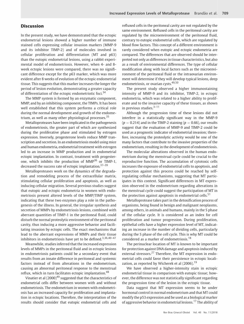
A significantlymore intense stain forMMP9, TIMP-2,MT, andp63 were observed in the ectopic endometria than in theeutopic endometria. (►Table 1 and ►Fig. 1). However, re-garding the ectopic lesions that were compared at 4 and8 weeks, no significant differences were observed, with theexception of the p63 indicator, whichwasmore evident after8 weeks of ectopic endometrial tissue progression. (►Table 2
and ►Fig. 1).
Rev Bras Ginecol Obstet Vol. 40 No. 11/2018
Increased Expression Levels of Metalloprotease Brandão et al. 707

Table 1 Cellular immunostaining according to endometrial types
Topicmean � SD
Ectopicmean � SD
p-value
MMP-9 0.273 � 0.147 0.364 � 0.223 0.0003
TIMP-2 0.249 � 0.126 0.274 � 0.152 0.02
Metallothionein 0.277 � 0.141 0.361 � 0.220 0.0003
p63 0.218 � 0.080 0.276 � 0.095 < 0.0001
Abbreviations: MMP-9, metalloprotease; SD, standard deviation;TIMP-2, tissue inhibitor of metalloprotease.
Fig. 1 Immunostainingby:metalloprotease in the eutopic (A) andectopicendometria (B); tissue inhibitor ofmetalloprotease in theeutopic (C) and ectopicendometria (D); metallothionein in the eutopic (E) and ectopic endometria (F); and p63 in the eutopic (G) and ectopic endometria (H). 40x enlargement.
Table 2 Cellular immunostaining in ectopic endometriumaccording to progression time
4 weeksmean � SD
8 weeksmean � SD
p-value
MMP-9 0.418 � 0.209 0.314 � 0.231 0.21
TIMP-2 0.288 � 0.152 0.262 � 0.157 0.66
Metallothionein 0.428 � 0.221 0.298 � 0.208 0.11
p63 0.240 � 0.058 0.309 � 0.113 0.05
Abbreviations: MMP-9, metalloprotease; SD, standard deviation;TIMP-2, tissue inhibitor of metalloprotease.
Rev Bras Ginecol Obstet Vol. 40 No. 11/2018
Increased Expression Levels of Metalloprotease Brandão et al.708
Discussion
In the present study, we have demonstrated that the ectopicendometrial lesions showed a higher number of immunestained cells expressing cellular invasion markers (MMP-9and its inhibitor TIMP-2) and of molecules involved incellular proliferation and differentiation (MT and p63)than the eutopic endometrial lesions, using a rabbit experi-mental model of endometriosis. However, when 4- and 8-week ectopic lesions were compared, there was no signifi-cant difference except for the p63 marker, which was moreevident after 8 weeks of evolution of the ectopic endometrialtissue. This suggests that thismarker increases the longer theperiod of lesion evolution, demonstrating a greater capacityof differentiation of the ectopic endometriotic foci.34
The MMP system is formed by an enzymatic component,MMP, and by an inhibiting component, the TIMPs. It has beenwell established that this system performs a critical roleduring the normal development and growth of the endome-trium, as well as many other physiological processes.35
Metalloproteases have been implicated in the pathogenesisof endometriosis, the greater part of which are synthesizedduring the proliferative phase and stimulated by estrogenexpression. Inversely, progesterone levels reduce MMP tran-scription and secretion. In an endometriosismodel usingmiceand humanendometria, endometrial treatmentwith estrogenled to an increase in MMP production and in endometrialectopic implantation. In contrast, treatment with progester-one, which inhibits the production of MMP36 or TIMP-1,decreased the success rate of ectopic implantation.37–39
Metalloproteases work on the dynamics of the degrada-tion and remodeling process of the extracellular matrix,stimulating cellular proliferation and apoptosis, as well asinducing cellular migration. Several previous studies suggestthat eutopic and ectopic endometria in women with endo-metriosis present altered levels of the MMP/TIMP system,indicating that these two enzymes play a role in the patho-genesis of the illness. In general, the irregular synthesis andsecretion of MMPs by endometriosis lesions, combined withaberrant quantities of TIMP-1 in the peritoneal fluid, coulddisturb the normal proteolytic environment of the peritonealcavity, thus inducing a more aggressive behavior and facili-tating invasion by ectopic cells. The exact mechanisms thatlead to the aberrant expressions of MMPs and their tissueinhibitors in endometriosis have yet to be defined.3,36,40–47
Meanwhile, studies inferred that the increased expressionlevels of MMPs in the peritoneal fluid and in ectopic lesionsin endometriosis patients could be a secondary event thatresults from an innate difference in peritoneal and systemicfactors instead of from alterations in the endometrium,causing an abnormal peritoneal response to the menstrualreflux, which in turn facilitates ectopic implantation.48
Vinatier et al (2000)49 suggested that the characteristics ofendometrial cells differ between women with and withoutendometriosis. The endometrium in womenwith endometri-osis has an increased capacity for proliferation and implanta-tion in ectopic locations. Therefore, the interpretation of theresults should consider that eutopic endometrial cells and
refluxed cells in the peritoneal cavity are not regulated by thesame environment. Refluxed cells in the peritoneal cavity areregulated by the microenvironment of the peritoneal fluid,contrary to eutopic endometrial cells, which are regulated byblood flow factors. This concept of a different environment israrely considered when eutopic and ectopic endometria arecompared. The differences that are observed should be inter-preted not only as differences in tissue characteristics, but alsoas a result of environmental differences. The type of cellularmodification along with local factors such as the microenvi-ronment of the peritoneal fluid or the intraovarian environ-ment will determine if they will develop typical lesions, deependometriosis, or ovarian cysts.49,50
The present study observed a higher immunostainingintensity of MMP-9 and its inhibitor, TIMP-2, in ectopicendometria, which was related to a higher ability to prolif-erate and to the invasive capacity of these tissues, as shownin previous studies.3–11
Although the progression time of these lesions did notinterfere in a statistically significant way in the MMP-9(p ¼ 0.214) and in the TIMP-2 staining (p ¼ 0.66), our resultssuggest that the evaluation of MMP-9 and TIMP-2 could beused as a prognostic indicator of endometrial invasion; there-fore, increasing the proteolytic activity would be one of themany factors that contribute to the invasive properties of theendometrium, resulting in the development of endometriosis.
The molecular alterations observed in the human endo-metrium during the menstrual cycle could be crucial to thereproductive function. The accumulation of cytotoxic cellsincreases the exposure of endometrial cells to apoptosis, andprotection against this process could be reached by self-regulating cellular mechanisms, suggesting that MT partic-ipates in this context. Significant differences in MT expres-sion observed in the endometrium regarding alterations inthe menstrual cycle could suggest the participation of MT inthe protection against apoptosis in endometrial cells.51
Metalloprotease takes part in the detoxification process oforganisms, being found in benign and malignant neoplasms,among others, in animals and humans, mainly in the S phaseof the cellular cycle. It is considered as an index for cellproliferation and tumor progression. During proliferation,epithelial cells have a higher expression level of MT, indicat-ing an increase in the number of dividing cells, particularlyduring the S phase of the cell cycle. This is why MT could beconsidered as a marker of endometriosis.18
The perinuclear location of MT is known to be importantfor protection against DNAdamage and apoptosis induced byexternal stressors.27 Therefore, the MT expression in endo-metrial cells could favor their persistence in ectopic locali-zation, as reported by Wicherek et al (2006).19
We have observed a higher-intensity stain in ectopicendometrial tissue in comparison with eutopic tissue; how-ever, the differencewas not statistically significant regardingthe progression time of the lesion in the ectopic tissue.
Data suggest that MT expression seems to be underhormonal control in normal endometrium and thatMT couldmodify the p53 expression and be used as a biologicalmarkerof aggressive behavior in endometrial lesions.17 The ability of
Rev Bras Ginecol Obstet Vol. 40 No. 11/2018
Increased Expression Levels of Metalloprotease Brandão et al. 709

the endometrium to distinguish cytotoxic activity fromincreased protection against DNA damage (MT expression),as well as concomitant changes in the number of cells in theimmune system and its activity, which are observed innormal endometrium during the phases of the menstrualcycle, seems to be fundamental for the pathological charac-teristics of endometriosis.52
The p63 protein has been described as a marker of basaland reserve cells in the female genital tract,53–55 beingstrongly relatedwith altered differentiation, includingmeta-plasia, either isolated or in combinationwith neoplasms.56,57
Some clinical and laboratory data provide evidence thatsuggests that ectopic endometrial lesions result in the dislo-cation of basal endometrial cells.58
Studies showed that endometriotic lesions express p63differently; however, whether the lack of p63 expression insome lesions is related to the extent of the illness, to itsclinical behavior, or to the exacerbation of the symptoms thataccompany it is unclear.13
We have noted in our experimental model that the stainwas more evident in ectopic endometria than in eutopicendometria (p < 0.0001), inferring a greater potential fordifferentiation in eutopic tissue, favoring the establishmentof endometriosis, as it has already been suggested that p63-positive cells in normal endometria represent cells with astem cell phenotype that have the potential for multidirec-tional differentiation.12–16
Another relevant fact was the interference of the progres-sion time of the lesion regarding the staining intensity,whichcould suggest that the longer the progression, the greater theability of endometriotic foci in the ectopic tissue todifferentiate.
Conclusion
Upon analyzing the differentmarkers of cell proliferation anddifferentiation, as well as of tissue invasion in eutopic andectopic endometria in the rabbits submitted to the inductionof endometriotic lesions by the experimental model, 4 and8 weeks after the endometrial implantation procedure, weconclude that the ectopic lesions seem to express a greaterability for cell proliferation and differentiation, as well as fortissue invasion when compared with eutopic endometria.This is evident in the greater intensity of the immunostainingfor the proteins involved in the capacity to invade tissues(MMP-9 and TIMP-2) in ectopic endometria compared to theeutopic endometrium and in the higher number of cells ofmolecules involved in cell proliferation and differentiation(MT and p63) that were stained in ectopic endometria, whichwas most evident by the p63 stain in the endometrium after8 weeks of progression. The ectopic endometrial lesionsseem to express a greater ability for cell differentiationand tissue invasion than eutopic endometrial lesions, char-acterizing endometriosis as a potentially invasive, progres-sive, and heterogeneous disease in its presentation.However, more studies are necessary to better clarify theparticipation of these markers in the complex pathophysio-logical mechanism of endometriosis.
ContributorsAll authors contributed to the conception and design, datacollection or analysis and interpretation of data, as well asto the writing of the article or critical review of theintellectual content and to the final approval of theversion to be published.
Conflicts of InterestThe authors have no conflicts of interest to declare.
AcknowledgmentsThe authors thank Rosangela Orlandin Lopes for the excel-lent technical assistance. This work was supported by theNational Council for Scientific and Technological Develop-ment (CNPq, in thePortugueseacronym),Universal Project:Procedure 47210 /2009–1–Research Support.
References1 Burney RO, Giudice LC. Pathogenesis and pathophysiology of
endometriosis. Fertil Steril 2012;98(03):511–519 Doi: 10.1016/j.fertnstert.2012.06.029
2 Augoulea A, Alexandrou A, Creatsa M, Vrachnis N, LambrinoudakiI. Pathogenesis of endometriosis: the role of genetics, inflamma-tion and oxidative stress. Arch Gynecol Obstet 2012;286(01):99–103 Doi: 10.1007/s00404-012-2357-8
3 Chung HW, Wen Y, Chun SH, Nezhat C, Woo BH, Lake Polan M.Matrix metalloproteinase-9 and tissue inhibitor of metallopro-teinase-3 mRNA expression in ectopic and eutopic endometriumin women with endometriosis: a rationale for endometrioticinvasiveness. Fertil Steril 2001;75(01):152–159 Doi: 10.1016/S0015-0282(00)01670-8
4 Chung HW, Lee JY, Moon HS, et al. Matrix metalloproteinase-2,membranous type 1 matrix metalloproteinase, and tissue inhi-bitor of metalloproteinase-2 expression in ectopic and eutopicendometrium. Fertil Steril 2002;78(04):787–795 Doi: 10.1016/S0015-0282(02)03322-8
5 Uzan C, Cortez A, Dufournet C, Fauvet R, Siffroi JP, Daraï E. Eutopicendometrium and peritoneal, ovarian and bowel endometriotictissues express a different profile of matrix metalloproteinases-2,-3 and -11, and of tissue inhibitor metalloproteinases-1 and -2.Virchows Arch 2004;445(06):603–609 Doi: 10.1007/s00428-004-1117-y
6 Ria R, Loverro G, Vacca A, et al. Angiogenesis extent and expres-sion ofmatrix metalloproteinase-2 and -9 agree with progressionof ovarian endometriomas. Eur J Clin Invest 2002;32(03):199–206Doi: 10.1046/j.1365-2362.2002.00960.x
7 Sotnikova NY, Antsiferova YS, Posiseeva LV, ShishkovDN, PosiseevDV, Filippova ES. Mechanisms regulating invasiveness and growthof endometriosis lesions in rat experimental model and inhumans. Fertil Steril 2010;93(08):2701–2705 Doi: 10.1016/j.fertnstert.2009.11.024
8 Wenzl RJ, Heinzl H. Localization of matrix metalloproteinase-2 inuterine endometrium and ectopic implants. Gynecol ObstetInvest 1998;45(04):253–257 Doi: 10.1159/000009978
9 BeckerCM, LouisG, ExarhopoulosA, et al.Matrixmetalloproteinasesare elevated in the urine of patientswith endometriosis. Fertil Steril2010;94(06):2343–2346 Doi: 10.1016/j.fertnstert.2010.02.040
10 Liu XJ, He YL, Peng DX. Expression of metalloproteinase-9 inectopic endometrium in women with endometriosis. J First MilMed Univ 2002;22(05):467–469
11 Collette T, Bellehumeur C, Kats R, et al. Evidence for an increasedrelease of proteolytic activity by the eutopic endometrial tissue inwomen with endometriosis and for involvement of matrix
Rev Bras Ginecol Obstet Vol. 40 No. 11/2018
Increased Expression Levels of Metalloprotease Brandão et al.710

metalloproteinase-9. Hum Reprod 2004;19(06):1257–1264 Doi:10.1093/humrep/deh290
12 O’Connell JT, Mutter GL, Cviko A, et al. Identification of a basal/reserve cell immunophenotype in benign and neoplastic endo-metrium: a study with the p53 homologue p63. Gynecol Oncol2001;80(01):30–36 Doi: 10.1006/gyno.2000.6026
13 Poli Neto OB, Ferreira HM, Ramalho LN, Rosa e Silva JC, Candidodos Reis FJ, Nogueira AA. Expression of p63 differs in peritonealendometriosis, endometriomas, adenomyosis, rectovaginal sep-tum endometriosis, and abdominal wall endometriosis. ArchPathol Lab Med 2007;131(07):1099–1102 Doi: 10.1043/1543-2165(2007)131[1099:EOPDIP]2.0.CO;2
14 Gargett CE, Masuda H. Adult stem cells in the endometrium. MolHum Reprod 2010;16(11):818–834 Doi: 10.1093/molehr/gaq061
15 Maruyama T, Yoshimura Y. Stem cell theory for the pathogenesisof endometriosis. Front Biosci (Elite Ed) 2012;4:2754–2763
16 Oliveira FR, Dela Cruz C, Del Puerto HL, Vilamil QT, Reis FM,Camargos AF. Stem cells: are they the answer to the puzzlingetiologyof endometriosis?Histol Histopathol 2012;27(01):23–29Doi: 10.14670/HH-27.23
17 Ioachim EE, Kitsiou E, Carassavoglou C, Stefanaki S, Agnantis NJ.Immunohistochemical localization of metallothionein in endo-metrial lesions. J Pathol 2000;191(03):269–273 Doi: 10.1002/1096-9896(2000)9999:9999<:AID-PATH616>3.0.CO;2-Q
18 Madej P, Madej JA, Kamiński K, et al. [Immunohistochemicallocalisation of metallothionein in ovarian endometriosis inwomen]. Ginekol Pol 2003;74(11):1456–1464
19 Wicherek L, Dutsch-Wicherek M, Galazka K, et al. Comparison ofRCAS1 and metallothionein expression and the presence andactivity of immune cells in human ovarian and abdominal wallendometriomas. Reprod Biol Endocrinol 2006;4:41
20 Salamonsen LA. Matrix metalloproteinases and their tissue inhi-bitors in endocrinology. Trends Endocrinol Metab 1996;7(01):28–34 Doi: 10.1016/1043-2760(95)00189-1
21 Busiek DF, Baragi V, Nehring LC, Parks WC, Welgus HG. Matrilysinexpression by humanmononuclear phagocytes and its regulationby cytokines and hormones. J Immunol 1995;154(12):6484–6491
22 Jeziorska M, Nagase H, Salamonsen LA, Woolley DE. Immunolo-calization of the matrix metalloproteinases gelatinase B andstromelysin 1 in human endometrium throughout the menstrualcycle. J Reprod Fertil 1996;107(01):43–51
23 Shi W, Mognetti B, Campana A, Bischof P. Metalloproteinase secre-tionbyendometrial leukocyte subsets. Am JReprod Immunol 1995;34(05):299–310 Doi: 10.1111/j.1600-0897.1995.tb00956.x
24 Yang A, Kaghad M, Wang Y, et al. p63, a p53 homolog at 3q27-29,encodes multiple products with transactivating, death-inducing,and dominant-negative activities. Mol Cell 1998;2(03):305–316Doi: 10.1016/S1097-2765(00)80275-0
25 Yang A, Schweitzer R, Sun D, et al. p63 is essential for regenerativeproliferation in limb, craniofacial and epithelial development.Nature 1999;398(6729):714–718 Doi: 10.1038/19539
26 Theocharis SE, Margeli AP, Klijanienko JT, Kouraklis GP. Metal-lothionein expression in human neoplasia. Histopathology 2004;45(02):103–118 Doi: 10.1111/j.1365-2559.2004.01922.x
27 Levadoux-Martin M, Hesketh JE, Beattie JH, Wallace HM. Influ-ence of metallothionein-1 localization on its function. Biochem J2001;355(Pt 2):473–479 Doi: 10.1042/bj3550473
28 Chiaverini N, De Ley M. Protective effect of metallothionein onoxidative stress-induced DNA damage. Free Radic Res 2010;44(06):605–613 Doi: 10.3109/10715761003692511
29 Manyak MJ, Nelson LM, Solomon D, DeGraff W, Stillman RJ, RussoA. Fluorescent detection of rabbit endometrial implants resultingfrommonodispersed viable cell suspensions. Fertil Steril 1990;54(02):356–359 Doi: 10.1016/S0015-0282(16)53720-0
30 Hahn DW, Carraher RP, Foldesy RG, McGuire JL. Development ofan animal model for quantitatively evaluating effects of drugs onendometriosis. Fertil Steril 1985;44(03):410–415 Doi: 10.1016/S0015-0282(16)48869-2
31 Schor E, Baracat EC, Simões MJ, de Freitas V, Giannotti Filho O, deLima GR. Effects of conjugated estrogens and progestogen insurgically induced endometriosis in oophorectomized rats. ClinExp Obstet Gynecol 1999;26(3-4):158–161
32 Rosa e Silva JC, Rosa e Silva ACJS, Coltro PS, Garcia SB, Cândido dosReis FJ, Nogueira AA. [Experimental endometriosis model inrabbits with follow-up of the lesions. Rev Bras Ginecol Obstet2004;26:715–719 Doi: 10.1590/S0100-72032004000900007
33 Schenken RS, Asch RH. Surgical induction of endometriosis in therabbit: effects on fertility and concentrations of peritoneal fluidprostaglandins. Fertil Steril 1980;34(06):581–587 Doi: 10.1016/S0015-0282(16)45199-X
34 Schor E, Freitas V, Soares JJ, Simões M, Baracat E. [Endometriosis:experimental model in rats]. Rev Bras Ginecol Obstet 1999;21:281–284 Doi: 10.1590/S0100-72031999000500006
35 Zhou HE, Nothnick WB. The relevancy of the matrix metallopro-teinase system to the pathophysiology of endometriosis. FrontBiosci 2005;10:569–575
36 Osteen KG, Bruner KL, Sharpe-Timms KL. Steroid and growthfactor regulation of matrix metalloproteinase expression andendometriosis. Semin Reprod Endocrinol 1996;14(03):247–255Doi: 10.1055/s-2007-1016334
37 Bruner-Tran KL, Eisenberg E, Yeaman GR, Anderson TA, McBean J,Osteen KG. Steroid and cytokine regulation of matrix metallo-proteinase expression in endometriosis and the establishment ofexperimental endometriosis in nude mice. J Clin EndocrinolMetab 2002;87(10):4782–4791 Doi: 10.1210/jc.2002-020418
38 Chen JL, Lin QH, Fang XL, Tao GS, Huang FY. [Effect of progesteroneon the secretion of matrix metalloproteinase-2 and matrixmetalloproteinase-9 in human ectopic endometrial stromalcells]. Zhong Nan Da Xue Xue Bao Yi Xue Ban 2005;30(03):307–311
39 Bruner KL, Matrisian LM, Rodgers WH, Gorstein F, Osteen KG.Suppression of matrix metalloproteinases inhibits establishmentof ectopic lesions by human endometrium in nude mice. J ClinInvest 1997;99(12):2851–2857 Doi: 10.1172/JCI119478
40 Gilabert-Estellés J, Ramón LA, España F, et al. Expression ofangiogenic factors in endometriosis: relationship to fibrinolyticand metalloproteinase systems. Hum Reprod 2007;22(08):2120–2127 Doi: 10.1093/humrep/dem149
41 Sillem M, Prifti S, Koch A, Neher M, Jauckus J, Runnebaum B.Regulation of matrix metalloproteinases and their inhibitors inuterine endometrial cells of patients with and without endome-triosis. Eur J Obstet Gynecol Reprod Biol 2001;95(02):167–174Doi: 10.1016/S0301-2115(00)00415-2
42 Szamatowicz J, Laudański P, Tomaszewska I. Matrix metallopro-teinase-9 and tissue inhibitor of matrix metalloproteinase-1: apossible role in the pathogenesis of endometriosis. Hum Reprod2002;17(02):284–288 Doi: 10.1093/humrep/17.2.284
43 Gilabert-Estellés J, Estellés A, Gilabert J, et al. Expression of severalcomponents of the plasminogen activator and matrix metallo-proteinase systems in endometriosis. Hum Reprod 2003;18(07):1516–1522 Doi: 10.1093/humrep/deg300
44 Li Y, Lang JH. [Expressions of matrix metalloproteinase-9 andtissue inhibitor of metalloproteinase-1 mRNA in endometriosis].Zhonghua Fu Chan Ke Za Zhi 2006;41(01):30–33
45 Collette T, Maheux R, Mailloux J, Akoum A. Increased expressionof matrix metalloproteinase-9 in the eutopic endometrial tissueof women with endometriosis. Hum Reprod 2006;21(12):3059-–3067 Doi: 10.1093/humrep/del297
46 Pitsos M, Kanakas N. The role of matrix metalloproteinases in thepathogenesis of endometriosis. Reprod Sci 2009;16(08):717–726Doi: 10.1177/1933719109333661
47 Amălinei C, Căruntu ID, Giuşcă SE, Bălan RA. Matrix metallopro-teinases involvement in pathologic conditions. Rom J MorpholEmbryol 2010;51(02):215–228
48 Malik S, Day K, Perrault I, Charnock-Jones DS, Smith SK.Menstrualeffluent in endometriosis shows no difference in volume, VEGF-A,
Rev Bras Ginecol Obstet Vol. 40 No. 11/2018
Increased Expression Levels of Metalloprotease Brandão et al. 711

MMP2 and MMP9 or sFLT. Reprod Biomed Online 2006;12(02):174–181
49 Vinatier D, Cosson M, Dufour P. Is endometriosis an endometrialdisease? Eur J Obstet Gynecol Reprod Biol 2000;91(02):113–125Doi: 10.1016/S0301-2115(99)00263-8
50 Nisolle M, Donnez J. Peritoneal endometriosis, ovarian endome-triosis, and adenomyotic nodules of the rectovaginal septum arethree different entities. Fertil Steril 1997;68(04):585–596 Doi:10.1016/S0015-0282(97)00191-X
51 Klimek M, Wicherek L, Galazka K, et al. Cycle dependent expres-sion of endometrial metallothionein. Neuroendocrinol Lett 2005;26(06):663–666
52 Wicherek L, Popiela TJ, Galazka K, et al. Metallothionein andRCAS1 expression in comparison to immunological cellsactivity in endometriosis, endometrial adenocarcinomaand endometrium according to menstrual cycle changes. Gyne-col Oncol 2005;99(03):622–630 Doi: 10.1016/j.ygyno.2005.07.003
53 Quade BJ, Yang A, Wang Y, et al. Expression of the p53 homologuep63 in early cervical neoplasia. Gynecol Oncol 2001;80(01):24–29Doi: 10.1006/gyno.2000.5953
54 Martens JE, Arends J, Van der Linden PJ, De Boer BA, HelmerhorstTJ. Cytokeratin 17 and p63 are markers of the HPV target cell, thecervical stem cell. Anticancer Res 2004;24(2B):771–775
55 Houghton O, McCluggage WG. The expression and diagnosticutility of p63 in the female genital tract. Adv Anat Pathol 2009;16(05):316–321 Doi: 10.1097/PAP.0b013e3181b507c6
56 DiComoCJ,UristMJ,BabayanI, etal.p63expressionprofiles inhumannormal and tumor tissues. Clin Cancer Res 2002;8(02):494–501
57 Poli Neto OB, Candido Dos Reis FJ, Zambelli Ramalho LN, NogueiraAA, de Andrade JM. p63 expression in epithelial ovarian tumors.Int J Gynecol Cancer 2006;16(01):152–155 Doi: 10.1111/j.1525-1438.2006.00290.x
58 Leyendecker G, HerbertzM, Kunz G,Mall G. Endometriosis resultsfrom the dislocation of basal endometrium. Hum Reprod 2002;17(10):2725–2736 Doi: 10.1093/humrep/17.10.2725
Rev Bras Ginecol Obstet Vol. 40 No. 11/2018
Increased Expression Levels of Metalloprotease Brandão et al.712