indian guidelines for copd
TRANSCRIPT

DR S RAGHU M.D.,
ASST PROF
DEPT. T B & CD
GUNTUR MEDICAL
COLLEGE
GUNTUR
Dr s. raghu m.d.,Associate professor Department of TB & CD
Guntur medical college&
Chest physicianGovt fever hospital
Guntur


Introduction • COPD poses enormous burden in terms of
morbidity and mortality globally and in India.
• COPD is a major public health problem in
India.
• Although several International guidelines for
diagnosis and management of COPD are
available, yet there are lot of gaps in
recognition and management of COPD in India
due to vast differences in availability and
affordability of healthcare facilities across the
country.

FORMULATION OF
GUIDELINES• The process of development of guidelines for
diagnosis and management of patients of
COPD in India was undertaken as a joint
exercise ICS and NCCP, by the Department of
Pulmonary Medicine, PGIMER, Chandigarh.
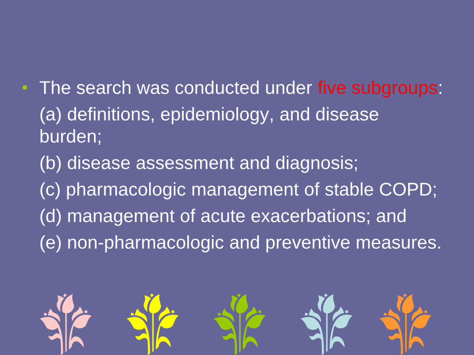
• The search was conducted under five subgroups:
(a) definitions, epidemiology, and disease
burden;
(b) disease assessment and diagnosis;
(c) pharmacologic management of stable COPD;
(d) management of acute exacerbations; and
(e) non-pharmacologic and preventive measures.
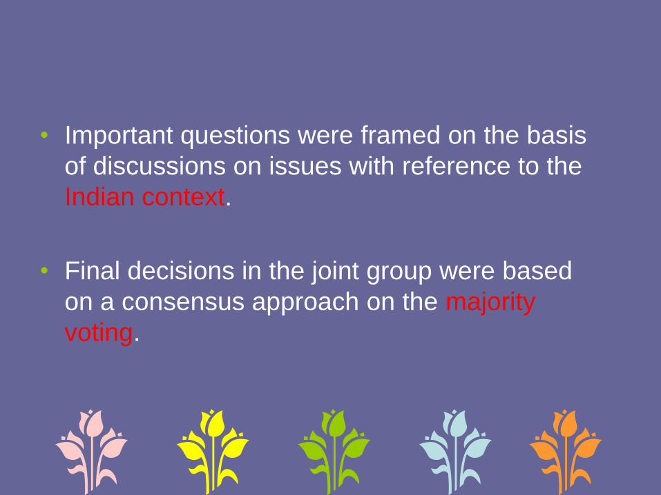
• Important questions were framed on the basis
of discussions on issues with reference to the
Indian context.
• Final decisions in the joint group were based
on a consensus approach on the majority
voting.

Classification of level of evidence and grading of
recommendation
• The modified grade system was used for
classifying the quality of evidence as 1, 2, 3, or
usual practice point (UPP).
• The strength of recommendation was graded
as A or B depending upon the level of evidence

Classification of level of evidence
Level 1 High-quality evidence backed by consistent results from
well-performed randomized controlled trials, or
overwhelming evidence from well-executed
observational studies with strong effects
Level 2 Moderate-quality evidence from randomized trials (that
suffer from flaws in conduct, inconsistency, indirectness,
imprecise estimates, reporting bias, or other limitations)
Level 3 Low-quality evidence from observational evidence or
from controlled trials with several serious limitations
Useful
practice
point
Not backed by sufficient evidence; however, a
consensus reached by working group, based on clinical
experience and expertise

Grading of recommendation based on the quality of
evidence
Grade
A
Strong recommendation to do (or not to do) where
the benefits clearly outweigh the risk (or vice versa)
for most, if not all patients
Grade
B
Weaker recommendation where benefits and risk
are more closely balanced or are more certain

• Grade A recommendations in the guidelines
should be interpreted as “recommended” and
the grade B recommendations as “suggested”.
• While making a recommendation, the issues of
practicality, costs, and feasibility in the country
at different levels of healthcare was also taken
into consideration.
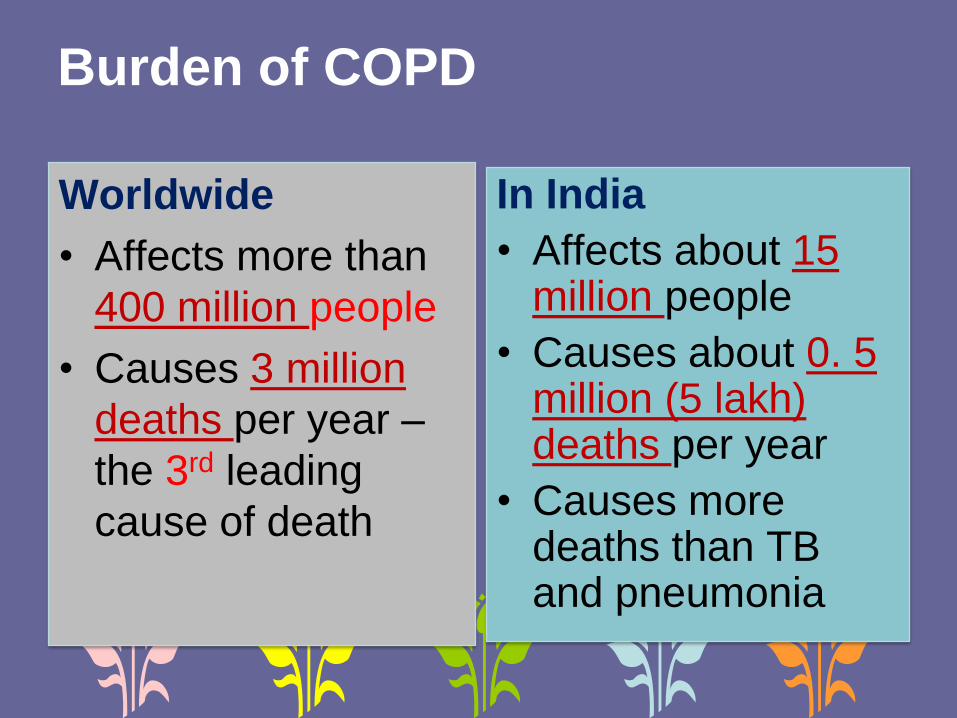
Burden of COPD
Worldwide
• Affects more than
400 million people
• Causes 3 million
deaths per year –
the 3rd leading
cause of death
In India
• Affects about 15 million people
• Causes about 0. 5 million (5 lakh) deaths per year
• Causes more deaths than TB and pneumonia


DEFINITION ???

• The GOLD definition in its latest edition is
comprehensive and elaborate, but complex.
• Retaining the key components of various definitions
and using simple terms, ICS & NCCP recommend
the following definition of COPD
“Chronic Obstructive Pulmonary Disease (COPD) is a
common, preventable lung disorder characterized by
progressive, poorly reversible airflow limitation often
with systemic manifestations, in response to tobacco
smoke and/or other harmful inhalational exposures.”

RISK FACTORS ?

Risk factors for COPD
Established
• Tobacco smoking
• Environmental tobacco smoke
• Exposure to biomass fuel smoke
• Occupational exposure
• Alpha-1 antitrypsin deficiency
Probable
• Outdoor air pollution
• Pulmonary tuberculosis
• Poorly treated asthma
• Intrauterine growth retardation
• Poor nourishment
• Repeated lower respiratory infections
• during childhood
• Others
• Age
• Male gender
• Low socioeconomic status

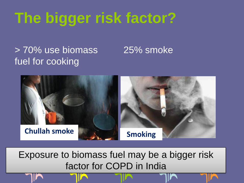
The bigger risk factor?
> 70% use biomass
fuel for cooking
25% smoke
Exposure to biomass fuel may be a bigger risk
factor for COPD in India
SmokingChullah smoke

• Both smokeless and smoking forms of tobacco are
associated with serious health hazards, although only
smoking tobacco is primarily responsible for COPD. (1A)
• Bidi and other indigenous forms of tobacco smoking are at
least as (or even more) harmful than cigarette smoking.
(1A)
• Low tar or filtered cigarettes are not “less harmful”. (2B)
• There is no minimum number of cigarettes/bidi per day
below which the risk for COPD decreases. (1A)
Smoking
• Tobacco smoking is the most
well established risk factor for
COPD. (1A)


Exposure to environmental tobacco smoke (ETS) is a definitive risk factor for COPD. (1A)
Exposure to biomass fuel smoke is a strong risk factor for COPD. (1A)
There is limited data on the association of ambient air pollution and COPD, and its causative role in COPD needs further evaluation.
There is insufficient evidence to attribute an etiological role of pulmonary tuberculosis in causing COPD.
A subgroup of chronic asthma may clinically behave like COPD; whether it is true COPD remains to be established. (UPP)

WHEN TO SUSPECT ?
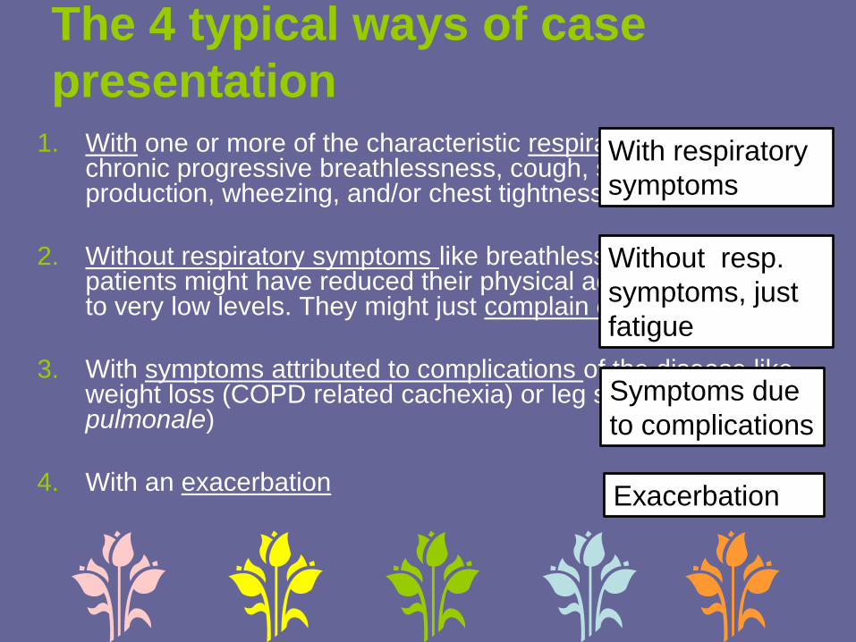
The 4 typical ways of case
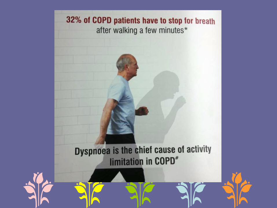
presentation1. With one or more of the characteristic respiratory symptoms of
chronic progressive breathlessness, cough, sputum production, wheezing, and/or chest tightness
2. Without respiratory symptoms like breathlessness, because patients might have reduced their physical activity unknowingly to very low levels. They might just complain of fatigue
3. With symptoms attributed to complications of the disease like weight loss (COPD related cachexia) or leg swelling (due to corpulmonale)
4. With an exacerbation
With respiratory
symptoms
Without resp.
symptoms, just
fatigue
Symptoms due
to complications
Exacerbation


Useful physical signs in the
diagnosis of COPD
Perc
ussio
n
Palp
ation
Au
scu
lta
tio
n
Inspe
ctio
n
Sp
ecia
l m
ane
uver
Pursed-lip breathing
Use of accessory muscles of respiration
Jugular venousdistension duringexpiration
Retraction of suprasternal, supraclavicular and intercostal spaces during
inspiration
Short trachea
Pulsus paradoxus
Increased anteroposteriordiameter of the chest (barrel-shapedchest)
Reduced chest movements
Peripheral edema
Dyspnea-relievingposture
Muscle wasting
Restricted chest expansion
Subxiphoidshift of maximum impulse of the heart
Chest hyperresonance
Obliteration of cardiac dullness
Lower level of liver dullness
Lower diaphragmatic levels
Diminished breath sounds
Early inspiratorycrackles
Loud pulmoniccomponent of second heart sound
Forced expiratory time
Snider’s match test
FET: Forced expiratory
time

• A diagnosis of COPD should be considered in
persons having chronic symptoms of cough,
sputum production, shortness of breath, and/or
wheezing, especially among those with
prolonged exposure to risk factors for the
disease. (1A)
• A diagnosis of COPD should not be excluded in
the absence of physical signs. (2A)
• Forced expiratory time (FET) of more than six
seconds is suggestive of airflow obstruction.
(2B)
ROLE OF SPIROMETRY IN
DIAGNOSIS ?

• Spirometry should be performed in all patients
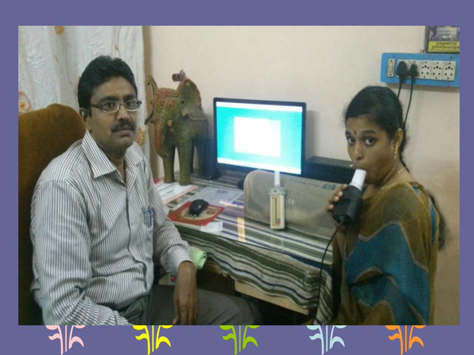
suspected of having COPD. (1A)
• In the absence of availability of spirometry,
patients suspected of having COPD should be
referred for spirometric evaluation to a center
with the facility. (UPP)

• A post-bronchodilator forced expiratory volume
in first second (FEV1)/forced vital capacity
(FVC) below the LLN (lower fifth percentile of
values from a reference population) should be
preferably used as the criterion for diagnosis of
airflow obstruction. (1A)
• However, in the absence of reference
equations for LLN, FEV1/FVC < 0.7 may be
used as the cutoff for defining airflow
obstruction. (1A)

ROLE OF REVERSIBILITY
TESTING ?
• Absence of bronchodilator reversibility
does not differentiate COPD from
asthma, and its presence does not
predict the response to treatment. (1A)
• However, all FEV1 values should be
reported post-bronchodilator.

ROLE OF SCREENING
SPIROMETRY?


• Spirometry should not be used as a screening
tool in asymptomatic individuals to detect airflow
obstruction. (2A)

ROLE OF PEFR ?


• PEF should not be routinely used for screening,
diagnosis, or monitoring of COPD. (1A)

CLASSIFICATION OF
SEVERITY?
• Classification of severity of the disease should
be done for all COPD patients based on the
FEV1 and exacerbation frequency. (1A)

Classification of severity of COPD
Severity * Post-
bronchodilator
FEV1, % predicted
mMRC
grade
Exacerbation
Frequency †
Complicatio
ns ‡
Mild ≥80 <2 <2 No
Moderate 50-79 ≥2 <2 No
Severe <50 ≥2 ≥2 Yes
*The category with the worst value should be used for severity classification, †number of
exacerbations in the last year, ‡complications include respiratory failure (defined by pO2
< 60 mmHg and/or SpO2 < 88% and/or pCO2 > 50 mmHg), cor pulmonale, and
secondary
polycythemia (hematocrit 55%)
• Level of patient’s disability due to symptoms
should be assessed using modified Medical
Research Council (mMRC) dyspnea
questionnaire or the COPD assessment test
(CAT) and recorded at each clinical visit. (1A)

mMRC grading of breathlessness
“I only get breathless with strenuous exercise”
“I get short of breath when hurrying on the level or walking up a slight hill”
“I walk slower than people of the same age on the level because of breathlessness or have to stop for breath when walking at my own pace on the level”
“I stop for breath after walking about 100 yards or after a few minutes on the level”
“I am too breathless to leave the house” or “I am breathless when dressing or undressing”
0
1
2
3
4

ROLE OF ADDITIONAL
INVESTIGATIONS ?

All new COPD suspects with cough of more than
2 weeks’ duration should undergo sputum smear
examination for acid fast bacilli to rule out
pulmonary tuberculosis as per the standard
practice of Revised National Tuberculosis
Control Program (RNTCP). (UPP)
Pulse oximetry should be used to screen for
hypoxemia in stable disease with FEV1 < 50%
and in the presence of clinical suspicion of
hypoxemia. (3A)

• An arterial blood gas analysis should be done if
arterial saturation by pulse oximetry is less than
90%. (2A)
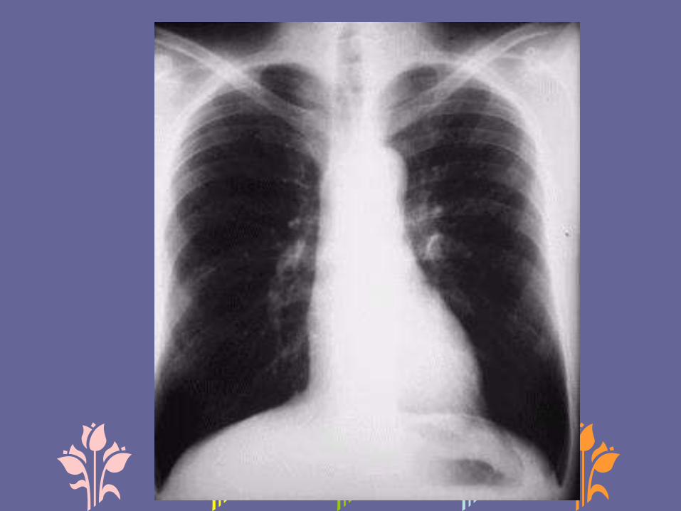
• Diagnosis of COPD should not be made on the
basis of a chest radiograph. (2A)
• Chest radiograph may be done during the initial
evaluation of COPD to look for comorbidities,
complications, and alternative diagnoses. (2B)

• Special investigations like high-resolution computed
tomography (HRCT) scan, lung volumes, diffusing
capacity for carbon monoxide (DLCO), and exercise
testing should be done in situations of diagnostic
difficulty or whenever clinically indicated. (2A)
• 6MWT may be used for monitoring of exercise
capacity in COPD. (1A)
• Testing for alpha-1 antitrypsin deficiency may be done
in young patients with lower lobe emphysema. (UPP)


Differential diagnosis of COPDFeatures
Asthma Early age of onset
Episodic symptoms with asymptomatic periods in
between
Wide variation of symptoms day to day
Symptoms worse at night/early morning
Chronic productive cough is uncommon
History of atopy may be present
Family history of asthma may be present
Reversibility of airway obstruction
Increased diffusing capacity for carbon monoxide
(DLCO)
Congestive
heart failure
Cardiomegaly/pulmonary edema in chest X-ray
PFT suggestive of restrictive abnormality
Bronchiectasis Copious purulent sputum
Clubbing, coarse crackles
HRCT shows bronchial dilatation and bronchial wall
thickening

Differential diagnosis of
COPDFeatures
Tuberculosis Fever, anorexia, weight loss
Chest X-ray opacity, fibrocavitary disease
Microbiological diagnosis
Constrictive
bronchiolitis
Non-smoker, young age
History of rheumatoid arthritis, fume exposure,
lung/bone marrow
transplantation
HRCT shows mosaic attenuation
Diffuse
panbronchiolitis
Non-smoker
Association with chronic sinusitis
HRCT shows centrilobular nodules,
hyperinflation and air-trapping


ROLE OF MULTI-DIMENSIONAL
ASSESSMENT TOOLS ?

• Composite scores including BODE (body mass
index (BMI), obstruction, dyspnea, exercise
capacity) and DOSE (dyspnea, obstruction,
smoking, exacerbation) should not be used to
assess severity or prognosis in COPD unless
they are validated in Indian patients. (2A)

ASSOCIATED
CO-MORBIDITIES ?

• Comorbid diseases in COPD are
independently associated with a higher risk of
hospitalization and mortality.
• COPD patients should be routinely evaluated
and appropriately treated for comorbid
conditions. (2A)
INDIAN STRATEGY FOR
MANAGEMENT OF COPD?


Treatment goals in stable
COPD
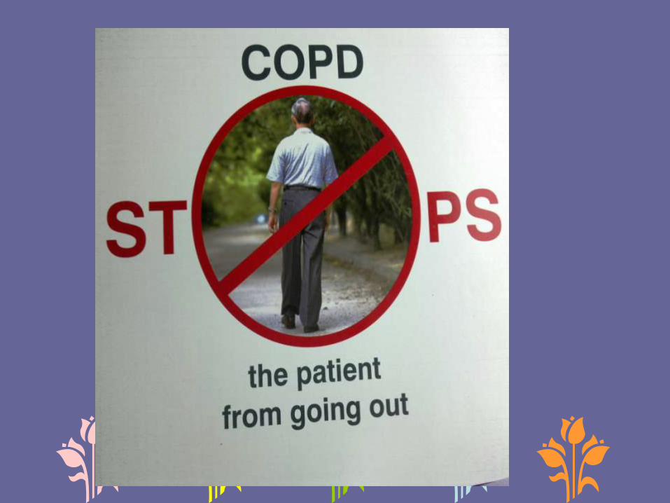
• Relief in breathlessness and other symptoms
• Improvement in exercise tolerance
• Improvement in overall health-related quality of life
Reduction in current symptoms
• Prevention (or slowing down) of disease progression
• Prevention of disease exacerbations
• Reduction in disease-related mortality
Reduction of future risk
Minimizing adverse effects from treatment
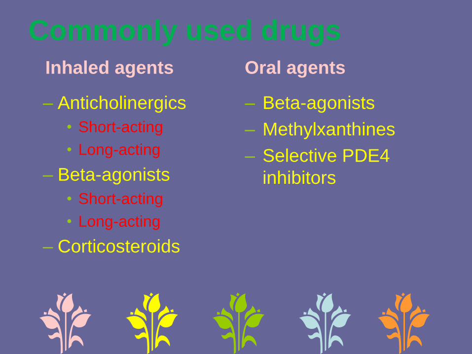
Commonly used drugs Inhaled agents Oral agents
– Anticholinergics
• Short-acting
• Long-acting
– Beta-agonists
• Short-acting
• Long-acting
– Corticosteroids
– Beta-agonists
– Methylxanthines
– Selective PDE4
inhibitors

Treating patients of stable COPD
Category Initial therapy Add-on
therapy (if
patient
continues to
have
symptoms)
First choice Alternative
choice
Mild SABA or
SAMA
Methyl
xanthines
-
Moderate LAMA LABA Methylxanthine
s to
LAMA/LABA
Severe ICS plus
LABA
LAMA Methylxanthine
s to LAMA or
ICS plus LABA

ROLE OF INHALATION
THERAPY ?

• Short-acting antimuscarinic agent (SAMA) can be used
as rescue medication to relieve patient symptoms. (1A)
• Long term SAMA monotherapy on regular basis is not
recommended. (1A)
• Long-acting antimuscarinic agents (LAMA) are useful in
stable COPD (FEV1 < 80%) to control symptoms and
decrease the risk of exacerbations. (1A)
• LAMA should be preferred over SAMA. (1A)
• We suggest close monitoring of patients with coronary
artery disease who are treated with LAMA. (UPP)
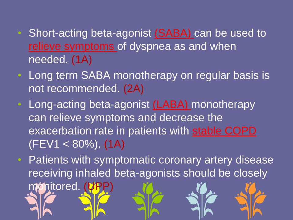
• Short-acting beta-agonist (SABA) can be used to
relieve symptoms of dyspnea as and when
needed. (1A)
• Long term SABA monotherapy on regular basis is
not recommended. (2A)
• Long-acting beta-agonist (LABA) monotherapy
can relieve symptoms and decrease the
exacerbation rate in patients with stable COPD
(FEV1 < 80%). (1A)
• Patients with symptomatic coronary artery disease
receiving inhaled beta-agonists should be closely
monitored. (UPP)
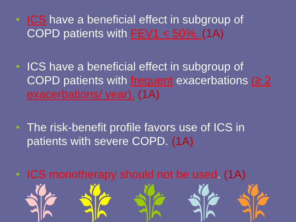
• ICS have a beneficial effect in subgroup of
COPD patients with FEV1 < 50%. (1A)
• ICS have a beneficial effect in subgroup of
COPD patients with frequent exacerbations (≥ 2
exacerbations/ year). (1A)
• The risk-benefit profile favors use of ICS in
patients with severe COPD. (1A)
• ICS monotherapy should not be used. (1A)

• LAMA is superior to LABA monotherapy. (1A)
• SABA and SAMA are equally effective when
used for COPD. (2A)
• LAMA plus LABA may be used in patients who
continue to have symptoms on monotherapy,
except for those with frequent exacerbations.
(1A)

• LABA plus ICS should be preferred over LABA
alone in patients with FEV1 < 50% or those having
frequent exacerbations. (1A)
• In patients of severe COPD (FEV1 < 50%), triple
therapy may be used in those who are symptomatic
despite single or dual bronchodilator therapy. (1B)
• There is lack of sufficient data to recommend
ICS-LABA or ICS-LAMA combination over LAMA
monotherapy.

ROLE OF ORAL
BRONCHODILATORS IN STABLE
COPD?
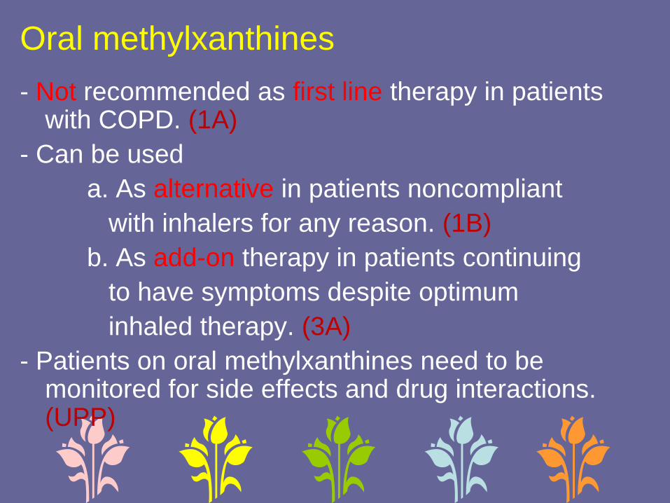
Oral methylxanthines
- Not recommended as first line therapy in patients with COPD. (1A)
- Can be used
a. As alternative in patients noncompliant
with inhalers for any reason. (1B)
b. As add-on therapy in patients continuing
to have symptoms despite optimum
inhaled therapy. (3A)
- Patients on oral methylxanthines need to be monitored for side effects and drug interactions. (UPP)

• Roflumilast (PDE4 inhibitor) may be used in
frequent exacerbators as an add-on or
substitute to ICS. (2B)
• Routine use of mucolytic agents is not
recommended in patients with COPD. (2A)
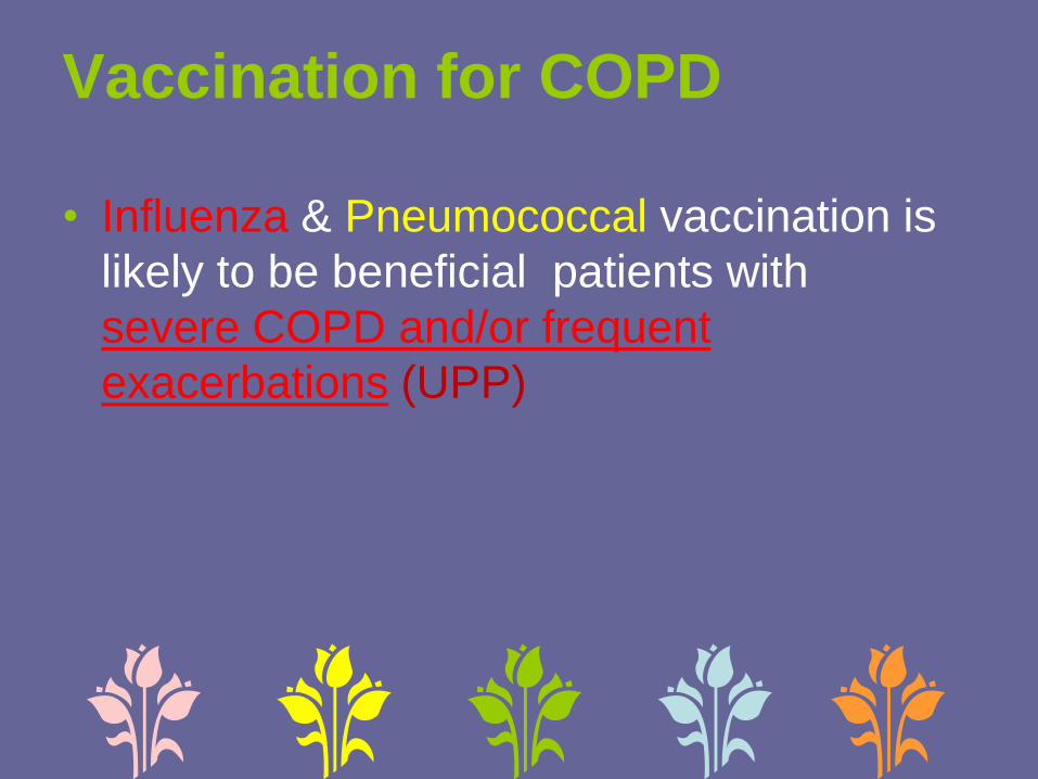
Vaccination for COPD
• Influenza & Pneumococcal vaccination is
likely to be beneficial patients with
severe COPD and/or frequent
exacerbations (UPP)
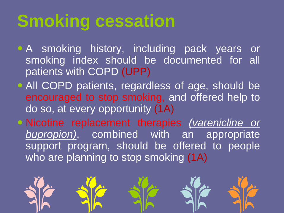
Smoking cessation
A smoking history, including pack years orsmoking index should be documented for allpatients with COPD (UPP)
All COPD patients, regardless of age, should beencouraged to stop smoking, and offered help todo so, at every opportunity (1A)
Nicotine replacement therapies (varenicline orbupropion), combined with an appropriatesupport program, should be offered to peoplewho are planning to stop smoking (1A)

Topics for patient education
Risks and benefits of stopping smoking and cessation strategies
Avoidance of potential risk factors in nonsmokers
The concept of normal lung function
Inhaler use
Use of other medications, including oxygen
Nutrition
Adequate physical exercise, especially if the patient is not enrolled in a pulmonary rehabilitation program
Breathing strategies
Advice related to travel and sexuality
End of life planning


Non-pharmacological
management of stable COPD
• Pulmonary rehabilitation
• Oxygen therapy
• Non-invasive ventilation
• Bronchoscopic techniques
• Surgery
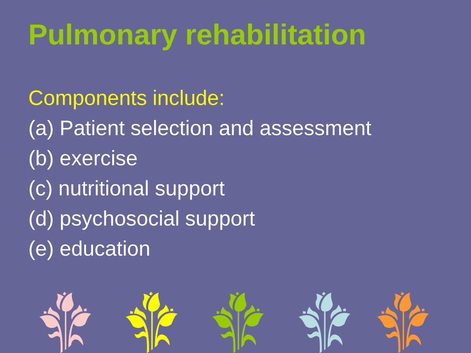
Pulmonary rehabilitation
Components include:
(a) Patient selection and assessment
(b) exercise
(c) nutritional support
(d) psychosocial support
(e) education


Pulmonary rehabilitation in stable
COPDStructured programs should be set up where
feasible (1A)
In the absence of structured programs, patients should be advised regarding unsupervised daily physical activity (3A)
All patients should be assessed for nutritional status at least by BMI at the initial visit and followed-up by serial BMI estimation at every visit. Any patient found to be malnourished should be referred for nutritional advice to a specialist (UPP)

Aerobic exercises – increase amount of oxygen delivered
to muscles which allows them to work longer.
Lower body exercises – Improve lower body muscle
strength and endurance, help move around easily for longer
period.
knee extension Leg lift Step - ups
PULMONARY REHABILITATION
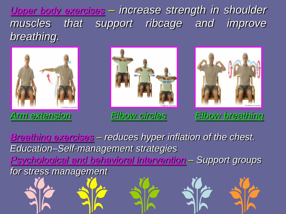
Upper body exercises – increase strength in shoulder
muscles that support ribcage and improve
breathing.
Arm extension Elbow circles Elbow breathing
Breathing exercises – reduces hyper inflation of the chest.
Education–Self-management strategies
Psychological and behavioral intervention – Support groups
for stress management

Oxygen therapyLong-term supplemental oxygen therapy is indicated for those with severe daytime resting hypoxemia (1A) defined as:
a) PaO2 of < 55 mmHg (or pulse oxygen saturation of < 88%), or
b) PaO2 of 56-60 mmHg (or pulse oxygen saturation of 88-92%) with evidence of end-organ dysfunction such as pulmonary hypertension, congestive cardiac failure, and erythrocytosis with hematocrit > 55%
c) Determined on two occasions at least 3 weeks apart in the stable patient.
Recommendations for oxygen therapy
The role of oxygen supplementation in other situations is currently not clear and should be decided on a case to case basis (2B)
Supplemental oxygen should be titrated to achieve a pulse oximetric saturation of 90-92% or a PaO2 of 60-65 mmHg (3A)
Patients should breathe supplemental oxygen for at least 16 hours a day (1A)
Patients on long-term oxygen therapy should be reviewed at regular intervals with either pulse oximetry or arterial blood gas analysis as indicated (UPP)
NIV in stable COPDIndications:1. Documentation of the diagnosis of COPD by a physician, optimization of other therapies, and exclusion of sleep apnea if required.
2. Presence of both symptoms (such as fatigue, dyspnea, morning headache, etc.) and physiologic criteria (one of the following):
a. PaCO2 ≥ 55 mmHg or PaCO2 of 50-54 mmHg and nocturnal desaturation (oxygen saturation by pulse oximeter ≤ 88% for continuous 5 min while receiving oxygen therapy at 2 L/min)
b. PaCO2 of 50-54 mmHg and hospitalization related to recurrent (≥ 2 in a 12-month period) episodes of hypercapnicrespiratory failure.

Recommendations for NIV
1. NIV may be used in patients with recurrent
exacerbations who require frequent use of
mechanical and noninvasive ventilation
during the acute episodes; the patient
should be referred to a specialist center for
management (3A)
2. The choice of the machine for NIV
depends on the presence of coexistent
sleep apnea syndromes (UPP)

Indications for surgical
treatments for COPD
Bullectomy
• presence of single large bullae compressing the remaining lung,
• breathlessness due to the bullae,
• hemoptysis,
• reduction in the FEV1 to < 50%.
Lung Volume Reduction Surgery
• predominantly upper lobe emphysema and
• low post-rehabilitation exercise capacity
Lung transplantation
• BODE index of 7-10 with an FEV1
< 20% predicted,
• or a DLCO < 20% and homogenous emphysema or cor pulmonale

Surgical treatments for COPD
Bullectomy may be carried out in properly
selected patients in appropriate centers
(3A)
LVRS may be offered to properly selected
patients at centers capable of performing
the procedure (1A)
Lung transplantation may be offered to
properly selected patients at centers
capable of doing the procedure (1A)

PROPHYLACTIC
ANTIBIOTICS IN COPD
• Antibiotics should not be prescribed as a routine
for the prevention of exacerbations of COPD.
(2A)
ADVICE REGARDING AIR-
TRAVEL
• Patients with severe COPD and those on
long-term oxygen therapy should be assessed
before air travel by a specialist. (UPP)


ACUTE EXACERBATION OF
COPD (AECOPD)
DEFINITION OF AECOPD
• An exacerbation of COPD is an acute event
characterized by sustained worsening of any of
the patient’s respiratory symptoms (cough,
sputum quantity and/or character, dyspnea)
that is beyond normal day-to-day variation and
leads to a change in medication, and where
other causes of acute breathlessness have
been clinically excluded.

HOW TO INVESTIGATE
AECOPD ?

• No investigations apart from pulse oximetry are
routinely required in patients with acute exacerbations
managed in an outpatient setting. (IIA)
• In those hospitalized with AECOPD, serum
electrolytes, LFT, RFT, CBP, CXR, ECG, ABG (if
available) should be performed. (IA)
• If an infectious exacerbation does not respond to the
initial antibiotic treatment, a sputum culture and an
antibiotic sensitivity test should be performed. (IIA)

Differential diagnosis of AECOPD
• You need to exclude the 6 P’s
– Pneumonia
– Pulmonary embolism
– Pneumothorax
– Pleural effusion
– Pulmonary edema (heart failure)
– Paroxysmal atrial tachycardia (arrhythmias)

Causes of AECOPDInfectious (60-80% of all
exacerbations)
• Frequent (70-85% of all infectious
• exacerbations)
• Viruses (influenza and parainfluenza
• viruses, rhinoviruses, coronaviruses)
• Haemophilus influenzae
• Streptococcus pneumoniae
• Moraxella catarrhalis
• Infrequent (15-30% of all infectious
• exacerbations)
• Pseudomonas aeruginosa
• Opportunistic gram-negative species
• Staphylococcus aureus
• Chlamydophila pneumoniae
• Mycoplasma pneumoniae
Environmental factors
• Air pollution
• Non-adherence to respiratory
• medication
• Cold air
• Allergens
• Tobacco smoking

SITE OF MANAGEMENT OF
AECOPD ?

Severity assessment of COPD exacerbation
Symptoms
• Marked reduction in activity of daily living due to dyspnea
• Altered sensorium
• New onset cyanosis
Signs
• Use of accessory respiratory muscles
• Paradoxical chest wall movements
• Central cyanosis
• Systolic blood pressure <90 mmHg
• RR >30/min
• Heart rate >110/min
• Asterixis
• Altered mental status
Others
• Presence of severe comorbid conditions
• Lack of social support
Pulse oximetry
• SpO2<90%

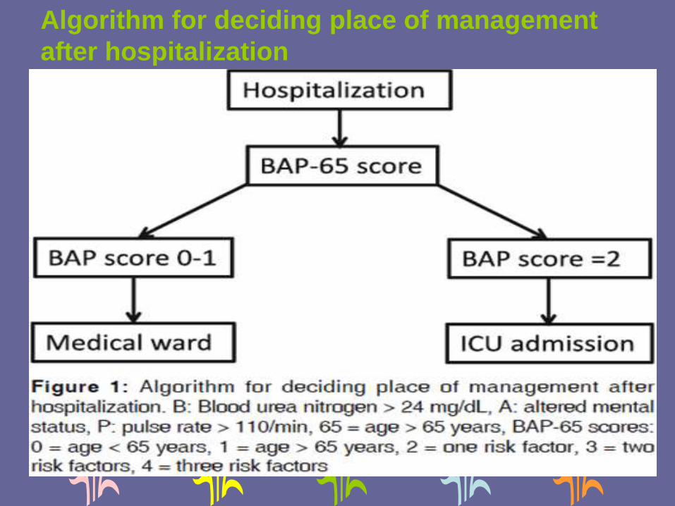
Algorithm for deciding place of management
after hospitalization

SHORT-ACTING BRONCHODILATORS IN
AECOPD • Inhaled route is the preferred route of administering
bronchodilators. (IA)
• Inhaled SABAs should be used as the first-line agent
because of quicker onset of action (IIIA). SAMAs are
however, in no way inferior to SABAs.
• Nebulized salbutamol at a dose of 2.5 mg every 20
min (or salbutamol pressurized metered dose inhaler
(pMDI) 100 μg 2-4 puffs every 20 min) for 1 h can be
given initially. (IIIA) Further dosing would depend on
the clinical response, generally every 4-6 h. (IIIA)

• If additional bronchodilatation is desired, a
combination of ipratropium (500 μg nebulized or 20
μg 2-4 puffs with pMDI) and salbutamol (2.5 mg
nebulized or salbutamol pMDI 100 μg 2-4 puffs)
every 4-6 h can be used. (IIIA)
• Nebulizer or pMDIs with spacer are equally effective.
(IIA)
• Nebulization should not be driven by oxygen; patients
should receive oxygen separately through nasal
cannula, with monitoring of oxygen saturation. (IIA)

• Intravenous methyl xanthines should not be
routinely used. (IA)
• The use of intravenous or subcutaneous route
of administering bronchodilators should be
reserved in the most seriously ill mechanically
ventilated patient demonstrating inadequate
response to inhaled therapy. (IIIB)

GLUCOCORTICOIDS IN
AECOPD • Systemic steroids
- shorten recovery time,
- improve lung function, oxygenation,
- reduce length of hospital stay
- fewer treatment failures. (IA)
• A short course of oral prednisolone (or
equivalent) at a dose of 30-40 mg/day is
recommended for managing acute
exacerbations. (IIA)
• The duration of systemic steroid therapy
should be 5-10 days. (IIA)
• Intravenous steroids should be given in
patients who are being mechanically ventilated
or cannot tolerate oral medication. (UPP)
• ICS are not routinely recommended in
management of AECOPD. (IA)

ANTIBIOTICS IN AECOPD
• Antibiotics should be prescribed for all
exacerbations of COPD. (IIA)
• The choice of antibiotics should be guided by
local flora and sensitivity pattern. (IIA)
• Fluoroquinolones should not be used routinely
in treating AECOPD. (IA)

• Patients with AECOPD being managed in the
outpatient setting may be treated with first line
antibiotics. (IIA)
• Hospitalized patients or those requiring mechanical
ventilation (noninvasive/invasive) should be treated
with second line drugs. (IIA)
• The duration of therapy should be 5-7 days. (IIA)

PROCALCITONIN
Any role in deciding for
antibiotic therapy?
• Biomarkers do not have any role in
management of acute exacerbation of
COPD. (IIA)
• Procalcitonin should not be used routinely in
guiding antibiotic usage in COPD. (IIA)


When to use O2 in AECOPD
• Oxygen should be prescribed to hypoxemic patients
with a target SpO2 between 88-92%. (IA)
• Oxygen should be delivered preferably by a Venturi
mask, and by nasal cannula upon recovery. (IIA)
• ABG monitoring is recommended in patients
receiving oxygen therapy, wherever available. (IIA)

Indication of NIV during
AECOPD
• NIV should be used early in the management of
respiratory failure due to AECOPD. (IA)
• NIV can be used even in settings where arterial
blood gas monitoring is not routinely available.
(UPP)

Indications and protocol of
invasive
mechanical ventilation

When should a patient be
discharged from hospital?
Patient should be clinically stable for at least 24-
48 hours,
Should be able to eat and sleep comfortably,
Should be ambulatory for activities of daily living.
In addition,
minimal requirement of short-acting bronchodilators,
and
the patient should be able to use long-acting
bronchodilators

Follow-up after discharge from hospital
Follow-up at 4-6 weeks after discharge
At every visit,
due emphasis should be laid on smoking cessation, the inhaler technique checked, and the effectiveness of each medication monitored
For patients who are hypoxemic during an exacerbation,
arterial blood gases, and/or pulse oximetryshould be evaluated prior to hospital discharge and in the following 3-6 weeks.
If the patient remains hypoxemic, long-term supplemental oxygen therapy may be required


Thank you