indirect pathogenicity of haemophilus influenzae and...
TRANSCRIPT

Indirect Pathogenicity of Haemophilus influenzae and Moraxellacatarrhalis in Polymicrobial Otitis Media Occurs via InterspeciesQuorum Signaling
Chelsie E. Armbruster, Wenzhou Hong,* Bing Pang, Kristin E. D. Weimer, Richard A. Juneau, James Turner, and W. Edward Swords
Department of Microbiology and Immunology, Wake Forest University Health Sciences, Winston-Salem, North Carolina, USA
* Present address: Department of Otolaryngology, Medical College of Wisconsin, Milwaukee, Wisconsin, USA.
ABSTRACT Otitis media (OM) is among the leading diseases of childhood and is caused by opportunists that reside within thenasopharynx, such as Haemophilus influenzae and Moraxella catarrhalis. As with most airway infections, it is now clear thatOM infections involve multiple organisms. This study addresses the hypothesis that polymicrobial infection alters the course,severity, and/or treatability of OM disease. The results clearly show that coinfection with H. influenzae and M. catarrhalis pro-motes the increased resistance of biofilms to antibiotics and host clearance. Using H. influenzae mutants with known biofilmdefects, these phenotypes were shown to relate to biofilm maturation and autoinducer-2 (AI-2) quorum signaling. In support ofthe latter mechanism, chemically synthesized AI-2 (dihydroxypentanedione [DPD]) promoted increased M. catarrhalis biofilmformation and resistance to antibiotics. In the chinchilla infection model of OM, polymicrobial infection promoted M. catarrha-lis persistence beyond the levels seen in animals infected with M. catarrhalis alone. Notably, no such enhancement of M. ca-tarrhalis persistence was observed in animals infected with M. catarrhalis and a quorum signaling-deficient H. influenzae luxSmutant strain. We thus conclude that H. influenzae promotes M. catarrhalis persistence within polymicrobial biofilms via inter-species quorum signaling. AI-2 may therefore represent an ideal target for disruption of chronic polymicrobial infections. More-over, these results strongly imply that successful vaccination against the unencapsulated H. influenzae strains that cause airwayinfections may also significantly impact chronic M. catarrhalis disease by removing a reservoir of the AI-2 signal that promotesM. catarrhalis persistence within biofilm.
IMPORTANCE Otitis media (OM) is one of the most common childhood infections and is a leading reason for antibiotic prescrip-tions to children. Chronic and recurrent OM involves persistence of bacteria within biofilm communities, a state in which theyare highly resistant to immune clearance and antibiotic treatment. While it is clear that most of these infections involve multiplespecies, the vast majority of knowledge about OM infections has been derived from work involving single bacterial species.There is a pressing need for better understanding of the impact of polymicrobial infection on the course, severity, and treatabil-ity of OM disease. In this study, we show that communication between bacterial species promotes bacterial persistence and resis-tance to antibiotics, which are important considerations in the diagnosis, prevention, and treatment of OM. Moreover, the re-sults of this study indicate that successful preventive measures against H. influenzae could reduce the levels of disease caused byM. catarrhalis.
Received 1 April 2010 Accepted 9 June 2010 Published 6 July 2010
Citation Armbruster, C. E., W. Hong, B. Pang, K. E. D. Weimer, R. A. Juneau, J. Turner, and W. E. Swords. 2010. Indirect pathogenicity of Haemophilus influenzae and Moraxellacatarrhalis in polymicrobial otitis media occurs via interspecies quorum signaling. mBio 1(3):e00102-10. doi:10.1128/mBio.00102-10.
Editor Larry McDaniel, University of Mississippi
Copyright © 2010 Armbruster et al. This is an open-access article distributed under the terms of the Creative Commons Attribution-Noncommercial-Share Alike 3.0 UnportedLicense, which permits unrestricted noncommercial use, distribution, and reproduction in any medium, provided the original author and source are credited.
Address correspondence to W. Edward Swords, [email protected].
Otitis media (OM) is one of the most common childhood in-fections (1–3) and is the leading reason for pediatric office
visits and new antibiotic prescriptions to children (4). OM infec-tions often persist for long periods of time and are frequentlyrecalcitrant to antibiotic treatment (5, 6). Due to the highly resis-tant nature of chronic and recurrent OM, these infections havelong been thought of as involving bacterial persistence within abiofilm (7–9). Clinical evidence of bacterial biofilms includes di-rect observation of biofilms in patient tissue samples (10) and inthe chinchilla experimental model of OM (11–15). Persistence ofbacteria within a biofilm community can also greatly increase re-
sistance to antibiotics (16, 17) through numerous mechanisms,including phenotypic heterogeneity and slower growth of bacteriawithin the biofilm, delayed antibiotic penetration through matrixmaterial/exopolysaccharide, and the presence of persister cells(18–21).
As with most upper airway infections, epidemiological dataindicate that the majority of chronic OM infections are polymi-crobial in nature (22). For example, Haemophilus influenzae andMoraxella catarrhalis are frequently present together in samplesobtained from patients with chronic and recurrent OM (22, 23).Interestingly, a recent study found M. catarrhalis to be more
RESEARCH ARTICLE
July/August 2010 Volume 1 Issue 3 e00102-10 mbio.asm.org 1
on July 8, 2018 by guesthttp://m
bio.asm.org/
Dow
nloaded from

frequently isolated from polymicrobial OM infections than fromsingle-species OM infections (24). This suggests that the presenceof other bacterial pathogens may impact the persistence of M. ca-tarrhalis or the severity of disease caused by this species. Addition-ally, M. catarrhalis is thought to confer passive antibiotic resistance
upon other OM pathogens via secretion ofbeta-lactamase (25–30). However, the im-pact of polymicrobial infection on bacterialpersistence, virulence, or response to treat-ment is not presently clear.
Interbacterial communication via quo-rum signaling is one factor which may im-pact the establishment of chronic polymi-crobial infection, as quorum signaling isknown to influence biofilm developmentfor many species (31, 32). Autoinducer-2(AI-2) is commonly referred to as an inter-species signal, as the genetic determinant forAI-2 production (luxS) is conserved amongnumerous bacterial species (33–35). AI-2 isknown to influence biofilms for many spe-cies, including H. influenzae (36), and insome instances, AI-2 can impact the devel-opment of polymicrobial biofilms (37, 38).In this study, we addressed the hypothesisthat polymicrobial infection impacts bio-film development and resistance duringOM disease. The results clearly show thatH. influenzae promotes M. catarrhalis per-sistence by means of interspecies quorumsignals that increase the resistance of M. ca-tarrhalis in biofilm.
RESULTSH. influenzae and M. catarrhalis formpolymicrobial biofilms in vitro. Basedon clinical evidence for the coexistence ofH. influenzae and M. catarrhalis in OMcases, it was hypothesized that these bac-terial species would coexist in culture andin vitro biofilms. Static biofilms of H. in-fluenzae, M. catarrhalis, or a mixture ofboth species were established in micros-copy chamber slides, and the surface-attached bacterial communities were ex-amined by scanning electron microscopy(SEM) (Fig. 1A) and confocal laser scan-ning microscopy (CLSM) (Fig. 1B) at var-ious times during biofilm development.As previously observed, H. influenzaeformed matrix-encased biofilm commu-nities on the chamber slide surfaces (13,36, 39). In contrast, M. catarrhalis formedsmaller surface-attached clusters. In co-culture, H. influenzae and M. catarrhalisformed polymicrobial biofilms with bothspecies incorporated into the biofilmstructure, as indicated by the presence oflarger M. catarrhalis diplococci inter-spersed with the smaller H. influenzae
coccobacilli (Fig. 1A). Immunostaining and confocal laser scan-ning microscopy showed that M. catarrhalis communities werepresent in discrete regions within the H. influenzae biofilm struc-ture (Fig. 1B). Based on these results, we concluded that H. influ-enzae and M. catarrhalis form polymicrobial biofilms.
FIG 1 H. influenzae and M. catarrhalis form polymicrobial biofilms in vitro. Stationary biofilms wereestablished in chamber slides for visualization of bacteria by SEM and confocal laser scanning micros-copy (CLSM). (A) Samples of H. influenzae and M. catarrhalis single-species or polymicrobial biofilmswere taken at 48 h and prepared for SEM. Images shown are at three different levels of magnification.(B) CLSM was performed on 24-, 48-, and 72-h biofilms following staining of H. influenzae (red) andM. catarrhalis (green).
Armbruster et al.
2 mbio.asm.org July/August 2010 Volume 1 Issue 3 e00102-10
on July 8, 2018 by guesthttp://m
bio.asm.org/
Dow
nloaded from
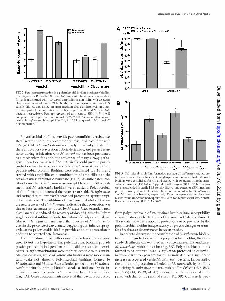
Polymicrobial biofilms provide passive antibiotic resistance.Beta-lactam antibiotics are commonly prescribed to children withOM (40). M. catarrhalis strains are nearly universally resistant tothese antibiotics via secretion of beta-lactamase, and passive resis-tance during coinfection with M. catarrhalis has been postulatedas a mechanism for antibiotic resistance of many airway patho-gens. Therefore, we asked if M. catarrhalis could provide passiveprotection for a beta-lactam-sensitive H. influenzae strain within apolymicrobial biofilm. Biofilms were established for 24 h andtreated with ampicillin or a combination of ampicillin and thebeta-lactamase inhibitor clavulanate (Fig. 2). As anticipated, bio-films formed by H. influenzae were susceptible to ampicillin treat-ment, and M. catarrhalis biofilms were resistant. Polymicrobialbiofilm formation increased the recovery of viable H. influenzae,indicating that M. catarrhalis provided protection against ampi-cillin treatment. The addition of clavulanate abolished the in-creased recovery of H. influenzae, indicating that protection wasdue to beta-lactamase produced by M. catarrhalis. As anticipated,clavulanate also reduced the recovery of viable M. catarrhalis fromsingle-species biofilms. Of note, formation of a polymicrobial bio-film with H. influenzae increased the recovery of M. catarrhalis,even in the presence of clavulanate, suggesting that inherent prop-erties of the polymicrobial biofilm provide antibiotic protection inaddition to secreted beta-lactamase.
A combination of trimethoprim-sulfamethoxazole was nextused to test the hypothesis that polymicrobial biofilms providepassive protection independent of diffusible resistance determi-nants. H. influenzae biofilms were more susceptible to this antibi-otic combination, while M. catarrhalis biofilms were more resis-tant (data not shown). Polymicrobial biofilms formed byH. influenzae and M. catarrhalis afforded protection to H. influen-zae from trimethoprim-sulfamethoxazole, as indicated by the in-creased recovery of viable H. influenzae from these biofilms(Fig. 3A). Control experiments indicated that bacteria recovered
from polymicrobial biofilms retained broth culture susceptibilitycharacteristics similar to those of the inocula (data not shown).These data show that antibiotic protection can be provided by thepolymicrobial biofilm independently of genetic changes or trans-fer of resistance determinants between species.
In order to determine the contribution of H. influenzae biofilmto antibiotic protection within a polymicrobial biofilm, the mac-rolide clarithromycin was used at a concentration that eradicatesM. catarrhalis within a biofilm (Fig. 3B). Polymicrobial biofilmsformed by M. catarrhalis and H. influenzae protected M. catarrha-lis from clarithromycin treatment, as indicated by a significantincrease in recovered viable M. catarrhalis bacteria. Importantly,the amount of protection afforded to M. catarrhalis by biofilmscontaining H. influenzae mutants with biofilm defects (siaB, licD,and luxS) (14, 36, 39, 41, 42) was significantly diminished com-pared with that of the parental strain (Fig. 3B). Conversely, pro-
FIG 2 Beta-lactam protection in a polymicrobial biofilm. Stationary biofilmsof H. influenzae Rd and/or M. catarrhalis were established on chamber slidesfor 24 h and treated with 100 �g/ml ampicillin or ampicillin with 25 �g/mlclavulanate for an additional 24 h. Biofilms were resuspended in sterile PBS,serially diluted, and plated on sBHI medium plus clarithromycin and BHImedium plates for enumeration of viable H. influenzae Rd and M. catarrhalisbacteria, respectively. Data are represented as means � SEM. *, P � 0.05compared to H. influenzae plus ampicillin; **, P � 0.05 compared to polymi-crobial H. influenzae plus ampicillin; ***, P � 0.05 compared to M. catarrhalisplus ampicillin.
FIG 3 Polymicrobial biofilm formation protects H. influenzae and M. ca-tarrhalis from antibiotic treatment. Single-species or polymicrobial stationarybiofilms were established for 4 h and treated with 60 �g/ml trimethoprim-sulfamethoxazole (TS) (A) or 6 �g/ml clarithromycin (B) for 24 h. Biofilmswere resuspended in sterile PBS, serially diluted, and plated on sBHI mediumplus clarithromycin or BHI medium for enumeration of viable H. influenzaeand M. catarrhalis bacteria, respectively. Data are represented as the meanresults from three combined experiments, with two replicates per experiment.Error bars represent SEM. *, P � 0.05.
Interspecies Quorum Signaling in Otitis Media
July/August 2010 Volume 1 Issue 3 e00102-10 mbio.asm.org 3
on July 8, 2018 by guesthttp://m
bio.asm.org/
Dow
nloaded from

tection of M. catarrhalis was increased in biofilms formed withH. influenzae licON, a mutant which forms thicker biofilms (13).Control experiments showed that bacteria recovered from allpolymicrobial biofilms retained broth susceptibility characteris-tics similar to those of the inocula, indicating that the increasedantibiotic resistance observed in polymicrobial biofilms was notdue to genetic changes or transfer of resistance determinants be-tween species (data not shown). Based on these data, we con-cluded that the maturation state and/or overall biomass of thepolymicrobial biofilm play integral roles in the antibiotic protec-tion provided by H. influenzae biofilms.
Autoinducer-2 (dihydroxypentanedione) promotes M. ca-tarrhalis biofilm thickness and antibiotic resistance. The de-creased clarithromycin protection of M. catarrhalis by H. influen-zae luxS could be due to differences in biofilm thickness or thematuration state of the luxS mutant, similar to the mechanism fordecreased protection by H. influenzae licD. However, another pos-sibility is that M. catarrhalis responds to AI-2 produced by H. in-fluenzae, and the decrease in protection observed with H. influen-zae luxS would thus be due to the loss of the AI-2 quorum signal.M. catarrhalis is not known to possess a luxS homolog and did notproduce detectable AI-2 during growth in a broth culture, as mea-sured by a Vibrio harveyi bioluminescence assay (Fig. 4A). How-ever, recent evidence suggests that bacterial species which do notmake AI-2 may still sense and respond to the AI-2 signal (43, 44).To test this hypothesis, M. catarrhalis was cultured in broth sup-plemented with the chemically synthesized AI-2 precursor dihy-droxypentanedione (DPD), and samples were taken to determinethe level of DPD remaining in the culture over time (Fig. 4A).M. catarrhalis depleted DPD over the course of 6 h, which indi-cates uptake and/or degradation of DPD, while an uninoculatedcontrol showed a minimal decrease in the AI-2 signal after 6 h.Notably, the amount of DPD depleted by M. catarrhalis was sim-ilar to the amount depleted by H. influenzae luxS. To determine ifdepletion of AI-2 requires live bacteria and/or active protein syn-thesis, M. catarrhalis cultures were incubated with tetracyclineovernight prior to the addition of DPD or incubated with tetracy-cline concurrent with the addition of DPD. Samples were takenover the course of 7 h for comparison of M. catarrhalis DPD de-pletion to that of untreated M. catarrhalis (Fig. 4B). Both of thetetracycline treatments completely inhibited depletion of DPD byM. catarrhalis, suggesting that depletion is an active process whichrequires protein synthesis. Additionally, incubation of M. ca-tarrhalis culture supernatant with DPD did not result in depletion(data not shown). Taken together, these data indicate that M. ca-tarrhalis is most likely depleting AI-2 by means of an uptake sys-tem rather than passive binding or external degradation of thissignaling molecule.
To assess the impact of exogenous AI-2 on M. catarrhalis bio-film formation, stationary M. catarrhalis biofilms were establishedin the presence or absence of DPD and stained with crystal violet at4, 6, 12, 24, and 48 h (Fig. 4C). Incubation with DPD resulted in anoverall increase in M. catarrhalis biofilm biomass that was partic-ularly evident at 24 and 48 h. Viability staining and confocal laserscanning microscopy (CLSM) of M. catarrhalis biofilms con-firmed the increased M. catarrhalis biofilm density in the presenceof DPD and further demonstrated an increase in bacterial viabilitywithin biofilm. M. catarrhalis biofilms established in media lack-ing DPD formed small clusters with mostly nonviable staining(Fig. 4D), while biofilms established in the presence of DPD were
thicker on average and showed an increased number of viablebacteria within the larger biomasses (Fig. 4E). Compressedz-series images confirmed the increased viable staining present inthe larger biomasses of DPD-treated M. catarrhalis (Fig. 4G) com-pared to those of untreated M. catarrhalis biofilms (Fig. 4F). SEMof M. catarrhalis biofilms similarly demonstrated the impact ofDPD on biofilm development, with treatment resulting in in-creased formation of bacterial clusters compared to those formedin M. catarrhalis biofilms established in media lacking DPD(Fig. 4H and I).
Based on the imaging results, we hypothesized that DPD couldincrease resistance of M. catarrhalis to antibiotic treatment. Totest this hypothesis, M. catarrhalis biofilms were established in thepresence or absense of DPD for 4 h and treated with clarithromy-cin. Treatment with DPD did not significantly alter the overallrecovery of viable M. catarrhalis from control wells (Fig. 4J). How-ever, M. catarrhalis biofilms established in the presence of DPDwere inherently more resistant to clarithromycin, as indicated bythe increased recovery of viable bacteria following incubation withantibiotic. Similar results were obtained using trimethoprim-sulfamethoxazole (data not shown). Taken together, these studiesindicate that while M. catarrhalis 7169 does not produce AI-2, thisstrain does respond to the interspecies quorum signaling moleculeby producing biofilms with increased biomass and resistance toantibiotic treatment.
Interspecies quorum signaling during polymicrobial infec-tion promotes persistence of M. catarrhalis. The in vitro studiesof polymicrobial biofilms support a prominent role for interspe-cies quorum signaling in the development of M. catarrhalis bio-films with increased resistance phenotypes. As M. catarrhalis isfrequently isolated from polymicrobial OM infections, we hy-pothesized that M. catarrhalis could utilize AI-2 produced byH. influenzae or other OM pathogens to persist in vivo. Therefore,infection studies were performed using the chinchilla model ofOM to test this hypothesis. As previously observed, high numbersof H. influenzae and H. influenzae luxS bacteria were detected inmiddle ear effusion fluid (Fig. 5A) and bullar homogenate(Fig. 5B) samples at both 7 days and 14 days postinfection forsingle-species and polymicrobial infection groups. Counts ofM. catarrhalis within middle ear effusion fluid samples were at orbelow the level of detection at both time points (Fig. 5A), regard-less of the type of infection. Animals infected with M. catarrhalisalone had bacterial loads within the bullar homogenates at both 7and 14 days postinfection that were consistent with the initialinocula. However, in the coinfected animals, significantly highernumbers of M. catarrhalis bacteria in bullar homogenate samplesat 14 days postinfection were observed (Fig. 5B). Based on theseresults, we conclude that M. catarrhalis survives exclusively insurface-attached communities within the chinchilla middle earchamber and that coinfection with H. influenzae provides a per-missive environment in which M. catarrhalis can proliferate. Wenext asked if the increase in M. catarrhalis bacterium numbersduring coinfection with H. influenzae were dependent on inter-species quorum signaling by performing similar coinfection stud-ies using H. influenzae luxS. Notably, no increase in M. catarrhalisbacterium counts was observed during coinfection with H. influ-enzae luxS at either time point. Taken in concert with the experi-ments showing increased M. catarrhalis biofilm density and resis-tance following treatment with synthetic AI-2, these experiments
Armbruster et al.
4 mbio.asm.org July/August 2010 Volume 1 Issue 3 e00102-10
on July 8, 2018 by guesthttp://m
bio.asm.org/
Dow
nloaded from

show that M. catarrhalis can utilize exogenous AI-2 provided byH. influenzae to establish a persistent infection.
DISCUSSION
According to the long-standing concept of indirect pathogenicity,bacterial disease and/or response to treatment is subject to influ-ence by other bacteria sharing the same environment (28, 29). In
this study, H. influenzae was shown to promote persistence andantibiotic resistance of M. catarrhalis via protection within thebiofilm structure in response to interspecies quorum signaling.The data presented in this study, therefore, provide concrete val-idation of the concept of indirect pathogenicity and provide amechanism to support how this can occur during polymicrobialotitis media infections.
FIG 4 AI-2 promotes M. catarrhalis biofilm development and antibiotic resistance. (A) M. catarrhalis was cultured in BHI medium or BHI mediumsupplemented with 0.2 �M synthetic AI-2 (DPD) to determine AI-2 production and depletion, as measured by Vibrio harveyi bioluminescence. H. influenzaeluxS was cultured in sBHI medium supplemented with DPD to measure depletion. An uninoculated control of BHI medium with DPD shows the minimaldegradation of the AI-2 signal during 6 h of incubation at 37°C. (B) Depletion of DPD by M. catarrhalis biofilms were established for 24 h following incubationwith 10 �g/ml tetracycline was measured by bioluminescence over a period of 7 h. (C) M. catarrhalis biofilms were established in the presence or absense of DPDand stained with crystal violet for determination of biofilm biomass at 4, 6, 12, 24, and 48 h. Data represent the mean results from three combined experiments,with three replicate wells per experiment. Error bars represent SEM. (D and E) M. catarrhalis biofilms were established for 24 h in the presence (E) or absence (D)of DPD and stained with a viability kit for CLSM visualization of surface coverage and biofilm thickness. (F and G) Z-series images from panels D and E werecompressed to show total viable and nonviable staining of biofilms established in the presence (G) or absence (F) of DPD. (H and I) SEM images of 24-hM. catarrhalis biofilms established with (I) or without (H) DPD. (J) M. catarrhalis biofilms were established for 4 h in the presence or absence of DPD and thentreated with 6 �g/ml clarithromycin for 24 h and plated for enumeration of viable bacteria. Data represent the means from three replicates � SEM. *, P � 0.05;**, P � 0.01; ***, P � 0.001.
Interspecies Quorum Signaling in Otitis Media
July/August 2010 Volume 1 Issue 3 e00102-10 mbio.asm.org 5
on July 8, 2018 by guesthttp://m
bio.asm.org/
Dow
nloaded from

In the context of infectious disease, biofilm formation has longbeen considered to be an important determinant of bacterial per-sistence. It has also long been appreciated that growth within abiofilm increases resistance to antibiotics through multiple mech-anisms, which include delayed antibiotic penetration of the bio-film and changes in the metabolic state of bacteria within biofilms(18–21). In this study, both in vitro and in vivo models were usedto ask how H. influenzae and M. catarrhalis polymicrobial biofilmformation impacts antibiotic resistance and bacterial persistence.While M. catarrhalis can be considered an indirect pathogenthrough the production of beta-lactamase, the results of this studyclearly prove that both M. catarrhalis and H. influenzae can pro-vide antibiotic protection to other pathogens within a polymicro-bial biofilm in a manner independent of diffusible resistance de-terminants. Additionally, the abrogation of antibiotic protectionobserved for polymicrobial biofilms formed with H. influenzaebiofilm mutants demonstrates a role for biofilm maturation/totalbiomass in antibiotic protection. This study and others supportthe hypothesis that biofilm can provide a barrier that protectssusceptible organisms contained within.
In addition to the impact of biofilm biomass on antibiotic sus-ceptibility, this study solidifies the role of interspecies communi-cation during the establishment of polymicrobial biofilms. Muta-tion of the luxS gene, the genetic determinant of AI-2 production,alters H. influenzae biofilm maturation and density as well as bac-terial persistence, indicating that AI-2 plays a critical role in thedevelopment and maturation of H. influenzae biofilms (36).While M. catarrhalis does not produce AI-2, the results clearlydemonstrate the critical role of interspecies quorum signaling viaAI-2 in the establishment of persistent polymicrobial biofilmscontaining this species and indicate the presence of an AI-2 trans-port system in M. catarrhalis. The main AI-2 transport system thathas been described outside Vibrio species is the Lsr ABC trans-porter. The Lsr system, identified in Salmonella enterica serovarTyphimurium, Escherichia coli, Sinorhizobium meliloti, and Aggre-gatibacter actinomycetemcomitans, has homology to the ribose
ABC transporter and involves binding of AI-2 by LsrB and trans-port through a heterodimeric membrane channel (44–47). It wasdetermined in A. actinomycetemcomitans that the ribose bindingprotein RbsB, in addition to LsrB, can bind AI-2 (48, 49). Themechanism for the sensing of DPD/AI-2 by M. catarrhalis has yetto be defined, but as the presence of this signaling molecule clearlyalters M. catarrhalis biofilm development, this will be an impor-tant topic for future studies. Additionally, the increased antibioticresistance following treatment with DPD also provides furthersupport that quorum signaling contributes to the establishment ofa diffusion barrier to delay or limit antibiotic penetration of thebiofilm.
The augmented persistence observed during infection byM. catarrhalis and the parental strain of H. influenzae but not byH. influenzae luxS further demonstrates the role of interspeciesquorum signaling in the establishment of polymicrobial OM. Onepossible explanation for the increase in M. catarrhalis persistenceis that M. catarrhalis becomes incorporated into the biofilm scaf-fold provided by H. influenzae, and this incorporation protectsM. catarrhalis from host factors, thus allowing for increased per-sistence. As H. influenzae luxS has a persistence defect in the chin-chilla model of OM, it is possible that the luxS mutant does notprovide sufficient biofilm structure for protection of M. catarrha-lis or, alternatively, that other factors lacking in H. influenzae luxSare required for the enhancement of M. catarrhalis persistence.However, the results demonstrating depletion of synthetic AI-2 byM. catarrhalis, as well as the increased M. catarrhalis biofilm den-sity and resistance elicited by synthetic AI-2, argue that AI-2 quo-rum signaling from H. influenzae promotes M. catarrhalis persis-tence in polymicrobial biofilm. Therefore, we conclude that theproduction of AI-2 by H. influenzae promotes M. catarrhalis re-sistance within biofilm and thereby promotes M. catarrhalis per-sistence within the middle ear chamber. It is notable that priorstudies involving infection of rodents with M. catarrhalis havehistorically failed to mimic the chronic and recurrent infectionsthat are typical of human patients with opportunistic airway in-
FIG 5 Polymicrobial infection augments M. catarrhalis persistence in vivo. Chinchillas were infected with 103 CFU of H. influenzae or H. influenzae luxS,104 CFU of M. catarrhalis, or a mixture of both species. (A) Middle ear effusion fluids were removed for enumeration of viable H. influenzae and M. catarrhalisbacteria by plating on sBHI medium plus clarithromycin or BHI medium, respectively. (B) Bullae were removed at each time point and homogenized forenumeration of viable H. influenzae and M. catarrhalis bacteria, as described above. Data represent the mean results from four experiments � SEM. ***, P � 0.005compared to the number of CFU from M. catarrhalis single-species bullar homogenate.
Armbruster et al.
6 mbio.asm.org July/August 2010 Volume 1 Issue 3 e00102-10
on July 8, 2018 by guesthttp://m
bio.asm.org/
Dow
nloaded from

fections; instead, rodent infections are typically transient in nature(50). The results presented here may indicate that this differencein bacterial persistence occurs as a consequence of the absence ofan “infection partner,” such as H. influenzae, to provide AI-2, asopposed to any species differences.
As we observed that both M. catarrhalis and H. influenzae candeplete AI-2/DPD, yet only one of these species produces the sig-nal, there could be competition between M. catarrhalis and H. in-fluenzae for available AI-2 during coinfection. Based on the AI-2requirement for H. influenzae to establish a chronic infection, anycompetition with M. catarrhalis would most likely have a negativeimpact on H. influenzae persistence. However, the numbers ofviable bacteria recovered from coinfected animals were similar tothe numbers recovered from those which received H. influenzaealone, indicating that AI-2 uptake by M. catarrhalis did not inter-fere with H. influenzae biofilm formation or persistence. Addi-tionally, the presence of M. catarrhalis during stationary biofilmformation was not observed to negatively impact H. influenzaebiofilm formation or antibiotic resistance. Taken together, theseobservations support a model wherein H. influenzae requires onlya certain threshold level of AI-2 but may produce AI-2 in excess ofthe threshold concentration needed to promote biofilm develop-ment. In this model, AI-2 depleted from the biofilm environmentby M. catarrhalis would not have a detrimental impact on H. in-fluenzae biofilm development. Another possible explanation isthat M. catarrhalis may require only a minimal concentration ofAI-2 to alter biofilm development. This would be advantageous, asM. catarrhalis could utilize any AI-2-producing species as an “in-fection partner” to promote its own persistence without directlycompeting for the AI-2 signal. Further research will be necessaryto determine the minimal AI-2 concentration required by bothM. catarrhalis and H. influenzae to promote biofilm development.
The data presented in this study and others provide substantialevidence for the influence of polymicrobial infection on severity ofdisease and the outcome of antibiotic treatment, particularly forchronic infections involving persistence of bacteria within bio-films. Notably, the results of this study imply that vaccinationagainst upper airway pathogens, such as the unencapsulatedstrains of H. influenzae, may have a greater impact than expected.For instance, successful vaccination against H. influenzae may alsodisrupt the establishment of disease by M. catarrhalis. Furtherresearch is necessary to elucidate the interactions between all threeof the leading causative agents of OM and the impact of otherpolymicrobial upper airway infections on resistance to relevantantibiotics. Knowledge of the bacterial species present duringhighly recalcitrant infections may provide insight into whichcourse of antibiotic treatment would be most effective. Addition-ally, AI-2 may represent an ideal target for disruption of numer-ous chronic and/or recurrent infections.
MATERIALS AND METHODSBacterial strains and culture conditions. A complete list of bacterialstrains used in this study is provided in Table 1. M. catarrhalis strains werecultivated in brain heart infusion (BHI) medium (Difco), and H. influen-zae strains were cultivated in BHI medium supplemented with hemin(ICN Biochemicals) and NAD (Sigma); this medium is referred to hereinas supplemented BHI (sBHI). For experiments using trimethoprim-sulfamethoxazole, H. influenzae and M. catarrhalis were cultured inMorse’s defined medium (51) supplemented with hemin and NAD. H. in-fluenzae siaB was constructed essentially as described previously for strain
2019 siaB (52) and confirmed by immunoblotting to have decreased re-activity with Limax flavus (LFA) lectin (EY Laboratories).
SEM. Stationary in vitro biofilm cultures were grown in Lab-Tek IIcover glass slides (Nunc). Each chamber was inoculated with~108 CFU/ml of H. influenzae, M. catarrhalis, or a 1:1 dilution mixture ofboth species and incubated for 48 h at 37°C and 5% CO2. Biofilm sampleswere fixed for 30 min with 2.5% glutaraldehyde in phosphate-bufferedsaline (PBS) and rinsed once. Samples were then dehydrated, fixed, andprepared for scanning electron microscopy (SEM) analysis as previouslydescribed (39). Biofilm samples were mounted onto stubs, sputter coatedwith palladium, and then viewed with a Philips SEM-515 scanning elec-tron microscope.
CLSM. In vitro biofilm cultures were grown using a continuous flowsystem as previously described (13). H. influenzae and M. catarrhalis werecultured overnight in sBHI broth and diluted to ~108 CFU/ml. Chamberslides were inoculated with each strain alone or a 1:1 dilution mixture ofboth species and incubated for 24, 48, and 72 h at 37°C and 5% CO2. Ateach time point, biofilms were fixed and stained with rabbit polyclonalanti-H. influenzae sera (41) and/or monoclonal antibody 4G5 (53). MAb4G5 was generously provided by Anthony Campagnari. All secondaryantibodies were purchased from Jackson Laboratories. Biofilms were vi-sualized using a Zeiss LSM 510 CLSM and Zeiss LSM Image Browsersoftware.
Antibiotics. Antibiotics used were ampicillin (Sigma), clavulanate(Sigma), clarithromycin (Abbott Laboratories), trimethoprim (Sigma),and sulfamethoxazole (Sigma). Trimethoprim-sulfamethoxazole experi-ments were conducted using a 1:5 dilution ratio. Concentrations listedrefer to trimethoprim.
Biofilm antibiotic protection studies. Bacteria were grown overnighton sBHI or BHI medium plates, suspended in sBHI medium, and dilutedto ~108 CFU/ml. A 24-well microtiter plate was inoculated with a single-species suspension diluted 1:1 with PBS or a 1:1 dilution mixture of bothbacterial suspensions. Cultures were incubated at 37°C and 5% CO2 foreither 4 h or 24 h to establish biofilms. Supernatants were then carefullyremoved and replaced with either fresh sBHI or sBHI medium with theantibiotic, and cultures were returned to 37°C and 5% CO2 for 24 h.Following incubation, supernatants were removed, and biofilms were re-suspended in PBS for serial dilution and plating to enumerate viable bac-teria. Polymicrobial biofilms were plated onto both BHI medium andsBHI medium containing 2 �g/ml clarithromycin to distinguish betweenM. catarrhalis and H. influenzae, respectively.
M. catarrhalis AI-2 studies. All studies were conducted using 0.2 �Mdihydroxypentanedione (DPD; Omm Scientific). This concentration ofDPD was chosen to simulate the amount of AI-2 produced by H. influen-zae, as it elicits luminescence from Vibrio harveyi that is approximatelyequal to that elicited by H. influenzae late-exponential-phase culture su-pernatant. For AI-2 depletion studies, BHI or sBHI medium was supple-mented with DPD when indicated, inoculated with ~108 CFU of M. ca-tarrhalis or H. influenzae luxS, and incubated at 37°C and 150 rpm for 6 h.Samples were taken at 0.25, 0.5, 0.75, 1, 2, 3, and 6 h, centrifuged, filtersterilized, and stored at �20°C for bioluminescence. Luminescence pro-duced by Vibrio harveyi BB170 (54) following a 3-h incubation with su-
TABLE 1 Bacterial strains
Strain/mutant Description Reference
H. influenzae86-028NP Nasopharyngeal isolate from child with OM 55licD mutant 86-028NP NTHI 1594 mutant 42licON mutant 86-028NP constitutive PCho� 13luxS mutant 86-028NP NTHI 0621 mutant 36siaB mutant 86-028NP NTHI 1891 mutant This studyRd Rd KW20 56
M. catarrhalis 7169 Serotype B strain 57a PCho�, phosphorylcholine positive.
Interspecies Quorum Signaling in Otitis Media
July/August 2010 Volume 1 Issue 3 e00102-10 mbio.asm.org 7
on July 8, 2018 by guesthttp://m
bio.asm.org/
Dow
nloaded from

pernatant samples was determined in a Turner Designs TD-20/20 lumi-nometer for 10 s. Data are reported as relative light units (counts per 10 s).Tetracylcine studies were conducted by incubating M. catarrhalis with10 �g/ml tetracycline (Sigma). M. catarrhalis cultures were incubatedwith tetracycline during overnight growth in a broth culture to completelyinhibit bacterial growth/viability as well as at the start of the 7-h AI-2depletion study to monitor the role of protein synthesis in AI-2 depletion.For crystal violet staining of M. catarrhalis biofilms, wells of a 24-welltissue culture plate containing sBHI medium or sBHI medium supple-mented with 0.2 �M DPD were inoculated with ~108 CFU of M. catarrha-lis and incubated at 37°C and 5% CO2. Supernatants were carefully re-moved at each time point, and biofilms were washed one time with H2O,stained with 0.1% crystal violet for 30 min, washed two times with H2O,and solubilized in ethanol for 10 min prior to measurement of the opticaldensity at 600 nm. For CLSM and SEM, M. catarrhalis biofilms wereestablished in sBHI medium or sBHI medium supplemented with 0.2 �MDPD for 24 h. Biofilms were then prepared for SEM as described above orwashed once with PBS and stained with a Live/Dead BacLight viability kit(Invitrogen) prior to imaging by CLSM, as described above.
Chinchilla infection studies. Bacterial persistence and biofilm forma-tion in the middle ear chamber were assessed as described previously (12,13). Chinchillas were purchased from Rauscher’s Chinchilla Ranch (La-Rue, OH) and allowed to acclimate to the vivarium for �7 days prior toinfection. No animals showed visible signs of illness by otoscopy prior toinfection. The animals were anesthetized with isofluorane and infected viatransbullar injection with ~103 CFU of H. influenzae or H. influenzae luxS,~104 CFU of M. catarrhalis, or a 1:1 mixture of both species. All inoculawere confirmed by plate counting. At 7 days or 14 days postinfection,animals (four per group) were euthanized, and middle ear chambers wereaseptically opened. Effusion fluid samples were recovered, and middle earlavage was performed using 1.0 ml sterile PBS. Viable bacteria were enu-merated by plate counting the combined retrieved fluids. Fluid samplesobtained from animals which received polymicrobial inocula were platedonto both sBHI medium containing 2 �g/ml clarithromycin and BHImedium lacking NAD and hemin. Bullae were excised and homogenizedin 10 ml sterile PBS and then plated to determine the number of CFU oftissue-associated bacteria (12).
Statistics. Significance was determined by the nonparametric t test,unpaired t test with Welsh’s correction, or two-way analysis of variance(ANOVA), with post hoc tests of significance. All P values are two tailed ata 95% confidence interval. Analyses were performed using GraphPadPrism, version 5 (GraphPad Software, San Diego, CA).
ACKNOWLEDGMENTS
We acknowledge excellent assistance by Gayle Foster and members of theWFUHS Microscopy Core Facility and helpful comments and critiquesfrom colleagues in the WFUHS Department of Microbiology and Immu-nology.
This work was supported by grants from NIH/NIDCD (DC007444and DC10051) and from the Whitaker Infectious Disease Foundationawarded to W.E.S. K.E.D.W. was supported by an NIH training grant(T32 AI07401; Steven Mizel, principal investigator).
REFERENCES1. Klein, J. O. 2000. The burden of otitis media. Vaccine 19:S2–S8.2. Mandel, E. M., W. J. Doyle, B. Winther, and C. M. Alper. 2008. The
incidence, prevalence and burden of OM in unselected children aged 1– 8years followed by weekly otoscopy through the “common cold” season.Int. J. Pediatr. Otorhinolaryngol. 72:491– 499.
3. Paradise, J. L., H. E. Rockette, D. K. Colborn, B. S. Bernard, C. G.Smith, M. Kurs-Lasky, and J. E. Janosky. 1997. Otitis media in 2253Pittsburgh-area infants: prevalence and risk factors during the first twoyears of life. Pediatrics 99:318 –333.
4. Finkelstein, J. A., R. L. Davis, S. F. Dowell, J. P. Metlay, S. B. Soumerai,S. L. Rifas-Shiman, M. Higham, Z. Miller, I. Miroshnik, A. Pedan, andR. Platt. 2001. Reducing antibiotic use in children: a randomized trial in12 practices. Pediatrics 108:1–7.
5. Pichichero, M. E. 2000. Recurrent and persistent otitis media. Pediatr.Infect. Dis. J. 19:911–916.
6. St. Geme, J. W. 2002. Molecular and cellular determinants of non-typeable Haemophilus influenzae adherence and invasion. Cell. Microbiol.4:191–200.
7. Bakaletz, L. O. 2007. Bacterial biofilms in otitis media: evidence andrelevance. Pediatr. Infect. Dis. J. 26:S17–S19.
8. Costerton, J. W., P. S. Stewart, and E. P. Greenberg. 1999. Bacterialbiofilms: a common cause of persistent infections. Science 284:1318 –1322.
9. Post, J. C., N. L. Hiller, L. Nistico, P. Stoodley, and G. D. Ehrlich. 2007.The role of biofilms in otolaryngologic infections: update 2007. Curr.Opin. Otolaryngol. Head Neck Surg. 15:347–351.
10. Hall-Stoodley, L., F. Z. Hu, A. Gieseke, L. Nistico, D. Nguyen, J. D.Hayes, M. Forbes, D. P. Greenberg, B. Dice, A. Burrows, P. Wackym, P.Stoodley, J. C. Post, G. D. Ehrlich, and J. E. Kerschner. 2006. Directdetection of bacterial biofilms on the middle ear mucosa of children withchronic otitis media. JAMA 296:202–211.
11. Ehrlich, G. D., R. Veeh, X. Wang, J. W. Costerton, J. D. Hayes, F. Z. Hu,B. J. Daigle, M. D. Ehrlich, and J. C. Post. 2002. Mucosal biofilmformation on middle-ear mucosa in the chinchilla model of otitis media.JAMA 287:1710 –1715.
12. Hong, W., K. Mason, J. A. Jurcisek, L. A. Novotny, L. O. Bakaletz, andW. E. Swords. 2007. Phosphorylcholine decreases early inflammation andpromotes the establishment of stable biofilm communities of nontypeableHaemophilus influenzae strain 86-028NP in a chinchilla model of otitismedia. Infect. Immun. 75:958 –965.
13. Hong, W., B. Pang, S. West-Barnette, and W. E. Swords. 2007. Phos-phorylcholine expression by nontypeable Haemophilus influenzae corre-lates with maturation of biofilm communities in vitro and in vivo. J. Bac-teriol. 189:8300 – 8307.
14. Jurcisek, J. A., L. Greiner, H. Watanabe, A. Zaleski, M. A. Apicella, andL. O. Bakaletz. 2005. Role of sialic acid and complex carbohydrate bio-synthesis in biofilm formation by nontypeable Haemophilus influenzae inthe chinchilla middle ear. Infect. Immun. 73:3210 –3218.
15. Post, J. C., J. J. Aul, G. J. White, R. M. Wadowsky, T. Zavoral, R. Tabari,B. Kerber, W. J. Doyle, and G. D. Ehrlich. 1996. PCR-based detection ofbacterial DNA after antimicrobial treatment is indicative of persistent,viable bacteria in the chinchilla model of otitis media. Am. J. Otolaryngol.17:106 –111.
16. Ceri, H., M. E. Olson, C. Stremick, R. R. Read, D. Morck, and A. Buret.1999. The Calgary Biofilm Device: new technology for rapid determina-tion of antibiotic susceptibilities of bacterial biofilms. J. Clin. Microbiol.37:1771–1776.
17. Starner, T. D., N. Zhang, G. Kim, M. A. Apicella, and P. B. McCray, Jr.2006. Haemophilus influenzae forms biofilms on airway epithelia: impli-cations in cystic fibrosis. Am. J. Respir. Crit. Care Med. 174:213–220.
18. Anderson, G. G., and G. A. O’Toole. 2008. Innate and induced resistancemechanisms of bacterial biofilms. Curr. Top. Microbiol. Immunol. 322:85–105.
19. Jefferson, K. K., D. A. Goldmann, and G. B. Pier. 2005. Use of confocalmicroscopy to analyze the rate of vancomycin penetration through Staph-ylococcus aureus biofilms. Antimicrob. Agents Chemother. 49:2467–2473.
20. Kaldalu, N., R. Mei, and K. Lewis. 2004. Killing by ampicillin andofloxacin induces overlapping changes in Escherichia coli transcriptionprofile. Antimicrob. Agents Chemother. 48:890 – 896.
21. Stewart, P. S. 2002. Mechanisms of antibiotic resistance in bacterial bio-films. Int. J. Med. Microbiol. 292:107–113.
22. Hendolin, P. H., A. Markkanen, J. Ylikoski, and J. J. Wahlfors. 1997.Use of multiplex PCR for simultaneous detection of four bacterial speciesin middle ear effusions. J. Clin. Microbiol. 35:2854 –2858.
23. Rayner, M. G., Y. Zhang, M. C. Gorry, Y. Chen, J. C. Post, and G. D.Ehrlich. 1998. Evidence of bacterial metabolic activity in culture-negativeotitis media with effusion. JAMA 279:296 –299.
24. Broides, A., R. Dagan, D. Greenberg, N. Givon-Lavi, and E. Leibovitz.2009. Acute otitis media caused by Moraxella catarrhalis: epidemiologicand clinical characteristics. Clin. Infect. Dis. 49:1641–1647.
25. Brook, I. 1986. Direct and indirect pathogenicity of Branhamella ca-tarrhalis. Drugs 31(Suppl. 3):97–102.
26. Budhani, R. K., and J. K. Struthers. 1998. Interaction of Streptococcuspneumoniae and Moraxella catarrhalis: investigation of the indirect patho-genic role of beta-lactamase-producing moraxellae by use of a
Armbruster et al.
8 mbio.asm.org July/August 2010 Volume 1 Issue 3 e00102-10
on July 8, 2018 by guesthttp://m
bio.asm.org/
Dow
nloaded from
continuous-culture biofilm system. Antimicrob. Agents Chemother. 42:2521–2526.
27. Hol, C., E. E. Van Dijke, C. M. Verduin, J. Verhoef, and H. van Dijk.1994. Experimental evidence for Moraxella-induced penicillin neutraliza-tion in pneumococcal pneumonia. J. Infect. Dis. 170:1613–1616.
28. Maddocks, J. L. 1980. Indirect pathogenicity. J. Antimicrob. Chemother.6:307–309.
29. Maddocks, J. L., and J. R. May. 1969. “Indirect pathogenicity” ofpenicillinase-producing enterobacteria in chronic bronchial infections.Lancet i:793–795.
30. Wardle, J. K. 1986. Branhamella catarrhalis as an indirect pathogen.Drugs 31(Suppl. 3):93–96.
31. Davies, D. G., M. R. Parsek, J. P. Pearson, B. H. Iglewski, J. W.Costerton, and E. P. Greenberg. 1998. The involvement of cell-to-cellsignals in the development of a bacterial biofilm. Science 280:295–298.
32. Hardie, K. R., and K. Heurlier. 2008. Establishing bacterial communitiesby “word of mouth”: LuxS and autoinducer 2 in biofilm development.Nat. Rev. Microbiol. 6:635– 643.
33. Jayaraman, A., and T. K. Wood. 2008. Bacterial quorum sensing: signals,circuits, and implications for biofilms and disease. Annu. Rev. Biomed.Eng. 10:145–167.
34. Surette, M. G., M. B. Miller, and B. L. Bassler. 1999. Quorum sensing inEscherichia coli, Salmonella typhimurium, and Vibrio harveyi: a new familyof genes responsible for autoinducer production. Proc. Natl. Acad. Sci.U. S. A. 96:1639 –1644.
35. Waters, C. M., and B. L. Bassler. 2005. Quorum sensing: cell-to-cellcommunication in bacteria. Annu. Rev. Cell Dev. Biol. 21:319 –346.
36. Armbruster, C. E., W. Hong, B. Pang, K. E. Dew, R. A. Juneau, M. S.Byrd, C. F. Love, N. D. Kock, and W. E. Swords. 2009. LuxS promotesbiofilm maturation and persistence of nontypeable Haemophilus influen-zae in vivo via modulation of lipooligosaccharides on the bacterial surface.Infect. Immun. 77:4081– 4091.
37. Alexander, H. R., J. P. Robert, Jr., S. B. David, R. C. Shawn, F. S.Martin, G. E. Paul, L. B. Bonnie, and E. K. Paul. 2006. Autoinducer 2:a concentration-dependent signal for mutualistic bacterial biofilmgrowth. Mol. Microbiol. 60:1446 –1456.
38. Rickard, A. H., S. R. Campagna, and P. E. Kolenbrander. 2008.Autoinducer-2 is produced in saliva-fed flow conditions relevant to nat-ural oral biofilms. J. Appl. Microbiol. 105:2096 –2103.
39. Swords, W. E., M. L. Moore, L. Godzicki, G. Bukofzer, M. J. Mitten, andJ. VonCannon. 2004. Sialylation of lipooligosaccharides promotes bio-film formation by nontypeable Haemophilus influenzae. Infect. Immun.72:106 –113.
40. American Academy of Pediatrics Subcommittee on Management ofAcute Otitis Media. 2004. Diagnosis and management of acute otitismedia. Pediatrics 113:1451–1465.
41. Jones, P. A., N. A. Samuels, N. J. Phillips, R. S. Munson, J. A. Bozue,J. A. Arseneau, W. A. Nichols, A. Zaleski, B. W. Gibson, and M. A.Apicella. 2002. Haemophilus influenzae type B strain A2 has multiple sia-lyltransferases involved in lipooligosaccharide sialylation. J. Biol. Chem.277:14598 –14611.
42. West-Barnette, S., A. Rockel, and W. E. Swords. 2006. Biofilm growthincreases phosphorylcholine content and decreases potency of nontype-able Haemophilus influenzae endotoxins. Infect. Immun. 74:1828 –1836.
43. Duan, K., C. Dammel, J. Stein, H. Rabin, and M. G. Surette. 2003.
Modulation of Pseudomonas aeruginosa gene expression by host microflorathrough interspecies communication. Mol. Microbiol. 50:1477–1491.
44. Pereira, C. S., J. R. McAuley, M. E. Taga, K. B. Xavier, and S. T. Miller.2008. Sinorhizobium meliloti, a bacterium lacking the autoinducer-2(AI-2) synthase, responds to AI-2 supplied by other bacteria. Mol. Micro-biol. 70:1223–1235.
45. Taga, M E., J. L. Semmelhack, and B. L. Bassler. 2001. The LuxS-dependent autoinducer AI-2 controls the expression of an ABC trans-porter that functions in AI-2 uptake in Salmonella typhimurium. Mol.Microbiol. 42:777–793.
46. Shao, H., R. J. Lamont, and D. R. Demuth. 2007. Autoinducer 2 isrequired for biofilm growth of Aggregatibacter (Actinobacillus) actinomy-cetemcomitans. Infect. Immun. 75:4211– 4218.
47. Xavier, K. B., and B. L. Bassler. 2005. Regulation of uptake and process-ing of the quorum-sensing autoinducer AI-2 in Escherichia coli. J. Bacte-riol. 187:238 –248.
48. James, D., H. Shao, R. J. Lamont, and D. R. Demuth. 2006. TheActinobacillus actinomycetemcomitans ribose binding protein RbsB inter-acts with cognate and heterologous autoinducer 2 signals. Infect. Immun.74:4021– 4029.
49. Shao, H., D. James, R. J. Lamont, and D. R. Demuth. 2007. Differentialinteraction of Aggregatibacter (Actinobacillus) actinomycetemcomitansLsrB and RbsB proteins with autoinducer 2. J. Bacteriol. 189:5559 –5565.
50. Fulghum, R. S., and H. G. Marrow. 1996. Experimental otitis media withMoraxella (Branhamella) catarrhalis. Ann. Otol. Rhinol. Laryngol. 105:234 –241.
51. Morse, S. A., and L. Bartenstein. 1980. Purine metabolism in Neisseriagonorrhoeae: the requirement for hypoxanthine. Can. J. Microbiol. 26:13–20.
52. Hood, D. W., M. E. Deadman, T. Allen, H. Masoud, A. Martin, J. R.Brisson, R. Fleischmann, J. C. Venter, J. C. Richards, and E. R. Moxon.1996. Use of the complete genome sequence information of Haemophilusinfluenzae strain Rd to investigate lipopolysaccharide biosynthesis. Mol.Microbiol. 22:951–965.
53. Zaleski, A., N. K. Scheffler, P. Densen, F. K. Lee, A. A. Campagnari,B. W. Gibson, and M. A. Apicella. 2000. Lipooligosaccharide P(k)(Galalpha1-4Galbeta1-4Glc) epitope of Moraxella catarrhalis is a factor inresistance to bactericidal activity mediated by normal human serum. In-fect. Immun. 68:5261–5268.
54. Bassler, B. L., M. Wright, and M. R. Silverman. 1994. Multiple signallingsystems controlling expression of luminescence in Vibrio harveyi: se-quence and function of genes encoding a second sensory pathway. Mol.Microbiol. 13:273–286.
55. Bakaletz, L. O., B. M. Tallan, T. Hoepf, T. F. DeMaria, H. G. Birck, andD. J. Lim. 1988. Frequency of fimbriation of nontypable Haemophilusinfluenzae and its ability to adhere to chinchilla and human respiratoryepithelium. Infect. Immun. 56:331–335.
56. Fleischmann, R. D., M. D. Adams, O. White, R. A. Clayton, E. F.Kirkness, A. R. Kerlavage, C. J. Bult, J. F. Tomb, B. A. Dougherty, J. M.Merrick, et al. 1995. Whole-genome random sequencing and assembly ofHaemophilus influenzae Rd. Science 269:496 –512.
57. Campagnari, A. A., T. F. Ducey, and C. A. Rebmann. 1996. Outermembrane protein B1, an iron-repressible protein conserved in the outermembrane of Moraxella (Branhamella) catarrhalis, binds human trans-ferrin. Infect. Immun. 64:3920 –3924.
Interspecies Quorum Signaling in Otitis Media
July/August 2010 Volume 1 Issue 3 e00102-10 mbio.asm.org 9
on July 8, 2018 by guesthttp://m
bio.asm.org/
Dow
nloaded from