individual inquiry topic a tissue engineering skin: regeneration
TRANSCRIPT

1E406
Individual Inquiry Topic A
Tissue Engineering
Skin:
Regeneration After
Full-Thickness
Wounds
FINAL REPORT
Name: Tony Scott
Student Number: 348 749-977
Supervisor: Dr. Lars Nielsen
Date: 27th October 2000
1E406 Individual Inquiry Topic A Tony Scott
Skin Regeneration
Abstract
This report considers the repair of full-thickness wounds. The anatomy
and aetiology of the wound is introduced, and current treatment
procedures are considered. A discussion of the advantages and
disadvantages of recently developed potential treatment procedures
precedes a proposed skin regeneration approach. The strategy believed to
be best suited to the task is proposed. For this strategy an in vivo
approach has been adopted, involving the implantation of a synthesised
bovine collagen extra-cellular matrix seeded with cultured allogenic
fibroblasts. Gelatin microspheres are to be utilised for the controlled
delivery of basic fibroblast growth factor at the wound site.

1E406 Individual Inquiry Topic A Tony Scott
Skin Regeneration
Table of Contents
1 PROBLEM STATEMENT ............................................................................................................ 1
1.1 Socio-Economic Impact ..................................................................................................... 1
2 ANATOMY AND AETIOLOGY.................................................................................................... 2
2.1 Anatomy of Skin................................................................................................................. 2
2.2 Aetiology of the burn wound............................................................................................... 3
3 EXISTING TREATMENT ............................................................................................................ 4
3.1 Natural repair ................................................................................................................... 4
3.2 Standard Treatment ........................................................................................................... 5
3.3 Commercial Products ........................................................................................................ 6
4 POTENTIAL TREATMENT PROCEDURES .................................................................................... 7
4.1 In vitro Vs. In vivo ............................................................................................................. 8
4.2 The Extra-Cellular Matrix ............................................................................................... 11
4.3 Growth Factors ............................................................................................................... 12
4.4 Growth factor delivery vectors ......................................................................................... 14
4.5 Seeding of the Collagen Matrix ........................................................................................ 14
4.6 Living Skin Equivalent ..................................................................................................... 16
5 REGENERATION STRATEGY ................................................................................................... 17
5.1 Key Parameters ............................................................................................................... 17
6 CONCLUSIONS AND RECOMMENDATIONS ............................................................................... 19
6.1 Recommendations for future work .................................................................................... 19
7 REFERENCES ........................................................................................................................ 20

1E406 Individual Inquiry Topic A Tony Scott
Skin Regeneration
1
1 PROBLEM STATEMENT
The skin is the body’s largest organ, and one of the least appreciated. It provides the body
with its first line of defence against both infection and dehydration. It also has a pivotal role
in temperature control, increasing the rate of heat loss by routing blood close to the surface
and exuding sweat, or decreasing heat loss with the aid of hair follicles as appropriate.
Unfortunately its position at the exterior of the body means that it is vulnerable, and is
frequently damaged. The ability of the skin to repair itself following minor injury is
remarkable, but when the injury is severe, medical intervention is required, both to speed the
recovery of the skin itself and to protect the body from infection and fluid loss in the
meantime. The burn is one of the most common types of skin damage to require medical
attention.
There exist four major types of burn: thermal, chemical, electrical and radiation (sunburn is a
type of radiation burn). The most common burn type requiring medical treatment is the
thermal burn. Burns are classified according to severity, as follows:
1. First-degree burns only affect the epidermis. These are painful, but heal rapidly, and
generally do not require medical attention.
2. Second-degree burns involve damage to both the epidermis and the dermis.
3. Third-degree burns are burns for which the skin has been damaged or destroyed to its
full depth, and may also involve damage to underlying tissues.
1.1 Socio-Economic Impact
Approximately 2,000,000 burns per year in the United States require medical attention1. Of
these, about 70,000 require hospitalisation, and 20,000 entail referral to a specialized burns
centre. Burns enable viruses and bacteria to breach the body’s defences at a time when it is
most vulnerable, and about 10,000 patients die each year of infections subsequent to
sustaining serious burns. This vulnerability to infection often dictates long-term admission to
an intensive care unit, at a very high cost.
1E406 Individual Inquiry Topic A Tony Scott
Skin Regeneration
2
2 ANATOMY AND AETIOLOGY
2.1 Anatomy of Skin
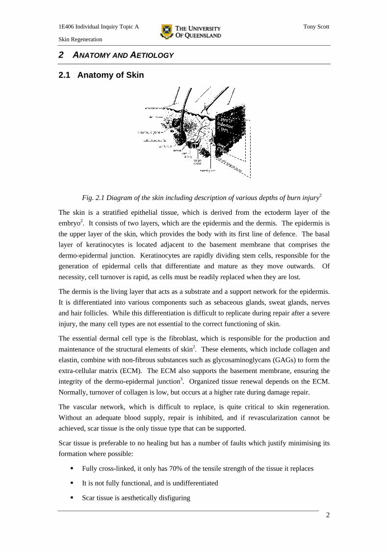
Fig. 2.1 Diagram of the skin including description of various depths of burn injury2
The skin is a stratified epithelial tissue, which is derived from the ectoderm layer of the
embryo2. It consists of two layers, which are the epidermis and the dermis. The epidermis is
the upper layer of the skin, which provides the body with its first line of defence. The basal
layer of keratinocytes is located adjacent to the basement membrane that comprises the
dermo-epidermal junction. Keratinocytes are rapidly dividing stem cells, responsible for the
generation of epidermal cells that differentiate and mature as they move outwards. Of
necessity, cell turnover is rapid, as cells must be readily replaced when they are lost.
The dermis is the living layer that acts as a substrate and a support network for the epidermis.
It is differentiated into various components such as sebaceous glands, sweat glands, nerves
and hair follicles. While this differentiation is difficult to replicate during repair after a severe
injury, the many cell types are not essential to the correct functioning of skin.
The essential dermal cell type is the fibroblast, which is responsible for the production and
maintenance of the structural elements of skin2. These elements, which include collagen and
elastin, combine with non-fibrous substances such as glycosaminoglycans (GAGs) to form the
extra-cellular matrix (ECM). The ECM also supports the basement membrane, ensuring the
integrity of the dermo-epidermal junction3. Organized tissue renewal depends on the ECM.
Normally, turnover of collagen is low, but occurs at a higher rate during damage repair.
The vascular network, which is difficult to replace, is quite critical to skin regeneration.
Without an adequate blood supply, repair is inhibited, and if revascularization cannot be
achieved, scar tissue is the only tissue type that can be supported.
Scar tissue is preferable to no healing but has a number of faults which justify minimising its
formation where possible:
§ Fully cross-linked, it only has 70% of the tensile strength of the tissue it replaces
§ It is not fully functional, and is undifferentiated
§ Scar tissue is aesthetically disfiguring
1E406 Individual Inquiry Topic A Tony Scott
Skin Regeneration
3
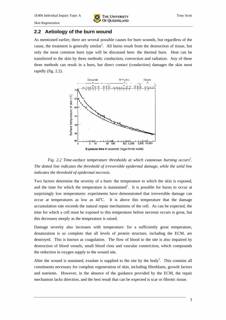
2.2 Aetiology of the burn wound
As mentioned earlier, there are several possible causes for burn wounds, but regardless of the
cause, the treatment is generally similar2. All burns result from the destruction of tissue, but
only the most common burn type will be discussed here: the thermal burn. Heat can be
transferred to the skin by three methods: conduction, convection and radiation. Any of these
three methods can result in a burn, but direct contact (conduction) damages the skin most
rapidly (fig. 2.2).
Fig. 2.2 Time-surface temperature thresholds at which cutaneous burning occurs2.
The dotted line indicates the threshold of irreversible epidermal damage, while the solid line
indicates the threshold of epidermal necrosis.
Two factors determine the severity of a burn: the temperature to which the skin is exposed,
and the time for which the temperature is maintained2. It is possible for burns to occur at
surprisingly low temperatures: experiments have demonstrated that irreversible damage can
occur at temperatures as low as 44oC. It is above this temperature that the damage
accumulation rate exceeds the natural repair mechanisms of the cell. As can be expected, the
time for which a cell must be exposed to this temperature before necrosis occurs is great, but
this decreases steeply as the temperature is raised.
Damage severity also increases with temperature: for a sufficiently great temperature,
denaturation is so complete that all levels of protein structure, including the ECM, are
destroyed. This is known as coagulation. The flow of blood to the site is also impaired by
destruction of blood vessels, small blood clots and vascular constriction, which compounds
the reduction in oxygen supply to the wound site.
After the wound is sustained, exudate is supplied to the site by the body3. This contains all
constituents necessary for complete regeneration of skin, including fibroblasts, growth factors
and nutrients. However, in the absence of the guidance provided by the ECM, the repair
mechanism lacks direction, and the best result that can be expected is scar or fibrotic tissue.

1E406 Individual Inquiry Topic A Tony Scott
Skin Regeneration
4
3 EXISTING TREATMENT
3.1 Natural repair
Natural wound healing typically consists of three phases: Inflammation and Debridement,
Repair and Maturation. (The information in section 3.1 was taken from Purves et al, Life: The
Science of Biology4 unless otherwise referenced)
1. Inflammation and Debridement
Immediately a wound is sustained, the body enters damage control. Platelets in the blood
are activated by contact with collagen outside the blood vessels, which causes them to
adhere to each other (the blood coagulates). They also release chemicals, which
encourage both vascular constriction to stem blood flow and platelet aggregation to
enlarge clot and plug the wound. White blood cells are mobilised to fight sources of
infection as they enter the wound, so capillaries surrounding the wound become
engorged. This engorgement has the effect of increasing the permeability of the capillary
walls. Lytic enzymes remove dead tissue to speed the healing process. The length of the
inflammation and debridement phase varies, and is prolonged by infection, lack of blood
supply to the wound site and the obstruction by necrotic tissue.
2. Repair
The repair phase normally begins within twelve hours of the wound occurrence. This
involves granulation, fibrification and epithelialisation. Granulation involves the
formation of a scab, which acts as a scaffold for new cells to attach to. Depending upon
the severity of the wound, fibrification may be required. Fibroblasts, which produce
fibrous tissue, enter the wound site during fibrification. Fibrous tissue acts as a barrier
against infection, and is required for wound contraction, during which intact tissue around
the wound contracts to bring the sides of the wound together. Revascularization occurs to
some extent during fibrification. Once a scaffold is in place, epithelialisation can take
place: the migration and multiplication of cells to form new tissue. The presence of
infection, too much granulation, hypothermia and insufficient blood supply are all factors
that protract the repair phase.
3. Maturation
Depending upon the severity of the wound, maturation takes between 14 days and 12
months to complete. The fibroblast concentration at the wound site is reduced, and
fibroclasps enter the site. Fibroclasps produce lytic enzymes that remove the irregularly
arranged collagen laid down during the initial phases. The wound site is then
strengthened through regeneration of the extra-cellular matrix. This is achieved in part by
the laying down and cross-linking of correctly oriented collagen fibres by fibroblasts.
Scarring occurs when the damage is so extensive that an adequate support system cannot be
regenerated. That is, the blood vessel network is not replaced. Scars typically exhibit an
1E406 Individual Inquiry Topic A Tony Scott
Skin Regeneration
5
inverted triangular cross-section, as epidermal tissue encroaches from each side above the
necrotic dermis, leaving a thin line of scar tissue visible at the surface.
3.2 Standard Treatment
First aid for all burns consists initially of cooling the wound (typically with cold water), as
damage accumulation will continue to occur until the wound is cooled to within a tolerable
temperature range2. Rapid cooling minimises the damage that occurs, and plays a large part
in determining the extent and depth of the burn. Dressing the wound is also important from
the point of view of preventing both infection and fluid loss.
To date, the standard treatment for severe burns has centred on the grafting of skin harvested
from an undamaged part of the patient’s own body (known as an autograft)5. This method
works reasonably well for the replacement of the epidermis, provided the damage is not too
extensive and sufficient undamaged skin is available. Usually, the skin is taken from the
same location on several occasions, and is allowed to regrow between harvesting. This
procedure is slow, and results in a ‘chequerboard’ effect both at the donor site and the burn
site. It has been found that burn healing can be accelerated by a number of factors, including:
§ The application of pressure to the burn site, which encourages the rapid reinforcement
and cross-linking of the new tissue. This serves two purposes: it increases the
strength of the skin, and it improves aesthetic appearance by providing an
environment that promotes correct healing and minimisation of scar tissue.
§ Application of topical antibiotics to the wound site to prevent infection of the wound
and allow the body to concentrate on wound healing rather than fighting disease.
Alternatively, it is possible to source skin either from other people or cadavers (an allograft),
or from an animal such as a cow or a pig (a xenograft)5. The body’s immune response means
that these grafts are almost inevitably rejected, so such grafts serve only to buy time while a
permanent solution is sought. It is generally not possible to administer immunosuppressive
drugs to prevent rejection of foreign skin grafts, as is normal procedure during the
transplantation of any other organ, as infections are common, and the immune system is
required to be fully functional. This means that alternatives such as autografts, or the
transplantation of an acellular collagen matrix (free from cells that would cause it to be
rejected) must be implemented.
Living tissue has an extremely limited shelf life outside the body, and preservation techniques
such as freezing must be used if the tissue is to remain viable. The growth of epidermal tissue
on the body as opposed to externally has the potential to defeat this problem, as well as to
greatly decrease the costs associated with the growth factors and other nutrients required for
growth in a laboratory.

1E406 Individual Inquiry Topic A Tony Scott
Skin Regeneration
6
3.3 Commercial Products
3.3.1 IntegraTM
Fig. 3.2.1 Schematic diagram of IntegraTM 2
IntegraTM is an artificial skin, which consists of two layers6. The lower layer is made up of an
ECM of synthetically cross-linked bovine collagen, forming a scaffold to which the dermal
cells may attach. The upper layer consists of a flexible silicon sheet to protect the site. Once
the dermis is established, the upper layer can be removed, and depending upon the size of the
wound, epidermal cells (either an autograft or an allograft) may be applied. Integra has a take
rate of 80%, compared with 95% for an autograft. One major advantage of Integra over
autografts is the reduced tissue thickness that must be harvested from donor sites, allowing
more rapid healing both at the burn site and at the donor site. However, it should be noted
that Integra does not provide any assistance to epidermal regeneration.
3.3.2 LifeCellTM
LifeCell7 have patented a method of removing cells from human dermal tissue while
maintaining the collagen matrix, under the trade name AlloDerm®. This is achieved through
chemical processing. The patent also extends to include a method of freeze-drying this ECM
without damaging the structure, for the purposes of preservation. This gives AlloDerm a shelf
life of approximately one year, but the product sells for about $10,000 per square foot8.
AlloDerm functions in a similar fashion to Integra. Arguably, healing is assisted by the fact
that the collagen is arranged in the form of a true human ECM.
LifeCell has a major disadvantage – its raw material is cadaver skin, the supply of which is
limited. There are also safety issues associated with the harvesting of the skin, which must be
performed soon after death to prevent sepsis from taking hold.
3.3.3 EpicelTM
EpicelTM 9 differs from the previous two treatment methods in that epicel is only suitable for
replacing the epidermis. Epicel involves the culturing of a small sample of the patient’s skin
in vitro until it is large enough to be meshed and grafted onto the patient. Meshing the skin
prior to application greatly increases the area that can be covered with a given graft. The
nutrients and growth factors required for epidermal regeneration are supplied by a substrate of
mouse fibroblasts that have been lethally irradiated to ensure that they do not divide and
multiply themselves. The utility of Epicel is limited by its inability to assist dermal
regeneration, which is frequently a more pressing concern than epidermal replacement. As a
1E406 Individual Inquiry Topic A Tony Scott
Skin Regeneration
7
living tissue, the shelf life of Epicel is just 2 hours, and the cost is $15,000 per square foot8.
However, there exists a very useful potential for the epidermis to be cultured in parallel with
dermal regrowth. Total healing time, along with the risk of infection, can thus be greatly
reduced.
3.3.4 DermagraftTM
One of the major disadvantages of many grafts is their lack of mechanical strength; hence the
approach taken by the developers of Dermagraft10. Essentially, this is a polyglactin-910
mesh, seeded with autologous fibroblasts. Polyglactin-910 is the polymer from which
biodegradable sutures are made. The benefits of increased mechanical strength include
§ Ease of handling avoiding the requirement for a backing material
§ Ability to hold sutures and staples
An interesting characteristic of dermagraft is the lack of an ECM. The fibroblasts seeded in
the mesh secrete the proteins, glycoproteins and collagen necessary for the formation of the
ECM in vivo. This avoids the susceptibility of the collagen matrix to both collagenases and
microorganisms in the wound bed.
The fibroblasts are essential for healing, however. It was demonstrated that when the mesh
was grafted to the wound when not seeded with fibroblasts, ingrowth of fibrovascular tissue
from the wound bed did not occur, and the mesh rapidly separated from the wound.
A disadvantage of this approach is the need for autologous fibroblasts, which means that this
treatment may not be administered until the requisite cells have been separated and cultured.
However, the low immunogenicity of fibroblasts is considered in section 4.5 of this report.
4 POTENTIAL TREATMENT PROCEDURES
There exist a number of potentially successful treatment procedures, all of which utilise an
extra-cellular matrix to provide a template for repair. Variations on the repair procedure
revolve around modification of this matrix to promote repair. The structural and mechanical
properties of the matrix can be altered during the matrix synthesis phase, controlling factors
such as the degree of cross-linking.
A good deal of research work has explored possible additions to the matrix that may aid both
the speed of healing and the completeness of the final repair. Such additions have included
growth factors to accelerate cell ingression and growth, and seeding of the matrix with cells to
overcome the constraint placed on cell ingression rate by diffusion. Consideration must also
be given to the promotion of revascularization. The ingression of new tissue will only occur
if it can be supported with a nutrient supply, and seeding cells is useless if the support
network cannot be regenerated within a short period of time.
The major variable remaining is the location of the repair, for which there exist two options:
in vivo and in vitro. While all regenerated tissue must be incorporated into the body
eventually, there are some repair procedures that can be initiated outside the body for reasons

1E406 Individual Inquiry Topic A Tony Scott
Skin Regeneration
8
of expediency. Needless to say, the choice for location of repair depends to a large extent on
the nature of the tissue: some tissue types are better suited to ex vivo repair than others.
For example, it is common to culture epidermal tissue ex vivo, for a number of reasons:
§ The epidermis is thin, allowing nutrients to be supplied to cells by the diffusion
mechanism only
§ The complexity of the epidermis is low, as there are few cell types and the dermis
provides the necessary substrate for growth
§ Vascularization is not required. It is not currently possible to stimulate angiogenesis
outside the body.
§ Integration into the wound site is relatively easy.
In vitro treatment is better suited to regeneration of the epidermis than of the dermis, as
differentiation in the dermis is too complex to achieve with current technology.
However, the dermis is not suitable for regeneration in vitro. Vascularization is essential for
complete integration of the dermis into the body. Currently, this is not possible outside the
body. This means that dermal tissue cultured outside the body would degrade into scar tissue
following transplantation, due to the absence of a nutrient supply.
For this reason, an in vivo procedure is to be adopted for dermal repair.
4.1 In vitro Vs. In vivo
(The information in section 4.1 was taken from Lanza et al, Principles of Tissue Engineering3
unless otherwise referenced)
The following table outlines the advantages of growing tissue inside and outside of the body.
In vivo In vitro
Many aspects of in vivo repair such asparticular growth hormones cannot bereplicated outside the body
Targeting of specific cells is possible (genetherapy for example) and risks for other cellswithin the body are minimised
In vivo repair is a lot less expensive than exvivo repair considering the cost of growthfactors, specialised labour and equipment
Recovery time can be decreased bysimultaneous development of dermis insidethe body and epidermis outside the body
Complete differentiation is possible includingvascularization
Rapid growth is possible through the use ofcytokines in concentrations which would betoxic within the body
Full integration into the body is simpler toachieve
Development of tissue from embryonic stemcells is possible, to make use of the rapidturnover in these cells
The body provides the correct physio-mechanical environment for structurally-adequate repair
Multiple experimental procedures can betested outside the body, and the most suitableprocedure can be selected
Table 4.1 Respective advantages of in vitro and in vivo treatment techniques

1E406 Individual Inquiry Topic A Tony Scott
Skin Regeneration
9
4.1.1 In vitro Treatment
Initially, design criteria must be established for the specific application for which the tissue is
intended. Skin consists of multiple layers, and currently each layer must be cultured
separately as cell differentiation outside the body is in its early stages (see section 4.6).
Design criteria for skin equivalents are:
§ Required dimensions (determined by the extent and depth of the damage)
§ Structural and functional properties, such as:
Ø The strength to function normally and resist cuts and abrasions
Ø Temperature control (sweating or routing blood close to the surface for cooling)
Ø Barrier to infection and to water loss
Ø The elasticity to permit the normal range of movement
Ø Aesthetic considerations dictate that the tissue should have the look and feel of
natural skin.
The epidermis is particularly suited to in vitro regeneration by its dimensions. The supply of
nutrients and oxygen to cells is limited by diffusional constraints, and the small thickness of
the skin ensures that in general, growth is not limited by starvation. However, mixing of the
growth medium is still required to maximise mass transfer, and this must be achieved without
mechanically interfering with tissue growth. Stress during growth is desirable, however, and
a tension/pressure cycle encourages the reinforcing of skin tissue so that adequate strength is
obtained.
Growing the skin replacement on an extra-cellular matrix makes the skin simpler to handle
when it comes time to transplant the skin to the wound. In addition, enzymatic treatment is
not required to separate the skin from the bioreactor, so the dermo-epidermal junction is not
disturbed. However, the presence of an ECM in vitro presents a limitation to the rate of mass
transfer of nutrients and oxygen to cells in the matrix, and of metabolic waste products from
cells in the matrix.
Finally, all tissue grown in vitro must at some stage be integrated into the body, and
consideration must be given as to how this is to be achieved. Major factors include
acceptance by the immune system, vascularization if required and the replacement of the graft
by natural cells such that structural properties are maintained.
4.1.2 In vivo Treatment
It is important to note that the epidermal layer will regenerate spontaneously, but this will
only occur if there is an adequate support network supplied by the dermal substrate
underneath. The dermis does not regenerate if the damage is too great. To this end, in vivo
treatment techniques for full thickness burns to date have concentrated on dermal repair.
In vivo repair techniques revolve around the implantation of a biodegradable extra-cellular
matrix (ECM) to act as a scaffold onto which cells can attach during wound repair. An

1E406 Individual Inquiry Topic A Tony Scott
Skin Regeneration
10
advantage of in vivo treatment is that the wound is supplied with nutrients, cells and cytokines
(growth factors) by the body, and all that is required is a matrix to guide the repair effort. The
stability of the temperature and pH of the living environment provide good conditions for
growth. Even though a precise knowledge of the chemical reactions and biological
interactions which take place during wound repair is not required for in vivo repair, the
original configuration must be known with reasonable accuracy so that the end goal is clearly
defined. Design requirements for an in vivo skin regeneration template include:
§ Interaction with components of the exudate supply to modify the kinetics which
normally convert the exudate to scar tissue
§ Easily accessible to migrating cells, including the adequate presence and distribution
of macropores for cell and nutrient transport.
§ Appropriate density, position and types of binding sites for cells must be present
§ Biodegradable so that it can be removed from the wound site by the body after
healing is complete
§ Biocompatible so that the immune system does not reject it.
In addition to these design requirements, there exists the difficulty of choosing an appropriate
performance measure by which success is defined. An example of such a measure is the
moisture evaporation rate. However, there are many performance measures to choose from,
and in many cases meeting one will mean failing to meet another.
The exudate is drawn into pores through mechanisms of capillarity and diffusion down the
concentration gradient. It has been found that the optimal effective pore size lies between
20-125µm, as these pores are large enough for rapid transport of exudate, but small enough to
provide sufficient sites for migrating cells to attach to2. Orientation of pore channels should
be random, as for natural skin3. Porosity may be controlled in a number of ways, most of
which involve manipulation of polymerisation conditions. An interesting variation was
utilised by Butler et al11. A freeze-drying process under vacuum conditions was used to
remove ice crystals by sublimation, leaving a highly porous matrix.
As noted above, one criterion for the suitability of the template is the biodegradability3.
Degradation must occur with reasonable rapidity, but not so fast that adequate repair does not
have a chance to take place. This degradation is ideally caused by lytic enzymes naturally
present during wound repair, and should be complete enough that the products of the
degradation (which must be non-toxic) can be transported from the site in the bloodstream.
The time for complete degradation should be of the order of several weeks, and depends upon
the severity of the wound. It is possible to use in vitro techniques to experiment with template
characteristics prior to implantation to determine degradation rate data.

1E406 Individual Inquiry Topic A Tony Scott
Skin Regeneration
11
4.2 The Extra-Cellular Matrix
Primarily, the ECM provides a scaffold to which fibroblasts can attach. The extra-cellular
matrix has been shown to be useful both as a carrier of cytokines and for quick closure of
wounds12.
When considering desirable properties for the ECM, it must be realised that the artificial
ECM is replaced by fibroblasts, so the initial structure is refashioned to a large extent. This
means that the synthesised ECM does not necessarily have to have the structure of natural
skin. However, it has been found that repair is promoted by an ECM structure close to that of
the original.
There exists a choice of materials from which the ECM can be created. Broadly speaking,
these materials can be divided into natural and synthetic groupings. It is generally easier to
control the properties of synthetics, and parameters such as strength, speed of degradation and
permeability can be readily manipulated during production. Natural materials such as
collagen have one decisive advantage however: they are usually easier for cells to stick to13.
The ECM is typically constructed from a combination of collagen and GlycosAminoGlycans
(GAGs). Collagen provides the template with structure and mechanical integrity, while the
GAGs slow in vivo degradation and encourage correct biological activity. It has been
suggested that hyaluronic acid, the largest GAG, acts as a transport mechanism for growth
factors, releasing these when it is degraded.
4.2.1 Mechanical Considerations
In order to create a skin replacement with properties that approach those of the real thing, the
physical requirements must be considered in addition to the biological requirements14. The
affinity of the skin for the wound bed must exceed the affinity of air for the wound bed;
otherwise pockets of air will build up in the interface. This is not only bad for healing; it also
exposes the wound to potential sources of infection. In addition, the modulus of elasticity of
the skin replacement must be close to that of natural skin so that the skin replacement can flex
with the natural movement of the body without pulling away from the wound. Typically,
once the modulus of elasticity is right (this is dictated by the material of construction) the
desired flexibility is achieved through manipulation of the graft thickness.
The balance between growth and differentiation is affected by the hardness of the ECM3.
Compliant gels promote differentiation, whereas stiffer gels support growth. Thus a gel that
is initially hard and becomes softer as it degrades is desirable for the promotion of complete
healing.
4.2.2 Cross-linking
Cross-linking of the collagen matrix is the primary means by which the mechanical properties
and degradation rate of the matrix can be controlled. Cross-links stabilise the conformation of
collagen fibres, and may be induced by chemical or physical methods. Natural collagen
consists of polypeptide chains that form triple-stranded helical units. Cross-links between
lysine residues mostly occur between the non-helical ends of the chains, and act to stabilize

1E406 Individual Inquiry Topic A Tony Scott
Skin Regeneration
12
the three-dimensional arrangement of these units. Unfortunately it is these non-helical ends
that generate an immune response from the host. Once these ends have been cleaved from the
chains, an alternative cross-linking method is required which links the helical regions of the
collagen strands. Siegel15 has patented such a method. After cleavage of the non-helical
ends, cross-linking is induced through incubation with pyridoxal-5-phosphate in the presence
of ionic copper or iron.
Glutaraldehyde (GTA), an efficient chemical agent, is the basis of the most widely used cross-
linking method. Unfortunately, while GTA polymerises when in solution, with time GTA
monomers are released, which are cytotoxic at concentrations as low as 10ppm. An
alternative cross-linking method patented by Petite et al16 involves the formation of amide
bonds through the addition of diphenylphosphorylazide.
4.3 Growth Factors
There exist a number of growth factors that may be added to the ECM to stimulate healing.
Specifically, the aspects of healing that should be assisted are vascularization and fibroblast
growth. The requirement for angiogenesis results in a chicken-and-egg situation during
wound healing. While fibroblasts are required to generate the physical support necessary for
blood vessel ingrowth, these fibroblasts require a blood supply or they will not survive. This
situation can lead to a petulant standoff, in which scar tissue is the only alternative. Through
the use of growth factors, it is possible to stimulate both aspects of regeneration
simultaneously.
Whilst growth factors are normally supplied to the wound site in vivo as part of the exudate, it
is desirable to supplement the exudate with pure forms of these growth factors for a number
of reasons:
§ Chemically defined media reduce variability in experiments, and improve
reproducibility.
§ Research into growth factors allows partial regeneration of skin in vitro prior to
transplantation.
§ Biological fluids may be a source of toxins or viruses
§ The growth factors may not be present in the quantities required for sufficiently rapid
healing.
Growth factors known to promote fibroblast growth include interleukin, TGF-α and -β, bFGF
and tumour necrosis factor α, along with many others. Unfortunately, while works exist
which examine the effects of various growth factors, no direct comparison of growth factor
types was located in the literature.
The main problem associated with growth factors is the delivery vector. Rapid diffusion
combines with a short half-life to ensure that regular dosing is necessary in the absence of an
adequate sustained-release mechanism.
1E406 Individual Inquiry Topic A Tony Scott
Skin Regeneration
13
4.3.1 Basic Fibroblast Growth Factor
Fibroblast growth factors have been shown to be modulators of cell proliferation, growth,
differentiation and survival17. In particular, bFGF (otherwise known as FGF-2) has been
shown to significantly inhibit wound contraction, particularly when applied in combination
with a collagen matrix12. This combination was shown to be more effective than either bFGF
or the matrix alone. In addition, bFGF stimulates the proliferation of fibroblasts, and
promotes angiogenesis through accelerating the growth of capillary endothelial cells. Dosage
has also been shown to affect healing: increasing the dose of bFGF was shown to improve
both the healing rate and the success of the end result.
4.3.2 Acidified Fibroblast Growth Factor
Acidified fibroblast growth factor, FGF-1, has been demonstrated to stimulate
revascularization, while inhibiting the formation of scar tissue18. As with many growth
factors, repeated dosing is required for effective healing. FGF-1 can be expressed in
recombinant E. coli, so large-scale production is not likely to present a problem.
In the study by Pandit et al., an FGF-1/collagen system was compared with both a control (no
treatment) and a collagen scaffold only, and was found to produce better blood vessel
regeneration than either of the other two cases. In addition, the FGF-1/collagen combination
produced the strongest and most elastic of the regenerated skins, with the smallest
inflammatory response during healing.
4.3.3 Keratinocyte Growth Factor
Keratinocyte Growth Factor is an important mediator of in vivo epidermal repair. KGF has
been shown to selectively induce keratinocyte proliferation and differentiation19. It has also
been shown to accelerate maturation of the dermo-epidermal junction, and to increase the
thickness of the epithelium.
4.3.4 Transforming Growth Factor ββ
TGF-β inhibits inflammation while promoting angiogenesis (revascularization) and
histogenesis20. A collagen sheet ECM impregnated with TGF-β was shown to stimulate the
multiplication rate of fibroblasts. It has also been demonstrated to regulate epithelial-
mesenchymal interactions. Until recently, a major disadvantage of TGF-β was the difficulty
associated with obtaining and isolating it: a ton of bone yields just one therapeutic application
of the growth factor. This patent20 also covers the production of TGF-β through implantation
of recombinant DNA into E.coli cells by means of plasmids.
4.3.5 Recombinant Human Growth Hormone
rHGH has been demonstrated to accelerate healing of burns through more rapid production of
tissue to replace that which has been lost21. This finding is fairly generic, in that it applies to
all types of wounds. There exists a concerning lack of specificity in this treatment, however,
as the hormone affects all areas of the patient’s body, and may lead to problems elsewhere in
the patient.

1E406 Individual Inquiry Topic A Tony Scott
Skin Regeneration
14
4.4 Growth factor delivery vectors
Consideration should be given to how the chosen growth factor is to be delivered to the
wound site. Many growth factors degrade rapidly within the body. A single large dose
initially is not sufficient if the growth factor concentration is to be maintained at the wound
site. One option is regular topical dosage, but this poses a number of problems. The wound
must be disturbed in order to apply the dose, and it is not possible to achieve an even
distribution of the growth factor throughout the wound site. The effects of a cyclic variation
in growth factor concentration must also be investigated.
However, a number of alternative delivery vectors exist.
4.4.1 Delayed Release
An interesting delivery vector was explored by Kawai et al22. Gelatin microspheres were
impregnated with bFGF and seeded into an acellular collagen matrix. This seeding was
possible as the average microsphere diameter was smaller than the matrix pore size. The
inconvenience of regular dosing was overcome by this sustained-release mechanism. Use of
microspheres was seen to increase the growth factor retention time by a factor of about ten.
4.4.2 Gene Therapy
An area in which little work has been carried out to date is the application of gene therapy to
dermal regeneration. It would be possible to genetically modify allogenic fibroblasts prior to
seeding these in the ECM to overproduce a growth factor such as bFGF. This method would
also eliminate the need for regular dosing. Gene therapy raises a number of significant
concerns however. While the genetic modification itself is relatively straightforward, the
primary difficulty is one of control. Unless the fibroblasts can be engineered so they do not
pass the genetic modification on when they replicate, it is not possible to stop the growth
factor production once treatment is complete, which will lead to unwanted side effects.
Ethical issues associated with gene therapy must also be taken into account. It is for these
reasons that gene therapy cannot be considered as a treatment option.
4.5 Seeding of the Collagen Matrix
4.5.1 Fibroblast Seeding
The presence of living fibroblasts in the dermal substitute means that vascularization is swift,
and the wound heals more rapidly than if the matrix was acellular. Also, no growth factors
explored promote the ingression of fibroblasts to the wound site, making seeding the preferred
method to expedite repair.
Fibroblasts serve a number of roles in the regeneration of damaged skin. The first is the
secretion and maintenance of components of the ECM that makes up the ‘backbone’ of the
skin, ensuring that the general morphology of the skin is maintained even when cells die. It is
only when this matrix is damaged that the skin has difficulty regenerating spontaneously.
Secondly, they are an integral component of the support network for the epidermis, allowing

1E406 Individual Inquiry Topic A Tony Scott
Skin Regeneration
15
for physical attachment through the basement membrane, which lies along the
dermo-epidermal junction. Support is also supplied by way of nutrients.
The reason for rejection of grafts by the immune system is the identification of that tissue as
foreign. This identification is always based upon the surface molecules of the transplanted
cells (as this is the only surface presented to the immune cells). It is important to realise that
there exist two classes of antigens, class I and class II. While MHC class II antigens23 are
present in most cells (including keratinocytes), fibroblasts lack these surface molecules. This
makes them, immunologically, relatively inert. This is fortunate from a tissue-engineering
point of view, as dermal regeneration matrices can be seeded with allogenic fibroblasts
without risking an immune response. Indeed, if allogenic fibroblasts are present, they will
generally inhibit the penetration of the host’s fibroblasts into the matrix24.
The lack of an immunogenic response to allogenic fibroblasts opens up a number of exciting
possibilities, one of which is the preparation of skin equivalent from volunteer donors, to be
stored until needed14. Fibroblasts can typically be isolated from healthy skin through punch
biopsies. This provides surgeons with a permanent skin replacement ‘off-the-shelf’.
Approval from the appropriate regulatory bodies must be sought to seed the collagen matrix
with allogenic human fibroblasts. While introducing foreign cells into the patient brings in
the possibility for transfer of disease, previous work in the screening of blood donations and
organs for transplant will hopefully minimise this risk.
Lamme et al.25 examined the relationship between fibroblast seeding density and wound
healing, using autologous fibroblasts. It was found that increasing the seeding density
improved not only the healing time but also the cosmetic appearance of the final result. The
time for which the fibroblasts were cultured in the matrix prior to transplantation was also
found to have a large effect: increasing this time dramatically improved healing, as the
fibroblasts had the opportunity to establish themselves within the ECM, laying down collagen
and elastin fibrils.
4.5.2 Keratinocyte Seeding
Seeding the skin equivalent with a layer of keratinocytes greatly increases the rapidity with
which the epidermal layer can be regenerated, given adequate support from the new dermal
layer. However, the source of the keratinocytes must be considered. Allogenic keratinocytes
are rejected by the immune system, so the keratinocytes seeded in the matrix must be drawn
from the patient.
The activity of the keratinocytes, however, is a function of the location from whence they are
removed. There is seen a decrease in proliferative potential as one moves from the basement
membrane to the cornified layer of the epidermis. Indeed, when cells leave the basal cell
layer they start down the path of terminal differentiation, and are thereafter limited to a
maximum of about five cell divisions. The basal cells have the greatest ability to divide and
grow: these are stem cells. Butler et al.11 hypothesised that terminal differentiation of
keratinocyte stem cells is triggered by the contact with other cells resulting from the
1E406 Individual Inquiry Topic A Tony Scott
Skin Regeneration
16
formation of a confluent layer of cells. It was demonstrated that when cultured to sub-
confluence prior to harvesting, keratinocytes had colony-forming efficiencies as high as 80%
(compared with less than 10% for uncultured keratinocytes). The keratinocytes thus
harvested proved to regenerate epidermis much more rapidly than uncultured keratinocytes.
The disadvantage of this technique is the time required for culturing. However, surgical
treatment is often postponed for a number of weeks so that healing can be observed, which
means that culturing of autologous keratinocytes is not out of the question.
4.6 Living Skin Equivalent
An exciting development in tissue regeneration is the culturing of a living skin equivalent
(LSE) in vitro. The goal of LSE is the development of an entire living tissue in vitro, the idea
being that, with the exception of nutrient and oxygen supply, the tissue is self-sufficient. To
this end, researchers attempt to seed the matrix with the potential for differentiation into all
cell types required for functional skin. This potential is found in stem cells, a popular source
of which in recent research has been human foreskins.
A major difficulty associated with the growth of a dermal equivalent in vitro is that of
contraction of the matrix. Once they have established themselves within the matrix,
connective cells such as fibroblasts pull collagen fibrils towards themselves, which results in
an overall reduction in the volume of the matrix. This tendency has been overcome through
the development of anchoring techniques that fix the length and breadth of the matrix,
allowing contraction in the thickness dimension only26. A second problem is the time
required to culture such an equivalent from autologous cells, as allogenic keratinocytes will
be rejected by the immune system.
These limitations mean that thus far, the main use for living skin equivalent is as a testing
vehicle for new treatments. Applications outside skin regeneration include assessing the
effectiveness of sunscreens, and also in investigating the diffusion rate of drugs through skin.

1E406 Individual Inquiry Topic A Tony Scott
Skin Regeneration
17
5 REGENERATION STRATEGY
The strategy believed to be best suited to the task is an in vivoapproach, involving the implantation of a collagen ECM seeded withcultured allogenic fibroblasts. Gelatin microspheres are to beutilised for the delivery of basic fibroblast growth factor to thewound site.
5.1 Key Parameters
5.1.1 In Vivo
The repair is to occur in vivo, mainly to facilitate integration into the body. Vascularization is
an aspect of integration that is essential for regeneration, and cannot be replicated in vitro. In
vivo repair has the additional advantage over ex vivo of low cost, both in terms of materials
and labour. The in vivo approach also reduces the trauma associated with multiple surgical
procedures. For in vitro methods, multiple procedures are dictated by the need to protect the
wound site while a more permanent solution is created outside the body.
5.1.2 Extra-cellular matrix
The matrix is to be constructed primarily from type I collagen and glycosaminoglycans
(GAGs). This material was deemed to be most appropriate as it is possesses the required
properties, and is relatively inexpensive. In addition, collagen is the main material from
which the natural extra-cellular matrix is constructed. Collagen is not the only constituent of
the natural ECM. However, the implanted matrix exists to provide a template for tissue
regeneration and to prevent wound contraction, tasks that are fulfilled by collagen alone.
During the repair process, the matrix is entirely replaced by the body as the fibroblasts
establish themselves.
The ECM is to be prepared through polymerisation of a solution containing approximately
6mg/mL of bovine collagen and 2mg/mL elastin at 37oC25. Implementing the freeze-drying
technique used by Butler et al11 can increase the porosity of the matrix. Ideally, the mean
pore size in the matrix is to be 80µm25.
Cross-linking of the matrix is to be achieved with glutaraldehyde. Further work is required,
however, to establish the cytotoxicity of the products of degradation of GTA. The extent of
cross-linking controls the rate of degradation. Residence time is to be approximately two
weeks, but this depends on the severity of the wound. The matrix should be firm initially to
promote growth, becoming more compliant as the cross-links break down. A softer matrix
encourages differentiation and maturation, to give a final product closer to natural skin.
In the style of IntegraTM, an outer silicon layer 0.3mm thick is to provide protection for the
wound and control moisture loss while the dermis establishes itself. While the dermal layer is
re-establishing itself, autologous epidermal tissue can be cultured in vitro, so that it can be
applied as soon as the silicon sheet is removed.

1E406 Individual Inquiry Topic A Tony Scott
Skin Regeneration
18
5.1.3 Growth factors
The growth factor chosen to promote healing and revascularization is basic fibroblast growth
factor. bFGF has also been shown to reduce the rate of wound contraction, which is
desirable from the point of view of minimising scarring. Porous gelatin microspheres are to
be utilised for the controlled delivery of the growth factor22. The advantages of this delivery
vector include
§ Delayed release of bFGF, which maintains the concentration of growth factor at the
wound site and removes the need for regular dosing. Controlled release is desirable
to counter the rapid degradation of bFGF in vivo, and diffusion away from the wound.
§ Ensures efficient use of growth factor, reducing the amount of bFGF required.
§ Prevents bFGF from causing unwanted effects elsewhere in the patient
The mean diameter of these microspheres is to be approximately 40µm. For the method of
microsphere production, refer to Kawai et al22. As this is smaller than the mean ECM pore
size, these microspheres should disperse evenly within the matrix
5.1.4 Fibroblast seeding
The ECM is to be seeded with allogenic fibroblasts. Advantages of allogenic cells include
§ Due to the low immunogenicity of fibroblasts, the matrix can be prepared for instant
application when required, so multiple surgical procedures are not necessary
§ Further trauma is not inflicted on the patient by extraction of autologous fibroblasts
§ Fibroblasts can be cultured in the matrix to establish themselves prior to application
The culturing of fibroblasts also reduces wound contraction through lessening their tendency
to differentiate into their contractile phenotype, the myofibroblast25. Fibroblasts are to be
collected from volunteer donors by means of punch biopsies, and seeded into the matrix at a
density of approximately 5 x 10⁵ cells/cm3. Following seeding, the cellular matrix is to be
cultured in the matrix for a minimum of ten days where possible to allow the fibroblasts to
establish themselves. The addition of ascorbic acid will promote the deposition of ECM
components in vitro25.
5.1.5 Surgical procedure
Implantation of the matrix into the patient involves the following steps
§ Excision of wound. The removal of necrotic tissue from the wound bed is desirable
in order to prevent infection and allow room for tissue regeneration
§ Implantation of ECM at wound site, held in position by elastic bandages. The ECM
will not be strong enough to hold sutures. As a result, the patient should be kept still
to minimise stresses on the wound.
§ Following establishment of the dermis, the silicon sheet is removed. An autograft can
then be applied to assist epidermal regeneration, cultured outside the body if required.
1E406 Individual Inquiry Topic A Tony Scott
Skin Regeneration
19
6 CONCLUSIONS AND RECOMMENDATIONS
Advances in the field of burns treatment have great potential both to speed recovery time and
to reduce suffering. The various components that make up this regeneration strategy have all
been demonstrated to work previously, so it is anticipated that no major problems will be
encountered in the engineering of this solution.
6.1 Recommendations for future work
As this solution is entirely based upon review of available literature, a great deal of
experimental work is required before it can be implemented. Experimental work should
concentrate on:
§ Growth factors. A direct comparison of the effectiveness of the growth factors
available should be conducted. Parameters to be quantified should include:
Ø Degree of differentiation
Ø Rate of growth
Ø Extent of angiogenesis
Ø Degree of wound contraction
§ Storage conditions. If this skin equivalent is to be available to surgeons as an
‘off-the-shelf’ solution, it is desirable to determine the following characteristics.
Ø Optimum storage conditions
§ Freezing
§ Low temperature
§ Under solvent solution
Ø Length of time fibroblasts will remain viable
Ø How storage will affect growth factor
Ø Whether gelatin microspheres will retain functionality during storage
§ Cross-linking
Ø Comparison of cross-linking methods
Ø Cytotoxicity of degradation products
Ø Effect on biodegradability
With serious burns as common as they are (although most are preventable), the field of tissue
engineering has much to contribute to this area of human injury. Research has demonstrated
that better methods for the assistance of skin regeneration do exist, and further work is
essential so that these may be applied in burns treatment centres as soon as possible.

1E406 Individual Inquiry Topic A Tony Scott
Skin Regeneration
20
7 REFERENCES
1 National Institute of Health (United States) website, http://www.nih.gov2 Herndon D.N., Total Burn Care, 1996, W.B. Saunders, London3 Lanza, Langer & Chick, Principles of Tissue Engineering, 1997, R.G. Landes Company4 Purves, Orians, Heller & Sadava, Life: The Science of Biology (5th ed.), 1998, Sinauer Associates, Sunderland5 Boswick J.A., The Art and Science of Burn Care, 1987, Aspen Publishers, Rockville6 Integra LifeSciences Corporation website, http://www.integra-ls.com/index_java.html7 Lifecell website, http://www.lifecell.com8 Lifecell Editorial, Biotechnology (1994) 12(4) 3389 Epicel website, http://www.genzyme.com/epicel/welcome.htm10 Hansbrough JF, Morgan JL, Greenleaf GE, Bartel R., Composite grafts of human keratinocytes grown on apolyglactin mesh-cultured fibroblast dermal substitute function as a bilayer skin replacement in full-thicknesswounds on athymic mice, J Burn Care Rehabil. 1993 Sep-Oct;14(5):485-94.11 Butler CE, Yannas IV, Compton CC, Correia CA, Orgill DP, Comparison of cultured and unculturedkeratinocytes seeded into a collagen-GAG matrix for skin replacements, Br J Plast Surg. 1999 Mar;52(2):127-32.12 Effects of collagen matrix containing bFGF on wound contraction, J. Biomed. Mater. Res. (1999) 48(5) 621-3013 Langer and Vacanti, Skin: The first tissue-engineered products, Scientific American, April 1999 280(4) 83-914 Maugh, Engineering Artificial Skin, Technology Review, Jan. 1985, 88 48-5815 Siegel, Synthesis of cross-links in the helical domain of collagen using pyridoxal 5-phosphate and copper oriron, Patent: US 4,544,63816 Petite, Menasche, Huc, Process for cross-linking of collagen by diphenylphosphorylazide the cross-linkedcollagen obtained thereby and collagen-based biomaterials thus cross-linked, Patent: US 5,264,55117 Hu & Rosen, Fibroblast growth factor 11, Patent: US 5,763,21418 Pandit, Ashar, Feldman, Thompson, Investigation of acidic FGF delivered through a collagen scaffold for thetreatment of full-thickness skin defects in a rabbit model, Plast. Reconstr. Surg., Mar. 1998, 101(3) 766-7519 Stimulation of all epithelial elements during skin regeneration by keratinocyte growth factor, Journal ofExperimental Medicine (1994) 179(3) 831-4020 Hall, Nimni, Tuan, Wu & Cheung, Artificial skin prepared from collagen matrix containing TransformingGrowth Factor β, US 5,800,81121 Recombinant HGH accelerates wound healing in children with large cutaneous burns, Annals of Surgery (1994)220(1) 19-2422 Kawai, Suzuki, Tabata, Ikada, Nishimura, Accelerated tissue regeneration through incorporation of bFGF-impregnated microspheres into artificial dermis, Biomaterials (2000) 21(5) 489-9923 Dickman, Strobel, Life in the tissue factory, New Scientist, Mar. 1995, 145 32-724 The role of fibroblasts in dermal revascularization and remodelling, Transplantation, Aug 1992, 54(2), 317-2625 Lamme, Van Leeuwen, Brandsma, Van Marle, Middlekoop, Higher numbers of autologous fibroblasts in anartificial dermal substitute improve tissue regeneration and modulate scar tissue formation, J. Path., Apr. 2000190(5) 595-60326 Bell, Rosenberg, Kemp, Gay, Green, Muthukumaran and Nolte, Recipes for reconstituting skin, J. Biomech.Eng., May 1991 113(2) 113-11927. Boyce ST, Glafkides MC, Foreman TJ, Hansbrough JF., Reduced wound contraction after grafting of full-thickness burns with a collagen and chondroitin-6-sulfate (GAG) dermal skin substitute and coverage withbiobrane, J Burn Care Rehabil. 1988 Jul-Aug;9(4):364-70.28. Boyce ST, Hansbrough JF., Biologic attachment, growth, and differentiation of cultured human epidermalkeratinocytes on a graftable collagen and chondroitin-6-sulfate substrate, Surgery. 1988 Apr;103(4):421-31.29. California Polytechnic State University artificial skin page,http://www.calpoly.edu/~drjones/medpoly/skin/Polyskin.htm30. Complex coacervate microculture for mammalian cell culture, Biotechnology Progress31. Compton CC, Butler CE, Yannas IV, Warland G, Orgill DP., Organized skin structure is regenerated in vivofrom collagen-GAG matrices seeded with autologous keratinocytes, J Invest Dermatol. 1998 Jun;110(6):908-16

1E406 Individual Inquiry Topic A Tony Scott
Skin Regeneration
21
32. Cooper ML, Andree C, Hansbrough JF, Zapata-Sirvent RL, Spielvogel RL., Direct comparison of a culturedcomposite skin substitute containing human keratinocytes and fibroblasts to an epidermal sheet graft containinghuman keratinocytes on athymic mice, J Invest Dermatol. 1993 Dec;101(6):811-933. Cooper ML, Hansbrough JF, Use of a composite skin graft composed of cultured human keratinocytes andfibroblasts and a collagen-GAG matrix to cover full-thickness wounds on athymic mice, Surgery. 1991Feb;109(2):198-20734. Coulomb B, Friteau L, Baruch J, Guilbaud J, Chretien-Marquet B, Glicenstein J, Lebreton-Decoster C, BellE, Dubertret L., Advantage of the presence of living dermal fibroblasts within in vitro reconstructed skin forgrafting in humans, Plast Reconstr Surg. 1998 Jun;101(7):1891-903.35. Dong-Youn Lee, Hi-Tae Ahn and Kwang-Hyun Cho, A new skin equivalent model: dermal substrate thatcombines de-epidermized dermis with fibroblast-populated collagen matrix, J. Dermatol. Sci., Jun 2000 23(2)132-736. Eaglstein and Falanga, Tissue engineering and the development of Apligraf, a human skin equivalent, Adv.Wound Care, Jul-Aug 1998 11(4 suppl) 1-837. Eming SA, Medalie DA, Tompkins RG, Yarmush ML, Morgan JR., Genetically modified humankeratinocytes overexpressing PDGF-A enhance the performance of a composite skin graft, Hum Gene Ther. 1998Mar 1;9(4):529-39.38. Hansbrough JF, Boyce ST, Cooper ML, Foreman TJ., Burn wound closure with cultured autologouskeratinocytes and fibroblasts attached to a collagen-glycosaminoglycan substrate, JAMA. 1989Oct 20;262(15):2125-3039. Intellectual Property Network, http://www.delphion.com40. Kuroyanagi Y, Kenmochi M, Ishihara S, Takeda A, Shiraishi A, Ootake N, Uchinuma E, Torikai K, ShioyaN., A cultured skin substitute composed of fibroblasts and keratinocytes with a collagen matrix: preliminaryresults of clinical trials, Ann Plast Surg. 1993 Oct;31(4):340-9; discussion 349-51.41. Lamme EN, de Vries HJ, van Veen H, Gabbiani G, Westerhof W, Middelkoop E., Extracellular matrixcharacterization during healing of full-thickness wounds treated with a collagen/elastin dermal substitute showsimproved skin regeneration in pigs, J Histochem Cytochem. 1996 Nov;44(11):1311-2242. Lopez Valle CA, Germain L, Rouabhia M, Xu W, Guignard R, Goulet F, Auger FA., Grafting on nude miceof living skin equivalents produced using human collagens, Transplantation. 1996 Aug 15;62(3):317-2343. Maruguchi T, Maruguchi Y, Suzuki S, Matsuda K, Toda K, Isshiki N., A new skin equivalent: keratinocytesproliferated and differentiated on collagen sponge containing fibroblasts, Plast Reconstr Surg. 1994 Mar;93(3):537-44; discussion 545-644. Matouskova E, Vogtova D, Konigova R., A recombined skin composed of human keratinocytes cultured oncell-free pig dermis, Burns. 1993 Apr;19(2):118-2345. Mayoclinic website, http://mayohealth.org/mayo/9709/htm/skin-sub.htm#ww5ro6546. Prokop, Hunkeler & Cherrington, Bioartificial Organs, 1997, New York Academy of Sciences, NY47. Rennekampff HO, Kiessig V, Griffey S, Greenleaf G, Hansbrough JF., Acellular human dermis promotescultured keratinocyte engraftment, J Burn Care Rehabil. 1997 Nov-Dec;18(6):535-4448. Shahabeddin L, Berthod F, Damour O, Collombel C., Characterization of skin reconstructed on a chitosan-cross-linked collagen-glycosaminoglycan matrix, Skin Pharmacol. 1990;3(2):107-1449. Shaw A.J., Epithelial Cell Culture, 1996, Oxford University Press, NY50. Taub M (ed.), Tissue Culture of Epithelial Cells, 1985, Plenum Press NY51. The role of fibroblasts in dermal revascularization and remodelling, Transplantation, Aug 1992, 54(2),317-2652. Ultrastructural features of composite skin cultures grafted onto athymic mice, J. Anat., Oct. 1994 185(Pt 2)325-3353. Watson SA, Pisansarakit P, Moore GP., Sheep vibrissa dermal papillae induce hair follicle formation inheterotypic skin equivalents, Br J Dermatol. 1994 Dec;131(6):827-35