induction and suppression of otc and urea cycle enzymes … · nucleus mitochondrion 1 ... 17+ ,~...
TRANSCRIPT
9
Induction and Suppression of OTC and Urea Cycle Enzymes in Bacteria, Fungi and Mammals
Human OTC is related phylogenetic ally to the OTCs of the rat and mouse, less so to that of the frog and distantly to the six fungal OTCs which have been sequenced, called OTCj3s by Labedan(172). The OTCs of E.coli and P.aeruginosa, which like hOTC have had their structure determined by X-ray diffraction, belong to the OTea group by phylogenetic analysis of their amino acid sequences (172 ).
The control of amounts of OTC enzyme protein differs in bacteria from controls in fungi and from those in mammals, although some mechanisms have carried over to hOTC in its induction and suppression in hepatocytes. The arginine biosynthetic pathway in E.coli consists of nine enzymes and 10 genes, one coding for the argR repressor protein. (662) OTC is the sixth enzyme in the E.coli pathway from glutamate to arginine. The ninth enzyme is a glutamine-dependent carbamate kinase that supplies CP for OTC and for pyrimidine synthesis. Arginine exerts feedback inhibition of the first enzyme and also binds to the arginine repressor protein preventing transcription of the eight genes(662). Thus in E.coli and most bacteria, induction of OTC is accomplished by de-repression when arginine levels are low. An excellent summary of the development of de-repression in the arginine pathway is that of Maas(663 ).
Among eukaryotic fungi, the cellular compartmentation of Neurospora crassa most resembles that in mammalian hepatocytes (Figure 9-1)1664 ). The 5 enzymes that convert glutamate to ornithine, and en-
P. J. Snodgrass, Ornithine Transcarbamylase© Kluwer Academic Publishers 2004
166 . Ornithine Transcarbamylase
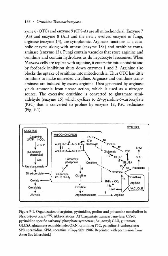
zyme 6 (OTC) and enzyme 9 (CPS-A) are all mitochondrial. Enzyme 7 (AS) and enzyme 8 (AL) and the newly evolved enzyme in fungi, arginase (enzyme 14), are cytoplasmic. Arginase functions as a catabolic enzyme along with urease (enzyme 18a) and ornithine transaminase (enzyme 15). Fungi contain vacuoles that store arginine and ornithine and contain hydrolases as do hepatocyte lysosomes. When N.crassa cells are replete with arginine, it enters the mitochondria and by feedback inhibition shuts down enzymes 1 and 2. Arginine also blocks the uptake of ornithine into mitochondria. Thus OTC has little ornithine to make unneeded citrulline. Arginase and ornithine transaminase are induced by excess arginine. Urea generated by arginase yields ammonia from urease action, which is used as a nitrogen source. The excessive ornithine is converted to glutamate semialdehyde (enzyme 15) which cyclizes to 8 C pyrroline-S-carboxylate (PSC) that is converted to proline by enzyme 12, PSC reductase (Fig. 9-1).
CYTOSOL NUCLEUS
MITOCHONDRION 1 Glutamine P5C,- I."
2ATP HC03 2)... 17+ ,~
• +3 4 5 •••••• ~ SPM I cps-p ACGLU_P_ACGLU~lutamate I Proline
[Carbamoyl AcGLUSA_AcORN ... Glutamate PSC t phosphate] 1;-;;"""0 t
GLU-P SPD I~ ~~~ ~ t • phosphate Ornithine.. 11 GLUSA
'- Ureidosuccinate .. 9 J' I~""""" 1sj 1~ Putrescine ~ '/ + ,# Ornithine ",
Dihydroorotate ---..., Glutamine ". 2ATP, HC03 Citrulline ~ ":'0 'th'
:- 14 I rm lne Orotate +----" . 2NH CO
• '-------: 3 + 2 Arginine C't" II' 18a t .;
Orotidylate I ru Ine U /. VACUOLE + + 7 8 rea ;/ \. Uridylate Argininosuccinate ~ Arginine
Figure 9-1. Organization of arginine, pyrimidine, proline and polyamine metabolism in Neurospora crassa(664). Abbreviations: ATC,aspartate transcarbamylase; CPS-P, pyrimidine-specific carbamyl phosphate synthetase; Ac-,acetyl; GLU, glutamate; GLUSA, glutamate semi aldehyde; ORN, ornithine; P5C, pyrroline-5-carboxylate; SPD,spermidine; SPM, spermine. (Copyright 1986. Reprinted with permission from Amer Soc Microbiol.)
Induction and Suppression of OTC and Urea Cycle Enzymes . 167
When arginine is severely limited in the growth medium, N.crassa has three means for generating it within the celli664). The first means is by drawing on storage from the vacuoles, releasing arginine and ornithine into the cytoplasm. The second means, derepression of arginine biosynthetic enzymes, in N.crassa requires an arginine concentration one quarter of that in a minimum medium where anabolic and catabolic processes are in balance(664). The third means is called the general amino acid control system, regulated by GCN genes 1-4(664). Deficiency of one amino acid derepresses the synthetic pathways of many other amino acids. In the arginine pathway of N.crassa, arginine deficiency, or that of leucine for example, derepresses synthesis of enzymes 1-9 (Fig. 9-1). The general amino acid control system is the major mechanism for correcting arginine or other amino acid deficiencies in N.crassa(664). Thus these complex controls in fungi shut off wasteful urea formation when arginine is in short supply, stop arginine synthesis when it is plentiful and channel excess arginine to ammonia, ornithine, proline and glutamate for growth and energy.
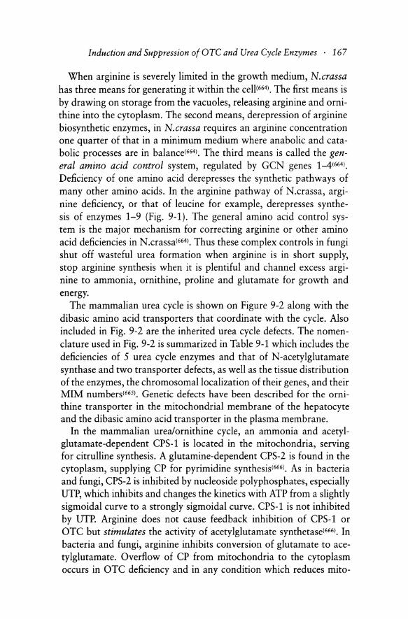
The mammalian urea cycle is shown on Figure 9-2 along with the dibasic amino acid transporters that coordinate with the cycle. Also included in Fig. 9-2 are the inherited urea cycle defects. The nomenclature used in Fig. 9-2 is summarized in Table 9-1 which includes the deficiencies of 5 urea cycle enzymes and that of N-acetylglutamate synthase and two transporter defects, as well as the tissue distribution of the enzymes, the chromosomal localization of their genes, and their MIM numbers(6651. Genetic defects have been described for the ornithine transporter in the mitochondrial membrane of the hepatocyte and the dibasic amino acid transporter in the plasma membrane.
In the mammalian urea/ornithine cycle, an ammonia and acetylglutamate-dependent CPS-1 is located in the mitochondria, serving for citrulline synthesis. A glutamine-dependent CPS-2 is found in the cytoplasm, supplying CP for pyrimidine synthesis(666). As in bacteria and fungi, CPS-2 is inhibited by nucleoside polyphosphates, especially UTP, which inhibits and changes the kinetics with ATP from a slightly sigmoidal curve to a strongly sigmoidal curve. CPS-1 is not inhibited by UTP. Arginine does not cause feedback inhibition of CPS-lor OTC but stimulates the activity of acetylglutamate synthetase(666). In bacteria and fungi, arginine inhibits conversion of glutamate to acetylglutamate. Overflow of CP from mitochondria to the cytoplasm occurs in OTC deficiency and in any condition which reduces mito-

~ •
..,o
Fig
ure
9-2.
Ure
a cy
cle
and
ass
ocia
ted
enzy
mes
in
th
e h
um
an li
ver
cell
, an
d t
heir
in
her
ited
de
fect
s(66
5).
Nu
mb
erin
g o
f th
e en
zym
es i
s sh
ow
n i
n T
able
9-1
. (C
opyr
ight
199
6.
Rep
rint
ed w
ith
perm
issi
on f
rom
Ox
ford
Uni
vers
ity
Pre
ss.)

Tab
le 9
-1.
No
men
clat
ure
for
Fig
ure
9-2
No
.
Ure
a cy
cle
enzy
mes
1 2 3 4 5 6
Dis
orde
r-af
fect
ed c
om
po
nen
t
Car
bam
yl
phos
phat
e sy
nthe
tase
I (
CP
S)
Orn
ith
ine
tran
scar
bam
ylas
e (O
TC
)
Arg
inin
osuc
cina
te s
ynth
etas
e (c
itru
llin
e mia
, C
ITR
) A
rgin
inos
ucci
nate
lya
se (
argi
nosu
ccin
ic
acid
uria
, A
SA)
Arg
inas
e (a
rgin
inem
ia,
AR
G)
N-A
cety
lglu
tam
ate
synt
hase
(N
AG
S)
Tra
nsp
ort
def
ects
of
diba
sic
amin
o ac
ids
7 L
ysin
uric
pro
tein
int
oler
ance
(L
PI)
(h
yper
diba
sic
amin
oaci
duri
a)
8 H
yp
ero
rnit
hin
emia
-hy
per
amm
on
emia
-ho
moc
itru
llin
uria
(H
HH
)
Tis
sue
dis
trib
uti
on
Liv
er,
gu
t (m
itoc
hond
ria)
L
iver
, gu
t, k
idn
ey
mit
och
on
dri
a)
Liv
er,
kid
ney
fib
robl
asts
(c
ytos
ol)
Liv
er,
kidn
ey f
ibro
blas
ts
RB
C,
bra
in (
cyto
sol)
L
iver
, ki
dney
, R
BC
(cy
toso
l)
Liv
er,
gut,
(m
itro
cho
nd
ria)
Liv
er,
gut,
kid
ney
(pl
asm
a m
emb
ran
e)
Liv
er,
fibr
obla
sts
( mit
och
on
dri
al
mem
bra
ne)
Ch
rom
oso
mal
lo
cali
zati
on
2p
X
p 2
1.1
9q
34
7cen
-q11
.2
6q
23
U
nk
no
wn
Un
kn
ow
n
Un
kn
ow
n
MIM
no
.
23
73
00
3
11
25
0
21
57
00
20
78
00
2
37
31
0
22
27
00
23
89
70
170 . Ornithine Transcarbamylase
chondrial ornithine levels, causing excessive synthesis of orotic acid and orotidine, as discussed in Chapter 8.
The major carry-over of a control mechanism for enzyme synthesis from the bacterial and fungal arginine synthetic pathways to mammalian hepatocytes is the derepression by arginine deficiency of CPS-l, OTC, argininosuccinate synthetase (AS) and lyase (AL). Schimke fed weanling male Osborne-Mendel rats a low arginine diet and "de-repressed" or induced these four enzymes and suppressed arginase levels(26). We confirmed the induction of the first four enzymes in Wistar male rats and partial responses in 3 other rat strains and in a mouse(27) but did not obtain a decrease in arginase by any baseline. This induction occurred even though liver concentrations of arg, om, citrulline and urea were slightly but not significantly lower on arginine-free diets(27). Synthesis of arginine from glutamine in the intestine/kidney axis(746) maintains arginine levels. In fungi the general amino acid control system turns on synthesis of arginine even though the cytosol level of arg is only slightly low: the postulated signal is the accumulation of uncharged tRNA(664).
The same phenomenon was shown more impressively by Schimke for AS and AL in HeLa cells and in two fibroblast lines in culture(28), where arginine concentrations in the growth media were widely varied. Arginase levels were suppressed by low arginine media and increased by high arginine media, as in fungi(28,664). Arginine-mediated repression of human AS was demonstrated in RPMI 2650 cells(25). Repression of AS occurred in these cells when cultured in citrullinecontaining medium lacking arginine(668). Morever expression of a transfected minigene containing the 5'-flanking region of AS was increased by leucine starvation, due to increased rates of transcription. However, AS enzyme activity only increased 2-fold in 72h of arginine starvation and required 3 weeks to show maximal derepression(667). This leucine effect resembles that of the general amino acid control system in fungi(664) where starvation for leucine derepresses its synthetic pathway and that of other unrelated amino acids, here the arginine pathway. Bruhat et al,(668,669) have found that leucine deprivation and that of arginine and other amino acids in HeLa cells and in HepG2 cells (an hepatocyte-derived tumor cell line) induces AS and a CCAATIenhancer-binding protein-related gene (CHOP), because of a cis-positive element in the promoter region of the human CHOP gene. This element contains a minimal core sequence 5'-

Induction and Suppression of aTC and Urea Cycle Enzymes . 171
ATIGCATCA-3' which has been named an amino acid response element (AARE). AARE binds CHOP and activating transcription factor 2 (ATF 2). CHOP protein-CIEBP heterodimers playa role in regulation of stress-induced cell responses concerning energy metabolism, cell proliferation and expression of cell type-specific genes(6691. Urea cycle enzyme genes eventually may be shown to be derepressed by CHOP-CIEBP binding to elements of their promoters when arginine starvation occurs. However, we could not identify the core AARE sequence on the promoter region out to -578bp of the hOTC gene(lOI or -800bp of the mouse gene(151 or -1011bp of the rat OTC gene(161.
We were unable to obtain any de-repression-like responses to media lacking arginine with rat liver cells cultured in monolayer for 72h or in a cultured hepatoma line maintaining all five urea cycle enzymes(271. The arg content of cells cultured in Minimal Essential Medium (MEM) without arginine was undetectable. These cells could still be induced by glucagon and dexamethasone as well as cells in argcontaining MEM. Perhaps 72h was too short a culture time to see derepression of urea cycle activities in arg-free MEM(6671.
The amounts of the 5 mammalian urea cycle enzymes in liver are related to the level of protein intake. In 1962, Schimke showed in rats that the activities of aIlS enzymes were proportional to the level of dietary casein intake(3411 and that the amounts of OTC and arginase were increased as well. The induction by the 60% casein diet peaked at 8 days for OTC and arginase. Addition of anyone of the intermediates of the pathway (ornithine, citrulline, arginine or urea) to a low casein diet did not alter enzyme levels(261. No induction occurred when the amounts of the following amino acids found in a 60% casein diet were added singly to a 15% casein diet: histidine, tryptophan, aspartate, methionine, alanine, threonine, lysine and leucine. Degradation as well as enzyme synthesis played a role in arginase levels(67ol. Using 14C-guanidinoarginine that is not re-utilized, the transition from a high to a low protein diet resulted in suppression of synthesis and a marked increase in arginase degradation until a new steady state was reached. During starvation, which is equivalent to a high protein diet because endogenous proteins are utilized, degradation ceased and synthesis continued at a constant rate until a higher steady state of arginase protein was achieved(6711. The half-life of arginase was 4-5 days by this method. It is remarkable that during starvation the urea cycle enzymes are increased in amount while the bulk of hepatocyte
172 . Ornithine Transcarbamylase
proteins are degraded, resulting in a smaller liver per body weight and a lowered total protein concentration(671).
We found that we could induce all 5 urea cycle enzymes in 48h when male Sprague-Dawley rats stabilized on a 15% casein diet were tube-fed casein hydrolysate three times a day at a dose of 315 mgN (2 gNlkg/day)(342). OTC increased l.4-fold expressed as units/liver per lOOg of rat; the increments of the other enzymes were CPS 1.5-, AS 1.9-, AL 1. 7- and arginase 1.3-fold. The casein feedings also increased liver weight per lOOg of rat and liver protein concentration. In enzymes like arginase where the amount of enzyme protein in liver is large and the rate of degradation on an adequate (15%) protein intake is slow, even a 4-fold increase in synthesis rate may lead to what appears to be small increases in activity per liver(670). We believe this same interpretation explains the small increases in OTe.
This two-day tube-feeding regimen was then applied to rats fed a 15% casein diet by giving them maximally tolerated doses of individual amino acids, avoiding toxicity(342). Alanine (2gN/kg), glycine (2gN/kg), methionine (O.2gN/kg) and cysteine (O.4gNlkg) each increased all 5 enzymes. A mixture of 8mgN of met, 12mgN of ala and 16mgN of gly, the amount of these amino acids present in 315mgN of casein hydrolysate, induced as well as the mixed amino acids of this dose of casein. Arginase increases were as great by immunoprecipitable protein as by activity. OTC was the least inducible of the five enzymes, ranging from 1.2-fold by cys to loS-fold by met. Tube feedings of pyruvate (85mmoles/kg) were compared with the same sized dose of ala (1.2gNlkg) but only ala induced all 5 enzymes (OTC lA-fold). The a-OH-acid of methionine (17 mmoles/kg) was compared with an equal dose of met but only the amino acid induced all 5 enzymes (OTC 1.3-fold). Thus the inducing effect of amino acids requires the presence of the a-amino group. Our success with induction by amino acids that did not induce in Schimke's study we attribute to our higher doses, for example, alanine(670). The explanation for the induction by ala, gly, met and cys plus some contributions by other amino acids(342) is not well understood but implies that there are positive amino acid response elements in the promoter regions of these five urea cycle enzyme genes, on five different chromosomes. One of these elements may be the urea cycle response element found in the 5' -region of OTC, AS, arginase and ornithine transaminase(17,18) which may control coordinate induction by dietary protein.
Induction and Suppression of aTC and Urea Cycle Enzymes . 173
Pitot and Peraino found that tube-feeding casein hydrolysate to rats induced hepatic threonine dehydratase and ornithine transaminase in 24h (the method we adopted)16721. When glucose was fed along with the casein the induction was blocked totallyl6731. Was this a carry-over of glucose repression in bacteria into mammals? We looked for this phenomenon in the urea cycle by stabilizing rats on an 8% casein diet, inducing all five enzymes with casein hydrolysate (3.9g/day) and comparing the responses to those when 3.9g/day of glucose was fed along with the casein. CPS, OTC and arginase induced as well as with casein alone but AS was suppressed by 20% and AL by 21 %, a barely significant non-coordinate suppressionl6741. During glucose repression of casein induction of threonine dehydratase, hepatocyte cAMP levels were not reduced as occurs in glucose repression in bacteriaI675,6761. Glucose fed to starved rats elevated serum insulin while suppressing glucagon, and hepatic cAMP levels were markedly reducedI6771. When casein hydrolysate was fed, both insulin and glucagon levels rose but the cAMP level in liver fell 25 % and returned to baseline.
To test whether ammonium ion is an inducer, we stabilized rats on a 27% casein diet and tube-fed ammonium citrate (2.2gN/kg), a maximum tolerated dose, three times daily for 8 days13421. Only AS increased, 1.2-fold. The urea excretion was the same as that of rats fed casein hydrolysate (2gN/kg) that induced all 5 enzymes. Thus processing ammonia through the cycle was not the signal for urea cycle induction.
A major evolutionary addition to the induction and suppression of animal enzymes is the role of hormones. We tested glucagon as an inducer of the 5 urea cycle enzymes because pharmacologic doses of soluble glucagon given to rats had been shown to increase CPS-1, AS and AL but not OTC or arginaseI6781. This report also showed that alloxan diabetes increased all five enzymes, and that insulin (4 units/day of protamine Zn insulin) did not affect any enzyme level in normal rats and did not restore enzyme levels to normal in diabetic rats. We used Zn glucagon because it has a prolonged action when given subcutaneously. When we gave pharmacologic doses of Zn glucagon to male rats, in 48h we increased CPS 2-fold, OTC 1.3-fold, AS 2.7-fold, AL 3.2-fold and arginase 2.2-foldI6791. Physiologic doses by an osmotic pump for 7 days induced aIlS enzymes: CPS 1.5-fold, OTC 1.2-fold, AS 2.7-fold, AL 1.8-fold and arginase 1.6-fold. Actinomycin-D or puromycin or cycloheximide blocked these increases, indicating that

174 . Ornithine Transcarbamylase
RNA and protein synthesis are necessary for the induction. Tubefeeding casein hydrolysate for 2 days induced the entire cycle while causing an increase in plasma glucagon equal.):o that of our physiologic dose of glucagons(679).
Schimke reported that adrenalectomy reduced all 5 urea cycle enzymes(26) and McLean and Gurney confirmed this(680). We found that adrenalectomy reduced all enzymes but AL when urea cycle activities were expressed as unitslliver per 100g of rat(343), a baseline which adjusts for changes in liver and body weight. In this report we gave continual high dose glucagon (0.3 mg/kg/day) to intact rats for 7 days and induced all enzymes but OTe. The only enzyme induced by glucagon in adrenalectomized rats was AL. Physiologic doses of dexamethasone induced arginase alone in intact and adrenalectomized rats. The combination of the two hormones induced all 5 enzymes synergistically in intact rats but the induction was additive or less than additive in adrenalectomy rats(343). What role other hormones might play is not clear. Thyroidectomy increased and excess thyroid hormone decreased OTC, CPS, AS and AL but arginase was decreased in both conditions(681).
We did not obtain a good correlation between portal vein plasma glucagon levels after tube-feeding various amino acids and their ability to induce the urea cycle, so we did not conclude that protein feeding induced the cycle only by release of glucagons(674). Eisenstein(682) found that high protein feedings elevated portal vein plasma glucagon levels but there was no correlation between glucagon and amino acid concentrations in the portal vein. He also studied the release of glucagon when rat islets were perifused with various amino acids: arginine and glutamine and less so ornithine were the major releasors(683). Ala, cys, gly and met, the amino acids we found induced the cycle(342), did not release glucagon well. In dogs, plasma glucagon levels were measured after amino acid infusions(684). The potencies of releasors were as follows: asn > gly > phe > ser > asp> cys > try> ala> glu > thr > gIn > arg > om > pro > met > lys > his. Val, leu and ile failed to stimulate glucagon release(684). Thus the induction of urea cycle enzymes by amino acids or by glucagon in whole rats seems to be by separate mechanisms and is not simply caused by amino acids releasing glucagon. To illustrate the complex interactions, dexamethasone injection in humans caused basal plasma glucagon and alanine

Induction and Suppression of OTC and Urea Cycle Enzymes . 175
levels to rise and alanine infusions or protein ingestion caused greater glucagon responses(685).
Mori et al,(686) first showed that the liver translatable mRNA levels in rats fed a 5% casein diet increased 4.2-fold for CPS and 2.2-fold for OTC when the rats were fed a 60% casein diet for 8 days. The mRNA increases were greater than the enzyme activity increases. Starvation for 7 days increased CPS and OTC activities but decreased mRNA levels by 40-50% of control animals, suggesting a decreased rate of protein degradation. Morris et al,(687) further showed that hybridizable levels of mRNA for all 5 enzymes in rat liver were proportional to dietary protein levels: OTC mRNA increased 4.4-fold 6 days after a dietary increase from a 27% to a 60% casein diet. Contrary to Mori(686), Morris reported a marked increase of mRNAs for all 5 enzymes after 5 days of starvation(687).
Controlled conditions are difficult to maintain in studies of whole animals. Results of enzyme or mRNA changes may be due to indirect effects of dietary changes(343,678-681,686-687) and various hormonal releases by amino acids add to the interpretive difficulties(682-685). We therefore developed a method to culture normal adult rat liver cells which maintained urea cycle enzymes in chemically-defined, serumfree medium for at least 7 days, using calf skin collagen to coat the plates(688). When the hepatocytes were cultured in 10% fetal calf serum during the first 21h the polygonal cells formed a monolayer which then stayed well attached in serum-free medium for 4d, long enough to do urea cycle induction studies. Adenylyl cyclase activities were stable and inducible for 72h. We used this cell culture system successfully from 1975 to 1993.
Gebhardt and Mecke used rat liver cells cultured in perifused mono layers and showed that l,uM glucagon or 20,uM dibutyrylcAMP induced all urea cycle enzymes but OT(689). Dexamethasone (O.l,uM) induced only AS and arginase. The combination of glucagon and dexamethasone induced all five enzymes in 24h in a manner called a permissive effect of dexamethasone, OTC increasing least at 1.3-fold(689) .
In our monolayer culture system we added bacitracin to protect glucagon from degradation and were able to increase cAMP levels for 7h or more after adding glucagon(690). We used physiologic concentrations of glucagon (lnM) added 4 times in 48h and induced only CPS-
176 . Ornithine Transcarbamylase
1. When 10nM dexamethasone was added twice in 48h to the glucagon regimen, all five enzymes were induced: CPS loS-fold, OTC 1.2-fold, AS 1.9-fold, AL 1.8-fold and arginase 1.7 -fold. Dexamethasone alone induced only AS and AL. Amounts of immunoprecipitable AL and arginase were increased as much as their activities. Glucagon induction was not affected by added insulin. Other hormones that did not affect any urea cycle activity with or without dexamethasone included epinephrine, pentagastrin, secretin, cholecystokinin octapeptide, dimethyl prostaglandin E2, triiodothyronine, estradiol and growth hormone(690). We concluded that glucagon, via cAMP, supported by a permissive role of glucocorticoids, is the only identified coordinate hormonal inducer of all five urea cycle enzymes.
Morris extended our understanding of hormonal induction by showing that intraperitoneal injection of dibutyryl cAMP (dbcAMP) in whole rats increased 4 urea cycle mRNAs at Sh but not that of OT0687). Transcriptional run-on assays showed 4-S-fold increases in CPS amd AS in 30 min. In cultured rat hepatocytes Nebes and Morris found that in 16h CPS, AL and arginase mRNAs increased during exposure to 8-(4-chlorophenylthio)cAMP (CPT-cAMP)(691). OTC mRNA did not respond. Dexamethasone increased CPS, AL and arginase mRNAs more slowly than with CPT-cAMP. Dexamethasone but not CPT-cAMP inductions were blocked by cycloheximide, suggesting that dexamethasone induced another gene product which led to urea cycle mRNA induction. The combination of both hormones gave synergistic mRNA responses for CPS, AS and arginase, additive responses for AL and no response for OTe. The hepatic mitochondrial ornithine/citrulline transporter was also induced by increased dietary protein(692). Moreover its mRNA was induced to a maximum in 12h by CPT-cAMP and in 4h by dexamethasone and the combination was synergistic, rising 34-fold after 16h of treatment.
Our own studies on mRNA induction by glucagon and dexamethasone demonstrated that the coordinate induction is accomplished by three different mechanisms(693). The transcription rate for arginase mRNA increased 9-fold in 7h, the mRNA 90-fold in 28h and the activity loS-fold at 48h when l,uM glucagon and 10nM dexamethasone were added to cultured rat liver cells. Arginase mRNA induction was minimal with either hormone alone. Cycloheximide pretreatment did not prevent the rise in mRNA levels with both hormones. CPS-l mRNA levels also responded synergistically to both hormones, peak-

Induction and Suppression of OTC and Urea Cycle Enzymes . 177
ing 240-fold above controls at 24h although activity only increased 1.4-fold at 48h. No transcriptional assays were done for CPS-I, but we assumed that transcription rate increases would not approach the 240-fold increase in mRNA levels. We interpreted these arginase responses to result from stabilization of mRNA by the two hormones because the steady-state mRNA increase was so much greater than the increase in transcription rate. AL and AS mRNAs were not induced by single hormones but responded to the combination by a 10-fold increase in AL and a 7-fold rise in AS transcription rates at 7h. AL mRNA peaked at 28h 7-fold above controls while AS mRNA peaked at S2h 14-fold above preinduction levels. AL enzyme activity increased 2.8-fold at 48h and AS 1.8-fold. We interpreted the induction mechanism for AS and AL as transcriptional control primarily. aTC mRNA levels were not increased by the combined hormones yet the activity increased 1.3-fold. We interpreted this result as consistent with aTC protein stabilization(693).
Matsuno et al.(694) found that dexamethasone and glucagon rapidly increased the transcription factor CCAAT/enhancer binding protein /3 (C/EBP/3) mRNA synergistically. The transcription rate and the DNAbinding activity of C/EBP/3 were induced as much as the mRNA. CI EBPa mRNA did not respond. CIEBP family members are found in the CPS-I promoter, in the arginase promoter and enhancer and in the aTC enhancer where they work in combination with hepatocyte nuclear factor-a (HNFa)(694). These factors may playa role in secondary glucocorticoid responses where the time course of induction is delayed and is sensitive to protein synthesis inhibitors(691).
Kimura et al.(698) made CIEBPa knockout mice and found decreased levels of mRNA for CPS, AS, AL and arginase in newborn or S-7h mice while aTC mRNA was variably low. Protein levels by Western blots were low for the same four enzymes whereas aTC was reduced by 60% and its lobular distribution was shifted to the midlobular from the usual peri-portal region. Kimura further showed that CIEBP/3 knockout mice still showed a normal basal level of mRNA and protein for aIlS enzymes(696). When glucagon and dexamethasone were given intra peritoneally to wild-type mice, the liver CPS, AS, AL and arginase mRNAs were induced normally but aTC did not increase. In cultured mouse hepatocytes the wild-type urea cycle responses to the combined hormones were lost. aTC showed no induction in wild-type or C/EBP/3 deficient cells(696). Apparently the

178 . Ornithine Transcarbamylase
deficiency of CIEBP,8 was compensated for in vivo by other transcription factors.
One new finding for gene regulation of urea cycle enzymes was that induction of CPS-l and AS mRNAs by dexamethasone was blocked by long-chain fatty acids, arachidonic acid being the most potent(697). No inhibition of induction by dbcAMP was seen with oleic acid. This finding may help explain the suppression of urea cycle enzyme activities and the hyperammonemia in hereditary carnitine-deficient JVS mice whose serum free fatty acid levels are very high(697).
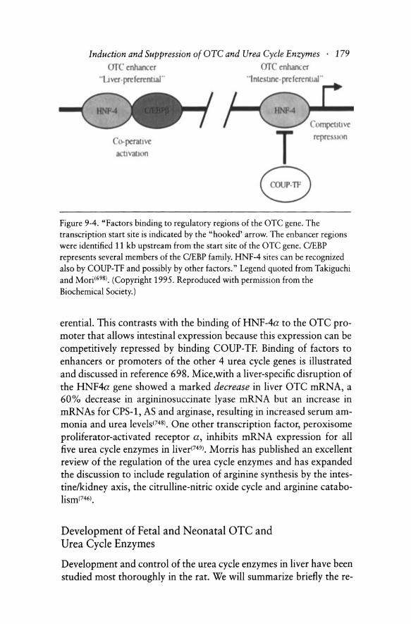
Based on the findings reported in references 20-22 concerning factors which bind to regulatory regions of the OTC gene and from expression of OTC in liver vs. intestine in transgenic mice(346-348), Takiguchi and Mori have constructed models of the OTC enhancer and promoter binding of the liver selective factors C/EBP and HNF-4a (Figure 9-3 )(698). They have illustrated in Figure 9-4 how these factors cooperate in the OTC enhancer to make transcription liver-pref-
140
arc
+JO
Figure 9-3. "Role and regulation of the OTC promoter and enhancer in tissueselective transcription. In transgenic mice the OTC promoter exhibits higher activity in the small intestine than in the liver; the enhancer inverts this tissueselectivity of the promoter, bringing about higher expression in the liver. The OTC promoter is activated by HNF-4, a liver- and small-intestine selective member of the steroid receptor superfamily and is competitively repressed by COUP-TF, a ubiquitous member of this family. For activation of the enhancer, the two liver-selective factors HNF-4 and OEBPP are required and neither alone is sufficient. This appears to enable these liver-selective, but not strictly liverspecific, factors to confer more restricted liver-specificity on expression of the target OTC gene." Legend quoted from Takiguchi and Mori(698).(Copyright 1995. Reproduced with permission from the Biochemical Society.)
Induction and Suppression of aTC and Urea Cycle Enzymes . 179 (Jf • enhancer enhancer
.. YCt. pre: f c:mIUaI" "1 nl.elUllC:-pre ferenc I.Ir
T 8
100
Figure 9-4. "Factors binding to regulatory regions of the OTC gene. The transcription start site is indicated by the "hooked' arrow. The enbancer regions were identified 11 kb upstream from the start site of the OTC gene. ClEBP represents several members of the ClEBP family. HNF-4 sites can be recognized also by COUP-TF and possibly by other factors." Legend quoted from Takiguchi and Mori16981. (Copyright 1995. Reproduced with permission from the Biochemical Society.)
erential. This contrasts with the binding of HNF-4a to the OTC promoter that allows intestinal expression because this expression can be competitively repressed by binding COUP-TF. Binding of factors to enhancers or promoters of the other 4 urea cycle genes is illustrated and discussed in reference 698. Mice.with a liver-specific disruption of the HNF4a gene showed a marked decrease in liver OTC mRNA, a 60% decrease in argininosuccinate lyase mRNA but an increase in mRNAs for CPS-1, AS and arginase, resulting in increased serum ammonia and urea levels17481. One other transcription factor, peroxisome proliferator-activated receptor a, inhibits mRNA expression for all five urea cycle enzymes in liver(7491. Morris has published an excellent review of the regulation of the urea cycle enzymes and has expanded the discussion to include regulation of arginine synthesis by the intestine/kidney axis, the citrulline-nitric oxide cycle and arginine catabolism17461.
Development of Fetal and Neonatal OTC and Urea Cycle Enzymes
Development and control of the urea cycle enzymes in liver have been studied most thoroughly in the rat. We will summarize briefly the re-

180 . Ornithine Transcarbamylase
sults of many reports, including what is known about the mouse and human urea cycle development.
OTC activity or protein becomes detectable in the rat fetus on the fourth day before birth (-Sd) or on day 17 of gestation, which is 21.5-22.5 days in the rat(700). OTC activity rises rapidly to a peak at 4d, then declines and rises slowly to a plateau from + 20 to + 35 days(700), equal to adult levels(699,702). During days -4 to birth, the mass of liver hepatocytes doubles while the hematopoietic cells recede(703). From -8 to -5 days of fetal life only 18 % of liver consists of hepatocytes. The OTC gene is substantially methylated at the 5' -end of the gene at -lSd and demethylation of one OTC allele occurs in liver but not kidney in the next few days(703). Thus the rise in activity is due to proliferation of hepatocytes, hypo-methylation of the 5' -end of OTC and new OTC synthesis. OTC mRNA in the rat liver becomes detectable at day -7, at 30% of adult levels(703). The mRNA rises steadily to birth, when a brief drop occurs and then rises steadily to adult levels by day + 7. This pattern is found similarly in CPS-l and arginase(703), the so-called late fetal type of maturation.
Hormonal changes in the fetus seem to playa role in this early fetal enzyme development. Corticosteroids are elevated at day -1, rise to a peak at - 2d to birth, then fall and rise again from +9 to + 16d(701). Insulin levels in the fetus are high days -2 to birth when they decrease to low levels and rise again after weaning. Glucagon levels are low before birth, rise sharply at birth, and then decline to low levels. Cyclic AMP levels follow the glucagon pattern(701). OTC, CPS and arginase are increased at + 13d by exogenous prednisolone, by glucagon and by the combination in a less than additive manner(701). AS mRNA was detected at 15.5 days (-7d) in the fetal rat liver and increased steadily to day +2, along with AS activity, due to an increased transcription rate(706). In cultured fetal hepatocytes dexamethasone increased AS mRNA, glucagon enhanced the dexamethasone effect and insulin suppressed it(706). AS activities in the rat liver were not reported anteor post-natally.
In the spiny mouse the activities of OTC, CPS and arginase activities increased from -Sd steadily to adult levels at +3Sd(702). Morris(704) found in Swiss-Webster mice that the mRNA levels for all 5 urea cycle enzymes were present at 2-14% of adult liver levels at day -6. OTC mRNA increased rapidly at days + 1 to +4. AL mRNA rose from -2d to + 2d. CPS, AS and arginase mRNAs increased rapidly from -4d to - 2d and then declined slightly.

Induction and Suppression of OTC and Urea Cycle Enzymes . 181
Human OTC activity and that of the other urea cycle enzymes were detected at 8 weeks of gestation, increased steadily to birth and up to 8 weeks of age(707). More extensive studies of human fetal livers from 13 to 36 weeks of gestation(705) showed that all five urea cycle activities were present at 13-16 weeks. OTC was then at 22% of adult liver levels, arginase at 28%, AL at 40%, CPS at 52% and AS at 55% of adult levels. By term human liver activities of all five enzymes were 65-90% of those in adult human liver.
Commentary. The human urea/ornithine cycle is an evolutionary descendant of the arginine synthetic and degradative pathways of fungi, and distantly of the arginine biosynthetic pathway of bacteria. The amounts of OTC protein, of CPS-1, AS, AL and arginase proteins, of N-acetylglutamate synthetase protein and of the ornithine/ citrulline transporter in liver cells usually increase or decrease in a coordinated manner. Two gene control mechanisms for OTC and other urea cycle enzymes in fungi have carried over into mammals, namely arginine-deficiency derepression and general amino acid control. The amounts of OTC and other urea cycle proteins adapt to the level of protein intake, increase with starvation and also increase with secretion of glucagon and glucocorticoids. Coordinate induction of the urea cycle by glucagon and dexamethasone is accomplished by three mechanisms: in CPS-1 and arginase by stabilization of mRNAs, in AS and AL by increased transcription rates and in OTC by protein stabilization. The transcription factor, HNF4a, is essential for OTC gene expression in liver while another factor, peroxisome proliferator-activated receptor a, inhibits mRNA expression of all five urea cycle enzymes. Development of OTC and the other urea cycle enzymes in fetal and post-natal livers is complex and modulated by glucagon, corticosteroids and insulin.