induction of akt phosphorylation in rat primary astrocytes by h2o2 occurs upstream of...
TRANSCRIPT
3
dscdwA3cl
(
Archives of Biochemistry and BiophysicsVol. 386, No. 2, February 15, pp. 275–280, 2001doi:10.1006/abbi.2000.2202, available online at http://www.idealibrary.com on
Induction of Akt Phosphorylation in Rat Primary Astrocytesby H2O2 Occurs Upstream of Phosphatidylinositol
-Kinase: No Evidence for Oxidative Inhibition of PTEN
Scott Salsman,*,† Nicole Felts,† Quentin N. Pye,† Robert A. Floyd,*,† and Kenneth Hensley†,1
*Department of Biochemistry and Molecular Biology, Oklahoma University Health Sciences Center, Oklahoma City,Oklahoma 73104; and †Free Radical Biology and Aging Program, Oklahoma Medical Research Foundation,Oklahoma City, Oklahoma 73104
Received October 18, 2000; published online January 11, 2001
lp(srsa
Phosphorylation of the serine/threonine kinase Akthas previously been shown to be increased by treat-ment of cells with H2O2; the target of H2O2 has not beenclearly identified. Here we show that treatment of ratprimary astrocytes with H2O2 resulted in increasedAkt phosphorylation that was blocked by wortmannin.The thiol-reducing agent N-acetylcysteine had only aslight inhibitory effect. Treatment with rotenone orantimycin A also resulted in increased wortmannin-sensitive Akt phosphorylation, probably by increasingintracellular H2O2 generation by blocking mitochon-
rial electron transport. Addition of phosphatidylino-itol 3,4-bisphosphate to cells also resulted in an in-rease in Akt phosphorylation. This increase was ad-itive to that induced by H2O2 and was also blocked byortmannin. These results suggest that activation ofkt by H2O2 occurs upstream of phosphatidylinositol-kinase (PI 3-K) activity in astrocytes. The data indi-ate that major oxidative effects do not occur at theevel of the PI 3-K-antagonizing phosphatase PTEN.
© 2001 Academic Press
Key Words: cell signaling; H2O2; PI 3-K; PTEN; Akt;EGF; astrocytes.
Reactive oxygen species (ROS)2 such as superoxideO2
•2) and hydrogen peroxide (H2O2) have been specu-
1 To whom correspondence should be addressed. Fax: (405) 271-1795. E-mail: [email protected].
2 Abbreviations used: ROS, reactive oxygen species; EGF, epider-mal growth factor; EGF-R, epidermal growth factor receptor; PI 3-K,phosphatidylinositol 3-kinase; PDK, phosphoinositide-dependent ki-nase; PBN, phenyl-tert-butylnitrone; PIP2, phosphatidylinositol-3,4-
bisphosphate; NAC, N-acetylcysteine; FCS, fetal calf serum; PBN,phenyl-tert-butylnitrone.0003-9861/01 $35.00Copyright © 2001 by Academic PressAll rights of reproduction in any form reserved.
ated to be second messengers in cellular signalingathways, e.g., by regulating tyrosine phosphorylation1, 2). Growth promotion by oxidants has been ob-erved in mammalian cells and is expected to play aole in tumorigenesis (3–5). Growth factor receptorsuch as the epidermal growth factor receptor (EGF-R)re thought to be targets of ROS (6). For instance, H2O2
induces an increase in tyrosine phosphorylation ofEGF-R in the absence of EGF (6–9). This presumablyleads to increased cell growth through pathways nor-mally regulated by EGF.
Akt (protein kinase B), a target of phosphatidylino-sitol 3-kinase (PI 3-K) activity, is a serine/threoninekinase activated in response to specific growth factors(10). Downstream targets of Akt include BAD (11, 12),caspase 9 (13), glycogen synthase kinase-3 (14), 6-phos-phofructo-2-kinase (15, 16), 4E-BP1 (17), GLUT4 (18),and p70 S6 kinase (19). Phosphorylation of proapop-totic factors BAD and caspase 9, which causes inhibi-tion of these proteins, promotes cell survival by de-creasing tendencies toward apoptosis. Unchecked acti-vation of Akt may therefore lead to tumor formation,and indeed tumor cells of many tissue types have beenshown to contain mutated forms of a PI 3-phosphatase,PTEN, which suppresses tumors by regulating Aktactivation (20).
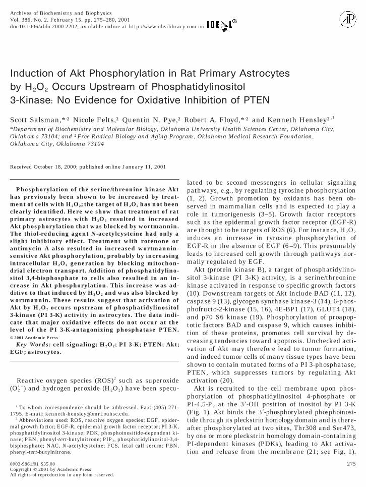
Akt is recruited to the cell membrane upon phos-phorylation of phosphatidylinositol 4-phosphate orPI-4,5-P2 at the 39-OH position of inositol by PI 3-K(Fig. 1). Akt binds the 39-phosphorylated phosphoinosi-tide through its pleckstrin homology domain and is there-after phosphorylated at two sites, Thr308 and Ser473,by one or more pleckstrin homology domain-containingPI-dependent kinases (PDKs), leading to Akt activa-
tion and release from the membrane (21; see Fig. 1).275
md
pm1m
tScft(
276 SALSMAN ET AL.
Wortmannin blocks Akt phosphorylation by inhibitingPI 3-K.
H2O2 has been shown to increase Akt activity inCOS-7 cells (22) and to increase Akt phosphorylationand activation in a wortmannin-sensitive fashion invascular smooth muscle cells (23). We present evidencethat wortmannin-sensitive Akt phosphorylation atSer473 in rat primary astrocytes was induced by H2O2
and by agents known to increase H2O2 generation byitochondria. Induction of Akt phosphorylation by ad-
ition of PI-3,4-P2 to the cell medium was additive tothe effect of H2O2 and was also inhibitable by wortman-nin, suggesting that Akt activation by H2O2 occursupstream of PI 3-K activity, presumably at the level ofreceptors such as EGF-R.
MATERIALS AND METHODS
Materials. DMEM/F12 medium and fetal calf serum (FCS) werepurchased from Mediatech CellGro (Herndon, VA). Epidermalgrowth factor (EGF) was purchased from R&D Systems (Minneapo-lis, MN). Wortmannin was purchased from Alexis (San Diego, CA).Phenyl-tert-butylnitrone (PBN) was synthesized at the OklahomaMedical Research Foundation. Phosphatidylinositol 3,4-bisphos-phate (PIP2) was purchased from Matreya, Inc. (Pleasant Gap, PA).Rabbit polyclonal anti-Akt and anti-phosphoAkt (Ser473) antibodieswere purchased from New England Biolabs (Beverly, MA). Rabbitpolyclonal anti-rat EGF-R antibody was purchased from Calbiochem(San Diego, CA). All other reagents were purchased from Sigma (St.Louis, MO) unless otherwise indicated.
Cell culture and treatments. Rat primary astrocytes were grownin monolayer culture in DMEM/F12 medium supplemented with10% FCS as described (24). Cells were seeded into 24-well plates inmedium supplemented with 5% FCS. Cells were incubated withEGF, H2O2, rotenone, or antimycin A for the indicated amounts oftime: wortmannin, PBN, or N-acetylcysteine (NAC) for 1 h or PIP2
for 30 min at 37°C in an atmosphere of 95% O2/5% CO2. PIP2 wasprepared by solubilization of synthetic PIP2 according to the manu-facturer’s instructions. Solubilized PIP2 was vacuum-dried and sus-pended in distilled water by sonication.
Western blot analysis. Cells were frozen, lysed in lysis buffer (10mM sodium acetate, 0.1% Triton X-100, 1:1000 mammalian proteaseinhibitor cocktail, 1 mM sodium orthovanadate, and 5 mM b-glycerol
hosphate), and scraped. Protein was normalized according to theethod of Lowry et al. (25). Lysates were subjected to SDS–PAGE on
FIG. 1. The EGF-R/PI 3-K/Akt pathway and its regulation byPTEN. See text for details. •, phosphate; —‹, activation/kinase ac-tivity; - - -‹, recruitment; – z– z‹, phosphatase activity; —u, inhibition.
2% polyacrylamide gels and transferred to polyvinylidene difluorideembranes. Membranes were incubated overnight at 4°C with agi-
ation with antibody raised against Akt, Akt phosphorylated ater473, or EGF-R. Bound antibody was detected with a peroxidase-onjugated secondary antibody and chemiluminescence (ECL1 kitrom Amersham Pharmacia Biotech, Buckinghamshire, UK). Quan-itative band density analysis was performed using GelExpertNucleoTech, San Mateo, CA).
RESULTS
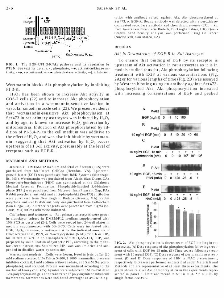
Akt Is Downstream of EGF-R in Rat Astrocytes
To ensure that binding of EGF by its receptor isupstream of Akt activation in rat astrocytes as it is inmodels studied thus far, Akt phosphorylation followingtreatment with EGF at various concentrations (Fig.2A) or for various lengths of time (Fig. 2B) was assayedby Western blotting using an antibody against Ser473-phosphorylated Akt. Akt phosphorylation increasedwith increasing concentrations of EGF and peaked
FIG. 2. Akt phosphorylation is downstream of EGF binding in ratastrocytes. (A) Dose response of Akt phosphorylation following treat-ment of cells with EGF for 15 min. (B) Time course following treat-ment with 10 ng/ml EGF. (C) Dose response of wortmannin pretreat-ment. (D and E) Dose responses of PBN or NAC pretreatment,respectively. Blots were performed as described under Materials andMethods and are representative of at least three experiments. Thegraph shows relative Akt phosphorylation in the experiments repre-
sented in panel E. Data are means 6 SE; n 5 3. *P , 0.05 bysingle-factor ANOVA.
bBc
tntMee
277PEROXIDE EFFECTS ON Akt
;5–10 min after treatment with 10 ng/ml EGF. TotalAkt content was not affected by EGF treatment asdetermined by Western blotting using an antibodyagainst an Akt epitope distinct from the sites of phos-phorylation (data not shown). EGF-induced Akt phos-phorylation was blocked by pretreatment of cells withless than 100 nM wortmannin (Fig. 2C). Akt phosphor-ylation was not affected by pretreatment with the ni-trone-based free radical trap PBN (Fig. 2D) but wasattenuated significantly by pretreatment with NAC, athiol-reducing agent and antioxidant (Fig. 2E). EGF-Rexpression was not affected by EGF as shown by West-ern blot with an anti-EGF-R antibody (data notshown).
Akt Phosphorylation Is Increased by H2O2
Astrocytes were treated with various concentrationsof H2O2 for various lengths of time followed by Westernlotting analysis of Akt phosphorylation (Figs. 3A and). The greatest amount of Akt phosphorylation oc-
urred ;15–30 min following treatment with 100 mMH2O2. Total Akt levels were not affected (data notshown). Phosphorylation of Akt induced by H2O2 wasblocked by pretreatment of cells with 10 nM wortman-nin (Fig. 3C). Akt phosphorylation was attenuatedslightly by NAC but not by PBN (Figs. 3D and E).Levels of phosphorylated Akt in control samplestended to vary from one experiment to another; thereason for this variation is not clear. The same trendsof activation and attenuation were seen in each exper-iment despite the variation in control levels. EGF-Rexpression was not affected by H2O2 (data not shown).
Although H2O2 diffuses across membranes, cells mayreact differently to H2O2 introduced exogenously thanto H2O2 generated intracellularly. For example, H2O2
in the cell medium may induce modification of theextracellular portion of EGF-R or other receptors lead-ing to intracellular effects that differ from those occur-ring in response to intracellular H2O2. To assay for Aktphosphorylation resulting from endogenous H2O2 gen-eration, Western blotting for phosphorylated Akt wasperformed following treatment of astrocytes for variouslengths of time with rotenone or antimycin A, both ofwhich are inhibitors of electron transport in the innermitochondrial membrane (26). Rotenone blocks elec-tron flow from NADH to ubiquinone by inhibitingNADH–ubiquinone reductase (complex I of the mito-chondrial electron transport chain). Antimycin Ablocks electron flow from cytochrome b 560 to ubiquinoneor ubisemiquinone in cytochrome reductase (complexIII). Inhibition of electron transport by either of thesemechanisms results in an increase in O2
•2 generationdue to incomplete reduction of O2 (i.e., one-electronreduction instead of four-electron reduction to H O
2that occurs as a result of unhindered electron flow; Ref.
26). Superoxide dismutates spontaneously or enzymat-ically via superoxide dismutase into H2O2.
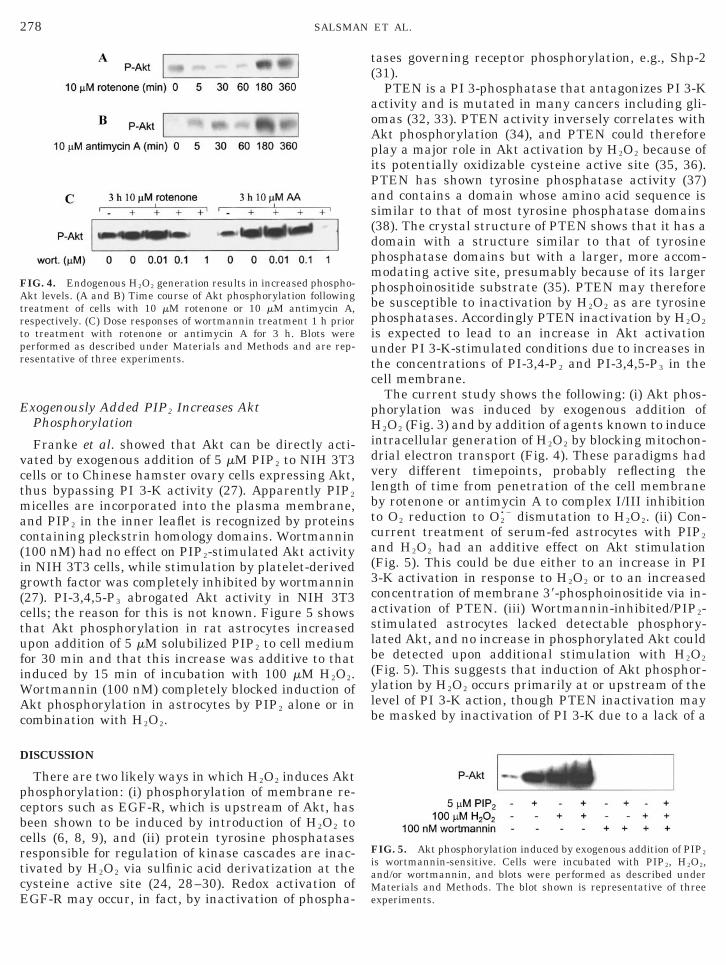
Treatment of astrocytes with 10 mM rotenone (Fig.4A) or 10 mM antimycin A (Fig. 4B) resulted in anincrease in Akt phosphorylation between 1 and 3 h;this increase was sensitive to wortmannin similarly tothe increase seen with EGF or exogenous H2O2, thougha higher concentration of wortmannin was necessary toreproduce the effect seen in those experiments (Fig.4C). This may be due to an intracellular concentrationof H2O2 in response to the mitochondrial inhibitorshigher than that produced during exogenous H2O2
treatment or in response to EGF, or it may be that thecells became less sensitive to wortmannin during the3-h treatment with the mitochondrial inhibitors (e.g.,perhaps overexpression of PI 3-K was induced). Againvariation in levels of phosphorylated Akt was seen inthe controls of some experiments for reasons that are
FIG. 3. Treatment of rat astrocytes with H2O2 induces Akt phos-phorylation. (A) Dose response of Akt phosphorylation followingtreatment of cells with H2O2 for 15 min. (B) Time course followingreatment of cells with 100 mM H2O2. (C) Dose response of wortman-in pretreatment. (D and E) Dose responses of PBN or NAC pre-reatment, respectively. Blots were performed as described underaterials and Methods and are representative of at least three
xperiments. The graph shows relative Akt phosphorylation in thexperiments represented in panel E. Data are means 6 SE; n 5 3.
unclear.
ct
fiWAc
iPas(dpmpb
tpr
278 SALSMAN ET AL.
Exogenously Added PIP2 Increases AktPhosphorylation
Franke et al. showed that Akt can be directly acti-vated by exogenous addition of 5 mM PIP2 to NIH 3T3ells or to Chinese hamster ovary cells expressing Akt,hus bypassing PI 3-K activity (27). Apparently PIP2
micelles are incorporated into the plasma membrane,and PIP2 in the inner leaflet is recognized by proteinscontaining pleckstrin homology domains. Wortmannin(100 nM) had no effect on PIP2-stimulated Akt activityin NIH 3T3 cells, while stimulation by platelet-derivedgrowth factor was completely inhibited by wortmannin(27). PI-3,4,5-P3 abrogated Akt activity in NIH 3T3cells; the reason for this is not known. Figure 5 showsthat Akt phosphorylation in rat astrocytes increasedupon addition of 5 mM solubilized PIP2 to cell mediumor 30 min and that this increase was additive to thatnduced by 15 min of incubation with 100 mM H2O2.
ortmannin (100 nM) completely blocked induction ofkt phosphorylation in astrocytes by PIP2 alone or in
ombination with H2O2.
DISCUSSION
There are two likely ways in which H2O2 induces Aktphosphorylation: (i) phosphorylation of membrane re-ceptors such as EGF-R, which is upstream of Akt, hasbeen shown to be induced by introduction of H2O2 tocells (6, 8, 9), and (ii) protein tyrosine phosphatasesresponsible for regulation of kinase cascades are inac-tivated by H2O2 via sulfinic acid derivatization at thecysteine active site (24, 28–30). Redox activation of
FIG. 4. Endogenous H2O2 generation results in increased phospho-Akt levels. (A and B) Time course of Akt phosphorylation followingtreatment of cells with 10 mM rotenone or 10 mM antimycin A,respectively. (C) Dose responses of wortmannin treatment 1 h prioro treatment with rotenone or antimycin A for 3 h. Blots wereerformed as described under Materials and Methods and are rep-esentative of three experiments.
EGF-R may occur, in fact, by inactivation of phospha-
tases governing receptor phosphorylation, e.g., Shp-2(31).
PTEN is a PI 3-phosphatase that antagonizes PI 3-Kactivity and is mutated in many cancers including gli-omas (32, 33). PTEN activity inversely correlates withAkt phosphorylation (34), and PTEN could thereforeplay a major role in Akt activation by H2O2 because ofts potentially oxidizable cysteine active site (35, 36).TEN has shown tyrosine phosphatase activity (37)nd contains a domain whose amino acid sequence isimilar to that of most tyrosine phosphatase domains38). The crystal structure of PTEN shows that it has aomain with a structure similar to that of tyrosinehosphatase domains but with a larger, more accom-odating active site, presumably because of its larger
hosphoinositide substrate (35). PTEN may thereforee susceptible to inactivation by H2O2 as are tyrosine
phosphatases. Accordingly PTEN inactivation by H2O2
is expected to lead to an increase in Akt activationunder PI 3-K-stimulated conditions due to increases inthe concentrations of PI-3,4-P2 and PI-3,4,5-P3 in thecell membrane.
The current study shows the following: (i) Akt phos-phorylation was induced by exogenous addition ofH2O2 (Fig. 3) and by addition of agents known to induceintracellular generation of H2O2 by blocking mitochon-drial electron transport (Fig. 4). These paradigms hadvery different timepoints, probably reflecting thelength of time from penetration of the cell membraneby rotenone or antimycin A to complex I/III inhibitionto O2 reduction to O2
•2 dismutation to H2O2. (ii) Con-current treatment of serum-fed astrocytes with PIP2
and H2O2 had an additive effect on Akt stimulation(Fig. 5). This could be due either to an increase in PI3-K activation in response to H2O2 or to an increasedconcentration of membrane 39-phosphoinositide via in-activation of PTEN. (iii) Wortmannin-inhibited/PIP2-stimulated astrocytes lacked detectable phosphory-lated Akt, and no increase in phosphorylated Akt couldbe detected upon additional stimulation with H2O2
(Fig. 5). This suggests that induction of Akt phosphor-ylation by H2O2 occurs primarily at or upstream of thelevel of PI 3-K action, though PTEN inactivation maybe masked by inactivation of PI 3-K due to a lack of a
FIG. 5. Akt phosphorylation induced by exogenous addition of PIP2
is wortmannin-sensitive. Cells were incubated with PIP2, H2O2,and/or wortmannin, and blots were performed as described under
Materials and Methods. The blot shown is representative of threeexperiments.
a
l
i
psi3stu
ottaoi
2
2
279PEROXIDE EFFECTS ON Akt
sufficient amount of PTEN substrate. As there is pre-cedence for membrane receptor activation by H2O2 (6–9), we are inclined to conclude that this is the predom-inant mechanism of Akt activation by H2O2 in primarystrocytes.It is not clear why wortmannin treatment reduced
evels of PIP2-stimulated Akt phosphorylation belowthe threshold of detection both in the absence and inthe presence of H2O2 (Fig. 5). Considering the amountof Akt phosphorylation induced by PIP2 in the absenceof wortmannin, it seems that some level of PIP2-stim-ulated Akt phosphorylation should be detectable whenPI 3-K is inhibited by wortmannin. Perhaps the addedPIP2 was dephosphorylated by PTEN at an increasedlevel due to inactivation of PI 3-K by wortmannin orperhaps Akt phosphorylation is absolutely dependenton PI 3-K activity. One could argue that PI 3-K or oneor more receptors that activate PI 3-K (e.g., EGF-R)may be stimulated by PIP2 or that these receptors aresomehow modified extracellularly by PIP2 and that thiss why PIP2-stimulated Akt phosphorylation is blocked
by wortmannin. It is possible that activation of PI 3-Kor the receptor(s) occurs via a pathway(s) involving oneor more pleckstrin-homology domain-containing pro-teins, e.g., a PDK, that recognize PIP2. Although Aktitself contains such a domain, its phosphorylation inresponse to exogenous PIP2 could be quite low com-
ared to activation of PI 3-K in response to the sametimulus. This explanation seems far-fetched becauset implies a positive feedback mechanism induced by PI-K activity that has not previously been described. Ithould be noted again that Franke et al. (27) showedhat the same concentration (100 nM) of wortmanninsed in our study had no effect on PIP2 stimulation of
Akt activation in NIH 3T3 cells.Interestingly, generation of a low concentration of
H2O2 (;25 mM) by exposure of insulin-stimulated3T3-L1 adipocytes to glucose oxidase for 2 h results ininhibition of insulin receptor substrate-1 and PI 3-Kactivities, a 90% reduction of Akt phosphorylation, andimpaired activation of two isoforms of Akt as measuredusing the Crosstide kinase substrate (39). This, too, isevidence against Akt activation by H2O2 occurringdownstream of PI 3-K activity or by inactivation of PI3-K-antagonistic enzymes such as PTEN; however,these results appear to be in opposition to the estab-lished role of H2O2 as an insulinomimetic agent (40–42). The reason for this discrepancy is unclear. Not-withstanding, H2O2 generated during oxidative stressr by normal cellular mechanisms in its role as a po-ential second messenger certainly affects different cellypes in different ways depending on the types andmounts of membrane receptors expressed as well asn the intracellular oxidant-sensitive proteins involved
n signal transduction.ACKNOWLEDGMENTS
This work was supported by NIH Grant NS35747 and OklahomaCenter for the Advancement of Science and Technology Grant HR97-067.
REFERENCES
1. Kamata, H., and Hirata, H. (1999) Cell Signal 11, 1–14.2. Rhee, S. G. (1999) Exp. Mol. Med. 31, 53–59.3. Cerutti, P., Shah, G., Peskin, A., and Amstad, P. (1992) Ann.
N. Y. Acad. Sci. 663, 158–166.4. Klaunig, J. E., Xu, Y., Isenberg, J. S., Bachowski, S., Kolaja,
K. L., Jiang, J., Stevenson, D. E., and Walborg, E. F., Jr. (1998)Environ. Health Perspect. 106(Suppl. 1), 289–295.
5. Suzuki, Y. J., and Ford, G. D. (1999) J. Mol. Cell. Cardiol. 31, 345–353.6. Goldkorn, T., Balaban, N., Matsukuma, K., Chea, V., Gould, R.,
Last, J., Chan, C., and Chavez, C. (1998) Am. J. Respir. Cell Mol.Biol. 19, 786–798.
7. Gamou, S., and Shimizu, N. (1995) FEBS Lett. 357, 161–164.8. Jope, R. S., Song, L., Grimes, C. A., and Zhang, L. (1999) J. Neu-
rosci. Res. 55, 329–340.9. Peus, D., Vasa, R. A., Meves, A., Pott, M., Beyerle, A., Squillace, K.,
and Pittelkow, M. R. (1998) J. Invest. Dermatol. 110, 966–971.10. Kandel, E. S., and Hay, N. (1999) Exp. Cell Res. 253, 210–229.11. Datta, S. R., Dudek, H., Tao, X., Masters, S., Fu, H., Gotoh, Y.,
and Greenberg, M. E. (1997) Cell 91, 231–241.12. del Peso, L., Gonzalez-Garcia, M., Page, C., Herrera, R., and
Nunez, G. (1997) Science 278, 687–689.13. Cardone, M. H., Roy, N., Stennicke, H. R., Salvesen, G. S.,
Franke, T. F., Stanbridge, E., Frisch, S., and Reed, J. C. (1998)Science 282, 1318–1321.
14. Cross, D. A., Alessi, D. R., Cohen, P., Andjelkovich, M., andHemmings, B. A. (1995) Nature 378, 785–789.
15. Deprez, J., Vertommen, D., Alessi, D. R., Hue, L., and Rider,M. H. (1997) J. Biol. Chem. 272, 17,269–17,275.
16. Lefebvre, V., Mechin, M. C., Louckx, M. P., Rider, M. H., andHue, L. (1996) J. Biol. Chem. 271, 22,289–22,292.
17. Gingras, A. C., Kennedy, S. G., O’Leary, M. A., Sonenberg, N.,and Hay, N. (1998) Genes Dev. 12, 502–513.
18. Kohn, A. D., Summers, S. A., Birnbaum, M. J., and Roth, R. A.(1996) J. Biol. Chem. 271, 31,372–31,378.
19. Burgering, B. M., and Coffer, P. J. (1995) Nature 376, 599–602.20. Wu, X., Senechal, K., Neshat, M. S., Whang, Y. E., and Sawyers,
C. L. (1998) Proc. Natl. Acad. Sci. USA 95, 15,587–15,591.1. Vanhaesebroeck, B., and Alessi, D. R. (2000) Biochem. J. 346,
561–576.2. Konishi, H., Matsuzaki, H., Tanaka, M., Takemura, Y., Kuroda,
S., Ono, Y., and Kikkawa, U. (1997) FEBS Lett. 410, 493–498.23. Ushio-Fukai, M., Alexander, R. W., Akers, M., Yin, Q., Fujio, Y.,
Walsh, K., and Griendling, K. K. (1999) J. Biol. Chem. 274,22,699–22,704.
24. Robinson, K. A., Stewart, C. A., Pye, Q. N., Nguyen, X., Kenney,L., Salsman, S., Floyd, R. A., and Hensley, K. (1999) J. Neurosci.Res. 55, 724–732.
25. Lowry, O. H., Rosebrough, N. J., Farr, A. L., and Randal, R. J.(1951) J. Biol. Chem. 193, 265–275.
26. Boveris, A. (1977) Adv. Exp. Med. Biol. 78, 67–82.27. Franke, T. F., Kaplan, D. R., Cantley, L. C., and Toker, A. (1997)
Science 275, 665–668.
28. Sullivan, S. G., Chiu, D. T., Errasfa, M., Wang, J. M., Qi, J. S.,and Stern, A. (1994) Free Radic. Biol. Med. 16, 399–403.

3
280 SALSMAN ET AL.
29. Caselli, A., Marzocchini, R., Camici, G., Manao, G., Moneti, G.,Pieraccini, G., and Ramponi, G. (1998) J. Biol. Chem. 273,32,554–32,560.
30. Denu, J. M., and Tanner, K. G. (1998) Biochemistry 37, 5633–5642.31. Qu, C. K., Yu, W. M., Azzarelli, B., and Feng, G. S. (1999) Proc.
Natl. Acad. Sci. USA 96, 8528–8533.32. Adachi, J., Ohbayashi, K., Suzuki, T., and Sasaki, T. (1999)
J. Neurosurg. 91, 822–830.33. Haas-Kogan, D., Shalev, N., Wong, M., Mills, G., Yount, G., and
Stokoe, D. (1998) Curr. Biol. 8, 1195–1198.34. Dahia, P. L., Aguiar, R. C., Alberta, J., Kum, J. B., Caron, S., Sill,
H., Marsh, D. J., Ritz, J., Freedman, A., Stiles, C., and Eng, C.(1999) Hum. Mol. Genet. 8, 185–193.
5. Lee, J. O., Yang, H., Georgescu, M. M., Di Cristofano, A.,
Maehama, T., Shi, Y., Dixon, J. E., Pandolfi, P., and Pavletich,N. P. (1999) Cell 99, 323–334.36. Scala, S., Bruni, P., Lo Muzio, L., Mignogna, M., Viglietto, G.,and Fusco, A. (1998) Int. J. Oncol. 13, 665–668.
37. Furnari, F. B., Lin, H., Huang, H. S., and Cavenee, W. K. (1997)Proc. Natl. Acad. Sci. USA 94, 12,479–12,484.
38. Li, J., Yen, C., Liaw, D., Podsypanina, K., Bose, S., Wang, S. I.,Puc, J., Miliaresis, C., Rodgers, L., McCombie, R., Bigner, S. H.,Giovanella, B. C., Ittmann, M., Tycko, B., Hibshoosh, H., Wigler,M. H., and Parsons, R. (1997) Science 275, 1943–1947.
39. Tirosh, A., Potashnik, R., Bashan, N., and Rudich, A. (1999)J. Biol. Chem. 274, 10,595–10,602.
40. Hayes, G. R., and Lockwood, D. H. (1987) Proc. Natl. Acad. Sci.USA 84, 8115–8119.
41. Kadota, S., Fantus, I. G., Deragon, G., Guyda, H. J., and Posner,B. I. (1987) J. Biol. Chem. 262, 8252–8256.
42. Koshio, O., Akanuma, Y., and Kasuga, M. (1988) Biochem. J.250, 95–101.