induction of persistent colitis by a human commensal ...iai.asm.org/content/77/4/1708.full.pdf ·...
TRANSCRIPT

INFECTION AND IMMUNITY, Apr. 2009, p. 1708–1718 Vol. 77, No. 40019-9567/09/$08.00�0 doi:10.1128/IAI.00814-08Copyright © 2009, American Society for Microbiology. All Rights Reserved.
Induction of Persistent Colitis by a Human Commensal, EnterotoxigenicBacteroides fragilis, in Wild-Type C57BL/6 Mice�
Ki-Jong Rhee,1* Shaoguang Wu,1 XinQun Wu,1 David L. Huso,2,6 Baktiar Karim,2Augusto A. Franco,1 Shervin Rabizadeh,4 Jonathan E. Golub,1 Lauren E. Mathews,1 Jai Shin,1
R. Balfour Sartor,7 Douglas Golenbock,8 Abdel R. Hamad,3 Christine M. Gan,5Franck Housseau,5,6 and Cynthia L. Sears1,5,6
Departments of Medicine,1 Molecular and Comparative Pathobiology,2 Pathology,3 Pediatrics,4 and Oncology5 and theSidney Kimmel Comprehensive Cancer Center,6 Johns Hopkins University School of Medicine, Baltimore, Maryland 21231;
Departments of Medicine, Microbiology, and Immunology, University of North Carolina School of Medicine, Chapel Hill,North Carolina7; and Department of Medicine, University of Massachusetts School of Medicine,
Worchester, Massachusetts8
Received 1 July 2008/Returned for modification 16 October 2008/Accepted 16 January 2009
Enterotoxigenic Bacteroides fragilis (ETBF) causes diarrhea and is implicated in inflammatory bowel dis-eases and colorectal cancer. The only known ETBF virulence factor is the Bacteroides fragilis toxin (BFT), whichinduces E-cadherin cleavage, interleukin-8 secretion, and epithelial cell proliferation. A murine model forETBF has not been characterized. Specific pathogen-free (SPF) C57BL/6J or germfree 129S6/SvEv mice wereorally inoculated with wild-type ETBF (WT-ETBF) strains, a nontoxigenic WT strain of B. fragilis (WT-NTBF),WT-NTBF overexpressing bft (rETBF), or WT-NTBF overexpressing a biologically inactive mutated bft(rNTBF). In SPF and germfree mice, ETBF caused colitis but was lethal only in germfree mice. Colonichistopathology demonstrated mucosal thickening with inflammatory cell infiltration, crypt abscesses, andepithelial cell exfoliation, erosion, and ulceration. SPF mice colonized with rETBF mimicked WT-ETBF,whereas rNTBF caused no histopathology. Intestinal epithelial E-cadherin was rapidly cleaved in vivo inWT-ETBF-colonized mice and in vitro in intestinal tissues cultured with purified BFT. ETBF mice colonizedfor 16 months exhibited persistent colitis. BFT did not directly induce lymphocyte proliferation, dendritic cellstimulation, or Toll-like receptor activation. In conclusion, WT-ETBF induced acute then persistent colitis inSPF mice and rapidly lethal colitis in WT germfree mice. Our data support the hypothesis that chroniccolonization with the human commensal ETBF can induce persistent, subclinical colitis in humans.
In 1984, a molecular subgroup of Bacteroides fragilis, en-terotoxigenic B. fragilis (ETBF), was identified to cause diarrhealillnesses in livestock (44) and, in 1992, in humans (40). Subse-quently, ETBF has been associated with diarrheal disease glo-bally and, in limited data, active inflammatory bowel diseases(IBD) (2, 36) and colorectal cancer (50). However, all humanstudies of ETBF infection identify a subset of individuals (�4to 30%) asymptomatically colonized with ETBF. Most re-cently, ETBF was shown to stimulate inflammatory diarrhea inhumans, similar to Shigella spp. (45). The only known virulencefactor for ETBF is a 20-kDa zinc-dependent metalloproteasecalled B. fragilis toxin (BFT) or fragilysin (10, 27, 52) that hasthree distinct molecular isoforms (bft-1, bft-2, and bft-3) (4, 10,19). All BFT isoforms exhibit similar biologic activity. Consis-tent with the clinical observations of ETBF disease, BFT in-jected into lamb, rabbit, or rat ligated intestinal loops inducedinflammation and fluid accumulation (33). In human colon cell(38) and colonic epithelial cell (CEC) lines in vitro (3, 32),BFT increases epithelial barrier permeability associated withE-cadherin shedding and changes CEC morphology and actin
structure as well as interleukin-8 (IL-8) secretion and CECproliferation (57). The lack of murine models for studyingETBF infections in vivo has impeded understanding of themolecular mechanisms of ETBF infections (29, 30). Recently,we described that ETBF colonization in C57BL/6J mice resultsin early colitis (by 1 to 2 weeks) and augments dextran sodiumsulfate (DSS)-induced colitis (37). One brief study limited tohistopathology reported that ETBF induces mild colitis in out-bred germfree mice (31). Pharmacologic doses of BFT arereported to induce fluid accumulation in ligated murine intes-tinal loops (17). None of these reports characterized the mu-rine models or provided insights into ETBF disease pathogen-esis. Herein, we detail both acute and persistent ETBF colitismodels in C57BL/6J mice and provide evidence that BFT ex-pression is essential to ETBF disease pathogenesis. Our datasuggest the hypothesis that asymptomatic ETBF colonizationin humans may result in persistent, potentially deleterious,colitis.
MATERIALS AND METHODS
Bacterial strains. Bacterial strains used in this study are listed in Table 1.Nontoxigenic wild-type B. fragilis (WT-NTBF) overexpressing bft (rETBF; bft-2)and WT-NTBF overexpressing a biologically inactive mutated bft (rNTBF; bft-2H352Y) differ by a single base pair resulting in a single amino acid change in theBFT-2 catalytic domain; rETBF and rNTBF secrete biologically active and in-active BFT-2, respectively (8). VPI13784 (�bft1) is an isogenic mutant ofVPI13784 containing an in-frame chromosomal deletion of bft-1 (see “Creationof bft-1 isogenic mutant”). Bacteroides thetaiotaomicron expressing bft-2 was
* Corresponding author. Present address: Section of Digestive Dis-eases and Nutrition (MC716), Department of Medicine, University ofIllinois at Chicago, Room 741, Clinical Sciences Building, 840 SouthWood Street, Chicago, IL 60612-7323. Phone: (312) 355-4945. Fax:(312) 996-5103. E-mail: [email protected].
� Published ahead of print on 2 February 2009.
1708
on July 20, 2018 by guesthttp://iai.asm
.org/D
ownloaded from
constructed as previously described (9). pFD340 is a plasmid vector conferringclindamycin resistance to transformed Bacteroides strains. All Bacteroides strainsused in this study are naturally resistant to gentamicin. Bacteroides strains weregrown overnight at 37°C under anaerobic conditions (Pack-Anaero; MitsubishiGas Chemical Co., Inc., NY) in brain heart infusion broth supplemented withhemin, vitamin K1, and cysteine; clindamycin was added into brain heart infusionbroth for transformed strains (46, 54).
Creation of bft-1 isogenic mutant. A bft-1 isogenic mutant was created usingthe method of Coyne et al. (5). Briefly, a primer internal to and orientedupstream of bft (primer 2; XhoI, 5�-GGAAGCTGTAACTCGAGTATCAATAGA) was used in a PCR analysis with primer 1 (BamHI, 5�-TTTACATTGGATCCCATGAGATTGGC) located approximately 3 kb upstream of primer 2(restriction sites are underlined). A second PCR analysis used a primer within bftoriented downstream (primer 3; XhoI, 5�-CATGCGGATGCTCGAGAAGATTTGAT) with a primer located approximately 3 kb downstream of primer 3(primer 4; BamHI, 5�-CTAAAAGTTGGATCCGTCCCACTGGA) (restrictionsites are underlined). PCRs with primers 1 and 2 and primers 3 and 4 wereperformed with high-fidelity DNA polymerase (Life Technologies, Gaithersburg,MD). The PCR products were digested with BamHI and XhoI cloned by three-way ligation into the Bacteroides suicide vector pNJR6 at the BamHI site (47).Ligation of the XhoI sites created an in-frame deletion, removing 90% of the1,191 bp of bft-1. A plasmid containing the correct orientation of the PCRproducts was selected by PCR and introduced into VPI13784 by mobilizationfrom Escherichia coli using the conjugal helper plasmid pRK231 (51). Singlehomologous recombination mutants were selected with clindamycin and, subse-quently, double homologous recombination mutants as being clindamycin sen-sitive. VPI13784 bft-1 isogenic mutants were confirmed by PCR analysis, se-quence analyses, and BFT biologic activity on HT29/C1 cells (28).
Mouse infection. Specific pathogen-free (SPF) 3-week-old male C57BL/6J and129S6/SvEv mice were purchased from Jackson Laboratories and housed underSPF conditions. Experimental protocols were approved by the Johns HopkinsUniversity Animal Care and Use Committee in accordance with the regulationsof the Association for the Assessment and Accreditation of Laboratory AnimalCare International. Based on other mouse enteric colonization models (15), micewere given water with clindamycin (100 mg/liter) and gentamicin (300 mg/liter)to promote B. fragilis colonization. Antibiotic water was initiated 7 days prior tobacterial inoculations and continued for the duration of the experiments. Bac-teria were washed with filter-sterilized 0.1 N sodium bicarbonate and adjusted to1 � 109 CFU/200 �l for mouse oral inoculations. Germfree 129S6/SvEv mice andgermfree IL-10 knockout (KO) 129S6/SvEv mice were maintained in the Gno-tobiotic Core of the Center for Gastrointestinal Biology and Disease at NorthCarolina State University (NCSU), College of Veterinary Medicine, and theNational Gnotobiotic Rodent Resource Center, University of North Carolina(UNC) at Chapel Hill. Gnotobiotic animal use protocols were approved by theInstitutional Animal Care and Use Committees, NCSU and UNC. Germfreemice were monoassociated with B. fragilis strains between �4 and 10 months ofage.
Fecal analysis. Total fecal bacteria were estimated using a bacterial countingkit (Molecular Probes). Briefly, a diluted stool sample was stained with a nucleicacid-specific fluorescent dye, mixed with a known number of microspheres, andanalyzed by flow cytometry. The bacterial density in the sample is calculated fromthe ratio of bacterial signals to microsphere signals. B. fragilis colonization
(CFU/g stool) was monitored microbiologically as previously described (37). B.fragilis was not present in baseline cultures of any mouse (�100 analyzed),indicating that B. fragilis bacteria are not normal colonic flora in C57BL/6J mice.ETBF and NTBF strains were verified by testing for BFT biologic activity onHT29/C1 cells or by analysis for the bft gene by PCR as previously described (37).
Histology. Formalin-fixed (10%), paraffin-embedded intestinal tissues weresectioned (5 �m) and stained with hematoxylin and eosin (H&E). Inflammationduring acute infection (up to 1 week) was graded blindly by a board-certifiedpathologist as follows: 0, normal; 1, mild increase in inflammatory cells and nomucosal epithelial changes (no proliferation or loss of crypt structure); 2, mod-erate increase in inflammatory cells and mild scattered mucosal epithelial pro-liferation with or without focal loss of crypt architecture; 3, moderate increase ininflammatory cells, diffuse or nearly diffuse (more than two sites) mucosal epi-thelial proliferation, and edema with or without focal loss of crypt architecture;4, severe increase in inflammatory cells and marked consistent proliferation withextensive loss (more than two sites) of crypt architecture; and 5, completedestruction of mucosa. Inflammation during chronic infection (�4 weeks) wasgraded as follows: 0, normal; 1, mild (few infiltrating inflammatory cells andedema); 2, moderate (focal area affected and mild to moderate infiltration ofinflammatory cells); and 3, marked (focally extensive or diffuse area affected,with extensive infiltration of inflammatory cells accompanied by either erosionand/or ulceration). Hyperplasia was graded as follows: 0, normal; 1, slight in-crease in crypt length and normal CEC; 2, moderate (less than twofold) cryptlength with hyperchromatic CEC and goblet cell loss; and 3, severe (more thantwofold) increase in crypt length with arborized crypts and high mitotic index.Images were taken using a Nikon E800 camera and rendered using AdobePhotoshop.
Anti-B. fragilis Western blot analysis. B. fragilis lysates or BFT (purified asdescribed previously [55]) were electrophoresed under reducing conditions onprecast 4 to 12% gradient sodium dodecyl sulfate-polyacrylamide gel electro-phoresis gels (NuPage; Invitrogen). Proteins were transferred to a 0.22-�mnitrocellulose membrane (Protran BA83; Whatman), probed overnight with di-luted murine sera (see Results), and developed with goat anti-mouse immuno-globulin horseradish peroxidase (ImmunoResearch Laboratories, Bar Harbor,ME) and SuperSignal West Pico chemiluminescent substrate (Pierce). B. fragilislysates were prepared by mechanically disrupting the bacterial cells with glassbeads (Mini-Beadbeater 3110BX; BioSpec Products, Bartlesville, OK) for 3 minat 5,000 rpm, clarified by centrifugation, and protein quantified using the bicin-choninic acid assay (Pierce Biotechnology, Rockford, IL).
In vitro and ex vivo E-cadherin cleavage. Mouse intestines were excised,opened longitudinally, washed extensively with cold phosphate-buffered saline toremove all fecal material, and laid flat onto a petri dish to obtain full-thicknesspunch biopsies (3 mm in diameter). Biopsies were incubated with or withoutpurified BFT (5 nM) in serum-free Dulbecco’s modified Eagle’s medium for 15min and then lysed on ice for 15 min in radioimmunoprecipitation assay lysisbuffer (Sigma). Clarified intestinal lysates and culture supernatants were storedat �20°C until analyzed by Western blot analysis using monoclonal antibodiesagainst E-cadherin (N-terminal specific; clone ECCD-2; Invitrogen), transferrinreceptor (clone H68.4; Invitrogen), pan-cytokeratin (clone C-11; Invitrogen), orglyceraldehyde-3-phosphate dehydrogenase (GAPDH; clone 6C5; Calbiochem).Ex vivo experiments to detect E-cadherin cleavage were also performed withceca from mice infected with B. fragilis strains as described above (“Mouse
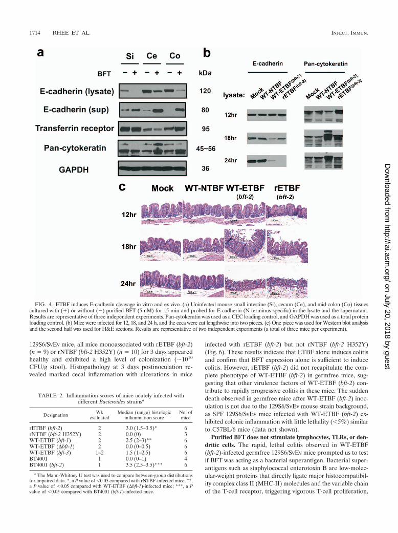
TABLE 1. Bacterial strains used in this study
Designation BFT isoformand activity Straina Relevant characteristics Source or
reference
WT-ETBF (bft-2) BFT-2 B. fragilis 86-5443-2-2 Piglet isolate 29WT-NTBF None B. fragilis NCTC 9343 Transformed with pFD340; human isolate 9rETBF (bft-2) BFT-2 B. fragilis NCTC 9343 Transformed with bft2::pFD340 9rNTBF (bft-2 H352Y) Noneb B. fragilis NCTC 9343 Transformed with inactive bft2 H352Y::pFD340 8WT-ETBF (bft-1) BFT-1 B. fragilis VPI 13784c Transformed with pFD340; lamb isolate This paperWT-ETBF (�bft1) Noneb B. fragilis VPI 13784c Transformed with pFD340; in-frame chromosomal
deletion of bft-1This paper
WT-ETBF (bft-3) BFT-3 B. fragilis Korea 570 Human blood isolate 4BT4001 None B. thetaiotaomicron 4001d Transformed with pFD340 This paperBT4001 (bft-2) BFT-2 B. thetaiotaomicron 4001d Transformed with bft-2::pFD340 This paper
a All strains used in this study are resistant to clindamycin either naturally or by introduction of pFD340.b Biologically inactive BFT or bft deletion mutant.c B. fragilis VPI 13784 was originally from Tracy Wilkins (51).d Bacteroides thetaiotaomicron strain 4001 was originally from Nadja Shoemaker (11). pFD340::bft-2 was transformed into B. thetaiotaomicron as described previously (9).
VOL. 77, 2009 INDUCTION OF PERSISTENT COLITIS BY B. FRAGILIS 1709
on July 20, 2018 by guesthttp://iai.asm
.org/D
ownloaded from

infection”) for 12, 18, and 24 h. Ceca were evaluated by histopathology andprocessed for Western blot analysis in parallel as described above.
TLR activation. HEK 293 stable transfectants expressing human Toll-likereceptor 2 (TLR-2), -3, -4, -7, or -9 were stimulated with either purified BFT (5nM), TLR ligands (positive controls), or unstimulated (negative controls) for24 h and conditioned medium frozen at �80°C until assayed for IL-8 secretionby enzyme-linked immunosorbent assay (R&D Systems) (14). HEK 293 cell TLRtransfectants secrete IL-8 upon stimulation with TLR ligands. HEK 293 cellsnaturally express TLR-5 (23).
Splenocyte proliferation assay. Splenocytes from C57BL/6J mice were pre-pared by gentle sieving on a nylon mesh (100 �m; BD Falcon) followed by lysisof red blood cells (ACK lysing buffer; Biosource). The cells were washed exten-sively, resuspended in 2% fetal bovine serum RPMI 1640 medium at a finalconcentration of 1 � 106 cells/ml and divided into aliquots (100 �l per well) intoflat-bottomed 96-well plates in triplicate. Purified BFT (5 nM [100 ng/ml final]),anti-mouse CD3ε (clone 145-2C11, 100 ng/ml), lipopolysaccharide (100 ng/ml;Alexis, San Diego, CA), or culture medium alone was added to the cells, followedby 48-h culture. Cells were pulsed with 1 �Ci of [3H]thymidine for the final 12 hof the culture period. After lysis, the [3H]thymidine incorporated into the spleno-cytes was counted with a liquid scintillation counter.
BMDCs. Bone marrow-derived dendritic cells (BMDCs) from C57BL/6J micewere generated as reported by Lu et al. (24). Briefly, bone marrow was collectedfrom tibias and femurs by flushing with RPMI 1640 culture medium. After lysingred blood cells, cells were cultured with recombinant granulocyte-macrophagecolony-stimulating factor (1,000 U/ml; R&D Systems) to induce dendritic celldifferentiation. After 6 days, BMDCs were incubated with purified BFT (5 nM[100 ng/ml final]), lipopolysaccharide (100 ng/ml), or culture medium alone (2%fetal bovine serum RPMI 1640 medium). After 20 h, cells were harvested for flowcytometric analysis (anti-CD11c, anti-MHC-II, anti-CD86, and anti-CD40; allfrom BD Biosciences), and conditioned medium was collected for cytokinesecretion analysis (BD cytometric bead array, catalogue no. 552364; detectsmurine IL-6, IL-10, monocyte chemoattractant protein 1, gamma interferon,tumor necrosis factor alpha, and IL-12 p70).
Data analysis. For statistical analysis of inflammation and hyperplasia scores,the Mann-Whitney U test was used to compare between-group distributions forunpaired data. All other data are presented as means standard errors of themeans. Comparison of means was done by unpaired Student’s t test. A P valueof �0.05 was considered statistically significant.
RESULTS
Acute ETBF infection (1 to 2 weeks). (i) Clinical character-istics. After 1 week of antibiotic pretreatment, stool bacterialcounts analyzed by fluorescence-activated cell sorting droppedby �10- to 100-fold to �1010 to 1011 bacterial cells/g stool.Acute infection experiments were initially performed usingWT-ETBF (bft-2), WT-NTBF, and rETBF (bft-2) (Table 1). At24 h postinoculation, all three B. fragilis strains were recoveredin high numbers from the stool (�109 to 1010 CFU/g stool),and this level of bacterial colonization density persisted for theduration of the experiment (data not shown). Therefore, a highlevel of colonization of mice with B. fragilis strains is rapid andstable in our mouse model.
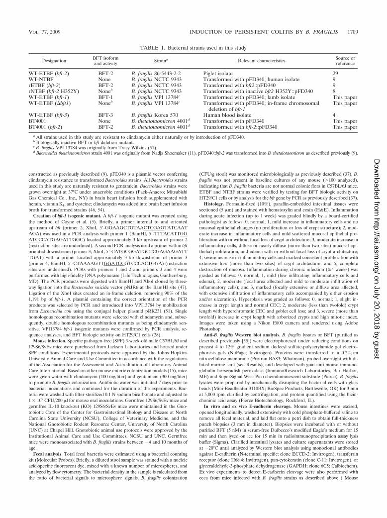
WT-ETBF (bft-2)-colonized mice were usually lethargic at�2 to 3 days postinoculation, with ruffled fur and a hunchedposture with transient weight loss (Fig. 1a) and occasionalrectal bleeding. A small percentage (5.4%; 3 of 55 mice) ofWT-ETBF (bft-2)-colonized mice died at 2 to 3 days postcolo-nization (Fig. 1b). Mortality did not appear to be due to bac-teremia as aerobic and anaerobic blood cultures of WT-ETBF(bft-2)-infected mice (n 5) at 2 days postinfection werenegative for B. fragilis or other bacterial species. The stool ofWT-ETBF (bft-2)-colonized mice ranged from moist, butformed, to transient diarrhea, usually at �1 to 3 days postcolo-nization. Thus, the deaths may be related to fluid or electrolyteimbalances secondary to enteric disease. All surviving WT-ETBF (bft-2)-colonized mice were healthy in appearance and
activity by approximately day 4 to 5 in association with theonset of weight gain (Fig. 1a). WT-NTBF-colonized and mock-infected mice appeared healthy at all times and, except for thefirst day in NTBF-colonized mice, continuously gained weight(Fig. 1a).
(ii) Gross morphology and histology. By 7 days postcoloni-zation, the ceca of WT-ETBF (bft-2)-colonized mice appearedsmaller and, in some mice, lacked cecal contents or containedblood clots compared to those of WT-NTBF-colonized mice ormock-infected mice (Fig. 1c and inset). Consistent with theobserved decrease in the size of ceca in WT-ETBF (bft-2)-colonized mice, cecal weights were significantly less in WT-ETBF (bft-2)-colonized mice compared to those in mock-in-fected and WT-NTBF-colonized mice. The decreased sizes ofceca in WT-ETBF (bft-2)-colonized mice occurred within 2days of oral colonization (data not shown). Decreases in thesizes of ceca with marked inflammation have also been notedin Citrobacter rodentium (25) and Salmonella enterica serovarTyphimurium (1) murine models and likely represent a re-sponse to cecal injury. Colon length decreased substantially inDSS-treated mice, and this correlates with the extent of in-flammation (34). The average colon length of WT-ETBF (bft-2)-colonized mice was similar to that of WT-NTBF-colonizedmice or mock-infected mice (data not shown). The only grossdifference between the colons of WT-ETBF (bft-2)-colonizedmice and those of WT-NTBF-colonized mice was that theproximal colons of WT-ETBF (bft-2)-colonized mice con-tained semisolid stool, whereas the WT-NTBF-colonized micehad well-formed stool. Although there is some variability be-tween individual ETBF-colonized mice, splenomegaly (P �0.0001) as well as enlarged mesenteric lymph nodes (data notshown) were noted in most WT-ETBF (bft-2)-colonized mice(Fig. 1d and inset).
Histopathology of the colons of WT-ETBF (bft-2)-colonizedmice revealed edema and inflammatory cell infiltration withrounding and detachment of enterocytes by day 2 postinfection(Fig. 2a and c). By day 7 postinfection, the WT-ETBF (bft-2)-colonized mice had mucosal thickening with mucosal and sub-mucosal edema, crypt hyperplasia, and elongation and exten-sive neutrophilic infiltration into both the mucosa andsubmucosa with epithelial cell destruction resulting in erosionsand ulceration (Fig. 2b and c). Scattered crypt abscesses wereobserved in the colons. Inflammation and hyperplasia wereusually more severe in the proximal than distal colons in WT-ETBF (bft-2)-colonized mice. The jejunums and ileums werenormal. WT-NTBF-colonized mice and mock-infected miceexhibited no colonic histopathology abnormalities.
Antibody responses to B. fragilis and BFT. Splenomegalyand enlarged mesenteric lymph nodes in WT-ETBF (bft-2)-colonized mice suggested activation of a systemic immune re-sponse. Thus, we examined sera for anti-Bacteroides antibodiesat day 7 postcolonization. Pooled sera from three WT-ETBF(bft-2)-colonized mice reacted specifically to diffuse high-mo-lecular-weight bands in WT-ETBF (bft-2), but not WT-NTBF,lysates; pooled sera from WT-NTBF-colonized and mock-in-fected mice did not react to either WT-ETBF (bft-2) or WT-NTBF lysates (Fig. 3). However, Western blots exposed for alonger amount of time (1 h) revealed that sera from WT-NTBF-colonized mice reacted specifically to WT-NTBF lysates(Fig. 3). These results suggest that WT-ETBF (bft-2)- and
1710 RHEE ET AL. INFECT. IMMUN.
on July 20, 2018 by guesthttp://iai.asm
.org/D
ownloaded from
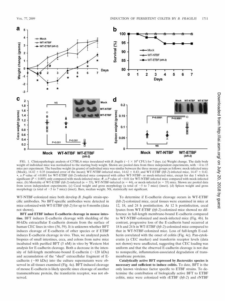
WT-NTBF-colonized mice both develop B. fragilis strain-spe-cific antibodies. No BFT-specific antibodies were detected inmice colonized with WT-ETBF (bft-2) for up to 8 months (datanot shown).
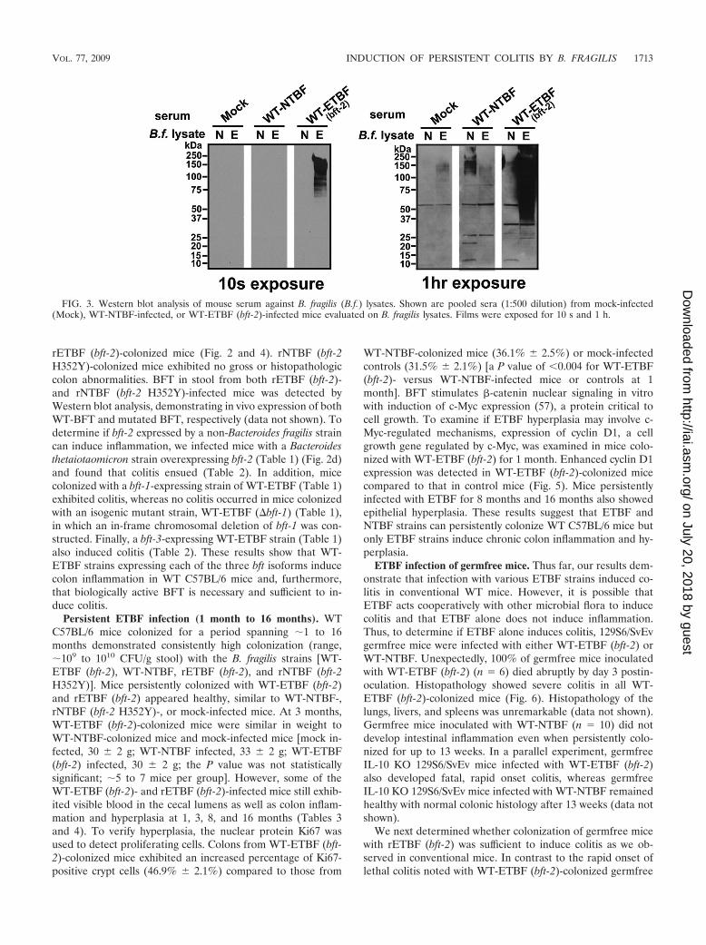
BFT and ETBF induce E-cadherin cleavage in mouse intes-tine. BFT induces E-cadherin cleavage with shedding of the80-kDa extracellular E-cadherin domain from the surface ofhuman CEC lines in vitro (56, 59). It is unknown whether BFTinduces cleavage of E-cadherin of other species or if ETBFinduces E-cadherin cleavage in vivo. Thus, we analyzed punchbiopsies of small intestines, ceca, and colons from naïve miceincubated with purified BFT (5 nM) in vitro by Western blotanalysis for E-cadherin cleavage. Both a decrease in the inten-sity of full-length membrane-bound E-cadherin (�120 kDa)and accumulation of the “shed” extracellular fragment of E-cadherin (�80 kDa) into the culture supernatants were ob-served in all tissues examined (Fig. 4a). BFT-induced cleavageof mouse E-cadherin is likely specific since cleavage of anothertransmembrane protein, the transferrin receptor, was not ob-served.
To determine if E-cadherin cleavage occurs in WT-ETBF(bft-2)-colonized mice, cecal tissues were examined in mice at12, 18, and 24 h postinfection. At 12 h postinfection, cecallysates from WT-ETBF (bft-2)-colonized mice showed no dif-ference in full-length membrane-bound E-cadherin comparedto WT-NTBF-colonized and mock-infected mice (Fig. 4b). Incontrast, progressive loss of the E-cadherin band occurred at18 h and 24 h in WT-ETBF (bft-2)-colonized mice compared tothat in WT-NTBF-colonized mice. Loss of full-length E-cad-herin correlated with the onset of colitis (Fig. 4c). Pan-cytok-eratin (a CEC marker) and transferrin receptor levels (datanot shown) were unaffected, suggesting that CEC loading wasuniform and that the observed E-cadherin cleavage is not dueto nonspecific, inflammation-associated degradation of trans-membrane proteins.
Catalytically active BFT expressed by Bacteroides species isnecessary and sufficient to induce colitis. To date, BFT is theonly known virulence factor specific to ETBF strains. To de-termine the contribution of biologically active BFT to ETBFcolitis, mice were colonized with rETBF (bft-2) and rNTBF
FIG. 1. Clinicopathologic analysis of C57BL/6 mice inoculated with B. fragilis (�1 � 109 CFU) for 7 days. (a) Weight change. The daily bodyweight of individual mice was normalized to the starting body weight. Shown are pooled data from three independent experiments, with �4 to 15mice per experiment. The baseline weight (in grams) of individual mice was similar between the three mouse groups as follows: mock-infected mice(Mock), 14.42 0.35 (standard error of the mean); WT-NTBF-infected mice, 14.62 0.43; and WT-ETBF (bft-2)-infected mice, 14.47 0.42.*, a P value of �0.001 for WT-ETBF (bft-2)-infected mice compared with either WT-NTBF- or mock-infected mice, except for day 1 which issignificant (P � 0.005) only compared with mock-infected mice; #, a P value of �0.01 for WT-NTBF-infected mice compared with mock-infectedmice. (b) Mortality of WT-ETBF (bft-2)-infected (n 55), WT-NTBF-infected (n 44), or mock-infected (n 35) mice. Shown are pooled datafrom seven independent experiments. (c) Cecal weight and gross morphology (a total of �5 to 7 mice) (inset). (d) Spleen weight and grossmorphology (a total of �5 to 7 mice) (inset). Bars, median weight. NS, statistically not significant.
VOL. 77, 2009 INDUCTION OF PERSISTENT COLITIS BY B. FRAGILIS 1711
on July 20, 2018 by guesthttp://iai.asm
.org/D
ownloaded from

(bft-2 H352Y) that differ by one amino acid, resulting in ex-pression of biologically active or inactive BFT-2, respectively(Table 1; see Materials and Methods). rETBF (bft-2)-colo-nized mice were similar to WT-ETBF (bft-2)-colonized mice,developing lethargy, ruffled fur, and, in several mice, rectal
bleeding. Mortality was usually similar to that of mice infectedwith WT-ETBF (bft-2), but it did reach 90% by 7 days in twoof five experiments (5 to 7 mice per group; data not shown).The time course and extent of colon inflammation as well asE-cadherin cleavage were similar in WT-ETBF (bft-2)- and
FIG. 2. ETBF induces acute colitis. H&E-stained tissue sections of mock-, WT-NTBF-, WT-ETBF (bft-2)-, and rETBF (bft-2)-infected mice for 2 days (a)and 7 days (b). Boxed areas were digitally magnified (�100). Bars, 100 �m. (c) Histologic inflammation scores of large bowels of infected mice at day 1, day 2,day 3, and day 7. Results of mock-infected mice were identical to those of WT-NTBF-infected mice (data not shown). Asterisks denote a lack of inflammation(an inflammation score of �0.2). (d) H&E-stained cecal tissue sections from BT4001- and BT4001 (bft-2)-infected mice.
1712 RHEE ET AL. INFECT. IMMUN.
on July 20, 2018 by guesthttp://iai.asm
.org/D
ownloaded from
rETBF (bft-2)-colonized mice (Fig. 2 and 4). rNTBF (bft-2H352Y)-colonized mice exhibited no gross or histopathologiccolon abnormalities. BFT in stool from both rETBF (bft-2)-and rNTBF (bft-2 H352Y)-infected mice was detected byWestern blot analysis, demonstrating in vivo expression of bothWT-BFT and mutated BFT, respectively (data not shown). Todetermine if bft-2 expressed by a non-Bacteroides fragilis straincan induce inflammation, we infected mice with a Bacteroidesthetaiotaomicron strain overexpressing bft-2 (Table 1) (Fig. 2d)and found that colitis ensued (Table 2). In addition, micecolonized with a bft-1-expressing strain of WT-ETBF (Table 1)exhibited colitis, whereas no colitis occurred in mice colonizedwith an isogenic mutant strain, WT-ETBF (�bft-1) (Table 1),in which an in-frame chromosomal deletion of bft-1 was con-structed. Finally, a bft-3-expressing WT-ETBF strain (Table 1)also induced colitis (Table 2). These results show that WT-ETBF strains expressing each of the three bft isoforms inducecolon inflammation in WT C57BL/6 mice and, furthermore,that biologically active BFT is necessary and sufficient to in-duce colitis.
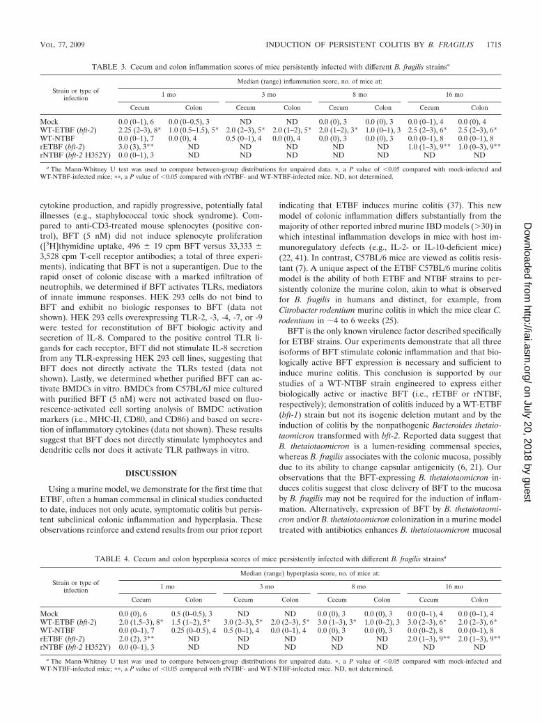
Persistent ETBF infection (1 month to 16 months). WTC57BL/6 mice colonized for a period spanning �1 to 16months demonstrated consistently high colonization (range,�109 to 1010 CFU/g stool) with the B. fragilis strains [WT-ETBF (bft-2), WT-NTBF, rETBF (bft-2), and rNTBF (bft-2H352Y)]. Mice persistently colonized with WT-ETBF (bft-2)and rETBF (bft-2) appeared healthy, similar to WT-NTBF-,rNTBF (bft-2 H352Y)-, or mock-infected mice. At 3 months,WT-ETBF (bft-2)-colonized mice were similar in weight toWT-NTBF-colonized mice and mock-infected mice [mock in-fected, 30 2 g; WT-NTBF infected, 33 2 g; WT-ETBF(bft-2) infected, 30 2 g; the P value was not statisticallysignificant; �5 to 7 mice per group]. However, some of theWT-ETBF (bft-2)- and rETBF (bft-2)-infected mice still exhib-ited visible blood in the cecal lumens as well as colon inflam-mation and hyperplasia at 1, 3, 8, and 16 months (Tables 3and 4). To verify hyperplasia, the nuclear protein Ki67 wasused to detect proliferating cells. Colons from WT-ETBF (bft-2)-colonized mice exhibited an increased percentage of Ki67-positive crypt cells (46.9% 2.1%) compared to those from
WT-NTBF-colonized mice (36.1% 2.5%) or mock-infectedcontrols (31.5% 2.1%) [a P value of �0.004 for WT-ETBF(bft-2)- versus WT-NTBF-infected mice or controls at 1month]. BFT stimulates �-catenin nuclear signaling in vitrowith induction of c-Myc expression (57), a protein critical tocell growth. To examine if ETBF hyperplasia may involve c-Myc-regulated mechanisms, expression of cyclin D1, a cellgrowth gene regulated by c-Myc, was examined in mice colo-nized with WT-ETBF (bft-2) for 1 month. Enhanced cyclin D1expression was detected in WT-ETBF (bft-2)-colonized micecompared to that in control mice (Fig. 5). Mice persistentlyinfected with ETBF for 8 months and 16 months also showedepithelial hyperplasia. These results suggest that ETBF andNTBF strains can persistently colonize WT C57BL/6 mice butonly ETBF strains induce chronic colon inflammation and hy-perplasia.
ETBF infection of germfree mice. Thus far, our results dem-onstrate that infection with various ETBF strains induced co-litis in conventional WT mice. However, it is possible thatETBF acts cooperatively with other microbial flora to inducecolitis and that ETBF alone does not induce inflammation.Thus, to determine if ETBF alone induces colitis, 129S6/SvEvgermfree mice were infected with either WT-ETBF (bft-2) orWT-NTBF. Unexpectedly, 100% of germfree mice inoculatedwith WT-ETBF (bft-2) (n 6) died abruptly by day 3 postin-oculation. Histopathology showed severe colitis in all WT-ETBF (bft-2)-colonized mice (Fig. 6). Histopathology of thelungs, livers, and spleens was unremarkable (data not shown).Germfree mice inoculated with WT-NTBF (n 10) did notdevelop intestinal inflammation even when persistently colo-nized for up to 13 weeks. In a parallel experiment, germfreeIL-10 KO 129S6/SvEv mice infected with WT-ETBF (bft-2)also developed fatal, rapid onset colitis, whereas germfreeIL-10 KO 129S6/SvEv mice infected with WT-NTBF remainedhealthy with normal colonic histology after 13 weeks (data notshown).
We next determined whether colonization of germfree micewith rETBF (bft-2) was sufficient to induce colitis as we ob-served in conventional mice. In contrast to the rapid onset oflethal colitis noted with WT-ETBF (bft-2)-colonized germfree
FIG. 3. Western blot analysis of mouse serum against B. fragilis (B.f.) lysates. Shown are pooled sera (1:500 dilution) from mock-infected(Mock), WT-NTBF-infected, or WT-ETBF (bft-2)-infected mice evaluated on B. fragilis lysates. Films were exposed for 10 s and 1 h.
VOL. 77, 2009 INDUCTION OF PERSISTENT COLITIS BY B. FRAGILIS 1713
on July 20, 2018 by guesthttp://iai.asm
.org/D
ownloaded from
129S6/SvEv mice, all mice monoassociated with rETBF (bft-2)(n 9) or rNTBF (bft-2 H352Y) (n 10) for 3 days appearedhealthy and exhibited a high level of colonization (�1010
CFU/g stool). Histopathology at 3 days postinoculation re-vealed marked cecal inflammation with ulcerations in mice
infected with rETBF (bft-2) but not rNTBF (bft-2 H352Y)(Fig. 6). These results indicate that ETBF alone induces colitisand confirm that BFT expression alone is sufficient to inducecolitis. However, rETBF (bft-2) did not recapitulate the com-plete phenotype of WT-ETBF (bft-2) in germfree mice, sug-gesting that other virulence factors of WT-ETBF (bft-2) con-tribute to rapidly progressive colitis in these mice. The suddendeath observed in germfree mice after WT-ETBF (bft-2) inoc-ulation is not due to the 129S6/SvEv mouse strain background,as SPF 129S6/SvEv mice infected with WT-ETBF (bft-2) ex-hibited colonic inflammation with little lethality (�5%) similarto C57BL/6 mice (data not shown).
Purified BFT does not stimulate lymphocytes, TLRs, or den-dritic cells. The rapid, lethal colitis observed in WT-ETBF(bft-2)-infected germfree 129S6/SvEv mice prompted us to testif BFT was acting as a bacterial superantigen. Bacterial super-antigens such as staphylococcal enterotoxin B are low-molec-ular-weight proteins that directly ligate major histocompatibil-ity complex class II (MHC-II) molecules and the variable chainof the T-cell receptor, triggering vigorous T-cell proliferation,
FIG. 4. ETBF induces E-cadherin cleavage in vitro and ex vivo. (a) Uninfected mouse small intestine (Si), cecum (Ce), and mid-colon (Co) tissuescultured with (�) or without (�) purified BFT (5 nM) for 15 min and probed for E-cadherin (N terminus specific) in the lysate and the supernatant.Results are representative of three independent experiments. Pan-cytokeratin was used as a CEC loading control, and GAPDH was used as a total proteinloading control. (b) Mice were infected for 12, 18, and 24 h, and the ceca were cut lengthwise into two pieces. (c) One piece was used for Western blot analysisand the second half was used for H&E sections. Results are representative of two independent experiments (a total of three mice per experiment).
TABLE 2. Inflammation scores of mice acutely infected withdifferent Bacteroides strainsa
Designation Wkevaluated
Median (range) histologicinflammation score
No. ofmice
rETBF (bft-2) 2 3.0 (1.5–3.5)* 6rNTBF (bft-2 H352Y) 2 0.0 (0) 3WT-ETBF (bft-1) 2 2.5 (2–3)** 6WT-ETBF (�bft-1) 2 0.0 (0–0.5) 6WT-ETBF (bft-3) 1–2 1.5 (1–2.5) 6BT4001 1 0.0 (0–1) 4BT4001 (bft-2) 1 3.5 (2.5–3.5)*** 6
a The Mann-Whitney U test was used to compare between-group distributionsfor unpaired data. *, a P value of �0.05 compared with rNTBF-infected mice; **,a P value of �0.05 compared with WT-ETBF (�bft-1)-infected mice; ***, a Pvalue of �0.05 compared with BT4001 (bft-1)-infected mice.
1714 RHEE ET AL. INFECT. IMMUN.
on July 20, 2018 by guesthttp://iai.asm
.org/D
ownloaded from
cytokine production, and rapidly progressive, potentially fatalillnesses (e.g., staphylococcal toxic shock syndrome). Com-pared to anti-CD3-treated mouse splenocytes (positive con-trol), BFT (5 nM) did not induce splenocyte proliferation([3H]thymidine uptake, 496 19 cpm BFT versus 33,333 3,528 cpm T-cell receptor antibodies; a total of three experi-ments), indicating that BFT is not a superantigen. Due to therapid onset of colonic disease with a marked infiltration ofneutrophils, we determined if BFT activates TLRs, mediatorsof innate immune responses. HEK 293 cells do not bind toBFT and exhibit no biologic responses to BFT (data notshown). HEK 293 cells overexpressing TLR-2, -3, -4, -7, or -9were tested for reconstitution of BFT biologic activity andsecretion of IL-8. Compared to the positive control TLR li-gands for each receptor, BFT did not stimulate IL-8 secretionfrom any TLR-expressing HEK 293 cell lines, suggesting thatBFT does not directly activate the TLRs tested (data notshown). Lastly, we determined whether purified BFT can ac-tivate BMDCs in vitro. BMDCs from C57BL/6J mice culturedwith purified BFT (5 nM) were not activated based on fluo-rescence-activated cell sorting analysis of BMDC activationmarkers (i.e., MHC-II, CD80, and CD86) and based on secre-tion of inflammatory cytokines (data not shown). These resultssuggest that BFT does not directly stimulate lymphocytes anddendritic cells nor does it activate TLR pathways in vitro.
DISCUSSION
Using a murine model, we demonstrate for the first time thatETBF, often a human commensal in clinical studies conductedto date, induces not only acute, symptomatic colitis but persis-tent subclinical colonic inflammation and hyperplasia. Theseobservations reinforce and extend results from our prior report
indicating that ETBF induces murine colitis (37). This newmodel of colonic inflammation differs substantially from themajority of other reported inbred murine IBD models (�30) inwhich intestinal inflammation develops in mice with host im-munoregulatory defects (e.g., IL-2- or IL-10-deficient mice)(22, 41). In contrast, C57BL/6 mice are viewed as colitis resis-tant (7). A unique aspect of the ETBF C57BL/6 murine colitismodel is the ability of both ETBF and NTBF strains to per-sistently colonize the murine colon, akin to what is observedfor B. fragilis in humans and distinct, for example, fromCitrobacter rodentium murine colitis in which the mice clear C.rodentium in �4 to 6 weeks (25).
BFT is the only known virulence factor described specificallyfor ETBF strains. Our experiments demonstrate that all threeisoforms of BFT stimulate colonic inflammation and that bio-logically active BFT expression is necessary and sufficient toinduce murine colitis. This conclusion is supported by ourstudies of a WT-NTBF strain engineered to express eitherbiologically active or inactive BFT (i.e., rETBF or rNTBF,respectively); demonstration of colitis induced by a WT-ETBF(bft-1) strain but not its isogenic deletion mutant and by theinduction of colitis by the nonpathogenic Bacteroides thetaio-taomicron transformed with bft-2. Reported data suggest thatB. thetaiotaomicron is a lumen-residing commensal species,whereas B. fragilis associates with the colonic mucosa, possiblydue to its ability to change capsular antigenicity (6, 21). Ourobservations that the BFT-expressing B. thetaiotaomicron in-duces colitis suggest that close delivery of BFT to the mucosaby B. fragilis may not be required for the induction of inflam-mation. Alternatively, expression of BFT by B. thetaiotaomi-cron and/or B. thetaiotaomicron colonization in a murine modeltreated with antibiotics enhances B. thetaiotaomicron mucosal
TABLE 3. Cecum and colon inflammation scores of mice persistently infected with different B. fragilis strainsa
Strain or type ofinfection
Median (range) inflammation score, no. of mice at:
1 mo 3 mo 8 mo 16 mo
Cecum Colon Cecum Colon Cecum Colon Cecum Colon
Mock 0.0 (0–1), 6 0.0 (0–0.5), 3 ND ND 0.0 (0), 3 0.0 (0), 3 0.0 (0–1), 4 0.0 (0), 4WT-ETBF (bft-2) 2.25 (2–3), 8* 1.0 (0.5–1.5), 5* 2.0 (2–3), 5* 2.0 (1–2), 5* 2.0 (1–2), 3* 1.0 (0–1), 3 2.5 (2–3), 6* 2.5 (2–3), 6*WT-NTBF 0.0 (0–1), 7 0.0 (0), 4 0.5 (0–1), 4 0.0 (0), 4 0.0 (0), 3 0.0 (0), 3 0.0 (0–1), 8 0.0 (0–1), 8rETBF (bft-2) 3.0 (3), 3** ND ND ND ND ND 1.0 (1–3), 9** 1.0 (0–3), 9**rNTBF (bft-2 H352Y) 0.0 (0–1), 3 ND ND ND ND ND ND ND
a The Mann-Whitney U test was used to compare between-group distributions for unpaired data. �, a P value of �0.05 compared with mock-infected andWT-NTBF-infected mice; ��, a P value of �0.05 compared with rNTBF- and WT-NTBF-infected mice. ND, not determined.
TABLE 4. Cecum and colon hyperplasia scores of mice persistently infected with different B. fragilis strainsa
Strain or type ofinfection
Median (range) hyperplasia score, no. of mice at:
1 mo 3 mo 8 mo 16 mo
Cecum Colon Cecum Colon Cecum Colon Cecum Colon
Mock 0.0 (0), 6 0.5 (0–0.5), 3 ND ND 0.0 (0), 3 0.0 (0), 3 0.0 (0–1), 4 0.0 (0–1), 4WT-ETBF (bft-2) 2.0 (1.5–3), 8* 1.5 (1–2), 5* 3.0 (2–3), 5* 2.0 (2–3), 5* 3.0 (1–3), 3* 1.0 (0–2), 3 3.0 (2–3), 6* 2.0 (2–3), 6*WT-NTBF 0.0 (0–1), 7 0.25 (0–0.5), 4 0.5 (0–1), 4 0.0 (0–1), 4 0.0 (0), 3 0.0 (0), 3 0.0 (0–2), 8 0.0 (0–1), 8rETBF (bft-2) 2.0 (2), 3** ND ND ND ND ND 2.0 (1–3), 9** 2.0 (1–3), 9**rNTBF (bft-2 H352Y) 0.0 (0–1), 3 ND ND ND ND ND ND ND
a The Mann-Whitney U test was used to compare between-group distributions for unpaired data. �, a P value of �0.05 compared with mock-infected andWT-NTBF-infected mice; ��, a P value of �0.05 compared with rNTBF- and WT-NTBF-infected mice. ND, not determined.
VOL. 77, 2009 INDUCTION OF PERSISTENT COLITIS BY B. FRAGILIS 1715
on July 20, 2018 by guesthttp://iai.asm
.org/D
ownloaded from

adherence. Although our data strongly suggest a central rolefor BFT secretion in ETBF disease pathogenesis, our germfreemouse studies using rETBF (bft-2) and rNTBF (bft-2 H352Y)suggest that other virulence factors may drive the earlymortality associated with colitis observed in this murinemodel (35, 39).
Our in vivo murine data confirm the relevance of prior invitro studies of the BFT mechanism of action to understandingthe pathogenesis of ETBF disease. In vitro studies indicatedthat BFT binds to a putative intestinal epithelial cell receptorstimulating signal transduction pathways, resulting in the fol-lowing: (i) E-cadherin cleavage with reduced barrier functionin CEC monolayers (59), (ii) �-catenin signaling with inductionof c-Myc expression and CEC proliferation (57), and (iii) ac-tivation of NF- B with expression and secretion of IL-8 andother cytokines (18, 42, 57, 58). The studies herein confirm forthe first time that E-cadherin cleavage occurs in vivo early inETBF-induced murine colitis. E-cadherin is important for in-testinal barrier function (61), and chimeric mice expressing adominant negative N-cadherin in CECs lose intercellular E-cadherin associations and develop colitis (13). Our preliminarydata (unpublished data) indicate that early ETBF colitis is also
associated with expression of the chemokine KC, the murineIL-8 analog. Our data support the concept that diminishedbarrier function is an early step in the development of colonicinflammation as proposed for human IBD (16, 48). Herein, wealso demonstrate ETBF-induced CEC hyperplasia (Fig. 5a)with increased Ki67 and cyclin D1 expression, the latter adownstream target of �-catenin signaling and c-Myc inductionknown to be triggered by BFT (12, 49). While crypt hyperplasiais common with experimental colitis, the colon histopathologyfrom mice colonized with ETBF for 16 months (Fig. 5b) re-veals hyperplasia without gross CEC barrier disruption andinflammation (compare Fig. 2a and 5b). More detailed studiesare needed to discern the mechanisms of ETBF-induced in-flammation and hyperplasia and to determine if these are mo-lecularly linked mucosal events. Lastly, our results did notshow direct activation of TLRs or immune cells by BFT con-sistent with in vitro data, indicating that BFT specifically bindsonly to epithelial cell lines that express E-cadherin and polar-ize in vitro (60). Additional studies are necessary to define themechanisms by which ETBF and BFT activate the mucosal andsystemic immune system and to identify the specific BFT cellreceptor. Our data suggest that the BFT receptor is present
FIG. 5. ETBF chronic infection enhances epithelial hyperplasia. (a) H&E staining and anti-cyclin D1 immunohistochemistry of ceca from micepersistently infected (for 1 month) with WT-NTBF and WT-ETBF (bft-2). Results for mock-infected mice are identical to those for WT-NTBF-infected mice (data not shown). (b) H&E staining of ceca from mice persistently infected for 8 months [WT-NTBF and WT-ETBF (bft-2)] and16 months [WT-NTBF and rETBF(bft-2)].
FIG. 6. ETBF induces colitis in germfree mice. Germfree 129S6/SvEv mice (�4 to 10 months old) were inoculated with WT-ETBF (bft-2),rETBF (bft-2), or rNTBF (bft-2 H352Y), and ceca were prepared for histology 3 days later. Bars, 100 �m.
1716 RHEE ET AL. INFECT. IMMUN.
on July 20, 2018 by guesthttp://iai.asm
.org/D
ownloaded from
throughout the intestine since BFT stimulated E-cadherincleavage in small intestinal and colonic tissues (Fig. 4a). Thus,the colonic localization of WT-ETBF-induced disease likelyrelates to the colonic environment and as-yet-uncharacterizedB. fragilis adherence factors.
The potent antibody response specific to WT-ETBF (bft-2)was unexpected considering the short-term infection but likelywas facilitated by BFT-induced colitis with translocation ofluminal antigens and systemic immune activation as evidencedby splenomegaly in ETBF-colonized mice. Although blood andperipheral tissue cultures of WT-ETBF (bft-2)- and rETBF(bft-2)-infected mice were negative (n 5), it is possible thatB. fragilis transiently invaded the periphery. The size and dif-fuse appearance of the WT-NTBF and WT-ETBF (bft-2) an-tigens detected by Western blot analysis (Fig. 3) suggest anti-body reactivity to the unique and antigenically potent B. fragilispolysaccharide capsules of the inoculated B. fragilis strains. Incontrast, murine BFT-specific antibodies were not detected de-spite the known immunogenicity of BFT in rabbits and humans(45). It is possible that Western blot analysis is too insensitive orthat only murine mucosal antibodies are produced.
Although WT-NTBF-colonized mice did not exhibit any co-lonic inflammation, we detected weak antibody responses toNTBF, indicating a systemic adaptive immune response toNTBF, a finding consistent with a recent report of peripheralCD4� T lymphocyte expansion in NTBF-colonized germfreemice (26). Our results extend these prior observations, showinginitiation of adaptive immune responses to NTBF within 1week of colonization, but differ from results of Konrad et al.(20) who reported that the murine immune responses to en-teric bacteria were tightly compartmentalized to the mucosa inconventional mice. These differences suggest that the interac-tion of B. fragilis with the host is complex and differs from othercommon fecal flora.
Recently, Nakano et al. (31) reported that outbred germfreeNIH (Swiss Webster) mice housed under short-term germfreeconditions and infected with a human strain of WT-ETBFdeveloped only mild, nonlethal colitis. Potential reasons for theexperimental discrepancies include differences in susceptibilityto colitis by different mouse strains and different amounts ofBFT secretion among clinical strains of ETBF (4, 43, 53).Nonetheless, our experiments indicate that monoinfection withat least some WT-ETBF strains is sufficient to induce profoundcolonic inflammation.
In small studies, ETBF colonization in humans has been asso-ciated with active IBD (2, 36) and colorectal cancer (50). Reportsestimate that �4 to 30% of healthy individuals harbor ETBFwithout obvious symptoms. We hypothesize that humans harbor-ing ETBF or potentially similar as-yet-unidentified commensalsmay be at risk for the deleterious consequences of persistent,subclinical colonic inflammation such as IBD, oncogenic se-quelae, and/or irritable bowel syndrome. Consistent with our hy-pothesis, we previously reported that mice given subclinical dosesof DSS followed by ETBF colonization show exacerbated colitis(37) and that in Bangladeshi patients with ETBF, persistent in-testinal inflammation was present 3 weeks postinfection even ifETBF was cleared with antibiotic therapy (45). To date, we havenot observed colonic neoplasms in mice persistently colonizedwith WT-ETBF (bft-2) or rETBF (bft-2) at up to 16 months of
age. Well-designed clinical studies are necessary to determine thelong-term consequences of ETBF colonization.
In summary, we describe ETBF-induced colitis in colitis-resis-tant antibiotic-treated SPF C57BL/6 mice that mirrors acute andpersistent infection observed in ETBF-infected individuals. Ourdata indicate that BFT is necessary and sufficient to induce colitisin both conventional and germfree mice. Studies to define theimmunopathogenesis of ETBF colitis are in progress. The futuretranslational challenge will be to identify if select members of theintestinal flora such as ETBF are key to the pathogenesis ofcolonic inflammatory conditions and their sequelae.
ACKNOWLEDGMENTS
We appreciate the technical assistance of Melanie Adams and thegifts of Bacteroides strains by Lyle Myers, Tracy Wilkins, and NadiaShoemaker.
This work was supported by the Crohn’s and Colitis Foundation ofAmerica (CCFA) Research Fellowship Award to K.-J.R. and througha CCFA Senior Investigator Award to C.L.S. as well as NIH grantsRO1 DK45496, RO1 DK080817 (to C.L.S.), NIH KO1 A1066994(to J.E.G.), CA62924 (to Scott Kern), R24 DK64388 (to MarkDonowitz), and the NCI Division of Cancer Prevention contractsHHSN261200433002C (to Bert Vogelstein), RR21362 (to B.K.), P40RR020764 (to R.B.S.), RO1 DK53347 (to R.B.S.), P30 DK34987 (toRobert S. Sandler), and RR00171 (to D.L.H.).
The authors have no financial conflict of interest.
REFERENCES
1. Barthel, M., S. Hapfelmeier, L. Quintanilla-Martinez, M. Kremer, M. Ro-hde, M. Hogardt, K. Pfeffer, H. Russmann, and W. D. Hardt. 2003. Pretreat-ment of mice with streptomycin provides a Salmonella enterica serovar Ty-phimurium colitis model that allows analysis of both pathogen and host.Infect. Immun. 71:2839–2858.
2. Basset, C., J. Holton, A. Bazeos, D. Vaira, and S. Bloom. 2004. Are Helico-bacter species and enterotoxigenic Bacteroides fragilis involved in inflamma-tory bowel disease? Dig. Dis. Sci. 49:1425–1432.
3. Chambers, F. G., S. S. Koshy, R. F. Saidi, D. P. Clark, R. D. Moore, and C. L.Sears. 1997. Bacteroides fragilis toxin exhibits polar activity on monolayers ofhuman intestinal epithelial cells (T84 cells) in vitro. Infect. Immun. 65:3561–3570.
4. Chung, G. T., A. A. Franco, S. Wu, G. E. Rhie, R. Cheng, H. B. Oh, and C. L.Sears. 1999. Identification of a third metalloprotease toxin gene in extraint-estinal isolates of Bacteroides fragilis. Infect. Immun. 67:4945–4949.
5. Coyne, M. J., W. Kalka-Moll, A. O. Tzianabos, D. L. Kasper, and L. E.Comstock. 2000. Bacteroides fragilis NCTC9343 produces at least three dis-tinct capsular polysaccharides: cloning, characterization, and reassignmentof polysaccharide B and C biosynthesis loci. Infect. Immun. 68:6176–6181.
6. Coyne, M. J., K. G. Weinacht, C. M. Krinos, and L. E. Comstock. 2003. Mpirecombinase globally modulates the surface architecture of a human com-mensal bacterium. Proc. Natl. Acad. Sci. USA 100:10446–10451.
7. Elson, C. O., Y. Cong, V. J. McCracken, R. A. Dimmitt, R. G. Lorenz, andC. T. Weaver. 2005. Experimental models of inflammatory bowel diseasereveal innate, adaptive, and regulatory mechanisms of host dialogue with themicrobiota. Immunol. Rev. 206:260–276.
8. Franco, A. A., S. L. Buckwold, J. W. Shin, M. Ascon, and C. L. Sears. 2005.Mutation of the zinc-binding metalloprotease motif affects Bacteroides fra-gilis toxin activity but does not affect propeptide processing. Infect. Immun.73:5273–5277.
9. Franco, A. A., R. K. Cheng, A. Goodman, and C. L. Sears. 2002. Modulationof bft expression by the Bacteroides fragilis pathogenicity island and its flank-ing region. Mol. Microbiol. 45:1067–1077.
10. Franco, A. A., L. M. Mundy, M. Trucksis, S. Wu, J. B. Kaper, and C. L.Sears. 1997. Cloning and characterization of the Bacteroides fragilis metal-loprotease toxin gene. Infect. Immun. 65:1007–1013.
11. Gupta, A., H. Vlamakis, N. Shoemaker, and A. A. Salyers. 2003. A newBacteroides conjugative transposon that carries an ermB gene. Appl. Environ.Microbiol. 69:6455–6463.
12. He, T. C., A. B. Sparks, C. Rago, H. Hermeking, L. Zawel, L. T. da Costa,P. J. Morin, B. Vogelstein, and K. W. Kinzler. 1998. Identification of c-MYCas a target of the APC pathway. Science 281:1509–1512.
13. Hermiston, M. L., M. H. Wong, and J. I. Gordon. 1996. Forced expression ofE-cadherin in the mouse intestinal epithelium slows cell migration and pro-vides evidence for nonautonomous regulation of cell fate in a self-renewingsystem. Genes Dev. 10:985–996.
14. Hise, A. G., K. Daehnel, I. Gillette-Ferguson, E. Cho, H. F. McGarry, M. J.
VOL. 77, 2009 INDUCTION OF PERSISTENT COLITIS BY B. FRAGILIS 1717
on July 20, 2018 by guesthttp://iai.asm
.org/D
ownloaded from

Taylor, D. T. Golenbock, K. A. Fitzgerald, J. W. Kazura, and E. Pearlman. 2007.Innate immune responses to endosymbiotic Wolbachia bacteria in Brugia malayiand Onchocerca volvulus are dependent on TLR2, TLR6, MyD88, and Mal, butnot TLR4, TRIF, or TRAM. J. Immunol. 178:1068–1076.
15. Hogenauer, C., C. Langner, E. Beubler, I. T. Lippe, R. Schicho, G. Gork-iewicz, R. Krause, N. Gerstgrasser, G. J. Krejs, and T. A. Hinterleitner. 2006.Klebsiella oxytoca as a causative organism of antibiotic-associated hemor-rhagic colitis. N. Engl. J. Med. 355:2418–2426.
16. Hollander, D., C. M. Vadheim, E. Brettholz, G. M. Petersen, T. Delahunty,and J. I. Rotter. 1986. Increased intestinal permeability in patients withCrohn’s disease and their relatives. A possible etiologic factor. Ann. Intern.Med. 105:883–885.
17. Kim, J. M., J. Y. Lee, Y. M. Yoon, Y. K. Oh, J. S. Kang, Y. J. Kim, and K. H.Kim. 2006. Bacteroides fragilis enterotoxin induces cyclooxygenase-2 andfluid secretion in intestinal epithelial cells through NF- B activation. Eur.J. Immunol. 36:2446–2456.
18. Kim, J. M., Y. K. Oh, Y. J. Kim, H. B. Oh, and Y. J. Cho. 2001. Polarizedsecretion of CXC chemokines by human intestinal epithelial cells in responseto Bacteroides fragilis enterotoxin: NF- B plays a major role in the regulationof IL-8 expression. Clin. Exp. Immunol. 123:421–427.
19. Kling, J. J., R. L. Wright, J. S. Moncrief, and T. D. Wilkins. 1997. Cloningand characterization of the gene for the metalloprotease enterotoxin ofBacteroides fragilis. FEMS Microbiol. Lett. 146:279–284.
20. Konrad, A., Y. Cong, W. Duck, R. Borlaza, and C. O. Elson. 2006. Tightmucosal compartmentation of the murine immune response to antigens ofthe enteric microbiota. Gastroenterology 130:2050–2059.
21. Krinos, C. M., M. J. Coyne, K. G. Weinacht, A. O. Tzianabos, D. L. Kasper,and L. E. Comstock. 2001. Extensive surface diversity of a commensal mi-croorganism by multiple DNA inversions. Nature 414:555–558.
22. Kuhn, R., J. Lohler, D. Rennick, K. Rajewsky, and W. Muller. 1993. Inter-leukin-10-deficient mice develop chronic enterocolitis. Cell 75:263–274.
23. Lu, W., A. Hisatsune, T. Koga, K. Kato, I. Kuwahara, E. P. Lillehoj, W.Chen, A. S. Cross, S. J. Gendler, A. T. Gewirtz, and K. C. Kim. 2006. Cuttingedge: enhanced pulmonary clearance of Pseudomonas aeruginosa by Muc1knockout mice. J. Immunol. 176:3890–3894.
24. Lu, Z., L. Yuan, X. Zhou, E. Sotomayor, H. I. Levitsky, and D. M. Pardoll.2000. CD40-independent pathways of T cell help for priming of CD8�
cytotoxic T lymphocytes. J. Exp. Med. 191:541–550.25. Luperchio, S. A., and D. B. Schauer. 2001. Molecular pathogenesis of
Citrobacter rodentium and transmissible murine colonic hyperplasia. Mi-crobes Infect. 3:333–340.
26. Mazmanian, S. K., C. H. Liu, A. O. Tzianabos, and D. L. Kasper. 2005. Animmunomodulatory molecule of symbiotic bacteria directs maturation of thehost immune system. Cell 122:107–118.
27. Moncrief, J. S., R. Obiso, Jr., L. A. Barroso, J. J. Kling, R. L. Wright, R. L.Van Tassell, D. M. Lyerly, and T. D. Wilkins. 1995. The enterotoxin ofBacteroides fragilis is a metalloprotease. Infect. Immun. 63:175–181.
28. Mundy, L. M., and C. L. Sears. 1996. Detection of toxin production byBacteroides fragilis: assay development and screening of extraintestinal clin-ical isolates. Clin. Infect. Dis. 23:269–276.
29. Myers, L. L., and D. S. Shoop. 1987. Association of enterotoxigenic Bacteroidesfragilis with diarrheal disease in young pigs. Am. J. Vet. Res. 48:774–775.
30. Myers, L. L., D. S. Shoop, J. E. Collins, and W. C. Bradbury. 1989. Diarrhealdisease caused by enterotoxigenic Bacteroides fragilis in infant rabbits. J. Clin.Microbiol. 27:2025–2030.
31. Nakano, V., D. A. Gomes, R. M. Arantes, J. R. Nicoli, and M. J. Avila-Campos. 2006. Evaluation of the pathogenicity of the Bacteroides fragilistoxin gene subtypes in gnotobiotic mice. Curr. Microbiol. 53:113–117.
32. Obiso, R. J., Jr., A. O. Azghani, and T. D. Wilkins. 1997. The Bacteroidesfragilis toxin fragilysin disrupts the paracellular barrier of epithelial cells.Infect. Immun. 65:1431–1439.
33. Obiso, R. J., Jr., D. M. Lyerly, R. L. Van Tassell, and T. D. Wilkins. 1995.Proteolytic activity of the Bacteroides fragilis enterotoxin causes fluid secre-tion and intestinal damage in vivo. Infect. Immun. 63:3820–3826.
34. Okayasu, I., S. Hatakeyama, M. Yamada, T. Ohkusa, Y. Inagaki, and R.Nakaya. 1990. A novel method in the induction of reliable experimentalacute and chronic ulcerative colitis in mice. Gastroenterology 98:694–702.
35. Pantosti, A., A. O. Tzianabos, B. G. Reinap, A. B. Onderdonk, and D. L.Kasper. 1993. Bacteroides fragilis strains express multiple capsular polysac-charides. J. Clin. Microbiol. 31:1850–1855.
36. Prindiville, T. P., R. A. Sheikh, S. H. Cohen, Y. J. Tang, M. C. Cantrell, andJ. Silva, Jr. 2000. Bacteroides fragilis enterotoxin gene sequences in patientswith inflammatory bowel disease. Emerg. Infect. Dis. 6:171–174.
37. Rabizadeh, S., K.-J. Rhee, S. Wu, D. Huso, C. M. Gan, J. E. Golub, X. Wu,M. Zhang, and C. L. Sears. 2007. Enterotoxigenic Bacteroides fragilis: apotential instigator of colitis. Inflamm. Bowel Dis. 13:1475–1483.
38. Riegler, M., M. Lotz, C. Sears, C. Pothoulakis, I. Castagliuolo, C. C. Wang,
R. Sedivy, T. Sogukoglu, E. Cosentini, G. Bischof, W. Feil, B. Teleky, G.Hamilton, J. T. LaMont, and E. Wenzl. 1999. Bacteroides fragilis toxin 2damages human colonic mucosa in vitro. Gut 44:504–510.
39. Robertson, K. P., C. J. Smith, A. M. Gough, and E. R. Rocha. 2006. Char-acterization of Bacteroides fragilis hemolysins and regulation and synergisticinteractions of HlyA and HlyB. Infect. Immun. 74:2304–2316.
40. Sack, R. B., L. L. Myers, J. Almeido-Hill, D. S. Shoop, W. C. Bradbury, R.Reid, and M. Santosham. 1992. Enterotoxigenic Bacteroides fragilis: epide-miologic studies of its role as a human diarrhoeal pathogen. J. DiarrhoealDis. Res. 10:4–9.
41. Sadlack, B., H. Merz, H. Schorle, A. Schimpl, A. C. Feller, and I. Horak.1993. Ulcerative colitis-like disease in mice with a disrupted interleukin-2gene. Cell 75:253–261.
42. Sanfilippo, L., C. K. Li, R. Seth, T. J. Balwin, M. G. Menozzi, and Y. R.Mahida. 2000. Bacteroides fragilis enterotoxin induces the expression of IL-8and transforming growth factor-beta (TGF-�) by human colonic epithelialcells. Clin. Exp. Immunol. 119:456–463.
43. Scotto d’Abusco, A. S., L. Sanfilippo, M. G. Menozzi, and A. Pantosti. 2000.Activity and role of BFT, an enterotoxin produced by Bacteroides fragilis J.Nat. Toxins 9:267–280.
44. Sears, C. L. 2001. The toxins of Bacteroides fragilis. Toxicon 39:1737–1746.45. Sears, C. L., S. Islam, A. Saha, M. Arjumand, N. H. Alam, A. S. Faruque,
M. A. Salam, J. Shin, D. Hecht, A. Weintraub, R. B. Sack, and F. Qadri.2008. Association of enterotoxigenic Bacteroides fragilis infection with in-flammatory diarrhea. Clin. Infect. Dis. 47:797–803.
46. Shanson, D. C., and J. Singh. 1981. Effect of adding cysteine to brain-heartinfusion broth on the isolation of Bacteroides fragilis from experimental bloodcultures. J. Clin. Pathol. 34:221–223.
47. Stevens, A. M., N. B. Shoemaker, and A. A. Salyers. 1990. The region of aBacteroides conjugal chromosomal tetracycline resistance element which isresponsible for production of plasmidlike forms from unlinked chromosomalDNA might also be involved in transfer of the element. J. Bacteriol. 172:4271–4279.
48. Teahon, K., P. Smethurst, A. J. Levi, I. S. Menzies, and I. Bjarnason. 1992.Intestinal permeability in patients with Crohn’s disease and their first degreerelatives. Gut 33:320–323.
49. Tetsu, O., and F. McCormick. 1999. Beta-catenin regulates expression ofcyclin D1 in colon carcinoma cells. Nature 398:422–426.
50. Toprak, N. U., A. Yagci, B. M. Gulluoglu, M. L. Akin, P. Demirkalem, T. Celenk,and G. Soyletir. 2006. A possible role of Bacteroides fragilis enterotoxin in theaetiology of colorectal cancer. Clin. Microbiol. Infect. 12:782–786.
51. Van Tassell, R. L., D. M. Lyerly, and T. D. Wilkins. 1992. Purification andcharacterization of an enterotoxin from Bacteroides fragilis. Infect. Immun.60:1343–1350.
52. Vines, R. R., S. S. Perdue, J. S. Moncrief, D. R. Sentz, L. A. Barroso, R. L.Wright, and T. D. Wilkins. 2000. Fragilysin, the enterotoxin from Bacteroidesfragilis, enhances the serum antibody response to antigen co-administered bythe intranasal route. Vaccine 19:655–660.
53. Weikel, C. S., F. D. Grieco, J. Reuben, L. L. Myers, and R. B. Sack. 1992.Human colonic epithelial cells, HT29/C1, treated with crude Bacteroidesfragilis enterotoxin dramatically alter their morphology. Infect. Immun. 60:321–327.
54. Wilkins, T. D., and S. Chalgren. 1976. Medium for use in antibiotic suscep-tibility testing of anaerobic bacteria. Antimicrob. Agents Chemother. 10:926–928.
55. Wu, S., L. A. Dreyfus, A. O. Tzianabos, C. Hayashi, and C. L. Sears. 2002.Diversity of the metalloprotease toxin produced by enterotoxigenic Bacte-roides fragilis. Infect. Immun. 70:2463–2471.
56. Wu, S., K. C. Lim, J. Huang, R. F. Saidi, and C. L. Sears. 1998. Bacteroidesfragilis enterotoxin cleaves the zonula adherens protein, E-cadherin. Proc.Natl. Acad. Sci. USA 95:14979–14984.
57. Wu, S., P. J. Morin, D. Maouyo, and C. L. Sears. 2003. Bacteroides fragilisenterotoxin induces c-Myc expression and cellular proliferation. Gastroen-terology 124:392–400.
58. Wu, S., J. Powell, N. Mathioudakis, S. Kane, E. Fernandez, and C. L. Sears.2004. Bacteroides fragilis enterotoxin induces intestinal epithelial cell secretion ofinterleukin-8 through mitogen-activated protein kinases and a tyrosine kinase-regulated nuclear factor- B pathway. Infect. Immun. 72:5832–5839.
59. Wu, S., K.-J. Rhee, M. Zhang, A. Franco, and C. L. Sears. 2007. Bacteroidesfragilis toxin stimulates intestinal epithelial cell shedding and �-secretase-dependent E-cadherin cleavage. J. Cell Sci. 120:1944–1952.
60. Wu, S., J. Shin, G. Zhang, M. Cohen, A. Franco, and C. L. Sears. 2006. TheBacteroides fragilis toxin binds to a specific intestinal epithelial cell receptor.Infect. Immun. 74:5382–5390.
61. Zbar, A. P., C. Simopoulos, and A. J. Karayiannakis. 2004. Cadherins: anintegral role in inflammatory bowel disease and mucosal restitution. J. Gas-troenterol. 39:413–421.
Editor: V. J. DiRita
1718 RHEE ET AL. INFECT. IMMUN.
on July 20, 2018 by guesthttp://iai.asm
.org/D
ownloaded from