inductionofdepressedmooddisruptsemotion ... fileinductionofdepressedmooddisruptsemotion...
TRANSCRIPT

IRUC
Bnmp
Ma
Rnncme
Ci
Kf
PdlpcstaaTeeccdm[s
F
A
R
0d
nduction of Depressed Mood Disrupts Emotionegulation Neurocircuitry and Enhances Painnpleasantness
hantal Berna, Siri Leknes, Emily A. Holmes, Robert R. Edwards, Guy M. Goodwin, and Irene Tracey
ackground: Depressed mood alters the pain experience. Yet, despite its clear clinical relevance, little is known about the cognitive andeural mechanisms underlying this phenomenon. We tested an experimental manipulation to unravel the interaction between depressedood and pain. We hypothesized that dysregulation of the neural circuitry underlying emotion regulation is the mechanism whereby pain
rocessing is affected during depressed mood.
ethods: Using functional magnetic resonance imaging, we compared the effects of sad and neutral cognitive mood inductions onffective pain ratings, pain-specific cognitions, and central pain processing of a tonic noxious heat stimulus in 20 healthy volunteers.
esults: The increase in negative pain-specific cognitions during depressed mood predicted the perceived increase in pain unpleasant-ess. Following depressed mood induction, brain responses to noxious thermal stimuli were characterized by increased activity in a broadetwork including prefrontal areas, subgenual anterior cingulate cortex, and hippocampus, as well as significantly less deactivation whenompared with pain responses in a neutral mood. The participants who reported the largest increase in pain unpleasantness after the sadood induction showed greater inferior frontal gyrus and amygdala activation, linking changes in emotion regulation mechanisms with
nhancement of pain affect.
onclusions: Our results inform how depressed mood and chronic pain co-occur clinically and may serve to develop and translate effective
nterventions using pharmacological or psychological treatment.ey Words: Cognitions, depressed mood, emotion regulation,MRI, pain
ain and depression have been reciprocally linked in manyexperimental and clinical studies. Chronic pain is morelikely in individuals with a history of depression (1) and
epression exacerbates the burden of painful diseases (2). Painends itself well to experimental investigation. Thus, depressedatients (without chronic pain) have altered prefrontal activityompared with healthy control subjects during brief noxioustimulation (3,4). Moreover, in patients with chronic pain, symp-oms of depression correlate with amygdalar and anterior insularctivity during experimental pain (5) and medial prefrontal cortexctivation during disease-relevant experimentally induced pain (6).hese recent studies support a general hypothesis of dysfunctionalmotion regulation during pain perception. However, these patientsxhibit significant comorbidity and enduring structural or functionalhanges that may confound experimental studies (7,8). For betterontrolled experiments, negative cognitive mood induction proce-ures allow us directly to manipulate mood. Although acute, theseood modulations have been used frequently in psychology (e.g.,
9]) to investigate cognitive processes relevant to chronic moodtates (10). While negative mood inductions can worsen affective
rom the Centre for Functional Magnetic Resonance Imaging of the Brain(CB, SL, IT), Department of Clinical Neurology and Nuffield Departmentof Anaesthetics, University of Oxford; and Department of Psychiatry (CB,EAH, GMG), University of Oxford, Warneford Hospital, Oxford, UnitedKingdom; and Department of Anesthesiology (RRE), Harvard MedicalSchool, Brigham & Women’s Hospital, Chestnut Hill, Massachusetts.
ddress correspondence to Chantal Berna, M.D., Centre for Functional Mag-netic Resonance Imaging of the Brain, Departments of Clinical Neurol-ogy and Anaesthetics, University of Oxford, Oxford OX3 9DU, UnitedKingdom; E-mail: [email protected].
eceived Jun 27, 2009; revised Jan 7, 2010; accepted Jan 10, 2010.
006-3223/$36.00oi:10.1016/j.biopsych.2010.01.014
pain ratings (11–14), the mechanisms underlying such modulationof pain perception are not yet established.
It is has been suggested that maladaptive thought processesmay mediate changes in pain perception in the context ofdepressed mood (15). Specifically, catastrophizing thoughts (i.e.,negative pain-related cognitions) are amplified in depressed indi-viduals (16) and depressed patients exhibit deficient emotion reg-ulation when exposed to negatively valenced stimuli (4,17,18);hence, we hypothesized that central pain processing duringdepressed versus neutral mood would be characterized byaltered activity in the dorsolateral and/or ventrolateral prefrontalcortex (dlPFC, vlPFC) and increased amygdala activation, reflect-ing ineffective emotion regulation. Finally, we predicted that thelevel of activity in these regions during painful stimulation in thesad condition would influence individual differences in painunpleasantness scores.
Accordingly, we used a well-established negative or sad moodinduction procedure and a matching neutral procedure for experi-mental comparison (19). Healthy volunteers received a tonic painfulstimulus after undergoing each mood induction inside the func-tional magnetic resonance imaging (fMRI) scanner. This allowed anexperimental test of hypotheses based on cognitive theories ofpain-mood interactions. Noxious stimuli, rated for pain unpleasant-ness, and mood reinforcers were given (Figure 1). We hypothesizedthat the effects of an induced depressed mood compared with aneutral mood would be: 1) an increase in negative pain-relatedthoughts (i.e., catastrophizing [20]), 2) an increase in the perceivedunpleasantness of the pain, and 3) neural evidence of disruption ofnormal emotion regulation.
Methods and Materials
ParticipantsTwenty-seven pain-free, nondepressed, right-handed volun-
teers were recruited. Invitations were sent to university students
asking for healthy volunteers who were not suffering from anyBIOL PSYCHIATRY 2010;67:1083–1090© 2010 Society of Biological Psychiatry

pamlc1mmc(BtpEw
E
msctri
tvmwm8W(hpi
FpV[mpafrbapcept
1084 BIOL PSYCHIATRY 2010;67:1083–1090 C. Berna et al.
w
ain condition, psychiatric disorder, or taking daily painkillers orntidepressants. The study received local Research Ethics Com-ittee approval (number C02.283) and conformed to the guide-
ines of the 1996 Declaration of Helsinki. The analysis wasonducted on a group of 20 volunteers (mean age: 28, range9–41; 11 male/9 female; Beck Depression Inventory-II [BDI-II]ean: 5.74 � SD 5.48) as postscanning exclusion criteria wereet by 7 participants (Supplement 1). To exclude those with a
urrent depressive episode, participants completed the BDI-II21) and a short interview based on DSM-IV criteria (22) if theDI-II score was above 12. No participant needed exclusion on
his basis. Negative affectivity was measured before the scanningrocedure with the short form of the neuroticism scale of theysenck Personality Questionnaire, which is highly associatedith anxiety (23).
xperimental DesignAll participants underwent both a negative and a neutral
ood induction in the scanner, each followed by a scanningession (Figure 1) (within-subjects design, runs presented inounterbalanced order across participants, with participants at-ributed to groups in a pseudorandomized way, 11 participantseceiving the order neutral-sad and 9 sad-neutral; this slightmbalance was due to postscanning exclusion criteria).
The mood induction procedure consisted of reading Velten-ype statements (24) while listening to mood-congruent musicia headphones (19). Velten-type neutral and sad statements,atched for number of words (e.g., “Cherries are fruits” vs. “I feelorthless”) were adapted from previous studies (24,25). Theood induction used 49 different statements presented each forseconds, in white writing on a black background, in a set order.hile presenting the sad mood induction statements, sad music
Prokofiev’s “Russia Under the Mongolian Yoke”) was played atalf speed (25,26). The largo movement from Dvorak’s “Sym-hony from the New World” was played with the neutral mood
igure 1. Design and timing of the experimental runs. In each run, partici-ants started by rating their current mood on visual analogue scales (moodAS: “At this moment I feel sad/happy” rated from not at all [0] to extremely
10]). The two separate scales were integrated into a composite depressedood score for analysis. Participants then underwent a mood induction
rocedure by Velten statements accompanied by mood-congruent musicfter which they re-rated their mood. A third mood rating followed theunctional imaging. During functional imaging, participants received 10epeats of the following sequence. The 21-second heat-pain was followedy a VAS for pain unpleasantness (not at all [0] to intensely unpleasant [10])nd a VAS for in vivo catastrophizing thoughts (e.g., “I worry about when theain will end” anchors: not at all [0] to all the time [10]). Then, two mood-ongruent Velten statements were presented that served as mood reinforc-rs, and finally, the sequence was concluded by a 12-second long rest. Eacharticipant underwent two runs, the order of presentation of mood induc-
ions being counterbalanced across participants. VAS, visual analogue scale.
nduction statements (27). Participants were not told which type
ww.sobp.org/journal
of mood they should be experiencing (28). As the effects ofmood inductions are of short duration (19), a “mood reinforcer”was presented between each painful stimulus. Two mood-congru-ent Velten-type statements presented for a total of 10 secondswithout music (the first one a repeat from the mood induction, thesecond one a new statement) served as mood reinforcers. This wasfollowed by a 12-second rest period (Figure 1).
Participants were deemed to have experienced a sad moodinduction if they achieved a greater than 40% increase indepressed mood scores and a concomitant less than 20% change(negative or positive) in the neutral mood manipulation (detailsregarding the mood ratings can be found in Supplement 1). Toensure a robust mood manipulation, these criteria were moreconservative than some of those described previously (see Clark[19] for a review).
Pain ProcedureTwo series of 10 tonic heat stimuli (21 sec each) were applied
on a patch of skin of the left forearm, pretreated with capsaicin.075% (Axsain, Zeneus Pharma, United Kingdom). The painfulstimulus was calibrated to an intensity rating of 6.5 (on anumerical rating scale of 0–10 with 0 � no pain, 1 � just painful,to 10 � extremely painful) at baseline, before the first run. Thesame temperature was applied in both runs. Pain unpleasantnessratings plus catastrophizing ratings were recorded as shown inFigure 1. The difference between what was meant by painintensity (sensory-discriminative rating) and pain unpleasantness(affective rating) was explained as in previous studies (29)(further details regarding the pain stimuli and pain scoring duringthe runs can be found in Supplement 1).
fMRI Image AcquisitionFunctional images were acquired using a 3 Tesla Siemens/Varian
Inova magnetic resonance system (Varian, Inc., Palo Alto, Califor-nia). The collection parameters are detailed in Supplement 1.
Data AnalysisBehavioral Data. A depressed mood composite score was
created, consisting of a mean of the ratings on the sad and(inverted) happy visual analogue scale {[(10 � happy) � sad]/2}.This score was computed for each participant at three time pointsin both mood conditions. Repeated-measures analyses of vari-ance (ANOVAs) were conducted on the depressed mood scores,with the within-subjects factors of time (at three time points sincethe mood induction: t0, t � 6, t � 20) and mood (sad/neutralmood induction) as between-subjects factors (Figure 2A). Posthoc t tests assessed the significance levels of the changes overtime in each mood separately. The difference of the depressedmood scores between the two runs (ratings in sad mood �ratings in neutral mood) was calculated at t � 6 (immediatelyafter the mood induction). To exclude an effect of group (orderof mood inductions neutral-sad vs. sad-neutral) on the moodratings at t � 6, an ANOVA was conducted on these measures,with mood as within-subjects factor and group as between-subjects factor.
Individual means and standard deviations of pain unpleasant-ness and in vivo catastrophizing ratings were calculated for eachcondition. Separate ANOVAs were conducted on each measure,with mood as within-subjects factor and group as between-subjects factor. One participant was excluded from the analysisof the catastrophizing data, as he was an outlier in the difference
of his ratings between moods (�2 SD). Then, post hoc compar-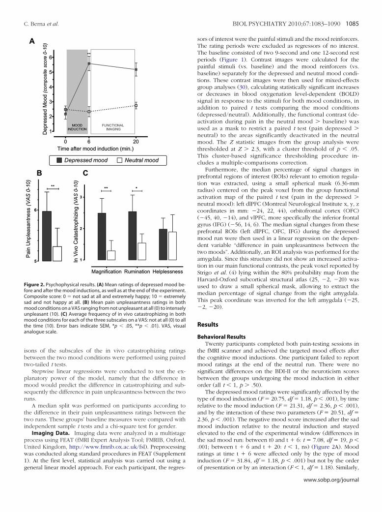
ibt
pmsr
tti
pUw1g
FfCsmumta
C. Berna et al. BIOL PSYCHIATRY 2010;67:1083–1090 1085
sons of the subscales of the in vivo catastrophizing ratingsetween the two mood conditions were performed using pairedwo-tailed t tests.
Stepwise linear regressions were conducted to test the ex-lanatory power of the model, namely that the difference inood would predict the difference in catastrophizing and sub-
equently the difference in pain unpleasantness between the twouns.
A median split was performed on participants according tohe difference in their pain unpleasantness ratings between thewo runs. These groups’ baseline measures were compared withndependent sample t tests and a chi-square test for gender.
Imaging Data. Imaging data were analyzed in a multistagerocess using FEAT (fMRI Expert Analysis Tool; FMRIB, Oxford,nited Kingdom, http://www.fmrib.ox.ac.uk/fsl). Preprocessingas conducted along standard procedures in FEAT (Supplement). At the first level, statistical analysis was carried out using a
igure 2. Psychophysical results. (A) Mean ratings of depressed mood be-ore and after the mood inductions, as well as at the end of the experiment.omposite score: 0 � not sad at all and extremely happy; 10 � extremelyad and not happy at all. (B) Mean pain unpleasantness ratings in both
ood conditions on a VAS ranging from not unpleasant at all (0) to intenselynpleasant (10). (C) Average frequency of in vivo catastrophizing in bothood conditions for each of the three subscales on a VAS: not at all (0) to all
he time (10). Error bars indicate SEM, *p � .05, **p � .01). VAS, visualnalogue scale.
eneral linear model approach. For each participant, the regres-
sors of interest were the painful stimuli and the mood reinforcers.The rating periods were excluded as regressors of no interest.The baseline consisted of two 9-second and one 12-second restperiods (Figure 1). Contrast images were calculated for thepainful stimuli (vs. baseline) and the mood reinforcers (vs.baseline) separately for the depressed and neutral mood condi-tions. These contrast images were then used for mixed-effectsgroup analyses (30), calculating statistically significant increasesor decreases in blood oxygenation level-dependent (BOLD)signal in response to the stimuli for both mood conditions, inaddition to paired t tests comparing the mood conditions(depressed/neutral). Additionally, the functional contrast (de-activation during pain in the neutral mood � baseline) wasused as a mask to restrict a paired t test (pain depressed �neutral) to the areas significantly deactivated in the neutralmood. The Z statistic images from the group analysis werethresholded at Z � 2.3, with a cluster threshold of p � .05.This cluster-based significance thresholding procedure in-cludes a multiple-comparisons correction.
Furthermore, the median percentage of signal changes inprefrontal regions of interest (ROIs) relevant to emotion regula-tion was extracted, using a small spherical mask (6.36-mmradius) centered on the peak voxel from the group functionalactivation map of the paired t test (pain in the depressed �neutral mood): left dlPFC (Montreal Neurological Institute x, y, zcoordinates in mm: �24, 22, 44), orbitofrontal cortex (OFC)(�45, 40, �14), and vlPFC, more specifically the inferior frontalgyrus (IFG) (�56, 14, 6). The median signal changes from theseprefrontal ROIs (left dlPFC, OFC, IFG) during the depressedmood run were then used in a linear regression on the depen-dent variable “difference in pain unpleasantness between thetwo moods”. Additionally, an ROI analysis was performed for theamygdala. Since this structure did not show an increased activa-tion in our main functional contrasts, the peak voxel reported byStrigo et al. (4) lying within the 80% probability map from theHarvard-Oxford subcortical structural atlas (25, �2, �20) wasused to draw a small spherical mask, allowing to extract themedian percentage of signal change from the right amygdala.This peak coordinate was inverted for the left amygdala (�25,�2, �20).
Results
Behavioral ResultsTwenty participants completed both pain-testing sessions in
the fMRI scanner and achieved the targeted mood effects afterthe cognitive mood inductions. One participant failed to reportmood ratings at the end of the neutral run. There were nosignificant differences on the BDI-II or the neuroticism scoresbetween the groups undergoing the mood induction in eitherorder (all t � 1, p � .50).
The depressed mood ratings were significantly affected by thetype of mood induction (F � 20.75, df � 1.18, p � .001), by timerelative to the mood induction (F � 21.31, df � 2.36, p � .001),and by the interaction of these two parameters (F � 20.51, df �2.36, p � .001). The negative mood score increased after the sadmood induction relative to the neutral induction and stayedelevated to the end of the experimental window (differences inthe sad mood run: between t0 and t � 6: t � 7.08, df � 19, p �.001; between t � 6 and t � 20: t � 1, ns) (Figure 2A). Moodratings at time t � 6 were affected only by the type of moodinduction (F � 31.84, df � 1.18, p � .001) but not by the order
of presentation or by an interaction (F � 1, df � 1.18). Similarly,www.sobp.org/journal

miF
oum.mppSpoi.n
mau
Frbr
1086 BIOL PSYCHIATRY 2010;67:1083–1090 C. Berna et al.
w
ood ratings at t � 20 were only affected by the type of moodnduction (F � 15.16, df � 1.17, p � .001; order of presentation:
� 1.62, df � 1.17, p � .22; interaction F � 1, p � .5).The painful stimuli, which were calibrated for a pain intensity
f 6.5 out of 10 (Supplement 1), were rated as significantly morenpleasant in the depressed (M � 5.97, SD � 1.71) than neutralood condition (M � 5.28, SD � 1.61, F � 7.7, df � 1.18, p �
01) (Figure 2B), with no effect of the order of presentation of theood inductions or interaction (F � 1 df � 1.18). Overall,articipants reported more catastrophizing thoughts in the de-ressed mood than in the neutral one (depressed: M � 2.30,D � 1.92 vs. neutral: M � 1.66, SD � 1.42, F � 9.76, df � 1.17,� .006), with no effect of order of mood induction proceduresr interaction (F � 1 df � 1.17). The mood effect on catastroph-zing was significant on the magnification (t � 3.34, df � 18, p �004) and rumination (t � 2.36, df � 18, p � .03) subscales butot on the helplessness subscale (t � 1, df � 18) (Figure 2C).
Stepwise linear regression showed that the model: depressedood ¡ increase in catastrophizing ¡ increase in pain unpleas-
ntness, explained 34% of the variability in the difference of painnpleasantness ratings (F � 4.11, df � 2.18, p � .04).
igure 3. Significant activations and deactivations during pain (red-blue, topest. Note the lack of significant deactivations in the pain contrast in the deprain. The Z coordinates shown below are on the MNI system in millimeter
esonance imaging; MNI, Montreal Neurological Institute.
ww.sobp.org/journal
Imaging Data: Whole-Brain AnalysisSignificant activation during pain (compared with rest) was
observed in a broad network of cerebral regions (Figure 3, red),including bilateral thalamus, insula, prefrontal cortex and con-tralateral somatosensory areas during both depressed and neutralconditions. In striking contrast, the pattern of simultaneousdeactivations revealed a marked and obvious qualitative differ-ence between depressed and neutral conditions. In the de-pressed mood condition, there was no significant deactivation atall. In the neutral condition, a network including the bilateralprecuneus, bilateral S1, and medial temporal lobe was deacti-vated (Figure 3, blue). A paired t test confirmed a statisticaldifference in deactivation patterns (Figure S1 and Table S2 inSupplement 1).
Quantitative increases in BOLD signal in response to painand mood reinforcers in the depressed compared with neutralconditions were investigated using a mixed effects paired t testgroup contrast. For pain, this revealed increased activation inthe subgenual anterior cingulate cortex (sACC), the left IFG,the left OFC, the left dlPFC, the left posterior insula, the lefthippocampus, the left thalamus, the left middle temporalgyrus, the left precuneus, and the left caudate, as well as the
the mood reinforcers (green-blue, bottom), each separately compared withmood. Group fMRI data of 20 participants, plotted on the average MNI 152
scales for Z test scores are shown in the middle. fMRI, functional magnetic
) andresseds. The

bm
patSrtst
sar
IB
enanS5u
FddB here;
C. Berna et al. BIOL PSYCHIATRY 2010;67:1083–1090 1087
ilateral supramarginal gyri (Figure 4A; Table S3 in Supple-ent 1).For the mood reinforcer condition, brain activity for de-
ressed mood compared with neutral was increased in the rostralnterior cingulate cortex (ACC) and perigenual ACC, as well ashe ventromedial prefrontal cortex and the OFC (Figure 4B; Table4 in Supplement 1). These areas are consistent with previouslyeported neural correlates of perceiving sad stimuli (31). The facthat there was no other activation suggests that the Veltentatements were well matched between the two mood induc-ions/reinforcers.
The opposite t test (neutral � depressed) provided noignificant results for the pain contrast and a unique confluentctivation in the left inferior temporal gyrus for the moodeinforcer contrast (Figure S2 and Table S5 in Supplement 1).
maging Data: Exploring Neural Activity Changes That Explainehavioral Variance
A linear regression revealed that the magnitude of the differ-nce in pain unpleasantness ratings between the sad and theeutral mood correlated with the left dlPFC (ß � �.65, p � .002)nd at trend level with the left IFG activity (ß � .44, p � .06) butot with the left OFC activity (ß � .23, p � .32) (Figure S3 inupplement 1). The activity in these prefrontal areas explained8% of the variability in the difference between reported pain
igure 4. Paired t tests between (depressed � neutral) run for pain (A, red) anduring pain in the depressed mood is presented (C). The % BOLD signal activepressed mood are plotted, showing differences between the strong versus nOLD, blood-oxygenation level dependent; IFG, inferior frontal gyrus; L, left hemisp
npleasantness in depressed versus neutral mood (Figure 5).
When adding the difference in catastrophizing and mood ratingsto this model, 69% of the variance in the pain ratings could beexplained.
ood reinforcers (B, green) are shown on the left. Below, the analysis of the ROIsange in the left inferior frontal gyrus and both amygdalae during pain in theunpleasantness modulation group. Error bars indicate SEM, *p � .05, **p � .01.
MNI, Montreal Neurological Institute; R, right hemisphere; ROI, region of interest.
Figure 5. Synopsis of findings and proposed model. At a cognitive level,pain experienced in an induced depressed mood leads to more negativepain-related thoughts (catastrophizing). The concurrent prefrontal neuralcorrelates are represented below. The contribution to explaining the differ-ence in affective pain ratings of these elements is reported to the right.Finally, in terms of outcome, the differences in the emotion regulation areasbetween the participants showing a strong versus no pain modulation bythe mood manipulation are shown. dlPFC, dorsolateral prefrontal cortex;
the mity cho pain
IFG, inferior frontal gyrus; OFC, orbitofrontal cortex.
www.sobp.org/journal

puM.mmsrnppTa.pm
aop
D
rttptppfsrnpbm
M
whwmn(tmrptacedit(tah
1088 BIOL PSYCHIATRY 2010;67:1083–1090 C. Berna et al.
w
To explore these associations further, a median split dividedarticipants according to the strength of the modulation of painnpleasantness by the mood manipulation (strong modulation:
� 1.52, SD � .75; weak or no modulation: M � �.17, SD �42, called no modulation group). The strong and no painodulation groups did not differ significantly on any baselineeasure or pain intensity ratings during the calibration of the
timulus (Table S1 in Supplement 1). The pain unpleasantnessatings did not differ between these two groups during theeutral mood, but the group showing strong modulation re-orted more pain unpleasantness in the depressed mood com-ared with the no modulation group (Table S1 in Supplement 1).hose participants who showed the strong effect had higherctivation in the left IFG (p � .009) and both amygdalae (left p �01, right p � .04) during pain in the depressed mood than thosearticipants with no modulation (Figure 4C; Table S1 in Supple-ent 1).Finally, activity in the left IFG correlated significantly with
ctivity in the left amygdala during pain in the depressed moodnly (depressed mood: r � .54, p � .014; neutral mood: r � .30,� .21, difference between the two correlation strengths ns).
iscussion
A sad cognitive mood induction had the predicted effects oneported depressed mood, increased the frequency of negativehoughts about the tonic pain stimulus, and increased its subjec-ive unpleasantness. A stepwise linear regression analysis sup-orted the hypothesis that depressed mood increases pain-relatedhoughts (catastrophizing), so increasing the unpleasantness of theainful experience, implying if not proving directionality. Com-ared with neutral mood, depressed mood increased the BOLDMRI signal to pain in nociceptive afferent areas and in cortico-ubcortical structures involved in emotional processing andegulation and also reduced deactivations (otherwise found ineutral mood). Participants experiencing the highest effect onain unpleasantness had higher activation in the left IFG andoth amygdalae in response to noxious stimuli during depressedood.
ood Induction, Pain Affect, and CognitionsPain was rated as more unpleasant after the sad mood induction,
hen compared with the neutral mood induction. This supports ourypothesis that depressed mood increases pain and is consistentith earlier findings (11,13,14) (Figure 2B). Furthermore, depressedood was associated with increases in negative pain-related cog-itions (catastrophizing), suggesting a mechanism for this effectFigure 5). The proposed model explained 34% of the variability inhe difference in affective pain ratings. While this might seemodest, only a few behavioral measures were collected. In this
espect, our findings were of similar magnitude to those reportedreviously (32). Furthermore, when including measures of prefron-al activity in the regression analysis, 69% of the variability inffective pain ratings could be explained. The model we propose isonsistent with a clinical model of pain in which negative affectivityxacerbates a vicious cycle of negative pain-related cognitions andistress and drives subsequent increases in the perception andmpact of pain (33). The mood manipulation specifically affectedhe worry-related subscales of the in vivo catastrophizing scalemagnification and rumination) (Figure 2C). This is interesting givenhe emphasis on rumination as a driving process in depression (34)nd recent cognitive models of chronic pain, which have also
ighlighted worry as an important maintaining factor (35).ww.sobp.org/journal
Neuroimaging FindingsIncreased Activation during Pain in Depressed Mood. The
areas that showed increased activity during tonic pain in thedepressed mood included the left insula, thalamus, hippocam-pus, IFG, dlPFC, OFC, and the sACC (Figure 4A). The thalamusand the insular cortex are part of the afferent nociceptivenetwork (36). The sACC is commonly activated during negativemood (31) and appears to be a key area for depression (37).Increased activity in the sACC has only rarely been reported infMRI studies of pain (e.g., [38]). It has been suggested that thisregion responds selectively to negative emotional processing ofpersonally relevant material; acute exogenous experimental stim-uli may not meet this criterion (39). This argument is alsosupported by the depression-related increase in activity in thehippocampal formation. This region is known to be involved inanxiety-induced hyperalgesia (40) and nocebo-induced hyperal-gesia (41). Furthermore, the hippocampus is connected with thesACC (42), and these structures have been proposed to be part ofa dysfunctional limbic-frontal circuitry in major depression (43).
Activations in Areas Relevant to Emotion Regulation. Given re-cent findings in depressed patients (4), we hypothesized thatdepressed mood would impair emotion regulation of pain affect.When presented with an aversive stimulus, different types ofautomatic or voluntary cognitive processes can help the individ-ual to cope emotionally. The prefrontal cortex is highly involvedin these emotion regulation processes (44). Effortful modulationis thought to be predominantly underpinned by lateral structures(vlPFC, dlPFC), while automatic emotion regulation appears tobe mostly mediated by medial structures; however, some struc-tures, such as the dorsal ACC and the OFC, are shared by the twosystems (45).
We found that both the activity in the dlPFC and IFG duringthe negative mood was correlated with measured differences inaffective pain ratings between the two mood conditions but inopposite ways (Figure S3 in Supplement 1). Increased activityin the left dlPFC predicted a smaller difference in pain unpleas-antness between the two moods, consistent with activationrelated to successful downregulation of pain unpleasantness.However, the left IFG activity was positively correlated with thedifference in pain unpleasantness ratings. The IFG peak coordi-nate in this study is located posterior to the vlPFC regionpreviously implicated in the downregulation of pain due toperceived control over the stimulus (46,47). In fact, our peakactivation closely corresponds to the area within the vlPFC,which has been identified in studies of cognitive reappraisal ofsad emotion and alternatively named IFG or vlPFC (17,48–51).Wager et al. (49) have suggested that two separate pathsoriginate from this functional area: the first one, linked to thenucleus accumbens, is involved in generating positive reap-praisal, while the second one, connected to the amygdala, isthought to generate or enhance negative appraisals.
Supporting this notion, activity in the amygdala and the IFGwas significantly correlated during pain in the depressed mood.Furthermore, results from the median split analysis showed thatthe participants with the greatest increase in pain unpleasantnessduring depressed mood also showed significantly higher amyg-dala activation during this condition (Figure 4C). This suggeststhat the IFG, a structure that could exert positive reappraisal,instead underpinned either ineffective or detrimental emotionregulation during depressed mood. This notion is supported bythe Wager et al. (49) findings and by a recent study of emotional
and attentional pain modulation, which similarly identified the
Ism
iFssllss
sdtmadn(asc
pmima
FdEgbsWao
BPMAaro
o
C. Berna et al. BIOL PSYCHIATRY 2010;67:1083–1090 1089
FG as a modulator in the emotional process (52). The lattertudy also provided strong evidence against the notion that aood modulation was merely a hidden attentional manipulation.Our fMRI results show that in a depressed mood, volunteers
ncreased activity in areas involved in emotional appraisal.urthermore, despite the left-sided pain stimulation used in thistudy, a majority of the emotion regulation circuitry recruitedeemed lateralized to the left hemisphere (Figure 4A). Neverthe-ess, this potential lateralization was not tested formally, asateralization of emotional processing was not the focus of thistudy and investigating this debated topic would require a morepecific design (53).
Depressed Mood Affects Deactivations During Pain. Noignificant task-induced deactivations (54–56) were observeduring pain in the depressed mood condition (Figure 3). Areashat were more deactivated in the neutral than the depressedood (Figure S1 in Supplement 1) included the left angular gyrus
nd the bilateral precuneus and posterior cingulate. This lack ofeactivation could be linked to changes in the default modeetwork during the negative mood, as previously demonstrated57). While the rest periods in this study were too short to allowproper analysis of resting state networks, previous research has
uggested that depressed mood states are associated with in-reased cognitive load (58).
In conclusion, the fact that mood and cognition can influenceain perception at a neural level suggests that interventions toodify these processes may indeed be useful to reduce pain. Such
nsights about mood and cognition will be critical for the develop-ent of better treatments for chronic pain, both psychological (such
s cognitive behavior therapy) and pharmacological.
CB is supported by a Lord Florey scholarship of the Berrowoundation (Lincoln College, University of Oxford, United King-om) and an Overseas Research Student Award Scheme (Higherducation Funding Council for England, United Kingdom). SL israteful for funding from the Wellcome Trust. EAH is supportedy a Royal Society Dorothy Hodgkin fellowship. RRE receivedupport from the International Association for the Study of Pain.e acknowledge the Medical Research Council of Great Britain
nd Northern Ireland (Functional Magnetic Resonance Imagingf the Brain Centre).
Professor Goodwin reports receiving honoraria from AstraZeneca,ristol-Myers Squibb, Eisai, Lundbeck, and Servier; holding shares in1Vital; and serving on advisory boards for AstraZeneca, Bristol-yers Squibb, Janssen Cilag, Lilly, Lundbeck, P1Vital, Sanofi-ventis, Servier, and Wyeth. Professor Goodwin has also serveds an expert witness for Lilly and Servier. The other authorseported no biomedical financial interests or potential conflictsf interest.
Supplementary material cited in this article is availablenline.
1. Bair MJ, Robinson RL, Katon W, Kroenke K (2003): Depression and paincomorbidity: A literature review. Arch Intern Med 163:2433–2445.
2. Geisser ME, Roth RS, Theisen ME, Robinson ME, Riley JL 3rd (2000):Negative affect, self-report of depressive symptoms, and clinical de-pression: Relation to the experience of chronic pain. Clin J Pain 16:110 –120.
3. Bar KJ, Wagner G, Koschke M, Boettger S, Boettger MK, Schlosser R, et al.(2007): Increased prefrontal activation during pain perception in majordepression. Biol Psychiatry 62:1281–1287.
4. Strigo IA, Simmons AN, Matthews SC, Craig AD, Paulus MP (2008): Asso-
ciation of major depressive disorder with altered functional brain re-sponse during anticipation and processing of heat pain. Arch GenPsychiatry 65:1275–1284.
5. Giesecke T, Gracely RH, Williams DA, Geisser ME, Petzke FW, Clauw DJ(2005): The relationship between depression, clinical pain, and experi-mental pain in a chronic pain cohort. Arthritis Rheum 52:1577–1584.
6. Schweinhardt P, Kalk N, Wartolowska K, Chessell I, Wordsworth P, TraceyI (2008): Investigation into the neural correlates of emotional augmen-tation of clinical pain. Neuroimage. 40:759 –766.
7. Apkarian AV, Sosa Y, Sonty S, Levy RM, Harden RN, Parrish TB, et al.(2004): Chronic back pain is associated with decreased prefrontal andthalamic gray matter density. J Neurosci 24:10410 –10415.
8. Campbell S, MacQueen G (2006): An update on regional brain volumedifferences associated with mood disorders. Curr Opin Psychiatry 19:25–33.
9. Williams JM, Barnhofer T, Crane C, Beck AT (2005): Problem solvingdeteriorates following mood challenge in formerly depressed patientswith a history of suicidal ideation. J Abnorm Psychol 114:421– 431.
10. Goodwin AM, Williams JMG (1982): Mood-induction research—its im-plications for clinical depression. Behav Res Ther 20:373–382.
11. Loggia ML, Mogil JS, Bushnell MC (2008): Experimentally induced moodchanges preferentially affect pain unpleasantness. J Pain 9:784 –791.
12. Villemure C, Slotnick BM, Bushnell MC (2003): Effects of odors on painperception: Deciphering the roles of emotion and attention. Pain 106:101–108.
13. Rainville P, Bao QVH, Chretien P (2005): Pain-related emotions modulateexperimental pain perception and autonomic responses. Pain 118:306 –318.
14. Zelman DC, Howland EW, Nichols SN, Cleeland CS (1991): The effects ofinduced mood on laboratory pain. Pain 46:105–111.
15. Sharp TJ (2001): Chronic pain: A reformulation of the cognitive-behav-ioural model. Behav Res Ther 39:787– 800.
16. Geisser ME, Robinson ME, Keefe FJ, Weiner ML (1994): Catastrophizing,depression and the sensory, affective and evaluative aspects of chronicpain. Pain 59:79 – 83.
17. Johnstone T, van Reekum CM, Urry HL, Kalin NH, Davidson RJ (2007):Failure to regulate: Counterproductive recruitment of top-down pre-frontal-subcortical circuitry in major depression. J Neurosci 27:8877–8884.
18. Beauregard M, Paquette V, Levesque J (2006): Dysfunction in the neuralcircuitry of emotional self-regulation in major depressive disorder. Neu-roreport 17:843– 846.
19. Clark DM (1983): On the induction of depressed mood in the laboratory:Evaluation and comparison of the Velten and musical procedures. AdvBehav Res Ther 5:27– 49.
20. Edwards RR, Smith MT, Stonerock G, Haythornthwaite JA (2006): Pain-related catastrophizing in healthy women is associated with greatertemporal summation of and reduced habituation to thermal pain. Clin JPain 22:730 –737.
21. Beck AT, Steer RA, Brown GK (1996): Manual for the Back DepressionInventory-II. San Antonio, TX: Psychological Corporation.
22. Sheehan DV, Lecrubier Y, Sheehan KH, Amorim P, Janavs J, Weiller E, etal. (1998): The Mini-International Neuropsychiatric Interview (M.I.N.I.):The development and validation of a structured diagnostic psychiatricinterview for DSM-IV and ICD-10. J Clin Psychiatry 59(suppl 20):22–33;quiz 34 –57.
23. Eysenck SBG, Eysenck HJ, Barrett P (1985): A revised version of thepsychoticism scale. Pers Individ Dif 6:21–29.
24. Velten E Jr (1968): A laboratory task for induction of mood states. BehavRes Ther 6:473– 482.
25. Richell RA, Anderson M (2004): Reproducibility of negative mood induction:A self-referent plus musical mood induction procedure and a controllable/uncontrollable stress paradigm. J Psychopharmacol 18:94–101.
26. Clark DM, Teasdale JD (1985): Constraints on the effects of mood onmemory. J Pers Soc Psychol 6:1595–1608.
27. Au Yeung C, Dalgleish T, Golden A-M, Schartau P (2006): Reduced spec-ificity of autobiographical memories following a negative mood induc-tion. Behav Res Ther 44:1481–1490.
28. Seibert PS, Ellis HC (1991): A convenient self-referencing mood induc-tion procedure. Bull Psychon Soc 29:121–124.
29. Price DD, McGrath PA, Rafii A, Buckingham B (1983): The validation ofvisual analogue scales as ratio scale measures for chronic and experi-mental pain. Pain 17:45–56.
30. Beckmann CF, Jenkinson M, Smith SM (2003): General multilevel linear
modeling for group analysis in fMRI. Neuroimage 20:1052–1063.www.sobp.org/journal

3
3
3
3
3
3
3
3
3
4
4
4
4
4
4
1090 BIOL PSYCHIATRY 2010;67:1083–1090 C. Berna et al.
w
1. Phan KL, Wager T, Taylor SF, Liberzon I (2002): Functional neuroanatomyof emotion: A meta-analysis of emotion activation studies in PET andfMRI. Neuroimage 16:331–348.
2. Rhudy JL, Williams AE, McCabe KM, Russell JL, Maynard LJ (2008): Emo-tional control of nociceptive reactions (ECON): Do affective valence andarousal play a role? Pain 136:250 –261.
3. Vlaeyen JWS, Linton SJ (2000): Fear-avoidance and its consequences inchronic musculoskeletal pain: A state of the art. Pain 85:317–332.
4. Nolen-Hoeksema S (2000): The role of rumination in depressive disor-ders and mixed anxiety/depressive symptoms. J Abnorm Psychol 109:504 –511.
5. Eccleston C, Crombez G (2007): Worry and chronic pain: A misdirectedproblem solving model. Pain 132:233–236.
6. Tracey I, Mantyh PW (2007): The cerebral signature for pain perceptionand its modulation. Neuron 55:377–391.
7. Mayberg HS, Liotti M, Brannan SK, McGinnis S, Mahurin RK, Jerabek PA,et al. (1999): Reciprocal limbic-cortical function and negative mood:Converging PET findings in depression and normal sadness. Am J Psy-chiatry 156:675– 682.
8. Bingel U, Schoell E, Herken W, Buchel C, May A (2007): Habituation topainful stimulation involves the antinociceptive system. Pain 131:21–30.
9. Vogt BA (2005): Pain and emotion interactions in subregions of thecingulate gyrus. Nat Rev Neurosci 6:533–544.
0. Ploghaus A, Narain C, Beckmann CF, Clare S, Bantick S, Wise R, et al.(2001): Exacerbation of pain by anxiety is associated with activity in ahippocampal network. J Neurosci 21:9896 –9903.
1. Kong J, Gollub RL, Polich G, Kirsch I, Laviolette P, Vangel M, et al. (2008):A functional magnetic resonance imaging study on the neural mecha-nisms of hyperalgesic nocebo effect. J Neurosci 28:13354 –13362.
2. Johansen-Berg H, Gutman DA, Behrens TEJ, Matthews PM, RushworthMFS, Katz E, et al. (2008): Anatomical connectivity of the subgenualcingulate region targeted with deep brain stimulation for treatment-resistant depression. Cereb Cortex 18:1374 –1383.
3. Seminowicz DA, Mayberg HS, McIntosh AR, Goldapple K, Kennedy S,Segal Z, et al. (2004): Limbic-frontal circuitry in major depression: A pathmodeling metanalysis. Neuroimage. 22:409 – 418.
4. Tucker DM, Luu P, Pribram KH (1995): Social and emotional self-regula-tiona. Ann N Y Acad Sci 769:213–240.
5. Phillips ML, Ladouceur CD, Drevets WC (2008): A neural model of volun-
tary and automatic emotion regulation: Implications for understandingww.sobp.org/journal
the pathophysiology and neurodevelopment of bipolar disorder. MolPsychiatry 13:833– 857.
46. Kalisch R, Wiech K, Critchley HD, Seymour B, O’Doherty JP, Oakley DA, etal. (2005): Anxiety reduction through detachment: Subjective, physio-logical, and neural effects. J Cogn Neurosci 17:874.
47. Wiech K, Kalisch R, Weiskopf N, Pleger B, Stephan KE, Dolan RJ (2006):Anterolateral prefrontal cortex mediates the analgesic effect of ex-pected and perceived control over pain. J Neurosci 26:11501–11509.
48. Dolcos F, McCarthy G (2006): Brain systems mediating cognitive inter-ference by emotional distraction. J Neurosci 26:2072–2079.
49. Wager TD, Davidson ML, Hughes BL, Lindquist MA, Ochsner KN (2008):Prefrontal-subcortical pathways mediating successful emotion regula-tion. Neuron 59:1037–1050.
50. Ochsner KN, Ray RD, Cooper JC, Robertson ER, Chopra S, Gabrieli JDE, etal. (2004): For better or for worse: Neural systems supporting the cogni-tive down- and up-regulation of negative emotion. Neuroimage 23:483–499.
51. Phan KL, Fitzgerald DA, Nathan PJ, Moore GJ, Uhde TW, Tancer ME(2005): Neural substrates for voluntary suppression of negative affect: Afunctional magnetic resonance imaging study. Biol Psychiatry 57:210 –219.
52. Villemure C, Bushnell MC (2009): Mood influences supraspinal painprocessing separately from attention. J Neurosci 29:705–715.
53. Wager TD, Phan KL, Liberzon I, Taylor SF (2003): Valence, gender, andlateralization of functional brain anatomy in emotion: A meta-analysisof findings from neuroimaging. Neuroimage 19:513–531.
54. Mazoyer B, Zago L, Mellet E, Bricogne S, Etard O, Houdé O, et al. (2001):Cortical networks for working memory and executive functions sustainthe conscious resting state in man. Brain Res Bull 54:287–298.
55. McKiernan KA, Kaufman JN, Kucera-Thompson J, Binder JR (2003): Aparametric manipulation of factors affecting task-induced deactivationin functional neuroimaging. J Cogn Neurosci 15:394 – 408.
56. Shulman GL, Fiez JA, Corbetta M, Buckner RL, Miezin FM, Raichle ME, etal. (1997): Common blood flow changes across visual tasks. II. Decreasesin cerebral cortex. J Cogn Neurosci 9:648 – 663.
57. Harrison BJ, Pujol J, Ortiz H, Fornito A, Pantelis C, Yucel M (2008): Mod-ulation of brain resting-state networks by sad mood induction. PLoSONE 3:e1794.
58. Wegner DM, Erber R, Zanakos S (1993): Ironic processes in the mental
control of mood and mood-related thought. J Pers Soc Psychol 65:1093–1104.