inductive ability of human developing and differentiated dental · pdf file ·...
TRANSCRIPT

E-Mail [email protected]
Original Paper
Cells Tissues Organs 2013;198:99–110 DOI: 10.1159/000353116
Inductive Ability of Human Developing and Differentiated Dental Mesenchyme
Liwei Zheng a, b Rungnapa Warotayanont a Jonathan Stahl a Ryo Kunimatsu a
Ophir Klein a Pamela K. DenBesten a Yan Zhang a
a Department of Orofacial Sciences, University of California, San Francisco, Calif. , USA; b State Key Laboratory ofOral Diseases, Sichuan University, Chengdu , China
developing enamel-like structures could be detected in the complexes resulting from in vitro and ex vivo co-culture of ES-ECs and FDMCs. In contrast, co-cultured ES-ECs andADMCs formed amorphous spherical structures and occa-sionally formed hair . Transcription factors were significantly upregulated in FDMCs compared to ADMCs including MSX1 , GLI1 , LHX6 , LHX8, LEF1 and TBX1 . In summary, FDMCs but not ADMCs had the capacity to induce differentiation of ES-ECs into ameloblast lineage cells. Further characterization of the functional differences between these two types of dental mesenchyme could enable reprogramming of ADMCs to en-hance their odontogenic inductive competence.
© 2013 S. Karger AG, Basel
Introduction
Ameloblasts, highly specialized enamel-forming den-tal epithelial cells (ECs), go through multiple stagesof differentiation during tooth development. As amelo-blasts differentiate from the presecretory stage to the se-cretory stage, enamel matrix proteins consisting primar-ily of amelogenins are synthesized and secreted. Amelo-genins are alternatively spliced matrix proteins required for nucleation and growth of enamel hydroxyapatite crys-
Key Words
Ameloblasts · Amelogenin · Dental mesenchymal cells · Human embryonic stem cells · Odontogenic competence · Reprogramming · Transcription factors
Abstract
The development of cell-based therapeutic strategies to bio-engineer tooth tissue is a promising approach for the treat-ment of lost or damaged tooth tissue. The lack of a readily available cell source for human dental epithelial cells (ECs) severely constrains the progress of tooth bioengineering. Previous studies in model organisms have demonstrated that developing dental mesenchyme can instruct nondental epithelium to differentiate into enamel-forming epithelium. In this study, we characterized the ability of fetal and adult human dental mesenchyme to promote differentiation of human embryonic stem cell (hESC)-derived ECs (ES-ECs) into ameloblast-lineage cells. ES-ECs were co-cultured either withhuman fetal dental mesenchymal cells (FDMCs) or with adult dental mesenchymal cells (ADMCs) in either a three-dimen-sional culture system, or in the renal capsules of SCID mice. When co-cultured with FDMCs in vitro, ES-ECs polarized and expressed amelogenin. Tooth organ-like structures as-sembled with epithelium and encased mesenchyme and
Accepted after revision: May 13, 2013 Published online: July 30, 2013
Dr. Yan Zhang Department of Orofacial Sciences University of California, 513 Parnassus Avenue San Francisco, CA 94143-0422 (USA) E-Mail yan.zhang2 @ ucsf.edu
© 2013 S. Karger AG, Basel1422–6405/13/1982–0099$38.00/0
www.karger.com/cto
Dow
nloa
ded
by:
UC
SF
Lib
rary
& C
KM
12
8.21
8.19
1.10
3 -
11/7
/201
3 6:
23:4
7 P
M
Zheng /Warotayanont /Stahl /Kunimatsu /Klein /DenBesten /Zhang
Cells Tissues Organs 2013;198:99–110 DOI: 10.1159/000353116
100
tals during amelogenesis [Fincham et al., 1994; Mora-dian-Oldak et al., 1998; Beniash et al., 2005]. In the matu-ration stage, these proteins are subsequently removed, and mineralization is completed. This process leads to the formation of tooth enamel, which is the hardest tissue in the human body, providing protection, strength and wear resistance to the teeth.
Tooth tissue formation is guided by reciprocal signal-ing interactions between the ectodermally derived dental epithelium and cranial neural crest-derived dental mes-enchyme [Thesleff and Hurmerinta, 1981; Ruch et al., 1983; Thesleff et al., 1995a; Thesleff and Sharpe, 1997; Thesleff and Mikkola, 2002]. The first sign of tooth tissue formation is the localized thickening of oral ECs lining the first branchial arch in the location where teeth will form. The presumptive dental ECs form an epithelial band (so-called dental placode) and invaginate into the underlying mesenchyme. In mice, ECs from the dental placode can instruct tooth formation ex vivo when re-combined with either dental mesenchyme or nondental mesenchyme [Cummings et al., 1981; Mina and Kollar, 1987; Thesleff et al., 1995a].
In response to the epithelially derived signals, which include bone morphogenetic protein (BMP)4 and fi-broblast growth factor (FGF)8 [Thomas et al., 1997; Bei and Maas, 1998; Hardcastle et al., 1998], the underlying mes enchymal cells proliferate and condense around the
thickened epithelium to form a bud-like structure. Recip-rocally, the dental mesenchyme secretes BMP4, WNT5a, FGF3 and other signaling molecules to instruct the dental epithelium to form a cap-like enamel organ, where polar-ized and columnar inner enamel ECs appear [Cai et al., 2011]. In the following bell stage of tooth development, inner enamel ECs further elongate to differentiate into pre-ameloblasts, and the adjacent layer of dental mesen-chyme differentiates into odontoblasts. After odonto-blasts start to deposit dentin matrix, presecretory ame-loblasts progress to secretory ameloblasts, then to tran-sition and maturation ameloblasts. In the fully formed teeth, dental mesenchyme forms dentin, odontoblasts and vital pulp tissue, in which mesenchymal stem cells still remain [Gronthos et al., 2000, 2002]. Secretory ame-loblasts produce stage-specific enamel matrix proteins, including amelogenin, ameloblastin and enamelin [Ro b-inson et al., 1998], to modulate enamel crystal formation. Maturation ameloblasts finalize the mineralization pro-cess by removing matrix proteins and depositing min-erals.
Evidence from developmental biology studies indi-cates that cell-based tooth tissue bioengineering will re-quire both a viable source of epithelial cells and a source of dental mesenchyme to reconstitute the cascade of re-ciprocal signaling interactions that can lead to tooth for-mation. Mesenchymal stem cells, available from dental pulp of human exfoliated deciduous teeth and extracted adult molars, as well as from root apical dental papilla, are capable of regenerating dentin and pulp complex [Gronthos et al., 2000, 2002; Miura et al., 2003; Sonoya-ma et al., 2008; Estrela et al., 2011]. In humans, enamel loses the capacity to regenerate itself since enamel-form-ing ame loblasts are lost once the teeth erupt into the oral cavity [Vaahtokari et al., 1996]. Therefore, the lack of readily available human dental ECs severely constrains the advance of enamel tissue and whole tooth bioengi-neering.
Human embryonic stem cells (hESCs) have been con-sidered as a potential source of ECs for skin tissue regen-eration [Green et al., 2003; Clark et al., 2004; Metallo et al., 2008]. We have shown that epithelial cells differenti-ated from hESCs express FGF8 , MSX 1 and cytokeratins (CKs) similar to those found in human ameloblast lineage cells [Zheng et al., 2013]. In this study, we determined the ability of human dental mesenchyme derived from devel-oping fetal primary tooth organs and adult dental mesen-chyme (including adult dental pulp stem cells) to differ-entiate hESC-derived ECs (ES-ECs) into ameloblast lin-eage cells.
Abbreviations used in this paper
ADMCs adult dental mesenchymal cells (derived from human permanent teeth)
BMP bone morphogenetic proteinCK cytokeratinDMC dental mesenchymal cellDMEM Dulbecco’s modified Eagle’s mediumEC epithelial cellES-ECs human embryonic stem cell-derived epithelial cellsF12 Ham’s F12FGF fibroblast growth factorFDMCs fetal dental mesenchymal cells (derived from human
developing tooth organs)hESCs human embryonic stem cellsKGM2 keratinocyte growth medium 2LEF1 lymphoid enhancer-binding factor 1LHX LIM/homeobox proteinMSX1 Msh homeobox 1qPCR quantitative polymerase chain reactionSHH sonic hedgehog homologTBX1 T-box 1VitC vitamin C
Dow
nloa
ded
by:
UC
SF
Lib
rary
& C
KM
12
8.21
8.19
1.10
3 -
11/7
/201
3 6:
23:4
7 P
M

Inductive Ability of Human Developing and Differentiated Dental Mesenchyme
Cells Tissues Organs 2013;198:99–110 DOI: 10.1159/000353116
101
Materials and Methods
Induction of hESCs The hESC line WA09 (WiCell Research Institute, Madison,
Wisc., USA) was cultured and maintained on a layer of mitotically inactivated mouse embryonic fibroblast cells in Dulbecco’s modi-fied Eagle’s medium/Ham’s F12 (DMEM/F12) containing 20% knockout serum replacement (Invitrogen, Carlsbad, Calif., USA), 1x MEM nonessential amino acids (Sigma-Aldrich, St. Louis, Mo., USA), 1 m M L -glutamine, 0.1 m M β-mercaptoethanol, 4 ng/ml ba-sic FGF (Invitrogen), and 50 mg/ml penicillin and streptomycin [Klimanskaya et al., 2004]. All procedures using the hESCs fol-lowed the guidelines set by the Human Gamete, Embryo and Stem Cell Research Comittee at USCF. Prior to induction, hESCs were passaged twice on Matrigel TM (BD Biosciences, San Jose, Calif., USA)-coated dishes to eliminate contamination of mouse embry-onic fibroblast cells. For epithelial induction, hESCs were cultured in DMEM/F12 supplemented with 1 μ M retinoid acid (Sigma-Aldrich) and 12.5 ng/ml BMP4 (Invitrogen) for 7 days. The me-dium was changed daily. Cell morphology was monitored under an inverted phase-contrast microscope. Expression of the EC marker, CK14, was analyzed by quantitative polymerase chain re-action (qPCR) using total RNA purified from induced hESCs.
Upon achieving an epithelial phenotype identified by cobble-stone cell morphology and increased CK14 expression, several cul-ture conditions were used to explore the possibility that enamel ma-trix proteins and Wnt signaling could induce further differentiation of ES-ECs toward an ameloblast lineage. ES-ECs were cultured for 3 days in either keratinocyte growth medium 2 (KGM2; Lonza Inc., Walkersville, Md., USA), KGM2 supplemented with 100 μ M LiCl (Sigma-Aldrich), KGM2 supplemented with 1 μg/ml purified re-combinant human amelotin protein, KGM2 supplemented with 1 μg/ml purified recombinant human amelogenin protein, or human fetal ameloblast lineage cell conditioned medium.
Co-culture of ES-ECs with Dental Mesenchymal Cells All human tissues were collected under regulations and guide-
lines set by the University of California San Francisco Committee on Human Research. Human tooth organs were dissected from 16- to 18-week-old fetal cadavers. Tooth organs were digested with Dispase II (Roche Applied Science, Indianapolis, Ind., USA) for 15 min at 37 ° C. Dental epithelia were then separated from dental mesenchyme using fine point forceps under a dissecting micro-scope (SMZ1000; Nikon). Epithelial and mesenchymal tissues were digested with 2 mg/ml collagenase/Dispase II (Roche Applied Science) at 37 ° C for 2 h, followed by digestion with 0.05% trypsin/EDTA for 5 min at 37 ° C. KGM2 supplemented with 0.05 m M cal-cium was used to grow ameloblast lineage cells as described previ-ously [Yan et al., 2006]. The medium was collected daily to be used as human fetal ameloblast lineage cell conditioned medium to in-duce the differentiation of ES-ECs as mentioned above.
Human fetal dental mesenchymal cells (FDMCs) isolated from developing primary tooth organs were grown in DMEM supple-mented with 15% fetal bovine serum, 50 mg/ml penicillin and streptomycin. Cells isolated from pulp tissue of extracted human third molars (adult dental mesenchymal cells, ADMCs) were cul-tured in DMEM as described previously [Gronthos et al., 2000; Miura et al., 2003].
Cultured FDMCs or ADMCs were harvested at 90% conflu-ence. ES-ECs were harvested 7 days after induction. ES-ECs and
either FDMCs or ADMCs were then co-cultured in a 3D Matrigel drop system to facilitate cell compartmentalization for multicel-lular assembly and reciprocal signaling crosstalk. In this system, one million FDMCs or ADMCs were resuspended in 25 μl DMEM, and then mixed with 25 μl ice-chilled Matrigel (BD Biosciences). The cell/Matrigel complex was dropped onto a Transwell mem-brane (BD Biosciences). One million ES-ECs were then resuspend-ed in 25 μl KGM2 and mixed with 25 μl ice-chilled Matrigel. The EC-Matrigel complex was laid on the top of either FDMC or ADMC Matrigel drop. KGM2 was added to the upper chamber of Transwell to culture ECs, and DMEM supplemented with 10% FBS was added to the lower chamber of Transwell ( fig. 1 a). Co-cultures were maintained at 37 ° C in 5% carbon dioxide. One week later, KGM2 was supplemented with 1 m M calcium, and DMEM was supplemented with 10 n M dexamethasone, 50 μg/ml ascorbic acid, 10 m M β-glycerophosphate and 2 m M L -glutamine. ES-ECs,ADMCs or FDMCs alone were cultured in Matrigel to serve as controls. Cell/Matrigel complexes were maintained in these sup-plemented media for 8 weeks.
After rinsing with cold PBS, cell/Matrigel complexes were har-vested, embedded in OCT, and cryosectioned for histological anal-ysis. For gene expression analysis, cells were recovered from Matri-gel using cell recovery solution (BD Biosciences) according to the manufacturer’s instruction.
Gene Expression Analysis by qPCR Total RNA was purified from the cells recovered from Matrigel
using the RNeasy plus micro kit (Qiagen, Valencia, Calif., USA). A cDNA library was generated from 0.1 μg of total RNA using the SuperScript TM III first-strand synthesis system (Invitrogen). Ex-pression of target genes was characterized by qPCR using the ABI 7500 system (Applied Biosystems, Carlsbad, Calif., USA). Primers and probes used to detect endogenous control 18S, and target genes were also purchased from Applied Biosystems. Relative ex-pression levels of target genes were measured by the ΔΔCt method as previously published [Thomsen et al., 2010]. Data were analyzed using one-way ANOVA followed by Tukey’s post hoc test; p < 0.05 was considered significant.
Histological and Immunohistochemical Studies Cell/Matrigel complexes were washed with PBS, incubated in
20% sucrose at 37 ° C for 6 h, embedded in OCT compound and then cryosectioned. The sections were either stained with hema-toxylin and eosin, or immunostained for amelogenin protein.
Amelogenin immunostaining was performed by first blocking the sections with 3% goat serum, 0.1% BSA and 0.1% Triton for1 h at room temperature. The sections were then incubated over-night at 4 ° C with polyclonal rabbit anti-recombinant human am-elogenin IgG, as previously described [Zhang et al., 2006]. The sec-tions were then thoroughly washed with PBS and incubated with FITC-conjugated anti-rabbit secondary antibody (Sigma-Aldrich) for 1 h. Cell nuclei were counterstained with 0.5 μg/ml Hoechst 33342 (Invitrogen) for 5 min. After mounting, the sections were photographed under a Nikon Eclipse 300 fluorescence microscope (Compix Inc., Sewickley, Pa., USA).
Renal Capsule Transplantation Nine-week-old SCID CD-1 male mice (Charles River, Wil-
mington, Mass., USA) were housed in the animal barrier facility of the University of California, San Francisco, Calif., USA, in accor-
Dow
nloa
ded
by:
UC
SF
Lib
rary
& C
KM
12
8.21
8.19
1.10
3 -
11/7
/201
3 6:
23:4
7 P
M

Zheng /Warotayanont /Stahl /Kunimatsu /Klein /DenBesten /Zhang
Cells Tissues Organs 2013;198:99–110 DOI: 10.1159/000353116
102
dance with the guideline approved by UCSF Laboratory Animal Resource Center.
ES-ECs and either FDMCs or ADMCs were co-cultured in Matrigel as described above, then maintained for 3 days in vitro prior to transplantation. ADMCs, FDMCs or ES-ECs cultured in Matrigel were included as controls. ADMCs/ES-ECs, FDMCs/ES-ECs and control cells, all in Matrigel, were then transplanted into renal capsules of SCID mice. Each renal capsule maintained 2 trans-plants. Transplants were recovered 8 weeks after transplantation, dehydrated in 20% sucrose overnight and fixed in 4% buffered paraformaldehyde, and then processed for hematoxylin and eosin staining and immunostaining.
Comparative Gene Expression Analysis of Human FDMCs and ADMCs Total RNA was purified from first-passage human FDMCs and
ADMCs using a Qiagen RNeasy mini kit, and cDNA was generated using the SuperScript III first-strand synthesis system (Invitrogen). Conventional PCR was used to amplify transcription factors, in-cluding MSX (Msh homeobox) 1, MSX2 , DLX1 , DLX2 , DLX5 , GLI1 , GLI2 , GLI3 , LHX (LIM/homeobox protein) 6, LHX8 , LEF1 (lym-phoid enhancer-binding factor 1), RUNX2 , PAX9 , PRX1 , PRX2 and TBX1 (T-box 1; see table 1 for primer sequences). After PCR ampli-fication, an equal volume of each PCR product was separated on agarose gel by electrophoresis. The intensities of DNA bands were measured using NIH ImageJ (version 1.46) software. The expression level of each gene was normalized to GAPDH serving as an endog-enous control. Student’s t test was performed to compare the relative expression level of each target gene in FDMCs and ADPCs.
Characterization of the Odontogenic Inductive Competence of LEF1-Transduced ADMCs The first passage of 1 × 10 5 ADMCs was placed into a 6-well
culture dish. The following day, 2 μg of CMV promoter-driven hu-man LEF1 mammalian expression vector (a gift from Dr. Marian Waterman, University of California, Irvine, Calif., USA) in 100 μl of serum-free DMEM was transfected into ADMCs with 6 μl of FuGENE HD transfection reagent (Roche Applied Science) fol-lowing the manufacturer’s instruction. The transfection efficiency was determined by measuring the relative expression levels of LEF1 in the ADMCs 24 and 48 h after transfection, respectively, by qPCR. Forty-eight hours after transfection, 1 × 10 5 ES-ECs were placed and cultured on the top of ADMCs. Co-cultured cells were grown in KGM2 with or without 50 μg/ml vitamin C (VitC) and 10 n M sonic hedgehog (SHH) recombinant protein (Abcam, Cam-bridge, Mass., USA) for 3 days. Total RNA was purified using the RNeasy mini kit (Qiagen), and cDNA was synthesized from 2 μg of total RNA per sample. Amelogenin expression levels in the co-cultured cells were determined by qPCR.
Results
Induction of an Epithelial Phenotype from hESCs After culture with 1 μ M retinoid acid and 12.5 ng/ml
BMP4 for 7 days, hESCs on Matrigel adopted a cobble-stone-like epithelial morphology. As reported previously [Zheng et al., 2013], expression levels of the EC marker
CK14 were upregulated to an average of 44-fold in ES-ECs compared to noninduced hESCs. Addition of exog-enous factors in the culture including LiCl, recombinant human amelogenin protein, human amelotin protein or human fetal ameloblast lineage cell conditioned media failed to promote ES-ECs to express amelogenin indicat-ed by qPCR analysis (data not shown).
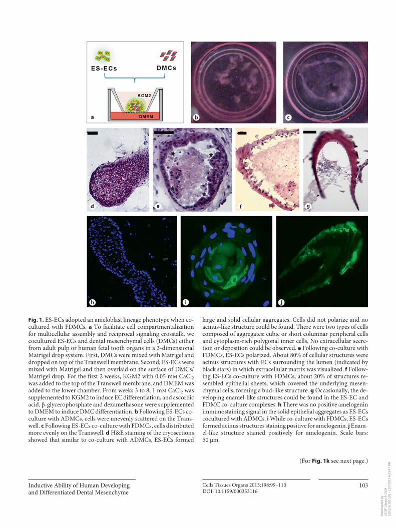
FDMCs, but Not ADMCs, Induced ES-ECs to Form Ameloblast Lineage Cells Co-cultured ES-ECs and ADMCs formed amorphous
scattered aggregates, which increased in size with longer incubation times ( fig. 1 b). Hematoxylin and eosin stain-ing showed that ES-ECs co-cultured with ADMCs formed large cell nests with no lumen ( fig. 1 d). These cell nests consisted of either cuboidal or polygonal ECs with low nuclear-to-cytoplasmic ratios. No extracellular protein was observed around cells under the microscope.
In contrast, when ES-ECs were co-cultured withFDMCs, cells lined up evenly in cultureware ( fig. 1 c)and reorganized to form acinus-like or structures ( fig. 1 e), or structures resembling the developing tooth organs ( fig. 1 f, g) or structures resembling the developing enam-el occasionally ( fig. 1 g). ES-ECs appeared polarized and formed acinar structures comprising a sheet of ECs sur-rounding a hollow lumen ( fig. 1 e). Approximately 20% of the ECs reorganized to form a sheet to encase the under-
Table 1. Sequences of primers used to characterize the expression levels of transcription factors in FDMCs and ADMCs
Forward primer Reverse primer Ampliconsize
MSX1 tcctcaagctgccagaagat tctccagctctgcctcttgt 342 bpMSX2 acacaagaccaatcggaagc gcagccattttcagcttttc 222 bpDLX1 ggaggaactctgttgcttcg gcttgttctcgcctgttttc 113 bpDLX2 ttcggatagtgaacgggaag gaagcacaaggtggagaagc 271 bpDLX5 ccaaccagccagagaaagaa gcaaggcgaggtactgagtc 150 bpGLI1 ccaggaatttgactcccaagag cagcatgtactgggctttgaa 136 bpGLI2 catggagcactacctccgttc cgagggtcatctggtggtaat 173 bpGLI3 gaagtgctccactcgaacga gtggctgcatagtgattgcg 125 bpLHX6 acttcagccgattcgggac atggtgtcgtagtggatgcg 196 bpLHX8 gaggagtttgctttggtggaa tgcttgcataacctgaagctg 204 bpLEF1 aacatggtggaaaacgaagc gggttggcagtgattgtctt 201 bpRUNX2 cagaccagcagcactccata cagcgtcaacaccatcattc 178 bpPAX9 acagtgctgctccttctggt ctttcaaggcagaagggttg 277 bpPRX1 gcccaattataggcccgattc ggcaagaagtcggaggcatc 247 bpPRX2 cgtgggctcttttaggaagca cctgacggaagcatccttcac 116 bp TBX1 acgacaacggccacattattc cctcggcatatttctcgctatct 102 bpGAPDH accacagtccatgccatcac tccaccaccctgttgctgta 452 bp
Dow
nloa
ded
by:
UC
SF
Lib
rary
& C
KM
12
8.21
8.19
1.10
3 -
11/7
/201
3 6:
23:4
7 P
M
Inductive Ability of Human Developing and Differentiated Dental Mesenchyme
Cells Tissues Organs 2013;198:99–110 DOI: 10.1159/000353116
103
a b c
d e f g
h i j
Fig. 1. ES-ECs adopted an ameloblast lineage phenotype when co-cultured with FDMCs. a To facilitate cell compartmentalization for multicellular assembly and reciprocal signaling crosstalk, we cocultured ES-ECs and dental mesenchymal cells (DMCs) either from adult pulp or human fe tal tooth organs in a 3-dimensional Matrigel drop system. First, DMCs were mixed with Matrigel and dropped on top of the Trans well membrane. Second, ES-ECs were mixed with Matrigel and then overlaid on the surface of DMCs/Matrigel drop. For the first 2 weeks, KGM2 with 0.05 m M CaCl 2 was added to the top of the Transwell membrane, and DMEM was added to the lower chamber. From weeks 3 to 8, 1 m M CaCl 2 was supplemented to KGM2 to induce EC differentiation, and ascorbic acid, β-glycerophos phate and dexamethasone were supplemented to DMEM to induce DMC differentiation. b Following ES-ECs co-culture with ADMCs, cells were unevenly scattered on the Trans-well. c Following ES-ECs co-culture with FDMCs, cells distributed more evenly on the Transwell. d H&E staining of the cryosections showed that similar to co-culture with ADMCs, ES-ECs formed
large and solid cellular aggregates. Cells did not polarize and no acinus-like structure could be found. There were two types of cells composed of aggregates: cubic or short columnar peripheral cells and cytoplasm-rich polygonal inner cells. No extracellular secre-tion or deposition could be observed. e Following co-culture with FDMCs, ES-ECs polarized. About 80% of cellular structures were acinus structures with ECs surrounding the lumen (indicated by black stars) in which extracellular matrix was visualized. f Follow-ing ES-ECs co-culture with FDMCs, about 20% of structures re-sembled epithelial sheets, which covered the underlying mesen-chymal cells, forming a bud-like structure. g Occasionally, the de-veloping enamel-like structures could be found in the ES-EC and FDMC co-culture complexes. h There was no positive amelogenin immunostaining signal in the solid epithelial aggregates as ES-ECs cocultured with ADMCs. i While co-culture with FDMCs, ES-ECs formed acinus structures staining positive for amelogenin. j Enam-el-like structure stained positively for amelogenin. Scale bars: 50 μm.
(For Fig. 1k see next page.)
Dow
nloa
ded
by:
UC
SF
Lib
rary
& C
KM
12
8.21
8.19
1.10
3 -
11/7
/201
3 6:
23:4
7 P
M

Zheng /Warotayanont /Stahl /Kunimatsu /Klein /DenBesten /Zhang
Cells Tissues Organs 2013;198:99–110 DOI: 10.1159/000353116
104
d e
f g
a b c
Fig. 2. ES-ECs formed tooth primordial-like structures and stained positive for am-elogenin after cotransplantation withFDMCs in the SCID mouse kidney cap-sules. Implanted ADMCs ( a ) or FDMCs ( b ) randomly scattered in the Matrigel scaffold and failed to form any specialized structure. c Implanted ES-ECs formed spherical epithelial nest structures. d Fol-lowing cotransplantation with ADMCs, ES-ECs formed tightly compacted aggre-gates consisting of cells with signs of kera-tinization. e Hair-like structures (indicated by the white arrows) were occasionally de-tected in the transplants with ES-ECs and ADMCs. f Cotransplanted ES-ECs/FDMCs formed tooth primordial-like structures. Cells were polarized (dotted line). g Epi-thelial sheets stained positive for amelo-genin.
Fig. 1. ES-ECs adopted an ameloblast lin-eage phenotype when co-cultured with FDMCs. k qPCR analysis showed that there was no detectable amelogenin ex-pression in ES-ECs grown in Matrigel alone. Amelogenin expression levels of ES-EC/FDMC complexes were approximately 15-fold greater than those in FDMCs, which had baseline expression levels of am-elogenin. The expression levels of amelo-genin in ES-ECs/ADMCs were 12.5% of the levels of FDMCs. Scale bars: 50 μm.
Cultured cells Amelogenin: relative expression levels vs. FDMCs (mean ± SD)
ES-ECs undetectedADMCs 0.125 ± 0.029FDMCs 1 ± 0.076ADMCs/ES-ECs 0.125 ± 0.046FDMCs/ES-ECs 14.75 ± 1.10
k
Dow
nloa
ded
by:
UC
SF
Lib
rary
& C
KM
12
8.21
8.19
1.10
3 -
11/7
/201
3 6:
23:4
7 P
M

Inductive Ability of Human Developing and Differentiated Dental Mesenchyme
Cells Tissues Organs 2013;198:99–110 DOI: 10.1159/000353116
105
lying mesenchymal tissues, similar to a developing tooth organ structure ( fig. 1 f).
Amelogenin immunostaining was negative in the structures formed by co-cultured ADMCs/ES-ECs ( fig. 1 h), whereas epithelial sheets from FDMC/ES-EC co-culture stained positive for amelogenin ( fig. 1 i, j). The center of acinus-like structures was amelogenin immu-nopositive ( fig. 1 i). qPCR showed that the co-cultured FDMCs/ES-ECs expressed the highest level of amelo-
genin compared to that of all other conditions. Amelo-genin expression levels in co-cultured FDMCs/ES-ECs increased approximately 15-fold compared to that of FDMCs, which expressed low baseline levels of am-elogenin ( fig. 1 k). Amelogenin was barely detected in ADMCs or in co-cultured ADMCs/ES-ECs.
In the kidney capsule transplants, ADMCs or FDMCs alone randomly scattered as fibroblast-like cells ( fig. 2 a, b). Epithelial nests were detected when ES-ECs were trans-
MSX1
MSX2
DLX1
DLX2
DLX5
GLI1
GLI2
GLI3
LHX6
LHX8
LEF1
RUNX2
PAX9
PRX1
PRX2
TBX1
GAPDH
FDMCs ADMCs
a
0.10 0.35 0.60
Relative expression level oftarget gene vs. GAPDH
0.85 1.10
FDMCs
ADMCs
b
Fig. 3. Conventional PCR analysis revealed the transcription factors that were signifi-cantly upregulated in the human FDMCs compared to ADMCs. a The PCR amplifi-cation products of target genes were visual-ized on agarose gel. Template total RNA was extracted from DMCs of 3 humanfetuses and 4 adult individuals. b Den-sitometric analysis on the electrophoretic bands showed that 6 transcription fac-tors were significantly upregulated in the FDMCs compared to ADMCs ( * p < 0.05; Student’s t test).
Dow
nloa
ded
by:
UC
SF
Lib
rary
& C
KM
12
8.21
8.19
1.10
3 -
11/7
/201
3 6:
23:4
7 P
M

Zheng /Warotayanont /Stahl /Kunimatsu /Klein /DenBesten /Zhang
Cells Tissues Organs 2013;198:99–110 DOI: 10.1159/000353116
106
planted alone ( fig. 2 c). ES-ECs cotransplanted withADMCs formed solid cell aggregates. In these aggregates, ECs had a low nuclear-to-cytoplasmic ratio and more trans-parent cytoplasm, adopting the morphology of keratinized squamous ECs ( fig. 2 d). Occasionally, hair-like structures (indicated by white arrows) were detected in the tissues de-rived from ES-ECs cotransplanted with ADMCs ( fig. 2 e).
Cellular structures resembling early stages of bud-shaped tooth primodia were observed in the tissues de-rived from FDMC/ES-EC transplantation ( fig. 2 f, g). Im-munofluorescent staining showed epithelium in the FDMC/ES-EC transplant started to express amelogenin (indicated by the white dotted line; fig. 2 f).
Genes Encoding Transcription Factors MSX1, GLI1, LHX6, LHX8, LEF1 and TBX1 Were Upregulated in FDMCs Compared to ADMCs Densitometry measurement of electrophoresis bands
of conventional PCR amplification products for a panel of 16 transcription factors known to be upregulated in mouse embryonic dental mesenchyme [Thesleff and Tummers, 2008] demonstrated that 6 factors, including MSX1 , GLI1 , LHX6 , LHX8 , LEF1 and TBX1 , were significantly upregu-lated in FDMCs compared to ADMCs ( fig. 3 ).
To further investigate the correlation of LEF1 tempo-ral upregulation in dental mesenchyme with its odon-togenic potential, we compared Lef1 expression in laser-microdissected and captured dental mesenchyme from embryonic day (E)14.5 and postnatal day (P)4 mouse mo-lars. E14.5 mouse dental mesenchyme had a 33-fold in-
crease in the expression of Lef1 compared to P4 cells. Sim-ilarly, FDMCs had a 35-fold greater LEF1 expression compared to ADMCs based on real-time PCR analysis (see online suppl. fig. 1; for all online suppl. material, see www.karger.com/doi/10.1159/000353116).
LEF1 Transfection Failed to Enhance the Odontogenic Instructive Competence of ADMCs The results from our and other studies suggested that
LEF1 was a stage-specific transcription factor, and as such was a candidate to induce odontogenic competence of ADMCs. Transfection of ADMCs with the LEF1 expres-sion vector resulted in upregulation of LEF1 : 830-fold at 24 h and 940-fold at 48 h after transfection on average. However, we could not detect amelogenin expressionin either co-cultured ES-ECs/ADMCs or ES-EC/LEF1-transduced ADMCs (LEF1ADMCs; fig. 4 ).
We found that supplementation of KGM2 with both VitC and recombinant SHH protein could induce ES-ECs to express amelogenin ( fig. 4 a). However, when VitC and SHH were added to the co-cultured ES-ECs/ADMCs and ES-ECs/LEF1ADMCs, amelogenin expression was not al-tered ( fig. 4 b).
Discussion
A number of previous studies have shown that starting from the bud stage, the odontogenic instructive compe-tence resides in dental mesenchyme [Kollar and Baird,
Control VitC SHHInducer
VitC + SHH0
1.02.03.04.05.06.0
Ratio
of a
mel
ogen
in e
xpre
ssio
n le
vels
in in
duce
d vs
.un
indu
ced
ES-E
Cs
7.0
ADMCs LEF1ADMCs ADMCs
VitC + SHHDMCs cocultured with ES-ECs
LEF1ADMCs0
Rela
tive
amel
ogen
in e
xpre
ssio
n in
ES-
ECs
as c
ocul
ture
dw
ith d
iffer
ent D
MCs
0.4
0.8
1.2
1.6
a b
Fig. 4. LEF1 -transduced ADMCs (LEF1ADMCs) could not induce co-cultured ES-ECs to express amelogenin. a VitC and SHH re-combinant protein could synergically upregulate amelogenin ex-pression in the ES-ECs, though VitC or SHH alone did not have
such an effect. However, this effect was suppressed as ES-ECs were cocultured with either ADMCs or LEF1ADMCs ( b ), since amelo-genin expression was not significantly different following co-cul-ture of ES-ECs with either ADMCs or LEF1ADMCs.
Dow
nloa
ded
by:
UC
SF
Lib
rary
& C
KM
12
8.21
8.19
1.10
3 -
11/7
/201
3 6:
23:4
7 P
M

Inductive Ability of Human Developing and Differentiated Dental Mesenchyme
Cells Tissues Organs 2013;198:99–110 DOI: 10.1159/000353116
107
1970; Yoshikawa and Kollar, 1981]. Mesenchymal cells isolated from bud and after bud stage mouse tooth organs can induce the differentiation of both dental ECs and nondental ECs to form tooth structures [Young et al., 2002; Duailibi et al., 2004; Ohazama et al., 2004; Hu et al., 2006; Yu et al., 2007]; however, this odontogenic compe-tence is lost in postnatal mouse mesenchymal cells [Kol-lar and Baird, 1970; Yoshikawa and Kollar, 1981]. There-fore, elucidation of the key molecular mechanisms re-sponsible for the odontogenic competence of developing dental mesenchyme may allow us to reprogram DMCs available from fully formed teeth.
Our studies are the first to investigate the relative odontogenic instructive competence of human dental mesenchyme from developing tooth organs and from ful-ly differentiated adult dental pulp tissues. They are also the first to explore the potential of ECs derived from hESCs to be used as an EC source for regeneration of am-eloblast lineage cells. To form the enamel matrix, amelo-blasts must secrete enamel matrix proteins, and in par-ticular amelogenin. Therefore, we used amelogenin ex-pression as a marker of ameloblast differentiation.
We found no effect on ES-EC differentiation when we added extracellular enamel matrix proteins, including full length recombinant amelogenin and amelotin, which is upregulated during the maturation stage, to the culture media. Likewise, addition of LiCl, a Wnt signaling activa-tor [Hedgepeth et al., 1997], or conditioned media col-lected from cultured primary human fetal ameloblast lin-eage cells did not induce ES-ECs to express amelogenin. Ning et al. [2010] found that ameloblast serum-free con-ditioned media could instruct mouse embryonic stem cells to commit into amelogenin-secreting cells. The lack of similar results using hESCs may imply that mouse em-bryonic stem cells possess different plasticity from the hESCs.
We explored the properties of developing dental mes-enchyme isolated from human fetal tooth organs com-pared to dental pulp cells derived from fully formed third molars in inducing ES-EC differentiation. Similar to clas-sic mouse tooth tissue recombination experiments [Kol-lar and Baird, 1970; Richman and Kollar, 1986; Mina and Kollar, 1987; Young et al., 2002; Duailibi et al., 2004;Nakao et al., 2007; Ikeda et al., 2009] which show the in-ductive capacity of the developing dental mesenchyme to differentiate either dental ECs or even nondental ECs to ameloblasts, we found that human developing mouse dental mesenchyme could also induce ECs derived from hESCs to differentiate into ameloblasts. Evidence of cell reorganization to form an ameloblast layer surrounding
mesenchymal cells was found in co-culture. Ex vivo co-culture of FDMCs and ES-ECs in SCID mouse kidney capsules showed some cell reorganization similar to a tooth bud phenotype, while other cells formed circular structures.
These results using human dental mesenchyme showed for the first time that human developing dental mesenchyme has properties consistent with observations from many classical recombination studies, in which mouse developing dental mesenchyme can induce epi-thelial differentiation. In mouse studies, this capacity was found to start from the bud stage, and decline through the cap and bell stage [Kollar and Baird, 1970; Yoshikawa and Kollar, 1981; Nakao et al., 2007; Takahashi et al., 2010]. The human fetal dental mesenchyme used for our recom-bination experiments was harvested from a collection of tooth organs ranging from early to late bell stage. There-fore, we would expect DMCs with different induction po-tentials in our co-cultures. Indeed, in approximately 20% of the cellular structures, the morphology is similar to that of developing tooth primordia, and the remaining structures were acinar structures, similar to what we have seen in previous in vivo studies when human adult pulp stem cells were recombined with human fetal ameloblast lineage cells [He et al., 2010]. Occasionally, we found hair-like structures formed in the tissues derived from ADMCs and ES-ECs cotransplantation. Both our results and other studies point to the potential use of adult dental mesenchymal stem cells in regenerating dermal papillae and hair follicles [Kollar and Baird, 1970; Thesleff et al., 1995b].
The achievement of inducing ameloblast differentia-tion by human developing dental mesenchyme enablesus to explore the possibility of restoring this capacity in more accessible human adult dental pulp stem cells. We compared the relative expression levels of 16 transcrip-tion factors that have previously been identified to beassociated with odontogenic competence of embryonic mouse dental mesenchyme [Thesleff and Tummers, 2008] in FDMCs versus ADMCs. We identified that LEF1 , MSX1 , GLI1 , LHX6 , LHX8 and TBX1 were signifi-cantly upregulated in FDMCs compared to ADMCs. LHX6 had the most profound upregulation in FDMCs versus ADMCs. Mice with deletion in the LHX6 gene alone have normal tooth development; however, LHX 6/ LHX7 -deficient mouse embryos lack molar teeth [Denaxa et al., 2009].
LEF1 is a transcription factor that mediates canonical Wnt signaling by interacting with β-catenin [Eastman and Grosschedl, 1999]. In mouse, Lef1 mutants lack hair,
Dow
nloa
ded
by:
UC
SF
Lib
rary
& C
KM
12
8.21
8.19
1.10
3 -
11/7
/201
3 6:
23:4
7 P
M
Zheng /Warotayanont /Stahl /Kunimatsu /Klein /DenBesten /Zhang
Cells Tissues Organs 2013;198:99–110 DOI: 10.1159/000353116
108
whiskers, mammary glands and teeth, which are arrested at the bud stage [van Genderen et al., 1994]. Previous studies showed a 37.4-fold decrease in Lef1 expression in mouse E18 (late bell stage) dental mesenchyme compared to mouse E16 (early bell stage) dental mesenchyme[Sasaki et al., 2010], similar to the 35-fold decrease in LEF1 expression that we found in ADMCs compared to FDMCs. Therefore, we investigated whether upregula-tion of LEF1 could enhance the ability of ADMCs to in-duce the recombined ECs to assume an ameloblast phe-notype. Though we could successfully upregulate LEF1 in ADMCs, overexpression of LEF1 alone did not en-hance the ability of ADMCs to induce the ameloblastic commitment of co-cultured ES-ECs. Interestingly, al-though we could increase amelogenin expression by di-rectly adding VitC and SHH to ES-ECs [Bronckers, 1983; Takahashi et al., 2007; Seidel et al., 2010], amelogenin was again downregulated when ES-ECs grown with VitC and SHH were subsequently co-cultured with LEF1 -trans-duced ADMCs.
We did not detect any significant difference between FDMCs and ADMCs in the expression levels of other transcription factors, including PAX9 , PRX1 and PRX2 . These results were unexpected, as previous loss-of-func-tion genetic mouse model studies have demonstrated the indispensable roles of these molecules [Satokata and Maas, 1994; Peters et al., 1998; D’Souza et al., 1999; Shiba-guchi et al., 2003; Mitchell et al., 2006; Mitsiadis et al., 2008; Denaxa et al., 2009] in regulating early tooth devel-opment. Expression of these genes may peak during bud stage and start to decline beyond cap stage, which may explain why we saw no change in their expression levels between FDMCs (mostly bell stage) and ADMCs.
References
Bei, M., R. Maas (1998) FGFs and BMP4 induce both Msx1-independent and Msx1-depen-dent signaling pathways in early tooth devel-opment. Development 125: 4325–4333.
Beniash, E., J.P. Simmer, H.C. Margolis (2005) The effect of recombinant mouse amelo-genins on the formation and organization of hydroxyapatite crystals in vitro. J Struct Biol 149: 182–190.
Bronckers, A.L. (1983) A histological and bio-chemical study of the effect of vitamin C-de-ficiency on induction of amelogenesis in hamster molars in vitro. Arch Oral Biol 28: 681–692.
Cai, J., N. Mutoh, J.O. Shin, N. Tani-Ishii, H. Ohshima, S.W. Cho, H.S. Jung (2011) Wnt5a plays a crucial role in determining tooth size during murine tooth development. Cell Tis-sue Res 345: 367–377.
Clark, A.T., R.T. Rodriguez, M.S. Bodnar, M.J. Abeyta, M.I. Cedars, P.J. Turek, M.T. Firpo, R.A. Reijo Pera (2004) Human STELLAR, NANOG, and GDF3 genes are expressed in pluripotent cells and map to chromosome 12p13, a hotspot for teratocarcinoma. Stem Cells 22: 169–179.
Cummings, E.G., P. Bringas, Jr., M.S. Grodin, H.C. Slavkin (1981) Epithelial-directed mes-enchyme differentiation in vitro model of murine odontoblast differentiation mediated by quail epithelia. Differentiation 20: 1–9.
D’Souza, R.N., T. Aberg, J. Gaikwad, A. Cavender, M. Owen, G. Karsenty, I. Thesleff (1999) Cbfa1 is required for epithelial-mesenchymal interactions regulating tooth development in mice. Development 126: 2911–2920.
Denaxa, M., P.T. Sharpe, V. Pachnis (2009) The LIM homeodomain transcription factors Lhx6 and Lhx7 are key regulators of mamma-lian dentition. Dev Biol 333: 324–336.
Duailibi, M.T., S.E. Duailibi, C.S. Young, J.D. Bartlett, J.P. Vacanti, P.C. Yelick (2004) Bio-engineered teeth from cultured rat tooth bud cells. J Dent Res 83: 523–528.
Eastman, Q., R. Grosschedl (1999) Regulation of LEF-1/TCF transcription factors by Wnt and other signals. Curr Opin Cell Biol 11: 233–240.
The reports of directly transdifferentiating somatic cells to functional cardiomyocytes and neurons with sev-eral cell type-specific transcription factors [Ieda et al., 2010; Vierbuchen et al., 2010] suggest the feasibility of reprogramming somatic adult dental mesenchyme for tooth bioengineering purposes. These strategies would facilitate the use of tissues from the same individual for regeneration of tooth tissue specifically, sidestepping is-sues related to ethics and tissue rejection.
Here, we have shown for the first time that ECs derived from hESCs can be induced to form polarized amelo-genin-generating cells by co-culturing with human devel-oping dental mesenchyme. Although odontogenic hu-man fetal and mouse bud stage dental mesenchyme ex-pressed relative higher levels of LEF1 , overexpressing LEF1 alone in the human ADMCs was not sufficient to reprogram adult pulp cells to gain their odontogenic in-ductive ability. Future studies will need to optimize the expression levels and combination of potential transcrip-tion factors sufficient to transdifferentiate these ADMCs.
Deciphering the repertoire of transcription factors as-sociated with the odontogenic potential of developing human dental mesenchyme will help pave the way for the regeneration of the odontogenic potential in the accessi-ble adult dental mesenchyme.
Acknowledgments
This study was supported by NIH/NIDCR grants R03 DE019507-02 to Yan Zhang, R21 DE018633 to Pamela K. Den-Besten and a New Faculty II Award from the California Institute of Regenerative Medicine to Ophir Klein.
Dow
nloa
ded
by:
UC
SF
Lib
rary
& C
KM
12
8.21
8.19
1.10
3 -
11/7
/201
3 6:
23:4
7 P
M

Inductive Ability of Human Developing and Differentiated Dental Mesenchyme
Cells Tissues Organs 2013;198:99–110 DOI: 10.1159/000353116
109
Estrela, C., A.H. Alencar, G.T. Kitten, E.F. Ven-cio, E. Gava (2011) Mesenchymal stem cells in the dental tissues: perspectives for tissue re-generation. Braz Dent J 22: 91–98.
Fincham, A.G., J. Moradian-Oldak, J.P. Simmer, P. Sarte, E.C. Lau, T. Diekwisch, H.C. Slavkin (1994) Self-assembly of a recombinant ame-logenin protein generates supramolecular structures. J Struct Biol 112: 103–109.
Green, H., K. Easley, S. Iuchi (2003) Marker succes-sion during the development of keratinocytes from cultured human embryonic stem cells. Proc Natl Acad Sci USA 100: 15625–15630.
Gronthos, S., J. Brahim, W. Li, L.W. Fisher, N. Cherman, A. Boyde, P. DenBesten, P.G. Robey, S. Shi (2002) Stem cell properties of human dental pulp stem cells. J Dent Res 81: 531–535.
Gronthos, S., M. Mankani, J. Brahim, P.G. Robey, S. Shi (2000) Postnatal human dental pulp stem cells (DPSCs) in vitro and in vivo. Proc Natl Acad Sci USA 97: 13625–13630.
Hardcastle, Z., R. Mo, C.C. Hui, P.T. Sharpe (1998) The Shh signalling pathway in tooth development: defects in Gli2 and Gli3 mu-tants. Development 125: 2803–2811.
He, P., Y. Zhang, S.O. Kim, R.J. Radlanski, K. Butcher, R.A. Schneider, P.K. DenBesten (2010) Ameloblast differentiation in the hu-man developing tooth: effects of extracellular matrices. Matrix Biol 29: 411–419.
Hedgepeth, C.M., L.J. Conrad, J. Zhang, H.C. Huang, V.M. Lee, P.S. Klein (1997) Activation of the Wnt signaling pathway: a molecular mechanism for lithium action. Dev Biol 185: 82–91.
Hu, B., A. Nadiri, S. Kuchler-Bopp, F. Perrin-Schmitt, H. Peters, H. Lesot (2006) Tissue en-gineering of tooth crown, root, and periodon-tium. Tissue Eng 12: 2069–2075.
Ieda, M., J.D. Fu, P. Delgado-Olguin, V. Vedan-tham, Y. Hayashi, B.G. Bruneau, D. Srivastava (2010) Direct reprogramming of fibroblasts into functional cardiomyocytes by defined factors. Cell 142: 375–386.
Ikeda, E., R. Morita, K. Nakao, K. Ishida, T. Na-kamura, T. Takano-Yamamoto, M. Ogawa, M. Mizuno, S. Kasugai, T. Tsuji (2009) Fully functional bioengineered tooth replacement as an organ replacement therapy. Proc Natl Acad Sci USA 106: 13475–13480.
Klimanskaya, I., J. Hipp, K.A. Rezai, M. West, A. Atala, R. Lanza (2004) Derivation and com-parative assessment of retinal pigment epi-thelium from human embryonic stem cells using transcriptomics. Cloning Stem Cells 6: 217–245.
Kollar, E.J., G.R. Baird (1970) Tissue interactions in embryonic mouse tooth germs. II. The in-ductive role of the dental papilla. J Embryol Exp Morphol 24: 173–186.
Metallo, C.M., L. Ji, J.J. de Pablo, S.P. Palecek (2008) Retinoic acid and bone morphogenet-ic protein signaling synergize to efficiently di-rect epithelial differentiation of human em-bryonic stem cells. Stem Cells 26: 372–380.
Mina, M., E.J. Kollar (1987) The induction of odontogenesis in non-dental mesenchyme combined with early murine mandibular arch epithelium. Arch Oral Biol 32: 123–127.
Mitchell, J.M., D.M. Hicklin, P.M. Doughty, J.H. Hicklin, J.W. Dickert, Jr., S.M. Tolbert, R. Pe-terkova, M.J. Kern (2006) The Prx1 homeo-box gene is critical for molar tooth morpho-genesis. J Dent Res 85: 888–893.
Mitsiadis, T.A., A.S. Tucker, C. De Bari, M.T. Co-bourne, D.P. Rice (2008) A regulatory rela-tionship between Tbx1 and FGF signaling during tooth morphogenesis and ameloblast lineage determination. Dev Biol 320: 39–48.
Miura, M., S. Gronthos, M. Zhao, B. Lu, L.W. Fisher, P.G. Robey, S. Shi (2003) SHED: stem cells from human exfoliated deciduous teeth. Proc Natl Acad Sci USA 100: 5807–5812.
Moradian-Oldak, J., J. Tan, A.G. Fincham (1998) Interaction of amelogenin with hydroxyapa-tite crystals: an adherence effect through am-elogenin molecular self-association. Biopoly-mers 46: 225–238.
Nakao, K., R. Morita, Y. Saji, K. Ishida, Y. Tomita, M. Ogawa, M. Saitoh, Y. Tomooka, T. Tsuji (2007) The development of a bioengineered organ germ method. Nat Methods 4: 227–230.
Ning, F., Y. Guo, J. Tang, J. Zhou, H. Zhang, W. Lu, Y. Gao, L. Wang, D. Pei, Y. Duan, Y. Jin (2010) Differentiation of mouse embryonic stem cells into dental epithelial-like cells in-duced by ameloblasts serum-free conditioned medium. Biochem Biophys Res Commun 394: 342–347.
Ohazama, A., S.A. Modino, I. Miletich, P.T. Sharpe (2004) Stem-cell-based tissue engi-neering of murine teeth. J Dent Res 83: 518–522.
Peters, H., A. Neubuser, K. Kratochwil, R. Balling (1998) Pax9-deficient mice lack pharyngeal pouch derivatives and teeth and exhibit cra-niofacial and limb abnormalities. Genes Dev 12: 2735–2747.
Richman, J.M., E.J. Kollar (1986) Tooth induction and temporal patterning in palatal epithelium of fetal mice. Am J Anat 175: 493–505.
Robinson, C., S.J. Brookes, R.C. Shore, J. Kirkham (1998) The developing enamel matrix: nature and function. Eur J Oral Sci 106(suppl 1): 282–291.
Ruch, J.V., H. Lesot, V. Karcher-Djuricic, J.M. Meyer, M. Mark (1983) Epithelial-mesenchy-mal interactions in tooth germs: mechanisms of differentiation. J Biol Buccale 11: 173–193.
Sasaki, H., T. Muramatsu, H.J. Kwon, H. Yama-moto, S. Hashimoto, H.S. Jung, M. Shimono (2010) Down-regulated genes in mouse den-tal papillae and pulp. J Dent Res 89: 679–683.
Satokata, I., R. Maas (1994) Msx1 deficient mice exhibit cleft palate and abnormalities of cra-niofacial and tooth development. Nat Genet 6: 348–356.
Seidel, K., C.P. Ahn, D. Lyons, A. Nee, K. Ting, I. Brownell, T. Cao, R.A. Carano, T. Curran, M. Schober, et al. (2010) Hedgehog signaling reg-ulates the generation of ameloblast progeni-tors in the continuously growing mouse inci-sor. Development 137: 3753–3761.
Shibaguchi, T., J. Kato, M. Abe, Y. Tamamura, M.J. Tabata, J.G. Liu, M. Iwamoto, S. Waki-saka, A. Wanaka, K. Kurisu (2003) Expression and role of Lhx8 in murine tooth develop-ment. Arch Histol Cytol 66: 95–108.
Sonoyama, W., Y. Liu, T. Yamaza, R.S. Tuan, S. Wang, S. Shi, G.T. Huang (2008) Character-ization of the apical papilla and its residing stem cells from human immature permanent teeth: a pilot study. J Endod 34: 166–171.
Takahashi, C., H. Yoshida, A. Komine, K. Nakao, T. Tsuji, Y. Tomooka (2010) Newly estab-lished cell lines from mouse oral epithelium regenerate teeth when combined with dental mesenchyme. In Vitro Cell Dev Biol Anim 46: 457–468.
Takahashi, S., N. Kawashima, K. Sakamoto, A. Nakata, T. Kameda, T. Sugiyama, K. Katsube, H. Suda (2007) Differentiation of an amelo-blast-lineage cell line (ALC) is induced by Sonic hedgehog signaling. Biochem Biophys Res Commun 353: 405–411.
Thesleff, I., K. Hurmerinta (1981) Tissue interac-tions in tooth development. Differentiation 18: 75–88.
Thesleff, I., M. Mikkola (2002) The role of growth factors in tooth development. Int Rev Cytol 217: 93–135.
Thesleff, I., P. Sharpe (1997) Signalling networks regulating dental development. Mech Dev 67: 111–123.
Thesleff, I., M. Tummers, M. (2008) Tooth organ-ogenesis and regeneration.
Thesleff, I., A. Vaahtokari, P. Kettunen, T. Aberg (1995a) Epithelial-mesenchymal signaling during tooth development. Connect Tissue Res 32: 9–15.
Thesleff, I., A. Vaahtokari, A.M. Partanen (1995b) Regulation of organogenesis. Common mo-lecular mechanisms regulating the develop-ment of teeth and other organs. Int J Dev Biol 39: 35–50.
Thomas, J.T., M.W. Kilpatrick, K. Lin, L. Erla-cher, P. Lembessis, T. Costa, P. Tsipouras,F.P. Luyten (1997) Disruption of human limb morphogenesis by a dominant negative muta-tion in CDMP1. Nat Genet 17: 58–64.
Thomsen, R., C.A. Solvsten, T.E. Linnet, J. Ble-chingberg, A.L. Nielsen (2010) Analysis of qPCR data by converting exponentially relat-ed Ct values into linearly related X0 values.J Bioinform Comput Biol 8: 885–900.
Vaahtokari, A., T. Aberg, I. Thesleff (1996) Apop-tosis in the developing tooth: association with an embryonic signaling center and suppres-sion by EGF and FGF-4. Development 122: 121–129.
Dow
nloa
ded
by:
UC
SF
Lib
rary
& C
KM
12
8.21
8.19
1.10
3 -
11/7
/201
3 6:
23:4
7 P
M

Zheng /Warotayanont /Stahl /Kunimatsu /Klein /DenBesten /Zhang
Cells Tissues Organs 2013;198:99–110 DOI: 10.1159/000353116
110
van Genderen, C., R.M. Okamura, I. Farinas, R.G. Quo, T.G. Parslow, L. Bruhn, R. Grosschedl (1994) Development of several organs thatrequire inductive epithelial-mesenchymalinteractions is impaired in LEF-1-deficient mice. Genes Dev 8: 2691–2703.
Vierbuchen, T., A. Ostermeier, Z.P. Pang, Y. Kokubu, T.C. Sudhof, M. Wernig (2010) Di-rect conversion of fibroblasts to functional neurons by defined factors. Nature 463: 1035–1041.
Yan, Q., Y. Zhang, W. Li, P.K. DenBesten (2006) Differentiation of human ameloblast-lineage cells in vitro. Eur J Oral Sci 114(suppl 1): 154–158, discussion 164–155, 380–151.
Yoshikawa, D.K., E.J. Kollar (1981) Recombina-tion experiments on the odontogenic roles of mouse dental papilla and dental sac tissues in ocular grafts. Arch Oral Biol 26: 303–307.
Young, C.S., S. Terada, J.P. Vacanti, M. Honda, J.D. Bartlett, P.C. Yelick (2002) Tissue engi-neering of complex tooth structures on biode-gradable polymer scaffolds. J Dent Res 81: 695–700.
Yu, J., Y. Wang, Z. Deng, L. Tang, Y. Li, J. Shi, Y. Jin (2007) Odontogenic capability: bone mar-row stromal stem cells versus dental pulp stem cells. Biol Cell 99: 465–474.
Zhang, Y., Q. Yan, W. Li, P.K. DenBesten (2006) Fluoride down-regulates the expression of matrix metalloproteinase-20 in human fetal tooth ameloblast-lineage cells in vitro. Eur J Oral Sci 114(suppl 1): 105–110, discussion 127–109, 380.
Zheng, L.W., L. Linthicum, P.K. Denbesten, Y. Zhang (2013) The similarity between human embryonic stem cell-derived epithelial cells and ameloblast-lineage cells. Int J Oral Sci 5: 1–6.
Dow
nloa
ded
by:
UC
SF
Lib
rary
& C
KM
12
8.21
8.19
1.10
3 -
11/7
/201
3 6:
23:4
7 P
M