inexpensive detection microbial lactamases detection ...jcm.asm.org/content/19/6/818.full.pdf ·...
TRANSCRIPT

JOURNAL OF CLINICAL MICROBIOLOGY, June 1984, p. 818-8250095-1137/84/060818-08$02.00/0Copyright © 1984, American Society for Microbiology
Vol. 19, No. 6
Rapid, Inexpensive Method for Specific Detection of Microbial -
Lactamases by Detection of Fluorescent End ProductsKIRK C. S. CHEN,"2* JOAN S. KNAPP,3' 4 AND KING K. HOLMES4
Biochemistry Laboratory1 and Neisseria Reference Laboratory,3 Division of Infectious Diseases, Seattle Public Health
Hospital, Seattle, Washington 98114, and Departments of Pathobiology2 and Medicine,4 University of Washington,Seattle, Washington 98195
Received 19 December 1983/Accepted 7 March 1984
A rapid method was developed for specific detection of microbial 1-lactamases which uses ampicillin andcephalexin as substrates. The end products (open 3-lactam ring forms) generated after separately incubatingeither substrate with 3-lactamase-producing organisms initially were separated from the unhydrolyzedsubstrates by high-voltage electrophoresis at pH 2.1. The end products of both antibiotics were highlyfluorescent and could be analyzed visually and semiquantitatively under a long-wave UV lamp. Applicationof 5 ,ul of the same incubation mixture onto filter paper without subsequent electrophoretic separation alsoresulted in development of fluorescence after brief heating at 120°C for 5 min. This spot test differentiatespenicillinase activity from cephalosporinase activity and distinguishes between 3-lactamase and acylaseactivities, since the end products of acylase [the common side chain, D(-)-a-aminophenylacetic acid, andthe intact 3-lactam nuclei, 6-aminopenicillanic acid and 7-aminodeacetoxycephalosporanic acid] are notfluorescent. This method was relatively rapid, inexpensive, and more sensitive than the chromogeniccephalosporin (nitrocefin) method when 21 strains of 7 gram-positive species and 77 strains of 29 gram-negative species of bacteria were tested.
P-lactamases which hydrolyze the amide bonds of the ,B-lactam ring of sensitive penicillins and cephalosporins (Fig. 1and 2) are widely distributed among microorganisms (14) andplay ah important role in microbial resistance to f-lactamantibiotics. Chemical methods used for detecting microbial1-lactamases include (i) the acidimetric method, which em-ploys a pH color indicator to detect the decrease in pHresulting from the formation of a new carboxyl group; (ii) theiodometric method, which is based on decolorization of astarch-iodine complex by the end products of 3-lactamasehydrolysis, which act as reducing agents to reduce iodine inthe complex; and (iii) the chromogenic cephalosporin meth-od, which is based on a color change after the hydrolysis of achromogenic cephalosporin substrate (16). The microbiolog-ical assay methods (16) are based on the loss of antibacterialactivity after the hydrolysis of the 1-lactam ring. Microbialacylases which remove the acyl side chains of susceptiblepenicillins or cephalosporins (Fig. 1 and 2) are also producedby many microorganisms (20). The cleavage of acyl sidechains from 1-lactam antibiotics often results in a decreasein pH and reduction of antibiotic activity, so acidimetric andmicrobiological methods may not differentiate 13-lactamaseactivity from acylase activity (18). Although microbial 1-lactamases do not act exclusively on penicillins or oncephalosporins, many show a predominance of either peni-cillinase or cephalosporinase activity (14, 16, 17); thus,chemical or microbiological methods which utilize either apenicillin alone or a cephalosporin alone can give false-negative results for 1-lactamase activity (1, 5, 6, 9, 10, 12,15).
In this study, we found that some penicillins and cephalo-sporins, such as ampicillin and cephalexin, yielded fluores-cent end products after hydrolysis by ,B-lactamase. Thesefluorescent end products could be detected on filter paperunder a long-wave UV lamp after brief heating at 120°C for 5
* Corresponding author.
818
min. For the detection of microbial r-lactamases, we usedampicillin to detect penicillinase activity and cephalexin todetect cephalosporinase activity. The nitrocefin test (13) isspecific and is the simplest and most rapid of the availabletests for 1-lactamase (16). We performed our fluorescentspot test concurrently with the nitrocefin test under the sameconditions. Since r-lactamase activity toward ampicillin andcephalexin generated fluorescent end products (D-phenylgly-cylpenicilloic acid and D-phenylglycyldeacetoxycephalo-sporoic acid) and acylase activity produced nonfluorescentend products [D(-)-ot-aminophenylacetic acid, 6-aminopeni-cillanic acid, and 7-aminodeacetoxycephalosporanic acid],we could distinguish 1-lactamase activity from acylase activ-ity. In addition, by using ,B-lactam substrates representingboth penicillin and cephalosporin antibiotics, we could de-termine the specificity of 1-lactamases of various species ofgram-positive and gram-negative bacteria.
(This work was reported in part at the 23rd InterscienceConference on Antimicrobial Agents and Chemotherapy,Las Vegas, Nevada, October 1983 [K. C. S. Chen, J. S.Knapp, and K. K. Holmes. Program Abstr. Intersci. Conf.Antimicrob. Agents Chemother. 23rd, Las Vegas, Nev.,abstr. no. 86, 1983].
MATERIALS AND METHODSChemicals. Compounds (acid forms) related to 3-lactam
antibiotics, including the acyl side chain and the 3-lactamnuclei, were purchased from Sigma Chemical Co., St. Louis,Mo,., and separately dissolved in 0.04 M sodium phosphatebuffer, pH 7.5, to a final concentration of 0.02 M, except foramoxicillin (0.01 M; prepared in 0.02 M sodium phosphatebuffer) and D(-)-ot-aminophenylacetic acid (0.005 M; pre-pared in 0.01 M sodium phosphate buffer). Nitrocefin was agift from Glaxo Research Ltd., Greenford, Middlesex, En-gland, prepared and used at a concentration of 50 pug/ml asdescribed previously (16).
Preparation of inocula for detection of 18-lactamase. Hae-mophilus ducreyi and Haemophilus influenzae were grown
on July 23, 2019 by guesthttp://jcm
.asm.org/
Dow
nloaded from

FLUORESCENT DETECTION OF MICROBIAL ,-LACTAMASES 819
R-C-NH-OCH-CHfH CHI_K-OCH,
O=C-N OH COOHPenicillin
Acylase
s CH' H20H N-CH-CH /iOH3O=C N H COOH
6-Aminopenicillanic acid
\ -lactamase
H20 0
R-C-NH-OCH-HCH 3_'CH
O=0 NH-CH COOHOH
Penicilloic acid
FIG. 1. Hydrolysis of penicillins by acylase and P-lactamase.
on GC agar base (BBL Microbiology Systems, Cockeysville,Md.) with supplements as described previously (19). Bacte-roides spp. were grown anaerobically on Columbia base agar(BBL) with supplements as described previously (21). Theother microorganisms described previously (2) were grownaerobically on GC medium base (Difco Laboratories, De-troit, Mich.) containing 1% defined supplement (22) at 37°Covernight, except for Neisseria gonorrhoeae, which wasgrown in a CO2 incubator (19).
Portions (50 ,lI) of each 3-lactam antibiotic were separate-ly placed in a microcentrifuge tube (200 ,lI; StockwellScientific, Monterey Park, Calif.). Approximately half of aloopful (diameter, 2 mm) of growth of each strain wasremoved from the agar plate, dispensed in each substrate bybrief agitation on a vortex apparatus, and incubated for 1 h at37°C or for 5 and 15 min at room temperature for the rapidspot test described below. Uninoculated substrate controlswere prepared in the same manner. After incubation, thetubes (except the uninoculated substrate control tubes andtubes for the rapid spot test) were centrifuged in a Microfuge(model 152; Beckman Instruments, Inc., Fullerton, Calif.)for 1 min.
Detection of open P-lactam ring end products by the spottest. After 1 h of incubation at 37°C, a 5-ptl volume ofsupernatant fluid from each tube, including each uninoculat-ed substrate control tube, was applied separately ontoWhatman 3MM paper and heated at 120°C in an oven for 5min. The fluorescent intensity of each test spot was thencompared with its uninoculated substrate control spot undera long-wave UV lamp and classified as negative, weaklypositive, or positive.For the rapid spot test, 5 pul of uncentrifuged bacterial
suspension from each inoculated tube after 5 or 15 min ofincubation at room temperature and 5 ,lI from each uninocu-lated substrate control tube were applied onto the paper and
heated at 120°C for 5 min. For microorganisms whichshowed strong autofluorescence (e.g., Pseudomonas spp.),we applied the tip of the Eppendorf pipettor containing 5 RIof suspension to the filter paper and allowed the fluid to bewithdrawn from the tip by capillary action. This caused thebacteria to remain concentrated at the point of application,so that central bacterial autofluorescence could be differenti-ated from peripheral fluorescence of the end products. Thefluorescent intensity of the rapid spot test was classified asdescribed for the 1-h spot test.
Detection of open B-lactam ring end products by HVE.After 1 h of incubation, a 5-,ul volume of supernatant fluidfrom each tube was separately applied onto Whatman 3MMpaper which was subjected to high-voltage electrophoresis(HVE) at pH 2.1 and 80 V/cm for 30 min (4). The paper wasdried at 90°C for 15 min and viewed under a long-wave UVlamp. The fluorescent intensity of each test was comparedwith that of its uninoculated substrate control and classifiedas negative, weakly positive, or positive. The paper was thenstained with ninhydrin-cadmium acetate (7) to reveal theunhydrolyzed substrate. The color intensities of the endproducts were further classified as negative or as weakly,moderately, or strongly positive.
Detection of I-lactamase by the nitrocefin test. The nitroce-fin test (13) was performed under the same conditions as thespot test. Approximately half of a loopful of growth of eachorganism was dispersed in 50 ,l of nitrocefin (50 ,ug/ml) in awell of a microtitration plate (Linbro Division, Flow Labora-toris, Inc., Hamden, Conn.) and incubated for 1 h at 37°C orfor 5 and 15 min at room temperature for the rapid test.
RESULTSDetection of P-lactamase by identification of fluorescent end
products. In initial studies, penicillins (ampicillin and amoxi-cillin) and cephalosporins (cephalosporin C, cephaloglycin,
0R-C-NH-CH-CH CH2 0
11 1~ ~~~~~~~~~~~~~~~~~~11O=C-N \ OC-CH2-0-O-CH3
COOHCephalosporin
Acylase
H20 H,
, SRCOOH H 2N-CH CH CH2 0 R-
O=C-N ,C-CH2-0-C-CH3
COOH
7-Aminocephalosporanic acid
3-lactamase
120
-C-NH-CH-CH CH2 011 1~ ~~~~~~~~~~~~~~~~~~11
O=C NH C-CH2-O-C-CH3OH
COOH
Cephalosporoic acid
FIG. 2. Hydrolysis of cephalosporins by acylase and P-lactamase.
VOL. 19, 1984
on July 23, 2019 by guesthttp://jcm
.asm.org/
Dow
nloaded from

820 CHEN, KNAPP, AND HOLMES
CDrigin| Dns-OH
APC
AXCNo organism CEG
CEXCEL
(APCAXC
C. freundii CEG
CEXCEL
lAntibiotic
FIG. 3. HVE analyses at pH 2.1 of the end products of ampicillin(APC), amoxicillin (AXC), cephaloglycin (CEG), cephalexin (CEX),and cefadroxil (CEL) produced by C. freundii NRL 5329. Eachuninoculated substrate control (100 nmol) and its reaction mixtureafter incubation with the organism for 1 h at 370C were appliedseparately on a Whatman 3MM paper, subjected to HVE at pH 2.1(4) at 80 V/cm for 30 min, and dried at 90°C for 15 min. The endproducts were visualized with a long-wave UV lamp. The electro-phoretic mobility was toward the cathode (-). Fluorescent markers(3) were dansyl acid (Dns-OH; neutral marker at pH 2.1) and dansylarginine (Dns-Arg). Similar patterns were obtained if each substrate(100 nmol) was incubated with 1 pLg each of purified 1-lactamasefrom E. cloacae or B. cereus for 1 h at 37°C.
cephalexin, and cefadroxil) containing a primary aminogroup on the acyl side chain were separately incubated withCitrobacter freundii for 1 h at 37°C. Supernatant fluid fromeach reaction mixture was separated by HVE at pH 2.1 (4).The product and the unhydrolyzed substrate were revealedby ninhydrin-cadmium acetate stain (7) after drying at 90°Cfor 15 min. The ninhydrin-cadmium acetate stain showeddistinct spots of the end product and the unhydrolyzedsubstrate for all P-lactam antibiotics tested except cephalo-glycin and cephalosporin C (the end products trailed towardthe cathode; data not shown). Subsequent studies showedthat before ninhydrin-cadmium acetate staining each majorend product (as detected later by ninhydrin-cadmium stain;cephaloglycin produced no distinct major end product) andsome minor end products, except from cephalosporin C,were highly fluorescent under a long-wave UV lamp, where-as the unhydrolyzed substrates were not fluorescent (Fig. 3).The fluorescent pattern produced by C. freundii (Fig. 3) for
each substrate was found to be identical to that produced bypurified 1-lactamase from Enterobacter cloacae or Bacilluscereus (Sigma; 100 nmol of each substrate incubated with 1,ug of each enzyme for 1 h at 37°C). Trace amounts of thefluorescent open 3-lactam-ring forms of ampicillin, amoxicil-lin, and cefadroxil detected in the uninoculated substratecontrol were attributable to spontaneous hydrolysis duringincubation and contamination of the commercial sourceswith the open-ring forms themselves, and this background offluorescence was easily distinguished from the amount offluorescent end product by the microbial ,B-lactamases. Theminor fluorescent end products of cephalexin and cefadroxilafter incubation were presumably due to acid degradation ofeach major end product during HVE at pH 2.1, since bettercooling of paper during HVE reduced their formation (re-sults not shown). Therefore, all five 3-lactam substrateswhich produced fluorescent end products (open 1-lactam-ring forms) during incubation with known 3-lactamasescould be used for detection of microbial 1-lactamases.We further found that the end product of each ,-lactam
substrate shown in Fig. 3 could be detected on the filterpaper without subsequent electrophoretic separation afterbrief heating at 120°C for 5 min. Thus, a simple spot testcould be employed for detection of microbial 3-lactamases.
Selection of j-lactam substrates for differentiation betweenpenicillinase and cephalosporinase activities of P-lactamase bythe spot test. To detect 1-lactamases with a predominance ofpenicillinase or cephalosporinase activity and to detect weakP-lactamase producers with the spot test, two substrateswhich produced the least fluorescent background (due tononenzymatic hydrolysis during incubation and the endproduct contaminants present in the commercial sources)were selected. Ampicillin produced less fluorescent back-ground than amoxicillin (results not shown) and was chosenas the substrate for penicillinase, despite the fact that itsopen ,B-lactam-ring form was less fluorescent than that ofamoxicillin (Fig. 3). Likewise, cephalexin was chosen as thesubstrate for cephalosporinase. A summary of the fluores-cent spot test method is provided in Fig. 4.
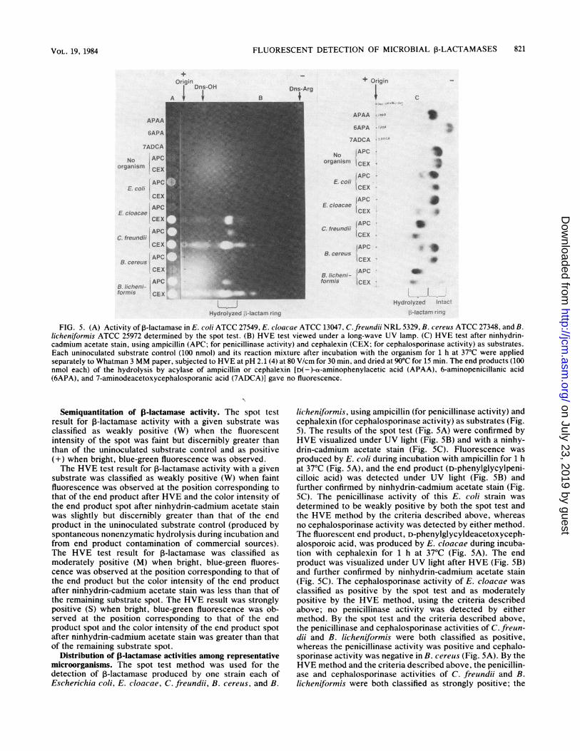
Differentiation of f-lactamase and acylase activities by thespot test and HVE. To determine whether the spot testmethod could distinguish 3-lactamase activity from acylaseactivity, the end products of acylase (the common sidechain, D(-)-ot-aminophenylacetic acid, and the intact P-lactam nuclei, 6-aminopenicillanic acid and 7-aminodeace-toxycephalosporanic acid, 100 nmol each) were separatelyapplied onto Whatman 3MM paper. None of the end prod-ucts of acylase were fluorescent either by the spot testmethod or the HVE method (Fig. 5A and B). However, allproduced color after ninhydrin-cadmium acetate staining(Fig. 5C). Therefore, both the spot test method and the HVEmethod can distinguish 3-lactamase activity from acylaseactivity.
Nonfluorescent substrateFluorescent
Hydrolysis product
Ampicillin(50 pul, 0.02 M, pH 7.0)
Cephalexin(50 ,ul, 0.02 M, pH 7.0)
1/2 loopful of bacteria
Penicillinase activity
1/2 loopful of bacteria
Cephalosporinase activity
D-Phenylglycyl-penicilloic acid
D-Phenylglycyl-deacetoxycephalo-sporoic acid
FIG. 4. Summary of the fluorescent spot test for detection of microbial 1-lactamases.
Fluorescencedevelopment
Long-waveUV lamp
5 R1I on paper
120°C, 5 min
5 p.l on paper
120°C, 5 min
Fluorescence
Fluorescence
J. CLIN. MICROBIOL.
on July 23, 2019 by guesthttp://jcm
.asm.org/
Dow
nloaded from
FLUORESCENT DETECTION OF MICROBIAL ,-LACTAMASES
Dns-ArgIr
+ Origin
APAA
6APA
7ADCA
No APCorganiism ICEX
E. cAPC
CEX
IAPCE. cloacae E
CEX
C. freundiiICEXFAPO
B. cereusCEX
APCB. licheni-formis iCEX
:,, ....,
Hydrolyzed ;-l"-actam ring
Hydrolyzed r'rt:-f
.-)-lactarr rin..
FIG. 5. (A) Activity of 1-lactamase in E. coli ATCC 27549, E. cloacae ATCC 13047, C. freundii NRL 5329, B. cereus ATCC 27348, and B.licheniformis ATCC 25972 determined by the spot test. (B) HVE test viewed under a long-wave UV lamp. (C) HVE test after ninhydrin-cadmium acetate stain, using ampicillin (APC; for penicillinase activity) and cephalexin (CEX; for cephalosporinase activity) as substrates.Each uninoculated substrate control (100 nmol) and its reaction mixture after incubation with the organism for 1 h at 37°C were appliedseparately to Whatman 3 MM paper, subjected to HVE at pH 2.1 (4) at 80 V/cm for 30 min, and dried at 90°C for 15 min. The end products (100nmol each) of the hydrolysis by acylase of ampicillin or cephalexin [D(-)-a-aminophenylacetic acid (APAA), 6-aminopenicillanic acid(6APA), and 7-aminodeacetoxycephalosporanic acid (7ADCA)] gave no fluorescence.
Semiquantitation of ,-lactamase activity. The spot testresult for ,-lactamase activity with a given substrate was
classified as weakly positive (W) when the fluorescentintensity of the spot was faint but discernibly greater thanthan of the uninoculated substrate control and as positive(+) when bright, blue-green fluorescence was observed.The HVE test result for 1-lactamase activity with a given
substrate was classified as weakly positive (W) when faintfluorescence was observed at the position corresponding tothat of the end product after HVE and the color intensity ofthe end product spot after ninhydrin-cadmium acetate stainwas slightly but discernibly greater than that of the endproduct in the uninoculated substrate control (produced byspontaneous nonenzymatic hydrolysis during incubation andfrom end product contamination of commercial sources).The HVE test result for 3-lactamase was classified as
moderately positive (M) when bright, blue-green fluores-cence was observed at the position corresponding to that ofthe end product but the color intensity of the end productafter ninhydrin-cadmium acetate stain was less than that ofthe remaining substrate spot. The HVE result was stronglypositive (S) when bright, blue-green fluorescence was ob-served at the position corresponding to that of the endproduct spot and the color intensity of the end product spotafter ninhydrin-cadmium acetate stain was greater than thatof the remaining substrate spot.
Distribution of ,-lactamase activities among representativemicroorganisms. The spot test method was used for thedetection of 1-lactamase produced by one strain each ofEscherichia coli, E. cloacae, C. freundii, B. cereus, and B.
licheniformis, using ampicillin (for penicillinase activity) andcephalexin (for cephalosporinase activity) as substrates (Fig.5). The results of the spot test (Fig. 5A) were confirmed byHVE visualized under UV light (Fig. SB) and with a ninhy-drin-cadmium acetate stain (Fig. SC). Fluorescence was
produced by E. coli during incubation with ampicillin for 1 hat 37°C (Fig. 5A), and the end product (D-phenylglycylpeni-cilloic acid) was detected under UV light (Fig. SB) andfurther confirmed by ninhydrin-cadmium acetate stain (Fig.SC). The penicillinase activity of this E. coli strain was
determined to be weakly positive by both the spot test andthe HVE method by the criteria described above, whereasno cephalosporinase activity was detected by either method.The fluorescent end product, D-phenylglycyldeacetoxyceph-alosporoic acid, was produced by E. cloacae during incuba-tion with cephalexin for 1 h at 37°C (Fig. 5A). The endproduct was visualized under UV light after HVE (Fig. 5B)and further confirmed by ninhydrin-cadmium acetate stain(Fig. 5C). The cephalosporinase activity of E. cloacae was
classified as positive by the spot test and as moderatelypositive by the HVE method, using the criteria describedabove; no penicillinase activity was detected by eithermethod. By the spot test and the criteria described above,the penicillinase and cephalosporinase activities of C. freun-dii and B. licheniformis were both classified as positive,whereas the penicillinase activity was positive and cephalo-sporinase activity was negative in B. cereus (Fig. SA). By theHVE method and the criteria described above, the penicillin-ase and cephalosporinase activities of C. freundii and B.licheniformis were both classified as strongly positive; the
Origin Dns-OHA I , I
APAA
6APA
7ADCA
No APC
organism CEXAPC
E. cobi'CEXAPC
E cloacaeCEX
.APCC. treundii
£:EX
APC FB-cereus 1
CEXtAPC
B. licheti-jformis CEX
c
)
S9
00
V.
821VOL. 19, 1984
on July 23, 2019 by guesthttp://jcm
.asm.org/
Dow
nloaded from

822 CHEN, KNAPP, AND HOLMES J. CLIN. MICROBIOL.
TABLE 1. Activities of 1-lactamases of representative microorganisms determined by the spot test, the HVE test, and the nitrocefin testafter incubation for 1 h at 37°C
,B-Lactamase activityb
Microorganisma Spot test HVE test NitrocefinPenicillinase Cephalosporinase Penicillinase Cephalosporinase test
Gram-negative entericCitrobacter freundiiNRL 5329 + + S S +ATCC 10787 + - M +
Enterobacter aerogenesNRL 9817, ATCC 13048 - + - M +
Enterobacter agglomeransNRL 9819; ATCC 29915 + - S +
Enterobacter cloacaeNRL 5335, 9818; ATCC 13047 - + - M +
Escherichia coliATCC 21986, 27549, 31027 W - W +
Klebsiella oxytocaNRL 9979 + - S +
Klebsiella pneumoniaeNRL 9976; ATCC 13883, + - S +27799
Morganella morganiiNRL 5334; ATCC 25830 W W W W +
Proteus mirabilisATCC 14273, 29855 W W W W
Proteus vulgaris ATCC 13315 W + W MProvidencia rettgeriATCC 9250, 31052
Salmonella typhimurium ATCC13311
Serratia marcescensATCC 8100, 17991 + - S +
Serratia rubidaea ATCC 181 W + W S +Shigella dysenteriaeATCC 13313 W - W +
Shigella sonneiATCC 11060 + - M +
Gram-negative nonentericBranhamella catarrhalisNRL 32674, 32681, 32763 + + S S +NRL 30069, 30071, 32589 - - -
Eikenella corrodensATCC 1073, 23834
Haemophilus ducreyiV-1157, 1158, 1169 + + S M +V-1152, 1168
Haemophilus influenzaeAS1115, 902 + + S S +AS1117, Ela; ATCC 19418
Neisseria gonorrhoeaeNRL 33044, 33047, 33050 + + S S +NRL 8327, 30483, F62
Pseudomonas aeruginosaSM31302-31311 W W W W
Pseudomonas cepaciaeBM1, 2 + + S S +
Pseudomonas fluorescensBM3; ATCC 25289 + - S +
Pseudomonas maltophiliaBM4, 5; ATCC 13637 + + S S +
Pseudomonas putidaBM6, 7; ATCC 25571 + - S +
Gram-negative anaerobicBacteroides biviusATCC 29303 + + S M +
Bacteroides capillosusATCC 29799 - - - -
Continued on following paige
on July 23, 2019 by guesthttp://jcm
.asm.org/
Dow
nloaded from

FLUORESCENT DETECTION OF MICROBIAL P-LACTAMASES 823
TABLE 1-Continued,B-Lactamase activityb
Microorganisma Spot test HVE test Nitrocefin
Penicillinase Cephalosporinase Penicillinase Cephalosporinase test
Bacteroides fragilisATCC 23745, 25285 W + W M +
Gram-positiveBacillus cereusATCC 13061 + - S - +ATCC 27348, 14579 + - M - -
Bacillus circulans ATCC 4513 + - SBacillus licheniformisATCC 9789, 14409 + W S W +ATCC 25972 + + S S +
Bacillus subtilisATCC 9799, 14410, 14415 W - W - -ATCC 14807 + - S - -
Staphylococcus aureusATCC 12598, 25923BM U17, Su3 + - S - +BM Me19 + - S
Staphylococcus epidermidisATCC 12228, 14990 + - S - +
Streptococcus faecalisATCC 11420, 12984, 19433 - - - -
a Strain numbers are those of the American Type Culture Collectiorn (ATCC), the Neisseria Reference Laboratory (NRL), Stephen A.Morse (SM), Barbara H. Minshew (BM), and Arnold L. Smith (AS). Strains of Haemophilus ducreyi were described previously (19). Thegrowth conditions for each microorganism are described in the text.
b The activities of penicillinase and cephalosporinase in each microorganism as determined by the spot test and by the HVE test wererecorded as follows. The spot test result for 1-lactamase activity with a given substrate was classified as weakly positive (W) when thefluorescent intensity of the spot was faint but discernibly greater than that of the uninoculated substrate control, and as positive (+) whenbright, blue-green fluorescence was observed. The HVE test results for 1-lactamase activity with a given substrate were classified as follows:weakly positive (W), faint fluorescence was observed at the position corresponding to that of the end product after HVE, and the colotintensity of the end product spot after ninhydrin-cadmium acetate stain was slightly but discernibly greater than that of the end product in theuninoculated substrate control (produced by spontaneous nonenzymatic hydrolysis and from contamination); moderately positive (M), bright,blue-green fluorescence was observed at the position corresponding to that of the end product spot, but color intensity of the end product spotafter ninhydrin-cadmium acetate stain was less than that of the remaining substrate spot; strongly positive (S), bright, blue-green fluorescencewas observed at the position corresponding to that of the end product spot, and the color intensity of the end product spot after ninhydrin-cad-mium acetate stain was greater than that of the remaining substrate spot.
penicillinase activity and cephalosporinase activity of B.cereus were classified as moderately positive and negative,respectively (Fig. 5B and C).
Activities of P-lactamases in 21 strains of 7 gram-positivespecies and 77 strains of 29 gram-negative species of bacteriawere determined by the spot test, the HVE test, and thenitrocefin test after incubation for 1 h at 37°C (Table 1). Theresults of the nitrocefin test agreed well with those of thespot test (confirmed by the HVE test; Table 1). Some 1B-lactamases which acted predominantly against ampicillin,such as a few species of gram-positive bacteria, were notdetected by the nitrocefin test (Table 1). Therefore, ourfluorescent method was more sensitive than the chromogen-ic cephalosporin (nitrocefin) method.
Activities of 1-lactamases in selected 3-lactamase produc-ers listed in Table 1 were further assessed by the rapid spottest and the nitrocefin test after incubation for 5 and 15 minat room temperature, and the results are listed in Table 2.The incubation time needed for a positive reaction for thefluorescent spot test appears to be the same as, or shorterthan, that needed for the nitrocefin test (Table 2). Someclinically important microorganisms such as N. gonorrhoeaeand H. influenzae produce ,B-lactamases which could bedetected by the rapid spot test immediately after organismswere suspended in the ampicillin substrate solution, without
further incubation (results not shown), just as they could bedetected by the nitrocefin test under similar conditions.
DISCUSSIONThis fluorescent spot test method can identify ,B-lactamase
activity per se and is not influenced by acylase activity. Forexample (Table 1) two strains of Providencia rettgeri whichproduce acylase (K. C. S. Chen and K. K. Holmes, unpub-lished data) but not 1-lactamase were nonreactive in all ,B-lactamase assays, including the nitrocefin test; a strain ofPseudomonas maltophilia (BM4) which produces both acy-lase and ,B-lactamase (K. C. S. Chen and K. K. Holmes,unpublished data) was reactive in all the tests shown in thetable. The test can also detect and differentiate betweenpenicillinase activity and cephalosporiniase activity. Thefluorescent spot test requires 5 min of heating at 1200C tomaximize the fluorescent potential of the end products; themechanism by which heating enhances fluorescence is notunderstood.
Stock solutions of the substrates (ampicillin and cepha-lexin, acid fotms) for the spot test were easily prepared as0.02 M solutions in 0.04 M sodium phosphate buffer, pH 7.5.These solutions both had a pH of 7.0, and the pHs of bothsolutions fall to 6.5 (within the optimal pH ranges of microbi-al ,B-lactamases [18]) upon complete hydrolysis by C. freun-
VOL. 19, 1984
on July 23, 2019 by guesthttp://jcm
.asm.org/
Dow
nloaded from

824 CHEN, KNAPP, AND HOLMES J. CLIN. MICROBIOL.
TABLE 2. Activities of 13-lactamases of selected microorganisms determined by the rapid spot test and the nitrocefin test after incubationfor 5 and 15 min at room temperature
3-Lactamase activity'Rapid spot test Nitrocefin test
Microorganisma 5 min 15 min5 min 15 min
Penicillinase Cepahlosporinase Penicillinase CephalosporinaseGram-negative enteric
Citrobacter freundiiNRL 5329 + + + + + +ATCC 10787 - - + - +
Enterobacter aerogenesNRL 9817, ATCC 13048 - - - + - -
Enterobacter agglomeransNRL 9819; ATCC 29915 + - + - - +
Enterobacter cloacaeNRL 533S, 9818; ATCC 13047 - - - + - -
Klebsiella oxytocaNRL 9979 + - + - - -
Klebsiella pneumoniaeNRL 9976; ATCC 13883 + - + - - -
Serratia marcescensATCC 8100, 17991 - - + - +
Serratia rubidaeaATCC 181
Shigella sonneiATCC 11060 - - + - -
Gram-negative nonentericBranhamella catarrahalisNRL 32674, 32681, 32763 + - + - + +
Haemophilus ducreyiV-1157, 1158, 1169 + - + - + +
Haemophilus influenzaeAS1115, 902 + - + - + +
Neisseria gonorrhoeaeNRL 33044, 33047, 33050 + - + - + +
Pseudomnonas cepaciaeBM1, 2 - - + - +
Pseudomonas fluorescensBM3; ATCC 25289 - - + - +
Pseudomonas maltophiliaBM4, 5 + + + + + +
Pseudomonas putidaBM6, 7; ATCC 25571 - - + - +
Gram-positiveBacillus cereusATCC 13061 + - + - + +ATCC 27348, 14579 - + - - -
Bacillus circulansATCC 4513 - W - - -
Bacillus licheniformisATCC 9789, 14409 - + W - +ATCC 25972 + - + + + +
Bacillus subtilisATCC 14807 - + - - -
Staphylococcus aureusBM U17, Su3 - + - - +BM Mel9 - + - - -
Staphylococcus epidermidisATCC 14990 + - + - + +ATCC 1228 - + - - +
a Strain numbers are those of the American Type Culture Collection (ATCC), the Neisseria Reference Laboratory (NRL), Barbara H.Minshew (BM) and Arnold L. Smith (AS). Strains of Haemophilus ducreyi were described previously (19). The growth conditions for eachmnicroorganism are described in the text.
b The activities of penicillinase and cephalosporinase in each microorganism as determined by the spot test were recorded as follows. Thespot test result for ,-lactamase activity using a given substrate was classified as weakly positive (W) when the fluorescent intensity of the spotwas faint but discernibly greater than that of the uninoculated substrate control; the result was positive when bright, blue-green fluorescencewas observed.
on July 23, 2019 by guesthttp://jcm
.asm.org/
Dow
nloaded from

FLUORESCENT DETECTION OF MICROBIAL 1-LACTAMASES 825
dii. The solutions could be stored at 4°C for 5 days or at-20°C for months without detectable increases in fluores-cent background as checked by the spot test. Some organ-isms fluoresced slightly on paper, but this fluorescence wasconfined to the center of the applied spot and could be easilydistinguished from the fluorescence of end products whichdiffused radially by capillary action of the paper from thecenter of application. Therefore, the spot test could beperformed without prior removal of the organisms by centrif-ugation. Unless the P-lactamase sought shows no substratespecificity, the ampicillin and cephalexin substrates shouldbe incubated separately with the organism. When penicillinand cephalosporin substrates are mixed together, the sub-strate which is not hydrolyzed may act as a competitiveinhibitor of the other substrate. For example, Serratiamarcescens ATCC 8100 and 17991 exhibited only cephalo-sporinase activity as detected by the spot test and the HVEtest when ampicillin and cephalexin were incubated sepa-rately with the organism (Table 1). However, cephalosporin-ase activity was not detected in either strain by the spot testor the HVE test when both strains were incubated with amixture of equal volumes of ampicillin and cephalexinsubstrate solution for 1 h at 37°C (results not shown).The spot test described in this paper, using ampicillin and
cephalexin as substrates, provides a rapid, inexpensivemethod for specific detection of microbial P-lactamases bydetecting the presence of fluorescent end products. The testmay have several applications. For example, incubation withhuman sera can result in a color change for nitrocefin (13).Therefore, nitrocefin is not suitable for detection of I-lactamase in the presence of certain body fluids. We foundthat eight of eight human sera mixed with an equal volume ofnitrocefin solution converted nitrocefin to a red color after10 min of incubation at 37°C, but produced no fluorescenceafter incubation with an equal volume of ampicillin orcephalexin solution for 1 h at 37°C (results not shown). Thus,the methods described here have the potential for directdetection of microbial ,B-lactamases in clinical specimens.The detection of fluorescent end products offers an eco-
nomical alternative for clinical laboratories which test largenumbers of microorganisms for P-lactamases. For example,the routine 1-lactamase nitrocefin tests of staphylococcalisolates require a noninhibitory concentration of a semisyn-thetic penicillin as an inducer for the enzyme (8). In ourfluorescent spot test method, with sufficient incubation time(e.g., -15 min; Table 2) amnpicillin could serve not only as asubstrate but also as an inducer for these gram-positivebacteria of clinical importance (11). The potential of thismethod for testing clinical isolates for 3-lactamases whichmay have a predominance of penicillinase or cephalosporin-ase activity deserves further evaluation.
ACKNOWLEDGMENTSThis study was supported by Public Health Service Research
Program Project grant A1-12192 from the National Institutes ofHealth.We thank Arnold L. Smith, Barbara H. Minshew, Betsy L.
Williams, Patricia A. Totten, and Stephen A. Morse who providedseveral of the organisms used in this study.
LITERATURE CITED1. Anhalt, J. P., and R. Nelson. 1982. Failure of Padac test strips to
detect staphylococcal ,B-lactamase. Antimicrob. Agents Che-mother. 21:993-994.
2. Chen, K. C. S., N. J. Culbertson, J. S. Knapp, G. E. Kenny, and
K. K. Holmes. 1982. Rapid method for simultaneous detection ofthe arginine dihydrolase system and amino acid decarboxylasesin microorganisms. J. Clin. Microbiol. 16:909-919.
3. Chen, K. C. S., T. J. Kindt, and R. M. Krause. 1975. Primarystructure of the L chain from a rabbit homogeneous antibody tostreptococcal carbohydrate I. Purification of antibody and se-quence determination of peptides from a-chymotryptic andthermolytic digests. J. Biol. Chem. 250:3280-3288.
4. Chen, K. C. S., and R. M. Krause. 1975. A peptide mappingtechnique-a three map system. Anal. Biochem. 69:180-186.
5. Durkin, J. P., G. I. Dmitrienko, and T. Viswanatha. 1977. N-(2-furyl) acryloyl penicillin: a novel compound for the spectropho-tometric assay of 3-lactamase I. J. Antibiot. 30:883-885.
6. Ericsson, H. 1978. The effect of P-lactamases of varying originon different types of cephalosporins. Scand. J. Infect. Dis.Suppl. 13:33-34.
7. Heilmann, J., J. Barrollier, and E. Watzke. 1957. Beitrag zurAminosaurebestimung auf Papierchromatogrammen. Hoppe-Seyler's Z. Physiol. Chem. 309:219-220.
8. Horton, K. A., B. R. Jennings, and V. S. Baselski. 1982. -Lactamase testing of staphylococci in a commercial brothmicrodilution minimum inhibitory concentration system. J.Clin. Microbiol. 16:406-407.
9. Jorgensen, J. H., S. A. Crawford, and G. A. Alexander. 1982.Pyridinium-2-azo-p-dimethylaniline chromophore, a new chro-mogenic cephalosporin for rapid beta-lactamase testing. Antimi-crob. Agents Chemother. 22:162-164.
10. Lee, D. T. F., and J. E. Rosenblatt. 1983. A comparison of fourmethods for detecting 1-lactamases in anaerobic bacteria.Diagn. Microbiol. Infect. Dis. 1:173-175.
11. Leitner, F., H. M. Sweeney, T. F. Martin, and S. Cohen. 1963.Induction of staphylococcal penicillinase by benzylpenicillin:effect of pH, concentration of ferrous ion and inducer, andduration of exposure of cells to inducer. J. Bacteriol. 86:717-727.
12. Mat-shall, M. J., G. W. Ross, K. V. Chanter, and A. M. Harris.1972. Comparison of the substrate specificities of the ,-lacta-mases from Klebsiella aerogenes 1082 E and Enterobactercloacae P99. Appl. Microbiol. 23:765-769.
13. O'Callaghan, C. H., A. Morris, S. M. Kirby, and A. H. Shingler.1972. Novel method for detection of 3-lactamases by using achromogenic cephosporin substrate. Antimicrob. Agents Che-mother. 1:283-288.
14. Ogawara, H. 1981. Antibiotic resistance in pathogenic andproducing bacteria, with special reference to P-lactam antibiot-ics. Microbiol. Rev. 45:591-619.
15. Ross, G. W., K. V. Chanter, A. M. Harris, S. M. Kirby, M. J.Marshall, and C. H. O'Callaghan. 1973. Comparison of assaytechniques for ,-lactamase activity. Anal. Biochem. 54:9-16.
16. Sykes, R. B., and K. Bush. 1982. Physiology, biochemistry, andinactivation of 3-lactamases, p. 155-207. In R. B. Morin and M.Gorman (ed.), Chemistry and biology of P-lactam antibiotics,vol. 3. Academic Press, Inc., New York.
17. Sykes, R. B., and M. Matthew. 1976. The P-lactamases of gram-negative bacteria and their role in resistance to 3-lactam antibi-otics. J. Antimicrob. Chemother. 2:115-157.
18. Sykes, R. B., and M. Matthew. 1979. Detection, assay andimmunology of ,-lactamases, p. 17-49. In J. M. T. Hamilton-Miller and J. T. Smith (ed.), Beta-lactamases. Academic Press,Inc., New York.
19. Totten, P. A., H. H. Handsfield, D. Peters, K. K. Holmes, and S.Falkow. 1982. Characterization of ampicillin resistance plasmidsfrom Haemophilus ducreyi. Antimicrob. Agents Chemother.21:622-627.
20. Vandamme, E. J. 1977. Enzymes involved in ,B-lactam antibioticbiosynthesis. Adv. Appl. Microbiol. 21:89-123.
21. Williams, B. L., K.-A. Osterberg, and J. Jorgensen. 1979.Subgingival microflora of periodontal patients on tetracyclinetherapy. J. Clin. Periodontol. 6:210-221.
22. White, L. A., and D. S. Kellogg, Jr. 1965. Neisseria gonor-rhoeae identification in direct smears by a fluorescent antibodycounterstain method. Appl. Microbiol. 13:171-174.
VOL. 19, 1984
on July 23, 2019 by guesthttp://jcm
.asm.org/
Dow
nloaded from