infiltrative and restrictive cardiomyopathy: recognition ... 15 (tuesday)/8. 1020am... ·...
TRANSCRIPT
10/7/2013
1
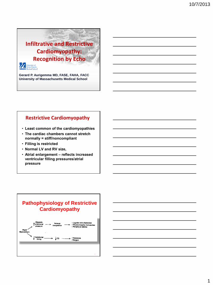
Gerard P. Aurigemma MD, FASE, FAHA, FACC
University of Massachusetts Medical School
Infiltrative and Restrictive Cardiomyopathy:
Recognition by Echo
Restrictive Cardiomyopathy
• Least common of the cardiomyopathies
• The cardiac chambers cannot stretch
normally = stiff/noncompliant
• Filling is restricted
• Normal LV and RV size,
• Atrial enlargement – reflects increased
ventricular filling pressures/atrial
pressure
3
Pathophysiology of Restrictive
Cardiomyopathy

10/7/2013
2
90% x small EDV = small SV
Rise in PCWP Flat stroke volume
response to exercise
6
10/7/2013
3
10/7/2013
4
10
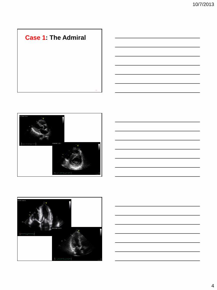
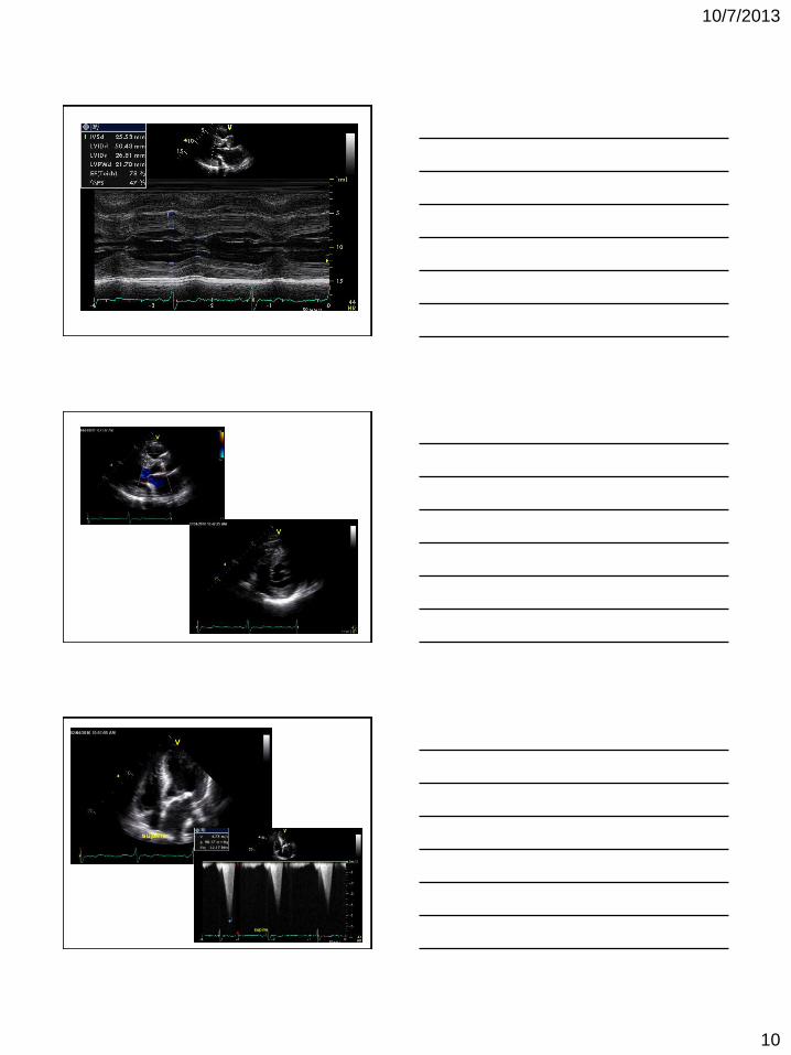
Case 1: The Admiral
11
12
10/7/2013
5
13
14
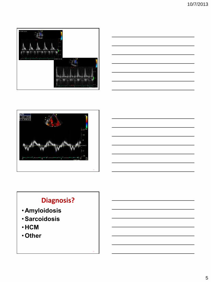
Diagnosis?
•Amyloidosis
•Sarcoidosis
•HCM
•Other
15
10/7/2013
6
Diagnosis?
•Amyloidosis
•Sarcoidosis
•HCM
•Other
16
The Systemic Amyloidoses
• Primary (AL) or light chain disease
– Plasma cell dyscrasia
– Immunoglobulin light chains
– 12 month survival without treatment
– 6 month survival with cardiac disease
• Familial (AF)
– Mutations in transthyretin (TTR)
– Ile 122 of particular interest
The Systemic Amyloidoses
• Senile systemic amyloid (SSA)
– TTR-based non-genetic (ie, TTR normal)
– Cardiac predilection
– Male gender, onset after age 60
• Secondary amyloidosis (AA)
– Chronic inflammatory states
10/7/2013
7
Amyloid Cardiomyopathy
• Very poor prognosis (6 mo survival)
• Restrictive cardiomyopathy with profound abnormalities of diastolic function
– Systolic dysfunction late manifestation
• Classic teaching
– biventricular thickening in a small ventricle
– valvular thickening
– Atrial enlargement
– Pericardial effusion/evidence of elevated filling pressures
Amyloid Cardiomyopathy • Patients do NOT respond to normal
medication for CHF
– ACE inhibitors, beta-blockers, dig
• There is a treatment for AL amyloid
– Autologous bone marrow transplant
• Patient selection critical
– assessment of cardiac involvement
Continuum of Amyloid • Advanced disease is too late
• Initial changes are abnormalities of
diastolic function
• As wall thickness progresses
restrictive physiology ensues
• Systolic dysfunction late stage
10/7/2013
8
22
23
Case 2: The Attorney
HPI • 58 year old male who presented with dizziness and presyncopal
symptoms with possible fall and left leg pain, numbness and swelling in both lower extremities.
• Patient reported that at 1:30 am, he had gotten out of bed, felt dizzy and went down to the floor due to weakness. He denied LOC but noted that he was on the floor for approx. 30 minutes before he got himself back to bed. He noticed that there was urine on the floor.
• He called his HCP who arrived at his home at 4am and sent him to the ED.
• The patient denied chest discomfort, SOB or palpitations.
• The patient also reported LBP radiating from his left buttock down through the back of his LLE for 3 weeks while he was in Florida. He had driven back to Worcester at 7pm the night of admission and continued to have left leg pain, numbness and swelling in both lower extremities.
10/7/2013
9
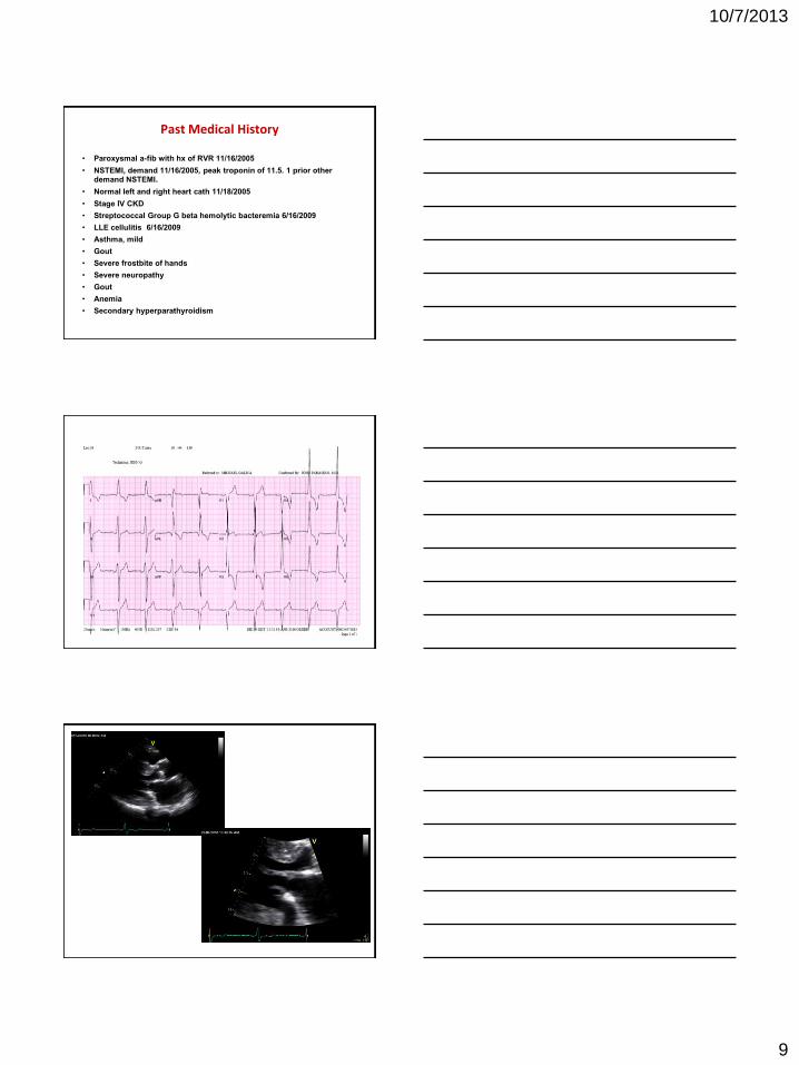
Past Medical History
• Paroxysmal a-fib with hx of RVR 11/16/2005
• NSTEMI, demand 11/16/2005, peak troponin of 11.5. 1 prior other demand NSTEMI.
• Normal left and right heart cath 11/18/2005
• Stage IV CKD
• Streptococcal Group G beta hemolytic bacteremia 6/16/2009
• LLE cellulitis 6/16/2009
• Asthma, mild
• Gout
• Severe frostbite of hands
• Severe neuropathy
• Gout
• Anemia
• Secondary hyperparathyroidism
10/7/2013
10
10/7/2013
11
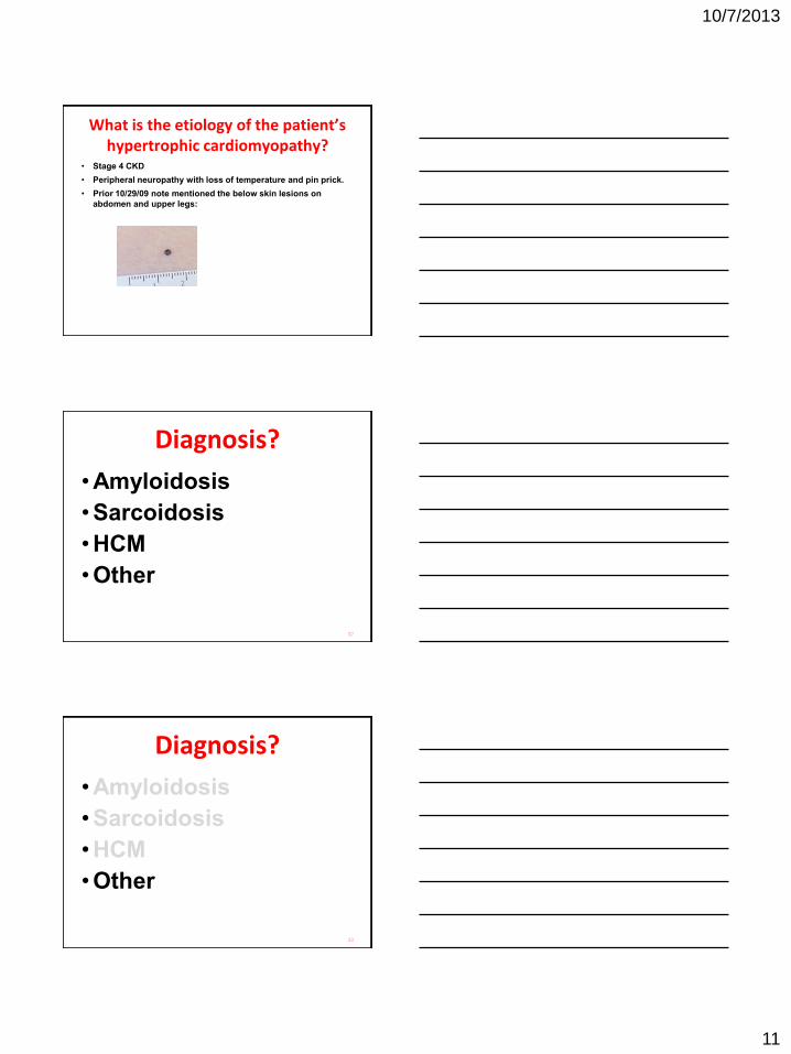
What is the etiology of the patient’s hypertrophic cardiomyopathy?
• Stage 4 CKD
• Peripheral neuropathy with loss of temperature and pin prick.
• Prior 10/29/09 note mentioned the below skin lesions on
abdomen and upper legs:
Diagnosis?
•Amyloidosis
•Sarcoidosis
•HCM
•Other
32
Diagnosis?
•Amyloidosis
•Sarcoidosis
•HCM
•Other
33
10/7/2013
12
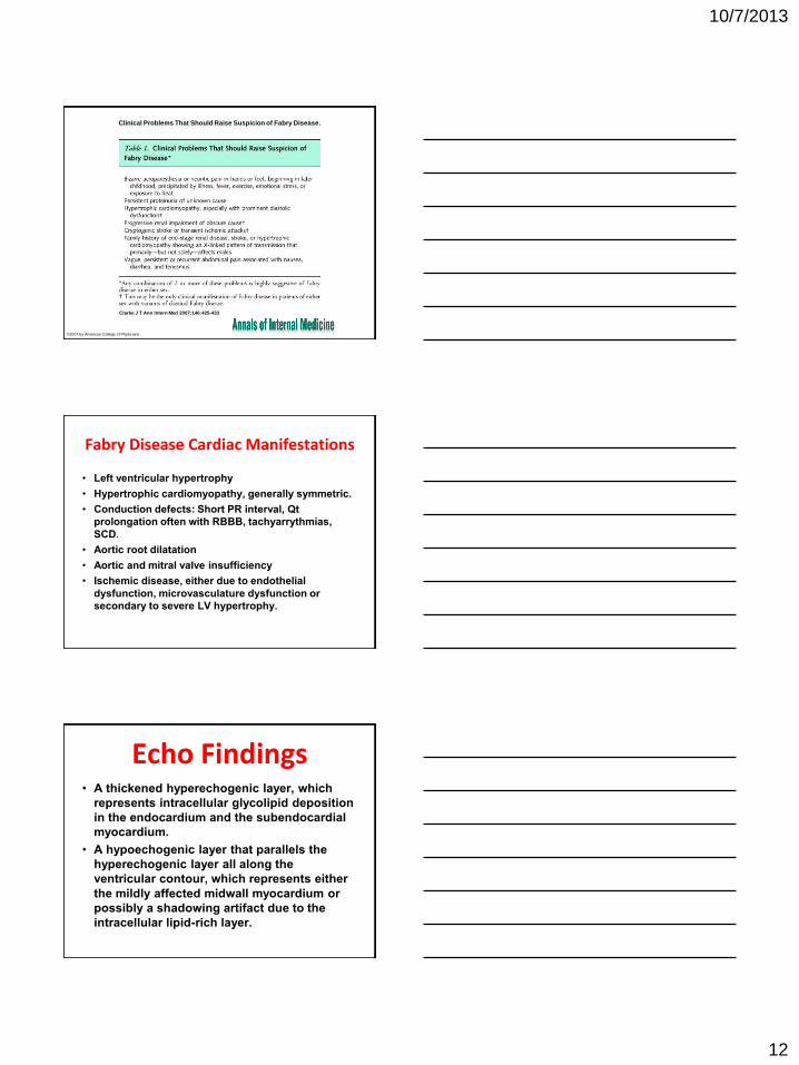
Clinical Problems That Should Raise Suspicion of Fabry Disease.
Clarke J T Ann Intern Med 2007;146:425-433
©2007 by American College of Physicians
Fabry Disease Cardiac Manifestations
• Left ventricular hypertrophy
• Hypertrophic cardiomyopathy, generally symmetric.
• Conduction defects: Short PR interval, Qt
prolongation often with RBBB, tachyarrythmias,
SCD.
• Aortic root dilatation
• Aortic and mitral valve insufficiency
• Ischemic disease, either due to endothelial
dysfunction, microvasculature dysfunction or
secondary to severe LV hypertrophy.
Echo Findings • A thickened hyperechogenic layer, which
represents intracellular glycolipid deposition
in the endocardium and the subendocardial
myocardium.
• A hypoechogenic layer that parallels the
hyperechogenic layer all along the
ventricular contour, which represents either
the mildly affected midwall myocardium or
possibly a shadowing artifact due to the
intracellular lipid-rich layer.
10/7/2013
13
LVH Screening • Those who also have a binary appearance of
the LV endocardial border.
• Among patients who have hypertrophic
cardiomyopathy, particularly if symmetric,
those with either no family history of HCM or
a family history consistent with X-linked
disease.
• Measurement of plasma alpha-galactosidase
A.
Histopathologic changes in a small cutaneous blood vessel showing vacuolation of endothelial and smooth-muscle cells (periodic acid–Schiff stain).
Clarke J T Ann Intern Med 2007;146:425-433
©2007 by American College of Physicians
Long Term effects of Enzyme Replacement Therapy on Fabry Cardiomyopathy: Evidence for a Better Outcome With Early Treatment
• 32 patients over 3 years receiving ERT
• Matched with 20 age-matched healthy controls
• Underwent MRI, echocardiography, color doppler
myocardial imaging studies and bicycle stress tests at
baseline and every year.
• Patients assigned to 3 groups depending on amount of
fibrosis
• Patients with fibrosis in one LV segment were in mild
fibrosis group
• 14 patients had hypertenison, all were almost
normotensive Circulation 2009:119;524-529.
10/7/2013
14
Fabry, LVH and HCM
• 3% (7/230) of middle aged men with LVH
had Fabry’s disease (10% of unexplained) Nakao S et al. N Engl J Med. 1995; 333: 288–293
• In patients with Fabry disease
– 50% no LVH
– 37% concentric LVH
– 10% asymmetric LVH
– 3% eccentric LVH Linhart A et al. Am Heart J. 2000; 139: 1101–1108
• 4% of patients (6/153) with HCM had
Fabry’s Disease Sachdev B. et al Circulation. 2002;105:1407-1411
Diagnosis
• Blood test to measure the level of α-
galactosidase A activity
– this may be misleading in female carriers
due to the random nature of X-inactivation
• Genetic Testing
– chromosomal analysis of the GLA gene
– many mutations (>300)
– gold standard
• Kidney biopsy
– for proteinuria
Copyright ©2009 American Heart Association
Weidemann, F. et al. Circulation 2009;119:524-529
MRI of 3 typical Fabry patients at baseline
10/7/2013
15
44
Case 3: The Industrial
Chemist
28 y/o M with PMHx of HTN p/w chest pain for last 4 days.
- Chest pain constant, dull, worse with inspiration.
- Denies SOB, orthopnea, PND, palpitations, pre-syncope, n/v.
- No URI sxs, but son has active URI.
EKG on arrival
NB: EKG completely normal 6 months ago
10/7/2013
16
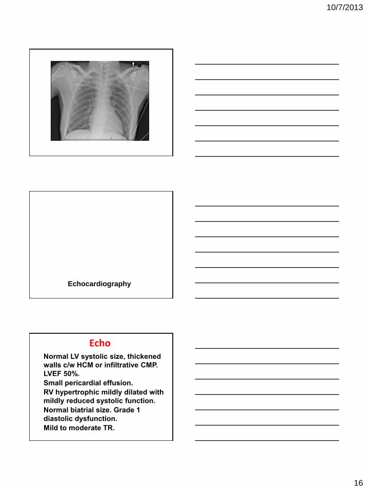
Echocardiography
Echo •Normal LV systolic size, thickened
walls c/w HCM or infiltrative CMP.
LVEF 50%.
•Small pericardial effusion.
•RV hypertrophic mildly dilated with
mildly reduced systolic function.
•Normal biatrial size. Grade 1
diastolic dysfunction.
•Mild to moderate TR.
10/7/2013
17
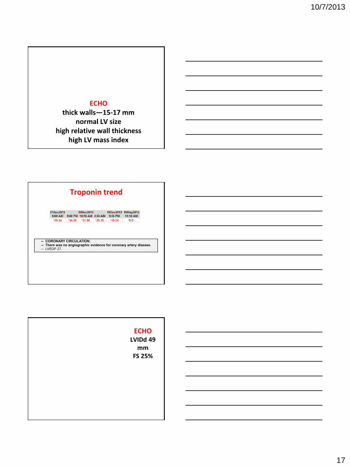
ECHO thick walls—15-17 mm
normal LV size high relative wall thickness
high LV mass index
Troponin trend
• -- CORONARY CIRCULATION: • -- There was no angiographic evidence for coronary artery disease. • -- LVEDP 27.
ECHO LVIDd 49
mm FS 25%
10/7/2013
18
21 mm
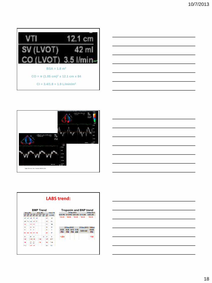
BSA = 1.8 m2
CO = π (1.05 cm)2 x 12.1 cm x 84
CI = 3.4/1.8 = 1.9 L/min/m2
Tighe DA et al. Am J Cardiol 2003;91;254.
LABS trend:
BMP Trend Troponin and BNP trend
10/7/2013
19
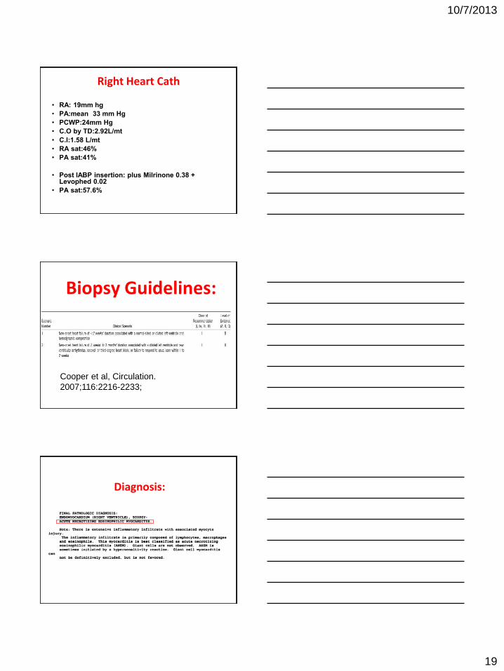
Right Heart Cath
• RA: 19mm hg
• PA:mean 33 mm Hg
• PCWP:24mm Hg
• C.O by TD:2.92L/mt
• C.I:1.58 L/mt
• RA sat:46%
• PA sat:41%
• Post IABP insertion: plus Milrinone 0.38 + Levophed 0.02
• PA sat:57.6%
Biopsy Guidelines:
Cooper et al, Circulation.
2007;116:2216-2233;
Diagnosis:
10/7/2013
20
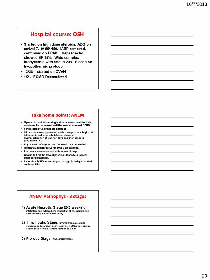
Hospital course: OSH
• Started on high dose steroids, ABG on
arrival 7.10/ 66/ 450. IABP removed,
continued on ECMO. Repeat echo
showed EF 15%. Wide complex
bradycardia with rate in 20s. Placed on
hyopothermic protocol.
• 12/28 – started on CVVH
• 1/2 - ECMO Decanulated.
Take home points: ANEM
• Myocardial wall thickening is due to edema and Not LVH, as shown by decreased wall thickness on repeat ECHO.
• Pericardial effusions more common.
• Initiate immunosuppression early if suspicion is high and infection is not suspected. Usual doses of Hydrocortisone 100 q8h for days and then taper to prednisone PO.
• Any amount of supportive treatment may be needed.
• Myocardium can recover in DAYS on steroids.
• Response is re-assessed with repeat biopsy
• Goal is to find the lowest possible doses to suppress eosinophilic activity.
• 6 monthly ECHO as end organ damage is independent of eosinophillia.
ANEM Pathophys - 3 stages
1) Acute Necrotic Stage (2-3 weeks): infiltration and extracellular deposition of eosinophils and
consequently IL-5 mediated injury.
2) Thrombotic Stage: layered thrombus along
damaged endocardium due to activation of tissue factor by
eosinophils, cerebral thromboemboli common
3) Fibrotic Stage: Myocardial fibrosis
10/7/2013
21
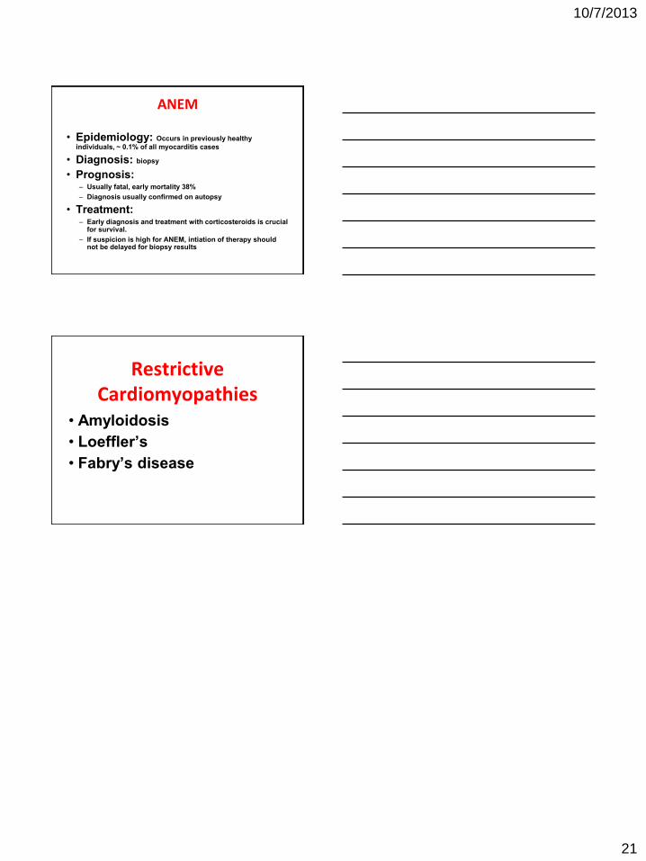
ANEM
• Epidemiology: Occurs in previously healthy
individuals, ~ 0.1% of all myocarditis cases
• Diagnosis: biopsy
• Prognosis: – Usually fatal, early mortality 38%
– Diagnosis usually confirmed on autopsy
• Treatment:
– Early diagnosis and treatment with corticosteroids is crucial for survival.
– If suspicion is high for ANEM, intiation of therapy should not be delayed for biopsy results
Restrictive Cardiomyopathies
• Amyloidosis
• Loeffler’s
• Fabry’s disease