inflammation and targeted nutrition€¦ · 3. review the science behind specialized pro-resolving...
TRANSCRIPT

© 2018 Metagenics Institute. All Rights Reserved.
Inflammation and Targeted Nutrition
Nutrition Masters Course
Annalouise O’Connor, PhD, RD

© 2018 Metagenics Institute. All Rights Reserved.
Learning Objectives
1. Understand the two distinct phases of inflammation: initiation and resolution.
2. Learn how inflammation initiation can be modulated with select nutritional bioactives EPA, DHA, curcumin, xanthohumol, Boswellia serrata, ginger, and THIAA.
3. Review the science behind specialized pro-resolving mediators (SPMs), their biosynthesis, the essential role they play in inflammation resolution, and the rationale for supplementation.
4. Discuss an integrated view of up-to-date research supporting the clinical management of inflammation in practice.

© 2018 Metagenics Institute. All Rights Reserved.© 2018 Metagenics Institute. All Rights Reserved.
Inflammation Overview

© 2018 Metagenics Institute. All Rights Reserved.
.
.
.
Inflammation: Friend or Foe?Inflammation is critical for survival, but excessive inflammation is linked with disease.
Adapted from: Miller AH, Raison CL. Nat Rev Immunol. 2016;16(1):22–34.Okin D, Medhitov R. Curr Biol. 2012;22(17):R733-R740.
Ancestral populations Modern life
.
..
Evolutionary time
↓ Infectious mortality
↑Inflammation ↑ Chronic conditions
↑Autoimmunity
• Avoidance• Alarm
Robust inflammatory response still
important and beneficial but cost increased due to
altered environment
Benefit of robust inflammatory
response outweighed potential cost
• Wound healing• Fighting infection
Infl
amm
ato
ry b
ias
Pathogens
Predators
Rivals
Pathogens
Obesogenic environment
Environmental toxins
Chronic stress

© 2018 Metagenics Institute. All Rights Reserved.
What Is Normal Inflammatory Process?
Inflammationinitiation
Inflammationresolution
HomeostasisNormal function
HomeostasisNormal function
Inflammation triggers
Goal Localize and eliminate trigger of inflammation
Counter-regulate initiation; subsidence of inflammation, tissue regeneration, and return to normal function
Predominant immune cells
Neutrophils, monocytes, PMNs
Resolving macrophages
Chemical mediators
Pro-inflammatory lipid mediators—e.g., PGs, LTs, chemokines, and pro-inflammatorycytokines)
Specialized pro-resolving mediators (SPMs)
Adapted from: Serhan CN. Nature. 2014;510:92-101.
© 2018 Metagenics Institute. All Rights Reserved.
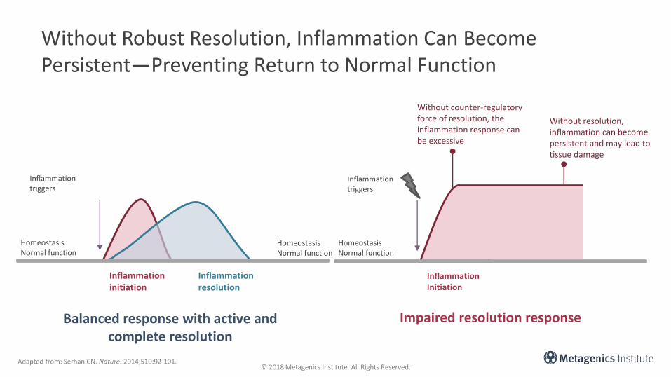
Without Robust Resolution, Inflammation Can Become Persistent—Preventing Return to Normal Function
Adapted from: Serhan CN. Nature. 2014;510:92-101.
Balanced response with active and complete resolution
InflammationInitiation
HomeostasisNormal function
Inflammation triggers
Impaired resolution response
Without counter-regulatory force of resolution, the inflammation response can be excessive
Without resolution, inflammation can become persistent and may lead to tissue damage
Inflammationinitiation
Inflammationresolution
HomeostasisNormal function
HomeostasisNormal function
Inflammation triggers

© 2018 Metagenics Institute. All Rights Reserved.
Inflammation Within Tissues Is at the Core of Chronic Disease
Handschin C & Spiegelman BM. Nature. 2008;454:463-469.
Neuroinflammation—Linked with Alzheimer’s disease, Huntington’s disease, Parkinson’s disease
Vascular inflammation—Core for development of atherosclerosis
Joint inflammation—Arthritis development
Hepatic and adipose inflammation—Linked with insulin resistance and type 2 diabetes
Systemic and local inflammatory environment
© 2018 Metagenics Institute. All Rights Reserved.
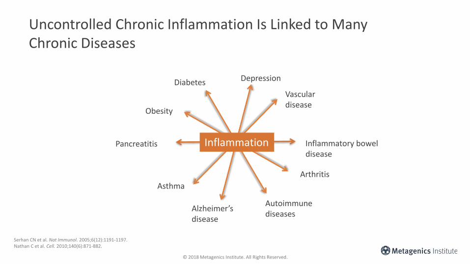
Uncontrolled Chronic Inflammation Is Linked to Many Chronic Diseases
Serhan CN et al. Nat Immunol. 2005;6(12):1191-1197.Nathan C et al. Cell. 2010;140(6):871-882.
Diabetes Depression
Vascular disease
Inflammatory bowel disease
Arthritis
Autoimmune diseases
Alzheimer’s disease
Obesity
Pancreatitis
Asthma
Inflammation

© 2018 Metagenics Institute. All Rights Reserved.
Resolution
Cardinal Signs of Inflammation Initiation and Resolution
Adapted from: Basil MC, Levy BD. Nat Rev Immunol. 2016;16(1):51–67.
Inflammation
Re
dn
ess
He
at
Swe
llin
g
Pai
n
Loss
of
fun
ctio
nProtective response Resolution response
Re
mo
val
Re
sto
rati
on
Re
gen
era
tio
n
Re
mis
sio
n
Re
lief

© 2018 Metagenics Institute. All Rights Reserved.© 2018 Metagenics Institute. All Rights Reserved.
Inflammation Initiation Mechanisms

© 2018 Metagenics Institute. All Rights Reserved.
Inflammation & Pain: Key Enzymes
Phospholipase A2 (PLA2)
• Liberates arachidonic acid (ARA) from the cell membrane
• ARA is then available as a substrate for COX-1, COX-2, and LOX (as well as other enzymes)
Dennis EA et al. Chem Rev. 2011;111(10):6130-6185.
Arachidonic acid (ARA)
Arachidonic acid (ARA)
Phospholipase A2
(PLA2)
Cell membrane lipid bi-layer

© 2018 Metagenics Institute. All Rights Reserved.
Inflammation & Pain: Key Enzymes
• Cyclooxygenase (COX-1 and COX-2)
o Converts ARA to prostaglandins (including PGE2)
o PGE2
– Increases pain perception
– Contributes to the destruction of cartilage in arthritic joints in both rheumatoid arthritis (RA) and osteoarthritis (OA)
Arachidonic acid (ARA)
Dennis EA et al. Chem Rev. 2011;111(10):6130-6185.Lee A et al. Gene. 2014;527(2):440-447.
Cyclooxygenase-1 & 2 (COX-1, COX-2)
PGE2
Phospholipase A2
(PLA2)
Arachidonic acid (ARA)

© 2018 Metagenics Institute. All Rights Reserved.
Inflammation & Pain: Key Enzymes
• Lipoxygenase (LOX)
o Converts ARA to leukotrienes (including LTB4)
o Leukotrienes are active during inflammation initiation
o Overproduction of leukotrienes plays a role in inflammatory conditions like asthma and allergic rhinitis
o Commonly used LOX inhibitors include medications used as analgesics for OA and RAand treatments for asthma
Coffey M, Peters-Golden M. Curr Opin Allergy Clin Immunol. 2003;3(1):57-63.Çobanoğlu B et al. Curr Allergy Asthma Rep. 2013;13(2):203-208.
LeukotrienesLBT4
Lipoxygenase (5-LOX)
Arachidonic acid (ARA)
Phospholipase A2
(PLA2)
Arachidonic acid (ARA)

© 2018 Metagenics Institute. All Rights Reserved.
ARA is released from the cell membrane through the action of PLA2.
Through the actions of COX and LOX enzymes, ARA is converted to the pro-inflammatory lipid mediators such as prostaglandins and leukotrienes.
These pro-inflammatory lipid mediators are key drivers of inflammation initiation, and they enhance vasodilation, attract pro-inflammatory immune cells to the affected tissue, and—in the case of prostaglandins—contribute to pain.
1
2
3
Inflammation & Pain: Key Enzymes
Dennis EA et al. Chem Rev. 2011;111(10):6130-6185.
LeukotrienesLBT4
Lipoxygenase (5-LOX)
Arachidonic acid (ARA)
Phospholipase A2
(PLA2)
Arachidonic acid (ARA)
Cyclooxygenase-1 & 2 (COX-1, COX-2)
ProstaglandinsPGE2

© 2018 Metagenics Institute. All Rights Reserved.
Inflammation & Pain: Key Regulator of Gene Expression
• Nuclear factor-kappa B (NF-κB)
o Protein complex that controls DNA transcription/genetic expression
o When activated, controls/regulates the expression of ~500 genes, including:– Pro-inflammatory enzymes: PLA2, COX-1,
COX-2, LOX
– Pro-inflammatory cytokines: IL-1β, IL-6, IL-12, TNFα
– Chemokines: CXCL8 (aka IL-8), MCP-1
Shih R et al. Front Mol Neurosci. 2015;8:77.

© 2018 Metagenics Institute. All Rights Reserved.
Nuclear Factor-Kappa B (NF-ĸB)
NF-κB (inactive)
NF-κB (activated)
Increased expression of:PLA2
COX-1COX-2LOXTNFα, IL-1β, IL-6, IL-12CXCL8 (IL-8)MCP-1
Cytosol
Nucleus
NF-κB (inactive)
NF-κB (activated)
Upstream
Downstream
Shih R et al. Front Mol Neurosci. 2015;8:77.

© 2018 Metagenics Institute. All Rights Reserved.
Inflammation & Pain: NF-κB & Pro-Inflammatory Cytokines
• Not only does NF-κB increase the expression of pro-inflammatory cytokines
• Some pro-inflammatory cytokines increase the activation of NF-ĸB
o e.g., TNFα, IL-1β
• Chronic inflammation persists
Pro-inflammatory
cytokine expression
e.g., TNFα, IL-1β
NF-κBActivation
Hayden MS, Ghosh S. Semin Immunol. 2014;26(3):253-266.

© 2018 Metagenics Institute. All Rights Reserved.
TNFα Activates NF-κB
Arachidonic Acid (ARA)NF-κB (inactive)
NF-κB (activated)
Increased expression of:PLA2
COX-1COX-2LOXTNFα, IL-1β, IL-6, IL-12CXCL8 (IL-8)MCP-1
TNFα
Cytosol
NF-κB (inactive)
NF-κB (activated)
Nucleus
TNFα
Upstream
Downstream
Hayden MS, Ghosh S. Semin Immunol. 2014;26(3):253-266.Shih R et al. Front Mol Neurosci. 2015;8:77.

© 2018 Metagenics Institute. All Rights Reserved.
Inflammation & Pain: Chemokines
• Examples of inflammatory chemokineso Chemokine ligand 8 (CXCL8, aka IL-8)
o Monocyte chemoattractant protein 1 (MCP-1)
o Interferon-γ activated protein (IP-10)
• Recruit white blood cells to local sites of inflammationo Promote joint pathology in patients with
arthritis
Szekanecz Z et al. Front Biosci. 2010;146(614):41-46. Kapoor M et al. Nat Rev Rheumatol. 2011;7(1):33-42.
© 2018 Metagenics Institute. All Rights Reserved.
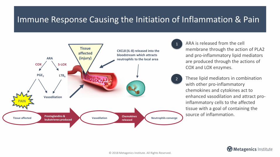
Immune Response Causing the Initiation of Inflammation & Pain
ARA is released from the cell membrane through the action of PLA2 and pro-inflammatory lipid mediators are produced through the actions of COX and LOX enzymes.
These lipid mediators in combination with other pro-inflammatory chemokines and cytokines act to enhanced vasodilation and attract pro-inflammatory cells to the affected tissue with a goal of containing the source of inflammation.
Tissue affected(injury)ARA
PGE2 LTB4
COX 5-LOX
Vasodilation
CXCL8 (IL-8) released into the bloodstream which attracts neutrophils to the local area
Vasodilation Neutrophils convergeProstaglandins & leukotrienes produced
Chemokines released
Tissue affected
PAIN
1
2

© 2018 Metagenics Institute. All Rights Reserved.© 2018 Metagenics Institute. All Rights Reserved.
Focus on Nutritional Bioactiveswith Inflammation Initiation Modulating Activity

© 2018 Metagenics Institute. All Rights Reserved.
Key Nutritional Bioactives for Modulation of Inflammation Initiation
• Omega-3 fatty acids EPA and DHA• Tetrahydro-iso-α acids (THIAA)• Curcumin• Xanthohumol• Boswellia serrata• Ginger

© 2018 Metagenics Institute. All Rights Reserved.
Omega-3 Fatty Acids EPA and DHA
Arachidonic acid omega-6 fatty acid
EPA omega-3 fatty acid
2-seriesprostaglandins
4-series leukotrienes
Pro-inflammatory lipid mediatorsLess potent
pro-inflammatory lipid mediators
3-seriesprostaglandins
5-series leukotrienes
Adapted from: Calder PC. Nutrients. 2010;2(3):355-374.

© 2018 Metagenics Institute. All Rights Reserved.
Tetrahydro-iso-α acids (THIAA) from Hops Have Anti-Inflammatory Properties
Mechanistic studies in endothelial cells, monocytes, macrophages, and human rheumatoid arthritis synovial fibroblasts indicate anti-inflammatory activities of THIAA.
THIAA
Pro-inflammatory cytokines e.g., TNF-a, IL-6
NF-kB expression and activity
COX-2 PGE2
Desai A et al. Inflamm Res. 2009;58(5):229-34.Konda VR et al. Arthritis Rheum. 2010;62(6):1683-1692.Desai A et al. Atherosclerosis. 2012;223:130-136.

© 2018 Metagenics Institute. All Rights Reserved.
Tetrahydro-iso-α acids (THIAA) from Hops Have Anti-Inflammatory Properties
In a mouse model of induced arthritis, treatment with THIAA
Reduced paw swelling
Reduced arthritis index
Reduced extent of joint damage
Reduced cartilage degradation
Reduced bone erosion
Reduced IL-6 levels
Konda VR et al. Arthritis Rheum. 2010;62(6):1683-1692.
© 2018 Metagenics Institute. All Rights Reserved.
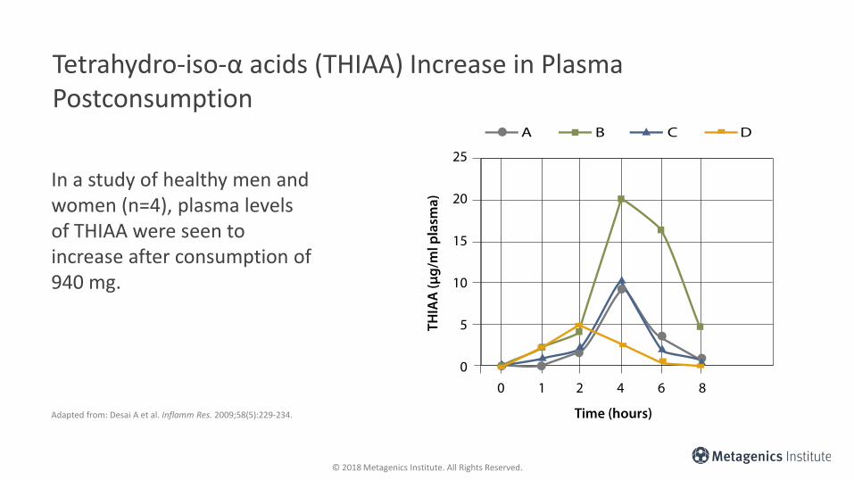
Tetrahydro-iso-α acids (THIAA) Increase in Plasma Postconsumption
Adapted from: Desai A et al. Inflamm Res. 2009;58(5):229-234.
In a study of healthy men and women (n=4), plasma levels of THIAA were seen to increase after consumption of 940 mg.

© 2018 Metagenics Institute. All Rights Reserved.
Curcuminoids: Isolated Constituents
• Primary active constituents in turmeric root (Curcuma longa)
• Turmeric has culinary and potential therapeutic uses
• Properties
o Analgesic: Reduces pain including neuropathic pain in mouse models1-2
o Anti-arthritic: e.g. Reduces joint inflammation and matrix metalloproteinase expression in mice3
o Anti-inflammatory4
o Antioxidant4
o Other: Inflammatory bowel disease (animal models and pilot human data),5 antidepressant (clinical data),6,7
antidiabetic (clinical data),8-10 cardiovascular risk markers (clinical data)11,12
1. Banafshe HR et al. Eur J Pharmacol. 2014;723:202-206.2. Zhu X et al. PLoS One. 2014;9(3):e91303.3. Mun SH et al. J Pharmacol Sci. 2009;111(1):13-21.4. Shezhad A et al. J Food Sci. 2017;82(9):2006-2015.5. Vecchi Brumatti L et al. Molecules. 2014;19(12):21127-21153.6. Sanmukhani J et al. Phytother Res. 2014;28(4):579-585.7. Lopresti AL et al. J Affect Disord. 2014;167:368-375.8. Maradana MR et al. Mol Nutr Food Res. 2013 ;57(9):1550-1556.9. Na LX et al. Mol Nutr Food Res. 2013;57(9):1569-1577. 10. Chuengsamarn S et al. Diabetes Care. 2012;35(11):2121-2127.11. Khurana S et al. Nutrients. 2013;5(10):3779-3827.12. Chuengsamarn S et al. J Nutr Biochem. 2014;25(2):144-150.

© 2018 Metagenics Institute. All Rights Reserved.
Curcumin Bioavailability Considerations
• Poor bioavailability
• Not well absorbed
• Animal studies have shown that the majority of curcumin (up to 85%) passes through the GI tract
• Absorption improves slightly when consumed with lipids/fat or piperine(in peppercorns)
Natural Medicines. Turmeric. https://naturalmedicines.therapeuticresearch.com/databases/food,-herbs-supplements/professional.aspx?productid=662. Accessed August 27, 2018.

© 2018 Metagenics Institute. All Rights Reserved.
Curcumin as CGM
• Curcumagalactomannoside (CGM)
• Combines curcumin with galactomannan fibers (from fenugreek seeds)
• Bioavailability1,2
o Enhanced absorption of curcuminoids into the bloodstream
o Exceptional delivery to target tissues
1. Sudheeran SP et al. J Clin Psychopharmacol. 2016;36(3):236-243. 2. IM K et al. J Funct Foods. 2015;14:215-225.

© 2018 Metagenics Institute. All Rights Reserved.
Circulating Free Curcuminoids in Human Plasma Are Higher with CGM Compared to Standard Curcumin Extract
• Studies have shown that CGM is exceptionally well-absorbed compared to standard curcumin
• Plasma curcuminoids were assayed after a single 500 mg dosage
Adapted from: Sudheeran P et al. J Clin Psychopharmacol. 2016;36(3):236-243.
5

© 2018 Metagenics Institute. All Rights Reserved.
Circulating Free Curcuminoidsin Rat Plasma Are Higher with CGM Compared to Standard Curcumin Extract
Adapted from: IM K et al. J Funct Foods. 2015;14:215-225.
CGM leads to higher total curcuminoids in circulation compared to standard curcumin
Total free curcuminoids in plasma of Wistar rats orally administrated with standard curcumin or CGM. Data presented as mean ± SD.
© 2018 Metagenics Institute. All Rights Reserved.
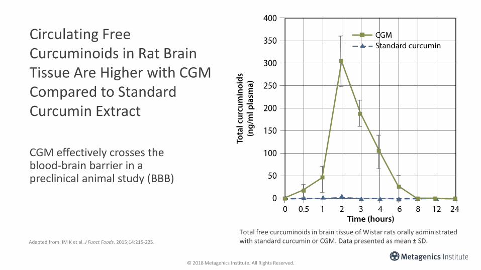
Circulating Free Curcuminoids in Rat Brain Tissue Are Higher with CGM Compared to Standard Curcumin Extract
Adapted from: IM K et al. J Funct Foods. 2015;14:215-225.
Total free curcuminoids in brain tissue of Wistar rats orally administrated with standard curcumin or CGM. Data presented as mean ± SD.
CGM effectively crosses the blood-brain barrier in a preclinical animal study (BBB)

© 2018 Metagenics Institute. All Rights Reserved.
Circulating Free Curcuminoids Across Rat Plasma Are Higher with CGM Compared to Standard Curcumin Extract
Adapted from: IM K et al. J Funct Foods. 2015;14:215-225.
TissueStandard curcumin
(200mg dose)CGM (200mg dose)
CGM compared to standard
AUC AUC Fold Increase
Plasma 70.18 1,758.00* 25.05
Liver 7.77 867.60* 111.6
Kidney 12.21 882.20* 72.25
Heart 8.7 476.90* 54.82
Spleen 9.87 543.00* 55.02
Brain 2.41 838.50* 347.93
Intestine 351,277 4,396,000* 12.51
Differences in Curcuminoid AUC in Rat Tissue Following CGM Compared with Standard Curcumin Preparation
AUC, area under the curve. Data as mean ± SD (n=3). * denotes p < 0.001 of CGM compared with standard curcumin preparation.

© 2018 Metagenics Institute. All Rights Reserved.
Curcumin/CGM—Key Takeaways
• Properties of curcumin:1-4
o Analgesic (reduced neuropathic pain in animal models)1,2
o Anti-arthritic (reduced production of MMP1 and 3 in mouse model of arthritis)3
o Anti-inflammatory signaling4
• CGM is highly bioavailable5,6
o Readily enters the blood and tissues1. Banafshe HR et al. Eur J Pharmacol. 2014;723:202-206.2. Zhu X et al. PLoS One. 2014;9(3):e91303.3. Mun SH et al. J Pharmacol Sci. 2009;111(1):13-21.4. Shezhad A et al. J Food Sci. 2017;82(9):2006-2015.5. Sudheeran SP et al. J Clin Psychopharmacol. 2016;36(3):236-243. 6. IM K et al. J Funct Foods. 2015;14:215-225.

© 2018 Metagenics Institute. All Rights Reserved.
Xanthohumol: An Isolated Constituent
• Xanthohumol is one of many active constituents in hops flowers (Humulus lupulus)
• Hops flowers have commercial uses (to flavor and preserve beer) and medicinal uses
• Properties1
o Anti-inflammatory
o Antioxidant
• Evidence:
o Anti-arrhythmic and anti-atherosclerotic properties in animal models,2-4 reduced platelet activation (human ex vivo),5 anti-obesity and glucose lowering effect in animal models,6 reduced DNA damage (clinical study)7
1. Liu M et al. Molecules. 2015;20(1):754-779.2. Arnaiz-Cot JJ et al. J Pharmacol Exp Ther. 2017;360(1):239-248.3. Liu et al. J Nat Prod. 2017;80(7):2146-2150. 4. Hirata et al. PLoS One. 2012;7(11):e49415.5. Lee YM et al. Evid Based Complement Alternat Med. 2012;2012:852362.6. Legette LL et al. Phytochemistry. 2013;91:236-241. 7. Ferk F et al. Mol Nutr Food Res. 2016;60(4):773-786.

© 2018 Metagenics Institute. All Rights Reserved.
Xanthohumol—Key Takeaways
• Properties:1,2
o Anti-inflammatory
o Antioxidant
• Xanthohumol delivered through a hops-protein matrix:
o Increased bioavailability3
1. Liu M et al. Molecules. 2015;20(1):754-779.2. Ferk F et al. Mol Nutr Food Res. 2016;60(4):773-786.3. O’Connor A et al. Mol Nutr Food Res. 2018;62(6):e1700692.

© 2018 Metagenics Institute. All Rights Reserved.
Xanthohumol Availability Is Enhanced by Delivery Through a Hops-Protein Matrix
• Xanthohumol is generally not well absorbed
• Xanthohumol given as part of a hops-protein matrix shows enhanced bioavailability compared to standard hops-xanthohumol preparations
Adapted from: O’Connor A. Mol Nutr Food Res. 2018;62(6):e1700692.
Hops-protein matrix
Standard hops preparation
Xan
tho
hu
mo
l

© 2018 Metagenics Institute. All Rights Reserved.
Boswellia Serrata
• Also known as Indian Frankincense
• A “gum resin” from the Boswellia serrata tree
• Contains a few active constituents:
o Beta-boswellic acids
o Alpha-boswellic acids
o Essential oils
o Flavonoids—quercetin
• Properties
o Analgesic (increased pain threshold in healthy subjects)1
o Anti-arthritic (reduced inflammatory markers and arthritis score in mouse models and reduced knee pain in subjects with OA)2,3
o Anti-inflammatory and anti-oxidant signaling4
1. Prabhavathi K et al. Indian J Pharmacol. 2014;46(5):475-479.2. Umar S et al. Phytomedicine. 2014;21(6):847-856. 3. Kimmatkar N et al. Phytomedicine. 2003;10(1):3-7.4. Du Z et al. Planta Med. 2015;81(4):259-267.
© 2018 Metagenics Institute. All Rights Reserved.
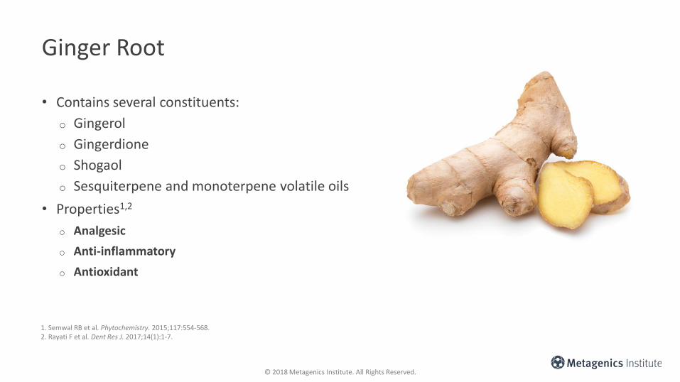
Ginger Root
• Contains several constituents:
o Gingerol
o Gingerdione
o Shogaol
o Sesquiterpene and monoterpene volatile oils
• Properties1,2
o Analgesic
o Anti-inflammatory
o Antioxidant
1. Semwal RB et al. Phytochemistry. 2015;117:554-568.2. Rayati F et al. Dent Res J. 2017;14(1):1-7.

© 2018 Metagenics Institute. All Rights Reserved.
Arachidonic acid (ARA)
Arachidonic acid (ARA)
Prostaglandins Leukotrienes
Phospholipase A2 (PLA2)
Lipoxygenase (5-LOX)Cyclooxygenase-1 & 2 (COX-1, COX-2)
PGE2 LTB4
CurcuminGinger
CurcuminBoswellic acidsGinger
CurcuminXanthohumolBoswellic acidsGingerTHIAA
Inflammation & Pain: Key Enzymes
Perrone D et al. Exp Ther Med. 2015;10(5):1615-1623. Zhou H et al. Curr Drug Targets. 2011;12(3):332-347.Nievergelt A et al. J Immunol. 2011;187(8):4140-50. Kunnumakkara AB et al. Br J Pharmacol. 2017;174(11)1325-1348.Weiskirchen R et al. Front Physiol. 2015;6:140.
Abdel-tawab M et al. Clin Pharmacokinet. 2011;50(6):349-369.Grzanna R et al. J Med Food. 2005;8(2):125-132.Desai A et al. Inflamm Res. 2009;58:1-6.Konda VR et al. Arthritis Rheum. 2010;62(6):1683-1692.Desai A et al. Atherosclerosis. 2012;223:130-136.
© 2018 Metagenics Institute. All Rights Reserved.
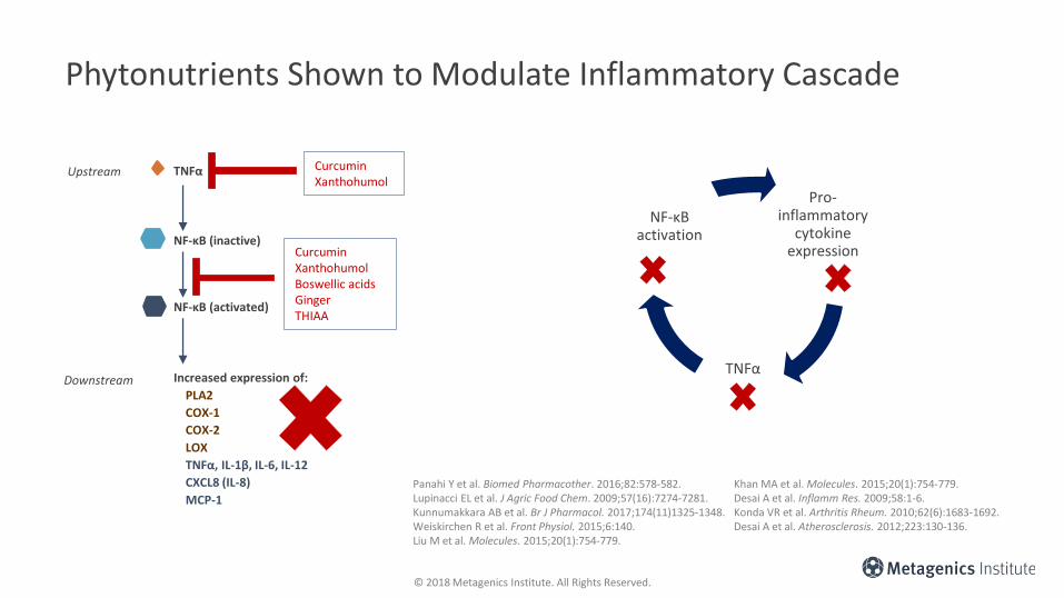
Phytonutrients Shown to Modulate Inflammatory Cascade
Arachidonic Acid (ARA)
CurcuminXanthohumolBoswellic acidsGingerTHIAA
NF-κB (inactive)
NF-κB (activated)
Increased expression of:
PLA2
COX-1
COX-2
LOX
TNFα, IL-1β, IL-6, IL-12
CXCL8 (IL-8)
MCP-1
TNFα CurcuminXanthohumol
Upstream
Downstream
Pro-inflammatory
cytokine expression
TNFα
NF-κBactivation
Panahi Y et al. Biomed Pharmacother. 2016;82:578-582. Lupinacci EL et al. J Agric Food Chem. 2009;57(16):7274-7281.Kunnumakkara AB et al. Br J Pharmacol. 2017;174(11)1325-1348.Weiskirchen R et al. Front Physiol. 2015;6:140. Liu M et al. Molecules. 2015;20(1):754-779.
Khan MA et al. Molecules. 2015;20(1):754-779. Desai A et al. Inflamm Res. 2009;58:1-6.Konda VR et al. Arthritis Rheum. 2010;62(6):1683-1692.Desai A et al. Atherosclerosis. 2012;223:130-136.

© 2018 Metagenics Institute. All Rights Reserved.
Inflammation & Pain: Chemokines
• Curcumin
o Reduces serum levels of MCP-11
o Reduces chondrocyte production of CXCL82
• Xanthohumol
o Reduces macrophage production of MCP-13
• Ginger
o Reduces IP-10, specifically in activated human synoviocytes4
1. Karimiana MS et al. Cytokine Growth Factor Rev. 2017;33:55-63. 2. Panahi Y et al. Biomed Pharmacother. 2016;82:578-582. 3. Lupinacci EL et al. J Agric Food Chem. 2009;57(16):7274-81. 4. Phan PV et al. J Altern Complement Med. 2005;11(1):149-154.

© 2018 Metagenics Institute. All Rights Reserved.
Mechanism of Action Summary
Curcumin Xanthohumol Boswellia serrata THIAA Ginger
Moderation of pro-
inflammatory cytokines,
chemokines, and
transcription factors
associated with
inflammation and pain
✓Moderates NFkB
✓ Reduces serum levels:
TNFα, IL-1β, IL-6,
MCP-1
✓ Diminishes
chondrocyte
production of CXCL8
(IL-8)
✓Moderates NF-κB
✓ Reduces WBC production
of
TNFα, IL-12, MCP-1
✓Moderates NF-κB
✓Moderates NF-κB
expression
and activity
✓ Reduces serum levels of
IL-6
✓ Reduces immune cell
production of
pro-inflammatory
cytokines
✓Moderates NF-κB
✓ Diminishes synoviocyte
production of IP-10
Moderation of enzymes
and prostaglandins
associated with
inflammation and pain
✓Moderates PLA2
✓Moderates COX-2
✓Moderates 5-LOX✓Moderates COX-1 & COX-2
✓Moderates COX-1 &
COX-2
✓Moderates 5-LOX
✓Moderates COX-2✓Moderates PLA2
✓Moderates COX-1 & COX-2
✓Moderates LOX
Mathy-Hartert M et al. Inflamm Res. 2009;58(12):899-908. Karimiana MS et al. Cytokine Growth Factor Rev. 2017;33:55-63. Panahi Y et al. Biomed Pharmacother. 2016;82:578-582. Lupinacci EL et al. J Agric Food Chem. 2009;57(16):7274-7281. Phan P V et al. J Altern Complement Med. 2005;11(1):149-154.Perrone D et al. Exp Ther Med. 2015;10(5):1615-1623. Zhou H et al. Curr Drug Targets. 2011;12(3):332-347.Nievergelt A et al. J Immunol. 2011;187(8):4140-4150. Kunnumakkara AB et al. Br J Pharmacol. 2017;174(11)1325-1348.Weiskirchen R et al. Front Physiol. 2015;6:140. Desai et al. Atherosclerosis. 2012;223:130-136.
Abdel-tawab M et al. Clin Pharmacokinet. 2011;50(6):349-369.Grzanna R et al. J Med Food. 2005;8(2):125-132.Liu M et al. Molecules. 2015:754-779. Khan MA et al. J Ethnopharmacol. 2016;191:315-323. Lee T et al. Biochem Biophys Res Commun. 2009;382(1):134-139. Lee HY et al. Br J Pharmacol. 2012;167(1):128-140.Ganjali S et al. Scientific World J. 2014;2014:898361. Desai et al. Inflamm Res. 2009;58:1-6.Konda et al. Arthritis Rheum. 2010;62(6):1683-1692.

© 2018 Metagenics Institute. All Rights Reserved.
Key Mediators of Inflammation & Pain Modulated by Curcumin, Xanthohumol, Boswellic Acids, & Ginger
o Enzymes– Phospholipase A2 (PLA2)
– Cyclooxygenase (COX-1 and COX-2)
– Lipoxygenase (LOX)
o Gene expression regulators– Nuclear factor-kappa B (NFkB)
o Pro-inflammatory cytokines– TNFα
– IL-1β
– IL-6
– IL-12
o Chemokines– Neutrophil chemotactic factor (CXCL8, aka IL-8)
– Monocyte chemoattractant protein 1 (MCP-1 )
– Interferon-γ activated protein (IP-10)
Panahi Y et al. Biomed Pharmacother. 2016;82:578-582. Lupinacci EL et al. J Agric Food Chem. 2009;57(16):7274-7281.Kunnumakkara AB et al. Br J Pharmacol. 2017;174(11)1325-1348.Weiskirchen R et al. Front Physiol. 2015;6:140. Liu M et al. Molecules. 2015;20(1):754-779.
Khan et al. Molecules. 2015;20(1):754-779.Desai A et al. Inflamm Res. 2009;58:1-6.Konda VR et al. Arthritis Rheum. 2010;62(6):1683-1692.Desai A et al. Atherosclerosis. 2012;223:130-136.
All are modulated by:CurcuminXanthohumolBoswellic acidsGingerTHIAA
© 2018 Metagenics Institute. All Rights Reserved.
Mechanistic Takeaways
• There are several nutritional bioactives that modulate inflammation initiation, including curcumin, xanthohumol, Boswellia serrata, ginger, and THIAA:
o Moderate several key mediators of inflammation & pain
– NF-κB—Upregulates cytokines, chemokines, and pro-inflammatory enzymes
– Pro-inflammatory cytokines
– Chemokines—WBC recruitment
– Enzymes involved in prostaglandin and leukotriene production
o Act upstream and downstream on inflammation & pain pathways
Panahi Y et al. Biomed Pharmacother. 2016;82:578-582. Lupinacci EL et al. J Agric Food Chem. 2009;57(16):7274-7281.Kunnumakkara AB et al. Br J Pharmacol. 2017;174(11)1325-1348.Weiskirchen R et al. Front Physiol. 2015;6:140. Liu M et al. Molecules. 2015;20(1):754-779.
Khan et al. Molecules. 2015;20(1):754-779.Desai A et al. Inflamm Res. 2009;58:1-6.Konda VR et al. Arthritis Rheum. 2010;62(6):1683-1692.Desai A et al. Atherosclerosis. 2012;223:130-136.

© 2018 Metagenics Institute. All Rights Reserved.
Other Key Takeaways
THIAA
Has anti-inflammatory properties, and has demonstrated anti-arthritic action in animal model of arthritis1-3
Curcumin
Has anti-inflammatory and analgesic action in pre-clinical models.4-6 Shown to have anti-arthritic properties in rodent models of arthritis7
Xanthohumol
Activates anti-oxidant and anti-inflammatory signaling pathways.8 Preclinical studies have demonstrated anti-arrhythmic and anti-atherosclerotic properties,9-11 anti-obesity and glucose lowering effect in animal models.12 Human studies showed reduced platelet activation (ex vivo model)13 and reduced DNA damage (clinical study)14
Boswellia serrata
Analgesic and anti-arthritic action in human studies,15,16 and modulate anti-inflammatory and anti-oxidant signaling pathways17
Ginger
Demonstrated anti-inflammatory anti-oxidant signaling effects,18 and analgesic effects19
1. Desai A et al. Atherosclerosis. 2012;223:130-136.2. Desai A et al. Inflamm Res. 2009;58(5):229-234.3. Konda VR et al. Arthritis Rheum. 2010;62(6):1683-1692.4. Banafshe HR et al. Eur J Pharmacol. 2014;723:202-206.5. Zhu X et al. PLoS One. 2014;9(3):e91303.6. Shezhad A et al. J Food Sci. 2017;82(9):2006-2015.7. Mun SH. J Pharmacol Sci. 2009;111(1):13-21.8. Liu M et al. Molecules. 2015;20(1):754-779.
9. Arnaiz-Cot JJ et al. J Pharmacol Exp Ther. 2017;360(1):239-248.10. Liu et al. J Nat Prod. 2017;80(7):2146-2150. 11. Hirata et al. PLoS One. 2012;7(11):e49415.12. Legette LL et al. Phytochemistry. 2013;91:236-241. 13. Lee YM et al. Evid Based Complement Alternat Med. 2012;2012:852362.14. Ferk F et al. Mol Nutr Food Res. 2016;60(4):773-786.
15. Prabhavathi K et al. Indian J Pharmacol. 2014;46(5):475-479.16. Kimmatkar N et al. Phytomedicine. 2003;10(1):3-7.17. Du Z et al. Planta Med. 2015;81(4):259-257.18. Semwal RB et al. Phytochemistry. 2015;117:554-568.19. Rayati F et al. Dent Res J. 2017;14(1):1-7.

© 2018 Metagenics Institute. All Rights Reserved.© 2018 Metagenics Institute. All Rights Reserved.
Inflammation Resolution
Mechanisms and the Role of
Specialized Pro-Resolving Mediators (SPMs)

© 2018 Metagenics Institute. All Rights Reserved.
What are Specialized Pro-Resolving Mediators?
AA
LXsLipoxins
EPA
RvEsE-series
resolvins
RvDsD-seriesresolvins
PDsProtectins
MaRsMaresins
DHA
Specialized Pro-Resolving Mediators
Adapted from: Serhan CN. FASEB J. 2017;31(4):1273-1288.
17-HDHA
1 Specialized pro-resolving mediators (SPMs) are a group of lipid mediators that drive the resolution of inflammation.
2SPMs can be produced endogenously in the body from precursor polyunsaturated fatty acids (AA, EPA, and DHA)
3SPMs are required for inflammation resolution to occur and for effective return to homeostasis or previously normal conditions
4There are several classes of SPMs (lipoxins, resolvins, protectinsand neuro-protectins, maresins) that all work together to resolve inflammation
18-HEPE
5 Key hydroxylated precursor SPMs such as 18-HEPE and 17-HDHA can give rise to an array of downstream SPMs that have been identified to date. 18-HEPE can be converted into 4 distinct E-series resolvins (RvE1-4) and 17-HDHA can be converted into 6 distinct D-series resolvins (RvD1-6)

© 2018 Metagenics Institute. All Rights Reserved.
SPMs Have Been Identified at Bioactive Levels Across Many Human Tissues and Fluids
Adapted from: Serhan CN. FASEB J. 2017;31(4):1273-1288.
Brain380–1800pg/mg protein
Cerebrospinal fluid11–390 pM
Lymph nodes3–112pg/100mg
Adipose tissue2–3pg/106 cells
Placenta1–8pg/mg
Synovial fluid0.2–0.7nM
Urine12–330pM
Exhaled breath concentrate6nM
Plasma3–30pM
Serum14pM–6nM
Spleen1–112pg/100mg
Breast milk10pM–27nM

© 2018 Metagenics Institute. All Rights Reserved.
Previous science perspective
• Inflammation faded out by itself
• Blocking inflammation was the goal
Current perspective
• Inflammation resolution is an active process required for homeostasis
• Resolution is coordinated by specialized pro-resolving mediators
2000: First published on resolvins1
2005: Concept of resolution interval introduced2
SPMs in models of IBD;3 lower SPMs in Alzheimer’s disease brain tissue identified4
1. Serhan CN et al. J Exp Med. 2000;192:1197–1204.2. Bannenberg GL et al. J Immunol. 2005;174:4345-4355.3. Arita M et al. Proc Natl Acad Sci USA. 2005;201:7671-7676.4. Lukiw WJ et al. J Clin Invest. 2005;115(10):2774-2783.5. Gonzalez-Periz A et al. FASEB J. 2009;23:1946-1957.6. Hellman J et al. FASEB J. 2011;25:2399-2407.7. Ho KJ et al. Am J Pathol. 2010;177:2116-2123.
2009–2011: SPMs in models of obesity, diabetes, atherosclerosis, and arthritis5-8
2000s: SPMs identified in models of self-limiting inflammation
2012: SPMs enhance B-cell response9 linked with tissue regeneration10
and shown to enhance uptake and killing of microbes and reduce antibiotics requirement in mice11
8. Lima-Garcia JF et al. Br J Pharmacol. 2011;164(2):278-293.9. Ramon S et al. J Immunol. 2012;189(2):1036-1042.10. Serhan CN et al. FASEB J 2012;26:1755-1765.11. Chiang N et al. Nature. 2012;484:524-528.12. Titos E et al. J Immunol. 2016;197:3360-3370.13. Fredman G et al. Nat Commun. 2016;23;7:12859.
Aulus CelsusRoman 30–45 BC: Redness, warmth, swelling pain, and Galen (physician) loss of function
1897: Aspirinand other NSAIDs
from 1950s
2001: NSAIDs accounted for 70M+
prescriptions in the US
2016–2018: Cohort studies highlight imbalance of lipid mediators in several conditions12-13

© 2018 Metagenics Institute. All Rights Reserved.
Hallmarks of SPM Activity and Inflammation Resolution
Adapted from: Serhan CN. FASEB J. 2017;31(4):1273-1288.
SPMs actively promote inflammation resolution• Enhance macrophage phagocytosis and efferocytosis, which clears up
dead or dying cells and cellular debris • Shorten the time to resolution • Increase the production of anti-inflammatory mediators • Increase the killing and clearance of microbes• Enhance tissue regeneration
Inflammationinitiation
Inflammationresolution
HomeostasisNormal function
HomeostasisNormal function
Inflammation triggers
SPMs act to curtail excessive inflammatory response• Limit further pro-inflammatory PMN cells coming
to the site of inflammation• Counter-regulate pro-inflammatory mediators• Limit tissue damage from excessive or persistent
inflammation

© 2018 Metagenics Institute. All Rights Reserved.
Resolution is Necessary to Prevent Tissue Damage—Associated with Chronic Inflammation
Progression
Unresolved immune response
SPMs
Resolution of immune response
Tissue impacted(e.g., over-exercising)
Tissue impacted
Kumar et al. Robbins & Cotran Pathologic Basis of Disease. 8th ed. Saunders;2009.

© 2018 Metagenics Institute. All Rights Reserved.
Pro-Resolving Versus Pro-Inflammatory Macrophages
Pro-inflammatory• M1 phenotype• Classically activated macrophages
Pro-resolving• M2 phenotype
• Alternatively activated macrophages
Functions:• Anti-microbial
activity• ROS production• Engulfs pathogens
Secretes:• Free radicals• Pro-inflammatory cytokines
e.g. TNF-α, IL-6• MHC-II
Secretes:• IL-10• TGF-β• VEGF• PDGF
End result = inflammation Resolution of inflammation and tissue regeneration
Functions:• Anti-inflammatory• Engulfs dead and
dying cells and other cell debris
SPMs promote
pro-resolving phenotype
Adapted from: Das A et al. Am J Pathol. 2015;185(10):2596-2606.

© 2018 Metagenics Institute. All Rights Reserved.
Why Use Anti-Inflammatory Agents?
Anti-inflammatory agents may be important to help reduce the magnitude of the inflammation initiation response.
Inflammation initiation
Most anti-inflammatory agents do not address inflammation resolution.
Inflammatory triggers
HomeostasisNormal function
© 2018 Metagenics Institute. All Rights Reserved.
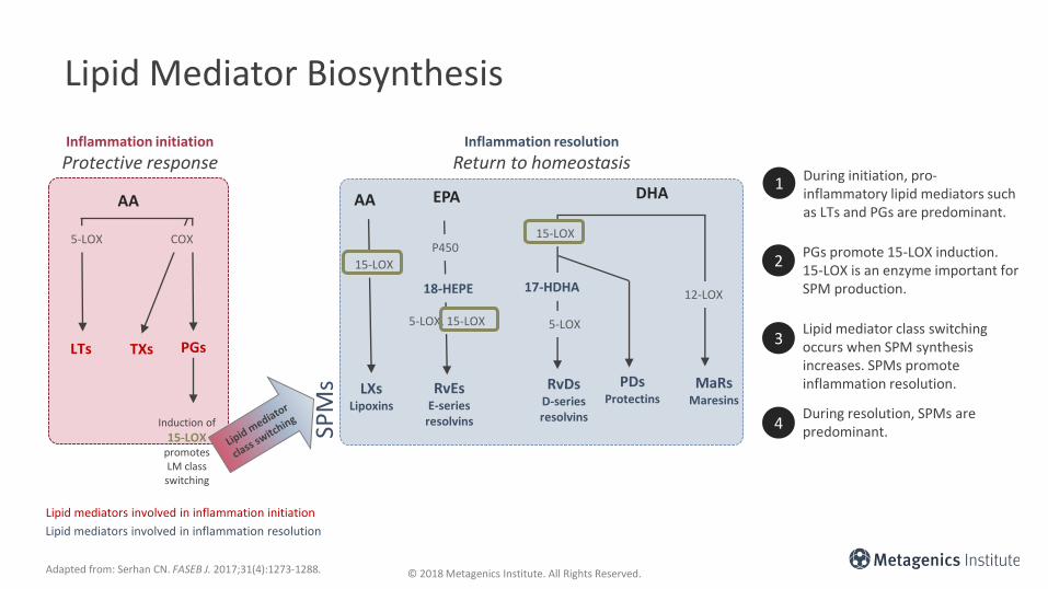
Lipid Mediator Biosynthesis
AA
LTs TXs PGs
AA
LXsLipoxins
EPA
RvEsE-series
resolvins
RvDsD-seriesresolvins
PDsProtectins
MaRsMaresins
DHA
Adapted from: Serhan CN. FASEB J. 2017;31(4):1273-1288.
5-LOX COX
15-LOX
15-LOX
17-HDHA
5-LOX
12-LOX
SPM
s
Inflammation initiation
Protective responseInflammation resolution
Return to homeostasis
Induction of
15-LOX promotes LM class switching
1 During initiation, pro-inflammatory lipid mediators such as LTs and PGs are predominant.
2PGs promote 15-LOX induction. 15-LOX is an enzyme important for SPM production.
3Lipid mediator class switching occurs when SPM synthesis increases. SPMs promote inflammation resolution.
4During resolution, SPMs are predominant.
P450
18-HEPE
5-LOX, 15-LOX
Lipid mediators involved in inflammation initiation
Lipid mediators involved in inflammation resolution

© 2018 Metagenics Institute. All Rights Reserved.
Impact of COX-2 Inhibitors on Lipid Mediator Biosynthesis
AA
LTs TXs PGs
AA
LXsLipoxins
EPA
RvEsE-series
resolvins
RvDsD-seriesresolvins
PDsProtectins
MaRsMaresins
DHA
Adapted from: Serhan CN. FASEB J. 2017;31(4):1273-1288.
5-LOX COX
SPM
s
Inflammation initiation
Protective responseInflammation resolution
Return to homeostasis
Induction of
15-LOXpromotes LM class switching
1 COX-2 inhibitors block the production of PGs.
2Because PGs promote the induction of 15-LOX needed for production of SPMs, SPM production can be reduced.
3 Lipid mediator class switching is impaired.
4COX-2 inhibitors are considered “resolution toxic.”
X
X X
15-LOX
15-LOX
17-HDHA
5-LOX
12-LOX
P450
18-HEPE
5-LOX, 15-LOX
XX
XX X
Lipid mediators involved in inflammation initiation
Lipid mediators involved in inflammation resolution

© 2018 Metagenics Institute. All Rights Reserved.
Impact of Low-Dose Aspirin on Lipid Mediator Biosynthesis
AA
LTs TXs PGs
AA
LXsLipoxins
EPA
RvEsE-series
resolvins
RvDsD-seriesresolvins
PDsProtectins
MaRsMaresins
DHA
Lipid mediators involved in inflammation initiation
Lipid mediators involved in inflammation resolution
Adapted from: Serhan CN. FASEB J. 2017;31(4):1273-1288.
5-LOX COX
SPM
s
Inflammation initiation
Protective responseInflammation resolution
Return to homeostasis
Induction of
15-LOXpromotes LM class switching
1 Low-dose aspirin blocks the activity of COX that leads to PG production.
2But low-dose aspirin changes the COX catalytic domain. Acetylated COX is involved in the production of aspirin-triggered SPMs.
3 Lipid mediator class switching can occur.
4Low-dose aspirin is considered “resolution friendly.”
X
X
17-HDHA
5-LOX
12-LOX18-HEPE
5-LOX, 15-LOXX
Acetylated COX
/5-LOX
Acetylated COX
Acetylated COX
Lipid mediator class switching
X X
Catalytic domain: the region where the chemical reaction takes place

© 2018 Metagenics Institute. All Rights Reserved.
Blocking Inflammation and Resolution Toxicity
Alpha signals omega—prostaglandin biosynthesis is critical to resolution, because PGEs stimulate induction of lipoxygenases necessary for LX and Rv synthesis. Blocking this cascade (e.g., COX-2 inhibitors) can be “resolution toxic.”
Adapted from: Chan MM et al. J Immunol. 2010;184:6418-6426.
Induced arthritis model
Induced arthritis + COX-2 inhibitor (therapeutic)
0
50
100
Day 70 swelling
VC NS-398
• TNFα and IL-17mRNA increased in NS-398
• Radiography showed greater degree of soft tissue swelling, digital misalignment, ankyloses ,and loss of bone density in NS-398
• Histological staining showed pannus of knee joint was proliferative and cartilage and bone more severely damaged in NS-398
• PGE2 analogue treatment helped restore resolution
87%
54%
% r
eso
luti
on
of
swel
ling

© 2018 Metagenics Institute. All Rights Reserved.
SPMs Are Not Considered Immunosuppressive
Infection Model SPM Tested Outcome in SPM Group
Candida albicans Mouse candidiasis RvE1 Stimulated clearance of the fungus from circulating blood
Sepsis Mouse CLP RvD2 Reduced mortality
Escherichia coli Mouse pneumonia RvE1 Reduced mortality
Herpes simplex virus Mouse keratitis RvE1 Reduced lesion severity
Escherichia coli Mouse peritonitis RvD1 Reduced mortality
Escherichia coli Mouse peritonitisRvD1 RvD5
Protections from hypothermia and enhanced antibiotic effectiveness
Staphylococcus aureus Mouse skin infectionRvD1 RvD5
Reduced bacterial titers and enhanced antibiotic effectiveness
SepsisMouse double injury of burn and sepsis
RvD2 Increased survival
Adapted from: Chiang N et al. Mol Aspects Med. 2017;58:114-129.
In a range of preclinical mouse models of infection, SPMs have been shown to improve outcomes—highlighting that SPMs do not reduce the protective inflammatory response and are therefore not considered immunosuppressive.

© 2018 Metagenics Institute. All Rights Reserved.© 2018 Metagenics Institute. All Rights Reserved.
Factors Impacting Levels of Endogenous Specialized
Pro-Resolving Mediators (SPMs)

© 2018 Metagenics Institute. All Rights Reserved.
Aging Delays Resolution and Alters the Balance of Pro-Inflammatory : Pro-Resolving Mediators
Adapted from: Arnordottir HH et al. J Immunol. 2014;193:4235-4244.
0
10
20
Ri
Resolution interval
Tmax(PMN infiltration)
Magnitude at 4 hrs(PMN infiltration)
0
5
Tmax0
10
20
Tim
e (h
ou
rs)
Tim
e (h
ou
rs)
Aged mice
Young mice
Peritonitis model(zymosan; i.p.)
Peritoneal exudates assessed over 24 hours P
MN
(x1
06 )
85% ↑ in aged
Ψ max
***
*** p<0.001 versus young mice
• IL-6 ↑ in exudates from aged mice• Macrophages from aged mice had reduced ability to clear apoptotic
PMNs• Distinct lipid mediator profile in young versus aged mice: reduced
lipoxins and DHA-derived SPMs and increased PGs/TXs
• RvD3 shorted Ri, reduced PMN, and enhanced ability of macrophage to clear PMNs

© 2018 Metagenics Institute. All Rights Reserved.
Less SPMs in Circulation Following Fish Oil in Metabolic Syndrome vs. Healthy
Adapted from: Barden AE et al. Am J Clin Nutr. 2015;102:1357-1364.
18
-HEP
E (m
mo
l/L)
0
31 2
Weeks
1
2
3
17
-HD
HA
(m
mo
l/L)
0
1
1 2 3
Weeks
2
3
4
5
ControlMetSyn
group * time W3 p=0.018
group * time W3 p=0.003
2.4g EPA+DHA 2.4g EPA+DHA
Metabolic syndrome blunts increase in 17-HDHA and 18-HEPE following EPA + DHA supplementation

© 2018 Metagenics Institute. All Rights Reserved.
People with Obesity Have a Lower Ratio of SPMs: Pro-Inflammatory Mediators Compared to People with Lower Body Weight
• Subjects with obesity (Ob) scheduled for bariatric surgery (n=41) and lean controls (CT) (n=7) scheduled for cholecystectomy. Omental adipose tissue samples collected at time of surgery.
• The ratio between SPMs and pro-inflammatory markers is lower in obese adipose tissue.
Adapted from: Titos E et al. J Immunol. 2016;197:3360-3370.
***
**
Re
lati
ve u
nit
s
Re
lati
ve u
nit
s
SPM/LTB4 SPM/PGs
100
80
60
40
20
0
40
30
20
10
CT CT ObOb
Data are mean ± SEM **p< 0.005 vs. CT subjects***p<0.001 vs. CT subjects
© 2018 Metagenics Institute. All Rights Reserved.
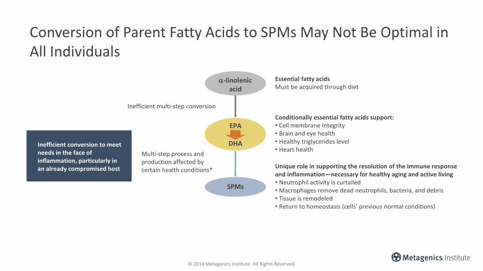
Conversion of Parent Fatty Acids to SPMs May Not Be Optimal in All Individuals
Essential fatty acidsMust be acquired through diet
Conditionally essential fatty acids support:• Cell membrane integrity• Brain and eye health• Healthy triglycerides level• Heart health
Unique role in supporting the resolution of the immune response and inflammation—necessary for healthy aging and active living• Neutrophil activity is curtailed• Macrophages remove dead neutrophils, bacteria, and debris• Tissue is remodeled• Return to homeostasis (cells’ previous normal conditions)
Inefficient conversion to meet needs in the face of inflammation, particularly in an already compromised host
Inefficient multi-step conversion
Multi-step process and production affected by certain health conditions*
a-linolenicacid
EPA
DHA
SPMs

© 2018 Metagenics Institute. All Rights Reserved.
Potential Groups Who May Benefit from SPM Supplementation
• Research has described areas in which SPM levels and the balance between the pro-inflammatory and pro-resolving lipid mediators are related. For example:
o In a preclinical mouse model of aging, SPM levels were lower and pro-inflammatory mediators were higher following an inflammatory challenge compared with younger mice, and giving exogenous SPMs could rescue this.1
o Circulating SPM (17-HDHA and 18-HEPE) levels increased to a lesser magnitude in people with metabolic syndrome vs. healthy controls following fish oil supplementation- indicating a potential need for more direct supplementation with SPMs.2
o In people with obesity, a lower level of SPMs and a higher level of pro-inflammatory lipid mediators is seen indicating some dysregulation of lipid mediators.3 Animal research in obesity has also shown that this occurs in diet-induced and genetic models of obesity,4,5 and that DHA-rich diets are less effective in raising tissue SPM levels in obese animals compared with leaner controls.5
1. Arnordottir HH et al. J Immunol. 2014;193:4235-4244.2. Barden AE et al. Am J Clin Nutr. 2015;102:1357-1364.3. Titos E et al. J Immunol. 2016;197:3360-3370.4. Claria et al. J Immunol. 2012;189:2597-2605.5. Neuhofer A et al. Diabetes. 2013;62:1945–1956.

© 2018 Metagenics Institute. All Rights Reserved.© 2018 Metagenics Institute. All Rights Reserved.
Emerging Areas of Specialized Pro-Resolving
(SPMs) Research• Preclinical and human observational cohort data
highlights potential areas of interest for resolution science

© 2018 Metagenics Institute. All Rights Reserved.
Emerging Areas of Specialized Pro-Resolving Mediator Research
Dry eye
Arthritis
Cardiovascular disease
Peripheral vascular disease
Cognition and neuroinflammation
Cancer
Inflammatory bowel disease
Response to surgery
Pain
Physical activity
Wound healingType 2 diabetes
Adaptive immunity
Endometriosis
Fibromyalgia
Dermatitis
Kidney disease
Sjogren’s syndrome
Autoimmune conditions
Mitochondrial function in inflammation
This emerging research is defined largely by pre-clinical and human observational data and provides an indicator of which aspects of resolution science may continue to grow

© 2018 Metagenics Institute. All Rights Reserved.
Emerging Area: Adaptive Immunity
B-cell differentiation and function1,2
• RvD1 and 17-HDHA promote the differentiation of IgG-secreting B cells and enhance antibody-mediated immune responses.
• RvD1 and 17-HDHA inhibit IgE production by human B cells and suppress the differentiation of naïve B cells into IgE-secreting cells by specifically blocking epsilon germline transcript.
T-cell response3,4
• Data suggests that RvD2 inhibits systemic and gingival Th1-type adaptive responses known to mediate alveolar bone loss in a mouse model of periodontitis.
• In human peripheral blood lymphocytes, RvD1, RvD2, and Mar1 reduced cytokine production by activated CD8(+) T cells and CD4(+) T helper 1 (TH1) and TH17 cells and prevented naïve CD4(+) T cell differentiation into TH1 and TH17 cells.
1. Kim N et al. Eur J Immunol. 2016;46(1):81-91.2. Ramon S et al. J Immunol. 2012;189(2):1036-1042.3. Mizraji G et al. Front Immunol. 2018;9:785.4. Chiurchiu V et al. Sci Transl Med. 2016;8(353):353ra111.

© 2018 Metagenics Institute. All Rights Reserved.
Emerging Area: Endometriosis
Dmitrieva N et al. Fertil Steril. 2014;102(4):1191-1196.
Objective: To study the effects of two resolvins of D series, RvD1 and 17(R)-RvD1, on inflammatory signs associated with endometriosis, in a rat model of endometriosis.
Design:Intravenous or intraperitoneal injections of RvD1 (300 ng/kg) or 17(R)-RvD1 (300 and 900 ng/kg) in rats with surgically induced endometriosis.
Results: Both resolvins, but not vehicle control (placebo), significantly decreased vascular permeability in ectopic endometrial tissue. 17(R)-RvD1 also significantly alleviated severity of vaginal hyperalgesia.
Conclusion: RvD1 and 17(R)-RvD1 can be considered for further investigation of their therapeutic potential for treating endometriosis.
© 2018 Metagenics Institute. All Rights Reserved.
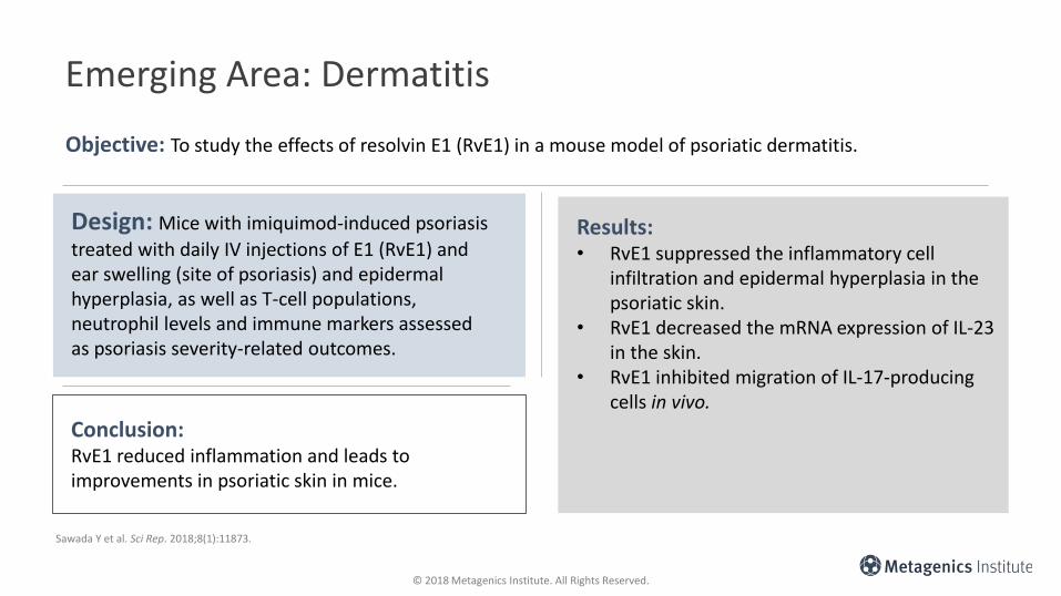
Emerging Area: Dermatitis
Objective: To study the effects of resolvin E1 (RvE1) in a mouse model of psoriatic dermatitis.
Design: Mice with imiquimod-induced psoriasis
treated with daily IV injections of E1 (RvE1) and ear swelling (site of psoriasis) and epidermal hyperplasia, as well as T-cell populations, neutrophil levels and immune markers assessed as psoriasis severity-related outcomes.
Results: • RvE1 suppressed the inflammatory cell
infiltration and epidermal hyperplasia in the psoriatic skin.
• RvE1 decreased the mRNA expression of IL-23 in the skin.
• RvE1 inhibited migration of IL-17-producing cells in vivo.
Conclusion: RvE1 reduced inflammation and leads to improvements in psoriatic skin in mice.
Sawada Y et al. Sci Rep. 2018;8(1):11873.

© 2018 Metagenics Institute. All Rights Reserved.
Emerging Area: Dermatitis
Objective: To determine the protective effects and the underlying mechanisms of RvD1 in a mouse model of induced psoriatic dermatitis.
Design: Mice were pretreated intraperitoneally (i.p.) with or without RvD1, and severity of induced dermatitis graded using a modified Psoriasis Area and Severity Index (PASI), histopathology, and cytokine analysis.
Results: • RvD1 treatment alleviated induced psoriasis
form dermatitis and improved skin pathological changes.
• RvD1 treatment inhibited pro-inflammatory signaling in this model.
Conclusion: RvD1 can improve skin inflammation in mice with induced psoriasiform dermatitis.
Xu J et al. J Dermatol Sci. 2018;89(2):127-135.

© 2018 Metagenics Institute. All Rights Reserved.
Emerging Area: Wound Healing
Rathod KS et al. J Clin Invest. 2017;127(1):169-182.
Objective: To explore sex differences in wound healing in lipid mediator profile over time-course of blister formation and resolution.
Design: Skin blister was induced by cantharidin in 16 men and 16 pre-menopausal women. Inflammatory exudate was collected and lipid mediators and cellular recruitment was measured at 24 and 72 hours.
Results: • At 24 hours, cantharidin formed blisters of
similar volume in both sexes; however, at 72 hours, blisters had only resolved in women.
• Monocyte and leukocyte counts were reduced, and the activation state of all major leukocytes was lower in blisters of females.
• This was associated with enhanced levels of the resolving lipids, particularly D-resolvin.
Conclusion: Pre-menopausal women appear to have greater capacity for resolution in this blister model than men, and this may be mediated by an elevation of the D-resolvin pathway.

© 2018 Metagenics Institute. All Rights Reserved.
Emerging Area: Wound Healing
Mennon R et al. Nano Life. 2017;7(1).pii:1750002.
Objective: Compare the effects of resolvins D1 (RvD1), D2 (RvD2), and E1 (RvE1) on their abilities to inhibit neutrophil migration in vitro and to promote wound healing in vivo in a mouse model.
Design: • The impact of vD1, RvD2 and RvE1 on neutrophil
migration was assessed in vitro (Transwell system).• RvD1, RvD2, RvE1 and vehicle control (placebo) were
then applied topically to mice with an excisional wound (1 cm x 1 cm), and time to wound closure was measured.
Results:• All three resolvins inhibited neutrophil migration, with
RvE1 being the most effective.• Topically applied Rvs accelerated wound closure. RvE1-
treated wounds healed within 19.4 ± 1.5, RvD2-treated within 22.8 ± 1.8 days, and RvD1-treated within 24.4 ±2.2 days. All resolvin-treated groups healed significantly faster than placebo (within 28.6 ± 1.5 days).
• There was a strong linear correlation (R2=0.9384) between each resolvin's potency in inhibiting neutrophil migration in vitro versus accelerating wound healing in vivo.
Conclusion:Topical application of specific resolvins shows promise for supporting wound healing.

© 2018 Metagenics Institute. All Rights Reserved.
Emerging Area: Mitochondrial Function in Inflammation
Hecker M et al. Biochim Biophys Acta. 2018;1863(9):1016-1028.
Objective: To investigate the impact of omega-6 (arachidonic acid) and omega-3 (EPA, DHA) fatty acids, and specialized pro-resolving mediators (18-HEPE and RvE1) on mitochondrial function in experimental inflammation.
Design: • Peripheral blood mononuclear cells (PBMCs) were isolated
from healthy individuals, pre-treated with TNF-a, and then treated with fatty acids (EPA, DHA, linoleic acid) or the SPMs (18-HEPE or RvE1).
• Pro-inflammatory mediators were measured, as were markers of mitochondrial function including respiration, membrane potential, and reactive oxygen species.
Results:
• The results revealed that, in contrast to n-6 and n-3 fatty acids, both 18R-HEPE and RvE1 possess anti-inflammatory and anti-apoptotic properties.
• Both mediators are able to restore inflammation-induced mitochondrial dysfunction, which is characterized by a decrease in mitochondrial respiration and membrane potential as well as an imbalance of mitochondrial fission and fusion.Conclusion:
These results suggest a novel functional mechanism for the beneficial effects of 18-HEPE and RvE1 in inflammatory reactions.
© 2018 Metagenics Institute. All Rights Reserved.
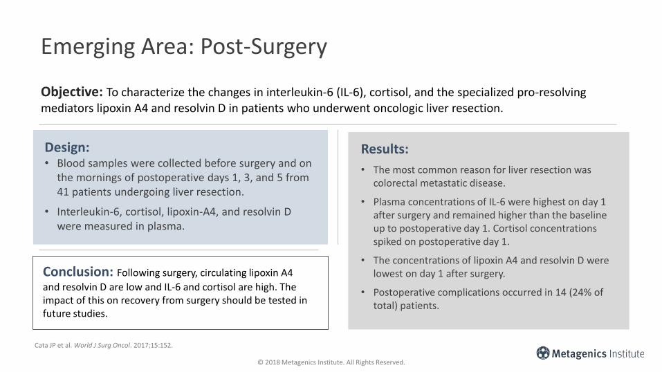
Emerging Area: Post-Surgery
Cata JP et al. World J Surg Oncol. 2017;15:152.
Objective: To characterize the changes in interleukin-6 (IL-6), cortisol, and the specialized pro-resolving mediators lipoxin A4 and resolvin D in patients who underwent oncologic liver resection.
Design: • Blood samples were collected before surgery and on
the mornings of postoperative days 1, 3, and 5 from 41 patients undergoing liver resection.
• Interleukin-6, cortisol, lipoxin-A4, and resolvin D were measured in plasma.
Results:
• The most common reason for liver resection was colorectal metastatic disease.
• Plasma concentrations of IL-6 were highest on day 1 after surgery and remained higher than the baseline up to postoperative day 1. Cortisol concentrations spiked on postoperative day 1.
• The concentrations of lipoxin A4 and resolvin D were lowest on day 1 after surgery.
• Postoperative complications occurred in 14 (24% of total) patients.
Conclusion: Following surgery, circulating lipoxin A4
and resolvin D are low and IL-6 and cortisol are high. The impact of this on recovery from surgery should be tested in future studies.

© 2018 Metagenics Institute. All Rights Reserved.
Emerging Area: Cancer
Sulciner ML et al. J Exp Med. 2018;215(1):115-140.
Objective: To determine the impact of tumor debris on cancer progression and assess whether this could be modulated by treatment with resolvins.
Design:• Tumor debris was generated by treating
tumor cells in vitro with chemotherapy or targeted therapy (e.g., cetuximab).
• Debris was then injected into an in vivomouse debris-stimulated tumor models.
Results: • Tumor cells killed by chemotherapy or
targeted therapy ("tumor cell debris") stimulate primary tumor growth.
• RvD1, RvD2, and RvE1 inhibited debris-stimulated tumors and cancer progression by enhancing clearance of debris via macrophage phagocytosis in multiple tumor types.
Conclusion: Enhancing endogenous clearance of tumor cell debris may be a valuable complement to cytotoxic cancer therapies.

© 2018 Metagenics Institute. All Rights Reserved.
Emerging Area: Fibromyalgia
Klein CP et al. Neuropharmacology. 2014;86:57-66.
Objective: To determine if spinal or systemic treatment with specialized pro-resolving mediators impacted the behavioral and neurochemical changes seen in a mouse model of fibromyalgia.
Design:Mice with induced fibromyalgia were treated with resolvin D1 (RvD1), aspirin-triggered resolvin D1 (AT-RvD1), and resolvin D2 (RvD2).
Results: • Acute administration of AT-RvD1 and RvD2
significantly inhibited mechanical allodynia and thermal sensitization.
• Chronic treatment with AT-RvD1 and RvD2 prevented depressive-like behavior (assessment of immobility time).
• RvD2 prevented 5-HT reduction in total brain, and AT-RvD1 led to a recovery of dopamine levels in cortex and 5-HT in thalamus.
Conclusion: D-series resolvins AT-RvD1, and mainly RvD2, reduced painful and depressive symptoms of fibromyalgia in mice.
© 2018 Metagenics Institute. All Rights Reserved.
Emerging Area: Sjögren’s Syndrome
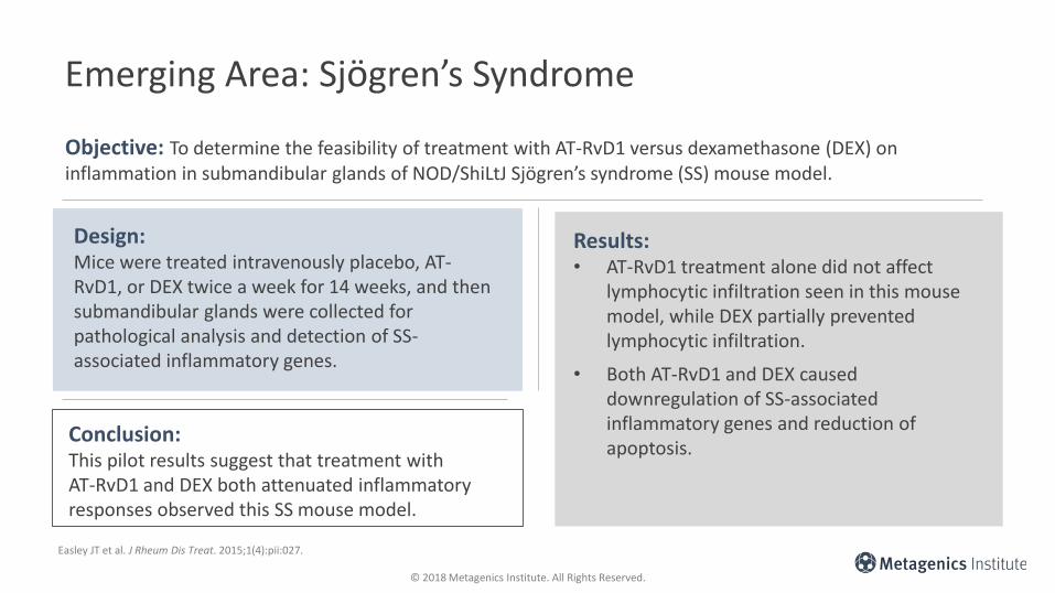
Easley JT et al. J Rheum Dis Treat. 2015;1(4):pii:027.
Objective: To determine the feasibility of treatment with AT-RvD1 versus dexamethasone (DEX) on inflammation in submandibular glands of NOD/ShiLtJ Sjögren’s syndrome (SS) mouse model.
Design:Mice were treated intravenously placebo, AT-RvD1, or DEX twice a week for 14 weeks, and then submandibular glands were collected for pathological analysis and detection of SS-associated inflammatory genes.
Results: • AT-RvD1 treatment alone did not affect
lymphocytic infiltration seen in this mouse model, while DEX partially prevented lymphocytic infiltration.
• Both AT-RvD1 and DEX caused downregulation of SS-associated inflammatory genes and reduction of apoptosis.
Conclusion: This pilot results suggest that treatment with AT-RvD1 and DEX both attenuated inflammatory responses observed this SS mouse model.

© 2018 Metagenics Institute. All Rights Reserved.
Emerging Area: Obesity and Metabolic Disease
1. Claria J et al. Mol Aspects Med. 2017;58:83-922. Spite M et al. Cell Metab. 2014;19(1):21-36.
Adipocytes
RvD1, RvD2, RvE1, LXA4 1,2
• Decrease secretion of pro-inflammatory adipokines
• Increase adiponectin secretion
• Increase macrophage phagocytosis
• Promote anti-inflammatory/pro-resolving M2 macrophage phenotype
• Reduce monocyte adhesion to adipocytes and reduce crown-like structures

© 2018 Metagenics Institute. All Rights Reserved.
Emerging Area: Vascular Disease
1. Elajami TK et al. FASEB J. 2016;30(8):2792-801.2. Claria J et al. Am J Physiol Cell Physiol. 2013;304:C1141-C1149.3. Ho KJ et al. Am J of Pathol. 2010;177(4):2116-2123.4. Hiram R et al. Am J Physiol Heart Circ Physiol. 2014;307:H1547-H1558.5. Akagi D et al. FASEB J. 2015;29(6):2504-2513.6. Salic K et al. Atherosclerosis. 2016:250:158-165.7. Fredman G et al. Nat Commun. 2016;23;7:12859.8. Chatterjee A et al. FASEB J. 2017;3393-3402.
RvD3 and RvD6
Promote macrophage phagocytosis of blood clots1
RvE1
Reduced smooth muscle cell migration2-5
Reduced atherosclerotic lesion size in ApoE*Leiden mice6
• Ratio of SPMs to pro-inflammatory leukotriene B4 (LTB4), is significantly decreased in the vulnerable compared with stable atherosclerotic plaque lesions.7
• Primary human vascular cells produce SPMs and express SPM receptors.3,8

© 2018 Metagenics Institute. All Rights Reserved.
Emerging Area: Arthritis
• Osteoarthritis (OA) is characterized by an increase in inflammatory cells and biomarkers in affected joints.1
• In patients with arthritis, lower levels of Rvs, 17-HDHA, and 18-HEPE were correlated with higher erythrocyte sedimentation rate and pain.2
• In animal models, treatment with Rvs reduced joint inflammation, ameliorated arthritis symptom and severity, and stimulated chondrocyte matrix production.3-5
1. Sellam J et al. Nat Rev Rheumatol. 2010;6(11):625-635.2. Barden AE et al. Prostaglandins Leukot Essent Fatty Acids. 2016;107:24-29.3. Norling LV et al. JCI Insight. 2016;1(5):e85922.4. Lima-Garcia JF et al. Br J Pharmacol. 2011;164(2):278-293.5. Arnardottir HH et al. J Immunol. 2016;197(6):2362-2368.

© 2018 Metagenics Institute. All Rights Reserved.
Emerging Area: Neurodegeneration
• Neuroinflammation has been associated with cognitive decline.1,2
• Measured in the postmortem brain tissues, lower levels of specific neuroprotectin and Rv in the brain and cerebrospinal fluid were seen in Alzheimer’s disease (AD)-related neurodegeneration.3,4
• Levels of lipoxin and Rv, measured in the postmortem brain tissues from AD patients, were positively correlated with cognitive function as determined by Mini-Mental State Examination scores.5
1. McGeer PL et al. J Leukoc Biol. 1999;65(4):409-415.2. Yaffe K et al. Neurology. 2003;61(1):76-80.3. Lukiw WJ et al. J Clin Invest. 2005;115(10):2774-2783.4. Zhu M et al. Mol Neurobiol. 2016;53(4):2733-2749.5. Wang X et al. Alzheimers Dement. 2015;11(1):40-50.e1-2.

© 2018 Metagenics Institute. All Rights Reserved.
Emerging Area: Inflammatory Bowel Disease
• Crohn’s disease and ulcerative colitis are IBD that lead to long-term and occasionally irreversible impairment of gastrointestinal structure and function.1
• In animal models, Rvs, Mar, and 17-HDHA have been shown to help reduce intestinal tissue damage, reduce inflammation and neutrophil infiltration, maintain body weight, and increase survival.2-5
1. Bouma G et al. Nat Rev Immunol. 2003;3(7):521-533.2. Arita M et al. Proc Natl Acad Sci U S A. 2005;102(21):7671-7676.3. Bento AF et al. J Immunol. 2011;187(4)1957-1969.4. Chiu CY et al. Inflamm Res. 2012;61(9):967-976.5. Marcon R et al. J Immunol. 2013;191(8):4288-4298.

© 2018 Metagenics Institute. All Rights Reserved.
Metagenics Institute: Educational Resources on SPMs
Access a discussion on emerging areas of SPM research with Charles Serhan, PhD, DSc.
Metagenics Institute. SPMs Now: Therapeutic Research Areas. https://www.metagenicsinstitute.com/video/spms-now-therapeutic-research-areas/. Accessed August 27, 2018.

© 2018 Metagenics Institute. All Rights Reserved.
Metagenics Institute: Educational Resources on SPMs
Access a discussion on the emerging link between SPMs and adaptive immunity with Charles Serhan, PhD, DSc.
Metagenics Institute. SPMs Now: Newly Discovered Role in Adaptive Immunity. https://www.metagenicsinstitute.com/video/spms-now-newly-discovered-role-adaptive-immunity. Accessed August 27, 2018.

© 2018 Metagenics Institute. All Rights Reserved.
Clinical Management of Inflammation: A Question of Balance
Address underlying triggersReduced inflammation triggers by addressing obesity, body composition, glucose control, diet, intestinal permeability and microbiome, allergy, and infection
1
1 2 3
Modulate initiationUtilize phytonutrients and long-chain omega-3 fatty acids that act on inter-cellular inflammatory signals that impact NF-κB, oxidative stress, and pro-inflammatory eicosanoid production
2
Push for resolutionUtilize specialized pro-resolving mediators and emerging science of inflammation resolution
3
Adapted from: Serhan CN. Nature. 2014;510:92-101.
Inflammationinitiation
Inflammationresolution
HomeostasisNormal function
HomeostasisNormal function
Inflammation triggers

© 2018 Metagenics Institute. All Rights Reserved.© 2018 Metagenics Institute. All Rights Reserved.© 2018 Metagenics Institute. All Rights Reserved.