inflammatory bowel diseases: genetic markers...
TRANSCRIPT
INFLAMMATORY BOWEL DISEASES:
Genetic Markers and their Use in Predicting Disease Course,
Response to Treatment, and Need for Surgery
Doctoral Dissertation
Dr. Simon Fischer
Semmelweis University
Doctoral School of Molecular Medical Sciences
Supervisor: Dr. Peter Laszlo Lakatos, PhD
Opponents: Assoc. Prof. Laszlo Herszenyi, PhD Prof. Janos Banai, PhD
Head of Final Exam Committee: Dr. Gabor Banhegyi, DsC Members of Final Exam Committee: Dr. Andras Kiss, PhD Dr. Molnar Bela, PhD Dr. Simon Karoly
Budapest
2008
2
Table of Contents: List of Figures ..................................................................................................................................4 List of Tables....................................................................................................................................5 List of Images...................................................................................................................................6 ABSTRACT.....................................................................................................................................7 ÖSSZEFOGLALÓ...........................................................................................................................8 ABBREVIATIONS..........................................................................................................................9 1. EPIDEMIOLOGY......................................................................................................................10 2. ETIOLOGY & PATHOGENESIS.............................................................................................20
2.1. Intestinal Permeability........................................................................................................ 20 2.2. Intestinal Flora.................................................................................................................... 21 2.3. Environmental Factors ....................................................................................................... 22
2.3.1. Microbial Agents......................................................................................................... 22 2.3.2. Cold Chain Hypothesis................................................................................................ 23 2.3.3. Smoking ...................................................................................................................... 24 2.3.4. Other Environmental Factors ...................................................................................... 24
2.4. Familial Studies.................................................................................................................. 25 3. CLINICAL PICTURE & DIAGNOSIS.....................................................................................28
3.1. Clinical Picture................................................................................................................... 28 3.2. Immunological Changes..................................................................................................... 30 3.3. Diagnosis............................................................................................................................ 31 3.4. Serological & Other Disease Markers................................................................................ 34
4. COMPLICATIONS & EXTRAINTESTINAL MANIFESTATIONS......................................38 4.1. Ocular Manifestations ........................................................................................................ 38 4.2. Dermatological Manifestations .......................................................................................... 39 4.3. Musculoskeletal Manifestations......................................................................................... 40 4.4. Thromboembolic & Hematologic Manifestations.............................................................. 41 4.5. Hepatobiliary Manifestations ............................................................................................. 42 4.6. Other Complications .......................................................................................................... 43 4.7. Colorectal Cancer in Inflammatory Bowel Disease........................................................... 43
5. TREATMENT............................................................................................................................44 5.1. Aminosalicylates ................................................................................................................ 44 5.2. Corticosteroids ................................................................................................................... 45 5.3. Immunomodulatory Agents................................................................................................ 46 5.4. Chimeric Monoclonal Antibodies ...................................................................................... 48
5.4.1. Infliximab and Crohn’s Disease.................................................................................. 48 5.4.2. Infliximab and Ulcerative Colitis ................................................................................ 49 5.4.3. Adalimumab ................................................................................................................ 50 5.4.4. Certolizumab Pegol ..................................................................................................... 50
5.5. Other Therapeutic Options ................................................................................................. 50 6. BACKGROUND & AIMS OF THE STUDIES ........................................................................52
6.1. Multidrug Resistance-1 Gene and Breast Cancer Resistance Protein................................ 52 6.2. Discs Large Homolog 5...................................................................................................... 53 6.3. Aims of Study I .................................................................................................................. 54 6.4. Aims of Study II................................................................................................................. 54
7. PATIENTS & METHODS.........................................................................................................55 7.1. Patient group - Study I ....................................................................................................... 55 7.2. Patient group – Study II ..................................................................................................... 55 7.3. Methods – Study I .............................................................................................................. 55
3
7.3.1. Clinical Data Collection, Statistics, Ethics.................................................................. 55 7.3.2. Genetic Methods ......................................................................................................... 56
7.4. Methods – Study II............................................................................................................. 56 7.4.1. Clinical Data Collection, Statistics, Ethics.................................................................. 56 7.4.2. Genetic Methods ......................................................................................................... 57
8. RESULTS...................................................................................................................................62 8.1. STUDY I ............................................................................................................................ 62 8.2. STUDY II........................................................................................................................... 67
9. DISCUSSION ............................................................................................................................71 9.1. Genetic Factors................................................................................................................... 71
9.1.1. Genes Involved in Bacterial Recognition.................................................................... 71 9.1.1.1. NOD2/CARD15 .................................................................................................... 71 9.1.1.2. NOD1/CARD4 ...................................................................................................... 77 9.1.1.3. Toll-like Receptors............................................................................................... 77
9.1.2. Genes Involved in Mucosal Transport and Integrity................................................... 80 9.1.3. Other IBD Susceptibility Loci..................................................................................... 81
9.1.3.1. HLA Genes & IBD............................................................................................... 83 9.1.3.2. Interferon Regulatory Factor 5............................................................................. 84 9.1.3.3. Genome-wide Association Scan........................................................................... 84 9.1.3.4. IL-23R .................................................................................................................. 85 9.1.3.5. Autophagy Genes ................................................................................................. 86 9.1.3.6. Prostaglandin Receptor EP4................................................................................. 87 9.1.3.7. Cytokines.............................................................................................................. 87
9.2. The Importance of the DLG5, MDR1 and ABCG2 Mutations in Hungarian Patients with IBD.................................................................................................................................... 90
10. SCIENTIFIC CONTRIBUTIONS TO THE FIELD................................................................97 ACKNOWLEDGEMENTS ...........................................................................................................98 PUBLICATIONS DIRECTLY RELATED TO THIS THESIS ....................................................99 PUBLICATIONS NOT DIRECTLY RELATED TO THIS THESIS ...........................................99 ABSTRACTS...............................................................................................................................102
4
List of Figures: Figure 1. Incidence of UC per 100 000 in Israeli Jews of Various Decent (1961-1985)......... 14 Figure 2. Localization of UC in Veszprem Province, Hungary (1977-2001) .......................... 15 Figure 3. Localization of CD in Veszprem Province, Hungary (1977-2001) .......................... 16 Figure 4. Incidence of CD in Europe, per 100 000 .................................................................. 16 Figure 5. Incidence of UC in Europe, per 100 000 .................................................................. 17 Figure 6. Multifactorial Involvement in IBD Pathogenesis ..................................................... 20 Figure 7. Families Concordant for Disease Location............................................................... 26 Figure 8. Families Concordant for Disease Type..................................................................... 26 Figure 9. Overview of the Pathogenesis of IBD ...................................................................... 27 Figure 10. Distribution of Various Forms of Uveitis ............................................................... 39 Figure 11. Metabolism of Azathioprine ................................................................................... 47 Figure 12. PCR Temperature Cycling Protocol ....................................................................... 60 Figure 13. LightCycler Melting Curve of MDR1 G2677T/A .................................................. 60 Figure 14. LightCycler Melting Curve of MDR1 C3435T....................................................... 61 Figure 15. LightCycler Melting Curves of ABCG2 V12M and Q141K .................................. 61 Figure 16. Association between the Presence of DLG5 R30Q and response to Infliximab
Induction Therapy (5 mg/bwkg at weeks 0, 2, and 6) Assessed at Week 8 in CD Patients ................................................................................................................... 66
Figure 17. Chromosomal Loci Identified with IBD Susceptibility Genes............................... 72 Figure 18. Schematic Organization of NOD2/CARD15.......................................................... 72 Figure 19. Pathways in the Activation of NF-кB..................................................................... 73 Figure 20. Three Most Common NOD2/CARD15 Mutations................................................. 74 Figure 21. Activation of Toll-like Receptors ........................................................................... 78 Figure 22. Odds Ratio of TLR4 Asp299Gly According to CD, UC or IBD ............................ 80 Figure 23. Findings of the Wellcome Trust Case Control Consortium, by Disease ................ 85 Figure 24. The Shifting Paradigms in the Pathogenesis of IBD .............................................. 89 Figure 25. Key Molecular Mechanisms in the Pathogenesis of IBD....................................... 90 Figure 26. Odds Ratios of DLG5 R30Q in Female CD Patients versus Female Control
Subjects .................................................................................................................. 95 Figure 27. Odds Ratios of DLG5 R30Q in Male CD Patients versus Male Control Subjects . 96
5
List of Tables: Table 1. Incidence and Prevalence Rates from Five Canadian Provinces ............................... 11 Table 2. Incidence Rates as Calculated by the EC-IBD........................................................... 12 Table 3. Incidence Rates in Select European and North American Locations (per 100 000).. 13 Table 4. Incidence of IBD in Cape Town, SA; Based on Race and Ethnicity (per 105).......... 14 Table 5. Prevalence of IBD in Europe and North America (100 000 person-years) ............... 18 Table 6. Standardized Mortality Ratios for IBD from Europe and North America................. 19 Table 7. Concordance Data in Monozygotic and Dizygotic Twins, ........................................ 25 Table 8. Differential Diagnoses of Acute and Chronic Diarrhea, subdivided by Presence of
Blood .......................................................................................................................... 29 Table 9. Comparison of Ulcerative Colitis versus Crohn’s Disease ........................................ 30 Table 10. Vienna Classification of Crohn’s Disease................................................................ 31 Table 11. Harvey-Bradshaw Index for Crohn’s Disease.......................................................... 32 Table 12. Modified Truelove-Witts Severity Index ................................................................. 33 Table 13. Simple Endoscopic Score for Crohn’s Disease........................................................ 33 Table 14. Prevalence of Serological Markers in IBD and their Clinical Relevance................ 35 Table 15. Hepatobiliary Complications in the Western Province of Veszprem, Hungary ...... 43 Table 16. Reaction Mixture for MDR1 Polymorphism Detection ........................................... 58 Table 17. Reaction Details for MDR1 by Polymorphism ........................................................ 58 Table 18. Reaction Mixture for the ABCG2 V12M Polymorphism Detection ........................ 59 Table 19. Reaction Mixture for the ABCG2 Q141K Polymorphism Detection....................... 59 Table 20. Clinical Data of Crohn’s Disease Patients divided by Presence of DLG5 R30Q .... 62 Table 21. Clinical Data of Ulcerative Colitis Patients divided by Presence of DLG5 R30Q .. 63 Table 22. Carrier Rates of DLG5 R30Q in CD, UC and Control Subjects .............................. 63 Table 23. Logistic Regression: Association between Clinical Phenotype, Carriage of DLG5
R30Q Variant Allele, and Steroid Resistance in Patients with Crohn’s Disease..... 64 Table 24. The Clinical Phenotypes of CD Patients in Infliximab Sub-study........................... 65 Table 25. Clinical Data of Patients with Crohn’s Disease divided by ABCG2 and MDR1
C3435T..................................................................................................................... 67 Table 26. Clinical Data of Patients with Ulcerative Colitis divided by ABCG2 and MDR1
C3435T..................................................................................................................... 68 Table 27. MDR1 C3435T and G2677T/A Genotypes and Allele Frequencies in Hungarian
IBD Patients versus Control Subjects ...................................................................... 69 Table 28. Genotypic Distribution of ABCG2 G34A and C421A in Hungarian IBD Patients
versus Control Subjects ............................................................................................ 69 Table 29. Association between MDR1 C3435T and G2677T/A in Hungarian IBD Patients .. 70 Table 30. IBD Susceptibility Loci............................................................................................ 82 Table 31. Distribution of C3435T Alleles in the Existing Literature....................................... 92 Table 32. Odds Ratios and 95% CIs for Different Outcomes Obtained through the Meta-
analysis for MDR1 Variants ..................................................................................... 93

6
List of Images: Image 1. Deep ulcers, stricturing, and the cobblestone effect seen in Crohn’s disease ........... 29 Image 2. Mucosal erosions visible in ulcerative colitis ........................................................... 30 Image 3. Pyoderma gangrenosum in IBD ................................................................................ 40 Image 4. Lysozyme staining..................................................................................................... 76 Image 5. Staining of NOD2/CARD15 RNA............................................................................ 76 Image 6. Staining of NOD2/CARD15 protein. ........................................................................ 76
7
ABSTRACT: Inflammatory bowel diseases (IBD), Crohn’s disease and ulcerative colitis, are well described medical conditions; however, the pathogenetic mechanisms behind them is still obscure. The three tenets of their development are environmental factors, luminal gut flora, and genetic susceptibility. While much research has been invested in deciphering the exact cause, no specific environmental factors have been found, variations in luminal gut flora have been found, and some genes have been positively identified with IBD. Certainly, much research is still needed. This thesis consisted of two separate, yet contextually related studies that investigated the association between five genetic polymorphisms and disease behavior, response to therapy, and need for surgery, in a Hungarian IBD cohort. The specific gene variants included: DLG5 R30Q, MDR1 C3435T and G2677T/A, and ABCG2 G34A and C421A. In the first study, focusing on DLG5 R30Q, we also tested for any association with other polymorphisms in IBD susceptibility genes, NOD2/CARD15 and TLR4. In the first study, a cohort of 773 unrelated IBD patients (CD: 639, UC: 134) and 150 healthy controls were genotyped for DLG5 R30Q, TLR4 D299G, and NOD2/CARD15 SNP8, SNP12, and SNP13. DLG5 and TLR4 variants were detected using polymerase chain reaction/restriction fragment length polymorphism, while NOD2/CARD15 mutations were detected using denaturing high-performance liquid chromatography. In the second study, MDR1 and ABCG2 polymorphisms were detected using real-time polymerase chain reaction with the LightCycler equipment. The protocol was developed specifically for this study. The study’s population consisted of 414 unrelated IBD patients (CD: 265, UC: 149) and 149 healthy control subjects. There were no significant differences in carriage frequencies of DLG5, ABCG2 or MDR1 variants. However, a trend was observed for the MDR1 G2677 allele to be associated with disease susceptibility in CD patients. The two MDR1 variants were linked to each other in IBD, CD, UC and controls. The MDR1 2677TT was associated with 3435CC, 2677GT with 3435CT, and 2677GG with 3435TT. In ulcerative colitis, carriage of the ABCG2 allele appeared to be protective against arthritis. The DLG5 R30Q variant was significantly associated with steroid resistance. Perianal disease and frequent relapses were independently associated with steroid resistance. While no genotype-phenotype associations could be made, a trend for the DLG5 variant was observed in extensive UC disease. PUBLICATIONS DIRECTLY RELATED TO THIS THESIS: 1. Lakatos PL, Fischer S (joint first authors), Claes K, Kovacs A, Molnar T, Altorjay I, Demeter P, Tulassay
Z, Palatka K, Papp M, Rutgeerts P, Szalay F, Papp J, Hungarian IBD Study Group, Vermeire S, Lakatos L. DLG5 R30Q is not associated with inflammatory bowel disease in Hungarian IBD patients, but predicts clinical response to steroids in Crohn’s disease. Inflamm Bowel Dis. 2006;12:362-368.
2. Fischer S, Lakatos PL, Lakatos L, Kovacs A, Molnar T, Altorjay I, Papp M, Szilvasi A, Tulassay Z, Osztovits J, Papp J, Demeter P, Schwab R, Tordai A, Andrikovics H. The ATP-binding Cassette Transporter ABCG2 (BCRP) and ABCB1 (MDR1) variants are not associated with disease susceptibility and disease phenotype in Hungarian patients with Inflammatory Bowel Diseases. Scand J Gastroenterol. 2007;42:726-733.
3. Lakatos PL, Fischer S, Lakatos L, Gal I, Papp J. Current concept on the pathogenesis of IBD: Crosstalk between genetic and microbial factors. Pathogenic bacteria, altered bacterial sensing or changes in mucosal integrity take “toll”? World J Gastroenterol. 2006;12:1829-1840.
8
ÖSSZEFOGLALÓ A gyulladásos bélbetegségek (IBD) két nagy csoportja, a Crohn betegség (CD) és a colitis ulcerosa (UC), mára már jól körülírt klinikai entitásnak számítanak, bár a patomechnizmusukat illetően még nem teljesen pontosak az ismereteink. A kórfolyamat elindításában szerepet játszó tényezők három nagy kategóriába sorolhatóak: környezeti faktorok, bélflóra összetétele és genetikai hajlam. Számos kutatás igyekezett feltárni a megbetegedések pontos okát. Ezek eredményeként ismerté váltak bizonyos IBD-vel összefüggést mutató gének, a bélflóra egyes jellemző variációi. Nem találtak azonban egyetlen specifikusnak mondható környezeti tényezőt sem. További kutatások szükségesek a részletek minél pontosabb megismeréséhez. Az értekezés két különálló, de tartalmilag kapcsolódó tanulmány anyagát foglalja össze. A magyarországi IBD-populációban vizsgáltuk öt genetikai polymorfizmus és a klinikum kapcsolatát. Elemeztük az összefüggésüket a betegség lefolyásával, a terápiás válaszkészséggel és a műtéti igénnyel. A DLG5 R30Q, MDR1 C3435T és G2677T/A, valamint ABCG2 G34A és C421A variáns allélek gyakoriságát vizsgáltuk. A DLG5 R30Q-ra koncentráló első vizsgálatban, más ismert IBD-re hajlamosító gének (NOD2/CARD15 és TLR4) polimorfizmusaival fennálló lehetséges kapcsolatot is kerestünk. Az első vizsgálatban 773 (CD: 639, UC: 134) IBD-ben szenvedő betegben határoztuk meg a DLG5 R30Q, a TLR4 D299G és a NOD2/CARD15 SNP8, SNP12, illetve SNP13 variánsok jelenlétét. A DLG5 és TLR4 polimorfizmusokat PCR/RFLP módszerrel határoztuk meg, míg a NOD2/CARD15 mutációk gyakoriságát HPLC-vel vizsgáltuk és szekvenálással erősítettük meg. A második tanulmányban 414 rokoni kapcsolatban nem álló, IBD-s beteget vontunk be (CD: 265, UC: 149). Náluk az MDR1 és a ABCG2 polymorfizmusát vizsgáltuk real-time PCR technikával LightCycler készülékkel, melyhez a használt módszert magunk fejlesztettük ki. Nem volt szignifikáns különbség a betegcsoportok és a kontorll egyének között a DLG5, ABCG2 illetve az MDR1 variánsok hordozási gyakoriságában. Tendenciaszerűen gyakoribb volt az MDR1 G2677 aléll Crohn betegségben. Az MDR1 két variánsa minden vizsgált csoportban kapcsolatot mutatott egymással: az MDR1 2677TT és 3435CC, a 2677GT és 3435CT, továbbá a 2677GG és 3435TT között egyaránt. Colitis ulcerosában az ABCG2 allél hordozói között kisebb arányban fordult elő arthritis. A DLG5 R30Q variáns jelenléte szignifikáns összefüggést mutatott a szeroid rezisztenciával. A perianális lokalizáció és a gyakori relapszusra való hajlam is összefüggésben álltnak a szteriod rezisztenciával. Végezetül, tendenciaszerűen gyakoribb volt a variáns DLG5 allél kiterjedt UC esetén. A TÉZISHEZ KÖZVETLENÜL KAPCSOLÓDÓ PUBLIKÁCIÓK:
1. Lakatos PL, Fischer S (joint first authors), Claes K, Kovacs A, Molnar T, Altorjay I, Demeter P, Tulassay Z, Palatka K, Papp M, Rutgeerts P, Szalay F, Papp J, Hungarian IBD Study Group, Vermeire S, Lakatos L. DLG5 R30Q is not associated with inflammatory bowel disease in Hungarian IBD patients, but predicts clinical response to steroids in Crohn’s disease. Inflamm Bowel Dis. 2006;12:362-368.
2. Fischer S, Lakatos PL, Lakatos L, Kovacs A, Molnar T, Altorjay I, Papp M, Szilvasi A, Tulassay Z, Osztovits J, Papp J, Demeter P, Schwab R, Tordai A, Andrikovics H. The ATP-binding Cassette Transporter ABCG2 (BCRP) and ABCB1 (MDR1) variants are not associated with disease susceptibility and disease phenotype in Hungarian patients with Inflammatory Bowel Diseases. Scand J Gastroenterol. 2007;42:726-733.
3. Lakatos PL, Fischer S, Lakatos L, Gal I, Papp J. Current concept on the pathogenesis of IBD: Crosstalk between genetic and microbial factors. Pathogenic bacteria, altered bacterial sensing or changes in mucosal integrity take “toll”? World J Gastroenterol. 2006;12:1829-1840.
9
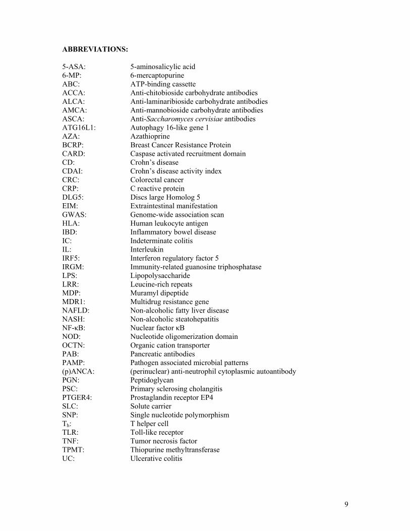
ABBREVIATIONS: 5-ASA: 5-aminosalicylic acid 6-MP: 6-mercaptopurine ABC: ATP-binding cassette ACCA: Anti-chitobioside carbohydrate antibodies ALCA: Anti-laminaribioside carbohydrate antibodies AMCA: Anti-mannobioside carbohydrate antibodies ASCA: Anti-Saccharomyces cervisiae antibodies ATG16L1: Autophagy 16-like gene 1 AZA: Azathioprine BCRP: Breast Cancer Resistance Protein CARD: Caspase activated recruitment domain CD: Crohn’s disease CDAI: Crohn’s disease activity index CRC: Colorectal cancer CRP: C reactive protein DLG5: Discs large Homolog 5 EIM: Extraintestinal manifestation GWAS: Genome-wide association scan HLA: Human leukocyte antigen IBD: Inflammatory bowel disease IC: Indeterminate colitis IL: Interleukin IRF5: Interferon regulatory factor 5 IRGM: Immunity-related guanosine triphosphatase LPS: Lipopolysaccharide LRR: Leucine-rich repeats MDP: Muramyl dipeptide MDR1: Multidrug resistance gene NAFLD: Non-alcoholic fatty liver disease NASH: Non-alcoholic steatohepatitis NF-κB: Nuclear factor κB NOD: Nucleotide oligomerization domain OCTN: Organic cation transporter PAB: Pancreatic antibodies PAMP: Pathogen associated microbial patterns (p)ANCA: (perinuclear) anti-neutrophil cytoplasmic autoantibody PGN: Peptidoglycan PSC: Primary sclerosing cholangitis PTGER4: Prostaglandin receptor EP4 SLC: Solute carrier SNP: Single nucleotide polymorphism Th: T helper cell TLR: Toll-like receptor TNF: Tumor necrosis factor TPMT: Thiopurine methyltransferase UC: Ulcerative colitis
10
1. EPIDEMIOLOGY The field of epidemiology studies, along with various other aspects of population, incidence,
prevalence, and mortality rates. The incidence of a disease or condition is defined as the
proportion of a population or group, initially free of a condition or disease, that develops it
over a set period of time, thus these are new cases. On the contrary, prevalence refers to the
fraction of a population or group which is already afflicted by a given condition or has
reached a clinical outcome at a stated time point.1 While these terms comprise the basics of
epidemiology, IBD studies have come a long way from merely valuating these terms.
Nowadays, they attempt to determine the prognostic outcome as well as various factors that
contribute to the disease process.2
In his paper, Binder describes three factors that are required for the completion of a
successful and useful epidemiological study. First, the investigated population must be well-
described. Next, the regional healthcare system must be organized in such a way as to allow
all patients in the area access to health care and be traceable. Finally, the healthcare system
should offer diagnostic and therapeutic facilities that will be able to exclude infections as well
as other differential diagnoses of IBD.2 Taking these requirements into consideration provides
an explanation for the reason that the majority of studies are carried out in Western countries,
where a public health system is well-rooted. Nonetheless, this chapter will examine
epidemiological data from around the globe, starting from the Americas and moving in an
eastward direction to Western and Eastern Europe, the Middle East and Africa, Asia and
southward to New Zealand.
In the United States, approximately 1.4 million people suffer from IBD, with 30 000
new cases diagnosed annually.3 In a study spanning several decades, Loftus and colleagues
followed the epidemiological features of IBD in Olmsted County, MD.4 Between 1940 and
2000 there were significant increases in both CD and UC. In particular, CD has increased
before the 1970s, while stabilizing later at ~ 8 cases per 100 000 person-years. In the case of
ulcerative colitis, stabilization in incidence was also observed after the 1970s, at 9 cases per
100 000 person-years. Overall, the incidence rates (age- and sex-adjusted) of CD and UC in
Olmsted County during the study period were 6.3 and 8.1 cases per 100 000 person-years,
respectively. The prevalence was calculated as of January 1, 2001 and resulted in 214 and 174
cases per 100 000 persons for UC and CD, respectively. Comparing these results to
measurements taken in 1991, CD’s prevalence increased by 31%, while the prevalence of UC
remained rather stable.5
11
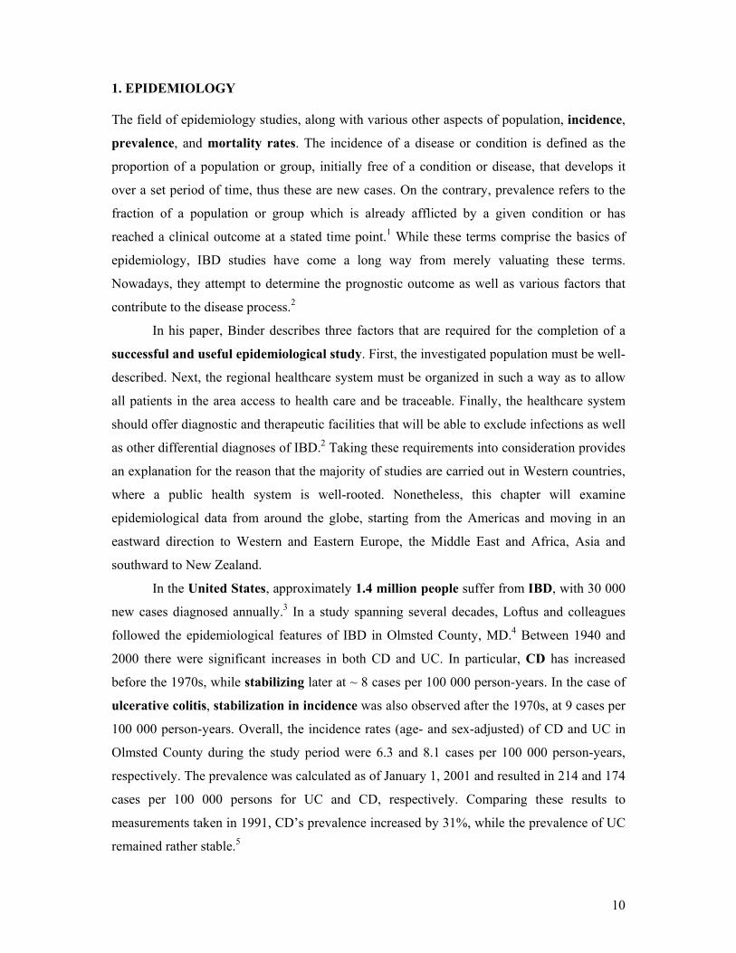
A Canadian group6 studied the epidemiological characteristics of IBD in five
provinces, Alberta (AB), British Columbia (BC), Manitoba (MB), Nova Scotia (NS), and
Saskatchewan (SK) between 1998 and 2000. Using provincial health insurance databases,
they accessed physician billing claims and hospital discharge summaries to create an IBD
database for each province, thus enabling them to calculate incidence and prevalence ratios
for each. Their demographic choice presented populations from the Pacific (BC) and Atlantic
(NS) Coasts, as well as the country’s central part, the prairies (AB, SK, MB). The incidence
and prevalence data from these five provinces are presented in Table 1.
Table 1. Incidence and Prevalence Rates from Five Canadian Provinces6
Crohn’s disease Ulcerative colitis
Province
(East West)
Incidencea
(per 100 000)
Prevalencea
(per 100 000)
Incidencea
(per 100 000)
Prevalencea
(per 100 000)
Nova Scotia 20.2 318.5 19.2 247.9
Manitoba 15.4 271.4 15.4 248.6
Saskatchewan 13.5 263.8 10.4 234.3
Alberta 16.5 283.0 11.0 185.0
British Columbia 8.8 160.7 9.9 162.1
Total 13.4 233.7 11.8 193.7
Total (without BC) 12.9 279.2 12.9 211.2
a Adjusted to the total population of the five investigated provinces.
Interestingly, while BC’s ethnic composition is slightly different from the rest of
Canada, it also presented the lowest incidence and prevalence rates of all five provinces.
Subsequently, the total values were calculated both with and without BC’s data.6
In Latin America, few studies have been conducted in this predominantly Hispanic
population. A recently published study found increasing incidence rates in a population
limited to the Southwest of Puerto Rico. The group identified 202 cases of IBD, comprised of
48 CD, 102 UC, and 52 indeterminate colitis (IC) patients. Over a 5-year period (1996-2000),
the group noticed a four-fold increase in both, CD and IC incidence rates, and an almost
doubled UC incidence rate. The incidence rate of Crohn’s disease increased from 0.49/105 in
1996 to 1.96/105 in 2000, while that for indeterminate colitis rose from 0.61/105 to 2.46/105
12
during the same time period. Ulcerative colitis had a 1.7-fold increase from 1.96/105 to
3.32/105. The study’s IBD prevalence rate was calculated to be 24.81 cases per 100 000.
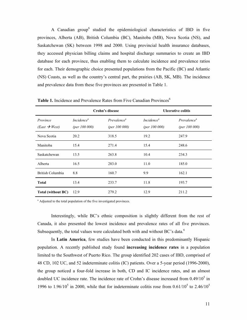
In 1988, a large scale experiment was initiated throughout the European Union that
saw the collection of data from 2201 IBD patients aged 15-64 between the years 1991-1993.
It was termed the European Collaborative study on IBD (EC-IBD). From this total, 706
patients suffered from Crohn’s disease and 1379 had ulcerative colitis. A further division was
performed based on a predetermined geographical axis whereby the Alps served as the
“equator”. A hypothesis was made that a North-South axis for IBD existed and the incidence
rates in the north were higher. In addition, Table 2 summarizes the mean values of IBD based
on the total cases as well as North-South distribution for CD and UC.7
Table 2. Incidence Rates as Calculated by the EC-IBD7
Crohn’s disease Ulcerative colitis
Mean Incidence 5.6/105 per year 10.4/105 per year
Centers north of the Alps:
Mean Incidence 7.0/105 11.8/105
Centers south of the Alps:
Mean Incidence 3.9/105 8.7/105
Overall, no axis was found. This was in spite of the finding that the average incidence
rate of UC was 40% higher, and CD 80% higher, in the northern centers. This can be
supported by the following findings:
The highest incidence of UC was found in Iceland (24.3/105), while the second
highest value was recorded in Crete, Greece (16.6/105).
The highest incidence rates of CD were detected in Holland and France, 9.2/105 in
each.
Two explanations were proposed for this failed “axial” hypothesis. The first refers to
the possibility that areas with high incidence rates maintained their levels at a steady plateau
while areas with lower incidence rates saw an increase in the incidence of IBD. The second
maintains that predominant levels of UC are being matched or replaced by higher incidence
levels of CD.8 Farrokhyar et al.9 have also concluded, based on the EC-IBD report, that no
13
axis of incidence can be identified, noting that Italy, Greece, and Northern Spain displayed
IBD incidence rates that were as high or higher than countries located further north.
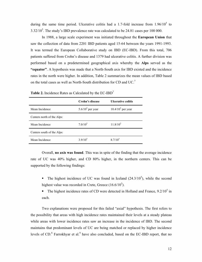
Table 3 presents data collected over several decades from European as well as North
American studies. Examining the results, one reaches the conclusion that UC incidence rates
are higher than CD in European locations, while being equal in North America.
Table 3. Incidence Rates in Select European and North American Locations (per 100 000)
Location Year Ulcerative colitis Crohn’s disease
Copenhagen, Denmark7 1991-1993 9.8 7.3
Florence, Italy7 1991-1993 8.7 3.3
Granada, Spain10 1979-1989 2.0 0.9
Leicestershire, UK7 1991-1993 10.0 3.8
North Tees, UK11 1995 22.1 7.36
Oslo, Norway7 1991-1993 15.6 7.9
Palermo, Italy7 1991-1993 11.0 6.6
Veszprem, Hungary8 1977-2001 5.89 2.23
Manitoba, Canada12 1989-1994 14.3 14.6
Minnesota, USA4 1940-2000 ♂: 9.8, ♀: 6.5 ♂: 6.7, ♀:6.1
In a study restricted solely to France, and based on national health insurance data,
Nerich et al.13 determined a North-South gradient for CD within the country. According to
their geographical mapping, the axis was drawn in the middle of France. A similar gradient
was not observed for UC. They noted two peak frequencies for CD, in the 20-24 years old age
group and 75-79 years old. The overall incidence rate was 8.2/105 for CD (men: 7.1/105;
women: 9.4/105). In UC, with an overall incidence rate of 7.2/105 (men: 7.7/105; women:
6.8/105), the peak was observed in the group of 30-34 years old. Thereafter, a steady decline
was observed.
Incidence rates can also be used to reflect IBD’s gender distribution, with respect to
age. Based on the EC-IBD, it was found that both UC and CD have a peak onset during late
adolescence or early adulthood while in some areas (UK, Scandinavia, USA) a second peak
is seen in the age group of 50-80 years.14,15,16,17 A more detailed inspection of data shows a
peak incidence for UC in women between 25-34 years of age, followed by a rapid decline.

14
However, men also show a continuous risk at the older age groups. In CD, both genders show
peak incidence in the age group of 15-24 years and a decline thereafter.2
Several research groups have examined the influence of race and ethnicity on the
development of IBD, hence its incidence, in various geographic areas. A hospital-based study
was conducted in Cape Town, South Africa, with data collected between 1970 and 1980,
while a second, population-based data set was collected in 1980-1984. Both studies suggested
that there is a higher incidence of IBD in the Jewish population as opposed to non-Jews and is
uncommon among Blacks.18,19,20 The results are shown in Table 4.
Table 4. Incidence of IBD in Cape Town, SA; Based on Race and Ethnicity (per 105)
Study period Ulcerative colitis Crohn’s disease
Hospital-based18,19 1970-1980 Jews (8.5-10.4)
Whites (2.1-2.4)
Colored (1.3-1.6)
Jews (5.0-7.2)
Whites (0.9-1.2)
Colored (0.4-1.3)
Population-based20 1980-1984 Jews (17.0)
Whites (5.0)
Colored (1.9)
Jews (10.4)
Whites (2.6)
Colored (1.8)
A separate study21 involving only permanent residents of Israel examined the
incidence of UC in individuals of various ethnic backgrounds. The study lasted from 1961
until 1985, and investigated three ethnic subgroups, those born in Europe-America, Asia-
Africa, and Israel. Of importance, the incidence rate of the Europe-America subgroup was
significantly higher than for the other two. The results are shown in Figure 1 as incidence
rates per 105 inhabitants of various backgrounds.
Figure 1. Incidence of UC per 100 000 in Israeli Jews of Various Decent (1961-1985)21
10.85.1
4.1
Europe-America bornAsia-Africa bornIsrael born
Another study conducted in northern Israel between 1965 and 1994 attempted to
reveal race and ethnic correlation to IBD by comparing the incidence rates between Israeli
Jews and Arabs.22 Ulcerative colitis was found to be 2.5 times greater in Israeli Jews than
15
in Arabs. Two possible explanations were suggested, the first claiming that lifestyle
differences may attribute to this inequality. In the last two decades, the Arab population
adopted a more Western lifestyle, as opposed to the traditional agricultural lifestyle, possibly
contributing to an increase in UC incidence. The second explanation proposed that genetic
differences were to blame for the higher incidence of UC in the Israeli Jews; however, this
was not clearly examined. Finally, more recent reports raised the possibility that IBD-
associated genes seem to occur more frequently in Ashkenazi Jews.23
Over the past several years, some new data has emerged from East Europe. In
Veszprem, a western province of Hungary, Lakatos et al.8 examined the incidence of IBD
during a 25-year period from 1977 to 2001. The raw data displayed 560 new cases of UC and
212 new cases of CD. The incidence rate of UC increased from 1.66 to 11.01 (6.6-fold
increase), while that of CD increased from 0.41 to 4.68 (11.4-fold increase) per 100 000.
Additionally, they investigated the localization of UC and CD, the results of which are
displayed in Figures 2 and 3. In Croatia, the earliest study to examine the incidence and
prevalence of IBD in the 1980s revealed a steady rate of CD and UC. For the former, the
incidence rate was 0.7/105, while the latter’s was 1.5/105 inhabitants.24,25 Interestingly, in a
separate, long-term study conducted during 1973-1994, an increase in the incidence of
Crohn’s disease was noted. In particular, it increased from 0.34/105 in 1973 to 3.47/105
inhabitants in 1994.26 From Romania, a single study was published on the epidemiology of
inflammatory bowel disease spanning a single year, from June 2002 until June 2003.
Certainly, limitations are inherent in epidemiological studies of such short duration.
Nonetheless, using data from 18 secondary and tertiary centers, the authors calculated an
incidence rate of 0.97/105 for UC and 0.50/105 for CD.27
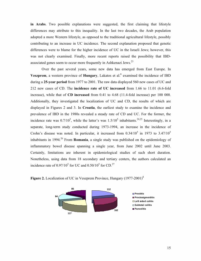
Figure 2. Localization of UC in Veszprem Province, Hungary (1977-2001)8
112
162117
84
85
ProctitisProctosigmoiditisLeft sided colitisSubtotal colitisPancolitis
16
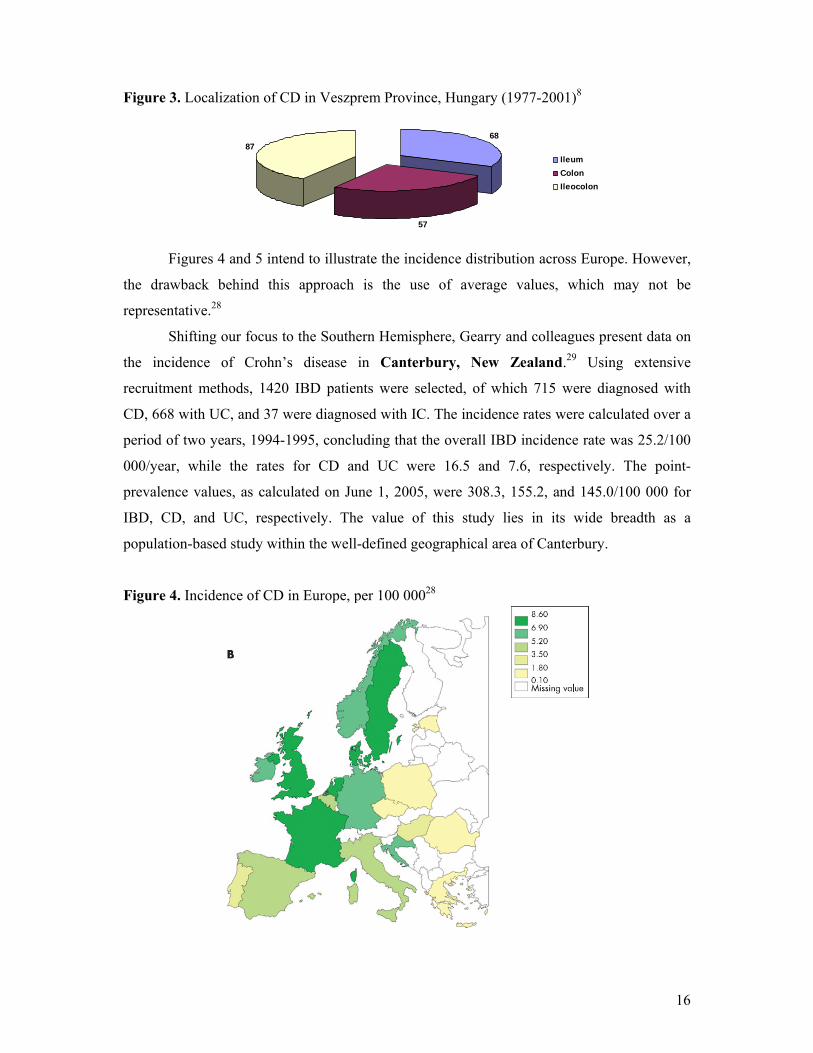
Figure 3. Localization of CD in Veszprem Province, Hungary (1977-2001)8
68
57
87IleumColonIleocolon
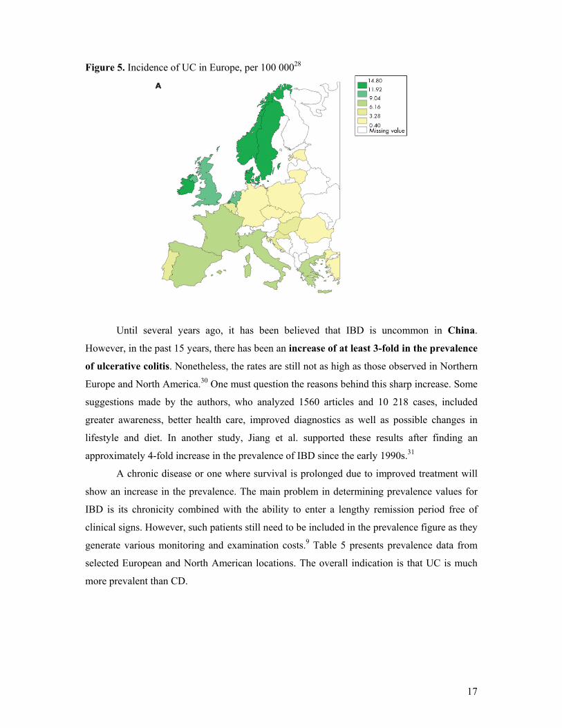
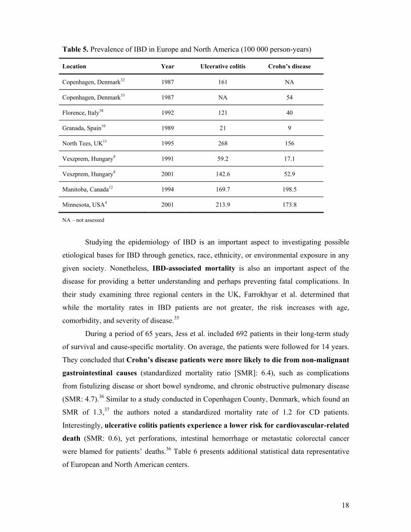
Figures 4 and 5 intend to illustrate the incidence distribution across Europe. However,
the drawback behind this approach is the use of average values, which may not be
representative.28
Shifting our focus to the Southern Hemisphere, Gearry and colleagues present data on
the incidence of Crohn’s disease in Canterbury, New Zealand.29 Using extensive
recruitment methods, 1420 IBD patients were selected, of which 715 were diagnosed with
CD, 668 with UC, and 37 were diagnosed with IC. The incidence rates were calculated over a
period of two years, 1994-1995, concluding that the overall IBD incidence rate was 25.2/100
000/year, while the rates for CD and UC were 16.5 and 7.6, respectively. The point-
prevalence values, as calculated on June 1, 2005, were 308.3, 155.2, and 145.0/100 000 for
IBD, CD, and UC, respectively. The value of this study lies in its wide breadth as a
population-based study within the well-defined geographical area of Canterbury.
Figure 4. Incidence of CD in Europe, per 100 00028
17
Figure 5. Incidence of UC in Europe, per 100 00028
Until several years ago, it has been believed that IBD is uncommon in China.
However, in the past 15 years, there has been an increase of at least 3-fold in the prevalence
of ulcerative colitis. Nonetheless, the rates are still not as high as those observed in Northern
Europe and North America.30 One must question the reasons behind this sharp increase. Some
suggestions made by the authors, who analyzed 1560 articles and 10 218 cases, included
greater awareness, better health care, improved diagnostics as well as possible changes in
lifestyle and diet. In another study, Jiang et al. supported these results after finding an
approximately 4-fold increase in the prevalence of IBD since the early 1990s.31
A chronic disease or one where survival is prolonged due to improved treatment will
show an increase in the prevalence. The main problem in determining prevalence values for
IBD is its chronicity combined with the ability to enter a lengthy remission period free of
clinical signs. However, such patients still need to be included in the prevalence figure as they
generate various monitoring and examination costs.9 Table 5 presents prevalence data from
selected European and North American locations. The overall indication is that UC is much
more prevalent than CD.
18
Table 5. Prevalence of IBD in Europe and North America (100 000 person-years)
Location Year Ulcerative colitis Crohn’s disease
Copenhagen, Denmark32 1987 161 NA
Copenhagen, Denmark33 1987 NA 54
Florence, Italy34 1992 121 40
Granada, Spain10 1989 21 9
North Tees, UK11 1995 268 156
Veszprem, Hungary8 1991 59.2 17.1
Veszprem, Hungary8 2001 142.6 52.9
Manitoba, Canada12 1994 169.7 198.5
Minnesota, USA4 2001 213.9 173.8
NA – not assessed
Studying the epidemiology of IBD is an important aspect to investigating possible
etiological bases for IBD through genetics, race, ethnicity, or environmental exposure in any
given society. Nonetheless, IBD-associated mortality is also an important aspect of the
disease for providing a better understanding and perhaps preventing fatal complications. In
their study examining three regional centers in the UK, Farrokhyar et al. determined that
while the mortality rates in IBD patients are not greater, the risk increases with age,
comorbidity, and severity of disease.35
During a period of 65 years, Jess et al. included 692 patients in their long-term study
of survival and cause-specific mortality. On average, the patients were followed for 14 years.
They concluded that Crohn’s disease patients were more likely to die from non-malignant
gastrointestinal causes (standardized mortality ratio [SMR]: 6.4), such as complications
from fistulizing disease or short bowel syndrome, and chronic obstructive pulmonary disease
(SMR: 4.7).36 Similar to a study conducted in Copenhagen County, Denmark, which found an
SMR of 1.3,37 the authors noted a standardized mortality rate of 1.2 for CD patients.
Interestingly, ulcerative colitis patients experience a lower risk for cardiovascular-related
death (SMR: 0.6), yet perforations, intestinal hemorrhage or metastatic colorectal cancer
were blamed for patients’ deaths.36 Table 6 presents additional statistical data representative
of European and North American centers.
19
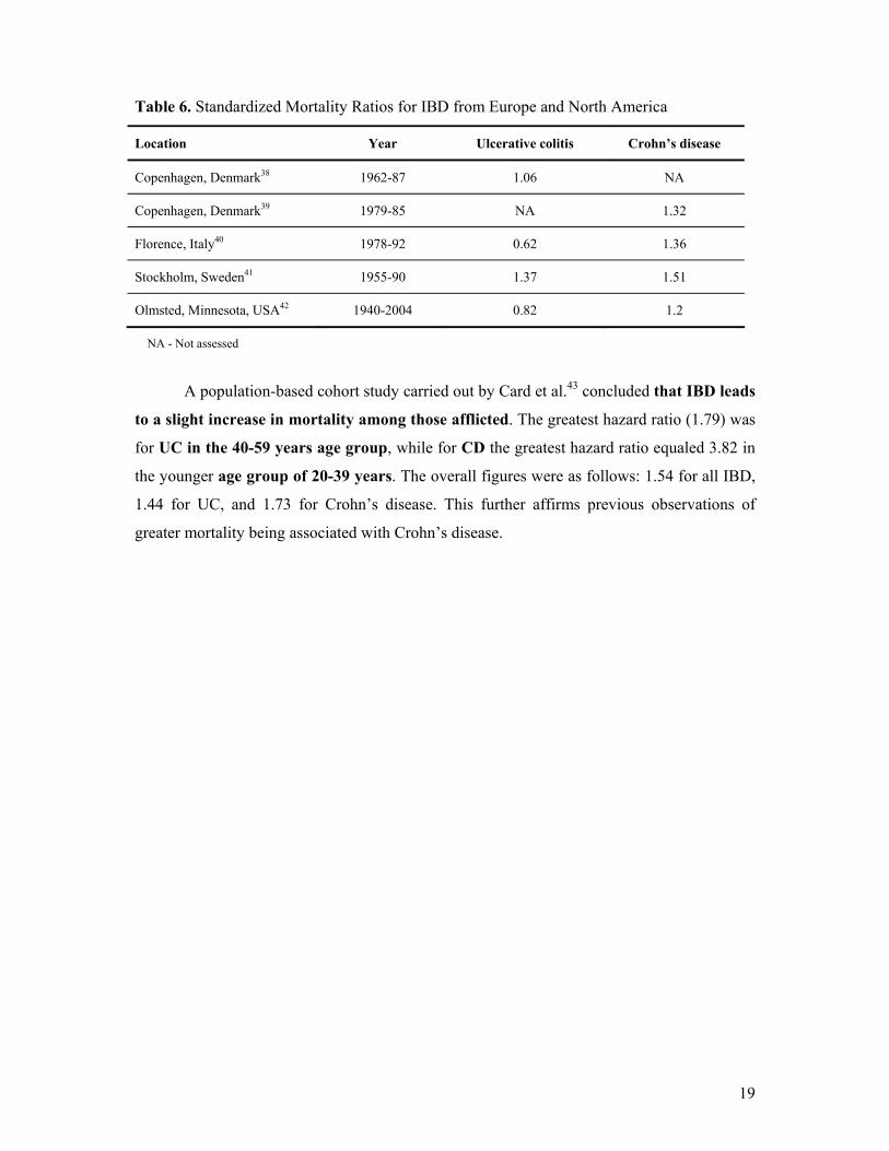
Table 6. Standardized Mortality Ratios for IBD from Europe and North America
Location Year Ulcerative colitis Crohn’s disease
Copenhagen, Denmark38 1962-87 1.06 NA
Copenhagen, Denmark39 1979-85 NA 1.32
Florence, Italy40 1978-92 0.62 1.36
Stockholm, Sweden41 1955-90 1.37 1.51
Olmsted, Minnesota, USA42 1940-2004 0.82 1.2
NA - Not assessed
A population-based cohort study carried out by Card et al.43 concluded that IBD leads
to a slight increase in mortality among those afflicted. The greatest hazard ratio (1.79) was
for UC in the 40-59 years age group, while for CD the greatest hazard ratio equaled 3.82 in
the younger age group of 20-39 years. The overall figures were as follows: 1.54 for all IBD,
1.44 for UC, and 1.73 for Crohn’s disease. This further affirms previous observations of
greater mortality being associated with Crohn’s disease.
20
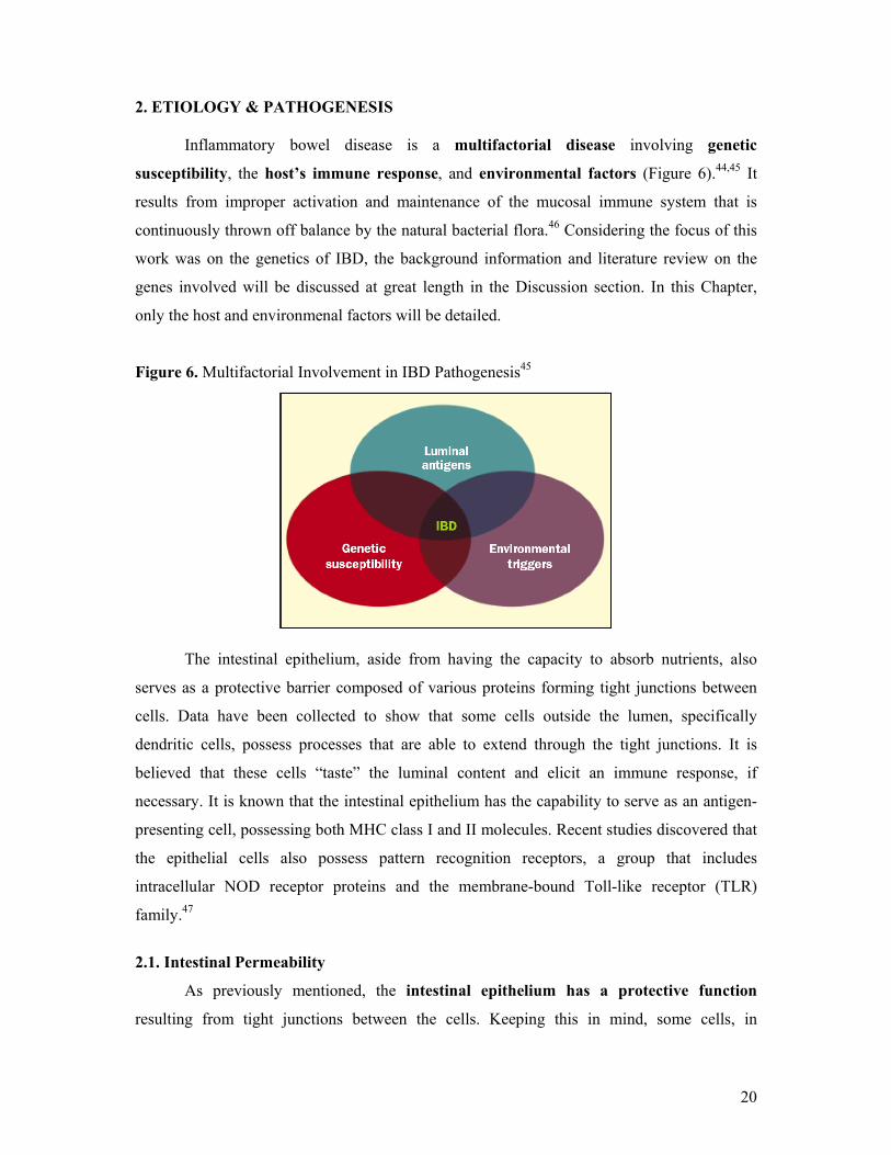
2. ETIOLOGY & PATHOGENESIS Inflammatory bowel disease is a multifactorial disease involving genetic
susceptibility, the host’s immune response, and environmental factors (Figure 6).44,45 It
results from improper activation and maintenance of the mucosal immune system that is
continuously thrown off balance by the natural bacterial flora.46 Considering the focus of this
work was on the genetics of IBD, the background information and literature review on the
genes involved will be discussed at great length in the Discussion section. In this Chapter,
only the host and environmenal factors will be detailed.
Figure 6. Multifactorial Involvement in IBD Pathogenesis45
The intestinal epithelium, aside from having the capacity to absorb nutrients, also
serves as a protective barrier composed of various proteins forming tight junctions between
cells. Data have been collected to show that some cells outside the lumen, specifically
dendritic cells, possess processes that are able to extend through the tight junctions. It is
believed that these cells “taste” the luminal content and elicit an immune response, if
necessary. It is known that the intestinal epithelium has the capability to serve as an antigen-
presenting cell, possessing both MHC class I and II molecules. Recent studies discovered that
the epithelial cells also possess pattern recognition receptors, a group that includes
intracellular NOD receptor proteins and the membrane-bound Toll-like receptor (TLR)
family.47
2.1. Intestinal Permeability
As previously mentioned, the intestinal epithelium has a protective function
resulting from tight junctions between the cells. Keeping this in mind, some cells, in
21
particular dendritic cells, are able to extend processes through the tight junctions, sampling
the luminal milieu, and eliciting an immune response, as needed.47
An interesting observation was made with regard to the unity of this barrier in CD
patients and even more so, their first-degree relatives. It was noted that there is an increased
permeability in the intestines of these patients. Additionally, their first-degree relatives,
without any signs and symptoms of CD, showed a similar increase.48 This brings forth the
idea that an increase in the epithelium’s permeability may play a role in the pathogenesis of
IBD. Certainly, increased permeability has been observed in patients prior to a flare-up,
signaling the presence of a state of subclinical disease.49
It was also discovered that some first-degree relatives who were administered
acetylsalicylic acid responded with increased epithelial permeability.50 In some cases of CD,
this may serve as a causative agent for a flare-up. The association between increased
permeability and CD has offered the possibility of an exaggerated recognition of antigens,
leading to an overactive immune response - as observed in CD. Additionally, this has been
systematically observed by an exaggerated level of circulating, mature B cells.51
2.2. Intestinal Flora
For many years, researchers have been examining the intestinal milieu for clues over
its role in the pathogenesis of IBD, without much success. In recent years, however, several
advances have been made. First, one study described a considerable decrease in the amount
of anaerobic bacteria and Lactobacillus in active IBD cases, in contrast to inactive cases,
which showed no remarkable change.52 It is very feasible that the resident flora of the
intestine is perpetuating and sustaining the continuous inflammatory reaction seen in IBD
patients. An ever-increasing amount of evidence is pointing towards the necessity in luminal
flora for the progress of IBD.47 This was clearly shown in a murine model where mutant mice,
deficient in IL-10,53 remained healthy as long as they were raised in a sterile environment.
Upon the introduction of commensal flora, the mice rapidly developed colitis.54
Second, there is a direct correlation between the site of an IBD lesion and the
concentration of luminal bacteria at that site, as observed by the benefit of antibiotic
treatment in some patients.55 Also, redirecting the flow of fecal stream and accompanying
flora through a stoma seems to have a healing effect on the inflammation. A flare-up will take
place if an anastomosis is connected.56
Finally, IBD-like enterocolitis was successfully provoked in murine models by the
injection of a purified sample of bacterial cell-wall PGN-LPS. This was coupled with
22
increased levels of collagen synthesis resulting in the characteristic fibrosis, as well as
increased levels of TGF-β1 and IL-6.57 Similarly, Kennedy et al. were also able to elicit
enterocolitis in IL-10-deficient mice, when exposed to intestinal bacteria, similar to the skip
lesions observed in Crohn’s disease.58 Also, as seen in the human form of IBD, the
inflammatory lesions in this model responded to corticosteroids.59
2.3. Environmental Factors
The genetics described thus far cannot account for 100% of all IBD cases; therefore,
one must conclude that the environment is also of great importance in the pathogenesis of
UC and CD. The environmental make-up of inflammatory bowel disease, or its flare-up, is
quite complex and consists of external factors such as smoking, childhood infections, and
non-steroidal anti-inflammatory drugs (NSAIDs), as well as internal ones such as the
intestinal flora.60 The use of oral contraceptives remains a controversial issue, and though
much research has been done, little has been found to either disprove or lend support to oral
contraceptives as a risk factor for IBD.61 However, there seems to be a correlation between
the use of oral contraceptives and smoking, resulting in an increased risk for IBD.62
Several studies were presented in recent years displaying cases of married couples that
developed IBD following their move to occupy a common establishment. One such particular
study was carried out in France and Belgium between 1989 and 2000 by Laharie et al.63 The
researchers grouped the subject couples into three groups, as follows:
A – couples where both partners exhibited IBD symptoms prior to cohabitation
B – if only one partner had IBD prior to cohabitation and both partners had it
following cohabitation
C – if neither partner displayed IBD before cohabitation yet both showed it afterwards.
In total, thirty cases were classified into these groups. Of importance, 22 of the cases were
registered in group C. Such high frequency is indicative of environmental factors having an
etiological role in IBD.
2.3.1. Microbial Agents
To date, no single microbial pathogen has been identified as the causative agent of
IBD but several bacteria and viruses were investigated at one point or another. At the
beginning of the last century, the first pathogen was brought under investigation,
Mycobacterium paratuberculosis. It was observed that Crohn’s disease displayed a
23
characteristic granulomatous inflammation similar to Johne’s disease of cattle, for which
M. paratuberculosis is a confirmed pathogen.64 Furthermore, the pathogenic agent of leprosy,
M. leprae, has also been targeted due to clinical, pathological and histological characteristics
that are similar to CD. On the other hand, ulcerative colitis was correlated with a special
variation of Escherichia coli, with adhesive properties. This form was found in UC patients
but was absent in control subjects.65
Among viruses, a major suspect is the measles virus infection at a young age.66 The
basis of this investigation is the virus’ ability to target the gastrointestinal tract as well as its
ability to induce granulomatous inflammation.67 A different study examined the involvement
of the measles vaccination in the pathogenesis of IBD; however, this has not been
conclusively confirmed.68 Other suspected viruses include the rotavirus and herpes virus.64
2.3.2. Cold Chain Hypothesis
This hypothesis is unique, as described by Hugot et al.69 It proposes that
psychrotrophic bacteria, for example, Yersinia species, are able to grow in a common
household appliance, the refrigerator. These agents are then ingested with food and mediate
inflammation and diarrhea through various mechanisms, as seen in CD.
First, Yersinia produces an invasin toxin which, along with the bacteria’s ability to
target M cells in lymphoid follicles, enables bacteria to penetrate cells. The notion of an
intracellular pathogen responsible for IBD pathogenesis has also been supported by
Karlinger et al.64 It is known that CD lesions are most frequently found in lymphoid follicles
of the intestine.
Next, yersiniosis produces similar clinical signs and symptoms as CD, which adds
to the list of differential diagnoses. Among others, these include ileitis, reactive arthritis,
erythema nodosum, and granuloma formation. Also, similar to Helicobacter pylori, the
causative agent of peptic ulcer, Yersinia produces an urease. This enzyme is an important
contributor to the formation of gastritis, and can be detected in a high number of CD patients.
Moreover, these microbial pathogens have also been linked to spondyloarthropathies, which
are, in turn, associated with Crohn’s disease.69
Lastly, at the molecular level, yet not fully understood, is a connection between
NOD2/CARD15, NF-кB, and Yersinia species-secreted proteins. These proteins are carried on
a plasmid, pYV, including the Yop and Ysc proteins. Selected Yop genes are involved in the
inhibition of NF-кB, while the possibility also exists that Yop proteins physically disrupt the
24
action of NOD2/CARD15 by some other pathway. Merging all of these data with the
bacteria’s tropism for M cells lends support to the cold chain hypothesis.69
2.3.3. Smoking
An enigmatic paradox exists between smoking and the individual forms of IBD.
Smoking appears more commonly in those patients afflicted by CD, while many UC
patients are non-smokers.70,71 To complicate matters further, nicotine seems to play a
curative role in UC. In one study, the application of a transdermal nicotine patch as an adjunct
to typical treatment regimen has eased the symptoms of mild and moderate UC cases.72 In
another study involving 154 UC patients, 52% of the patients quit smoking within three years
preceding the development of colitis.73 An Australian study found the odds ratio to be 3.45 for
smoking cessation prior to the onset of symptoms, leading the team to suggest that smoking
hides the symptoms of UC. Alternatively, removing the immunosuppressive effect of
smoking triggers the development of colitis in genetically susceptible individuals.74 This may
be due to nicotine’s effect on Th2 functions, which are a main constituent of UC, but lack of
effect on the Th1 functions that are present in CD.75 Additional possible factors of nicotine
and smoking include: effects on cellular and humoral immune cells,76,77 increased colonic
mucous production,78 and reduced colonic motility.79 Finally, smokers who show abstinence
from smoking experience a milder form of disease than if these individuals had never
smoked.80
2.3.4. Other Environmental Factors
Extrapolating on the role of the immune system in the pathogenesis of IBD brings one
to the subject of monocytes/macrophages. In the early 1990s, immunohistochemical
procedures showed that macrophages are present in the early stages of IBD in lesions formed
as aphthous lesions.81 Additionally, macrophages in IBD cases are blamed for the
overproduction of reactive oxygen and nitrogen species, capable of causing extensive
damage to cells and tissues. This is due to the inability by protective enzymes to eliminate
these injurious substances.82
Lastly, returning to the role of bacteria in IBD pathogenesis, Lodes et al.83 described
bacterial proteins, flagellins, as possible culprits. These proteins are recognized for their
ability to elicit an innate immune response via TLR5. Their presence is characterized by a
strong IgG2a response in murine models, in addition to causing colitis when flagellin-specific
25
CD4+ T-cells are transferred into naïve SCID (severe combined immunodeficiency) mice. Of
note, there is specific recognition of these molecules in CD patients, unlike in UC.
2.4. Familial Studies
It is frequently observed that family members of IBD probands present the disease
(CD or UC), or some other signs, indicative of an increased risk of developing it.
Transmissions from parents to offspring as well as sibling-sibling associations occur. As
many as 40% of first-degree relatives of IBD patients will suffer from the disease.84
Additional evidence to support the genetic basis of IBD comes from concordance twin
studies, where monozygotic twins have a much higher concordance rate for IBD than
dizygotic. These data are presented in Table 7.85,86
Table 7. Concordance Data in Monozygotic and Dizygotic Twins85,86
Crohn’s disease Ulcerative colitis
Monozygotic twins 44-50% 6-14%
Dizygotic twins 0-4% 0-5%
No doubt, genetics play a role in the pathogenesis of CD, as do other factors, since the
concordance rate for monozygotic twins is 50%, at best.45
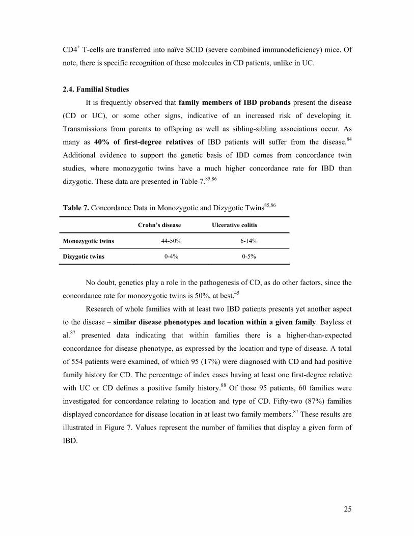
Research of whole families with at least two IBD patients presents yet another aspect
to the disease – similar disease phenotypes and location within a given family. Bayless et
al.87 presented data indicating that within families there is a higher-than-expected
concordance for disease phenotype, as expressed by the location and type of disease. A total
of 554 patients were examined, of which 95 (17%) were diagnosed with CD and had positive
family history for CD. The percentage of index cases having at least one first-degree relative
with UC or CD defines a positive family history.88 Of those 95 patients, 60 families were
investigated for concordance relating to location and type of CD. Fifty-two (87%) families
displayed concordance for disease location in at least two family members.87 These results are
illustrated in Figure 7. Values represent the number of families that display a given form of
IBD.
26
Figure 7. Families Concordant for Disease Location87
1
3013
8
IleojejunitisIleitisIleocolitisColitis
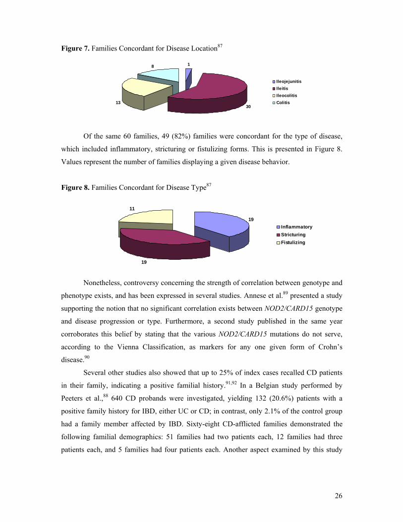
Of the same 60 families, 49 (82%) families were concordant for the type of disease,
which included inflammatory, stricturing or fistulizing forms. This is presented in Figure 8.
Values represent the number of families displaying a given disease behavior.
Figure 8. Families Concordant for Disease Type87
19
19
11
InflammatoryStricturingFistulizing
Nonetheless, controversy concerning the strength of correlation between genotype and
phenotype exists, and has been expressed in several studies. Annese et al.89 presented a study
supporting the notion that no significant correlation exists between NOD2/CARD15 genotype
and disease progression or type. Furthermore, a second study published in the same year
corroborates this belief by stating that the various NOD2/CARD15 mutations do not serve,
according to the Vienna Classification, as markers for any one given form of Crohn’s
disease.90
Several other studies also showed that up to 25% of index cases recalled CD patients
in their family, indicating a positive familial history.91,92 In a Belgian study performed by
Peeters et al.,88 640 CD probands were investigated, yielding 132 (20.6%) patients with a
positive family history for IBD, either UC or CD; in contrast, only 2.1% of the control group
had a family member affected by IBD. Sixty-eight CD-afflicted families demonstrated the
following familial demographics: 51 families had two patients each, 12 families had three
patients each, and 5 families had four patients each. Another aspect examined by this study
27
was the age at diagnosis. It was found that offspring were diagnosed at an earlier age than
their parents; however, there was no significant difference between siblings.
Some studies investigated the reason behind the apparent earlier age-at-onset in
offspring of IBD patients. The first, suggested by Satsangi et al.,93 indicated genetic
anticipation. This phenomenon refers to the earlier symptomatic onset of a given inherited
disease or a more severe clinical presentation of the disease.94 Others proposed that the
offspring were thoroughly examined at a younger age based on the known presence of IBD in
their parents.95 Finally, a Swedish study published in 2003 also demonstrated that genetics
had a greater influence in CD pathogenesis as opposed to UC, including a similarity in disease
phenotype.96 The team found a concordance rate of approximately 50% in monozygotic twins
afflicted with CD, compared with only 3.8% of CD dizygotic twins.
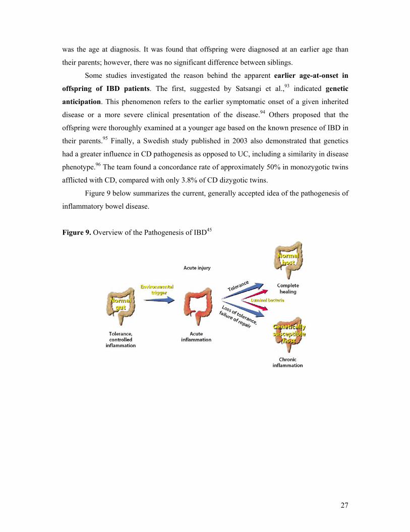
Figure 9 below summarizes the current, generally accepted idea of the pathogenesis of
inflammatory bowel disease.
Figure 9. Overview of the Pathogenesis of IBD45
28
3. CLINICAL PICTURE & DIAGNOSIS
3.1. Clinical Picture
The clinical picture can be divided into two separate but related entities. The first
involves the signs and symptoms as perceived by the patient and the clinician; the second
involves the cytological changes as demonstrated in laboratory examinations. The most
common complaints in both forms of IBD include increased stool frequency and decreased
stool consistency, though in 5-10% of patients, constipation may be the presenting symptom.
The second most common symptom is abdominal pain, which in some cases may help
localize the site of inflammation. In patients suffering from ileal Crohn’s disease, pain is
commonly felt in the lower right quadrant, while UC patients often complain of pain in the
left lower quadrant. In the latter case, cramping prior to and relieved by defecation is also
common. In the case of ulcerative colitis, the presence of tenesmus hints towards proctitis.
Finally, the presence of gross blood in the stool makes UC more likely than CD.97 Table 8
presents the differential diagnoses that must be ruled out during the diagnostic work-up for
inflammatory bowel disease.98
Crohn’s disease, in 60% of cases, affects the terminal ileum (ileitis), while colitis
and anorectal inflammation are the majority of the remaining cases. A small percentage of
cases will show inflammation anywhere throughout the gastrointestinal tract.297 Importantly,
with passing time, both disease location and behaviour are unstable and tend to change
with an increasing proportion of patients developing complications.99 Regardless of the site,
CD is characterized by a granulomatous form of inflammation.100
Two characteristic findings in CD are strictures and fistulae. Despite research, the
mechanism behind these observations is still unknown. Some support the idea of mechanical
forces within the lumen of the bowel, that is, intraluminal pressure. Others claim that the
inflammatory nature of CD and the associated host immune response are at work.101 Finally,
some suggest that these variations are a basis for classifying CD into perforating (fistulae
formation) and non-perforating (inflammation with or without strictures) disease.102
Ulcerative colitis, on the other hand, is defined as “continuous endoscopic or radiologic
mucosal inflammation affecting the rectum and colon together with the typical histology of
ulcerative colitis and the absence of granuloma.”9 Table 9 compares the various observations
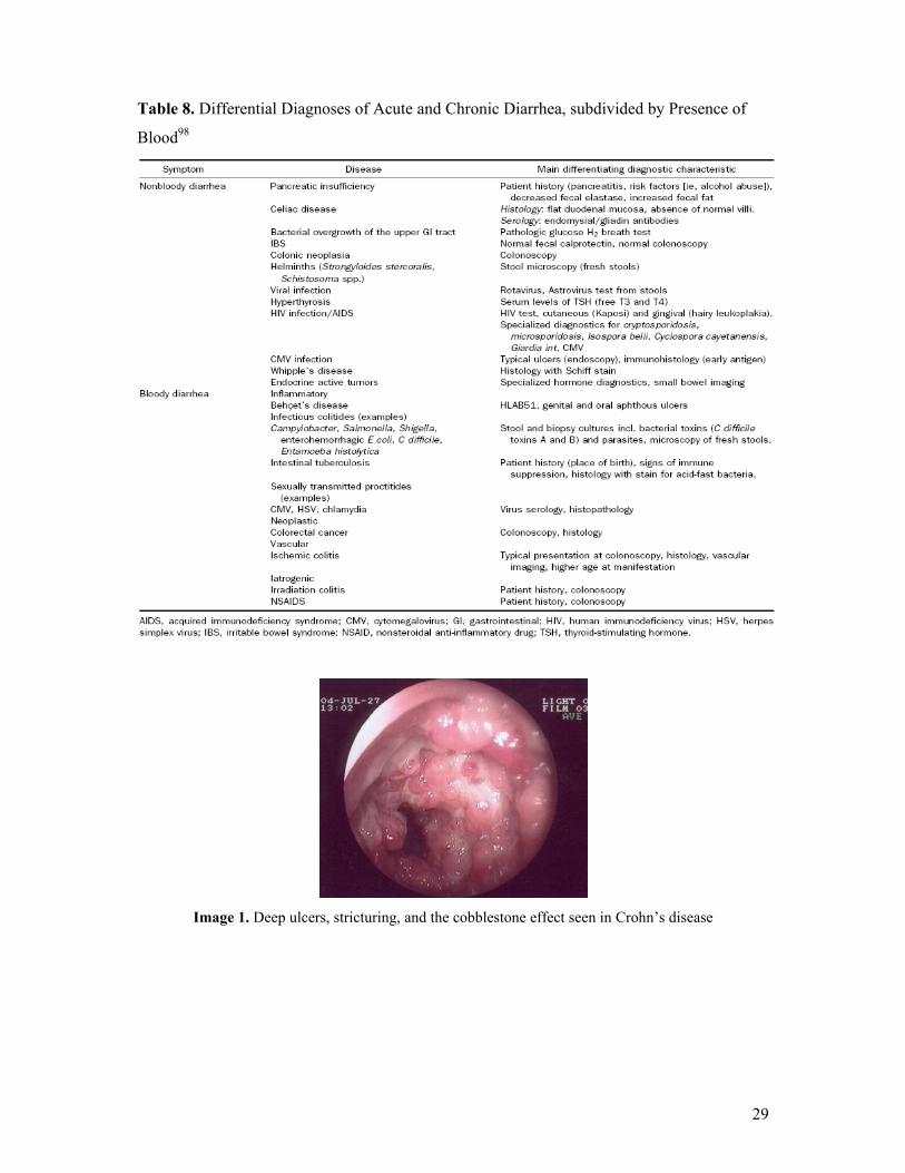
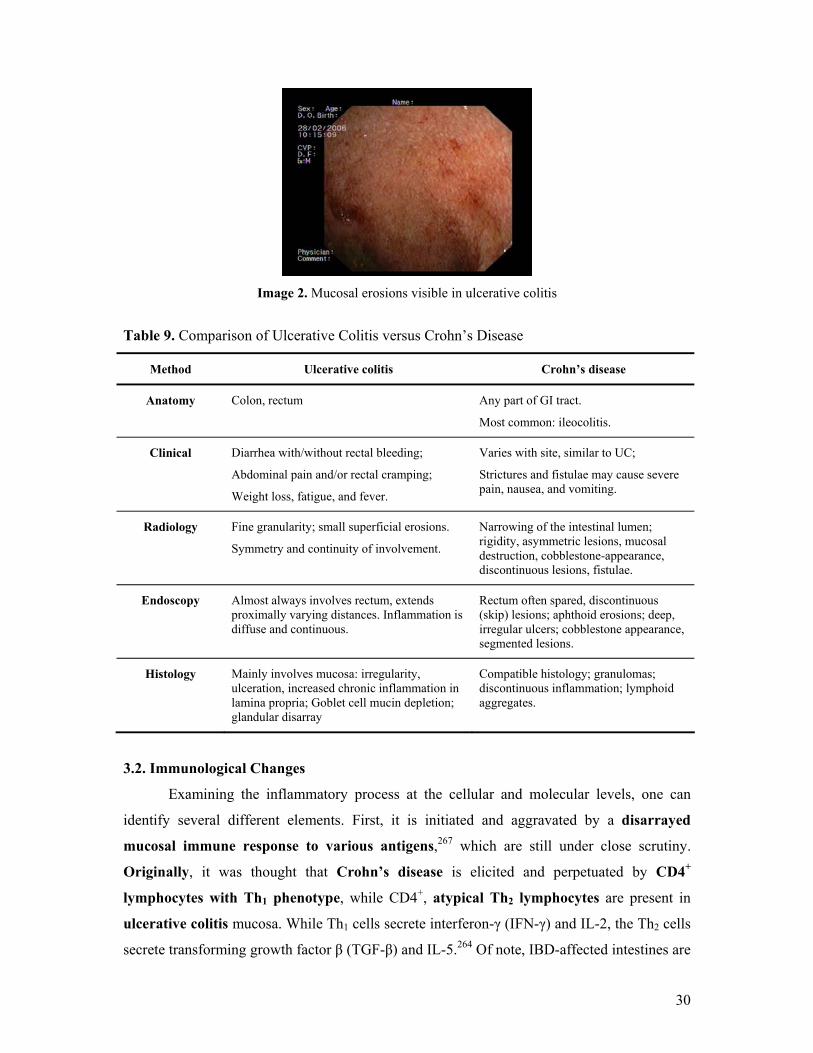
in UC and CD,103,104,105 while Images 1 and 2 illustrate active CD and ulcerative colitis,
respectively, as visualized during colonoscopy (Courtesy of Dr. Lakatos, 1st Department of
Internal Medicine, Semmelweis University, Budapest, Hungary).
29
Table 8. Differential Diagnoses of Acute and Chronic Diarrhea, subdivided by Presence of
Blood98
Image 1. Deep ulcers, stricturing, and the cobblestone effect seen in Crohn’s disease
30
Image 2. Mucosal erosions visible in ulcerative colitis
Table 9. Comparison of Ulcerative Colitis versus Crohn’s Disease
Method Ulcerative colitis Crohn’s disease
Anatomy Colon, rectum Any part of GI tract.
Most common: ileocolitis.
Clinical Diarrhea with/without rectal bleeding;
Abdominal pain and/or rectal cramping;
Weight loss, fatigue, and fever.
Varies with site, similar to UC;
Strictures and fistulae may cause severe pain, nausea, and vomiting.
Radiology Fine granularity; small superficial erosions.
Symmetry and continuity of involvement.
Narrowing of the intestinal lumen; rigidity, asymmetric lesions, mucosal destruction, cobblestone-appearance, discontinuous lesions, fistulae.
Endoscopy Almost always involves rectum, extends proximally varying distances. Inflammation is diffuse and continuous.
Rectum often spared, discontinuous (skip) lesions; aphthoid erosions; deep, irregular ulcers; cobblestone appearance, segmented lesions.
Histology Mainly involves mucosa: irregularity, ulceration, increased chronic inflammation in lamina propria; Goblet cell mucin depletion; glandular disarray
Compatible histology; granulomas; discontinuous inflammation; lymphoid aggregates.
3.2. Immunological Changes
Examining the inflammatory process at the cellular and molecular levels, one can
identify several different elements. First, it is initiated and aggravated by a disarrayed
mucosal immune response to various antigens,267 which are still under close scrutiny.
Originally, it was thought that Crohn’s disease is elicited and perpetuated by CD4+
lymphocytes with Th1 phenotype, while CD4+, atypical Th2 lymphocytes are present in
ulcerative colitis mucosa. While Th1 cells secrete interferon-γ (IFN-γ) and IL-2, the Th2 cells
secrete transforming growth factor β (TGF-β) and IL-5.264 Of note, IBD-affected intestines are
31
infiltrated by a large number of immune cells without any indication of inflammation, which
is particularly true in the case of Crohn’s disease.44 Furthermore, the cytokines released by
Th1 helper cells activate macrophages that release additional cytokines and proinflammatory
substances, such as IL-12, IL-18, and macrophage migration inhibitor factor. However, as will
be explained in the Discussion, it is now believed that IL-23, which was only identified in
2000, is responsible for the inflammatory response observed in Crohn’s disease. Due to its
structural similarity to IL-12, it is clear why such initial “confusion” is possible,
simultaneously over-estimating the actual role of IL-12 in IBD.
Finally, there are also changes in the profile of B-cells. This is mirrored by
exaggerated production of mainly IgG, but also IgA and IgM.106
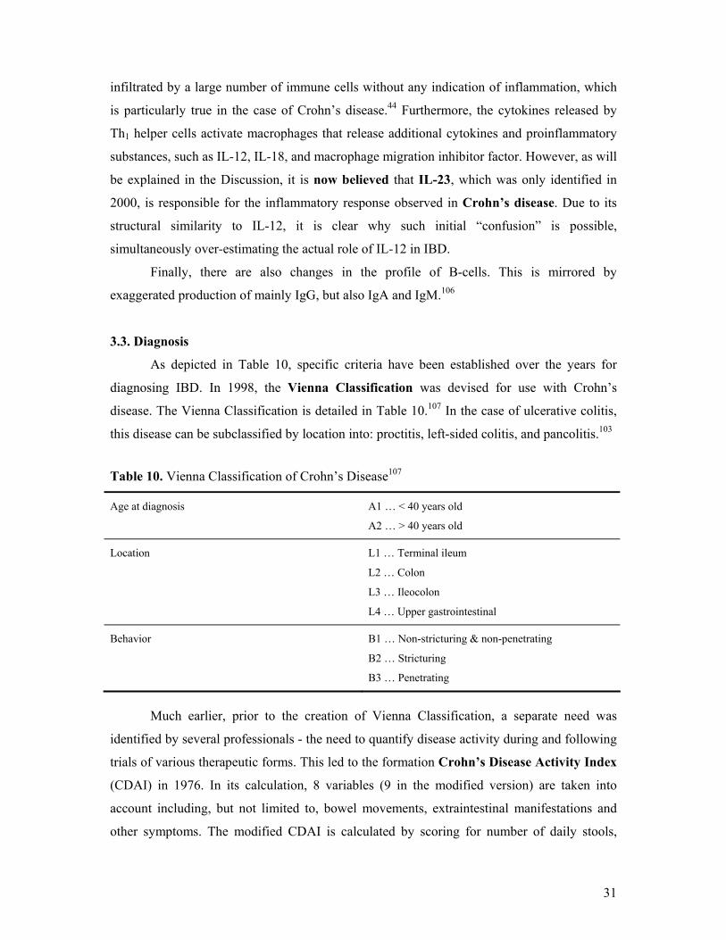
3.3. Diagnosis
As depicted in Table 10, specific criteria have been established over the years for
diagnosing IBD. In 1998, the Vienna Classification was devised for use with Crohn’s
disease. The Vienna Classification is detailed in Table 10.107 In the case of ulcerative colitis,
this disease can be subclassified by location into: proctitis, left-sided colitis, and pancolitis.103
Table 10. Vienna Classification of Crohn’s Disease107
Age at diagnosis A1 … < 40 years old
A2 … > 40 years old
Location L1 … Terminal ileum
L2 … Colon
L3 … Ileocolon
L4 … Upper gastrointestinal
Behavior B1 … Non-stricturing & non-penetrating
B2 … Stricturing
B3 … Penetrating
Much earlier, prior to the creation of Vienna Classification, a separate need was
identified by several professionals - the need to quantify disease activity during and following
trials of various therapeutic forms. This led to the formation Crohn’s Disease Activity Index
(CDAI) in 1976. In its calculation, 8 variables (9 in the modified version) are taken into
account including, but not limited to, bowel movements, extraintestinal manifestations and
other symptoms. The modified CDAI is calculated by scoring for number of daily stools,
32
presence of pain, well-being, manifested symptoms, use of antidiarrheals (Lomotil), presence
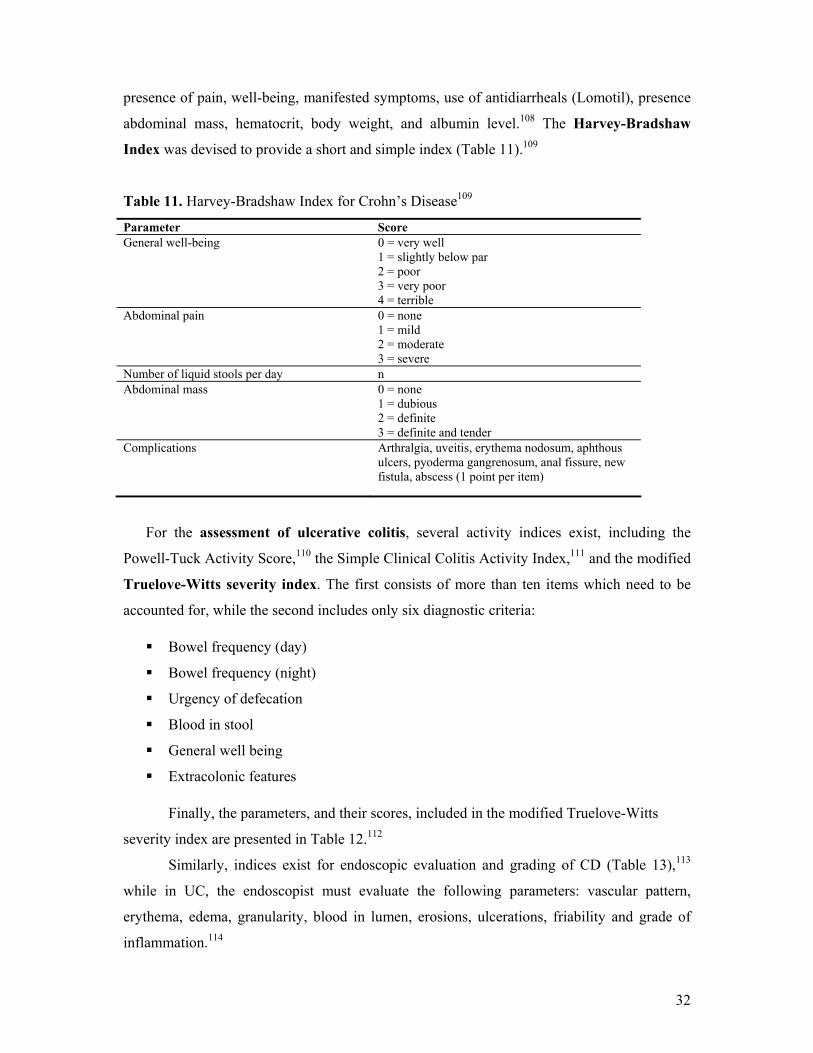
abdominal mass, hematocrit, body weight, and albumin level.108 The Harvey-Bradshaw
Index was devised to provide a short and simple index (Table 11).109
Table 11. Harvey-Bradshaw Index for Crohn’s Disease109
Parameter Score General well-being 0 = very well
1 = slightly below par 2 = poor 3 = very poor 4 = terrible
Abdominal pain 0 = none 1 = mild 2 = moderate 3 = severe
Number of liquid stools per day n Abdominal mass 0 = none
1 = dubious 2 = definite 3 = definite and tender
Complications Arthralgia, uveitis, erythema nodosum, aphthous ulcers, pyoderma gangrenosum, anal fissure, new fistula, abscess (1 point per item)
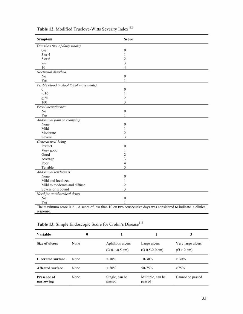
For the assessment of ulcerative colitis, several activity indices exist, including the
Powell-Tuck Activity Score,110 the Simple Clinical Colitis Activity Index,111 and the modified
Truelove-Witts severity index. The first consists of more than ten items which need to be
accounted for, while the second includes only six diagnostic criteria:
Bowel frequency (day)
Bowel frequency (night)
Urgency of defecation
Blood in stool
General well being
Extracolonic features
Finally, the parameters, and their scores, included in the modified Truelove-Witts
severity index are presented in Table 12.112
Similarly, indices exist for endoscopic evaluation and grading of CD (Table 13),113
while in UC, the endoscopist must evaluate the following parameters: vascular pattern,
erythema, edema, granularity, blood in lumen, erosions, ulcerations, friability and grade of
inflammation.114
33
Table 12. Modified Truelove-Witts Severity Index112
Symptom Score
Diarrhea (no. of daily stools) 0-2 3 or 4 5 or 6 7-9 10
0 1 2 3 4
Nocturnal diarrhea No Yes
0 1
Visible blood in stool (% of movements) 0 < 50 ≥ 50 100
0 1 2 3
Fecal incontinence No Yes
0 1
Abdominal pain or cramping None Mild Moderate Severe
0 1 2 3
General well-being Perfect Very good Good Average Poor Terrible
0 1 2 3 4 5
Abdominal tenderness None Mild and localized Mild to moderate and diffuse Severe or rebound
0 1 2 3
Need for antidiarrheal drugs No Yes
0 1
The maximum score is 21. A score of less than 10 on two consecutive days was considered to indicate a clinical response.
Table 13. Simple Endoscopic Score for Crohn’s Disease113
Variable 0 1 2 3
Size of ulcers None Aphthous ulcers
(Ø 0.1-0.5 cm)
Large ulcers
(Ø 0.5-2.0 cm)
Very large ulcers
(Ø > 2 cm)
Ulcerated surface None < 10% 10-30% > 30%
Affected surface None < 50% 50-75% >75%
Presence of narrowing
None Single, can be passed
Multiple, can be passed
Cannot be passed
34
3.4. Serological & Other Disease Markers
Along with the above mentioned criteria, serological markers are deployed as
possible means for diagnosis. These range from antibodies and autoantibodies,115 to various
products secreted by cells116 or their metabolites.117 Samples include serum115 as well as
urine.117 It is possible that some of these markers can also be used for the monitoring of
disease progression as well as the detection of subclinical disease in susceptible families.115
Surely, identifying markers in IBD is a difficult task for several reasons. First, different
markers might be specific for either form of IBD, or common to both. Second, several
markers may be indicative of disease phenotype, course or progression. Lastly, non-specific
markers of inflammation might also be present.
The list of markers isolated from patients’ sera is a comprehensive one with over
twenty antibodies and autoantibodies, some of which include: ASCA, anti-neutrophil
cytoplasmic autoantibody (ANCA), antigoblet cell (GAB) and antipancreas autoantibodies
(PAB).115 Table 14 presents the prevalence of various serological markers in IBD and their
clinical relevance.
Saccharomyces cerevisiae is yeast frequently used in baking and the making of many
foods, including bread and wine.118 Several studies were able to isolate Anti-Saccharomyces
cerevisiae antibodies (ASCA) from CD patients at far greater frequency and consistency than
from UC patients or healthy controls. One author pegged a value, for circulating ASCA, at
70% of CD patients, 10-15% of UC patients, and 0-5% in control subjects.119 Darroch et al.120
found extreme increase in the titers of IgG and IgA ASCA in CD cases. The same study
group also correlated the IgG titer with small bowel inflammation, while the IgA titer had a
direct relationship with disease duration. Another study related the presence of ASCA with
disease location - this correlation occurred more often in proximal disease.121
In a Hungarian study, Papp et al. used an ELISA assay for detecting antibodies against
porin protein C of E. coli (OmpC) and ASCA, while indirect immunofluorescence was used
for ANCA detection. A total of 653 IBD patients were investigated for the antibodies as well
as characterized for TLR4 and NOD2/CARD15 mutations. The results demonstrated an
increased risk for CD with the presence of ASCA (OR: 7.65) and anti-Omp antibodies (OR:
1.81). In addition, these two markers were also independently associated with ileal and non-
inflammatory disease. Finally, a serology dosage effect was detected.122
35
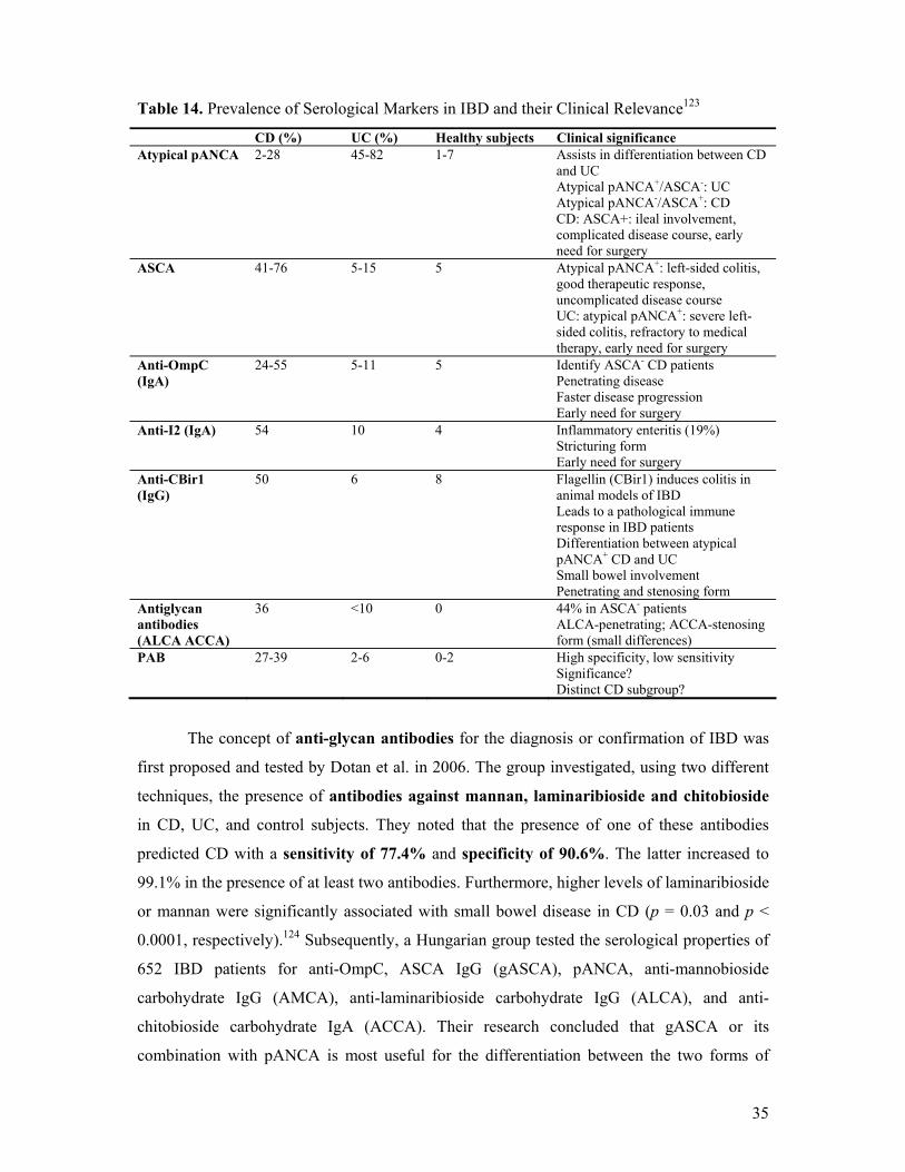
Table 14. Prevalence of Serological Markers in IBD and their Clinical Relevance123
CD (%) UC (%) Healthy subjects Clinical significance Atypical pANCA 2-28 45-82 1-7 Assists in differentiation between CD
and UC Atypical pANCA+/ASCA-: UC Atypical pANCA-/ASCA+: CD CD: ASCA+: ileal involvement, complicated disease course, early need for surgery
ASCA 41-76 5-15 5 Atypical pANCA+: left-sided colitis, good therapeutic response, uncomplicated disease course UC: atypical pANCA+: severe left-sided colitis, refractory to medical therapy, early need for surgery
Anti-OmpC (IgA)
24-55 5-11 5 Identify ASCA- CD patients Penetrating disease Faster disease progression Early need for surgery
Anti-I2 (IgA) 54 10 4 Inflammatory enteritis (19%) Stricturing form Early need for surgery
Anti-CBir1 (IgG)
50 6 8 Flagellin (CBir1) induces colitis in animal models of IBD Leads to a pathological immune response in IBD patients Differentiation between atypical pANCA+ CD and UC Small bowel involvement Penetrating and stenosing form
Antiglycan antibodies (ALCA ACCA)
36 <10 0 44% in ASCA- patients ALCA-penetrating; ACCA-stenosing form (small differences)
PAB 27-39 2-6 0-2 High specificity, low sensitivity Significance? Distinct CD subgroup?
The concept of anti-glycan antibodies for the diagnosis or confirmation of IBD was
first proposed and tested by Dotan et al. in 2006. The group investigated, using two different
techniques, the presence of antibodies against mannan, laminaribioside and chitobioside
in CD, UC, and control subjects. They noted that the presence of one of these antibodies
predicted CD with a sensitivity of 77.4% and specificity of 90.6%. The latter increased to
99.1% in the presence of at least two antibodies. Furthermore, higher levels of laminaribioside
or mannan were significantly associated with small bowel disease in CD (p = 0.03 and p <
0.0001, respectively).124 Subsequently, a Hungarian group tested the serological properties of
652 IBD patients for anti-OmpC, ASCA IgG (gASCA), pANCA, anti-mannobioside
carbohydrate IgG (AMCA), anti-laminaribioside carbohydrate IgG (ALCA), and anti-
chitobioside carbohydrate IgA (ACCA). Their research concluded that gASCA or its
combination with pANCA is most useful for the differentiation between the two forms of
36
IBD. Furthermore, a greater antibody response against gASCA, ACCA, ALCA, AMCA, and
OmpC were significantly associated with complicated disease behaviour and need for surgery
in Crohn’s disease (p < 0.0001 and p = 0.023, respectively).125
Perinuclear anti-neutrophil cytoplasmic autoantibodies (pANCA), in general, are used
for the diagnosis of various vasculitides. Specifically, they target the granule constituents of
neutrophils.115 An atypical pANCA pattern is observed in some conditions, including UC.126
As a result, it can serve as a useful marker to discriminate between UC and CD. It was shown
to react with epitopes such as: Escherichia coli,127 Bacteriodes caccae,127,128 Mycobacteria129
and others, further supporting the opinion that microbial agents play a role in the pathogenesis
of IBD and the subsequent abnormal immune response.130 In a study from Belgium, these
results are confirmed and further noted that a combination of gASCA and pANCA can be
effectively used for the differentiation between Crohn’s disease and ulcerative colitis.131
To date, ANCA has been recognized as the marker of UC in the task of distinguishing
between the forms of IBD.115
pANCA is also detected in 65-75% of primary sclerosing cholangitis (PSC)
patients.118 PSC is the most frequent chronic liver disease found in IBD patients, and in
particular, in ulcerative colitis. It is found in 2-7% of UC patients, affecting more men than
women. 132,133 At the cellular level, Terjung et al.134 found a 50-kilodalton nuclear envelope
protein, which is the target of pANCA in PSC, as well as in UC.
Pancreatic autoantibodies are extremely specific for CD, demonstrating 97-100%
specificity in comparison with UC; however, a much poorer sensitivity of only 27-
38%.135,136 In a study published by Seibold et al., 27% of CD patients were shown to have
PAB, while none of the controls were positive for these autoantibodies. The team concluded
that PAB is a specific marker in CD.135 Another group, Folwaczny et al., demonstrated a
higher number of patients with circulating PAB, 38%, and concluded it was insufficient as a
genetic marker for IBD.136 Finally, in a more recent Belgian study, encompassing 575
subjects (289 IBD patients, 108 unaffected first-degree relatives, 78 subjects with non-IBD
gastrointestinal disorders, and 100 healthy control subjects), similar rates were noted. The
prevalence of pancreatic autoantibodies was 32%, 23.3%, and 22.2% in CD, UC, and
unaffected first-degree relatives, respectively. Of importance, the prevalence of PABs in the
non-IBD gastrointestinal disorders group and the healthy controls was 1.3% and 0%,
respectively. Furthermore, the study group noticed two staining patterns, intracellular and
extracellular, and detected a significant association between the extracellular patter and CD,
37
as compared with UC patients (p = 0.014). However, the intracellular pattern was
significantly associated with both, familial UC (p = 0.0003) and familial CD (p = 0.0009).137
In 2002, Kayazawa et al. published their study on possible markers collected from
whole gut lavage fluid.116 As it is known that neutrophils are involved in inflammatory
reactions, the researchers examined four different proteins released from neutrophilic granules
to assess their correlation with disease activity in UC and CD. For UC, the disease activity
was graded by colonoscopy, while CD patients were graded according to the CDAI. The four
proteins examined included lactoferrin, polymorphonuclear neutrophil elastase (PMN-E),
myeloperoxidase (MPO), and lysozyme. The concentration of each protein was assessed in
whole gut lavage fluid (WGLF) collected at the beginning of the colonoscopy procedure.116
It was found that in ulcerative colitis, patients’ lactoferrin levels showed the best
relationship with colonscopic grading of disease status. However, in Crohn’s disease, in
addition to a strong correlation between lactoferrin and CDAI, MPO was also elevated
accordingly. It seemed that lactoferrin was most suitable as a marker and was quite stable in
WGLF.116
A different method uses fecal matter to test for calprotectin, serving as a marker for
inflammatory bowel disease. Calprotectin is a calcium-binding protein found in neutrophils
and on the surface of monocytes. Following the activation of neutrophils or adhesion of
monocytes to endothelium, calprotectin is released and can be detected in serum or other
bodily fluids. Pelli et al. demonstrated that patients with active IBD were all positive for this
protein, as opposed to 18% of healthy individuals. 138,139
38
4. COMPLICATIONS & EXTRAINTESTINAL MANIFESTATIONS
Inflammatory bowel disease is confined, by definition, to the gastrointestinal tract;
however, extraintestinal manifestations (EIM) are far-reaching. Many organs and organ
systems are affected including the eye, skin, liver and biliary system, musculoskeletal system,
and the hematopoietic system.132 Some conditions arise by nature of the disease process
itself,64 while others, such as uveitis and some forms of arthritis, have a genetic background
closely associated with HLA genes.45 A range of values are available for the frequency of
EIMs. According to one, geographically-localized study by Lakatos et al.,140 36.6% of CD
patients and 15.0% of UC had extraintestinal manifestations in the province of Veszprem,
Hungary. Various authors report different values for the occurrence of the extraintestinal
manifestations, making it is somewhat difficult to identify the most prevalent extraintestinal
manifestation.
4.1. Ocular Manifestations
IBD-related ocular disease was already described in 1925 by Crohn.141 Uncertainty
exists as to the real incidence value, but estimates range from 3.5% to 11.8% with some
gender-related variation.142 Data about the prevalence of uveitis within each inflammatory
disease is conflicting. Bernstein et al.132 found it more common in UC than CD, reporting a
3.8% incidence rate. Lakatos et al., however, reported of equal incidence rates of ocular
complications in UC and CD. Finally, Orchard et al. observed a greater incidence rate (6.4%)
in CD than in UC (3.1%).153 Nonetheless, agreement exists that ocular manifestations are
more common in women.132,140,143 The study by Lakatos et al.140 further asserts that most
ocular manifestations develop during the earlier years of the disease. This is supported by
the observation that occasionally, ocular EIMs are diagnosed prior to the inflammatory bowel
disease itself.144
The three most common ophthalmic conditions encountered in IBD patients are
uveitis, scleritis, and episcleritis. Almost 50% of IBD patients with ocular manifestations
will have more than one ocular complication, but as many as 68% of ophthalmic patients will
have other extraintestinal manifestations not involving the eyes. Other disorders, which
usually appear alongside ocular manifestations, include arthritis and ankylosing spondylitis.
The latter two, as well as uveitis, are associated with HLA-B27.145 Episcleritis has been
reported in as many as 29% of IBD patients with ocular complications, while only 18%
display scleritis.142 Of note, episcleritis correlates well with disease activity and as such, has
been used as a marker.
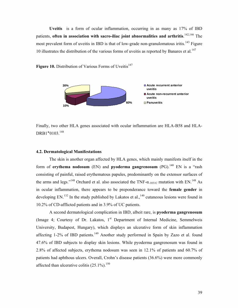
39
Uveitis is a form of ocular inflammation, occurring in as many as 17% of IBD
patients, often in association with sacro-iliac joint abnormalities and arthritis.142,146 The
most prevalent form of uveitis in IBD is that of low-grade non-granulomatous iritis.143 Figure
10 illustrates the distribution of the various forms of uveitis as reported by Banares et al.147
Figure 10. Distribution of Various Forms of Uveitis147
60%10%
30% Acute recurrent anterioruveitisAcute non-recurrent anterioruveitisPanuveitis
Finally, two other HLA genes associated with ocular inflammation are HLA-B58 and HLA-
DRB1 0103.148
4.2. Dermatological Manifestations
The skin is another organ affected by HLA genes, which mainly manifests itself in the
form of erythema nodosum (EN) and pyoderma gangrenosum (PG).140 EN is a “rash
consisting of painful, raised erythematous papules, predominantly on the extensor surfaces of
the arms and legs.”148 Orchard et al. also associated the TNF-α-1031C mutation with EN.148 As
in ocular inflammation, there appears to be preponderance toward the female gender in
developing EN.132 In the study published by Lakatos et al.,140 cutaneous lesions were found in
10.2% of CD-afflicted patients and in 3.9% of UC patients.
A second dermatological complication in IBD, albeit rare, is pyoderma gangrenosum
(Image 4; Courtesy of Dr. Lakatos, 1st Department of Internal Medicine, Semmelweis
University, Budapest, Hungary), which displays an ulcerative form of skin inflammation
affecting 1-2% of IBD patients.149 Another study performed in Spain by Zazo et al. found
47.6% of IBD subjects to display skin lesions. While pyoderma gangrenosum was found in
2.8% of affected subjects, erythema nodosum was seen in 12.1% of patients and 60.7% of
patients had aphthous ulcers. Overall, Crohn’s disease patients (36.6%) were more commonly
affected than ulcerative colitis (25.1%).150
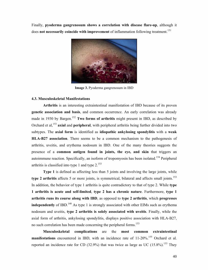
40
Finally, pyoderma gangrenosum shows a correlation with disease flare-up, although it
does not necessarily coincide with improvement of inflammation following treatment.151
Image 3. Pyoderma gangrenosum in IBD
4.3. Musculoskeletal Manifestations
Arthritis is an interesting extraintestinal manifestation of IBD because of its proven
genetic association and basis, and common occurrence. An early correlation was already
made in 1930 by Bargen.152 Two forms of arthritis might present in IBD, as described by
Orchard et al,153 axial and peripheral, with peripheral arthritis being further divided into two
subtypes. The axial form is identified as idiopathic ankylosing spondylitis with a weak
HLA-B27 association. There seems to be a common mechanism to the pathogenesis of
arthritis, uveitis, and erythema nodosum in IBD. One of the many theories suggests the
presence of a common antigen found in joints, the eye, and skin that triggers an
autoimmune reaction. Specifically, an isoform of tropomyosin has been isolated.154 Peripheral
arthritis is classified into type 1 and type 2.153
Type 1 is defined as affecting less than 5 joints and involving the large joints, while
type 2 arthritis affects 5 or more joints, is symmetrical, bilateral and affects small joints.153
In addition, the behavior of type 1 arthritis is quite contradictory to that of type 2. While type
1 arthritis is acute and self-limited, type 2 has a chronic nature. Furthermore, type 1
arthritis runs its course along with IBD, as opposed to type 2 arthritis, which progresses
independently of IBD.140 As type 1 is strongly associated with other EIMs such as erythema
nodosum and uveitis, type 2 arthritis is solely associated with uveitis. Finally, while the
axial form of arthritis, ankylosing spondylitis, displays positive association with HLA-B27,
no such correlation has been made concerning the peripheral forms.153
Musculoskeletal complications are the most common extraintestinal
manifestations encountered in IBD, with an incidence rate of 11-20%.155 Orchard et al.
reported an incidence rate for CD (32.9%) that was twice as large as UC (15.8%).153 They
41
consistently found larger values for type 1 and type 2 arthritis affecting CD and UC patients.
Type 1 peripheral arthritis affected 3.6% and 6.0% of UC and CD patients, respectively. Type
2 arthritis was seen in 2.5% and 4.0% of UC and CD patients, respectively. Of significance,
some patients will display arthropathic signs prior to a diagnosis of IBD, or arthropathic
processes might already be present at the time of diagnosis. This phenomenon is more
common for type 1 peripheral arthritis than type 2, as the former can also be used to gauge
disease activity.153 On the contrary, spinal involvement in IBD does not reflect the stage or
severity of the disease.155
4.4. Thromboembolic & Hematologic Manifestations
Two common problems affecting IBD patients are thromboembolism and anemia,
with the former leading to substantial disease and death in IBD patients.156 A study by Grip et
al.157 found an increased risk for venous thrombosis at a younger age among IBD patients
as opposed to the general population at risk for venous thrombosis. Interestingly, inherited
diseases of coagulopathy, such as hemophilia, seem to have a protective effect against IBD,
as the odds of developing UC or CD in such patients is lower than healthy subjects with
normal coagulation pathways.158 Additionally, while most EIMs are encountered during
active bouts of IBD, a study found 77% of peripheral venous thromboses occurred during
remission.159 The mechanism or mutation behind thrombus mutation is still debated, as is the
role of Factor V Leiden.156,160
The second most common cause of thrombus formation is described as a point
mutation in the prothrombin gene of patients with deep-vein thrombosis (6.2%).161
Heterozygote carriers express 30% higher prothrombin levels than healthy subjects.162
However, no studies have found conclusive evidence linking this mutation to an increased
risk of thrombosis in IBD patients.163
Anemia is defined, by most studies, as a hemoglobin level lower than 135.0 g/L in
men and 115.0 g/L in women.167,168 A hematocrit level below 0.40 also reflects an anemic
status.164 In IBD, anemia can arise as the result of two main mechanisms. The first and more
important one is that of iron deficiency caused by reduced dietary intake or malabsorption. In
addition, it might also be caused by increased loss through chronic gastrointestinal bleeding.
Of note, although iron absorption is usually normal in IBD patients, the loss of iron may be
overwhelming. The second mechanism involved in IBD anemia is a result of the chronic
disease process. This type results from the general activation of the proinflammatory cytokine
42
system, finally resulting in a reduced red blood cell lifespan as well as decreased and/or
inefficient erythropoiesis.165,166
It is estimated that the incidence of anemia in IBD varies from 8.8% to 73.7%;
however, reports are conflicting and, at times, no definition for anemia is given.164 One study
published by Harries et al.167 examined 55 CD patients in England. The patient pool was
divided into two groups based on the nourishment level of patients, which was a reflection of
the prevalence rate of anemia among the patients. Of the 25 undernourished patients, 64%
showed signs of anemia, while only 26.7% of well-nourished patients were anemic. In another
British study of 63 hospitalized CD patients, 63.5% were anemic.168 When considering the
prevalence rate of anemia in UC patients, a broad range of values is found depending on the
study. An Israeli study found only 8.8% anemia cases among 147 patients.169 On the high
side, another study conducted in Israel found 66.6% prevalence among the females examined
and 27.5% among the males.170
4.5. Hepatobiliary Manifestations
Primary sclerosing cholangitis (PSC) is at the forefront of EIMs affecting the
hepatobiliary system, predominantly in UC. One study found a prevalence rate of 7.5%.171
PSC is a condition affecting both intra- and extrahepatic bile ducts.172 It is an idiopathic
disease featuring regions of inflammation in the biliary tree, which may advance to fibrosis
and strictures.173 PSC, like many other EIMs, displays a gender preference for females,132
and is proposed to be an independent risk factor and a precancerous condition for colorectal
cancer in UC patients.174,175 This risk increases with the progress of the disease as
demonstrated by Broomé et al., who found that the average duration of disease until detection
of malignancy or dysplasia was 18 years. They also found that none of the twelve patients
who underwent colectomy due to dysplasia or carcinoma, died as a consequence of the
cancer. Finally, UC patients with PSC had 9% risk of developing colorectal dysplasia or
carcinoma after 10 years of disease, 31% after 20 years and 50% after 25 years with UC.175
Lakatos et al.140 collected extensive data about the distribution of hepatobiliary
complications in UC and CD. They classified three conditions affecting the hepatobiliary
system: PSC, small duct PSC (if ERCP could not confirm PSC finding), and non-alcoholic
fatty liver disease (NAFLD)/non-alcoholic steatohepatitis (NASH). The majority of
hepatobiliary EIM cases in CD and UC consisted of NAFLD/NASH. Table 15 summarizes
the results, for both for CD and UC. As shown in the table, PSC is twice as common in UC as
it is CD, while the opposite can be concluded for NAFLD/NASH.
43
Table 15. Hepatobiliary Complications in the Western Province of Veszprem, Hungary140
PSC Small duct PSC NAFLD/NASH
Crohn’s disease 0.8% 2.4% 19.3%
Ulcerative colitis 1.6% 1.3% 9.4%
NAFLD, non-alcoholic fatty liver disease; NASH, non-alcoholic steatohepatitis
4.6. Other Complications
Inflammatory bowel disease can also present itself in the form of surgical
emergencies. These may include: toxic colitis, massive hemorrhage, free perforation, and an
abscess accompanied by sepsis.176 Toxic megacolon is an emergency situation in UC. In fact,
in up to 30% of patients, toxic megacolon may be the presentation of the disease itself.177 It is
well known that gastrointestinal bleeding is a common feature in IBD; however, severe
bleeding is a rare occurrence. Most studies estimate the incidence value at 2-3%, while it is
responsible for about 10% of emergency colectomy procedures in ulcerative colitis.178 Free
perforation is a rare emergency occurring in only 2% of UC patients, usually occurring
together with a megacolon or toxic colitis.179 Finally, in Crohn’s disease, abscesses occur
most commonly in the right lower quadrant, neighboring the terminal ileum.176
4.7. Colorectal Cancer in Inflammatory Bowel Disease
The notion of colorectal cancer (CRC) was originally documented in 1925, very
shortly after the first report on Crohn’s disease.180 Until now, much of the knowledge
concerning CRC has been derived from UC patients where it is found more frequently, and
as stated above, the risk increases with a longer disease duration.173 Other risk factors
include greater anatomic extent,181 presence of primary sclerosing cholangitis, dysplasia
found in specimen,182 a family history of CRC, and degree of bowel inflammation.183
Furthermore, colitis-associated CRC has a younger age of onset.183 Accordingly, British and
American guidelines indicate that surveillance endoscopy needs to be initiated in the UC
patient population 8-10 years after diagnosis, or in the case of extensive or left-sided colitis,
after 15-20 years.184
44
5. TREATMENT
Over the years, it has become clear that the absence of a clear etiology for IBD
necessitated the use of treatment regimens that remedy the symptoms alone.100 The arsenal of
possible methods includes 5-aminosalicylic acid (5-ASA), corticosteroids,185
immunomodulatory agents,264 chimeric monoclonal antibodies,186 antibiotics,272 probiotics187
and gene therapy.188 Here, aspects of each treatment are presented. The majority of treatments
aim to control or change the immune response and the composition of the intestinal flora.272
Consequently, all treatment options have undesirable side-effects.189
5.1. Aminosalicylates
This family of drugs is at the forefront of treatment, with the primary agent being
sulfasalazine. These are appropriate for the treatment of moderate ulcerative colitis and
Crohn’s disease; however, different drug forms exist depending on the site of disease and the
method of administration.264 Two pharmaceutical forms of 5-ASA are available: sulfa-
containing and sulfa-free 5-ASA.190 These agents appear to have immunomodulatory
functions, even though their mechanism has not yet been fully elucidated. 5-ASAs are
capable of halting the production of prostaglandins and leukotrienes, thus preventing
neutrophil chemotaxis. Also, they scavenge reactive oxygen metabolites and might play a role
in the inhibition of NF-кB.264
It is important to select the proper form of 5-ASA as well as mode of delivery; these
will affect efficacy and the resultant side-effects. The newer sulfa-free 5-ASAs pose a
substantial improvement in this class of drugs, since they do not lead to the side effects seen
with drugs such as sulphasalazine,190 in particular, agranulocytosis, neutropenia, male
infertility, and neuropathy.191 Finally, depending on disease location, the form of
administration needs to be appropriately chosen, for example, azo-compounds, microspheres,
or pH-dependent compounds.190 The latter, for instance, lead to 5-ASA release in the distal
ileum while slow-release compounds release 5-ASA in the proximal small intestine.264
Several intolerance reports have been completed that included headache, dyspepsia,
nausea, vomiting, anorexia, and fatigue. These signs and symptoms might appear in as many
as 45% of patients, which is an extremely high fraction of the treated population. Of note, the
prevalence of these side effects is dose-dependent and also genetically determined, owing to
the hepatic degradation of a given side chain on the drug molecule.192 It is important to realize
that sulfa-free 5-ASAs (for example, mesalamine) do not come without their own arsenal of
45
side-effects, including paradoxical exacerbation of colitis, pancreatitis, pneumonitis, hepatitis,
pericarditis, and nephritis.193
The ASCEND I trial investigated the safety and efficacy of two different delayed-
release mesalamine dosages in 301 patients suffering from mildly to moderately active UC.
The cohort was randomly assigned to delayed release 2.4 g/day or 4.8 g/day mesalamine and
followed for 6 weeks, with complete remission or clinical improvement from baseline serving
as primary endpoint. In the mildly active UC group, there was no significant difference
between the two dosages, with 51% of patients assigned to the delayed-release 2.4 g/day
mesalamine and 56% of patients in the 4.8 g/day group reached the efficacy endpoint (p =
0.441). However, in the moderate UC group, a significant difference was observed between
the delayed-release 2.4 g/day mesalamine and 4.8 g/day. In the former, 57% of patients
reached the efficacy endpoint compared with 72% of patients in the latter group (p =
0.0384).194
A recent meta-analysis showed a significant reduction in the CDAI in patients
receiving mesalamine compared with a cohort that received a placebo (p = 0.04), concluding
that mesalamine 4 g/day is superior to placebo in decreasing the CDAI, yet the clinical
improvement is still unclear.195 Since there is insufficient information on the benefit of any
particular subgroup, the European Crohn’s and Colitis Organisation Consensus states that 5-
ASA “should be considered clinically no more effective than placebo for active ileal or
ileocolonic CD.”196 Maintenance therapy may also be considered for 5-ASA though data
regarding its use and efficacy is conflicting.196
5.2. Corticosteroids
This group is one of the longest used drugs in IBD treatment, their efficacy cannot
be denied, and despite their side effects, are still needed.196,197 Topical corticosteroids, in the
form of enema, may be used to treat patients with ulcerative colitis or distal UC, who have
shown adverse effects to 5-ASA. Oral forms of corticosteroids, such as budesonide, 9
mg/day, may be administered to patients with mild, moderate or severe forms of UC or CD.196
Other proven options include prednisone (0.5-0.75 mg/kg/day)198 and 6-methylprednisolone
(1 mg/kg/day).199 Finally, an intravenous route of administration may be used for
hospitalized patients with severe stages of the disease. Even in this latter group of patients,
improvement is already observed within 7-10 days. Importantly, these agents should be used
only as long as necessary to control an acute phase of the disease, as they have no indications
for maintenance therapy. 200
46
Their effects have been shown to act through the inhibition of proinflammatory
cytokine production. These include, but are not limited to: IL-1, IL-2, IL-4, IL-6, and IFN-
γ.201 Possibly, a disruption in the activity of NF-кB is responsible for this broad inhibitory
function by corticosteroids.202
Corticosteroids, with their popularity and efficiency in reducing immunologic and
inflammatory reactions, produce severe side effects. As with most therapeutic agents, these
effects depend on dosage and duration of treatment. Some of the more severe conditions
include osteoporosis, hypertension, diabetes, as well as a detrimental effect on the
hypothalamic-pituitary-adrenal axis.203,264 In addition, dependence also accompanies
corticosteroid use. Moreover, negative effects might be observed in the hepatic,
cardiovascular, mental or musculoskeletal systems. Corticosteroids administered for a short
period of time can also result in fluid and electrolyte imbalances, acid-base disturbances, and
metabolic abnormalities. Long-term corticosteroid therapy will induce changes in fat
distribution, ecchymoses, and abdominal striae.192 Corticosteroid administration suppresses
the immune system, thus exposing the patient to a greater risk of opportunistic infections.264
5.3. Immunomodulatory Agents
As discussed previously, and is well known, inflammatory bowel disease results from
an aberrant immune response and it is only logical that some treatment regimens will attempt
to control the immune system, thus affecting the extent and duration of disease. One very
important compound in this class is azathioprine (AZA), which is a thioguanine derivative
with the active metabolite being 6-mercaptopurine (6-MP).204 This derivative is the most
commonly used immunosuppressive agent in IBD treatment.264 It is a highly successful drug,
as two-thirds of IBD patients enter remission following treatment. Furthermore, 80-90% of
patients who are able to tolerate AZA are capable of enduring a long-term treatment period.204
Another advantage for using AZA is its capacity to wean off corticosteroid-dependent
patients. It serves as a continuation therapy, while the corticosteroid dose is simultaneously
and gradually decreased. In an important recent trial, Ardizzone et al. provided the evidence
to support this notion. They investigated 72 individuals who were steroid-dependent UC
patients, and the result from administering 2 mg/kg/day azathiopurine or 3.2 g/day of 5-ASA.
The results showed that AZA was significantly better than 5-ASA at inducing remission as
well as discontinuing the use of steroids, during 6 months of therapy.205 The pathway in
which these drugs function is still unknown; however, one theory exists whereby a specific
and long-lived subgroup of T-cells is suppressed.264 In general, AZA 1.5-2.5 mg/kg/day or
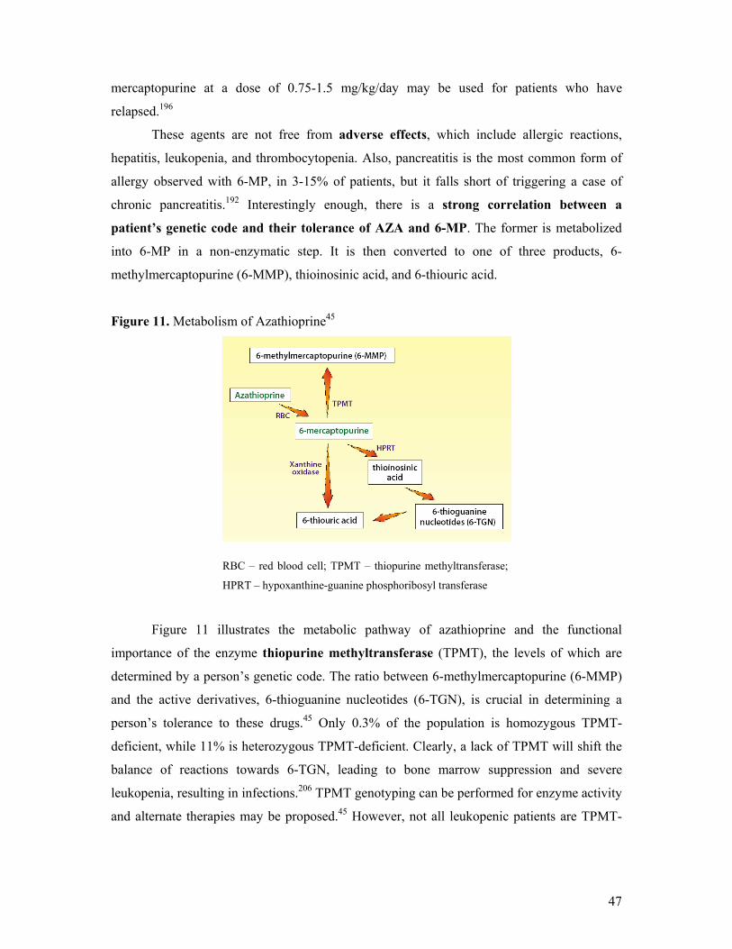
47
mercaptopurine at a dose of 0.75-1.5 mg/kg/day may be used for patients who have
relapsed.196
These agents are not free from adverse effects, which include allergic reactions,
hepatitis, leukopenia, and thrombocytopenia. Also, pancreatitis is the most common form of
allergy observed with 6-MP, in 3-15% of patients, but it falls short of triggering a case of
chronic pancreatitis.192 Interestingly enough, there is a strong correlation between a
patient’s genetic code and their tolerance of AZA and 6-MP. The former is metabolized
into 6-MP in a non-enzymatic step. It is then converted to one of three products, 6-
methylmercaptopurine (6-MMP), thioinosinic acid, and 6-thiouric acid.
Figure 11. Metabolism of Azathioprine45
RBC – red blood cell; TPMT – thiopurine methyltransferase;
HPRT – hypoxanthine-guanine phosphoribosyl transferase
Figure 11 illustrates the metabolic pathway of azathioprine and the functional
importance of the enzyme thiopurine methyltransferase (TPMT), the levels of which are
determined by a person’s genetic code. The ratio between 6-methylmercaptopurine (6-MMP)
and the active derivatives, 6-thioguanine nucleotides (6-TGN), is crucial in determining a
person’s tolerance to these drugs.45 Only 0.3% of the population is homozygous TPMT-
deficient, while 11% is heterozygous TPMT-deficient. Clearly, a lack of TPMT will shift the
balance of reactions towards 6-TGN, leading to bone marrow suppression and severe
leukopenia, resulting in infections.206 TPMT genotyping can be performed for enzyme activity
and alternate therapies may be proposed.45 However, not all leukopenic patients are TPMT-
48
deficient. One study showed that 73% of patients suffering from bone marrow suppression
displayed no known TPMT mutations.206
While the next section illustrates the efficacy of various chimeric and human
monoclonal antibodies, one must still always consider azathioprine as first-line therapy
because of its proven track record for the treatment of both CD and UC.
5.4. Chimeric Monoclonal Antibodies
5.4.1. Infliximab and Crohn’s Disease
Infliximab is the prototypical anti-TNF-α monoclonal antibody.207 It falls under the
category of biological treatment, since it focuses on inhibiting or downregulating specific
cytokines or chemokines involved in the immune response.186 At a dose of 5 mg/kg it is an
effective tool in patients with active CD.196 Similarly to other treatment modalities, the exact
mechanism of action is not fully known. By one account, infliximab binds soluble TNF-
α186,264; by another account, shown in vitro, it acts by inducing apoptosis in lymphocytes and
other cells.46,208 It is possible that its efficacy is a result from a combination of these two
functions. Structurally, infliximab is composed of a human IgG1 constant region attached to a
murine antigen-binding variable segment, hence forming a chimera.264 By composition, it is
about 75% human, while the rest is of murine origin.209
Two clinical trials, ACCENT I and II were conducted to test the efficacy of
infliximab. ACCENT I had a complex design, whereby patients were allowed to use other
treatment regimens in addition to the chimeric antibody. This made result interpretation
somewhat complicated but nonetheless useful results were produced. There was sufficient
evidence linking remission with a maintenance therapy of intravenous infliximab, where 24%
of patients remained in remission, in contrast to 9% of placebo-treated CD patients.210 The
ACCENT II randomized trial inspected the response of patients’ enterocutaneous disease to
infliximab treatment, with 69% of patients demonstrating a positive outcome.211
Despite infliximab’s clinically significant record, it does harbor several significant
side effects, consisting of acute and delayed infusion reactions, autoimmunity and
autoantibodies, and an increased risk of infections.212 Infusions administered too quickly may
lead to symptoms of an acute infusion reaction, in particular, dyspnea, chest pain, headache,
and fever. Also, since infliximab is not 100% human protein, development of antibodies is a
feasible and concrete phenomenon.213 A delayed infusion reaction might occur if a time gap
larger than 12 weeks occurs between subsequent infusions. Increasing levels of antinuclear
49
and anti-double-stranded-DNA antibodies have also been reported in long term use of
infliximab but only in rare cases does a lupus-like syndrome develop.212,214
5.4.2. Infliximab and Ulcerative Colitis
Until recently, infliximab was only indicated for Crohn’s disease without proven
efficacy in patients with ulcerative colitis. Two studies, Active Ulcerative Colitis Trials 1
and 2 (ACT 1 and 2), set out to determine the efficacy of infliximab in inducing and
maintaining remission of ulcerative colitis. These two randomized, double-blind, placebo-
controlled studies investigated 364 patients each, with moderate-to-severe UC despite
medical therapy. The subject received either intravenous infliximab (5 or 10 mg/kg body
weight) at weeks 0, 2, and 6, or a placebo. In ACT 1, following the first three doses, the
patients received placebo or infliximab every 8 weeks until week 46, while in ACT 2 the
administration continued every 8 weeks until week 22. All groups were followed for eight
additional weeks following the last administration. Response to therapy was defined as a
decrease in the Mayo Score215 of at least 3 points, or 30%, as well as a decline in the subscore
of rectal bleeding by 1 point or an absolute subscore of 0 or 1 for rectal bleeding.216
Both studies presented statistically significant improvements in the patient groups. In
the first study, 69% of patients who received 5 mg infliximab and 61% of those patients who
received 10 mg showed a clinical response at week 8. This was in comparison with 37% of
the placebo group (p < 0.001) (Figure 12). In ACT 2, 64% of patients who received 5 mg and
69% of those who received 10 mg infliximab demonstrated a clinical response at week 8,
compared with 29% of the placebo group (p < 0.001) (Figure 12). Furthermore, patients in
both study groups were more likely to show a clinical response at week 30, as compared with
the placebo-controlled group (p ≤ 0.002).216
Finally, Järnerot and colleagues conducted a randomized, placebo-controlled study on
the use of infliximab in patients with severe or moderately severe UC. The group reasoned
that such patients often become candidates for colectomy and was searching for alternative
rescue therapy when steroid use was ineffective. They enrolled 45 patients and randomized
them into two separate arms – placebo and infliximab. The primary endpoint was colectomy
or death at 3 months after randomization. Secondary endpoints included clinical and
endoscopic remission after 3 months in patients who did not undergo a colectomy. None of
their patients died, while 7 in the infliximab group and 14 in the placebo group underwent
colectomy (OR: 4.9; p = 0.019). Thus, they concluded that infliximab is indeed an appropriate
rescue therapy in patients not responding to conventional therapy.217
50
5.4.3. Adalimumab
Similar to infliximab, this IgG1 monoclonal antibody is a TNF antagonist, though its
structure varies from that of infliximab; it is completely of human origin.218 Numerous
controlled studies have demonstrated adalimumab to be safe and effective in treating immune-
mediated conditions such as rheumatoid arthritis, psoriatic arthritis, psoriasis, and ankylosing
spondylitis.219,220,221,222,223 Shifting the focus to Crohn’s disease, several trials have been
conducted to test the efficacy of adalimumab in this condition. The CLASSIC I and II trials
investigated the efficacy and safety of adalimumab in inducing and maintaining clinical
remission in CD, respectively. The CHARM trial tested the efficacy and safety of
adalimumab in maintaining response and remission in CD patients with moderate to severe
disease. Finally, the GAIN study examined the use of adalimumab in CD patients who were
previously treated with infliximab and either did not respond or could not tolerate the
treatment regimen.
5.4.4. Certolizumab Pegol
Certolizumab pegol is a pegylated humanized Fab’ fragment capable of binding
TNF-α with high affinity. While functioning as a TNF antagonist, its structure lacks the Fc
segment, thus being unable to induce in vitro complement activation, antibody-dependent
cellular cytotoxicity, or apoptosis.224,225 The PRECISE trials (I and II) set out to investigate
the efficacy of Certolizumab pegol for the induction and maintenance of remission in
moderate to severe Crohn’s disease.
In conclusion, while the efficacious mechanism behind TNF antagonists is a sound
solution for IBD, substantial research is still required to evaluate their long-term effects and
complications.
5.5. Other Therapeutic Options
Several studies have already established a role for the intestinal flora in the
pathogenesis of IBD.52,53,54,55,56 Consequently, attempts have been made in regulating the
presence of these bacteria with the hope of curbing the disease. Two options are available
following this train of thought: the first is antibiotics, while the second is probiotics. The
former, while efficient in CD, shows little improvement in UC patients. A commonly used
antibiotic in CD is metronidazole, which has shown efficacy in treating patients with perianal
fistulae, yet the required dose is quite high and can lead to neurotoxicity.264
51
Probiotics constitute a biological way for treating Crohn’s disease as well as
maintaining a clinical remission in ulcerative colitis.187 They are live, non-pathogenic
organisms that improve the gut’s microbial balance leading to a healthier status. These can be
divided into two groups226:
Bacteria – usually lactic acid producers, such as lactobacilli strain GG227 or
Escherichia coli Nissle.228
Yeast – eg, Saccharomyces boulardii.
It is important that probiotics adhere to strict criteria in order to be safe and effective.
In addition to safety concerns, the following criteria must also be met229:
be of human origin
resist bile and acid
have the ability to adhere, colonize, and survive in the human intestine.
Nonetheless, the majority of data supporting probiotics comes from animal models,
whereby colitis-induced mice are cured by certain Lactobacillus strains. In humans, trials
have been conducted using a non-pathogenic strain of Escherichia coli.230 Finally, a
published study tested the use of helminthic ova in altering the immune response of the
human gut. The ova of Trichuris suis, pork whipworm, were used to shift the Th1 profile
observed in CD to Th2. In this study, three of the four tested CD patients entered remission,
while the three UC subjects had a decrease of 43% in their Clinical Colitis Activity Index.
This method certainly seems like a feasible solution with much needed research.231
The ever-expanding field of gene therapy is yet another possible solution for the
treatment of IBD. The theory behind gene therapy in IBD, and other chronic inflammatory
diseases, is the expression of proteins that are important in the downregulation of the
pathogenic immune response. On the contrary, gene therapy may also lead to expression of
proteins that will confer protection against an illicit immune response. To date, there are no
human clinical trials involving specific gene therapy protocols for IBD, though animal models
are being investigated.232
52
6. BACKGROUND & AIMS OF THE STUDIES
6.1. Multidrug Resistance-1 Gene and Breast Cancer Resistance Protein
The multidrug resistance-1 (MDR1/ABCB1) gene is located on chromosome 7 and
consists of 28 exons. Its transmembrane protein product, P-glycoprotein 170 (Pgp-170),
belongs to the ATP-binding cassette (ABC) transporters and is thought to be responsible for
the removal of xenobiotics and resistance to multiple chemotherapeutic agents. It
performs this function by limiting the absorption from the intestinal tract, while encouraging
the removal of these compounds through the bile and urine.233 Supporting the role of Pgp-170
is its co-localization with CYP3A4, an important enzyme for drug metabolism.234 MDR1 was
first identified in cancer cells, followed by its discovery also in normal tissue including liver,
kidney, brain, testis, and placenta.235,236,237
MDR1, through its product Pgp-170, is thought to play an important role in response
to treatment of IBD as well as disease susceptibility. First, it is known that Pgp-170 has a
wide range of substrates, including but not limited to, anticancer drugs, cardiac drugs,
immunosuppressants, antiemetics, antibiotics, and steroids.233 It follows accordingly that Pgp-
170’s capacity to remove steroids, for example, dexametahosone,238 and immunosuppressants,
such as cyclosporine239 and methotrexate,240 from cells could also be the basis for some cases
of failed medical therapy in IBD necessitating surgical approaches. Farrell and colleagues
have indeed found a significant elevation (p < 0.0001) of peripheral blood lymphocytes’
MDR1 in CD patients who failed medical therapy and required bowel resection. Similarly, a
significant increase (p = 0.001) was also found in UC patients who underwent
proctocolectomy due to failed medical therapy.241
The role of MDR1 in bacterial-host interaction arose from a knockout mouse model.
As it became known that a possible susceptibility locus for IBD was located on chromosome
7q21.1,324 Panwala et al. created the mdr1a-/- knockout mouse model. They demonstrated that
these knockout mice, raised in pathogen-free conditions, developed colitis with features of
both CD and UC, which could be reversed with antibiotics. Subsequently, they concluded that
the intestinal flora was necessary for the inflammatory process. Therefore, it seems that
MDR1 not only has a protective function against xenobiotics but also bacterial
products.242 Nonetheless, its exact function has not yet been completely defined.
One must also consider the role of mutated Pgp-170 in the susceptibility of IBD, as at
the moment, at least 50 single nucleotide polymorphisms (SNPs) and 3 insertion/deletion
mutations have been described.243 The first SNP was found in exon 26 (C3435T), a silent
mutation that does not change the encoded amino acid (Ile). Nonetheless, it displays a
53
functional variance in individual homozygous for the variant T allele; Pgp-170 expression
was lower.244 A second mutation, G2677T/A in exon 21, has also been correlated with
reduced Pgp-170 expression levels.245
The breast cancer resistance protein (BCRP/ABCG2) is also a member of the ATP-
binding cassette transporters superfamily, which has been implicated and proven to play a role
in multidrug resistance. It is a half-transporter with a wide range of substrates and is located
in the small and large intestines as well as in the kidney and liver. The protein, which was
originally described in a multidrug-resistant breast cancer cell line, also secretes drugs and
xenotoxins into breast milk, posing a risk to the infant.246,247,248
Due to the protein’s close association with MDR1, and having a similar function and
localization, it is plausible that ABCG2 may also have an effect on medical therapy in IBD,
or its failure. Of note, while half-transporter are normally located to intracellular membranes,
and full-transporters to the plasma membrane, this particular transporter is also found at the
plasma membrane.247
6.2. Discs Large Homolog 5
In 1999, using a genome-wide scan, Hampe et al. discovered a susceptibility locus on
chromosome 10q23.336 Five years later, the discs large homolog 5 (DLG5) gene was added to
the growing list of IBD susceptibility loci by Stoll et al. who used positional cloning for this
task.249 DLG5 is a member of the membrane-associated guanlyate kinase (MAGUK) family
and, as a scaffold protein, has an important role in maintaining epithelial cell
integrity.250,251 Consequent to its location at cell-cell contact sites, it maintains cell shape
and polarity.252,253
The DLG5 SNP G113A results in the amino acid substitution R30Q, possibly having
functional consequences.249 It is interesting to note that ethnic variation has been uncovered
in the transmission of this polymorphism among IBD patients, though it is not surprising and
unusual. A similar trend has been observed with CARD15 in Caucasian versus Asian
populations.254 Likewise, an analysis of Japanese CD patients revealed no association with the
DLG5 R30Q polymorphism; however, a different variant, rs3758462, was detected in this
particular group.255 Further support for genetic heterogeneity of the R30Q variant comes from
a Greek CD study population, where the variant was completely absent.256
Recently, a protective role for DLG5 R30Q polymorphism in females has been
published. In the first such study, Biank and colleagues identified a significant negative
association (OR: 0.39; p = 0.006) between CD and female children. Studying a group of 281
54
CD patients against 479 control subjects, they initially found no association between the
R30Q variant and Crohn’s disease. In a second statistical step, they stratified the groups by
gender and found a protective effect for DLG5 R30Q in female children but not males (OR:
0.39; p = 0.006).257
6.3. Aims of Study I
In view of the limited data on the prevalence of DLG5 mutations in East European
countries, our aim was to investigate the presence of the DLG5 R30Q variant allele in a large
cohort of Hungarian patients with IBD. We also aimed to investigate a possible association
between genotype and clinical phenotype, need for surgery and response to medical therapy;
as well, the possible association between DLG5, NOD2/CARD15 and TLR4 D299G
polymorphism in CD patients was studied. As mentioned previously, DLG5’s function as a
scaffolding gene provides a plausible argument for its role in IBD secondary to intestinal
barrier dysfunction. Our secondary aim was to specifically investigate whether there is an
association between the efficacy of infliximab therapy and the DLG5 variant.
6.4. Aims of Study II
The MDR1 gene encodes an ATP-binding cassette transporter (Pgp-170) that confers
resistance to multiple drugs from a very broad range of classes including antibiotics, anti-
cancer drugs, cardiac drugs and steroids. However, it addition to its role, research has shown
that its localization to the intestinal epithelial layer may be also protective against bacterial
products. Subsequently, a dysfunction in the Pgp-170 protein may play a role in IBD
pathogenesis and resistance to medical therapy. In view of the limited data on the prevalence
of MDR1 and the absence of data on the relatively frequent (G34A and C421A) ABCG2
variants in East European countries, our aim was to investigate the presence of the variant
alleles of these genes in a large cohort of Hungarian patients with IBD. We also aimed to
investigate a possible association between genotype and clinical phenotype, need for surgery
and response to medical therapy.
55
7. PATIENTS & METHODS
7.1. Patient Group - Study I
Our study cohort consisted of 773 unrelated IBD patients of which 639 patients were
afflicted by CD. The average age of all IBD patients was 38.1 ± 10.3 years with a disease
duration of 8.8 ± 7.5 years. More specifically, our CD patients (m/f = 309/330) had a duration
of 8.4 ± 7.1 years, while among the 134 UC patients (m/f = 63/71) disease duration was 10.6
± 8.9 years. In addition, we also had 150 healthy control subjects, who did not suffer form any
gastrointestinal or liver disorders. They were age- and gender-matched with an average age of
37.6 ± 10.3 years and a male-to-female ratio of 72/78.
7.2. Patient Group – Study II
This study consisted of 414 unrelated IBD patients (265 CD, 149 UC) and 149 healthy
control subjects. The average of our CD cohort was 35 ± 12.1 years and for UC 44.5 ± 15.4
years. The male-to-female ratio was 129/136 and 73/76 for CD and UC, respectively. For our
CD cohort, disease duration was 8.7 ± 7.6 years, while in UC it was 10.7 ± 8.9 years.
7.3. Methods – Study I
7.3.1. Clinical Data Collection, Statistics, Ethics
The patients’ diagnoses were made according to the Lennard-Jones criteria.258
Background demographic and clinical information was collected by reviewing their medical
charts and a questionnaire. This data included: age, age at onset, presence of extraintestinal
manifestations (arthritis, ocular manifestations, dermatological lesions, primary sclerosing
cholangitis), frequency of flare-ups, therapeutic effectiveness (prescribed medication and
efficacy, steroid resistance), surgery associated with IBD, familial IBD cases, and smoking
habits. Additionally, CD patients were questioned about perianal involvement. The Vienna
Classification was used to determine disease phenotype (age at onset, duration, location, and
behavior).107 Our cohort’s clinical data are presented in Tables 20 and 21 for CD and UC,
respectively.
The Shapiro-Wilk’s W test was used for testing the normality of our sample, while the
t test with separate variance estimates, χ2 test, and χ2 with Yates’ correction were used for
calculating differences within IBD subgroups and between IBD and controls. A logistic
regression was used to find an association between clinical and genetic data, expressed as
odds ratio (OR) with 95% confidence interval (95% CI). We considered a p value < 0.05 as
statistically significant.
56
Each patient was informed of the nature of the study and signed the informed consent
form. Furthermore, the protocol was approved by the Ethical and Science Committee of the
Ministry of Health (ETT-TUKEB, 321-81/2005-1018EKU) and the Semmelweis University
Regional and Institutional Committee of Science and Research Ethics (81/2003).
7.3.2. Genetic Methods
Detection of the DLG5 R30Q polymorphism
To detect the R30Q variant of DLG5, we followed the process described by Török et
al.,314 using polymerase chain reaction/restriction fragment length polymorphism
(PCR/RFLP) with the MspI restriction enzyme. For this purpose, genomic DNA was isolated
from whole blood using the QIAamp DNA Blood Mini Kit (QIAgen GmbH, Germany). The
forward primer was 5’-GGA AGG CGC AGT CCC CAC CAC CCC TCC TCA C, while the
reverse primer was 5’-AAG GCC AGG CGC TTG CGG AGC TCG TTT CTC TCC TGG.
3% agarose gel was used for detection.
Detection of NOD2/CARD15 and TLR4 polymorphisms
The NOD2 SNP 8 (R702W), 12 (G908R), and 13 (3020insC) polymorphisms have
already been genotyped for 475 CD patients, as has the TLR4 D299G.259 NOD2 mutations
were detected using denaturing high-performance liquid chromatography (Wave DNA
Fragment Analysis System, Transgenomic Ltd., Cramlington, UK), followed by sequencing
of both strands to confirm variations. Sequencing reactions were performed with the ABI
BigDye v1.1 Terminator Cycle Sequencing Kit followed by electrophoresis on the ABI Prism
310 Genetic Analyzer (Applied Biosystems, Foster City, CA). The D299G polymorphism was
detected using the PCR/RFLP technique and visualized on 3% agarose GTG gel (NuSieve,
BMA, Rockland, ME).
7.4. Methods – Study II
7.4.1. Clinical Data Collection, Statistics, Ethics
As in the previous study, the diagnosis was based on the Lennard-Jones criteria258 with
the demographics and clinical data collected through a medical chart review and a
questionnaire. These parameters included age, age at onset, presence of extraintestinal
manifestations (arthritis, ocular manifestations, dermatological lesions, primary sclerosing
cholangitis), frequency of flare-ups, therapeutic effectiveness (prescribed medication and
efficacy, steroid resistance), surgery associated with IBD, familial IBD cases, and smoking
57
habits. In addition, CD patients were asked about perianal disease. Similarly, the Vienna
Classification was used to confirm disease phenotype according to age at onset, duration,
location, and behavior.107 Our cohort’s clinical data are presented in Tables 24 and 25.
In addition to the Shapiro-Wilk’s W test for normality, we also used t-tests with
separate variance estimates and χ2, both with and without Yates’ correction to compare data
between the IBD group and the control subjects, and within the IBD subgroups. Logistic
regression was used to link the genetic with the clinical data, with the results being expressed
as OR with 95% CI.
As already noted in the previous study, each patient was informed of the nature of the
study and those in agreement signed an informed consent form. The protocol was approved by
the Ethics and Science Committee of the Ministry of Health and the Semmelweis University
Regional and Institutional Committee of Science and Research Ethics.
7.4.2. Genetic Methods
Whole DNA was processed using QIAamp DNA Blood Mini Kit (QIAgen GmbH,
Germany) in order to retrieve genomic DNA. For each of the transporter genes, MDR1 and
ABCG2, we tested for the presence and frequency of two variants. For the former, G2677T/A
(Ala893Ser/Thr on exon 21) and C3435T (a silent base substitution of I1145I on exon 26)
were examined. For ABCG2, we investigated G34A (Val12Met, exon 2) and C421A
(Gln141Lys, exon 5). The hybridization probes for both MDR1 polymorphisms, used with the
LightCycler (Roche Diagnostics, Basle, Switzerland) for genotyping, were previously
published.260,261 The technique described here was developed particularly for this study
for investigating these polymorphisms. For the G2677T/A, the forward and reverse primers
were 5’-GCA GGA GTT GTT GAA ATG AAA ATG and 5’-CGC CTG CTT TAG TTT
GAC TCA, respectively.261 The forward and reverse primers for C3435T were 5’-TGT TTT
CAG CTG CTT GAT GG, and 5’-AAG GCA TGT ATG TTG GCC TC, respectively.260
Additional amplification primers and hybridization probes were designed by the LightCycler
Probe Design software (Roche Diagnostics) and Primer3.262 Tables 16 and 17 present the
reaction mixture and details for the MDR1 variants, while Tables 18 and 19 summarize the
reaction mixtures used for the ABCG2 variant amplification.
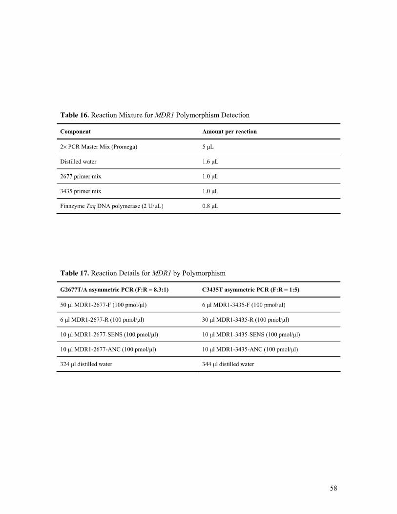
58
Table 16. Reaction Mixture for MDR1 Polymorphism Detection
Component Amount per reaction
2% PCR Master Mix (Promega) 5 μL
Distilled water 1.6 μL
2677 primer mix 1.0 μL
3435 primer mix 1.0 μL
Finnzyme Taq DNA polymerase (2 U/μL) 0.8 μL
Table 17. Reaction Details for MDR1 by Polymorphism
G2677T/A asymmetric PCR (F:R = 8.3:1) C3435T asymmetric PCR (F:R = 1:5)
50 μl MDR1-2677-F (100 pmol/μl) 6 μl MDR1-3435-F (100 pmol/μl)
6 μl MDR1-2677-R (100 pmol/μl) 30 μl MDR1-3435-R (100 pmol/μl)
10 μl MDR1-2677-SENS (100 pmol/μl) 10 μl MDR1-3435-SENS (100 pmol/μl)
10 μl MDR1-2677-ANC (100 pmol/μl) 10 μl MDR1-3435-ANC (100 pmol/μl)
324 μl distilled water 344 μl distilled water
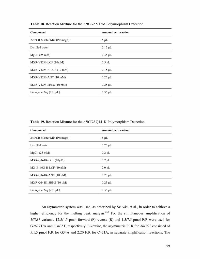
59
Table 18. Reaction Mixture for the ABCG2 V12M Polymorphism Detection
Component Amount per reaction
2% PCR Master Mix (Promega) 5 μL
Distilled water 2.15 μL
MgCl2 (25 mM) 0.35 μL
MXR-V12M-LCF (10mM) 0.5 μL
MXR-V12M-R-LCR (10 mM) 0.15 μL
MXR-V12M-ANC (10 mM) 0.25 μL
MXR-V12M-SENS (10 mM) 0.25 μL
Finnzyme Taq (2 U/μL) 0.35 μL
Table 19. Reaction Mixture for the ABCG2 Q141K Polymorphism Detection
Component Amount per reaction
2% PCR Master Mix (Promega) 5 μL
Distilled water 0.75 μL
MgCl2 (25 mM) 0.2 μL
MXR-Q141K-LCF (10μM) 0.2 μL
MX-E166Q-R-LCF (10 μM) 2.0 μL
MXR-Q141K-ANC (10 μM) 0.25 μL
MXR-Q141K-SENS (10 μM) 0.25 μL
Finnzyme Taq (2 U/μL) 0.35 μL
An asymmetric system was used, as described by Szilvási et al., in order to achieve a
higher efficiency for the melting peak analysis.263 For the simultaneous amplification of
MDR1 variants, 12.5:1.5 pmol forward (F):reverse (R) and 1.5:7.5 pmol F:R were used for
G2677T/A and C3435T, respectively. Likewise, the asymmetric PCR for ABCG2 consisted of
5:1.5 pmol F:R for G34A and 2:20 F:R for C421A, in separate amplification reactions. The
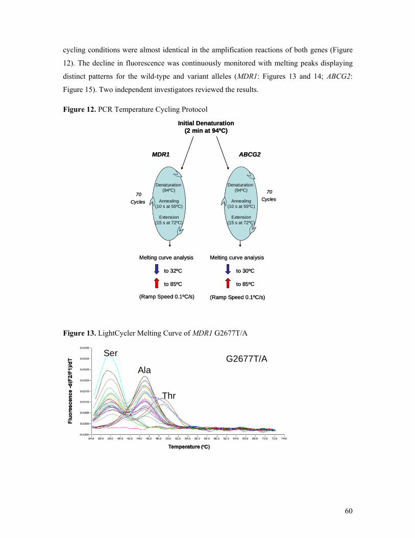
60
cycling conditions were almost identical in the amplification reactions of both genes (Figure
12). The decline in fluorescence was continuously monitored with melting peaks displaying
distinct patterns for the wild-type and variant alleles (MDR1: Figures 13 and 14; ABCG2:
Figure 15). Two independent investigators reviewed the results.
Figure 12. PCR Temperature Cycling Protocol
Denaturation (94ºC)
Annealing(10 s at 55ºC)
Extension(15 s at 72ºC)
Initial Denaturation(2 min at 94ºC)
MDR1 ABCG2
70Cycles
70Cycles
Melting curve analysis
to 32ºC
to 85ºC
Melting curve analysis
to 30ºC
to 85ºC
Denaturation (94ºC)
Annealing(10 s at 55ºC)
Extension(15 s at 72ºC)
(Ramp Speed 0.1ºC/s) (Ramp Speed 0.1ºC/s)
Denaturation (94ºC)
Annealing(10 s at 55ºC)
Extension(15 s at 72ºC)
Initial Denaturation(2 min at 94ºC)
MDR1 ABCG2
70Cycles
70Cycles
Melting curve analysis
to 32ºC
to 85ºC
Melting curve analysis
to 30ºC
to 85ºC
Denaturation (94ºC)
Annealing(10 s at 55ºC)
Extension(15 s at 72ºC)
(Ramp Speed 0.1ºC/s) (Ramp Speed 0.1ºC/s)
Figure 13. LightCycler Melting Curve of MDR1 G2677T/A
Ser
Ala
Thr
G2677T/ASer
Ala
Thr
G2677T/A
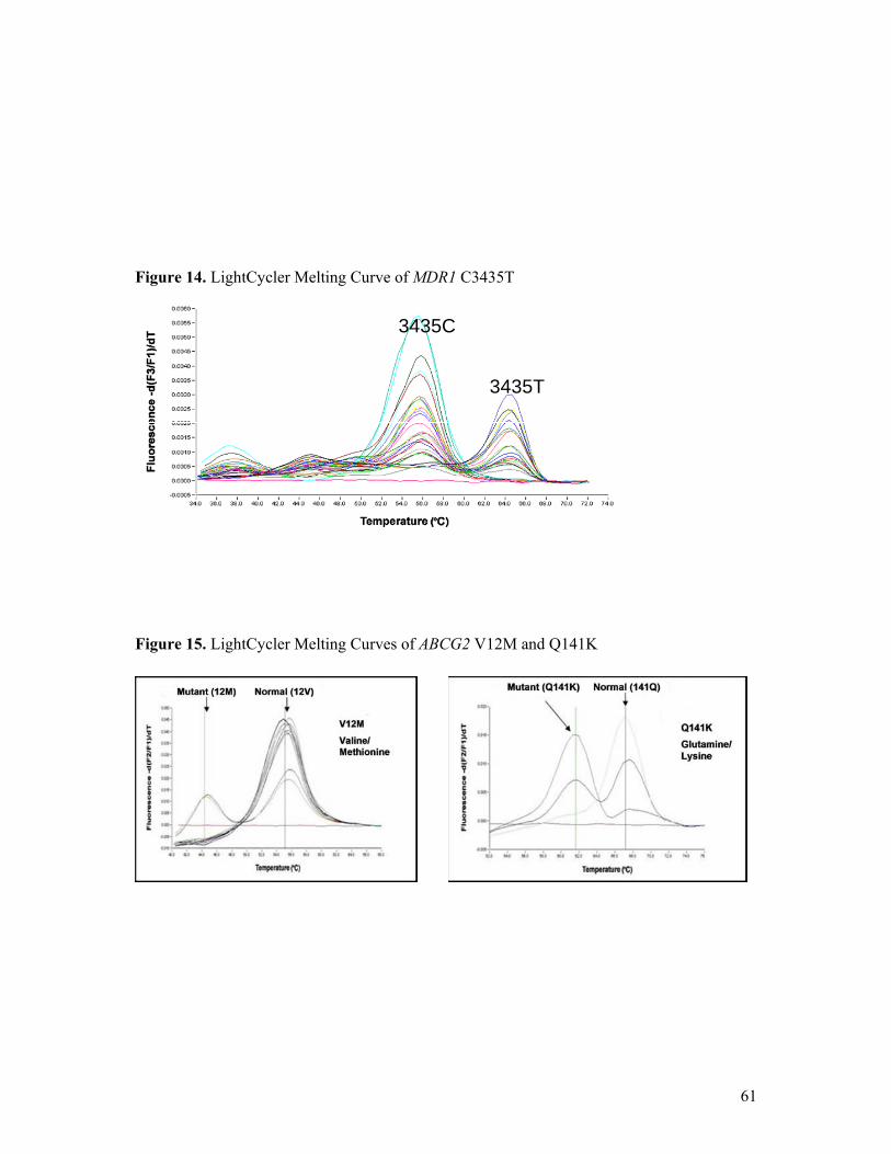
61
Figure 14. LightCycler Melting Curve of MDR1 C3435T
3435C
3435T
3435C
3435T
Figure 15. LightCycler Melting Curves of ABCG2 V12M and Q141K
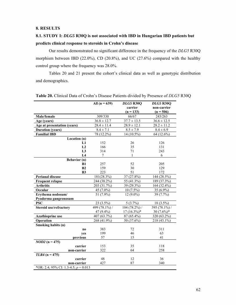
62
8. RESULTS
8.1. STUDY I: DLG5 R30Q is not associated with IBD in Hungarian IBD patients but
predicts clinical response to steroids in Crohn’s disease
Our results demonstrated no significant difference in the frequency of the DLG5 R30Q
morphism between IBD (22.0%), CD (20.8%), and UC (27.6%) compared with the healthy
control group where the frequency was 28.0%.
Tables 20 and 21 present the cohort’s clinical data as well as genotypic distribution
and demographics.
Table 20. Clinical Data of Crohn’s Disease Patients divided by Presence of DLG5 R30Q
All (n = 639) DLG5 R30Q carrier
(n = 133)
DLG5 R30Q non-carrier
(n = 506) Male/female 309/330 66/67 243/263 Age (years) 36.8 ± 12.7 37.7 ± 13.5 36.6 ± 12.5 Age at presentation (years) 28.4 ± 11.4 28.9 ± 12.1 28.2 ± 11.2 Duration (years) 8.4 ± 7.1 8.5 ± 7.9 8.4 ± 6.9 Familial IBD 78 (12.2%) 14 (10.5%) 64 (12.6%)
Location (n) L1
L2 L3 L4
152 166 314
7
26 35 71 1
126 131 243
6 Behavior (n)
B1 B2 B3
257 159 223
52 30 51
205 129 172
Perianal disease 181(28.3%) 37 (27.8%) 144 (28.5%) Frequent relapse 244 (38.2%) 55 (41.3%) 189 (37.3%) Arthritis 203 (31.7%) 39 (29.3%) 164 (32.4%) Occular 45 (7.0%) 10 (7.5%) 35 (6.9%) Erythema nodosum/ Pyoderma gangrenosum
51 (7.9%) 12 (9.0%) 39 (7.7%)
PSC 23 (3.5%) 5 (3.7%) 18 (3.5%) Steroid use/refractory 499 (78.1%) /
47 (9.4%) 104 (78.2%) / 17 (16.3%)*
395 (78.1%) / 30 (7.6%)*
Azathioprine use 407 (63.7%) 87 (65.4%) 320 (63.2%) Operation 268 (41.9%) 50 (37.6%) 218 (43.1%) Smoking habits (n)
no yes
previous
383 199 57
72 46 15
311 63 41
NOD2 (n = 475) carrier
non-carrier
153 322
35 64
118 258
TLR4 (n = 475) carrier
non-carrier
48 427
12 87
36 340
*OR: 2.4; 95% CI: 1.3-4.5; p = 0.013
63
Table 21. Clinical Data of Ulcerative Colitis Patients divided by Presence of DLG5 R30Q
All (n = 134)
DLG5 R30Q carrier (n = 37)
DLG5 R30Q non-carrier
(n = 97) Male/female 63/71 18/19 45/52 Age (years) 44.1 ± 15.1 44.5 ± 15.5 44.1 ± 14.9 Age at presentation (years) 33.9 ± 14.1 34.1 ± 14.7 33.8 ± 13.9 Duration (years) 10.6 ± 8.9 10.3 ± .18 10.7 ± 9.3 Familial IBD 8 (5.9%) 2 (5.4%) 6 (6.1%) Maximum extent (n) proctitis left-sided extensive
5
77 52
1 17
19*
4 60
33* Chronic continuous 25 (18.7%) 7 (18.9%) 18 (18.5%) Arthritis 35 (26.1%) 9 (24.3%) 26 (26.8%) Ocular 6 (4.5%) 3 (8.1%) 3 (3.1%) Erythema nodosum/ Pyoderma gangrenosum
6 (4.5%) 2 (5.4%) 4 (4.1%)
PSC 5 (3.7%) 2 (5.4%) 3 (3.1%) Steroid use/refractory 69 (51.5%) /
10 (14.5%) 20 (54.1%) /
2 (10.0%) 49 (50.5%) /
8 (16.3%) Azathioprine use 27 (20.2%) 8 (21.6%) 19 (19.6%) Operation 5 (3.7%) 3 (8.1%) 2 (2.1%) Smoking habits (n)
no yes
previous
103 15 16
28 3 6
75 12 10
*OR: 2.1; 95% CI: 0.95-4.4; p = 0.07
The following table presents the carriage distribution of R30Q in all groups, both in
absolute values and by percentage.
Table 22. Carrier Rates of DLG5 R30Q in CD, UC and Control Subjects
DLG5 R30Q Genotype, n (%)
Non-carrier All carriers Heterozygous Homozygous Variant allele frequency
CD (n = 639)
UC (n = 134)
Control (n = 150)
506 (79.2)
97 (72.4)
108 (72)
133 (20.8)*
37 (27.6)
42 (28)†
122 (19.1)
35 (26.1)
39 (26)
11 (1.7)
2 (1.5)
3 (2)
144 (11.3)*
39 (14.6)
45 (15)†
n (%) *OR = 0.67; 95% CI: 0.45-1.013; p = 0.06 vs. control subjects †OR = 0.72; 95% CI: 0.50-1.03; p = 0.07 vs. control subjects
A tendency was observed in the CD group toward a protective role by 113A (OR:
0.67; 95% CI: 0.45-1.013), as well as an association with steroid resistance (16.3% carriers
64
vs. 7.6% non-carriers; OR: 2.4; 95% CI: 1.3-4.5; p = 0.013). In addition, steroid resistance
was independently associated with penetrating disease (OR: 2.0; p = 0.046), perianal
manifestations (OR: 2.5; p = 0.07), and frequent relapses (OR: 4.2; p < 0.00001) but not with
location, familial disease, or need for surgery in univariate analysis. Using a logistic
regression, steroids resistance was independently associated with R30Q carriage, perianal
disease, and frequent relapse. In addition, after correcting for various confounding factors
(Table 23) by logistic regression, we noted a significant association between the DLG5 113A
allele and steroid resistance. Again, disease location was not associated with the DLG5
polymorphism using logistic regression. As noted in Table 20, 78.1% of patients received
steroid treatment during the course of their disease. Finally, no association was found between
carriage of the DLG5 R30Q and any of the NOD2 and TLR4 mutations genotyped in this
cohort. In ulcerative colitis patients, we noted a trend towards a more aggressive form of
disease (51.4% vs. 34.0% in controls; OR: 2.1; 95% CI: 0.95-4.4; p = 0.07) with no additional
associations.
Table 23. Logistic Regression: Association between Clinical Phenotype, Carriage of DLG5
R30Q Variant Allele, and Steroid Resistance in Patients with Crohn’s Disease
Factor Coefficient P value OR 95% CI
Female gender 0.09 0.78 1.09 0.57-2.09
DLG5 113A allele 0.807 0.02 2.24 1.14-4.43
Perianal disease 0.84 0.02 2.31 1.15-4.67
Penetrating disease -0.08 0.82 0.92 0.45-1.87
Frequent relapses 1.30 0.001 3.66 1.71-7.84
Azathioprine use 1.35 0.03 3.85 1.14-12.93
Current smoking 0.48 0.14 1.62 0.85-3.11
In a separate part of our study, we also investigated the association between DLG5
R30Q and response to infliximab therapy (5 mg/kg body weight). The sub-study’s cohort
consisted of 25 male and 35 female CD patients with an average age of 34.3 ± 11.1 years and
disease duration of 7.9 ± 5.1 years (Table 24). In just over half of the patients (32), the disease
was localized to the ileocolon, while 22 had colonic disease and 6 patients displayed ileal
form. Three patients had upper gastrointestinal disease. The disease was classified as
65
penetrating in 33 patients and inflammatory in 27, with perianal involvement in 33 patients.
Approximately half of the patients (51.7%) had some arthritic manifestations, while the rate
of ocular and dermatological manifestations was also elevated (16.7% and 13.3%,
respectively). The greater majority of the cohort was prescribed steroids (95.0%) or
azathioprine (93.3%). While 23 patients received infliximab treatment for fistulizing disease,
37 patients were subscribed infliximab for inflammatory disease that did not respond to
conventional therapy. Within the infliximab group, 17 patients were carriers of the 113A
variant. Assessing the response in the short-term, at week 8, no association was found
between carriers and non-carriers with regards to treatment response (Figure 16).
Table 24. The Clinical Phenotypes of CD Patients in Infliximab Sub-study
Male/female 25/35
Age at presentation (years) 28.4 ± 11.4
Age (years) 36.8 ± 12.7
Duration (years) 8.4 ± 7.1
Location L1
L2
L3
L4
6
22
32
(3)
Behaviour B1
B2
B3
27
-
33
Perianal involvement 33 (55%)
Extraintestinal manifestations:
- Arthritis
- Ocular
- Cutaneous
- PSC
31 (51.7%)
10 (16.7%)
8 (13.3%)
2 (3.3%)
Steroid use/refrectory
Azathiprine use
57 (95%) / 17(29.8%)
56 (93.3%)
DLG5 R30Q 17 (28.3%) vs. controls (28.0%)

66
Figure 16. Association between the Presence of DLG5 R30Q and response to Infliximab Induction Therapy (5 mg/bwkg at weeks 0, 2, and 6) Assessed at Week 8 in CD Patients
0
10
20
30
40
50
wild type carrier
no responsepartial response
remission
partial response: CDAI decrease by ≥70 points and/or ≥50% decrease in the number of draining fistulae,
remission: CDAI<150 or closure of all fistulas
There was no significant association between DLG5 R30Q and CD or UC;
however, a protective trend was observed in Crohn’s disease. No interaction was
observed between DLG5 R30Q and either NOD2 or TLR4 polymorphisms in our cohort.
In CD patients, a positive association was observed between DLG5 R30Q carriage and
steroid resistance. In UC patients, a positive trend was observed between DLG5 R30Q
carriage and extensive disease. No association was found between DLG5 R30Q carriage
and response to short-term infliximab treatment in a sub-cohort of CD patients.
67
8.2. STUDY II: The ATP-binding cassette transporter ABCG2 (BCRP) and ABCB1
(MDR1) variants are not associated with disease susceptibility, disease phenotype,
response to medical therapy or need for surgery in Hungarian patients with
inflammatory bowel disease
Our cohort’s clinical data as well as demographic and genotypic distribution are
presented in Tables 25 and 26.
Table 25. Clinical Data of Patients with Crohn’s Disease divided by ABCG2 and MDR1
C3435T
All (n = 265) ABCG2 carrier (n = 72)
ABCG2 non-carrier
(n = 193)
MDR1 3435CC (n = 66)
MDR1 3435 CT or TT (n = 199)
Male/female 129/136 36/36 93/100 27/39 102/97 Age (years) 35.1 ± 12.1 36.0 ± 13.1 34.9 ± 11.8 34.1 ± 11.5 35.5 ± 12.3 Age at presentation (years) 26.6 ± 10.5 27.2 ± 11.6 26.4 ± 10.1 26.8 ± 10.8 26.6 ± 10.4 Duration (years) 8.7 ± 7.6 8.9 ± 8.1 8.6 ± 7.3 7.4 ± 6.0 9.1 ± 7.9 Familial IBD 31 (11.7%) 7 (9.7%) 24 (12.4%) 8 (12.1%) 23 (11.6%)
Location (n) L1
L2 L3 L4
59 67 133
6
17 18 36 1
42 49 97 5
16 19 30 1
43 48 103
5 Behavior (n)
B1 B2 B3
93 73 99
24 19 29
69 54 70
18 18 30
75 55 69
Perianal disease 97(36.6%) 26 (36.1%) 71 (36.8%) 29 (43.9%) 68 (34.1%) Frequent relapse 118 (44.52%) 32 (44.4%) 86 (44.6%) 32 (48.5%) 86 (43.2%) Arthritis 108 (40.7%) 29 (40.2%) 79 (40.9%) 29 (43.9%) 79 (36.7%) Steroid use/refractory 192 (72.4%)/
36 (18.7%) 51 (70.8%)/ 10 (19.6%)
141 (73.1%)/ 26 (18.4%)
45 (68.1%)/ 10 (22.2%)
147 (73.9%)/ 26 (17.6%)
Azathioprine use 184 (69.4%) 45 (62.5%) 139 (72.0%) 50 (75.7%) 134 (67.4%) Surgery 115 (43.4%) 33 (45.8%) 82 (42.5%) 31 (46.9%) 84 (42.2%) Response to infliximab
remission partial
no effect
24 17 6
7 3 1
19 12 5
9 6 1
15 11 5
Smoking habits (n) no
yes previous
156 88 22
40 26 16
116 62 6
33 25 8
123 63 14
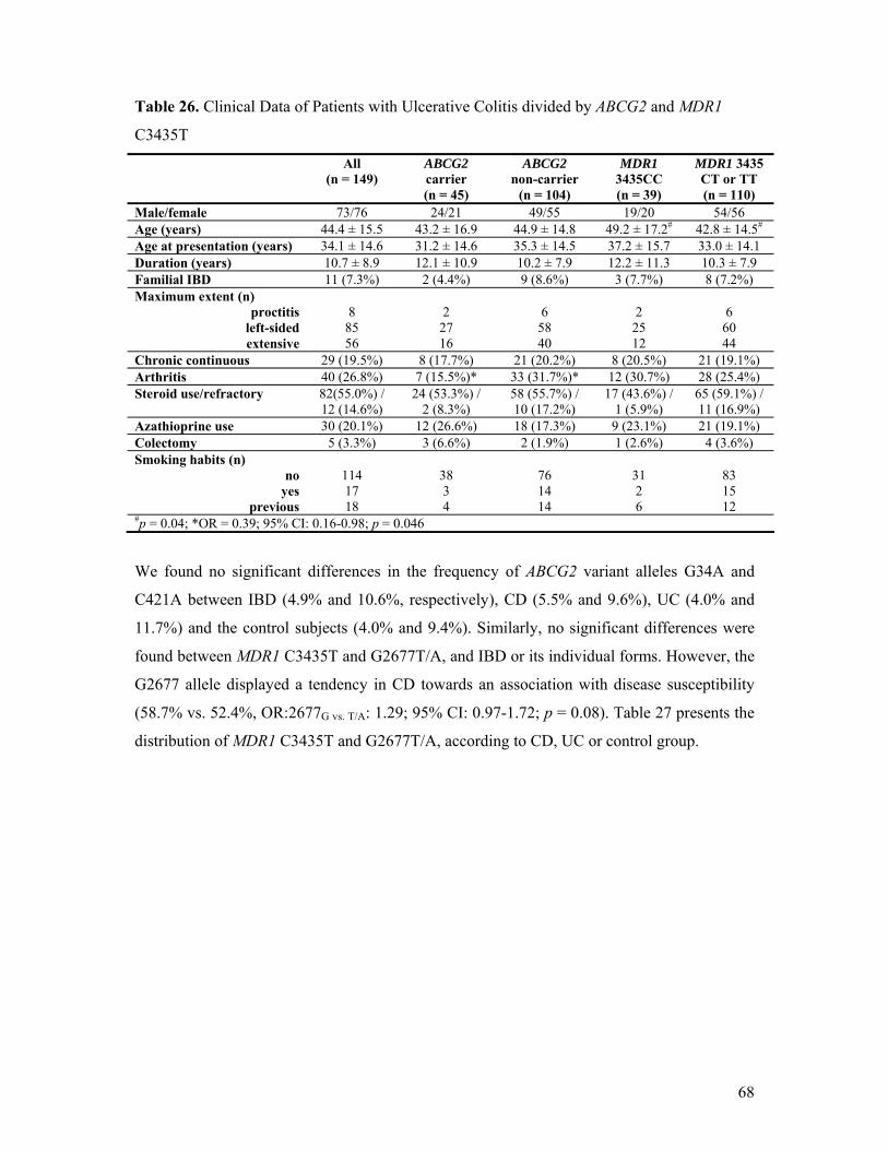
68
Table 26. Clinical Data of Patients with Ulcerative Colitis divided by ABCG2 and MDR1
C3435T
All (n = 149)
ABCG2 carrier (n = 45)
ABCG2 non-carrier
(n = 104)
MDR1 3435CC (n = 39)
MDR1 3435 CT or TT (n = 110)
Male/female 73/76 24/21 49/55 19/20 54/56 Age (years) 44.4 ± 15.5 43.2 ± 16.9 44.9 ± 14.8 49.2 ± 17.2# 42.8 ± 14.5# Age at presentation (years) 34.1 ± 14.6 31.2 ± 14.6 35.3 ± 14.5 37.2 ± 15.7 33.0 ± 14.1 Duration (years) 10.7 ± 8.9 12.1 ± 10.9 10.2 ± 7.9 12.2 ± 11.3 10.3 ± 7.9 Familial IBD 11 (7.3%) 2 (4.4%) 9 (8.6%) 3 (7.7%) 8 (7.2%) Maximum extent (n)
proctitis left-sided extensive
8
85 56
2
27 16
6
58 40
2
25 12
6
60 44
Chronic continuous 29 (19.5%) 8 (17.7%) 21 (20.2%) 8 (20.5%) 21 (19.1%) Arthritis 40 (26.8%) 7 (15.5%)* 33 (31.7%)* 12 (30.7%) 28 (25.4%) Steroid use/refractory 82(55.0%) /
12 (14.6%) 24 (53.3%) /
2 (8.3%) 58 (55.7%) / 10 (17.2%)
17 (43.6%) / 1 (5.9%)
65 (59.1%) / 11 (16.9%)
Azathioprine use 30 (20.1%) 12 (26.6%) 18 (17.3%) 9 (23.1%) 21 (19.1%) Colectomy 5 (3.3%) 3 (6.6%) 2 (1.9%) 1 (2.6%) 4 (3.6%) Smoking habits (n)
no yes
previous
114 17 18
38 3 4
76 14 14
31 2 6
83 15 12
#p = 0.04; *OR = 0.39; 95% CI: 0.16-0.98; p = 0.046
We found no significant differences in the frequency of ABCG2 variant alleles G34A and
C421A between IBD (4.9% and 10.6%, respectively), CD (5.5% and 9.6%), UC (4.0% and
11.7%) and the control subjects (4.0% and 9.4%). Similarly, no significant differences were
found between MDR1 C3435T and G2677T/A, and IBD or its individual forms. However, the
G2677 allele displayed a tendency in CD towards an association with disease susceptibility
(58.7% vs. 52.4%, OR:2677G vs. T/A: 1.29; 95% CI: 0.97-1.72; p = 0.08). Table 27 presents the
distribution of MDR1 C3435T and G2677T/A, according to CD, UC or control group.
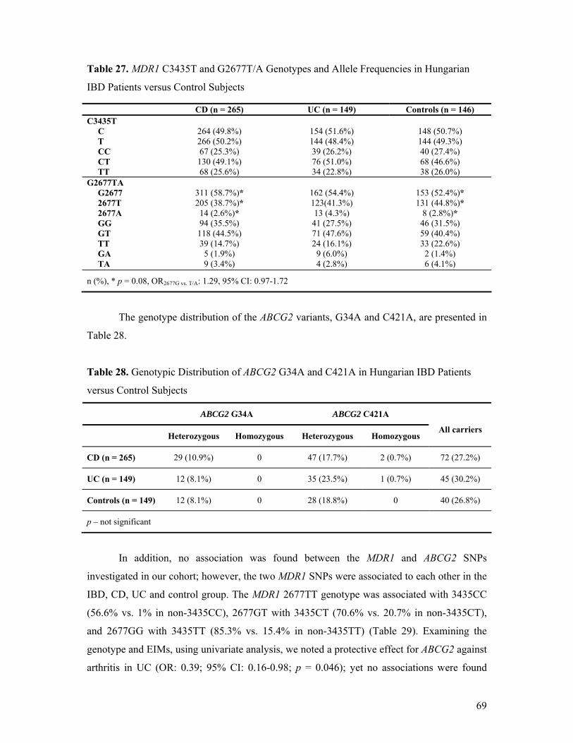
69
Table 27. MDR1 C3435T and G2677T/A Genotypes and Allele Frequencies in Hungarian
IBD Patients versus Control Subjects
The genotype distribution of the ABCG2 variants, G34A and C421A, are presented in
Table 28.
Table 28. Genotypic Distribution of ABCG2 G34A and C421A in Hungarian IBD Patients
versus Control Subjects
ABCG2 G34A ABCG2 C421A
Heterozygous Homozygous Heterozygous Homozygous
All carriers
CD (n = 265) 29 (10.9%) 0 47 (17.7%) 2 (0.7%) 72 (27.2%)
UC (n = 149) 12 (8.1%) 0 35 (23.5%) 1 (0.7%) 45 (30.2%)
Controls (n = 149) 12 (8.1%) 0 28 (18.8%) 0 40 (26.8%)
p – not significant
In addition, no association was found between the MDR1 and ABCG2 SNPs
investigated in our cohort; however, the two MDR1 SNPs were associated to each other in the
IBD, CD, UC and control group. The MDR1 2677TT genotype was associated with 3435CC
(56.6% vs. 1% in non-3435CC), 2677GT with 3435CT (70.6% vs. 20.7% in non-3435CT),
and 2677GG with 3435TT (85.3% vs. 15.4% in non-3435TT) (Table 29). Examining the
genotype and EIMs, using univariate analysis, we noted a protective effect for ABCG2 against
arthritis in UC (OR: 0.39; 95% CI: 0.16-0.98; p = 0.046); yet no associations were found
CD (n = 265) UC (n = 149) Controls (n = 146) C3435T C T CC CT TT
264 (49.8%) 266 (50.2%) 67 (25.3%) 130 (49.1%) 68 (25.6%)
154 (51.6%) 144 (48.4%) 39 (26.2%) 76 (51.0%) 34 (22.8%)
148 (50.7%) 144 (49.3%) 40 (27.4%) 68 (46.6%) 38 (26.0%)
G2677TA G2677 2677T 2677A GG GT TT GA TA
311 (58.7%)* 205 (38.7%)* 14 (2.6%)* 94 (35.5%) 118 (44.5%) 39 (14.7%)
5 (1.9%) 9 (3.4%)
162 (54.4%) 123(41.3%) 13 (4.3%) 41 (27.5%) 71 (47.6%) 24 (16.1%)
9 (6.0%) 4 (2.8%)
153 (52.4%)* 131 (44.8%)*
8 (2.8%)* 46 (31.5%) 59 (40.4%) 33 (22.6%)
2 (1.4%) 6 (4.1%)
n (%), * p = 0.08, OR2677G vs. T/A: 1.29, 95% CI: 0.97-1.72
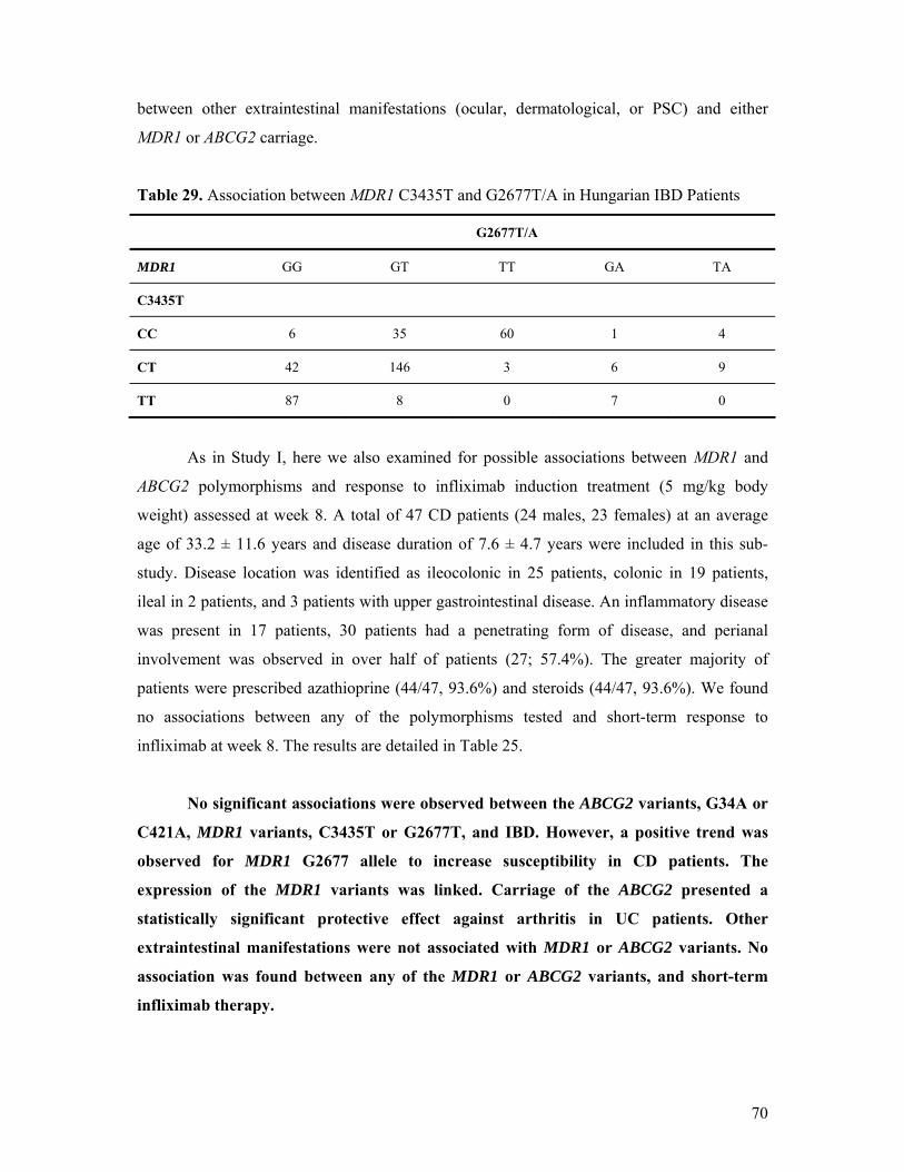
70
between other extraintestinal manifestations (ocular, dermatological, or PSC) and either
MDR1 or ABCG2 carriage.
Table 29. Association between MDR1 C3435T and G2677T/A in Hungarian IBD Patients
G2677T/A
MDR1 GG GT TT GA TA
C3435T
CC 6 35 60 1 4
CT 42 146 3 6 9
TT 87 8 0 7 0
As in Study I, here we also examined for possible associations between MDR1 and
ABCG2 polymorphisms and response to infliximab induction treatment (5 mg/kg body
weight) assessed at week 8. A total of 47 CD patients (24 males, 23 females) at an average
age of 33.2 ± 11.6 years and disease duration of 7.6 ± 4.7 years were included in this sub-
study. Disease location was identified as ileocolonic in 25 patients, colonic in 19 patients,
ileal in 2 patients, and 3 patients with upper gastrointestinal disease. An inflammatory disease
was present in 17 patients, 30 patients had a penetrating form of disease, and perianal
involvement was observed in over half of patients (27; 57.4%). The greater majority of
patients were prescribed azathioprine (44/47, 93.6%) and steroids (44/47, 93.6%). We found
no associations between any of the polymorphisms tested and short-term response to
infliximab at week 8. The results are detailed in Table 25.
No significant associations were observed between the ABCG2 variants, G34A or
C421A, MDR1 variants, C3435T or G2677T, and IBD. However, a positive trend was
observed for MDR1 G2677 allele to increase susceptibility in CD patients. The
expression of the MDR1 variants was linked. Carriage of the ABCG2 presented a
statistically significant protective effect against arthritis in UC patients. Other
extraintestinal manifestations were not associated with MDR1 or ABCG2 variants. No
association was found between any of the MDR1 or ABCG2 variants, and short-term
infliximab therapy.
71
9. DISCUSSION
9.1. Genetic Factors
Many groups have established the existence of IBD susceptibility loci in the human
genome264; however, no clear cause-and-effect has been identified with certainty. The genes
associated with IBD can be divided into three different subgroups: 1. genes involved in
bacterial sensing and autophagy; 2. Mucosal integrity and transport; and 3. cytokine genes. In
this thesis, we investigated members from the second group, as well as possible associations
between the studied DLG5 variant and NOD2/CARD15 or TLR4.
The primary susceptibility locus was localized to chromosome 16q and named NOD2
(nucleotide oligomerization domain)/CARD15 (caspase activation recruitment domain).265
Mutations in the wild-type NOD2/CARD15 proteins are associated with early onset,266,267
involvement of the terminal ileum,268,269 and a fibrostenosing disease form.270 Additionally,
NOD2/CARD15 mutations have a functional effect by decreasing the activity of NF-кB in
response to LPS.271 In general, it seems that CD has a greater genetic predisposition than
UC, as expressed by a familial pattern of inheritance.272 However, some susceptibility loci
(eg, on chromosome 6p) appear in both CD and UC, indicating a small overlap in IBD’s
genetic etiology.273,274,275 In ulcerative colitis, HLA genes are associated with disease
pathogenesis as well as extraintestinal manifestations.44,46 Finally, evidence is
accumulating for the involvement of various cytokines (eg, IL-2, IL-6, IL-12, IL-17, TNF) in
the pathogenesis, maintenance or aggravation of IBD.44,264,276 Figure 17 illustrates the
identified chromosomal loci with their respective IBD genes.46
9.1.1. Genes Involved in Bacterial Recognition
9.1.1.1. NOD2/CARD15
In 2001, three independent groups identified the first susceptibility gene for CD; it
was localized to chromosome 16q (IBD1) and named NOD2 – later renamed as
CARD15.277,278,279 The NOD2/CARD15 gene belongs to the Apaf-1/CARD superfamily of
cytosolic proteins involved in apoptosis and activation of nuclear factor-кB (NF-кB).280
The wild-type NOD2/CARD15 protein consists of 1040 amino acids and is composed of two
CARDs on the N-terminus, a nucleotide binding domain (NBD), and a sequence of leucine-
rich repeats (LRR) on the carboxy-terminus,270 as depicted in Figure 18.281 The LRR are
structurally similar to a portion of the Toll-like receptors (TLR) and might serve a similar
function in the recognition of exogenous microbial components, for example,
lipopolysaccharide (LPS). The NOD2/CARD15 protein is expressed with the highest
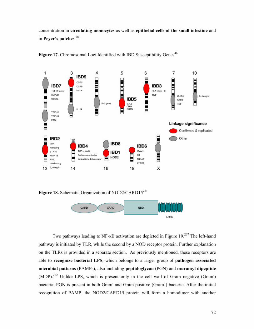
72
concentration in circulating monocytes as well as epithelial cells of the small intestine and
in Peyer’s patches.280
Figure 17. Chromosomal Loci Identified with IBD Susceptibility Genes46
Figure 18. Schematic Organization of NOD2/CARD15281
Two pathways leading to NF-кB activation are depicted in Figure 19.267 The left-hand
pathway is initiated by TLR, while the second by a NOD receptor protein. Further explanation
on the TLRs is provided in a separate section. As previously mentioned, these receptors are
able to recognize bacterial LPS, which belongs to a larger group of pathogen associated
microbial patterns (PAMPs), also including peptidoglycan (PGN) and muramyl dipeptide
(MDP).282 Unlike LPS, which is present only in the cell wall of Gram negative (Gram-)
bacteria, PGN is present in both Gram- and Gram positive (Gram+) bacteria. After the initial
recognition of PAMP, the NOD2/CARD15 protein will form a homodimer with another
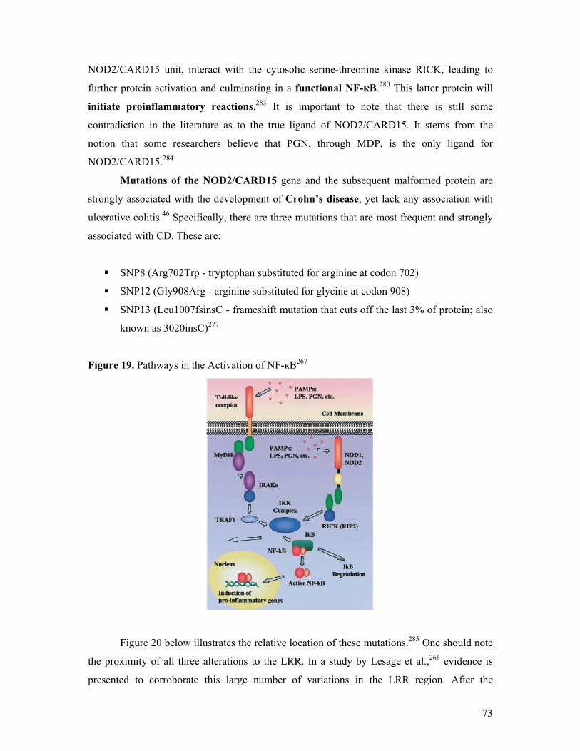
73
NOD2/CARD15 unit, interact with the cytosolic serine-threonine kinase RICK, leading to
further protein activation and culminating in a functional NF-кB.280 This latter protein will
initiate proinflammatory reactions.283 It is important to note that there is still some
contradiction in the literature as to the true ligand of NOD2/CARD15. It stems from the
notion that some researchers believe that PGN, through MDP, is the only ligand for
NOD2/CARD15.284
Mutations of the NOD2/CARD15 gene and the subsequent malformed protein are
strongly associated with the development of Crohn’s disease, yet lack any association with
ulcerative colitis.46 Specifically, there are three mutations that are most frequent and strongly
associated with CD. These are:
SNP8 (Arg702Trp - tryptophan substituted for arginine at codon 702)
SNP12 (Gly908Arg - arginine substituted for glycine at codon 908)
SNP13 (Leu1007fsinsC - frameshift mutation that cuts off the last 3% of protein; also
known as 3020insC)277
Figure 19. Pathways in the Activation of NF-кB267
Figure 20 below illustrates the relative location of these mutations.285 One should note
the proximity of all three alterations to the LRR. In a study by Lesage et al.,266 evidence is
presented to corroborate this large number of variations in the LRR region. After the
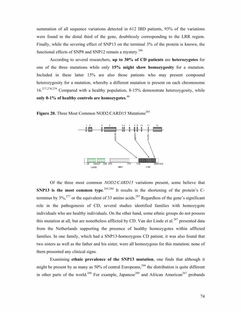
74
summation of all sequence variations detected in 612 IBD patients, 93% of the variations
were found in the distal third of the gene, doubtlessly corresponding to the LRR region.
Finally, while the severing effect of SNP13 on the terminal 3% of the protein is known, the
functional effects of SNP8 and SNP12 remain a mystery.286
According to several researchers, up to 30% of CD patients are heterozygotes for
one of the three mutations while only 15% might show homozygosity for a mutation.
Included in these latter 15% are also those patients who may present compound
heterozygosity for a mutation, whereby a different mutation is present on each chromosome
16.277,278,279 Compared with a healthy population, 8-15% demonstrate heterozygosity, while
only 0-1% of healthy controls are homozygotes.46
Figure 20. Three Most Common NOD2/CARD15 Mutations285
Of the three most common NOD2/CARD15 variations present, some believe that
SNP13 is the most common type.265,284 It results in the shortening of the protein’s C-
terminus by 3%,277 or the equivalent of 33 amino acids.287 Regardless of the gene’s significant
role in the pathogenesis of CD, several studies identified families with homozygote
individuals who are healthy individuals. On the other hand, some ethnic groups do not possess
this mutation at all, but are nonetheless afflicted by CD. Van der Linde et al.287 presented data
from the Netherlands supporting the presence of healthy homozygotes within afflicted
families. In one family, which had a SNP13-homozygous CD patient, it was also found that
two sisters as well as the father and his sister, were all homozygous for this mutation; none of
them presented any clinical signs.
Examining ethnic prevalence of the SNP13 mutation, one finds that although it
might be present by as many as 50% of central Europeans,266 the distribution is quite different
in other parts of the world.288 For example, Japanese289 and African American267 probands
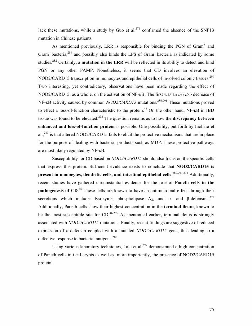
75
lack these mutations, while a study by Guo et al.271 confirmed the absence of the SNP13
mutation in Chinese patients.
As mentioned previously, LRR is responsible for binding the PGN of Gram+ and
Gram- bacteria,284 and possibly also binds the LPS of Gram- bacteria as indicated by some
studies.282 Certainly, a mutation in the LRR will be reflected in its ability to detect and bind
PGN or any other PAMP. Nonetheless, it seems that CD involves an elevation of
NOD2/CARD15 transcription in monocytes and epithelial cells of involved colonic tissues.290
Two interesting, yet contradictory, observations have been made regarding the effect of
NOD2/CARD15, as a whole, on the activation of NF-кB. The first was an in vitro decrease of
NF-кB activity caused by common NOD2/CARD15 mutations.286,291 These mutations proved
to effect a loss-of-function characteristic to the protein.46 On the other hand, NF-кB in IBD
tissue was found to be elevated.292 The question remains as to how the discrepancy between
enhanced and loss-of-function protein is possible. One possibility, put forth by Inohara et
al.,283 is that altered NOD2/CARD15 fails to elicit the protective mechanisms that are in place
for the purpose of dealing with bacterial products such as MDP. These protective pathways
are most likely regulated by NF-кB.
Susceptibility for CD based on NOD2/CARD15 should also focus on the specific cells
that express this protein. Sufficient evidence exists to conclude that NOD2/CARD15 is
present in monocytes, dendritic cells, and intestinal epithelial cells.280,293,294 Additionally,
recent studies have gathered circumstantial evidence for the role of Paneth cells in the
pathogenesis of CD.46 These cells are known to have an antimicrobial effect through their
secretions which include: lysozyme, phospholipase A2, and α- and β-defensins.295
Additionally, Paneth cells show their highest concentration in the terminal ileum, known to
be the most susceptible site for CD.46,296 As mentioned earlier, terminal ileitis is strongly
associated with NOD2/CARD15 mutations. Finally, recent findings are suggestive of reduced
expression of α-defensin coupled with a mutated NOD2/CARD15 gene, thus leading to a
defective response to bacterial antigens.288
Using various laboratory techniques, Lala et al.297 demonstrated a high concentration
of Paneth cells in ileal crypts as well as, more importantly, the presence of NOD2/CARD15
protein.
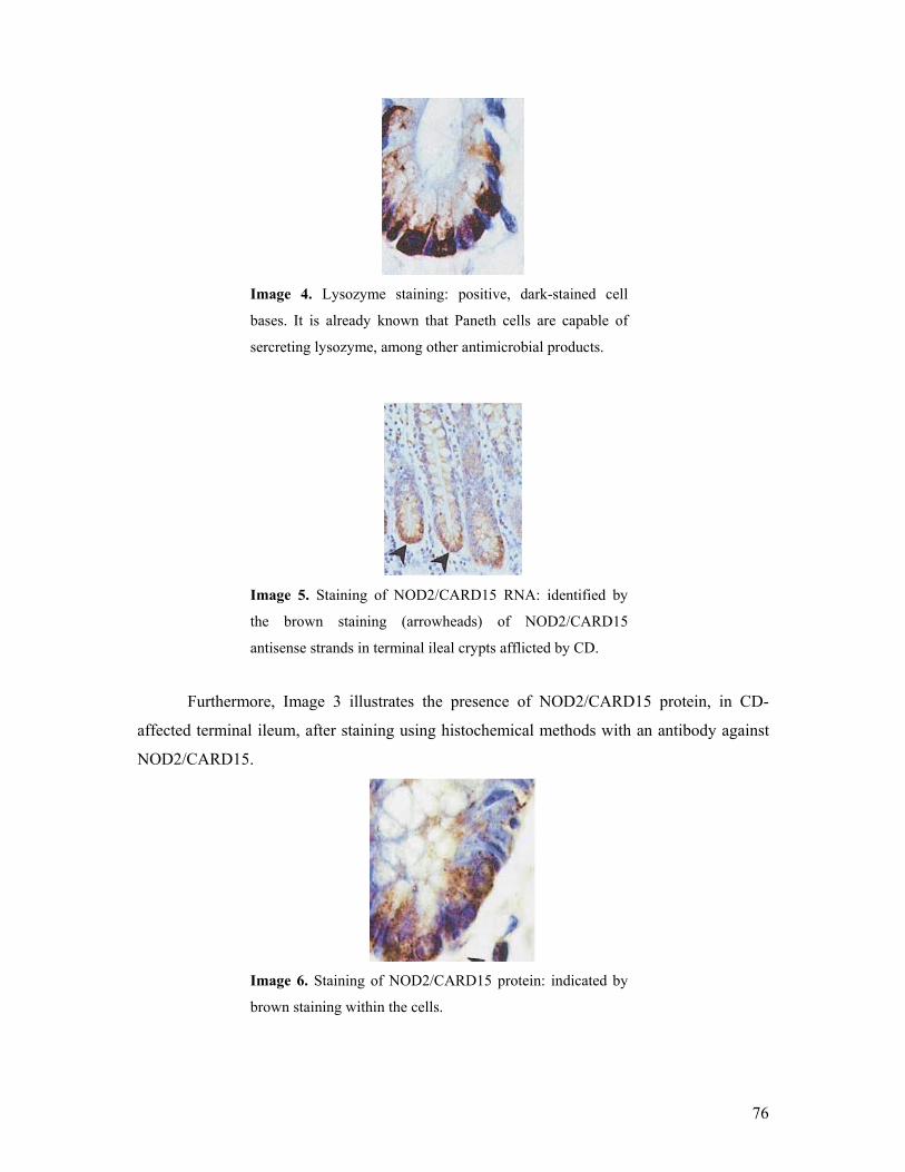
76
Image 4. Lysozyme staining: positive, dark-stained cell
bases. It is already known that Paneth cells are capable of
sercreting lysozyme, among other antimicrobial products.
Image 5. Staining of NOD2/CARD15 RNA: identified by
the brown staining (arrowheads) of NOD2/CARD15
antisense strands in terminal ileal crypts afflicted by CD.
Furthermore, Image 3 illustrates the presence of NOD2/CARD15 protein, in CD-
affected terminal ileum, after staining using histochemical methods with an antibody against
NOD2/CARD15.
Image 6. Staining of NOD2/CARD15 protein: indicated by
brown staining within the cells.
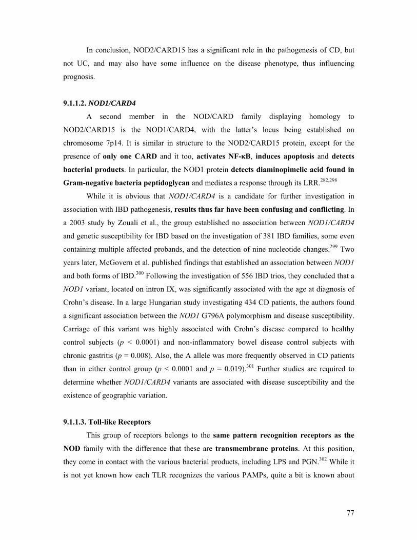
77
In conclusion, NOD2/CARD15 has a significant role in the pathogenesis of CD, but
not UC, and may also have some influence on the disease phenotype, thus influencing
prognosis.
9.1.1.2. NOD1/CARD4
A second member in the NOD/CARD family displaying homology to
NOD2/CARD15 is the NOD1/CARD4, with the latter’s locus being established on
chromosome 7p14. It is similar in structure to the NOD2/CARD15 protein, except for the
presence of only one CARD and it too, activates NF-κB, induces apoptosis and detects
bacterial products. In particular, the NOD1 protein detects diaminopimelic acid found in
Gram-negative bacteria peptidoglycan and mediates a response through its LRR.282,298
While it is obvious that NOD1/CARD4 is a candidate for further investigation in
association with IBD pathogenesis, results thus far have been confusing and conflicting. In
a 2003 study by Zouali et al., the group established no association between NOD1/CARD4
and genetic susceptibility for IBD based on the investigation of 381 IBD families, some even
containing multiple affected probands, and the detection of nine nucleotide changes.299 Two
years later, McGovern et al. published findings that established an association between NOD1
and both forms of IBD.300 Following the investigation of 556 IBD trios, they concluded that a
NOD1 variant, located on intron IX, was significantly associated with the age at diagnosis of
Crohn’s disease. In a large Hungarian study investigating 434 CD patients, the authors found
a significant association between the NOD1 G796A polymorphism and disease susceptibility.
Carriage of this variant was highly associated with Crohn’s disease compared to healthy
control subjects (p < 0.0001) and non-inflammatory bowel disease control subjects with
chronic gastritis (p = 0.008). Also, the A allele was more frequently observed in CD patients
than in either control group (p < 0.0001 and p = 0.019).301 Further studies are required to
determine whether NOD1/CARD4 variants are associated with disease susceptibility and the
existence of geographic variation.
9.1.1.3. Toll-like Receptors
This group of receptors belongs to the same pattern recognition receptors as the
NOD family with the difference that these are transmembrane proteins. At this position,
they come in contact with the various bacterial products, including LPS and PGN.302 While it
is not yet known how each TLR recognizes the various PAMPs, quite a bit is known about
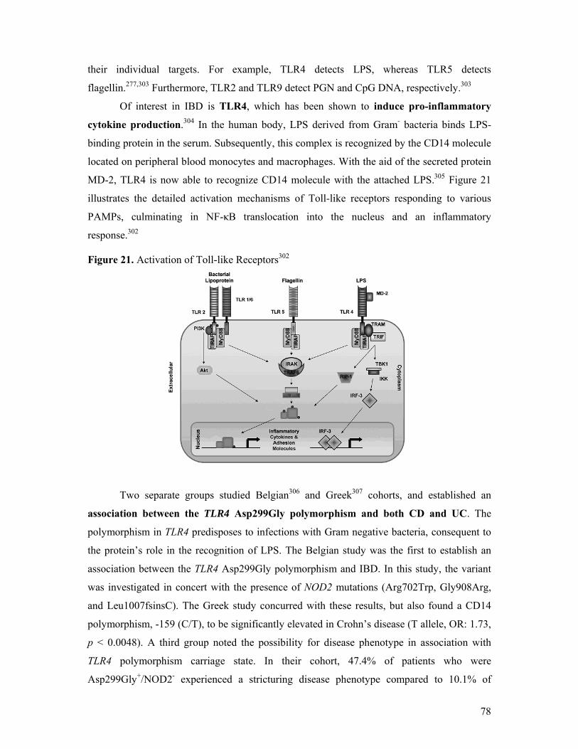
78
their individual targets. For example, TLR4 detects LPS, whereas TLR5 detects
flagellin.277,303 Furthermore, TLR2 and TLR9 detect PGN and CpG DNA, respectively.303
Of interest in IBD is TLR4, which has been shown to induce pro-inflammatory
cytokine production.304 In the human body, LPS derived from Gram- bacteria binds LPS-
binding protein in the serum. Subsequently, this complex is recognized by the CD14 molecule
located on peripheral blood monocytes and macrophages. With the aid of the secreted protein
MD-2, TLR4 is now able to recognize CD14 molecule with the attached LPS.305 Figure 21
illustrates the detailed activation mechanisms of Toll-like receptors responding to various
PAMPs, culminating in NF-κB translocation into the nucleus and an inflammatory
response.302
Figure 21. Activation of Toll-like Receptors302
Two separate groups studied Belgian306 and Greek307 cohorts, and established an
association between the TLR4 Asp299Gly polymorphism and both CD and UC. The
polymorphism in TLR4 predisposes to infections with Gram negative bacteria, consequent to
the protein’s role in the recognition of LPS. The Belgian study was the first to establish an
association between the TLR4 Asp299Gly polymorphism and IBD. In this study, the variant
was investigated in concert with the presence of NOD2 mutations (Arg702Trp, Gly908Arg,
and Leu1007fsinsC). The Greek study concurred with these results, but also found a CD14
polymorphism, -159 (C/T), to be significantly elevated in Crohn’s disease (T allele, OR: 1.73,
p < 0.0048). A third group noted the possibility for disease phenotype in association with
TLR4 polymorphism carriage state. In their cohort, 47.4% of patients who were
Asp299Gly+/NOD2- experienced a stricturing disease phenotype compared to 10.1% of
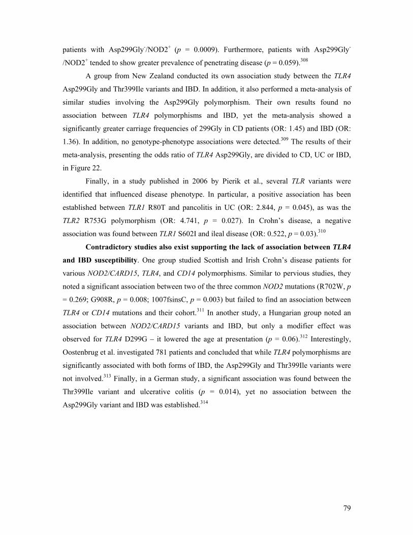
79
patients with Asp299Gly-/NOD2+ (p = 0.0009). Furthermore, patients with Asp299Gly-
/NOD2+ tended to show greater prevalence of penetrating disease (p = 0.059).308
A group from New Zealand conducted its own association study between the TLR4
Asp299Gly and Thr399Ile variants and IBD. In addition, it also performed a meta-analysis of
similar studies involving the Asp299Gly polymorphism. Their own results found no
association between TLR4 polymorphisms and IBD, yet the meta-analysis showed a
significantly greater carriage frequencies of 299Gly in CD patients (OR: 1.45) and IBD (OR:
1.36). In addition, no genotype-phenotype associations were detected.309 The results of their
meta-analysis, presenting the odds ratio of TLR4 Asp299Gly, are divided to CD, UC or IBD,
in Figure 22.
Finally, in a study published in 2006 by Pierik et al., several TLR variants were
identified that influenced disease phenotype. In particular, a positive association has been
established between TLR1 R80T and pancolitis in UC (OR: 2.844, p = 0.045), as was the
TLR2 R753G polymorphism (OR: 4.741, p = 0.027). In Crohn’s disease, a negative
association was found between TLR1 S602I and ileal disease (OR: 0.522, p = 0.03).310
Contradictory studies also exist supporting the lack of association between TLR4
and IBD susceptibility. One group studied Scottish and Irish Crohn’s disease patients for
various NOD2/CARD15, TLR4, and CD14 polymorphisms. Similar to pervious studies, they
noted a significant association between two of the three common NOD2 mutations (R702W, p
= 0.269; G908R, p = 0.008; 1007fsinsC, p = 0.003) but failed to find an association between
TLR4 or CD14 mutations and their cohort.311 In another study, a Hungarian group noted an
association between NOD2/CARD15 variants and IBD, but only a modifier effect was
observed for TLR4 D299G – it lowered the age at presentation (p = 0.06).312 Interestingly,
Oostenbrug et al. investigated 781 patients and concluded that while TLR4 polymorphisms are
significantly associated with both forms of IBD, the Asp299Gly and Thr399Ile variants were
not involved.313 Finally, in a German study, a significant association was found between the
Thr399Ile variant and ulcerative colitis (p = 0.014), yet no association between the
Asp299Gly variant and IBD was established.314
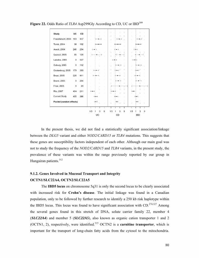
80
Figure 22. Odds Ratio of TLR4 Asp299Gly According to CD, UC or IBD309
In the present thesis, we did not find a statistically significant association/linkage
between the DLG5 variant and either NOD2/CARD15 or TLR4 mutations. This suggests that
these genes are susceptibility factors independent of each other. Although our main goal was
not to study the frequency of the NOD2/CARD15 and TLR4 variants, in the present study, the
prevalence of these variants was within the range previously reported by our group in
Hungairan patients.312
9.1.2. Genes Involved in Mucosal Transport and Integrity
OCTN1/SLC22A4, OCTN2/SLC22A5
The IBD5 locus on chromosome 5q31 is only the second locus to be clearly associated
with increased risk for Crohn’s disease. The initial linkage was found in a Canadian
population, only to be followed by further research to identify a 250 kb risk haplotype within
the IBD5 locus. This locus was found to have significant association with CD.274,327 Among
the several genes found in this stretch of DNA, solute carrier family 22, member 4
(SLC22A4) and member 5 (SLC22A5), also known as organic cation transporter 1 and 2
(OCTN1, 2), respectively, were identified.315 OCTN2 is a carnitine transporter, which is
important for the transport of long-chain fatty acids from the cytosol to the mitochondria.
81
These fatty acids are then β-oxidized316 and a disturbance in this pathway might hamper the
oxidation burst-mediated mechanisms responsible for pathogen destruction. 317 On the other
hand, OCTN1 has a lower affinity for carnitine but transports ergothioneine, which is
exclusively synthesized by mycobacteria and fungi; the transporter’s physiological function
has not yet been completely discovered.318
In 2004, Peletkova et al. proposed that the polymorphism L503F in OCTN1 and the
promoter variant G-207C in the OCTN2 gene were associated with IBD5 findings.
Homozygotes for the so-called TC haplotype had an odds ratio of 3.43-5.14, while patients
carrying both the TC haplotype and a CARD15 risk allele had an odds ratio of 7.28-10.50.328
In a later study, Noble et al. tested three SNPs, IGR2096, IGR2198, and IGR2230, as
well as the OCTN1 L503F (C1672T) polymorphism and OCTN2 G-207C.319 All five
polymorphisms are within the extended IBD5 haplotype first described by Daly et al.320 In
CD patients, the group found a linkage disequilibrium between each investigated SNP and the
disease. Significant associations were found between the OCTN1 (p = 0.0008) and OCTN2
promoter variant (p = 0.0092) and homozygosity for the TC-haplotype with CD. TC-
haplotype homozygosity was observed more often in the CD group than in the control
subjects (80% vs. 68.5%, OR: 1.8, p = 0.0016).319 Contrary to previous reports,274,321 they
found no association between OCTN1/2 polymorphisms and earlier age at onset, disease
phenotype, or disease behavior.319 However, a significant association was noted between the
need for surgery due to CD complications and IGR2198 (OR: 1.91; p = 0.037), OCTN1 (OR:
2.7; p = 0.0007), OCTN2 (OR: 1.77; p = 0.031), and the TC haplotype (OR: 2.2; p = 0.0023).
No associations with UC could be made.319
9.1.3. Other IBD Susceptibility Loci
Following the discovery of NOD2/CARD15, it was clear that other susceptibility loci
must be present, as its mutations accounted for no more than 20% of CD patients.322 Table 8
lists other susceptibility loci as well as the form of IBD to which they are linked. Although
nine genetic loci are depicted in Figure 17, only the first seven loci (IBD1-7) have been
confirmed by strong linkage analysis obeying strict criteria.323 As demonstrated in Table 30
below, some loci are exclusively linked to either CD or UC, while others play a role in both
forms of IBD. It is also quite clear that each locus codes for different genes that play a role in
the pathogenesis of IBD.
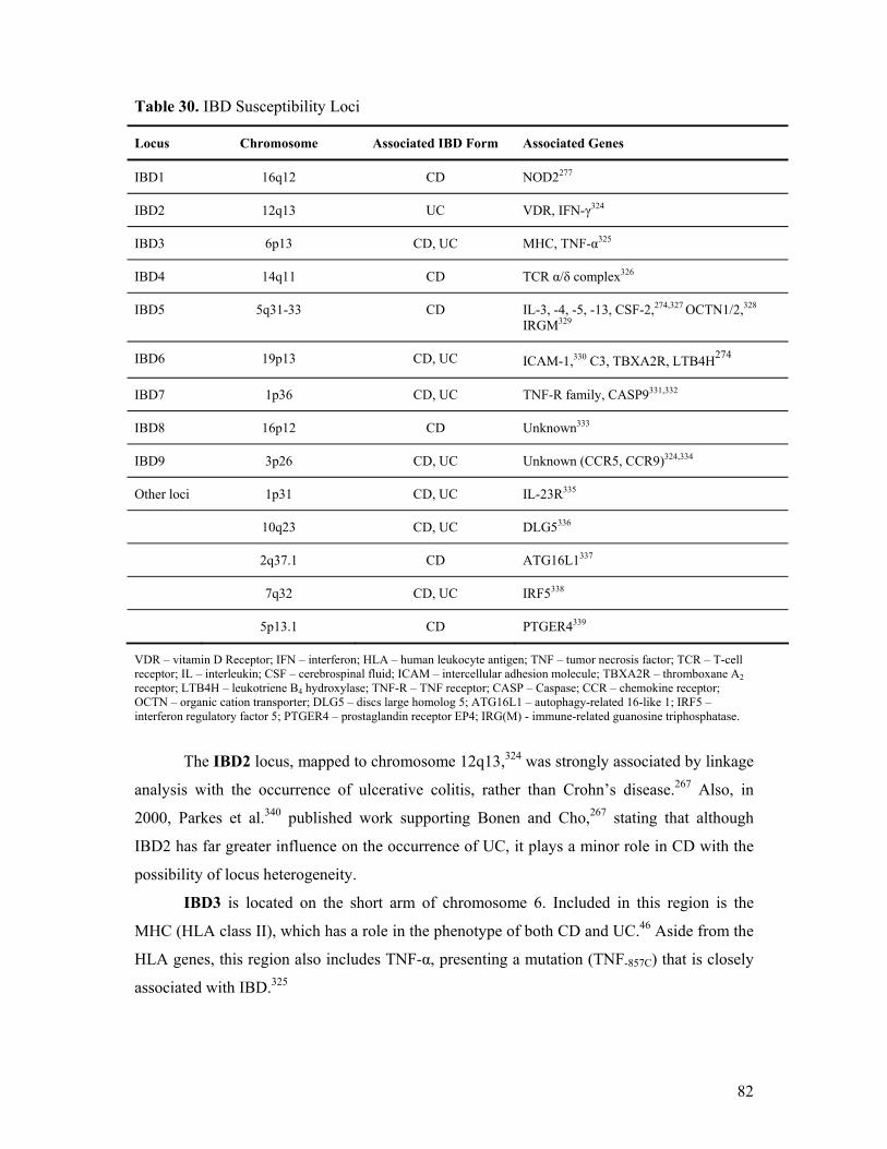
82
Table 30. IBD Susceptibility Loci
Locus Chromosome Associated IBD Form Associated Genes
IBD1 16q12 CD NOD2277
IBD2 12q13 UC VDR, IFN-γ324
IBD3 6p13 CD, UC MHC, TNF-α325
IBD4 14q11 CD TCR α/δ complex326
IBD5 5q31-33 CD IL-3, -4, -5, -13, CSF-2,274,327 OCTN1/2,328 IRGM329
IBD6 19p13 CD, UC ICAM-1,330 C3, TBXA2R, LTB4H274
IBD7 1p36 CD, UC TNF-R family, CASP9331,332
IBD8 16p12 CD Unknown333
IBD9 3p26 CD, UC Unknown (CCR5, CCR9)324,334
Other loci 1p31 CD, UC IL-23R335
10q23 CD, UC DLG5336
2q37.1 CD ATG16L1337
7q32 CD, UC IRF5338
5p13.1 CD PTGER4339
VDR – vitamin D Receptor; IFN – interferon; HLA – human leukocyte antigen; TNF – tumor necrosis factor; TCR – T-cell receptor; IL – interleukin; CSF – cerebrospinal fluid; ICAM – intercellular adhesion molecule; TBXA2R – thromboxane A2 receptor; LTB4H – leukotriene B4 hydroxylase; TNF-R – TNF receptor; CASP – Caspase; CCR – chemokine receptor; OCTN – organic cation transporter; DLG5 – discs large homolog 5; ATG16L1 – autophagy-related 16-like 1; IRF5 – interferon regulatory factor 5; PTGER4 – prostaglandin receptor EP4; IRG(M) - immune-related guanosine triphosphatase.
The IBD2 locus, mapped to chromosome 12q13,324 was strongly associated by linkage
analysis with the occurrence of ulcerative colitis, rather than Crohn’s disease.267 Also, in
2000, Parkes et al.340 published work supporting Bonen and Cho,267 stating that although
IBD2 has far greater influence on the occurrence of UC, it plays a minor role in CD with the
possibility of locus heterogeneity.
IBD3 is located on the short arm of chromosome 6. Included in this region is the
MHC (HLA class II), which has a role in the phenotype of both CD and UC.46 Aside from the
HLA genes, this region also includes TNF-α, presenting a mutation (TNF-857C) that is closely
associated with IBD.325
83
The IBD4 locus, located on the long arm of chromosome 14 and identified as 14q11-
12, is exclusively associated with CD.326
9.1.3.1. HLA Genes & IBD
The first studies to suggest genetic susceptibility in IBD investigated the association
between certain HLA regions and IBD. Some of the most thoroughly researched genes of IBD
included those of the major histocompatability complex (MHC), particularly the human
leukocyte antigen (HLA) cluster on chromosome 6. The HLA genes are divided into two
classes:
Class I: found in all cell types and includes gene products of HLA-A, -B, and -C.
Class II: found only on specialized immune cells and expresses products of the genes
HLA-DP, -DQ, and -DR.
It is the latter, HLA class II, which is of particular interest in the pathogenesis of IBD and
especially of UC.267 A 1999 study by Stokkers et al. 275 found evidence for the following
associations between IBD and HLA genes:
Crohn’s disease:
Positive association with HLA-DR7, -DRB3*0301, -DQ4.
Negative association with HLA-DR2 and -DR3.
Ulcerative colitis:
Positive association with HLA-DR2, -DR9, -DRB1*0103.
Negative association with HLA-DR4.
This study proved especially powerful as it spanned over three decades of publications
concerned with IBD and HLA.
Another aspect in the investigation of HLA genes, involving both classes, is the
correlation with disease phenotype, severity, and extraintestinal manifestations (EIM) of
IBD (eg, arthropathies).46,341 A common EIM is arthritis, of various forms. The first,
migratory pauciarticular large-joint arthritis has been shown to be related to HLA-
DRB1 0103, while chronic, small-joint, symmetrical arthritis is associated with HLA-B 44.
HLA-DRB1 0103 is also coupled with uveitis, as is HLA-B 27.342 In addition, the presence
of the DRB1 0103 allele, though rare, is related to a severe state of disease. This observation
is supported by an early need for colectomy.343
84
9.1.3.2. Interferon Regulatory Factor 5
The interferon regulator factors (IRFs) constitute a family of nine transcription factors
possessing several functions, including defense against pathogens, hematopoietic
development, and cellular survival.338 Structurally, they are all similar at the N-terminal
DNA-binding domain; however, they differ in the activating signals and interaction with
DNA regulatory elements.344 While some IRFs are activated by viral infections, such as IRF 3
and 7, others, for example, IRF5, do not.344,345,346 The latter, is activated by Toll-like
receptors and stimulates the release of pro-inflammatory cytokines, in particular, IL-6, -12,
and TNF-α.338,347 Given the established association between cytokines and IBD as well as the
role of IRF5 in other autoimmune disorders, systemic lupus erythematosus and rheumatoid
arthritis,348,349 Dideberg et al. investigated a possible link between IRF5 and IBD. The group
studied two separate Belgian IBD and control cohorts (Wallonia – CD: 748; UC: 254; control:
241; Leuven – CD: 488; UC: 192; control: 311) against 12 polymrphisms in the IRF5 gene,
and found a significant association between a 5bp insertion-deletion mutation (CGGGG) in
the promoter region and IBD in the Wallonia cohort. The same result was confirmed in the
Leuven patient group. While this polymorphism was associated with both forms of IBD, it
was far stronger in UC (OR: 2.42; p = 5.3 % 10-8) than in CD (p = 6.8 % 10-4). Overall in the
Wallonia cohort, the OR equaled 1.81 (p = 1.9 % 10-5). In the second cohort, from Leuven, the
OR value was 1.59 (p = 3.2 % 10-5).338 It is believed that this particular mutation leads to an
additional, third binding site for the SP1 transcription factor.350
9.1.3.3. Genome-wide Association Scan
The genome-wide association scan (GWAS) is a novel method used to find
associations between certain diseases or conditions and SNPs. It requires a large number of
affected and control subjects, who are genotyped for thousands of polymorphisms across
the entire genome. One condition that must be met is the identification of probable SNPs
related to the investigated condition. Statistical tools are then used to sift through the data and
locate significant associations.351 The Wellcome Trust Case Control Consortium has
exemplified such a wide-breadth and extensive study involving 14 000 patients, 2000 for each
of seven conditions/diseases and 3000 control subjects. Using GWAS, the Consortium
investigated coronary artery disease, hypertension, diabetes mellitus types 1 and 2, bipolar
disorder, rheumatoid arthritis, and Crohn’s disease. Twenty-four independent associations
were found among 6 of the 7 studied patient groups, with Crohn’s disease having nine
associations, the most of any of the investigated diseases or conditions. The figure below
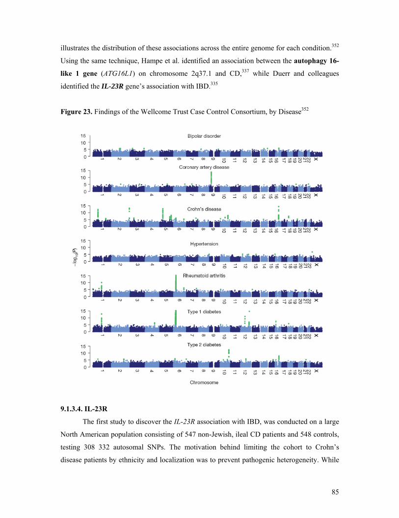
85
illustrates the distribution of these associations across the entire genome for each condition.352
Using the same technique, Hampe et al. identified an association between the autophagy 16-
like 1 gene (ATG16L1) on chromosome 2q37.1 and CD,337 while Duerr and colleagues
identified the IL-23R gene’s association with IBD.335
Figure 23. Findings of the Wellcome Trust Case Control Consortium, by Disease352
9.1.3.4. IL-23R
The first study to discover the IL-23R association with IBD, was conducted on a large
North American population consisting of 547 non-Jewish, ileal CD patients and 548 controls,
testing 308 332 autosomal SNPs. The motivation behind limiting the cohort to Crohn’s
disease patients by ethnicity and localization was to prevent pathogenic heterogeneity. While
86
they found several significant associations, only three were of far greater significance than
the rest. The first two included known CARD15 polymorphisms, while the third was the
Arg381Gln polymorphism in the IL-23R gene (p = 1.56 % 10-3). Nine other markers were
found both in the IL-23R gene and in the intergenic region, adjacent to the IL-12 receptor, β-2
gene. All had significant association values of p < 0.0001. Of interest, the glutamine allele of
Arg381Gln is rarer than the arginine allele, being present in 1.9% of the non-Jewish, ileal CD
cohort versus 7.0% of the control subjects, implying a protective effect.335 In a subsequent
study, 1902 CD, 975 UC, and 1345 control subjects were genotyped for eight SNPs identified
in the North American study. Similarly to the previous study, the research group also found a
significant association between Arg381Gln and CD, versus controls (p = 1.1 % 10-12; OR:
0.38). Furthermore, by examining all phenotypes of CD and UC, and finding no associations
to any single form, they concluded that the IL-23R variant influences susceptibility but not
disease phenotype. Finally, the study further affirmed the power and viability of genome-wide
association scans.353
9.1.3.5. Autophagy Genes
For the detection of autophagy 16-like 1 gene (ATG16L1), a protein in the
autophagosome pathway that processes intracellular bacteria, Hampe et al. screened 735
CD and 368 control subjects for 19 779 nonsynonymous SNPs. Next, they narrowed their
search to 72 SNPs which demonstrated a significance level of p ≤ 0.01. Subsequently, they
were able to find a significant association to the ATG16L1 locus (p = 4.0 % 10-8), for the
T300A SNP.337 A subsequent study by Prescott and colleagues revealed a similar and
significant association between T300A and CD (p = 2.4 % 10-6), as detected in 1236 cases.
In addition, the group also found a specific and significant association with the ileal form of
Crohn’s disease. Finally, the study found a modest but significant association between
ATG16L1 T300A genotype and ulcerative colitis.354
The IRGM gene located on chromosome 5q33.1 was identified through a genome-
wide association scan to be significantly associated with Crohn’s disease (p = 6.6 % 10-4).
Like ATG16L1, it is also involved in the autophagy pathway. Nonetheless, the exact SNPs
that are responsible for the increased disease susceptibility still need to be elucidated.329
87
9.1.3.6. Prostaglandin Receptor EP4
The prostaglandin receptor EP4 (PTGER4) was associated with Crohn’s disease
using a genome-wide association scan. In the study, Libioulle and colleagues first tested 547
CD patients and 928 controls for over 300 000 SNPs. They found three chromosomal regions
with significant p-values ranging from 10-6 to 10-9. Two of the three loci were the IL-23R and
CARD15 genes, while the third was located to a gene desert on 5p13.1. The group further
tested the results involving 5p13.1 on a second cohort consisting of 1266 CD patients, 559
control subjects, and 428 trios, again, establishing a p-value of < 4 % 10-4.339
9.1.3.7. Cytokines
Cytokines are substances produced by various cells and tissue types, even epithelial
cells,355 and have immunomodulating and proinflammatory functions, including the ability to
attract leukocytes, through a chemoattractant gradient, to various tissues.356,357 Over the years,
numerous cytokines and chemokines have come under scrutiny for their role in the
pathogenesis of IBD. This wide array of cytokines includes: IL-1,358 IL-2,44 IL-6,322 IL-8,356
IL-12,276 IL-16,359 IL-17,276 IL-23,360 and TNF-α.361
IL-2 is a mainstay immunoregulatory cytokine.44 Circulating IL-2 levels are below
detection levels during health and disease states; however, IBD T-cells display significant
variation in their response to IL-2. CD T-cells will show exaggerated response while UC T-
cells will respond only slightly.362 Moreover, the IL-2 receptor has also been found to be
upregulated in active cases of IBD.363
A second important cytokine in the pathogenesis of IBD has been identified to be IL-
8, which has been extensively studied for its role. The cytokine is expressed by neutrophils,
macrophages and intestinal epithelial cells, and has been found to be upregulated in both
forms of active disease. Daig et al., using mucosal biopsy specimens, noted a significant
increase in IL-8 expression in macroscopically inflamed bowel segments in CD (p < 0.0001),
UC (p < 0.001) and inflammatory control subjects (p = 0.010).364 Additionally, IL-8 was also
significantly elevated in un-inflamed CD biopsy samples (p < 0.001). Finally, over a decade
ago, Mazzucchelli et al. demonstrated a non-specific but important association of IL-8 with
the histological grade in both forms of active IBD disease.365
IL-12 and -17 are another pair of immunoregulatory cytokines, as reported by Nielsen
et al.276 While their exact role is unknown, mounting evidence suggests an increase in their
mRNA concentration in IBD. Fujino et al. conducted a small-scale study that consisted of
40 tissue samples, 20 each from CD and UC patients, retrieved endoscopically or surgically.
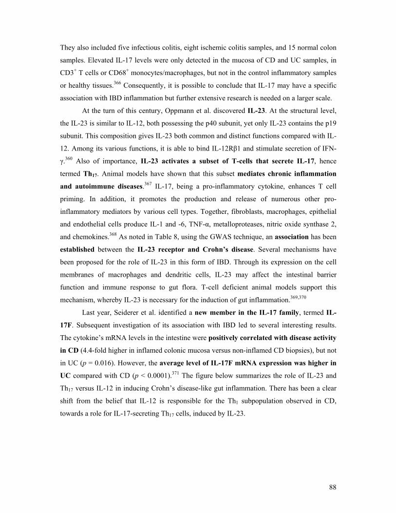
88
They also included five infectious colitis, eight ischemic colitis samples, and 15 normal colon
samples. Elevated IL-17 levels were only detected in the mucosa of CD and UC samples, in
CD3+ T cells or CD68+ monocytes/macrophages, but not in the control inflammatory samples
or healthy tissues.366 Consequently, it is possible to conclude that IL-17 may have a specific
association with IBD inflammation but further extensive research is needed on a larger scale.
At the turn of this century, Oppmann et al. discovered IL-23. At the structural level,
the IL-23 is similar to IL-12, both possessing the p40 subunit, yet only IL-23 contains the p19
subunit. This composition gives IL-23 both common and distinct functions compared with IL-
12. Among its various functions, it is able to bind IL-12Rβ1 and stimulate secretion of IFN-
γ.360 Also of importance, IL-23 activates a subset of T-cells that secrete IL-17, hence
termed Th17. Animal models have shown that this subset mediates chronic inflammation
and autoimmune diseases.367 IL-17, being a pro-inflammatory cytokine, enhances T cell
priming. In addition, it promotes the production and release of numerous other pro-
inflammatory mediators by various cell types. Together, fibroblasts, macrophages, epithelial
and endothelial cells produce IL-1 and -6, TNF-α, metalloproteases, nitric oxide synthase 2,
and chemokines.368 As noted in Table 8, using the GWAS technique, an association has been
established between the IL-23 receptor and Crohn’s disease. Several mechanisms have
been proposed for the role of IL-23 in this form of IBD. Through its expression on the cell
membranes of macrophages and dendritic cells, IL-23 may affect the intestinal barrier
function and immune response to gut flora. T-cell deficient animal models support this
mechanism, whereby IL-23 is necessary for the induction of gut inflammation.369,370
Last year, Seiderer et al. identified a new member in the IL-17 family, termed IL-
17F. Subsequent investigation of its association with IBD led to several interesting results.
The cytokine’s mRNA levels in the intestine were positively correlated with disease activity
in CD (4.4-fold higher in inflamed colonic mucosa versus non-inflamed CD biopsies), but not
in UC (p = 0.016). However, the average level of IL-17F mRNA expression was higher in
UC compared with CD (p < 0.0001).371 The figure below summarizes the role of IL-23 and
Th17 versus IL-12 in inducing Crohn’s disease-like gut inflammation. There has been a clear
shift from the belief that IL-12 is responsible for the Th1 subpopulation observed in CD,
towards a role for IL-17-secreting Th17 cells, induced by IL-23.
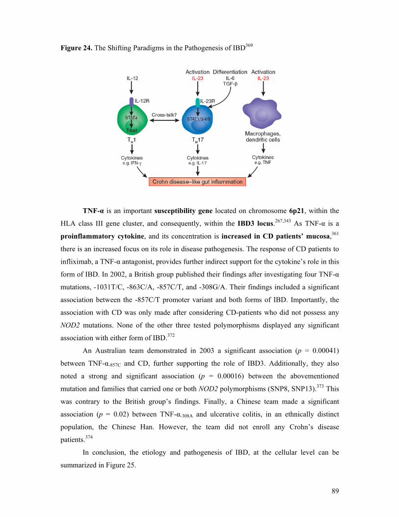
89
Figure 24. The Shifting Paradigms in the Pathogenesis of IBD369
TNF-α is an important susceptibility gene located on chromosome 6p21, within the
HLA class III gene cluster, and consequently, within the IBD3 locus.267,343 As TNF-α is a
proinflammatory cytokine, and its concentration is increased in CD patients’ mucosa,361
there is an increased focus on its role in disease pathogenesis. The response of CD patients to
infliximab, a TNF-α antagonist, provides further indirect support for the cytokine’s role in this
form of IBD. In 2002, a British group published their findings after investigating four TNF-α
mutations, -1031T/C, -863C/A, -857C/T, and -308G/A. Their findings included a significant
association between the -857C/T promoter variant and both forms of IBD. Importantly, the
association with CD was only made after considering CD-patients who did not possess any
NOD2 mutations. None of the other three tested polymorphisms displayed any significant
association with either form of IBD.372
An Australian team demonstrated in 2003 a significant association (p = 0.00041)
between TNF-α-857C and CD, further supporting the role of IBD3. Additionally, they also
noted a strong and significant association (p = 0.00016) between the abovementioned
mutation and families that carried one or both NOD2 polymorphisms (SNP8, SNP13).373 This
was contrary to the British group’s findings. Finally, a Chinese team made a significant
association (p = 0.02) between TNF-α-308A and ulcerative colitis, in an ethnically distinct
population, the Chinese Han. However, the team did not enroll any Crohn’s disease
patients.374
In conclusion, the etiology and pathogenesis of IBD, at the cellular level can be
summarized in Figure 25.
90
Figure 25. Key Molecular Mechanisms in the Pathogenesis of IBD375
9.2. The Importance of the DLG5, MDR1 and ABCG2 Mutations in Hungarian Patients
with IBD
These were the first studies to be published on the frequency of DLG5, MDR1, and
ABCG2 variant alleles in Hungarian IBD patients. In total, we tested five polymorphisms
against several variables including: disease susceptibility, genotype-phenotype correlation,
response to infliximab therapy or steroids, epistasis with NOD2/CARD15 risk alleles, and
need for surgery. We established numerous positive and negative associations and, while
some concurred with previous studies, others were in stark contradiction to formerly
published results.
In our studies, we found no genetic susceptibility associations between either form of
IBD and any of the tested polymorphisms, DLG5 R30Q, MDR1 C3435T or G2677T/A, and
ABCG2 G34A or C421A. The MDR1 C3435T and G2677T/A did not display any significant
association with disease susceptibility in our cohort; however, a tendency was observed for
the G2677 variant allele to be overtransmitted in CD patients versus control subjects (OR:
91
1.29; 95% CI: 0.97-1.72,; p = 0.08). These results are in agreement with Brant et al. who have
noted a significant association between the common allele (893Ser) and IBD in a case-control
analysis (p = 0.002) and a pedigree disequilibrium test (p = 0.00020-0.00030). Similar to our
results, the group did not associate C3435T with IBD.376 However, our results do not concur
with two other studies conducted in Scottish Caucasian and Spanish Caucasian populations.
In the former, Ho et al. found a significant association between the 3435TT genotype and
ulcerative colitis (34.6% vs. 26.5%; OR: 1.60; 95% CI: 1.04-2.44; p = 0.04) versus control
subjects, yet no association with CD was detected. Furthermore, they correlated this variant
allele with extensive UC (42.4% vs. 26.5%; OR: 2.64; 95% CI: 1.34-4.99; p = 0.003). Unlike
in our study, the G2677T SNP was not associated with either form of IBD.377 In the Spanish
population, a different conclusion was reached. A significant association (p = 0.021) was
made between C3435T and CD patients versus the control group. In addition, no associations
were made with either disease behavior or location.378 Schwab et al. also reported of increased
frequencies of the T-allele (p = 0.049) and TT-genotype (p = 0.045) in 149 UC patients but
found not association with CD, in their German cohort.379 While all of these studies have
reached different results, the possibility also exists for other MDR1 gene variants to be
associated with IBD.
An important meta-analysis was conducted by Annese and colleagues in 2006, where
they examined data concerning the two MDR1 polymorphisms of IBD, C3435T and
G2677T/A. The group collected data on the distribution of C3435T from six studies and on
G2677T/A from five studies. The data for C3435T are presented in Table 31. Taking these
data into consideration, as well as that for G2677T/A (not shown), the group calculated the p
values and odds ratios for the alleles and genotypes (Table 32).
Evaluating the pooled data in Table 32, one can observe a significant difference in the
association between C3435T, and CD and UC. In Crohn’s disease, no association was found
with this variant (T vs. C: OR: 0.968; p = 0.519). On the contrary, ulcerative colitis studies
demonstrated a significant difference between the T and C alleles (OR: 1.170; p = 0.002), as
well as between the TT and CC genotypes (OR: 1.332; p = 0.008). This is in partial
contradiction to our study, where no association was found between C3435T and IBD. With
regards to the G2677T/A variant, no significant association was found with either form of
IBD. Nevertheless, our study presented a trend for greater frequency of the G2677 variant
allele in CD.
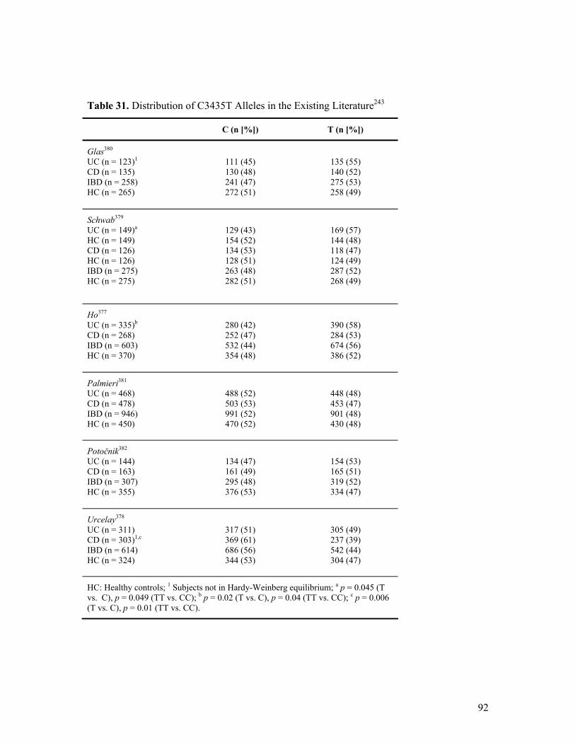
92
Table 31. Distribution of C3435T Alleles in the Existing Literature243
C (n [%]) T (n [%])
Glas380 UC (n = 123)1 CD (n = 135) IBD (n = 258) HC (n = 265)
111 (45) 130 (48) 241 (47) 272 (51)
135 (55) 140 (52) 275 (53) 258 (49)
Schwab379 UC (n = 149)a HC (n = 149) CD (n = 126) HC (n = 126) IBD (n = 275) HC (n = 275)
129 (43) 154 (52) 134 (53) 128 (51) 263 (48) 282 (51)
169 (57) 144 (48) 118 (47) 124 (49) 287 (52) 268 (49)
Ho377 UC (n = 335)b CD (n = 268) IBD (n = 603) HC (n = 370)
280 (42) 252 (47) 532 (44) 354 (48)
390 (58) 284 (53) 674 (56) 386 (52)
Palmieri381 UC (n = 468) CD (n = 478) IBD (n = 946) HC (n = 450)
488 (52) 503 (53) 991 (52) 470 (52)
448 (48) 453 (47) 901 (48) 430 (48)
Potočnik382 UC (n = 144) CD (n = 163) IBD (n = 307) HC (n = 355)
134 (47) 161 (49) 295 (48) 376 (53)
154 (53) 165 (51) 319 (52) 334 (47)
Urcelay378 UC (n = 311) CD (n = 303)1,c IBD (n = 614) HC (n = 324)
317 (51) 369 (61) 686 (56) 344 (53)
305 (49) 237 (39) 542 (44) 304 (47)
HC: Healthy controls; 1 Subjects not in Hardy-Weinberg equilibrium; a p = 0.045 (T vs. C), p = 0.049 (TT vs. CC); b p = 0.02 (T vs. C), p = 0.04 (TT vs. CC); c p = 0.006 (T vs. C), p = 0.01 (TT vs. CC).
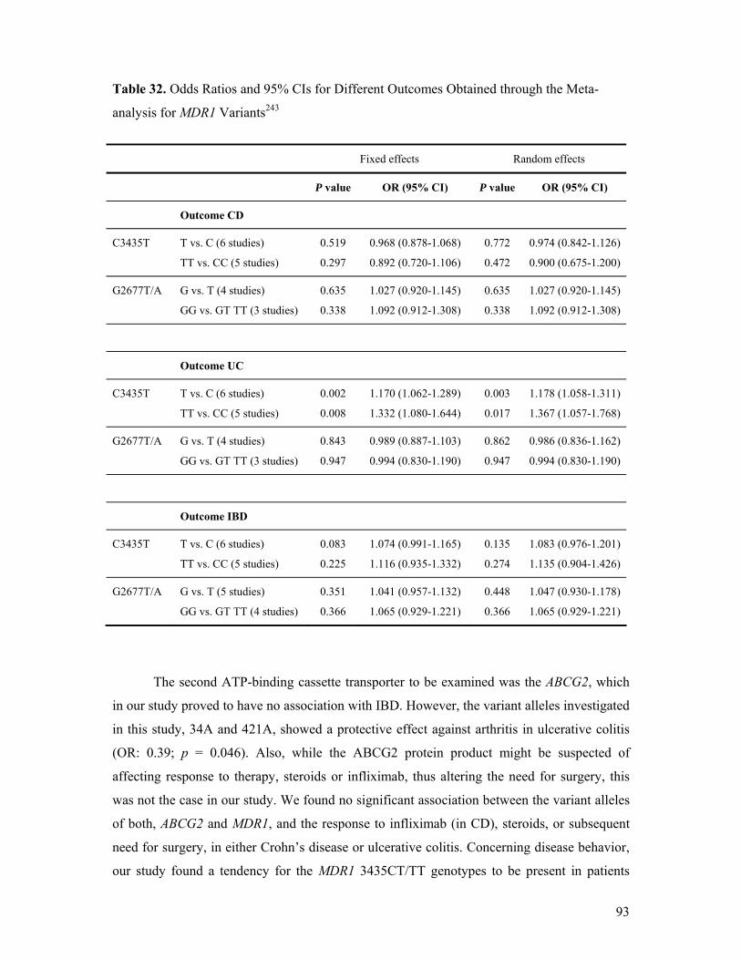
93
Table 32. Odds Ratios and 95% CIs for Different Outcomes Obtained through the Meta-
analysis for MDR1 Variants243
Fixed effects Random effects
P value OR (95% CI) P value OR (95% CI)
Outcome CD
C3435T T vs. C (6 studies)
TT vs. CC (5 studies)
0.519
0.297
0.968 (0.878-1.068)
0.892 (0.720-1.106)
0.772
0.472
0.974 (0.842-1.126)
0.900 (0.675-1.200)
G2677T/A G vs. T (4 studies)
GG vs. GT TT (3 studies)
0.635
0.338
1.027 (0.920-1.145)
1.092 (0.912-1.308)
0.635
0.338
1.027 (0.920-1.145)
1.092 (0.912-1.308)
Outcome UC
C3435T T vs. C (6 studies)
TT vs. CC (5 studies)
0.002
0.008
1.170 (1.062-1.289)
1.332 (1.080-1.644)
0.003
0.017
1.178 (1.058-1.311)
1.367 (1.057-1.768)
G2677T/A G vs. T (4 studies)
GG vs. GT TT (3 studies)
0.843
0.947
0.989 (0.887-1.103)
0.994 (0.830-1.190)
0.862
0.947
0.986 (0.836-1.162)
0.994 (0.830-1.190)
Outcome IBD
C3435T T vs. C (6 studies)
TT vs. CC (5 studies)
0.083
0.225
1.074 (0.991-1.165)
1.116 (0.935-1.332)
0.135
0.274
1.083 (0.976-1.201)
1.135 (0.904-1.426)
G2677T/A G vs. T (5 studies)
GG vs. GT TT (4 studies)
0.351
0.366
1.041 (0.957-1.132)
1.065 (0.929-1.221)
0.448
0.366
1.047 (0.930-1.178)
1.065 (0.929-1.221)
The second ATP-binding cassette transporter to be examined was the ABCG2, which
in our study proved to have no association with IBD. However, the variant alleles investigated
in this study, 34A and 421A, showed a protective effect against arthritis in ulcerative colitis
(OR: 0.39; p = 0.046). Also, while the ABCG2 protein product might be suspected of
affecting response to therapy, steroids or infliximab, thus altering the need for surgery, this
was not the case in our study. We found no significant association between the variant alleles
of both, ABCG2 and MDR1, and the response to infliximab (in CD), steroids, or subsequent
need for surgery, in either Crohn’s disease or ulcerative colitis. Concerning disease behavior,
our study found a tendency for the MDR1 3435CT/TT genotypes to be present in patients
94
with fistulizing disease (fistulizing: 30.3% vs. non-fistulizing: 21.7%; OR: 1.57; 95% CI:
0.92-2.76; p = 0.09). However, this was the only association we found for C3435T and could
not replicate a previous association with extensive UC.377
The DLG5 protein is believed to be a scaffolding protein, playing a role in the
intestinal barrier function. In our study, we found no association between the DLG5 R30Q
variant and IBD, or its sub-types. Rather, we noted a lower carriage frequency in CD patients,
contrary to Stoll and colleagues who reported overtransmission of this variant in IBD as a
whole, and in CD patients.249 Daly et al., in a joint Canada-UK study, succeeded in replicating
the DLG5 R30Q association with IBD using a Canadian/Italian case-control cohort, yet a
second cohort from the UK failed to present the same association. Finally, a third Canadian
family cohort demonstrated significant overtransmission of the risk allele (p = 0.018).383 Two
additional studies, one in a Scottish population384 and another in a German cohort,385 were not
able to establish the variant allele’s overtransmission, as noted in our study. Importantly, the
variant allele’s frequency in the control group was higher (15%) than in previous reports.
As has been demonstrated for NOD2/CARD15 mutations in Japanese patients,386 it is
possible that ethnic variation is a factor in DLG5 polymorphisms. While this has not yet been
proven, it can be observed in the frequency differences between Caucasian patients and other
ethnic groups. For example, in one study that examined 484 Japanese CD patients and 345
control subjects, the G113A polymorphism was completely absent, yet a significant
association (p = 0.023) was noted for another DLG5 variant (rs3758462).
We were unable to identify any significant genotype-phenotype associations in CD, in
agreement with a previous study by Török et al.385 Still, carriage of G113A, while not
associated with response to infliximab, was independently associated with steroid resistance.
One possible explanation for this observation may be derived from reviewing DLG5’s
function. The protein is believed to be a scaffolding protein responsible for maintaining
epithelial cell structure and integrity, as well as intracellular signal transduction an
intercellular contact. Consequently, it may be hypothesized that DLG5 is involved in the
dysfunction of the epithelial barrier function, thus influencing intestinal permeability and the
response to luminal antigens. As such, it is likely that response to steroid treatment is also
influenced in this manner. In silico research has suggested that the G113A variant prevents
the protein’s proper folding. Similarly to the observations made in CD, no genotype-
phenotype associations were made between the DLG5 variant and UC, yet a trend towards
more extensive disease was noted (OR: 2.1; 95% CI: 0.95-4.4; p = 0.07). Finally, contrary to
the findings by two other groups,249,387 we could not identify epistasis between DLG5
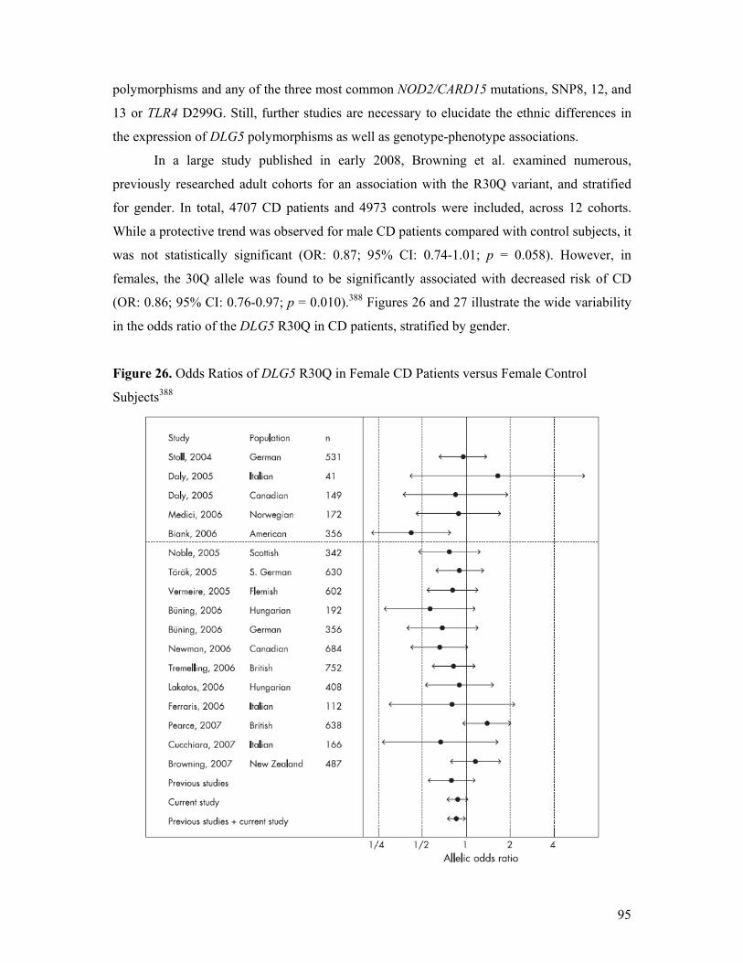
95
polymorphisms and any of the three most common NOD2/CARD15 mutations, SNP8, 12, and
13 or TLR4 D299G. Still, further studies are necessary to elucidate the ethnic differences in
the expression of DLG5 polymorphisms as well as genotype-phenotype associations.
In a large study published in early 2008, Browning et al. examined numerous,
previously researched adult cohorts for an association with the R30Q variant, and stratified
for gender. In total, 4707 CD patients and 4973 controls were included, across 12 cohorts.
While a protective trend was observed for male CD patients compared with control subjects, it
was not statistically significant (OR: 0.87; 95% CI: 0.74-1.01; p = 0.058). However, in
females, the 30Q allele was found to be significantly associated with decreased risk of CD
(OR: 0.86; 95% CI: 0.76-0.97; p = 0.010).388 Figures 26 and 27 illustrate the wide variability
in the odds ratio of the DLG5 R30Q in CD patients, stratified by gender.
Figure 26. Odds Ratios of DLG5 R30Q in Female CD Patients versus Female Control
Subjects388
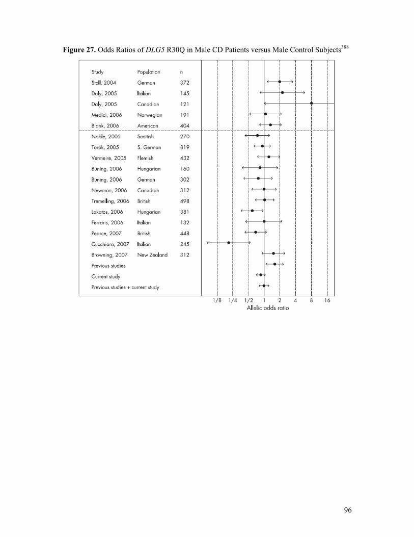
96
Figure 27. Odds Ratios of DLG5 R30Q in Male CD Patients versus Male Control Subjects388
97
10. SCIENTIFIC CONTRIBUTIONS TO THE FIELD:
1. These are the first studies to examine the DLG5, ABCG2, and MDR1 polymorphisms
in the Hungarian IBD population. In one respect, it is yet another addition to the
growing body of knowledge concerning ethnic differences in IBD susceptibility.
2. In our cohort, we did not find any association between the variants of ABCG2, MDR1
or DLG5 and disease susceptibility. However, one variant of the MDR1 gene, G2677,
showed a trend for association with Crohn’s disease (OR: 1.29, p = 0.08).
3. Carriage of ABCG2 showed a significant protective effect against arthritis in patients
with ulcerative colitis (OR: 0.39; p = 0.046).
4. DLG5 113A carriage appeared to have a protective effect in Crohn’s disease patients
(OR: 0.67; p = 0.06).
5. DLG5 demonstrated a trend for association with extensive disease in patients with
ulcerative colitis (OR: 2.1; p = 0.07). However, no other genotype-phenotype
associations could be made.
6. DLG5 R30Q was associated with steroid resistance in patients with Crohn’s disease
(OR: 2.4; p = 0.013). After correcting for various confounding factors, the 30Q variant
was still significantly associated with steroid resistnace (p = 0.02). In addition,
perianal disease and frequent relapses were independently associated with steroid
resistance (p = 0.01 and p = 0.001, respectively). Contrary to other reports, we could
not associate MDR1 polymorphisms with steroid resistance.
7. None of the tested variants demonstrated any association with clinical response to
short-term infliximab therapy in the sub-cohort of CD patients.
98
ACKNOWLEDGEMENTS: I would like to thank Dr Peter Lakatos for all his guidance and support during the past several
years. Without him, this dissertation would not have been realized. I am also grateful for his
inspiration that drove me to further my medical and academic achievements. I would also like
to extend my sincere appreciation to the members of the Hungarian IBD Study group who
contributed through their expertise, assistance, and hard work. Prof Mandl and Prof Szathmari
deserve many thanks for accepting me to their PhD School and the 1st Dept of Internal
Medicine, respectively. Finally, I wish to thank my fiancée, Anais, for her ongoing support,
input, and assistance.
99
PUBLICATIONS DIRECTLY RELATED TO THIS THESIS: 1. Lakatos PL, Fischer S (joint first authors), Claes K, Kovacs A, Molnar T, Altorjay I, Demeter P,
Tulassay Z, Palatka K, Papp M, Rutgeerts P, Szalay F, Papp J, Hungarian IBD Study Group, Vermeire S, Lakatos L. DLG5 R30Q is not associated with inflammatory bowel disease in Hungarian IBD patients, but predicts clinical response to steroids in Crohn’s disease. Inflamm Bowel Dis. 2006;12:362-368.
Citations: 11 IF: 3.912
2. Fischer S, Lakatos PL, Lakatos L, Kovacs A, Molnar T, Altorjay I, Papp M, Szilvasi A, Tulassay Z, Osztovits J, Papp J, Demeter P, Schwab R, Tordai A, Andrikovics H. The ATP-binding Cassette Transporter ABCG2 (BCRP) and ABCB1 (MDR1) variants are not associated with disease susceptibility and disease phenotype in Hungarian patients with Inflammatory Bowel Diseases. Scand J Gastroenterol. 2007;42:726-733.
Citations: 1 IF: 1.869
3. Lakatos PL, Fischer S, Lakatos L, Gal I, Papp J. Current concept on the pathogenesis of IBD: Crosstalk between genetic and microbial factors. Pathogenic bacteria, altered bacterial sensing or changes in mucosal integrity take “toll”? World J Gastroenterol. 2006;12:1829-1840.
Citations: 16
PUBLICATIONS NOT DIRECTLY RELATED TO THIS THESIS:
1. Szamosi T, Lakatos PL (joint first authors), Hungarian IBD Study Group (Study group: Fischer S), Szilvasi A, Lakatos L, Kovacs A, Molnar T, Altorjay I, Papp M, Szabo O, Satori A, Tulassay Z, Miheller P, Horvath HC, Papp J, Tordai A, Andrikovics H. NFKBIA 3’UTR and NFKB1 -94ins/delATTG variants in Hungarian IBD patients are not associated with disease susceptibility, response to medical therapy or need for surgery; the 3’UTR variant is associated with extensive colitis. Dig Dis Sci. (Accepted for publication)
IF: 1.448
2. Lakatos PL, Szamosi T, Szilvasi A, Molnar E, Lakatos L, Kovacs A, Molnar T, Altorjay I, Papp M, Tulassay Z Miheller P, Papp J, Hungarian IBD Study Group (Study group: Fischer S), Tordai A, Andrikovics H. ATG16L1 and IL-23 receptor (IL-23R) genes are associated with disease susceptibility in Hungarian CD patients. Dig Liver Dis. (Epub ahead of print)
IF: 2.000
3. Lakatos PL, Altorjay I, Mándi Y, Lakatos L, Tumpek J, Kovacs A, Molnar T, Tulassay Z, Miheller P, Palatka K, Tamas Szamosi T, Fischer S, Papp J, the Hungarian IBD Study Group, Papp M. Interaction between seroreactivity to microbial antigens and genetics in Crohn’s disease: is there a role for defensins? Tissue Antigens. 2008;71(6):552-559.
IF: 2.462
4. Papp M, Altorjay I, Dotan N, Palatka K, Ildiko Foldi I, Judit Tumpek J, Sipka S, Udvardy M, Dinya T, Lakatos L, Kovacs A, Molnar T, Zsolt Tulassay Z, Miheller P, Gary L. Norman GL, Szamosi T, Janos Papp J, Hungarian IBD Study Group (Study group: Fischer S), Lakatos PL. New serological markers for inflammatory bowel disease are associated with earlier age at onset, complicated disease behavior, risk for surgery, and NOD2/CARD15 genotype in a Hungarian IBD cohort. Am J Gastroenterol. 2008;103:665-681. IF: 5.608
100
5. Kocsis AK, Lakatos PL, Somogyvari F, Fuszek P, Papp J, Fischer S, Szamosi T, Lakatos L, Kovacs A, Hofner P, Mandi Y. Association of β-defensin 1 single nucleotide polymorphisms with Crohn’s disease. Scand J Gastroenterol. 2008;43:299-307.
IF: 1.869
6. Molnar T, Hofner P, Nagy F, Lakatos PL, Hungarian IBD Study Group, Fischer S, Lakatos L, Kovacs A, Altorjay I, Papp M, Palatka K, Demeter P, Tulassay Z, Nyari T, Miheller P, Papp J, Mandi Y, Lonovics J. NOD1 gene E266K polymorphism is associated with disease susceptibility but not with disease phenotype or NOD2/CARD15 in Hungarian patients with Crohn’s disease. Dig Liver Dis. 2007;39;1064-1070.
IF: 2.000
7. Lakatos PL, Hitre E, Szalay F, Zinober K, Fuszek P, Lakatos L, Fischer S, Osztovits J, Gemela O, Veres G, Papp J, Ferenci P. Common NOD2/CARD15 variants are not associated with susceptibility or the clinicopathologic characteristics of sporadic colorectal cancer in Hungarian patients. BMC Cancer. 2007;7:54.
Citations: 1 IF: 2.360
8. Papp M*, Lakatos PL* (joint first authors), Hungarian IBD Study Group, Palatka K, Foldi I, Udvardy M, Harsfalvi J, Tornai I, Vitalis Z, Dinya T, Kovacs A, Molnar T, Demeter P, Papp J, Lakatos L, Altorjay I (Study group: Fischer S). Haptoglobin polymorphisms are associated with Crohn’s disease, disease behaviour and extraintestinal manifestations in Hungarian patients. Dig Dis Sci. 2007;52:1279-1284.
Citations: 1 IF: 1.448
9. Papp M, Lakatos PL, Palatka K, Foldi I, Udvardy M, Harsfalvi J, Tornai I, Vitalis Z, Dinya T, Kovacs A, Molnar T, Demeter P, Papp J, Lakatos L, Altorjay I (Study group: Fischer S). Haptoglobin polimorfizmus vizsgálata gyulladásos bélbetegségekben. Orv Hetil. 2006;147:1745-1750.
10. Papp M, Altorjay I, Norman GL, Shums Z, Palatka K, Vitalis Z, Foldi I, Lakos G, Tumpek J, Udvardy ML, Harsfalvi J, Fischer S, Lakatos L, Kovacs A, Bene L, Tamas Molnar T, Tulassay Z, Miheller P, Veres G, Janos Papp J, Lakatos PL. Seroreactivity to microbial components in Crohn’s disease is associated with ileal involvement, non-inflammatory disease behaviour and NOD2/CARD15 genotype, but not with risk for surgery in a Hungarian cohort of IBD patient. Inflamm Bowel Dis. 2007;13:984-992.
Citations: 4 IF: 3.912
11. Lakatos PL, Fischer S, Lakatos L. Is the epidemiology of inflammatory bowel disease changing in Eastern Europe? Scand J Gastroenterol. 2006;41:870-872.
Citations: 1 IF: 1.869
12. Lakatos L, Mester G, Erdelyi Z, David G, Pandur T, Balogh M, Fischer S, Vargha P, Lakatos PL. Risk factors for ulcerative colitis-associated colorectal cancer in a Hungarian cohort of ulcerative colitis patients. Results of a population-based study. Inflamm Bowel Dis. 2006;12:205-211.
Citations: 10 IF: 3.912
13. Lakatos L, Mester G, Erdelyi Z, David G, Pandur T, Balogh M, Fischer S, Vargha P, Lakatos PL. A colorectális rák rizikófaktorai colitis ulcerosas betegekben populációs alapú vizsgálat eredményei alapján. Orv Hetil. 2006;147:175-181.
Citations: 3
14. Fuszek P, Horvath HC, Speer G, Papp J, Haller P, Fischer S, Halasz J, Jaray B, Szekely E, Schaff Z, Papp A, Bursics A, Harsanyi L, Lukovich P, Kupcsulik P, Hitre E, Lakatos PL. Lokalizáció és életkor változás a colorectalis rákos betegben 1993 és 2004. Az előrehaladott
101
esetek magas száma alátámasztja a colorectalis rákszűrés szükségességét Magyarországon. Orv Hetil. 2006;147:741-746.
Citations: 1
15. Fuszek P, Horvath HC, Speer G, Papp J, Haller P, Fischer S, Halasz J, Jaray B, Szekely E, Schaff Z, Papp A, Bursics A, Harsanyi L, Lukovich P, Kupcsulik P, Hitre E, Lakatos PL. Location and age at onset of colorectal cancer in Hungarian patients between 1993-2004. The high number of advanced cases supports the need for a colorectal cancer screening program in Hungary. Anticancer Res. 2006;26:527-532.
Citations: 3 IF: 1.479
16. Lakatos PL, Fekete S, Horanyi M, Fischer S, Abonyi ME. Development of multiple myeloma in a patient with chronic hepatitis C: A case report and review of the literature. World J Gastroenterol. 2006;12:2297-2300.
Impact Factors without abstracts: 36.148 Citations: 52
102
ABSTRACTS: 1. Papp M, Altorjay I, Norman GL, Sums Z, Palatka K, Vitalis Z, Foldi I, Lakos G, Tumpek J,
Udvardy ML, Harsfalvi J, Fischer S, Lakatos L, Kovacs A, Bene L, Molnar T, Tulassay Z, Miheller P, Hungarian IBD Study Group, Lakatos PL. New serological markers for inflammatory bowel disease are associated with earlier age at onset, complicated disease behavior, risk for surgery and NOD2/CARD15 genotype in a Hungarian IBD cohort. JCC 2008; 2(suppl1):253.
2. Lakatos PL, Kocsis AK, Somogyvari F, Fuszek P, Papp J, Fischer S, Szamosi T, Lakatos L, Kovacs A, Hofner P, Mandi Y. Association of β-defensin 1 Single Nucleotide Polymorphisms with Crohn’s disease. JCC 2008; 2(suppl1):236.
3. Lakatos PL, Fischer S, Hitre E, Szalay F, Zinober K, Fuszek P, Lakatos L, Gemela O, Papp J, Ferenci P. Common NOD2/CARD15 variants are not associated with susceptibility or clinicopathologic characteristics of sporadic colorectal cancer in Hungarian patients. Gut 2007; 56(supplIII):A247.
4. Papp M, Altorjay I, Norman GL, Sums Z, Palatka K, Vitalis Z, Foldi I, Lakos G, Tumpek J, Udvardy ML, Harsfalvi J, Fischer S, Lakatos L, Kovacs A, Bene L, Molnar T, Tulassay Z, Miheller P, Hungarian IBD Study Group, Lakatos PL. Sero-reactivity to microbial components in Crohn’s disease is associated with ileal involvement, non-inflammatory disease behaviour and NOD2/CARD15 genotype, but not response to medical therapy or risk for surgery. Gut 2007; 56(supplIII):A141.
5. Molnar T, Hofner P, Nagy F, Lakatos PL, Fischer S, Lakatos L, Kovacs A, Altorjay I, Papp M, Palatka K, Demeter P, Demeter P, Tulassay Z, Miheller P, Papp J, Mandi Y, Lonovics J. NOD1 gene E266K polymorphism is associated with disease susceptibility but not with disease phenotype or NOD2/CARD15 in Hungarian patients with Crohn’s disease. Gut 2007; 56(supplIII):A134.
6. Molnar T, Hofner P, Nagy F, Lakatos PL and the Hungarian IBD Study Group, Fischer S, Lakatos L, Kovacs A, Altorjay I, Papp M, Palatka K, Demeter P, Tulassay Z, Nyari T, Miheller P, Papp J, Mandi Y, Lonovics J. NOD1 gene E266K (G796A) polymorphism is associated with disease susceptibility but not with disease phenotype or NOD2/CARD15 in Hungarian patients with Crohn’s disease. Z Gastroenterol. 2007;45:437.
7. Gemela O, Hitre E, Szalay F, Zinober K, Fuszek P, Lakatos L, Fischer S, Osztovits J, Veres G, Papp J, Ferenci P, Lakatos PL. Common NOD2/CARD15 variants are not associated with susceptibility or the clinicopathologic characteristics of sporadic colorectal cancer in Hungarian patients. Z Gastroenterol. 2007;45:427.
8. Fischer S, Lakatos PL, Hungarian IBD Study Group, Lakatos L, Kovacs A, Molnar T, Altorjay I, Papp M, Szilvasi A, Tulassay Z, Osztovits J, Papp J, Demeter P, Schwab R, Tordai A, Andrikovics H. The ATP-binding Cassette Transporter ABCG2 (BCRP) and ABCB1 (MDR1) variants are not associated with disease susceptibility, disease phenotype, response to medical therapy or need for surgery in Hungarian patients with Inflammatory Bowel Diseases. Z Gastroenterol. 2007;45:427.
9. Fischer S, Lakatos PL, Hungarian IBD Study Group, Lakatos L, Kovacs A, Molnar T, Altorjay I, Papp M, Szilvasi A, Tulassay Z, Osztovits J, Papp J, Demeter P, Schwab R, Tordai A, Andrikovics H. The ATP-binding Cassette Transporter ABCG2 (BCRP) and ABCB1 (MDR1) variants are not associated with disease susceptibility, disease phenotype, response to medical therapy or need for surgery in Hungarian patients with Inflammatory Bowel Diseases. Gastroenterology. 2007; 132(suppls):M2098.
10. Lakatos PL, Fischer S, Lakatos L, Kovacs A, Molnar T, Altorjay I, Papp M, Szilvasi A, Tulassay Z, Osztovits J, Papp J, Demeter P. The ATP-binding Cassette Transporter ABCG2 (BCRP) and ABCB1 (MDR1) variants are not associated with disease susceptibility, disease phenotype, response to medical therapy or need for surgery in Hungarian patients with Inflammatory Bowel Diseases. Journal of Crohn’s and Colitis. 2007; 1(suppl1):35.
11. Papp M, Altorjay I, Norman GL, Palatka K, Lakos G, Fischer S, Lakatos L, Bene L, Molnar T, Tulassay Z, Miheller P, Lakatos PL. Sero-reactivity to microbial components in Crohn’s disease is associated with ileal involvement, non-inflammatory disease behaviour and NOD2/CARD15
103
genotype, but not response to medical therapy or risk for surgery. Journal of Crohn’s and Colitis. 2007; 1(suppl1): 4.
12. Fischer S, Lakatos PL, Lakatos L, Kovacs A, Molnar T, Altorjay I, Papp M, Szilvasi A, Demeter P, Papp J, Schwab R, Tordai A, Andrikovics H. The ATP-binding Cassette Transporter ABCG2 (BCRP) V12M and Q141K variants in Hungarian patients with Inflammatory Bowel Diseases. Gut. 2006; 55(supplV):A112.
13. Lakatos PL, Claes K, Molnar T, Fischer S, Kovacs A, Altorjay I, Willheim-Polli C, Tulassay Z, Szalay F, Papp J, Hungarian IBD Study Group, Rutgeerts P, Ferenci P, Vermeire S, Lakatos L. DLG5 R30Q is not associated with inflammatory bowel disease in Hungarian IBD patients, but predicts clinical response to steroids in Crohn’s disease. Gut 2006; 55(supplV):A112.
14. Lakatos PL, Fischer S, Claes K, Lakatos L, Kovacs A, Molnar T, Altorjay I, Demeter P, Palatka K, Papp M, Tulassay Z, Szalay F, Vermeire S, Papp J. DLG5 R30Q is not associated with inflammatory bowel disease in Hungarian IBD patients, but predicts clinical response to steroids in Crohn’s disease. Z Gastroenterol 2006; 44:430.
15. Fischer S, Lakatos PL, Claes K, Lakatos L, Altorjay I, Molnar T, Palatka K, Papp M, Osztovits J, Rutgeerts P, Hungarian IBD Study Group, Vermeire S, Papp J. DLG5 R30Q does not predict response to infliximab therapy in Hungarian patients with Crohn’s disease. Z Gastroentrol. 2006; 44:419.
16. Fuszek P, Fischer S, Lakatos PL, Lakatos L, Kovacs A, Molnar T, Altorjay I, Papp M, Szilvasi A, Demeter P, Papp J, Schwab R, Tordai A, Andrikovics H. The ATP-binding Cassette Transporter ABCG2 (BCRP) V12M and Q141K variants in Hungarian patients with Inflammatory Bowel Diseases. Z Gastroenterol 2006; 44:420.
17. Lakatos PL, Claes K, Molnar T, Fischer S, Kovacs A, Altorjay I, Willheim-Polli C, Tulassay Z, Szalay F, Papp J, Hungarian IBD Study Group, Rutgeerts P, Ferenci P, Vermeire S, Lakatos L. Is there a crosstalk between DLG5 R30Q and NOD2/CARD15 or TLR4 mutations in Hungarian patients with Crohn’s disease? Phenotype-genotype correlations. Gastroenterology. 2006; 130(SupplS):T2000.
18. Fischer S, Lakatos PL, Claes K, Lakatos L, Altorjay I, Molnar T, Palatka K, Papp M, Osztovits J, Rutgeerts P, Hungarian IBD Study Group, Vermeire S, Papp J. DLG5 R30Q does not predict response to infliximab therapy in Hungarian patients with Crohn’s disease. Gastroenterology. 2006; 130(SupplS):S1362.
19. Lakatos L, Erdelyi Z, David G, Pandur T, Balogh M, Fischer S, Vargha P, Lakatos PL. Risk factors for ulcerative colitis-associated colorectal cancers in a Hungarian cohort of ulcerative colitis patients. Results of a population-based study. Gastroenterology. 2006; 130(SupplS):M1126.
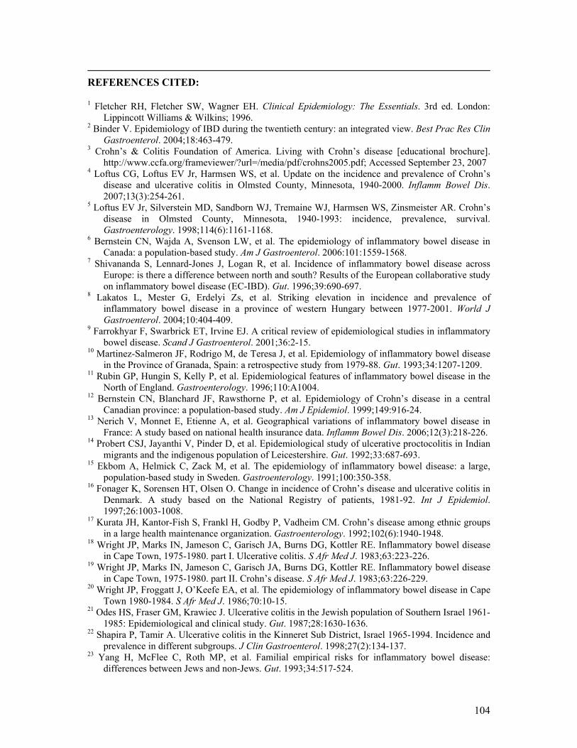
104
REFERENCES CITED: 1 Fletcher RH, Fletcher SW, Wagner EH. Clinical Epidemiology: The Essentials. 3rd ed. London:
Lippincott Williams & Wilkins; 1996. 2 Binder V. Epidemiology of IBD during the twentieth century: an integrated view. Best Prac Res Clin
Gastroenterol. 2004;18:463-479. 3 Crohn’s & Colitis Foundation of America. Living with Crohn’s disease [educational brochure].
http://www.ccfa.org/frameviewer/?url=/media/pdf/crohns2005.pdf; Accessed September 23, 2007 4 Loftus CG, Loftus EV Jr, Harmsen WS, et al. Update on the incidence and prevalence of Crohn’s
disease and ulcerative colitis in Olmsted County, Minnesota, 1940-2000. Inflamm Bowel Dis. 2007;13(3):254-261.
5 Loftus EV Jr, Silverstein MD, Sandborn WJ, Tremaine WJ, Harmsen WS, Zinsmeister AR. Crohn’s disease in Olmsted County, Minnesota, 1940-1993: incidence, prevalence, survival. Gastroenterology. 1998;114(6):1161-1168.
6 Bernstein CN, Wajda A, Svenson LW, et al. The epidemiology of inflammatory bowel disease in Canada: a population-based study. Am J Gastroenterol. 2006:101:1559-1568.
7 Shivananda S, Lennard-Jones J, Logan R, et al. Incidence of inflammatory bowel disease across Europe: is there a difference between north and south? Results of the European collaborative study on inflammatory bowel disease (EC-IBD). Gut. 1996;39:690-697.
8 Lakatos L, Mester G, Erdelyi Zs, et al. Striking elevation in incidence and prevalence of inflammatory bowel disease in a province of western Hungary between 1977-2001. World J Gastroenterol. 2004;10:404-409.
9 Farrokhyar F, Swarbrick ET, Irvine EJ. A critical review of epidemiological studies in inflammatory bowel disease. Scand J Gastroenterol. 2001;36:2-15.
10 Martinez-Salmeron JF, Rodrigo M, de Teresa J, et al. Epidemiology of inflammatory bowel disease in the Province of Granada, Spain: a retrospective study from 1979-88. Gut. 1993;34:1207-1209.
11 Rubin GP, Hungin S, Kelly P, et al. Epidemiological features of inflammatory bowel disease in the North of England. Gastroenterology. 1996;110:A1004.
12 Bernstein CN, Blanchard JF, Rawsthorne P, et al. Epidemiology of Crohn’s disease in a central Canadian province: a population-based study. Am J Epidemiol. 1999;149:916-24.
13 Nerich V, Monnet E, Etienne A, et al. Geographical variations of inflammatory bowel disease in France: A study based on national health insurance data. Inflamm Bowel Dis. 2006;12(3):218-226.
14 Probert CSJ, Jayanthi V, Pinder D, et al. Epidemiological study of ulcerative proctocolitis in Indian migrants and the indigenous population of Leicestershire. Gut. 1992;33:687-693.
15 Ekbom A, Helmick C, Zack M, et al. The epidemiology of inflammatory bowel disease: a large, population-based study in Sweden. Gastroenterology. 1991;100:350-358.
16 Fonager K, Sorensen HT, Olsen O. Change in incidence of Crohn’s disease and ulcerative colitis in Denmark. A study based on the National Registry of patients, 1981-92. Int J Epidemiol. 1997;26:1003-1008.
17 Kurata JH, Kantor-Fish S, Frankl H, Godby P, Vadheim CM. Crohn’s disease among ethnic groups in a large health maintenance organization. Gastroenterology. 1992;102(6):1940-1948.
18 Wright JP, Marks IN, Jameson C, Garisch JA, Burns DG, Kottler RE. Inflammatory bowel disease in Cape Town, 1975-1980. part I. Ulcerative colitis. S Afr Med J. 1983;63:223-226.
19 Wright JP, Marks IN, Jameson C, Garisch JA, Burns DG, Kottler RE. Inflammatory bowel disease in Cape Town, 1975-1980. part II. Crohn’s disease. S Afr Med J. 1983;63:226-229.
20 Wright JP, Froggatt J, O’Keefe EA, et al. The epidemiology of inflammatory bowel disease in Cape Town 1980-1984. S Afr Med J. 1986;70:10-15.
21 Odes HS, Fraser GM, Krawiec J. Ulcerative colitis in the Jewish population of Southern Israel 1961-1985: Epidemiological and clinical study. Gut. 1987;28:1630-1636.
22 Shapira P, Tamir A. Ulcerative colitis in the Kinneret Sub District, Israel 1965-1994. Incidence and prevalence in different subgroups. J Clin Gastroenterol. 1998;27(2):134-137.
23 Yang H, McFlee C, Roth MP, et al. Familial empirical risks for inflammatory bowel disease: differences between Jews and non-Jews. Gut. 1993;34:517-524.
105
24 Vucelić B, Korać B, Sentić M, et al. Epidemiology of Crohn’s disease in Zagreb, Yugoslavia: a ten-
year prospective study. Int J Epidemiol. 1991;20(1):216-220. 25 Vucelić B, Korać B, Sentić M, et al. Ulcerative colitis in Zagreb, Yugoslavia: incidence and
prevalence 1980-1989. Int J Epidemiol. 1991;20(4):1043-1047. 26 Jovanović Z. Epidemiology of Crohn’s disease in the Rijeka-Istra region. Lijec Vjesn. 1999;121(1-
2):8-13. 27 Gheorghe C, Pascu O, Gheorghe L, et al. Epidemiology of inflammatory bowel disease in adults
who refer to gastroenterology care in Romania: a multicentre study. Eur J Gastroenterol Hepatol. 2004;16(11):1153-1159.
28 Frangos CC, Frangos CC. Inflammatory bowel disease: reviewing an old study under a new perspective. Gut. 2007;56:1638-1639.
29 Gearry RB, Richardson A, Frampton CMA, et al. High incidence of Crohn’s disease in Canterbury, New Zealand: Results of an epidemiologic study. Inflamm Bowel dis. 2006;12(10):936-943.
30 Jiang XL, Cui HF. An analysis of 10218 ulcerative colitis cases in China. World J Gastroenterol. 2002;8(1):158-161.
31 Jiang L, Xia B, Li J, et al. Retrospective study of 451 patients with inflammatory bowel disease in Wuhan City, Central China. Inflamm Bowel Dis. 2006;12:212-217,
32 Langholz E, Munkholm P, Nielsen OH, et al. Incidence and prevalence of ulcerative colitis in Copenhagen County from 1962 to 1987. Scand J Gastroenterol. 1991;26:1247-1256.
33 Munkholm P, Langholz E, Nielsen OH, et al. Incidence and prevalence of Crohn’s disease in the county of Copenhagen, 1962 to 1987: a sixfold increase in incidence. Scand J Gastroenterol. 1992; 27:609-614.
34 Trallori G, Palli D, Saieva C, et al. A population-based study of inflammatory bowel disease in Florence over 15 years (1978-92). Scand J Gastroenterol. 1996;31:892-899.
35 Farrokhyar F, Swarbrick ET, Grace RH, et al. Low mortality of Crohn’s disease (CD) and ulcerative colitis (UC) in three regional centres in England. Gastroenterology. 1999;116:A710.
36 Jess T, Loftus V Jr, Harmsen WS, et al. Survival and cause specific mortality in patients with inflammatory bowel disease: a long term outcome studying in Olmsted County, Minnesota, 1940-2004. Gut. 2006;55:1248-1254.
37 Jess T, Winther KV, Munkholm P, Langholz E, Binder V. Mortality and causes of death in Crohn’s disease: follow-up of a population-based cohort in Copenhagen County, Denmark. Gastroenterology. 2002;122:1808-1814.
38 Langholz E, Munkholm P, Davidson M, et al. Colorectal cancer risk and mortality in patients with ulcerative colitis. Gastroenterology. 1992;103:1444-1451.
39 Munkholm P, Langholz E, Davidson M, et al. Intestinal cancer risk and mortality in patients with Crohn’s disease. Gastroenterology. 1993; 105:1716-1723.
40 Palli D, Trallori G, Saieva C, et al. General and cancer specific mortality of a population based cohort of patients with inflammatory bowel disease: The Florence study. Gut. 1998;42:175-179.
41 Persson P-G, Bernell O, Lejionmarck C-E, et al. Survival and cause-specific mortality in inflammatory bowel disease: A population-based cohort study. Gastroenterology. 1996;110:1339-1345.
42 Gollop JH, Phillips SF, Melton LJ, et al. Epidemiologic aspects of Crohn’s disease: a population-based study in Olmsted County, Minnesota, 1943-82. Gut. 1988;29:49-56.
43 Card T, Hubbard R, Logan RFA. Mortality in inflammatory bowel disease: a population-based cohort study. Gastroenterology. 2003;125:1583-1590.
44 Fiocchi C. Inflammatory bowel disease: etiology and pathogenesis. Gastroenterology. 1998;115:182-205.
45 Sartor RB. Clinical applications of advances in the genetics of IBD. Rev Gastroenterol Disord. 2003;3(suppl 1):S9-S17.
46 Ahmad T, Tamboli CP, Jewell D, et al. Clinical relevance of advances in genetics and pharmacogenetics of IBD. Gastroenterology. 2004;126:1533-1549.
47 Podolsky DK. The current future understanding of inflammatory bowel disease. Best Prac Res Clin Gastroenterol. 2002;16:933-943.
106
48 Hollander D, Vadheim C, Brettholz E, et al. Increased intestinal permeability in patients with
Crohn’s disease and their relatives. Ann Intern Med. 1986;105:883-885. 49 Wyatt J, Vogelsang H, Hubl W, et al. Intestinal permeability and the prediction of relapse in
Crohn’s disease. Lancet. 1993;341:1437-1439. 50 Hildsen RJ, Meddings JB, Sutherland LR. Intestinal permeability changes in response to
acetylsalicylic acid in relatives of patients with Crohn’s disease. Gastroenterology. 1996;110:1395-1403.
51 Yacyahyn BR, Meddings JB. CD45RO expression on circulating CD19+ B cells in Crohn’s disease correlates with intestinal permeability. Gastroenterology. 1995;108:132-137.
52 Fabia R, Ar’Rajab A, Andersson ML, et al. Impairment of bacterial flora in human ulcerative colitis and experimental colitis in the rat. Digestion. 1993;54:248-255.
53 Sellon RK, Tonkonogy S, Schultz M, et al. Resident enteric bacteria are necessary for development of spontaneous colitis and immune system activation in interleukin-10 deficient mice. Infect Immun. 1998;66(11):5224-5231.
54 Rath HC, Schultz M, Freitag R, et al. Different subsets of enteric bacteria induce and perpetuate experimental colitis in rats and mice. Infect Immun. 2001;69:2277-2285.
55 Shanahan F. Probiotics and inflammatory bowel disease: is there a scientific rationale? Inflamm Bowel Dis. 2000;6(2):107-115.
56 D’Haens GR, Geboes K, Peeters M, et al. Early lesions of recurrent Crohn’s disease caused by infusion of intestinal contents in excluded ileum. Gastroenterology. 1998;114:262-267.
57 van Tol EA, Holt L, Li FL, et al. Bacterial cell wall polymers promote intestinal fibrosis by direct stimulation of myofibroblasts. Am J Physiol. 1999;277:G245-255.
58 Kennedy RJ, Hoper M, Deodhar K, Erwin PJ, Kirk SJ, Gardiner KR. Interleukin 10-deficient colitis: new similarities to human inflammatory bowel disease. Br J Surg. 2000;87:1346-1351.
59 Herfarth HH, Bocker U, Janardhanam R, Sartor RB. Subtherapeutic corticosteroids potentiate the ability of interleukin 10 to prevent chronic inflammation in rats. Gastroenterology. 1998;115:856-865.
60 Koutrobakis I, Manousos ON, Mewwissen SGM, et al. Environmental risk factors in inflammatory bowel disease. Hepatogastroenterology. 1996;43:381-393.
61 Katschinski B, Fingerle D, Scherbaum B, et al. Oral contraceptive use and cigarette smoking in Crohn’s disease. Dig Dis Sci. 1993;38:1596-1600.
62 Sandler RS, Wurzelmann JI, Lyles CM. Oral contraceptive use and the risk of inflammatory bowel disease. Epidemiology. 1992;3:374-378.
63 Laharie D, Debeugny S, Peeters M, et al. Inflammatory bowel disease in spouses and their offspring. Gastroenterology. 2001;120:816-819.
64 Karlinger K, Györke T, Makö E, et al. The epidemiology and the pathogenesis of inflammatory bowel disease. Eur J Radiol. 2000;35:154-167.
65 Burke DA, Axon ATR. Ulcerative colitis and Escherichia coli with adhesive properties. J Clin Pathol. 1987;40:782-786.
66 Pardi DS, Tremaine WJ, Sandborn WJ, et al. Early measles virus infection is associated with the development of inflammatory bowel disease. Am J Gastroenterol. 2000;95:1480-1485.
67 Wakefield AJ, Ekbom A, Dhillon AP, et al. Crohn’s disease: pathogenesis and persistent measles virus infection. Gastroenterology. 1995;108:911-916.
68 Thompson NP, Montgomery SM, Pounder RE, et al. Is measles vaccination a risk factor for inflammatory bowel disease? Lancet. 1995;345:1071-1074.
69 Hugot JP, Alberti C, Berrebi D, et al. Crohn’s disease: The cold chain hypothesis. Lancet. 2003;362:2012-2015.
70 Franceschi S, Panza E, La Vecchia C, et al. Nonspecific inflammatory bowel disease and smoking. Am J Epidemiol. 1987;125:445-452.
71 Lindberg E, Tysk C, Anderson K, et al. Smoking and inflammatory bowel disease: a case control study. Gut. 1988;29:352-357.
72 Pullan RD, Rhodes J, Ganesh S, et al. Transdermal nicotine for active ulcerative colitis. N Engl J Med. 1994;330:811-815.
107
73 Motley RJ, Rhodes J, Ford GA, et al. Time relationships between cessation of smoking and onset of
ulcerative colitis. Digestion. 1987;37:125-127. 74 Abraham N, Selby W, Lazarus R, Solomon M. Is smoking an indirect risk factor for the
development of ulcerative colitis? An age- and sex-matched case-control study. J Gastroenterol Hepatol. 2003;18:139-146.
75 Madretsma S, Wolters LM, van Dijk JP. In-vivo effect of nicotine on cytokine production by human non-adherent mononuclear cells. Eur J Gastroenterol Hepatol. 1996;8:1017-1020.
76 Srivastava ED, Barton JR, O’Mahony S. Smoking, humoral immunity, and ulcerative colitis. Gut. 1991;32:1016-1019.
77 Miller LG, Goldstein G, Murphy M, et al. Reversible alterations in immunoregulatory T cells in smoking: analysis by monoclonal antibodies and flow cytometry. Chest. 1982;82:526-529.
78 Cope GF, Heatly RV, Kelleher JK. Smoking and colonic mucous in ulcerative colitis. BMJ. 1986;293:481.
79 Coulie B, Camilleri M, Bharucha AE, et al. Colonic motility in chronic ulcerative colitis proctosigmoiditis and the effects of nicotine on colonic motility in patients and healthy subjects. Aliment Pharmacol Ther. 2001;15:653-663.
80 Cosnes J, Beaugerie L, Carbonnel F, et al. Smoking cessation and the course of Crohn’s disease: An intervention study. Gastroenterology. 2001;120:1093-1099.
81 Morise K, Yamaguchi T, Kuroiwa A, et al. Expression of adhesion molecules and HLA-DR by macrophages and dendritic cells in aphthoid lesions of Crohn’s disease: an immunohistochemical study. J Gastroenterol. 1994;29:257-264.
82 Pavlick KP, Laroux FS, Fuseler J, et al. Serial Review: Reactive oxygen and nitrogen in inflammation. Role of reactive metabolites of oxygen and nitrogen in inflammatory bowel disease. Free Radic Biol Med. 2002;33:311-322.
83 Lodes MJ, Cong Y, Elson CO, et al. Bacterial flagellin is a dominant antigen in Crohn disease. J Clin Invest. 2004;113:1296-1306.
84 Farmer RG, Michener WM. Association of inflammatory bowel disease in families. Front Gastrointest Res. 1986;11:17-26.
85 Orholm M, Binder V, Sorensen TI, et al. Concordance of inflammatory bowel disease among Danish twins: Results of a nationwide study. Scand J Gastroenterol. 2003;35:1075-1081.
86 Tysk C, Lindberg E, Jarnerot G, et al. Ulcerative colitis and Crohn’s disease in an unselected population of monozygotic and dizygotic twins: a study of heritability and the influence of smoking. Gut. 1988;29:990-996.
87 Bayless TM, Tokayer AZ, Polito JM, et al. Crohn’s disease: concordance for site and clinical type in affected family members – potential hereditary influences. Gastroenterology. 1996;111:573-579.
88 Peeters M. Nevens H, Baert F, et al. Familial aggregation in Crohn’s disease: increased age-adjusted risk and concordance in clinical characteristics. Gastroenterology. 1996;111:597-603.
89 Annese V, Latiano A, Forabosco A, et al. Linkage to IBD1 locus does not identify specific clinical phenotypes in familial IBD patients. Dig Liver Dis. 2002; 32 (Suppl 1):A6.
90 Murillo L, Crusius JB, van Bodegraven AA, et al. CARD15 gene and the classification of Crohn’s disease. Immunogenetics. 2002;54:59-61.
91 Monsen U, Bernell O, Johansson C, et al. Prevalence of inflammatory bowel disease among relatives of patients with Crohn’s disease. Scand J Gastroenterol. 1991;26:302-306.
92 Meucci G, Vecchi M, Torgano G, et al. Familial aggregation of inflammatory bowel disease in northern Italy: a multicenter study. Gastroenterology. 1992;103:514-519.
93 Satsangi J, Parkes M, Jewell DP, et al. Genetics of inflammatory bowel disease. Clin Sci. 1998;94:473-478.
94 Faybush EM, Blanchard JF, Rawsthorne P, et al. Generational differences in the age at diagnosis with IBD: Genetic anticipation, bias, or temporal effects. Am J Gastroenterol. 2002;97:636-640.
95 Lee WCJ, Bridger S, McGregor C, et al. Why children with inflammatory bowel disease are diagnosed at a younger age than their affected parents. Gut. 1999;44:808-811.
96 Halfvarson J, Bodin L, Tysk C, et al. Inflammatory bowel disease in a Swedish twin cohort: a long-term follow-up of concordance and clinical characteristics. Gastroenterology. 2003;124:1767-1773.
108
97 Sands BE. From symptom to diagnosis: clinical distinctions among various forms of intestinal
inflammation. Gastroenterology. 2004;126:1518-1532. 98 Nikolaus S, Schreiber S. Diagnostics of inflammatory bowel disease. Gastroenterology.
2007;133:1670-1689. 99 Louis E, Collard A, Oger AF, et al. Behaviour of Crohn’s disease according to the Vienna
classification: Changing pattern over the course of the disease. Gut. 2001;49:777-782. 100 Hugot JP. Inflammatory bowel disease: causes and consequences. Best Prac Res Clin
Gastroenterol. 2004;18:447-449. 101 Oberhuber G, Stangl PC, Vogelsang H, et al. Significant association of strictures and internal
fistula formation in Crohn’s disease. Virchows Arch. 2000;437:293-297. 102 Greenstein AJ, Sachar DB, Mann D, et al. Spontaneous free indications and perforated abscesses in
30 patients with Crohn’s disease. Ann Surg. 1987;205:72-76. 103 Yang H, Taylor KD, Rotter JI. Minireview. Inflammatory bowel disease. I. Genetic Epidemiology.
Mol Genet Metab. 2001;74:1-21. 104 Tarján Zs, Tóth G, Györke T, et al. Ultrasound in Crohn’s disease of the small bowel. European J
Radiol. 2000;35:176-182. 105 Hommes DW, van Deventer SJH. Endoscopy in inflammatory bowel disease. Gastroenterology.
2004;126:1561-1573. 106 MacDermott RP, Nasg GS, Bertovich MJ, et al. Alterations of IgM, IgG, and IgA synthesis and
secretion by peripheral blood and intestinal mononuclear cells from patients with ulcerative colitis and Crohn’s disease. Gastroenterology. 1981;81:844-852.
107 Gasche C, Scholmerich J, Brynskov J, et al. A simple classification of Crohn’s disease: Report of the Working Party for the World Congresses of Gastroenterology, Vienna 1998. Inflamm Bowel Dis. 2000;6:8-15.
108 Best WR, Becktel JM, Singleton JW, et al. Development of a Crohn’s disease activity index. National Cooperative Crohn’s Disease Study. Gastroenterology. 1976;70(3):439-444.
109 Harvey RF, Bradshaw JM. A simple index of Crohn’s-disease activity. Lancet. 1980;1:514. 110 Powell-Tuck J, Brown RL, Lennard-Jones JE. A comparison of oral prednisolone given as a single
or multiple daily doses for active proctocolitis. Scand J Gastroenterol. 1978;13:833-837. 111 Walmsley RS, Ayres RCS, Pounder RE, et al. A simple clinical colitis activity index. Gut.
1998;43:29-32. 112 Lichtiger S, Present DH, Kornbluth A, et al. Cyclosporine in severe ulcerative colitis refractory to
steroid therapy. N Engl J Med. 1994;330:1841-1845. 113 Daperno M, D’Haens G, Van Assche G, et al. Development and validation of a new, simplified
endoscopic activity score for Crohn’s disease: the SES-CD. Gastrointest Endosc. 2004;60:505-512.
114 De Lange T, Larsen S, Aabakken L. Inter-observer agreement in the assessment of endoscopic findings in ulcerative colitis. BMC Gastroenterol. 2004;4:9.
115 Reumaux D, Sendid B, Poulian D, et al. Serological markers in inflammatory bowel diseases. Best Prac Res Clin Gastroenterol. 2003;17:19-35.
116 Kayazawa M, Saitoh O, Kojima K, et al. Lactoferrin in whole gut lavage fluid as a marker for disease activity in inflammatory bowel disease: comparison with other neutrophil-derived proteins. Am J Gastroenterol. 2002;97:360-369.
117 Winterkamp S, Weidenhiller M, Otte P, et al. urinary excretion of N-methylhistamine as a marker of disease activity in inflammatory bowel disease. Am J Gastroenterol. 2002;97:3071-3077.
118 Norman GL. Anti-Saccharomyces cerevisiae antibodies in inflammatory bowel disease. Clin Applied immunol Rev. 2001;2:45-63.
119 Peeters M, Joossens S, Vermeire S, et al. Diagnostic value of Anti-Saccharomyces cerevisiae and antineutrophil cytoplasmic antibodies in inflammatory bowel disease. Am J Gastroenterol. 2001;96:730-734.
120 Darroch CJ, Barnes RMR, Dawson J. Circulating antibodies to Saccharomyces cerevisiae (bakers’/brewers’ yeast) in gastrointestinal disease. J Clin Pathol. 1999;52:47-53.
109
121 Walker LJ, Aldhous MC, Drummond HE, et al. Anti-Saccharomyces cerevisiae antibodies (ASCA)
in Crohn’s disease are associated with disease severity but not NOD2/CARD15 mutations. Clin Exp Immunol. 2004;135:490-496.
122 Papp M, Altorjay I, Norman GL, et al. Seroreactivity to microbial components in Crohn’s disease is associated with ileal involvement, noninflammatory disease behavior and NOD2/CARD15 genotype, but not with risk for surgery in a Hungarian cohort of IBD patients. Inflamm Bowel Dis. 2007;13(8):984-992.
123 Papp M, Norman GL, Altorjay I, Lakatos PL. Utility of serological markers in inflammatory bowel disease: gadget or magic? World J Gastroenterol. 2007;13:2028-2036.
124 Dotan I, Fishman S, Dgani Y, et al. Antibodies against laminaribioside and chitobioside are novel serologic markers in Crohn’s disease. Gastroenterology. 2006;131:366-378.
125 Papp M, Altorjay I, Dotan N, et al. New serological markers for inflammatory bowel disease are associated with earlier age at onset, complicated disease behavior, risk for surgery, and NOD2/CARD15 genotype in a Hungarian IBD cohort. Am J Gastroenterol. 2007;102:1-17.
126 Hardarson S, Labrecque DR, Mitros FA, et al. Antineutrophil cytoplasmic antibody in inflammatory bowel disease and hepatobiliary disease. High prevalence in ulcerative colitis, primary sclerosing cholangitis, and autoimmune hepatitis. Am J Clin Pathol. 1993;3:277-281.
127 Cohavy O, Bruckner D, Gordon LK, et al. Colonic bacteria express an ulcerative colitis pANCA-related protein epitope. Infect Immun. 2000;68:1542-1548.
128 Wei B, Dalwadi H, Gordon LK, et al. Molecular cloning of a Bacteriodes caccae TonB-linked outer membrane protein identified by an inflammatory bowel disease marker antibody. Infect Immun. 2001;69:6044-6054.
129 Cohavy O, Harth G, Horwitz M, et al. Identification of a novel mycobacterial histone H1 homologue (HupB) as an antigenic target of pANCA monoclonal antibody and serum immunoglobulin A from patients with Crohn’s disease. Infect Immun. 1999;67:6510-6517.
130 Nakamura RM, Matsutani M, Barry M. Advances in clinical laboratory tests for inflammatory bowel disease. Clinica Chimica Acta. 2003;335:9-20.
131 Ferrante M, Henckaerts L, Joossens M, et al. New serological markers in inflammatory bowel disease are associated with complicated disease behaviour. Gut. 2007;56(10):1394-1403.
132 Olsson R, Danielsson A, Jarnerot G, et al. Prevalence of primary sclerosing cholangitis in patients with ulcerative colitis. Gastroenterology. 1991;100:1319-1323.
133 Bernstein CN, Blanchard JF, Rawsthorne P, et al. The prevalence of extraintestinal diseases in inflammatory bowel disease: a population-based study. Am J Gastroenterol. 2001;96:1116-1122.
134 Terjung B, Spengler U, Sauerbruch T, et al. “Atypical p-ANCA” in IBD and hepatobiliary disorders react with a 50-kilodalton nuclear envelope protein of neutrophils and myeloid cell lines. Gastroenterology. 2000;119:310-322.
135 Seibold F, Mörk H, Tanza S, et al. Pancreatic antibodies in Crohn’s disease: A family study. Gut. 1997;40:481-484.
136 Folwaczny C, Noehl N, Endres SP, et al. Antineutophil cytoplasmic antibody in first degree relatives of patients with inflammatory bowel disease. Scand J Gastroenterol. 1998;33:523-528.
137 Joossens S, Vermeire S, van Steen K, et al. Pancreatic autoantibodies in inflammatory bowel disease. Inflamm Bowel Dis. 2004;10:771-777.
138 Pelli MA, Maldini M, Contini C, et al. Use of Calprest© in the diagnosis of inflammatory bowel disease (IBD). Clin Applied Immunol Rev. 2002;3:5-16.
139 Stříž I, Trebžichavský I. Calprotectin – a pleiotropic molecule in acute and chronic inflammation. Physiol Rev. 2004;53:245-253.
140 Lakatos L, Pandur T, David G, et al. Association of extraintestinal manifestation of inflammatory bowel disease in a province of western Hungary with disease phenotype: Results of a 25-year follow-up study. World J Gastroenterol. 2003;9:2300-2307.
141 Crohn BB. Ocular lesions complicating ulcerative colitis. Amer J Med Sci. 1925;169:260-267. 142 Ghanchi FD, Rembacken BJ. Inflammatory bowel disease and the eye. Surv Ophthamol.
2003;48:663-676. 143 Salmon JF, Wright JP, Murray AND. Ocular inflammation in Crohn’s disease. Ophthalmology.
1997;98:480-484.
110
144 Lyons JL, Rosenbaum JT. Uveitis associated with inflammatory bowel disease compared with
uveitis associated with spondyloarthropathy. Arch Ophthalmol. 1997;115:61-64. 145 Soukiasian SH, Foster CS, Raizman MB. Treatment strategies for scleritis and uveitis associated
with inflammatory bowel disease. Am J Ophthalmol. 1994;118:601-611. 146 Wright R, Mea LKL. Abnormalities of the sacroiliac joints and uveitis in ulcerative colitis. Quart J
Med. 1965;34:229-236. 147 Banares A, Jover JA, Fernandex-Gutierrez B, et al. Pattern of uveitis as guide in making
rheumatologic and immunologic diagnoses. Arthritis Rheum. 1997;40:358-370. 148 Orchard TR, Chua CN, Ahmad T, et al. Uveitis and erythema nodosum in inflammatory bowel
disease: Clinical features and the role of HLA genes. Gastroenterology. 2002;123:714-718. 149 Regueiro M, Valentine J, Plevy S, et al. Infliximab for treatment of pyoderma gangrenosum
associated with inflammatory bowel disease. Am J Gastroenterol. 2003;98:1821-1826. 150 Zazo V, Hernanz JM, Parera A, et al. Skin in inflammatory bowel disease. J Eur Acad Dermatol
Venereol. 1997;9(Suppl 1):S196-S197. 151 Menachem Y, Gotsman I. Clinical manifestations of pyoderma gangrenosum associated with
inflammatory bowel disease. Isr Med Assoc J. 2004;6:88-90. 152 Bargen JA. Complication and sequelae of chronic ulcerative colitis. Ann Int Med. 1930;3:335-339. 153 Orchard TR, Wordsworth BP, Jewell DP. Peripheral arthropathies in inflammatory bowel disease:
Their articular distribution and natural history. Gut. 1998;42:387-391. 154 Bhagat S, Das KM. A shared and unique peptide in the human colon, eye, and joint detected by a
monoclonal antibody. Gastroenterology. 1994;107:103-108. 155 Salvarani C, Fornaciari G, Beltrami M, et al. Musculoskeletal manifestations in inflammatory
bowel disease. Eur J Intern Med. 2000;11:210-214. 156 Koutroubakis IE. Unraveling the mechanisms of thrombosis in inflammatory bowel disease. Am J
Gastroenterol. 2001;96:1325-1327. 157 Grip O, Svensson PJ, Lindgren S. Inflammatory bowel disease promotes venous disease earlier in
life. Scand J Gastroenterol. 2000;35:377-382. 158 Thompson NP, Wakefield AJ, Pounder RE. Inherited disorders of coagulation appear to protect
against inflammatory bowel disease. Gastroenterology. 1995;108:1011-1015. 159 Talbot RW, Heppell J, Dozois RR, et al. Vascular complications of inflammatory bowel disease.
Mayo Clin Proc. 1985;61:140-145. 160 Turri D, Rosselli M, Simioni P, et al. Factor V Leiden and prothrombin gene mutation in
inflammatory bowel disease in Mediterranean area. Digest Liver Dis. 2001;33:559-562. 161 Huisman MV, Rosendaal F. Thrombophilia. Curr Opin Hematol 1999;6:291-297. 162 Poort SR, Rosendaal FR, Reitsma PH, et al. A common genetic variation in the 3’-untranslated
region of the prothrombin gene is associated with elevated plasma prothrombin levels and an increase in venous thrombosis. Blood. 1996;88:3698-3703.
163 Papa A, Danses S, Grillo A, et al. Review Article: Inherited thrombophilia in inflammatory bowel disease. Am J Gastroenterol 2003;98:1247-1251.
164 Wilson A, Reyes E, Ofman J. Prevalence and outcome of anemia in inflammatory bowel disease: A systematic review of the literature. Am J Med. 2004;116:44S-49S.
165 Gasché C. Anemia in IBD: The overlooked villain. Inflamm Bowel Dis. 2000;6:142-150. 166 Cronin CC, Shanahan F. Anemia in patients with chronic inflammatory bowel disease. Am J
Gastroenterol. 2001;96:2296-2298. 167 Harries AD, Fitzsimons E, Dew MJ, et al. Association between iron deficiency anemia and mid-
arm circumference in Crohn’s disease. Hum Nutr Clin Nutr. 1984;38:47-53. 168 Dyer NH, Child JA, Mollin DL, et al. Anemia in Crohn’s disease. Q J Med. 1972;41:419-436. 169 Niv Y, Abukasis G. Prevalence of ulcerative colitis in the Israeli kibbutz population. J Clin
Gastroenterol. 1991;13:98-101. 170 Niv Y, Torten D, Tamir A, et al. Incidence and prevalence of ulcerative colitis in the upper Galilee,
Northern Israel, 1967-1986. Am J Gastroenterol. 1990;85:1580-1583. 171 Ahmad J, Slivka A. Hepatobiliary disease in inflammatory bowel disease. Gastroenterol Clin North
Am. 2002;31:329-345.
111
172 Wee A, Ludwig J. Pericholangitis in chronic ulcerative colitis: primary sclerosing cholangitis of the
small bile ducts? Ann Intern Med. 1985;102:581-587. 173 Rodrigues HJ, Bass NM. Primary sclerosing cholangitis. Semin Gastrointest Dis. 2003;14:189-198. 174 Loftus EV, Sandborn WJ, Tremaine WJ, et al. Risk of colorectal neoplasia in patients with primary
sclerosing cholangitis. Gastroenterology. 1996;110:432-440. 175 Broomé U, Löfberg R, Veress B, et al. Primary sclerosing cholangitis and ulcerative colitis:
Evidence for increased neoplastic potential. Hepatology. 1995;22:1404-1408. 176 Berg DF, Bahadursingh AM, Kaminski DL, et al. Acute surgical emergencies in inflammatory
bowel disease. Am J Surg. 2002;184:45-51. 177 Roy MA. Inflammatory bowel disease. Surg Clin North Am. 1997;77:1419-1431. 178 Robert JH, Sachar DB, Aufses AH, et al. Management of severe hemorrhage in ulcerative colitis.
Am J Surg. 1990;159:550-555. 179 Keighley MRB. Acute fulminating colitis and emergency colectomy. In: Keighley MRB, Williams
NS, editors. Surgery of the anus, rectum and colon. London:WB Saunders, 1993:1379-1397. 180 Crohn BB, Rosenberg H. The sigmoidoscopic picture of chronic ulcerative colitis. Am J Med Sci.
1925;170:220-228. 181 Eaden JA, Abrams KR, Mayberry JF. The risk of colorectal cancer in ulcerative colitis: a meta-
analysis. Gut. 2001;48:526-535. 182 Lakatos L, Mester G, Erdelyi Z, et al. Risk factors for ulcerative colitis-associated colorectal cancer
in a Hungarian cohort of patients with ulcerative colitis: results of a population-based study. Inflamm Bowel Dis. 2006;12(3):205-211.
183 Xie J, Itzkowitz SH. Cancer in inflammatory bowel disease. World J Gastroenterol. 2008;14(3):378-389.
184 Vleggaar FP, Lutgens MWMD, Claessen MMH. Review article: the relevance of surveillance endoscopy in long-lasting inflammatory bowel disease. Aliment Pharmacol Ther. 2007;26(Suppl 2):47-52.
185 Mascheretti S, Croucher PJP, Schreiber S. Pharmacogenetics of inflammatory bowel disease. Best Prac Re Clin Gastroenterol. 2004;18:597-609.
186 Blam ME, Stein RB, Lichtenstein GR. Integrating anti-tumor necrosis factor therapy in inflammatory bowel disease: current and future perspectives. Am J Gastroenterol. 2001;96:1977-1997.
187 Hart AL, Stagg AJ, Kamm MA. Use of probiotics in the treatment of inflammatory bowel disease. J Clin Gastroenterol. 2003;36:111-119.
188 Wirtz S, Neurath MF. Gene transfer approaches for the treatment of inflammatory bowel disease. Gene Ther. 2003;10:854-860.
189 Carty E, Rampton DS. Evaluation of new therapies for inflammatory bowel disease. Br J Clin Pharmacol. 2003;56:351-361.
190 Qasim A, Seery J, O’Morain CA. 5-Aminosalicylates in inflammatory bowel disease: Choosing the right dose. Digest Liver Dis. 2001;33:393-398.
191 Azad Khan AK, Piris J, Truelove SC. An experiment to determine the active therapeutic moiety of sulphasalazine. Lancet. 1977;2:892-895.
192 Navarro F, Hanauer SB. Treatment of inflammatory bowel disease: Safety and tolerability issues. Am J Gastroenterol. 2003;98:S18-S23.
193 Sands BE. Therapy of inflammatory bowel disease. Gastroenterology. 2000;S68-S82. 194 Hanauer SB, Sandborn WJ, Dallaire C, et al. Delayed-release oral mesalamine 4.8 g/day (800 mg
tablets) compared to 2.4 g/day (400 mg tablets) for the treatment of mildly to moderately active ulcerative colitis: the ASCEND I trial. Can J Gastroenterol. 2007;21:827-834.
195 Hanauser SB, Strömberg U. Oral Pentasa in the treatment of active Crohn’s disease: a meta-analysis of double-blind, placebo-controlled trials. Clin Gastroenterol Hepatol. 2004;2:379-388.
196 Travis SPL, Stange EF, Lémann M, et al. European evidence based consensus on the diagnosis and management of Crohn’s disease: current management. Gut. 2006;55(Suppl I):i16-i35.
197 Truelove SC, Witts LJ. Cortisone in ulcerative colitis. Final report on a therapeutic trial. BMJ. 1955;2:1041-1048.
112
198 Summers RW, Switz DM, Sessions JT, et al. national co-operative Crohn’s disease study group:
results of drug treatment. Gastroenterology. 1979;77:847-869. 199 Malchow H, Ewe K, Bandes JW, et al. European co-operative Crohn’s disease study (ECCDS):
results of drug treatment. Gastroenterology. 1984;86:249-266. 200 Faubion WA, Loftus EV, Harmsen WS, et al. The natural history of corticosteroid therapy for
inflammatory bowel disease: A population-based study. Gastroenterology. 2001;121:255-260. 201 Angeli A, Masera RG, Sartori ML, et al. Modulation by cytokines of glucocorticoid action. Ann N
Y Acad Sci. 1999;876:210-220. 202 Barnes PJ. Nuclear factor-кB. Int J Biochem Cell Biol. 1997;29:867-870. 203 Desramé J, Sabaté JM, Agher R, et al. Assessment of hypothalamic-pituitary-adrenal axis function
after corticosteroid therapy in inflammatory bowel disease. Am J Gastroenterol. 2002;97:1785-1791.
204 Louis E, Belaiche J. Optimizing treatment with thioguanine derivatives in inflammatory bowel disease. Best Prac Res Clin Gastroenterol. 2003;17:37-46.
205 Ardizzone S, Maconi G, Russo A, Imbesi V, Colombo E, Bianchi Porro G. Randomised controlled trial of azathioprine and 5-aminosalicylic acid for treatment of steroid dependent ulcerative colitis. Gut. 2006;55:47-53.
206 Colombel JF, Ferrari N, Debuysere H, et al. Genotypic analysis of thiopurine S-methyltransferase in patients with Crohn’s disease and severe myelosuppression during azathioprine therapy. Gastroenterology. 2000;118:1025-1030.
207 van Dullemen HM, van Deventer SJ, Hommes DW, et al. Treatment of Crohn’s disease with anti-tumor necrosis factor chimeric monoclonal antibody (cA2). Gastroenterology. 1995;109:129-135.
208 Scallon BJ, Moore MA, Trinh H, et al. Chimeric anti-TNF-α monoclonal antibody cA2 binds recombinant transmembrane TNF-α and activates immune effector functions. Cytokine. 1995;7:251-259.
209 Sandborn WJ, Hanauer SB. Antitumor necrosis factor therapy for inflammatory bowel disease: a review of agents, pharmacology, clinical results, and safety. Inflamm Bowel Dis. 1999;5:119-133.
210 Hanauer SB, Feagan BG, Lichtenstein GR, et al. Maintenance infliximab for Crohn’s disease: The ACCENT I randomized trial. Lancet. 2002;359:1541-1549.
211 Sands B, van Deventer S, Blank M, et al. maintenance infliximab is safe and effective in fistulizing Crohn’s disease (CD); Results from the ACCENT II trial. Am J Gastroenterol. 2002;97:S258.
212 Sandborn WJ, Hanauer SB. Infliximab in the treatment of Crohn’s disease: a user’s guide for clinicians. Am J gastroenterol. 2002;97:2962-2972.
213 Cheifetz A, Smedley M, Martin S, et al. The incidence and management of infusion reactions to infliximab: A large center experience. Am J Gastroenterol. 2003;98:1315-1324.
214 Klapman JB, Ene-Stroescu D, Becker MA, et al. A lupus-like syndrome associated with infliximab therapy. Inflamm Bowel Dis. 2003;9:176-178.
215 Schroeder KW, Tremaine WJ, Ilstrup DM. Coated oral 5-aminosalicylic acid therapy for mildly to moderately active ulcerative colitis: a randomized trial. N Engl J Med. 1987;317:1625-1629.
216 Rutgeerts P, Sandborn WJ, Feagan BG, et al. Infliximab for induction and maintenance therapy for ulcerative colitis. N Engl J Med. 2005;353:2462-2476.
217 Järnerot G, Hertervig E, Friis-Liby I, et al. Infliximab as rescue therapy in severe to moderately severe ulcerative colitis: a randomized, placebo-controlled study. Gastroenterology. 2005;128:1805-1811.
218 Hanauer SB, Sandborn WJ, Rutgeerts P, et al. Human anti-tumor necrosis factor monoclonal antibody (adalimumab) in Crohn’s disease: the CLASSIC-I trial. Gastroenterology. 2006;130:323-333.
219 Den Broeder A, van de Putte L, Rau R, et al. A single dose, placebo controlled study of the fully human anti-tumor necrosis factor-alpha antibody adalimumab (D2E7) in patients with rheumatoid arthritis. J Rheumatol. 2002;29(11):2288-2298.
220 Mease PJ, Gladman DD, Ritchlin CT, et al; Adalimumab Effectiveness in Psoriatic Arthritis Trial Study Group. Adalimumab for the treatment of patients with moderately to severely active psoriatic arthritis: results of a double-blind, randomized, placebo-controlled trial. Arthritis Rheum. 2005;52(10):3279-3289.

113
221 Menter A, Tyring SK, Gordon K, et al. Adalimumab therapy for moderate to severe psoriasis: a
randomized, controlled phas III trial. J Am Acad Dermatol. 2008;58(1):106-115. 222 Gordon KB, Langley RG, Leonardi C, et al. Clinical response to adalimumab treatment in patients
with moderate to severe psoriasis: double-blind, randomized, controlled trial and open-label extension study. J Am Acad Dermatol. 2006;55(4):598-606.
223 Lambert RG, Salonen D, Rahman P, et al. Adalimumab significant reduces both spinal and sacroiliac joint inflammation in patients with anylosing spondylitis: a multicenter, randomized, double-blind, placebo-controlled study. Arthritis Rheum. 2007;56(12):4005-4014.
224 Nesbitt A, Fossati G, Bergin M, et al. Mechanism of action of cerolizumab pegol (CDP870): in vitro comparison with other anti-tumor necrosis factor alpha agents. Inflamm Bowel Dis. 2007;13(11):1323-1332.
225 Fossati G, Nesbitt A. In-vitro complement-dependent cellular cytotoxicity by the anti-TNF agents adalimumab, etanercept, infliximab, and certolizumab pegol (CDP870). Am J Gastroenterol. 2005;100 (Suppl):S299. abstract.
226 Fuller R. Probiotics in man and animals. J Appl Bacteriol. 1989;66:365-378. 227 Malin M, Suomalainen H, Saxelin M, et al. Promotion of IgA immune response in patients with
Crohn’s disease by oral bacteriotherapy with Lactobacillus GG. Ann Nutr Metab. 1996;40:137-145.
228 Fellermann K, Wehkamp J, Herrlinger KR, et al. Crohn’s disease: A defensin deficiency syndrome? Eur J Gastroenterol Hepatol. 2003;15:627-634.
229 Salminen S, Isolauri E, Salminen E. Clinical use of probiotics for stabilizing the gut mucosal barrier: Successful strains and future challenges. Antonie Van Leeuwenhoek 1996;70:347-358.
230 Gionchetti P, Amadini C, Rizzello F, et al. Probiotics – role in inflammatory bowel disease. Digest Liver Dis. 2002;34:S58-S62.
231 Summers RW, Elliott DE, Qadir K, et al. Trichuris suis seems to be safe and possibly effective in the treatment of inflammatory bowel disease. Am J Gastroenterol. 2003;98:2034-2041.
232 Barbara G, Xing Z, Hogaboam CM, Gauldie J, Collins SM. Interleukin 10 gene transfer prevents experimental colitis in rats. Gut. 2000;46:344-349.
233 Fromm MF. The influence of MDR1 polymorphisms on P-glycoprotein expression and function in humans. Adv Drug Deliv Rev. 2002;54:1295-1310.
234 Watkins PB. The barrier function of CYP3A4 and P-glycoprotein in the small bowel. Adv Drug Deliv Rev. 1997;27:161-170.
235 Gottesman MM, Pastan I. Biochemistry of multidrug resistance mediated by the multidrug transporter. Annu Rev Biochem. 1993;62:385-427.
236 Thiebaut F, Tsuruo T, Hamada H, Gottesman MM, Pastan I, Willingham MC. Cellular localization of the multidrug-resistance gene product P-glycoprotein in normal human tissues. Proc Natl Acad Sci USA. 1987;84:7735-7738.
237 Atkinson DE, Greenwood SL, Sibley CP, Glazier JD, Fairbairn LJ. Role of MDR1 and MRP1 in trophoblast cells, elucidated using retroviral gene transfer. Am J Physiol Cell Physiol. 2003;285:C584-591.
238 Schinkel AH, Wagenaar E, van Deemter L, Mol CA, Borst P. Absence of the mdr1a P-glycoprotein in mice affects tissue distribution and pharmcokinetics of dexamethasone, digoxin, and cyclosporin A. J Clin Invest. 1995;96:1698-1705.
239 Sakaeda T, Nakamura T, Okumura K. MDR1 genotype-related pharmacokinetics and pharmacodynamics. Biol Pharm Bull. 2002;25(11):1391-1400.
240 Norris MD, De Graaf D, Haber M, et al. Involvement of MDR1 P-glycoprotein in multifactorial resistance to Methotrexate. Int J Cancer. 1996;65:613-619.
241 Farrell RJ, Murphy A, Long A, et al. High multidrug resistance (P-glycoprotein 170) expression in inflammatory bowel disease patients who fail medical therapy. Gastroenterology. 2000;118:279-288.
242 Panwala CM, Jones JC, Viney JL. A novel model of inflammatory bowel disease: mice deficient for the multiple drug resistance gene, mdr1a, spontaneously develop colitis. J Immunol. 1998;161:5733-5744.
114
243 Annese V, Valvano MR, Palmieri O, Latiano A, Bossa F, Andriulli A. Multidrug resistance 1 gene
in inflammatory bowel disease: a meta-analysis. World J Gastroenterol. 2006;12(23):3636-3644. 244 Hoffmeyer S, Murk O, von Richter O, et al. Functional polymorphisms of the multidrug-resistance
gene: multiple sequence variations and correlation of one allele with P-glycoprotein expression and activity in vivo. Proc Natl Acad Sci USA. 2000;97:3473-3478.
245 Kim RB, Leake BF, Choo EF, et al. Identification of functionally variant MDR1 alleles among European Americans and African Americans. Clin Pharmacol Ther. 2001;70:189-199.
246 Krishnamurthy P, Schuetz JD. Role of ABCG2/BCRP in biology and medicine. Annu Rev Pharmacol Toxicol. 2006;46:381-410.
247 Rocchi E, Khodjakov A, Volk EL, et al. The product of the ABC half-transporter gene ABCG2 (BCRP/MXR/ABCP) is expressed in the plasma membrane. Biochem Biophys Res Commun. 2000;271:42-46.
248 Van Herwaarden AE, Schinkel AH. The function of breast cancer resistance protein in epithelial barriers, stem cells and milk secretion of drugs and xenotoxins. Trends Pharm Sci. 2006;27(1):10-16.
249 Stoll M, Corneliussen B, Costello CM, et al. Genetic variation in DLG5 is associated with inflammatory bowel disease. Nat Genet. 2004;36(5)476-480.
250 Friedrichs F, Stoll M. Role of discs large homolog 5. World J Gastroenterol. 2006;12(23):3651-3656.
251 Nakamura H, Sudo T, Tsuiki H, et al. Identification of a novel human homolog of the Drosophilia dlg, P-dlg, specifically expressed in the gland tissues and interacting with p55. FEBS Lett. 1998;433(1-2):63-67.
252 Wakabayashi M, Ito T, Mitsushima M, et al. Interaction of lp-dlg/KIAA0583, a membrane-associated guanylate kinase family protein, with vinexin and beta-catenin at sites of cell-cell contact. J Biol Chem. 2003;278:21709-21714.
253 Humbert P, Russell S, Richardson H. Dlg, Scribble and Lgl in cell polarity, cell proliferation and cancer. Bioessays. 2003;25:542-553.
254 Croucher PJP, Mascheretti S, Hampe J, et al. Haplotype structure and association to Crohn’s disease of CRAD15 mutations in two ethnically divergent populations. Eur J Hum Genet. 2003;11:6-16.
255 Yamazaki K, Takazoe M, Tanaka T, et al. Association analysis of SLC22A4, SLC22A5, and DLG5 in Japanese patients with Crohn disease. J Hum Genet. 2004;49:664-668.
256 Gazouli M, Mantzaris G, Archimandritis AJ, Nasioulas G, Anagnou NP. Single nucleotide polymorphisms of OCTN1, OCTN2, and DLG5 genes in Greek patients with Crohn’s disease. World J Gastroenterol. 2005;11:7525-7530.
257 Biank V, Friedrichs F, Babusukumar U, et al. DLG5 R30Q variant is a female specific protective factor in pediatric onset Crohn’s disease. Am J Gastroenterol. 2007;102:391-398.
258 Lennard-Jones JE. Classification of inflammatory bowel disease. Scand J Gastroenterol. 1989;24(Suppl 170):2-6.
259 Lakatos PL, Lakatos, Szalay F, et al. Toll-like receptor 4 and NOD2/CARD15 mutations in Hungarian patients with Crohn disease: phenotype-genotype correlations. World J Gastroenterol. 2005;11:1489-1495.
260 Oselin K, Gerloff T, Mrozikiewicz PM, Pahkla R, Roots I. MDR1 polymorphisms G2677T in exon 21 and C3435T in exon 26 fail to affect rhodamine 123 efflux in peripheral blood lymphocytes. Fundam Clin Pharmacol. 2003;17:463-469.
261 Arjomand-Nahad F, Diefenbach K, Landt O, Gaikovitch E, Roots I. Genotyping of the triallelic variant G2677T/A in MDR1 using LightCycler with locked-nucleic-acid-modified hybridization probes. Anal Biochem. 2004;334:201-203.
262 Rozen S, Skaletsky H. Primer3 on the WWW for general users and for biologist programmers. Methods Mol Biol. 2000;132:365-386.
263 Szilvási A, Andrikovics H, Kalmár L, Bors A, Tordai A. Asymmetric PCR increases efficiency of melting point analysis on the LightCycler. Clin Biochem. 2005;38:727-730.
264 Podolsky DK. Inflammatory bowel disease. N Engl J Med. 2002;347:417-429.
115
265 Hampe J, Grebe J, Nikolaus S, et al. Association of NOD2 (CARD15) genotype with clinical
course of Crohn’s disease: a cohort study. Lancet. 2002;359:1661-1665. 266 Lesage S, Zouali H, Cézard JP, et al. CARD15/NOD2 mutational analysis and genotype-phenotype
correlation in 612 patients with inflammatory bowel disease. Am J Hum Genet. 2002;70:845-857. 267 Bonen DK, Cho JH. The genetics of inflammatory bowel disease. Gastroenterology.
2003;124:521:536. 268 Ahmad T, Armuzzi A, Bunce M, et al. The molecular classification of the clinical manifestations of
Crohn’s disease. Gastroenterology. 2002;122:854-866. 269 Cuthbert AP, Fischer SA, Mirza MM, et al. The contribution of NOD2 gene mutations to the risk
and site of disease in inflammatory bowel disease. Gastroenterology. 2002;122:867-874. 270 Abreu MT, Taylor KD, Lin YC, et al. Mutations in NOD2 are associated with fibrostenosing
disease in patients with Crohn’s disease. Gastroenterology. 2002;123:679-688 271 Guo QS, Xia B, Jiang Y, et al. NOD2 3020insC frameshift mutation is not associated with
inflammatory bowel disease in Chinese people of Han nationality. World J Gastroenterol. 2004;10:1069-1071.
272 Shanahan F. Inflammatory bowel disease: immunodiagnostics, immunotherapeutics, and ecotherapeutics. Gastroenterology. 2001;120:622-635.
273 Hampe J, Shaw SH, Saiz R, et al. Linkage of inflammatory bowel disease to human chromosome 6p. Am J Hum Genet. 1999;65:1647-1655.
274 Rioux JD, Silverberg MS, Daly MJ, et al. Genomewide search in Canadian families with inflammatory bowel disease reveals two novel susceptibility loci. Am J Hum Genet. 2000;66:1863-1870.
275 Stokkers PC, Reitsma PH, Tytgat GN, et al. HLA-DR and –DQ phenotypes in inflammatory bowel disease: a meta-analysis. Gut. 1999;45:395-401.
276 Nielsen OH, Kirman I, Rüdiger N, et al. Upregulation of Interleukin-12 and -17 in active inflammatory bowel disease. Scand J Gastroenterol. 2003;38:180-185.
277 Hugot JP, Chamaillard M, Zouali H, et al. Association of NOD2 leucine-rich repeat variants with susceptibility to Crohn’s disease. Nature. 2001;411:599-603.
278 Ogura Y, Bonen DK, Inohara N, et al. A frameshift mutation in NOD2 associated with susceptibility to Crohn’s disease. Nature. 2001;411:603-606.
279 Hampe J, Cuthbert A, Croucher PJ, et al. Association between insertion mutation in NOD2 gene and Crohn’s disease in German and British populations. Lancet. 2001;357:1925-1928.
280 Ogura Y, Inohara N, Benito A, et al. Nod2, a Nod1/Apaf-1 family member that is restricted to monocytes and activates NF-кB. J Biol Chem. 2001;276:4812-4818.
281 Satsangi J, Morecroft J, Shah NB, et al. Genetics of inflammatory bowel disease: Scientific and clinical implications. Best Prac Res Clin Gastroenterol. 2003;17:3-18.
282 Inohara N, Ogura Y, Chen FF, et al. Human Nod1 confers responsiveness to bacterial polysaccharides. J Biol Chem. 2001;276:2551-2554.
283 Inohara N, Ogura Y, Nuñez G. Nods: A family of cytosolic proteins that regulate host response to pathogens. Curr Opin Microbiol. 2002;5:76-80.
284 Girardin SE, Boneca IG, Viala J, et al. Nod2 is a general sensor of peptidoglycan through muramyl dipeptide (MDP) detection. J Biol Chem. 2003;278:8869-8872.
285 Russell RK, Nimmo ER, Satsangi J. Molecular genetics of Crohn’s disease. Curr Opin Genet Dev. 2004;14:264-270.
286 Bonen DK, Ogura Y, Nicolae DL, et al. Crohn’s disease-associated NOD2 variants share a signaling defect in response to lipopolysaccharide and peptidoglycan. Gastroenterology. 2003;124:140-146.
287 van der Linde K, Boor PPC, Houwing-Duistermaat JJ, et al. CARD15 and Crohn’s disease: healthy homozygous carriers of the 3020insC frameshift mutation. Am J Gastroenterol. 2003; 98;3:613-617.
288 Aldhous MC, Nimmo ER, Satsangi J. NOD2/CARD15 and the Paneth cell: another piece in the genetic jigsaw of inflammatory bowel disease. Gut. 2003;52:1533-1535.
289 Inoue N, Tamura K,Kinouchi Y, et al. Lack of common NOD2 variants in Japanese patients with Crohn’s disease. Gastroenterology. 2002;123:86-91.
116
290 Berrebi D, Maudians R, Hugot JP, et al. Card15 gene overexpression in mononuclear and epithelial
cells of the inflamed Crohn’s disease colon. Gut. 2003;52:840-846. 291 Chamaillard M, Philpott D, Girardin SE, et al. Gene-environment interaction modulated by allelic
heterogeneity in inflammatory bowel diseases. Proc Natl Acad Sci USA. 2003;100:3455-3460. 292 Schreiber S, Nikolaus S, Hampe J. Activation of nuclear factor kappa B in inflammatory bowel
disease. Gut. 1998; 42:477-484. 293 Gutierrez O, Pipaon C, Inohara N, et al. Induction of Nod2 in myelomonocytic cells and intestinal
epithelial cells via Nuclear Factor-кB activation. J Biol Chem. 2002;277:41701-41705. 294 Rosenstiel P, Fantini M, Brautigam K, et al. TNF-α and IFN-γ regulate the expression of the NOD2
(CARD15) gene in human intestinal epithelial cells. Gastroenterology. 2003;124:1001-1009. 295 Ayabe T, Satchell DP, Wilson CL, et al. Secretion of microbial α-defensins by intestinal Paneth
cells in response to bacteria. Nat Immunol. 2001;1:113-118. 296 Porter EM, Bevins CL, Ghosh D, et al. The multifaceted Paneth cell. Cell Mol Life Sci.
2002;59:156-170. 297 Lala S, Ogura Y, Osborne C, et al. Crohn’s disease and the NOD2 gene: a role for Paneth cells.
Gastroenterology. 2003;125:47-57. 298 Chamaillard M, Hashimoto M, Horie Y, et al. An essential role for NOD1 in host recognition of
bacterial peptidoglycan containing diaminopimelic acid. Nat Immunol. 2003;4(7):702-707. 299 Zouali H, Lesage S, Merlin F, et al. CARD4/NOD1 is not involved in inflammatory bowel disease.
Gut. 2003;52:71-74. 300 McGovern DPB, Hysi P, Ahmad T, et al. Association between a complex insertion/deletion
polymorphism in NOD1 (CARD4) and susceptibility to inflammatory bowel disease. Hum Mol Genet. 2005;14(10):1245-1250.
301 Molnar T, Hofner P, Nagy F, et al. NOD1 gene E266K polymorphism is associated with disease susceptibility but not with disease phenotype or NOD2/CARD15 in Hungarian IBD patients with Crohn’s disease. Dig Liver Dis. 2007;39(12):1064-1070.
302 Arancibia SA, Beltran CJ, Aguirre IM, et al. Toll-like receptors are key participants in innate immune responses. Biol Res. 2007;40:97-112.
303 Girardin SE, Hugot JP, Sansonetti PJ. Lessons from Nod2 studies: towards a link between Crohn’s disease and bacterial sensing. Trends Immunol. 2003;24:652-658.
304 Medzhitov R, Preston-Hurlburt P, Janeway CA Jr. A human homologue of the Drosophilia Toll protein signals activation of adaptive immunity. Nature. 1997;388(6640):394-397.
305 Da Silva Correia J, Soldau K, Christen U, Tobias PS, Ulevitch RJ. Lipopolysaccharide is in close proximity to each of the proteins in its membrane receptor complex. Transfer from CD14 to TLR and MD-2. J Biol Chem. 2001;276(24):21129-21135.
306 Franchimont D, Vermeire S, El Housni H, et al. Deficient host-bacteria interactions in inflammatory bowel disease? The toll-like receptor (TLR)-4 Asp299Gly polymorphism is associated with Crohn’s disease and ulcerative colitis. Gut. 2004;53:987-992.
307 Gazouli M, Mantzaris G, Kotsinas A, et al. Association between polymorphisms in the Toll-like receptor 4, CD14, and CARD15/NOD2 and inflammatory bowel disease in the Greek population. World J Gastroenterol. 2005;11(5):681-685.
308 Brand S, Staudinger T, Schnitzler F, et al. The role of Toll-like receptor 4 Asp299Gly and Thr399Ile polymorphisms and CARD15/NOD2 mutations in the susceptibility and phenotype of Crohn’s disease. Inflamm Bowel Dis. 2005;11(7):645-652.
309 Browning BL, Huebner C, Petermann I, et al. Has Toll-like receptor 4 been prematurely dismissed as an inflammatory bowel disease gene? Association study combined with meta-analysis shows strong evidence for association. Am J Gastroenterol. 2007;102:1-9.
310 Pierik M, Joossens S, Van Steen K, et al. Toll-like receptor-1, -2, and -6 polymorphisms influence disease extension in inflammatory bowel diseases. Inflamm Bowel Dis. 2006;12:1-8.
311 Arnott ID, Nimmo ER, Drummond HE, et al. NOD2/CARD15, TLR4 and CD14 mutations in Scottish and Irish Crohn’s disease patients: evidence for genetic heterogeneity within Europe? Genes Immun. 2004;5(5):417-425.
117
312 Lakatos PL, Lakatos L, Szalay F, et al. Toll-like receptor 4 and NOD2/CARD15 mutations in
Hungarian patients with Crohn’s disease: phenotype-genotype correlations. World J Gastroenterol. 2005;11:1489-1495.
313 Oostenbrug LE, Drenth JP, de Jong DJ, et al. Association between Toll-like receptor 4 and inflammatory bowel disease. Inflamm Bowel Dis. 2005;11:567-575.
314 Török HP, Glas J, Tonenchi L, et al. Polymorphisms of the lipopolysaccharide-signaling complex in inflammatory bowel disease: association od a mutation in the Toll-like receptor 4 gene with ulcerative colitis. Clin Immunol. 2004;112:85-91.
315 Silverberg MS. OCTNs: will the real IBD5 gene please stand up? World J Gastroeneterol. 2006;12(23):3678-3681.
316 Wang Y, Taroni F, Garavaglia B, et al. Functional analysis of mutations in the OCTN2 transporter causing primary carnitine deficiency: lack of genotype-phenotype correlation. Hum Mutat. 2000;16:401-407.
317 Peletkova VD, Wintle RF, Rubin LA, et al. Functional variants of OCTN cation transporter genes are associated with Crohn disease. Nat Genet. 2004;36(5):471-475.
318 Grundemann D, Harlfinger S, Golz S, et al. Discovery of the ergothioneine transporter. Proc Natl Acad Sci USA. 2005;102:5256-5261.
319 Noble CL, Nimmo ER, Drummond H, et al. The contribution of OCTN1/2 variants within the IBD5 locus to disease susceptibility and severity in Crohn’s disease. Gastroenterology. 2005;129:1854-1864.
320 Daly MJ, Rioux JD, Schaffner SF, Hudson TJ, Lander ES. High-resolution haplotype structure in the human genome. Nat Genet. 2001;29:229-232.
321 Vermeire S, Pierk M, Hlavaty T, et al. Association of organic cation transporter risk haplotype with perianal penetrating Crohn’s disease but not with susceptibility to IBD. Gastroenterology. 2005;129:1845-1853.
322 Ardizzone S, Porro GB. Inflammatory bowel disease: new insights into pathogenesis and treatment. J Int Med. 2003;252:475-496.
323 Lander E, Kruglyak L. Genetic dissection of complex traits: guidelines for interpreting and reporting linkage results. Nat Genet. 1995;11:241-247.
324 Satsangi J, Parkes M, Louis E, et al. Two-stage genome-wide search in inflammatory bowel disease provides evidence for susceptibility loci on chromosomes 3, 7, and 12. Nat Genet. 1996;14:199-202.
325 van Heel DA, Udalova IA, de Silva AP, et al. Inflammatory bowel disease is associated with a TNF polymorphism that affects an interaction between the OCT1 and NF-кB transcription factors. Hum Mol Genet. 2002; 11(11):1281-1289.
326 Duerr RH, Barmada MM, Zhang L, et al. High-density genome scan in Crohn disease shows confirmed linkage to chromosome 14q11-12. Am J Hum Genet. 2000;66:1857-1862.
327 Rioux JD, Daly MJ, Silverberg MS, et al. Genetic variation in the 5q31cytokine gene cluster confers susceptibility to Crohn disease. Nat Genet. 2001;29:223-228.
328 Peltekova VD, Wintle RF, Rubin LA, et al. Functional variants of OCTN cation transporter genes are associated with Crohn’s disease. Nat Genet. 2004;36:471-475.
329 Parkes M, Barrett JC, Prescott NJ, et al; Wellcome Trust Case Control Consortium. Sequence variants in the autophagy gene IRGM and multiple other replicating loci contribute to Crohn’s disease susceptibility. Nat Genet. 2007;39(7):830-832.
330 Low JH, Williams FA, Yang X, et al. Inflammatory bowel disease is linked to 19p13 and associated with ICAM-1. Inflamm Bowel Dis. 2004;10(3):173-181.
331 Cho JH, Nicolae DL, Gold LH, et al. Identification of novel susceptibility loci for inflammatory bowel disease on chromosomes 1p, 3q, and 4q: Evidence for epistasis between 1p and IBD1. Proc Natl Acad Sci USA. 1998;95:7502-7507.
332 Cho JH, Nicholae DL, Ramos R, et al. Linkage and linkage disequilibrium in chromosome band 1p36 in American Chaldeans with inflammatory bowel disease. Hum Mol Genet. 2000;22(9);1425-1432.
333 Hampe J, Frenzel H, Mirza MM, et al. Evidence for a NOD2-independent susceptibility locus for inflammatory bowel disease on chromosome 16p. Proc Natl Acad Sci USA. 2002;99(1):321-326.
118
334 Lakatos PL, Fischer S, Lakatos L, Gal I, Papp J. Current concept on the pathogenesis of
inflammatory bowel disease – crosstalk between genetic and microbial factors: pathogenic bacteria and altered bacterial sensing or changes in mucosal integrity take “toll”? World J Gastroenterol. 2006;12(12):1829-1841.
335 Duerr RH, Taylor KD, Brant SR, et al. A genome-wide association study identifies IL23R as an inflammatory bowel disease gene. Science. 2006;341:1461-1463.
336 Hampe J, Schreiber S, Shaw SH, et al. A genomewide analysis provides evidence for novel linkages in inflammatory bowel disease in a large European cohort. Am J Hum Genet. 1999;64(3):808-816.
337 Hampe J, Franke A, Rosentiel P, et al. A genome-wide association scan of nonsynonymous SNPs identifies a susceptibility variant for Crohn disease in ATG16L1. Nat Genet. 2007;39(2):207-211.
338 Dideberg V, Krisjansdottir G, Milani L, et al. An insertion-deletion polymorphism in the interferon regulatory factor 5 (IRF5) gene confers risk of inflammatory bowel diseases. Hum Mol Genet. 2007;16(24):3008-3016.
339 Libioulle C, Louis E, Hansoul S, et al. Novel Crohn disease locus identified by genome-wide association maps to a gene desert on 5p13.1 and modulates expression of PTGER4. PLoS Genet. 2007;3(4):e58.
340 Parkes M, Barmada MM, Satsangi J, et al. The IBD2 locus shows linkage heterogeneity between ulcerative colitis and Crohn disease. Am J Hum Genet. 2000;67:1605-1610.
341 Bouma G, Crusius JB, Garcia-Gonzales MA, et al. Genetic markers in clinically well defined patients with ulcerative colitis (UC). Clin Exp Immun. 1999;115:294-300.
342 Orchard TR, Thiyagaraja S, Welsh KI, et al. Clinical phenotype is related to HLA genotype in the peripheral arthropathies of inflammatory bowel disease. Gastroenterology. 2000;118:274-278.
343 Roussomoustakaki M, Satsangi J, Welsh K, et al. Genetic markers may predict disease behaviour in patients with ulcerative colitis. Gastroenterology. 1997;112:1845-1853.
344 Cheng TF, Brzostek S, Ando O, Van Scoy S, Kumar KP, Reich NC. Differential activation of IFN regulatory factor (IRF)-3 and IRF-5 transcription factors during viral infection. J Immunol. 2006;176(12):7462-7470.
345 Lin R, Heylbroeck C, Pitha PM, Hiscott J. Virus-dependent phosphorylation of the IRF-3 transcription factor regulates nuclear translocation, transactivation potential, and proteasome-mediated degradation. Mol Cell Biol. 1998;18:2986-2996.
346 Zhang L, Wu, L, Hong K, Pagano JS. Intracellular signaling molecules activated by Epstein-Barr virus for induction of interferon regulatory factor 7. J Virol. 2001;75:12393-12401.
347 Honda K, Taniguchi T. IRFs: master regulators of signallying by Toll-like receptors and cytosolic pattern-recognition receptors. Nat Rev Immunol. 2006;6:644-658.
348 Sigurdsson S, Nordmark G, Goring HH, et al. Polymorphisms in the tyrosine kinase 2 and interferon regulatory factor 5 genes are associated with systemic lupus erythematosus. Am J Hum Genet. 2005;76:528-537.
349 Sigurdsson S, Padyukov L, Kurreeman F, et al. Association of a haplotype in the promoter region of the interferon regulatory factor 5 gene with rheumatoid arthritis. Arthritis Rheum. 2007;56:2202-2210.
350 Heinemeyer T, Wingender E, Reuter I, et al. Databases on transcriptional regulation: TRANSFAC, TRRD, and COMPEL. Nucleic Acids Res. 1998;26:364-370.
351 Risch N, Merikangas K. The future of genetic studies of complex human disease. Science. 1996;273:1516-1517.
352 The Wellcome Trust Case Control Consortium. Genome-wide association study of 14,000 cases of seven common diseases and 3,000 shared controls. Nature. 2007;447:661-678.
353 Tremelling M, Cummings F, Fisher SA, et al. IL23R variation determines susceptibility but not disease phenotype in inflammatory bowel disease. Gastroenterology. 2007;132:1657-1664.
354 Prescott NJ, Fisher SA, Franke A, et al. A nonsynonymous SNP in ATG16L1 predisposes to ileal Crohn’s disease and is independent of CARD15 and IBD5. Gastroenterology. 2007;132:1665-1671.
355 Jung HC, Eckmann L, Yang SK, et al. A distinct array of proinflammatory cytokines is expressed in human colon epithelial cells in response to bacterial invasion. J Clin Invest. 1995;95:55-65.
119
356 Papadakis KA, Targan SR. The role of chemokines and chemokines receptors in mucosal
infmallation. Inflamm Bowel Dis. 2000;6(4):303-313. 357 Luster AD. Chemokines – chemotactic cytokines that mediate inflammation. N Engl J Med.
1998;338:436-445. 358 Ardizzone S, Porro GB. Inflammatory bowel disease: new insights into pathogenesis and treatment.
J Int Med. 2003;252:475-496. 359 Seegert D, Rosenstiel P, Pfahler H, Pfefferkorn P, Nikolaus S, Schreiber S. Increased expression of
IL-16 in inflammatory bowel disease. Gut. 2001;48:326:332. 360 Oppmann B, Lesley R, Blom B, et al. Novel p19 protein engages IL-12p40 to form a cytokine, IL-
23, with biological activities similar as well as distinct from IL-12. Immunity. 2000;13:715-725. 361 Reinecker HC, Steffen M, Witthoeft T, et al. Enhanced secretion of tumor necrosis factor-alpha, IL-
6, and IL-1β by isolated lamina propria mononuclear cells from patients with ulcerative colitis and Crohn’s disease. Clin Exp Immunol. 1993;94:174-181.
362 Kusugami K, Youngman KR, West GA, et al. Intestinal immune reactivity to interleukin 2 differs among Crohn’s disease, ulcerative colitis and control. Gastroenterology. 1989;97:1-9.
363 Matsuura T, West GA, Youngman KR, et al. Immune activation genes in inflammatory bowel disease. Gastroenterology. 1993;104:448-458.
364 Daig R, Andus T, Aschenbrenner E, Falk W, Schölmerich J, Gross V. Increased interleukin 8 expression in the colon mucosa of patients with inflammatory bowel disease. Gut. 1996;38(2):216-222.
365 Mazzucchelli L, Hauser C, Zgraggen K, et al. Expression of interleukin-8 gene in inflammatory bowel disease is related to the histological grade of active disease. Am J Pathol. 1994;144(5):997-1007.
366 Fujino S, Andoh A, Bamba S, et al. Increased expression of interleukin 17 in inflammatory bowel disease. Gut. 2003;52:65-70.
367 Ivanov I, McKenzie BS, Zhou L, et al. The orphan nuclear receptor RORγt directs the differentiation program of proinflammatory IL-17+ T helper cells. Cell. 2006;126:1121-1133.
368 Kolls JK, Lindén A. Interleukin-17 family members and inflammation. Immunity. 2004;21:467-476.
369 Neurath MF. IL-23 a master regulator in Crohn disease. Nat Med. 2007;13:26-28. 370 Uhlig HH, McKenzie BS, Hue S, et al. Differential activity of IL-12 and IL-23 in mucosal and
systemic innate immune pathology. Immunity. 2006;25:309-318. 371 Seiderer J, Elben I, Diegelmann J, et al. Role of the novel Th17 cytokine IL-17F in inflammatory
bowel disease (IBD): upregulated colonic IL-17F expression in active Crohn’s disease and analysis of the IL17F p.His161Arg polymorphism in IBD. Inflamm Bowel Dis. 2007. Epub ahead of print.
372 van Heel DA, Udalova IA, De Silva AP, et al. Inflammatory bowel disease is associated with a TNF polymorphism that affects an interaction between OCT1 and NF-κB transcription factors. Hum Mol Genet. 2002;11(11):1281-1289.
373 O’Callaghan NJ, Adams KE, van Heel DA, Cavanaugh JA. Association of TNF-α-857C with inflammatory bowel disease in the Australian population. Scand J Gastroenterol. 2003;38:533-534.
374 Cao Q, Zhu Q, Wu ML, Hu WL, Gao M, Si JM. Genetic susceptibility to ulcerative colitis in the Chinese Han ethnic population: association with TNF polymorphisms. Chin Med J (Engl). 1006;119:1198-1203.
375 Goyette P, Labbé C, Trinh TC, Xavier RJ, Rioux JD. Molecular pathogenesis of inflammatory bowel disease: genotypes, phenotypes and personalized medicine. Ann Med. 2007;39:177-199.
376 Brant SR, Panhuysen CIM, Nicolae D, et al. MDR1 Ala893 polymorphism is associated with inflammatory bowel disease. Am J Hum Genet. 2003;73:1282-1292.
377 Ho GT, Nimmo ER, Tenesa A, et al. Allelic variations of the multidrug resistance gene determine susceptibility and disease behavior in ulcerative colitis. Gastroenterology. 2005;128:288-296.
378 Urcelay E, Mendoza JL, Martin MC, et al. MDR1 Gene: susceptibility in Spanish Crohn’s disease and ulcerative colitis patients. Inflamm Bowel Dis. 2006;12:33-37.
120
379 Schwab M, Schaeffler E, Marx C, et al. Association between the C3435T MDR1 polymorphism
and susceptibility for ulcerative colitis. Gastroenterology. 2003;124:26-33. 380 Glas J, Török HP, Schiemann U, Folwaczny C. MDR1 gene polymorphism in ulcerative colitis.
Gastroenterology. 2004;126:367. 381 Palmieri O, Latiano A, Valvano R, et al. Multidrug resistance 1 genepolymorphisms are not
associated with inflammatory bowel disease ad respose to therapy in Italian patients. Aliment Pharmacol Ther. 2005;22:1129-1138.
382 Potočnik U, Ferkolj I, Glavac D, Dean M. Polymorphisms in multidrug resistance 1 (MDR1) gene are associated with refractory Crohn disease and ulcerative colitis. Genes Immun. 2004;5:530-539.
383 Daly MJ, Pearce AV, Farwell L, et al. Association of DLG5 R30Q variant with inflammatory bowel disease. Eur J Hum Genet. 2005;13:835-839.
384 Noble CL, Nimmo ER, Drummond H, et al. DLG5 variants do not influence susceptibility to inflammatory bowel disease in the Scottish population. Gut. 2005;54:1416-1420.
385 Török HP, Glas J, Tonenchi L, et al. Polymorphisms in the DLG5 and OCTN cation transporter genes in Crohn’s disease. Gut. 2005;54:1421-1427.
386 Yamazaki K, Takazoe M, Tanaka T, Ichimori T, Nakamura Y. Absence of mutations in the NOD2/CARD15 gene among 483 Japanese patients with Crohn’s disease. J Hum Genet. 2002;47:469-472.
387 Vermeire S, Pierik M, Hlavaty T, et al. Association of organic cation transporter risk haplotype with perianal penetrating Crohn’s disease but not with susceptibility to IBD. Gastroenterology. 2005; 129:1845-1853.
388 Browning BL, Annese V, Barclay ML, et al. Gender-stratified analysis of DLG5 R30Q in 4707 patients with Crohn disease and 4973 controls from 12 Caucasian cohorts. J Med Genet. 2008;45:36-42.