influence of cell envelope components on the …
TRANSCRIPT

INFLUENCE OF CELL ENVELOPE COMPONENTS ON THE
ATTACHMENT OF ESCHERICHIA COLI O157:H7 TO LETTUCE AND
SPINACH UNDER DIFFERENT IONIC ENVIRONMENTS: EFFECT ON
THEIR RESISTANCE TO ENVIRONMENTAL STRESS
by
Chi-Ching Lee
(Under the Direction of Joseph F. Frank)!
ABSTRACT
Produce-related outbreaks of Escherichia coli O157:H7 are the second most
common foodborne outbreaks since it was first recognized an enteric pathogen in 1982.
The mechanism of attachment of E. coli O157: H7 on leafy greens is complex and
dependent on a variety of factors including properties of the cell surface, substratum and
bulk fluid. The objective of this study was to determinate the role of cell envelope
components on attachment of E. coli O157:H7 to lettuce and spinach leaf surface and cut
edge under different environments. Cell surface charge, hydrophobicity, and capsule
characteristics were investigated. In addition, cellulose-deficient derivatives of Shiga
toxin–producing Escherichia coli (STEC) have been used. The lower surface
hydrophobicity, less negative charge, as well as capsule containing more D-Mannose and
!-Fucose of cells grown in tryptic soy broth combined to increase attachment to spinach
leaves. Cellulose-producing STEC cells attached significantly 0.5 log greater on lettuce
leaf surface than cellulose-deficient cells. However, cellulose-deficient cells attached
significantly 0.7 log greater to cut edge of lettuce than parental cells. In high water

!
hardness environment, attachment of wild type cells to leafy greens surface also could be
enhanced. In addition, chlorine treatment reduced the population of cellulose-deficient
cells at 1.2 log units more than the wild type in 150ppm of chlorine on spinach leaves
surface. However, the population of cellulose-producing cells was reduced significantly
by 1.5 log units more than its mutant when cells also produced colanic acid. Extra-
cellular cellulose production protects STEC cells attached to leafy greens from the effects
of chlorine on spinach leave surface.
INDEX WORDS: Escherichia coli O157:H7; STEC; Attachment; Lettuce; Spinach;
Capsule; Cellulose; Colanic Acid; Water hardness; Chlorine;
Surface Charge; Hydrophobicity

!
INFLUENCE OF CELL ENVELOPE COMPONENTS ON THE
ATTACHMENT OF ESCHERICHIA COLI O157:H7 TO LETTUCE AND
SPINACH UNDER DIFFERENT IONIC ENVIRONMENTS: EFFECT ON
THEIR RESISTANCE TO ENVIRONMENTAL STRESS
by
Chi-Ching Lee
B.S., National Taiwan University, Taiwan, 2003
M.S., Taipei Medical University, Taiwan, 2005
A Thesis Submitted to the Graduate Faculty of The University of Georgia in Partial
Fulfillment of the Requirements for the Degree
DOCTOR OF PHILOSOPHY
ATHENS, GEORGIA
2014

!
© 2014
CHI-CHING LEE
ALL Rights Reserved
!
INFLUENCE OF CELL ENVELOPE COMPONENTS ON THE
ATTACHMENT OF ESCHERICHIA COLI O157:H7 TO LETTUCE AND
SPINACH UNDER DIFFERENT IONIC ENVIRONMENTS: EFFECT ON
THEIR RESISTANCE TO ENVIRONMENTAL STRESS
by
CHI-CHING LEE
Major Professor: Joseph F. Frank
Committee: Jennifer L. Cannon
Jinru Chen
William C. Hurst
Electronic Version Approved:
Maureen Grasso
Dean of the Graduate School
The University of Georgia
May 2014

! "#!
DEDICATION
To my parents, brother and major adviser, Dr. Joseph Frank
! "!
ACKNOWLEDGEMENTS
I would like to thank my major professor, Dr. Joseph Frank. He has guided and
supported me very well for these years. He always shows me the right direction and gives
me a lot of useful advice whenever I encounter challenges in my research work. I am not
able to finish my dissertation without him. I also would like to thank all members of my
graduate committee, Dr. Jinru Chen, Dr. Jennifer Cannon and Dr. William C. Hurst for
all of their encouragement and guidance in these years. I deeply appreciate Dr. Chen for
providing the strains, mutants and technical advice. I would like to thank Dr. Robert
Shewfelt for helping my dissertation and doctoral comprehensive examination.
I would like to appreciate my laboratory manager: Revis Chmielewski, Ruth Ann
Morrow, Gwen Hirsch, Vickie Wentzel, ; post-doctor: Bwalya Lungu, Rowaida Khalil;
all my undergraduate laboratory helpers including Priya Naik during my study. You all
are very helpful and kept my study move forward smoothly. My perfect food micro
labmates: Amudhan Ponrajan, Shawn Lyons, Antonio Lourenco, Dvijal Patel,
Yanjie Tang, Christabelle Piansay, Sofia Farakos and Winnie Lim. We used to spend so
many nights and weekends together just for one decent table or figure. My research life
can not get through perfectly without you. Besides, I want to thank Ana Rodriguez,
Arena Richardson, Bruno Giri, Charlotte Steininger, Helga Doering, Jillian Fishburn,
Lauren Hudson, Lee Carella, Lisa Trimble, Maria Anum Sohail, Michael Johnston,
Mohammad Obaidat, Nakieta McCullum, Stephanie Barnes, Stephanie Bolton,
Stephanie Mako, Suejee Park, Venessa Chandra. You all have been around me to help
and encourage me for my study.
! "#!
I really would like to thank Bilal Kırmacı, Priyadarshi Puranjay, Christopher
Sharps, Jaideep Sidhu, Todd Fisk, Vikramaditya Yandapalli, Deepika Karnik,
Gaana Nagaraj, Garima Pande, Grishma Kotwal, Tina Phongpa-ngan, Yuanyuan Ma. My
life in graduate school can not be completed without you. All my food science geeks:
Allison Bechman, Angela Rincon, Dhivya Lakshmi, Haiqin Dong, Jing Chen,
Jaideep Sidhu, Krantikumar Yemmireddy, Michael Paul, Ola Mide, Sailaja Chintagari,
Solandre Almeida, Wei Kang, Xiaomeng Wu. You all have kept company with me and
inspired me for my entire graduate student life here.
All my friends from Taiwan: Kuan-Wu Yeh, Hao-Min Chen, Hsiao-Hui Chen,
Hsien-Tzer Tseng, Ming-Lan Kuo, Ming-Yi Huang, Si-Ying Chen, Wei-Chia Tseng,
Wei-Ren Huang, Yen-Jun Chuang, Yen-Ting Liu, Yi-Yin Lin, Yu-Wei Chen. You all let
me feel like that I am still in Taiwan even though I am so far away from home.
All my friends I have lived with: Ömer Ta!getiren, Mustafa Nural, Sedat "en,
Atakan Ata, Elkin Nurmammadov, Selçuk Tekin, Mehmet Durkan, Muhammet Arican.
You take care and love me just like family. You are very helpful, generous and have
unconditional tolerance to me in daily life.
In addition, I would like to thank Bochra Bahri, Selcuk Acar, Okta Ervianto, Ira
Nurmala, Ulugbek Bekchanov, Ozlem Kaya, Biswajit banerjee, Giasuddin shameem,
Adrijo chakraborty, Anirban Mukhopadhyay and all friends I met at UGA Athens. You
all make my stay much more pleasurable and meaningful.
Finally, I would like to thank my family. Although they are very far away from me
and we rarely have chance to get together, they are still with me all the time.

! "##!
TABLE OF CONTENTS
Page
ACKNOWLEDGEMENTS...............................................................................................v
LIST OF TABLES..........................................................................................................viii
LIST OF FIGURES............................................................................................................x
CHAPTER
1. INTRODUCTION..........................................................................................................1
2. LITERATURE REVIEW...............................................................................................5
3. INFLUENCE OF CELL SURFACE PROPERTIES ON ATTACHMENT OF
ESCHERICHIA COLI O157:H7 TO SPINACH AND LETTUCE LEAVES..............28
4. THE ROLE OF CELLULOSE IN ATTACHMENT OF SHIGA TOXIN–
PRODUCING ESCHERICHIA COLI TO LETTUCE AND SPINACH IN
DIFFERENT WATER HARDNESS ENVIRONMENTS............................................61
5. INFLUENCE OF EXTRA-CELLULAR CELLULOSE PRODUCTION ON THE
SURVIVAL OF SHIGA TOXIN-PRODUCING ESCHERICHIA COLI ON
SPINACH AND LETTUCE AFTER CHLORINE TREATMENT..............................83
6. SUMMARY CONCLUSION......................................................................................104
! "###!
LIST OF TABLES
Page
Table 2.1: Number of cases of bacterial and parasitic infection, hospitalizations, and
deaths by pathogen from 1996 to 2012........................................................... 25
Table 2.2: Variables important in cell attachment and biofilm formation……………….26
Table 2.3: Potential attachment factors in human pathogens that are similar to those
described for plant bacteria. ............................................................................27
Table 3.1: Attachment of E.coli O157:H7 grown in three media to the surface of lettuce
in the presence of different ionic environments................................................51
Table 3.2: Attachment of E.coli O157:H7 grown in three media to the cut edge of lettuce
in the presence of different ionic environments.……………………………...52
Table 3.3: Attachment of E.coli O157:H7 grown in three media to the surface of spinach
in the presence of different ionic environments………………………………53
Table 3.4: Attachment of E.coli O157:H7 grown in three media to the cut edge of spinach
in the presence of different ionic environments................................................54
Table 3.5: Carbohydrate-binding specificities of fluorescein isothiocyanate (FITC) and
FITC-conjugated lectins…..………………………………………………….55
Table 3.6: The relative fluorescence of E.coli O157:H7 resulting from FITC-lectin
binding in the presence of competitive sugars. ...............................................56
Table 3.7. Hydrophobicity of E. coli O157:H7 determined by adherence to hydrocarbons
(Xylene) and Zeta potential (mV) of cell surface charge of E. coli O157:H7
grown in tryptic soy broth (TSB), Luria broth base Millar (LB) and nutrient

! "#!
Page
broth (NB) in different ionic environments………………………….………57
Table 4.1: Attachment of four Shiga toxin–producing E. coli (STEC) strains to lettuce
and spinach on surface and cut edge. .…………………..……………..…….78
Table 4.2: Hydrophobicity of four Shiga toxin–producing E. coli (STEC) strains
determined by adherence to hydrocarbons (Xylene)………………...………79
Table 5.1: The population of sublethally-injured cells on surface and cut edge of spinach
after chlorine treatment..……………………………………………………100
Table 5.2: The log population of sublethally-injured cells on surface and cut edge of
lettuce after chlorine treatment……………………………...……………...101

! "!
LIST OF FIGURES
Page
Figure 3.1: Comparison of E. coli O157:H7 cells grown in TSB, LB and NB for
24hrs.…………………………………………………………………………58
Figure 3.2: Epifluorescence microscopic images of E. coli O157:H7 ………………......59
Figure 4.1: Attachment of E. coli O157:H7 strain 994 and STEC strains to iceberg
lettuce surface and cut edge in the different water hardness
environments…………………………………………………………….…80
Figure 4.2: Attachment of E. coli O157:H7 strain 994 and STEC strains to spinach
lettuce surface and cut edge in the different water hardness
environments.……………………………………………………………....81
Figure 4.3: Zeta potential (mV) of cell surface charge of E. coli O157:H7 strain 994
and STEC strains in different water hardness environments.……………..82
Figure 5.1: Survival of E. coli O157:H7 strain 994 (T) and STEC strains to iceberg
lettuce surface and cut edge after 50 and 150ppm chlorine treatment.…….102
Figure 5.2: Survival of E. coli O157:H7 strain 994 (T) and STEC strains to baby
spinach surface and cut edge after 50 and 150ppm chlorine reatment…….103
! "!
CHAPTER 1
INTRODUCTION
Fresh produce is the second most common transmission vehicle for Escherichia
coli O157:H7 in 38 (21%) of 183 foodborne outbreaks and 34% of 5269 foodborne
outbreak-related cases. It has been associated with apples cider or juice, salad, alfalfa,
clover spout, coleslaw, cantaloupes, radish, spinach and leaf lettuce (Beuchat, 2002;
Hillborn et al., 1999; Rangel et al., 2005). Lettuce-related outbreaks account for 34% of
produce-related E. coli O157:H7 outbreaks which is significantly more frequent than
other produce.
Cell attachment is dependent on the hydrodynamics of the fluid, composition of the
aqueous medium, ionic and chemical properties of the cell surface and physical
properties of the substratum (Donlan, 2002). Many factors have been identified to have
an influence on attachment and detachment of E. coli O157:H7 on lettuce, such as
hydrophobicity, capsule production and surface charge (Hassan et al., 2003; Hassan et
al., 2004). The hydrophobic and electrokinetic properties of cells are crucial influences
for cell attachment. The hydrophobicity is affected by bacterial exopolysaccharides (EPS)
components (Gross and Logan, 1995). Besides, many viable E. coli O157:H7 cells can be
observed on the cut edges and stomata of lettuce leaves after chlorine treatment (Seo and
Frank, 1999).
! #!
Furthermore, extracellular structures also influence cell attachment and the resistance
of embedded cells to many biocide treatments. These structures are protective matrices at
the cell surface, such as cellulose, colanic acid, fimbriae, flagella and EPS. The properties
of cell surface, the media of attachment and other environmental factors play important
roles on bacterial attachment (Costerton et al., 1987; Frank, 2000; Kumar, 1998).
Determination of the mechanism of cellulose and colanic acid in E. coli O157:H7
attachment to foods and food contact surfaces, and cell protection against sanitizers will
provide essential information for developing associated strategies to eliminate or control
pathogens in food processing and foodservice environments (Ryu and Beuchat, 2005).
The defense mechanism of microorganisms induced by environmental stress is complex.
The several stress response and modification of cell structure are performed by E. coli
which can produce the bacterial EPS including celloluse and colanic acid on the cell
surfaces. Cellulose composition with a linear long D-glucose units chain could protect
Shiga toxin-producing E. coli O157:H7 (STEC) strains against oxidative and acidic
treatments (Yoo and Chen, 2012). Colanic acid has been observed to offer E. coli
O157:H7 protection against acidic, heat, osmotic and oxidative stress (Chen et al., 2004).
This study investigated the effect of three growth media, tryptic soy broth (TSB),
Luria broth base, Miller (LB) and nutrient broth (NB) under different ionic environments
on the attachment of E. coli O157:H7 to lettuce and spinach leaves surface and cut edge.
Surface charge, hydrophobicity, and capsule characteristics were determined. In addition,
water hardness is an important factor in food safety and processing. In households and
manufacturing facilities it can influence the quality of a product and sanitation. In
addition, chlorine solution is the most commonly used sanitizer in food processing plants
! $!
due to low-cost, safe and easy to use. Therefore, this study also investigated the influence
of cellulose on attachment of STEC to iceberg lettuce and baby spinach leaves under
different water hardness environments and cell survival under chlorine solution treatment.
Mutants lacking cellulose production were used. The hypothesis for this study is that the
specific cell envelope components and certain environment conditions will have
significant effects on attachment and survival of E. coli O157:H7.
REFERENCES
Beuchat LR. 2002. Ecological factors influencing survival and growth of human
pathogens on raw fruits and vegetables. Microbes Infect. 4:413-23.
Chen, J.R., Lee, S.M., and Mao, Y., 2004. Protective effect of exopolysaccharide colanic
acid of Escherichia coli O157 : H7 to osmotic and oxidative stress. Int J Food
Microbiol 93:281-286.
Costerton, J. W., K. J. Cheng, G. G. Geesey, T. I. Ladd, J. C. Nickel, M. Dasgupta, and
T. J. Marrie. 1987. Bacterial biofilms in nature and diseas. Annu. Rev. Microbiol.
41:435-464.
Donlan, R.M., 2002. Biofilms: Microbial life on surfaces, Emerg. Inf. Dis. 8: 881–890.
Gross, M.J. and Logan, B.E., 1995. Influence of different chemical treatments on
transport of Alcaligenes paradoxus in porous media. Appl Environ Microbiol
61:1750-1756.
Hassan, A.N. and Frank, J.F., 2003. Influence of surfactant hydrophobicity on the
detachment of Escherichia coli O157:H7 from lettuce. Int J Food Microbiol
87:145-152.

! %!
Hassan, A.N. and Frank, J.F., 2004. Attachment of Escherichia coli O157 : H7 grown in
tryptic soy broth and nutrient broth to apple and lettuce surfaces as related to cell
hydrophobicity, surface charge, and capsule production. Int J Food Microbiol
96:103-109.
Hillborn, E. D., J. H. Mermin, P. A. Mshar, J. L. Hadler, A. Voetsch, C. Wojtkunski, M.
Swartz, R. Mshar, M. A. Lambert-Fair, J. A. Farrar, M. K. Glynn, and L. Slutsker.
1999. A multistate outbreak of Escherichia coli O157:H7 infections associated
with consumption of mesclun lettuce. Arch Intern Med 159:1758-1764.
Kumar, C. G., and S. K. Anand. 1998. Significance of microbial biofilms in food
industry: a review. Int J Food Microbiol 42:9-27.
Rangel, J.M., Sparling, P.H., Crowe, C., Griffin, P.M., Swerdlow, D.L. 2005
Epidemiology of Escherichia coli O157:H7 outbreaks, United States, 1982–2002.
Emerg Infect Dis 11(4):603-9.
Ryu, J.H. and Beuchat, R.L,. 2005 Biofilm formation by Escherichia coli O157:H7 on
Stainless Steel: Effect of Exopolysaccharide and Curli Production on Its
Resistance to Chlorine. Applied And Environmental Microbiology, Jan. 247-254
Seo, K.H. and Frank, J.F., 1999. Attachment of Escherichia coli O157:H7 to lettuce leaf
surface and bacterial viability in response to chlorine treatment as demonstrated
by using confocal scanning laser microscopy. J Food Prot 62:3-9
Yoo, B.K. and Chen, J.R., 2012. Role of cellulose in protecting Shiga toxin producing
Escherichia coli against oxidative and acidic stress. Food Control 23:289-292.
! &!
CHAPTER 2
LITERATURE REVIEW
I. The foodborne outbreak of E. coli O157:H7 associated with leafy greens
In United States, E. coli O157:H7 causes 73,000 illnesses annually and has been
characterized as causing thrombotic thrombocytopenic purpura, hemorrhagic colitis,
hemolytic-uremic syndrome (HUS) in susceptible people and children (Griffin et al.,
1991). It is Gram-negative, rod-shaped and predominant facultative anaerobic bacterium
in the gastrointestinal tracts of mammals, especially cattle. They have been implicated as
the most important a reservoir of E. coli O157:H7 and enable be asymptomatic carriers
due to lack of Shiga toxin receptor, globotriaosylceramide (Pruimboom-Brees et al.,
2000). The prevalence of E. coli O157:H7 in feedlot cattle herds of North American
ranges from 0 to 61% (Jeon et al., 2013).
E. coli O157:H7 was first isolated from ground beef and recognized as a pathogen
in 1982. Ground beef is the most common vehicle among foodborne outbreaks (75 [41%]
of 183 outbreak) and 33% of 5,269 foodborne-related cases between 1982 and 2002.
Produce was the transmission vehicle for E. coli O157:H7 in 38 (21%) of 183 foodborne
outbreaks and 34% of 5,269 foodborne outbreak-related cases from 1982 to 2002 with the
1st recorded incident occurring in 1991. In addition, a total of 350 outbreaks in this
period were reported from 49 states, accounting for 8,598 cases of E. coli O157 infection,
representing 1493 (17%) hospitalizations, 354 (4%) HUS cases, and 40 (0.5%) deaths.
Food remained the predominant transmission route and is associated with 52% of 350
! '!
outbreaks and 61% of 8,598 outbreak-related cases. Produce-associated outbreaks most
commonly occurred in restaurants for 15 outbreaks (39%) (Rangel et al., 2005).
According to 2011 Centers for Disease Control and Prevention (CDC) report, the
31 known major pathogens of foodborne outbreak in Unite States caused 9.4 million
illnesses, 55,961 hospitalizations and 1,351 deaths each year. Norovirus caused the most
illnesses (58%), and nontyphoidal Salmonella spp. (11%), Clostridium perfringens
(10%), Campylobacter spp. (9%). Primary causes of hospitalization were nontyphoidal
Salmonella spp. (35%), norovirus (26%), Campylobacter spp. (15%), and Toxoplasma
gondii (8%). Most death were caused by nontyphoidal Salmonella spp. (28%), followed
by T.gondii (24%), Listeria monocytogenes (19%), and norovirus (11%). STEC O157
were estimated to cause 63,153 illnesses, 2,138 hospitalizations and 20 (1%) deaths every
year (Scallan et al., 2011).
The means by which E. coli O157: H7 is introduced into the lettuce and spinach is
not well understood. Nevertheless, one hypothesis states that the lettuce plant is
contaminated when grown in fields fertilized with improperly treated manure (Ethan et
al., 2002). E. coli O157:H7 has been isolated from fresh produce including apples cider
or juice, bean sprouts, salad, cantaloupes and leaf lettuce (Hillforn et al., 1999; Ackers et
al., 1998). Produce-related outbreaks were usually the 2nd most common identified
outbreaks of E. coli O157 and were associated with apple cider, lettuce, radish, alfalfa
sprouts, and other mixed salads since 1991 (Beuchat, 2002). As of March 21, 2012, 58
persons infected with the outbreak strain of E. coli O157:H7 linked to romaine lettuce
were reported to Centers for Disease Control and Prevention (CDC) from 9 states. The
causes of an E. coli O157:H7 outbreak in 2006 that was associated with contaminated

! (!
Dole brand bagged spinach and resulted in 205 confirmed illnesses and three deaths.
Among the ill persons, 102 (51%) were hospitalized and 31 (16%) developed a type of
kidney failure called hemolytic-uremic syndrome (HUS) (USDA / CDC, 2007). As of
December 10 2012, a total of 33 persons infected with the outbreak of STEC O157:H7
related to organic spinach and spring mix blend were reported from five states.
In 2012, 19,531 laboratory-confirmed cases of infection were identified by FoodNet
(Table 2.1). The number of STEC O157 for infections and incidence per 100,000
populations are 531 and 1.12. However, outbreaks of E. coli O157:H7 are not only in US
but also in European countries. In 2007, an international outbreak was contaminated
lettuce in the Netherlands and Iceland (Friesema et al., 2008). In addition, lettuce-linked
outbreaks of pathogenic E. coli O157 were been reported during 2005 Sweden and
Enterotoxigenic E. coli in Denmark 2010 (Ethelberg et al., 2010; Soderstrom, et al.,
2008). Furthermore, produce-associated outbreaks peaked in summer and fall; 74%
occurred from July to October. 34% produce-associated outbreaks were from lettuce, 7
(18%) from apple cider or apple juice, 6 (16%) from salad, 4 (11%) from coleslaw, 4
(11%) from melons, 3 (8%) from sprouts, and 1 (3%) from grapes (Rangel et al., 2005).
II. The development of biofilm
Biofilms can be defined as structured communities of microorganisms enclosed in
a self-produced polymeric matrix and attached to an inert or living surface in any
environment (Costerton et al., 1999). The transition of cell biofilm formation undergoes
profound change from planktonic (free-swimming) lifestyle to develop a complex,
surface-attached community. A common bacterial model of biofilm development has
! )!
been proposed and characterized into five stages: reversible attachment, irreversible
attachment, microcolony formation, maturation and dispersal (Davey et al., 2000;
Donlan, 2002; Dunne et al., 2002; Stoodley et al., 2002).
1. Reversible attachment:
Planktonic cells approach the solid surface by motility or flowing bulk fluid. Once
moving across the surface, they overcome the hydrodynamic boundary layer and
repulsive forces at the surface. In this stage, several cell envelope structures are involved,
such as pili, capsule, flagella, S layer proteins and extracellular polymeric substances.
2. Irreversible attachment:
Aggregated cells on the surface begin to lose their flagella and synthesize curli as well as
colanic acid to integrate the biofilm structure. The increasing expression of extracellular
polymeric substances, induction of quorum sensing and induction of c-di-GMP
modulation can be observed.
3. Microcoloy formation:
The biofilm keeps developing by recruiting planktonic cells from the surrounding fluid to
aggregate attached cells.
4. Maturation:
Mature biofilms structure channels by cell columns for transport nutrients and remove
waste products to maintain viability of cell communities. EPS also provide adhesive
matrix and trap nutrients from surroundings. Several complex architectures and gene
expression occur, such as pedestal-like structures, water channels and regulation of Bap
protein. Quorum sensing facilitates inter-and intra-cells communication and organizes
biofilm growth.
! *!
5. Dispersal
Cell dispersion from the biofilms into environments is related with either auto-induction
or external-induction. Auto-induction for biofilm dispersing occurs by gene regulation
when architectures have matured or cells respond to environmental signals. Fluid sheer
force, disinfection of cleaning chemicals and physical washing can cause desperation by
these external induction factors. Cells dispersed from biofilms return to planktonic state
in natural environment. However, biofilm dispersal in food processing plants preferably
is disrupted by cleaning and sanitation processes.
III. Cell Attachment
Attachment is a complex process regulated by various characteristics of the cell
surface, medium and substratum. The solid-liquid interface between aqueous medium,
such as water or blood, and cell surface provides an ideal environment for the attachment
and growth of microorganisms. In Table 2.2, the cell attachment is dependent on the
hydrodynamics of the aqueous medium, composition of the medium, ionic and chemical
properties of the cell surface, physical properties of the substratum, and conditioning
films forming on the substratum (Donlan, 2002).
In aqueous media, ionic strength, temperature, pH, nutrient levels may play a role
in the mechanism of microbial attachment to a substratum. Some studies have shown a
seasonal effect on bacterial attachment and biofilm formation in different aqueous
systems (Donlan et al., 1994; Frank, 2000; Fera et al., 1989). Water temperature or other
seasonally unmeasured parameters may be the factors. Cowan and his colleagues in 1991
showed that an increase in the number of attached bacterial cells was correlated with an
! "+!
increase in nutrient concentration. Fletcher and other researchers in 1988 found that the
attachment of Pseudomonas fluorescens to glass surfaces was affected by an increase in
the concentration of several cations (calcium, ferric iron, lanthanum, sodium), seemingly
by reducing the repulsive forces between the negatively charged bacterial cells and the
glass surfaces. In the previous study it was found that E.coli O157:H7 can colonize the
interior of alfalfa (Medicago sativa) seedlings better than E.coli K-12. (Dong et al., 2003)
In addition, internalized pathogens in produce would be more difficult to be removed
during post-harvesting handling than surface contaminants.
Potential foodborne pathogens and the corresponding attachment factors to their
animal hosts (Table 2.3). Multiple types of fimbriae, curli, pili and flagella are the major
known attachment factors. Besides, Gram-negative bacteria in Table 2.3: E.coli,
Salmonella, Shigella, Vibrio can express LPS and CPS. Both are major surface
glycoconjugates (plant lectins receptor) and similar to the mechanisms of plant nitrogen-
fixing, epiphytic, and pathogenic bacteria. (Gerald et al., 2006)
Fimbriae
At the cell surface, many factors influence the rate and extent of attachment of
microbial cells, such as presence of fimbriae and flagella, hydrophobicity and
electrostatic charge (surface free energy) and production of EPS (Donlan, 2002). Most
enteric bacteria are negatively charged (i.e. E. coli O157:H7) and also contain
hydrophobic surface components (Rosenberg and Kjelleberg, 1986). The hydrophobicity
in the cell surface is very crucial during adhesion because hydrophobic interactions can
be enhanced with an increasing nonpolar nature between the cell and substratum surface.
! ""!
Previous study indicated the role of fimbriae in the mechanism of bacterial attachment to
surfaces (Rosenburg et al., 1982; Bullitt and Makowski, 1995). Fimbriae, such as the
nonflagellar appendages involved in transferring bacterial plasmids (called pili),
contribute to greater hydrophobicity of the cell surface. A number of aquatic bacteria
possess fimbriae, which have also been shown to be involved in bacterial attachment to
animal cells. Fimbriae play an important role in attachment and hydrophobicity of cell
surfaces, presumably by helping to overcome the initial electrostatic repulsion barrier that
exists between the substratum and cell surface (Corpe, 1980). Most fimbriae that have
been examined have a high proportion of hydrophobic amino acid residues (Rosenberg
and Kjelleberg, 1986).
Curli
A variety of mechanisms are involved in attachment of bacteria to complex
biological surfaces such as the edible leaves of fresh produce. E. coli and other enteric
bacteria assemble highly aggregative fibers on their cell surfaces called curli, a thin,
coiled fimbriae-like extracellular structure. Gram-negative bacteria assemble functional
amyloid surface fibers. Curli fibers are not only potent inducers of the host immune
response, but also play a critical role in attachment during biofilm formation (Elisabeth et
al., 2009). Curli fibers are adhesive surface fibers and have an important role in
pathogenesis. However unlike nonpathogenic E. coli, E. coli O157:H7 producing curli is
unusual and can occur in relation with csgD promoter point mutations (Uhlich et al.,
2001). They increase the attachment of cells on the surface of polystyrene (Prigent-
Combaret et al., 2000). They are expressed by E. coli and Salmonella enterica that
! "#!
contact phase proteins and bind several host extracellular matrixes. In addition, they also
promote bacterial internalization, which has a role in pathogenesis (Gophna et al., 2001).
Two csgDEFG and csgABC operons, which are two divergently transcribed units,
operate the biosynthesis of curli fimbriae that is composed of CsgA, the structural protein
subunit. CsgD is required for the biosynthesis of curli and cellulose (Bokranz et al.,
2005). It is also a transcriptional response regulator of the LuxR superfamily. The
Salmonella rdar morphotype is adhesive extracellular matrix adhesive components
cellulose and curli fimbriae. The genetic expression of rdar morphotype has been
investigated primarily in Salmonella enterica serovar Typhimurium (ATCC 1402).
Regulation of curli expression was also studied in E. coli K-12 strains (Hammar et al.,
1995; Römling et al., 1998a, 2000; Zogaj et al., 2001).
Flagella
Most E. coli are capable of motion by multiple peritrichous flagella, which have a
role in cell attachment and colonization of plant tissue. Flagellar filaments are important
in motility for chemotaxis of biofilm formation (Brisset et al., 1991). Pseudomonas
putida in soil promotes plant growth promoter and suppresses fungal pathogens. Motility
was related with efficiency of P. putida attachment to sterile wheat roots in a simplified
model system, indicating flagellin as a potential attachment factor (Turnbull et al., 2001).
P. putida colonized roots are used for biocontrol of plant pathogens, serving as important
competitors (Rainey et al., 1999) In P. putida, flagellin acts as an essential factor for
chemotaxis and colonization of potato roots (De Weger et al., 1987). Non-piliated

! "$!
variants of P. putida strains are not able to bind to roots of corn seedlings very well in
comparison with piliated/fimbriated strains (Vesper, 1987).
Cellulose
Cellulose is a polysaccharide consisting of a linear chain of ! (1"4) linked D-
glucose units. It can be found in primary cell wall of plants. Cellulose production is
important for biofilm formation on S. enteritidis (Solano et al., 2002). Studies by Zogaj
and his colleagues in 2003 showed a fecal isolate of Enterobacter sakazakii to produce
cellulose but not curli and fimbriae. The presence of cellulose synthase, the catalytic
subunit of which is encoded by bcsA, was confirmed and expressed constitutively by E.
sakazakii. Structural genes (csgBA) for curli fimbriae and a transcriptional activator
(csgD) were present and intact even though the fecal isolate of E. sakazakii did not
produce curli fimbria (Zogaj et al., 2003). In the cellulose biosynthesis, the bcsABZC
operon encodes structural genes with the catalytic subunit of the cellulose synthase
encoded by bcsA (Zogaj et al., 2001). The other genes in the bacterial cellulose synthase
(bcsABCZ) operon were intact, including a regulatory subunit (bcsB), an oxidoreductase
(bcsC), and an endoglucanase (bcsZ) (Solano et al., 2002).
Capsule
The capsule is a layer of surface-associated polysaccharide, covering most stains
of E. coli. It is demonstrated in the high amount of variation in the cell surface
polysaccharides produced by different strains. The primary cell surface polysaccharides
with recognized virulence determinants are serotype-specific, containing the LPS O
! "%!
antigens and capsular K antigens. In a previous study, the capsules of some pathogenic
strains of E. coli produced K1 antigens (polysialic acid) related with septicemia, urinary
tract infections, and meningitis (Whitfield et al., 1999). Several studies have indicated
that biofilm formation was inhibited by capsule expression. Schembri et al. (2004) found
that capsule formation suppressed the function of bacterial adhesion in E. coli K12 and
Klebsiella pneumoniae. Joseph and Wright (2004) reported that expression of capsular
polysaccharide in Vibrio vulnificus inhibits attachment and biofilm formation. Capsule
expression also blocks the regulation of CF29K adhesion (Favre-Bonte et al., 1999). In
addition, there is an inverse relationship between expression of capsule and type 1
fimbriae in K. pneumoniae (Matatov et al., 1999). All of these studies showed that
expression of capsular polysaccharide inhibits bacterial adhesins. However, capsule
production influences attachment of E. coli O157:H7 to lettuce leaves (Hassan et al.,
2004).
Exopolysaccharides
Exopolysaccharide (EPS) production is correlated with cell adhesion and biofilm
formation (Weiner et al., 1995) and EPS act as a conditioning film on inert surfaces
(Allison et al., 1987). Moreover, cell attachment and formation of three-dimensional
biofilm structures have an influence by functioning as an adhesive or anti-adhesive (Ofek
et al., 1994; Danese et al., 2000). EPS produced by E. coli O157:H7 can also prevent
cells from environmental stresses, serving as a physical barrier (Junkins et al, 1992). In a
nutrient-limited environment, the EPS-overproducing mutant in biofilm had increased

! "&!
resistance against stresses in lettuce juice when compared to a strain that did
notoverproduce EPS (Ryu et al., 2004).
Ryu and Beuchat in 2005 investigated the effect of EPS and curli production of E.
coli O157:H7 on the surface of stainless steel. The study indicated that EPS produced by
E. coli O157:H7 is like an anti-adhesive and inhibits the initial attachment of E. coli
O157:H7 cells. However, EPS did not inhibit the development and maturation of biofilm.
On the other hand, even though curli production was not involved in the initial
attachment of cells to stainless steel coupons, it increased biofilm development. Biofilm
formation and EPS as well as curli production enhanced the resistance of E. coli
O157:H7 to chlorine.
IV. Chlorinated Water
Chlorinated water is widely used in food industry for sanitizing purpose because of
its availability, low-cost, and acceptable level of efficacy. Its most potent bactericidal
form is hypochlorous acid (HOCl) which is produced from the active compound, sodium
hypochlorite (NaClO), in water. However, HClO also partially dissociates to
hypochlorite ion (ClO-) and hydrogen ion (H+) in aqueous solution under basic pH. The
concentration of chlorine commonly used in food processing plants ranges from 50 to
200 ppm, based on practicality and federal or state regulations.
E. coli exposed to HOCl lose cell viability due to inactivation of many vital
mechanisms (Rakita et al, 1989; Rakita et al, 1990; Rosen et al, 1985; Rosen et al, 1987).
The mode of HOCl bactericidal activity includes inhibition of DNA replication and
glucose oxidation (Barrette et al, 1987; Chesney et al, 1996; Mcfeters et al, 1983; Rosen

! "'!
et al, 1998), protein inactivation and aggregation (Winter et al, 2008), as well as cell
membrane destruction (Camper et al, 1979). In addition, chlorine resistance is related to
cellulose production in Salmonella enteritidis. After exposure to 30 ppm NaClO for 20
min, only 0.3% of cellulose-deficient S. enteritidis mutants survived in comparison to
75% of wild-type cellulose producing S. enteritidis (Solano et al., 2002). A similar
increase in chlorine resistance may be observed if Enterobacter sakazakii biofilms
primarily consist of cellulose (Heredia et al., 2009).
REFERENCES
Ackers, M.L., Mahon, B.E., Leahy, E., Goode, B., Damrow, T., Hayes, P.S., Bibb, W.F.,
Rice, D.H., Barrett, T.J., Hutwagner, L., Griffin, P.M., and Slutsker, L., 1998. An
outbreak of Escherichia coli O157:H7 infections associated with leaf lettuce
consumption. J Infect Dis 177:1588-1593.
Allison, D. G., Sutherland, I. W., 1987. The role of exopolysaccharides in adhesion of
freshwater bacteria. J Gen Microbiol 133:1319–1327.
Barrette Jr, W.C., Albrich, J.M., Hurst, J.K., 1987. "Hypochlorous acid-promoted loss of
metabolic energy in Escherichia coli". Infect Immun 55 (10): 2518–25.
Beuchat, L.R., 2002. Ecological factors influencing survival and growth of human
pathogens on raw fruits and vegetables. Microbes Infect 4:413–23.
Brisset, M.N., Rodriguez-Palenzuela, P., Burr, T. J., Collmer, A., 1991. Attachment,
chemotaxis, and multiplication of Agrobacterium tumefaciens biovar 1 and
biovars 3 on grapevine and pea. Appl Environ Microbiol 57:3178-82.
Bokranz, W., Wang, X., Tschäpe, H., Römling. U., 2005. Expression of cellulose and
! "(!
curli fimbriae by Escherichia coli isolated from the gastrointestinal tract. J Med
Microbiol 54 (Pt 12):1171-82.
Bullitt, R., Makowski, L., 1995. Structural polymorphism of bacterial adhesion pili.
Nature 373:164-7.
Camper, A.K., McFeters, G.A., 1979. "Chlorine injury and the enumeration of
waterborne coliform bacteria". Appl Environ Microbiol 37 (3):
633–41.
Chesney, J.A., Eaton, J.W., Mahoney Jr, J.R., 1996. Bacterial glutathione: a sacrificial
defense against chlorine compounds. J Bacteriol 178 (7): 2131–5.
Centers for Disease Control and Prevention (CDC). Update on Multi-State Outbreak of E.
coli O157:H7 Infections From Fresh Spinach, October 6, 2006.
Corpe, W.A., 1980. Microbial surface components involved in adsorption of
microorganisms onto surfaces. In: Bitton G., Marshall K.C., editors. Adsorption
of microorganisms to surfaces. New York: John Wiley & Sons. 105-44.
Costerton, J.W., Stewart, P.S., Greenberg, E.P., 1999. Bacterial biofilms: A common
cause of persistent infections. Science 284, 1318–1322.
Cowan, M.M., Warren, T.M., Fletcher, M., 1991. Mixed species colonization of solid
surfaces in laboratory biofilms. Biofouling 3:23-34.
Davey, M.E., O’Toole, G.A., 2000. Microbial biofilms: From ecology to molecular
genetics. Microbiol Mol Biol Rev 64: 847-867.
Danese, P.N., Pratt L.A. , Kolter, R., 2000. Exopolysaccharide production is required for
development of Escherichia coli K-12 biofilmarchitecture. J Bacteriol 182:3593-
3596.
! ")!
De Weger, L.A., Van Der Vlugt, C.I., Wijfjes, A.H., Bakker, P.A., Schippers, B.,
Lugtenberg, B., 1987. Flagella of a plant-growth-stimulating Pseudomonas
fluorescens strain are required for colonization of potato roots. J Bacteriol
169(6):2769-2773.
Dong Y, Iniguez, A.L., Ahmer, B.M.M., Triplett, E.W., 2003. Kinetics and strain
specificity of rhizosphere and endophytic colonization by enteric bacteria on
seedlings of Medicago sativa and Medicago truncatula. Appl EnvironMicrobiol
69(3):1783–90.
Donlan R.M., Pipes W.O., Yohe T.L., 1994. Biofilm formation on cast iron substrata in
water distribution systems. Water Res 28:1497-1503.
Donlan, R.M., 2002. Biofilms: Microbial life on surfaces. Emerg Inf Dis 8: 881–890.
Dunne Jr., M.W., 2002. Bacterial adhesion: Seen any good biofilms lately? Clin
Microbiol Rev 15: 155–166.
Epstein,E.A., Reizian M.A., Chapman, M.R., 2009. Spatial Clustering of the Curlin
Secretion Lipoprotein Requires Curli Fiber Assembly. J Bacteriol 191(2): 608-
615.
Ethelberg, S., Lisby, M., Bottiger, B., Schultz, A.C., Villif, A., Jensen, T., Olsen, K.E.,
Scheutz, F., Kjelso, C., Muller, L., 2010. Outbreaks of gastroenteritis linked to
lettuce, Denmark, January 2010. Euro Surveill 15.
Fera, P., Siebel, M.A., Characklis, W.G., Prieur, D., 1989. Seasonal variations in
bacterial colonization of stainless steel, aluminum, and polycarbonate surfaces in
a seawater flow system. Biofouling 1:251-61.
Fletcher, M., 1988. The applications of interference reflection microscopy to the study of
! "*!
bacterial adhesion to solid surfaces. In: Houghton, D.R., Smith, R.N., Eggins,
H.O.W. editors. Biodeterioration 7. London: Elsevier Applied Science; 31-5.
Fletcher, M. 1988 Attachment of Pseudomonas fluorescens to glass and influence of
electrolytes on bacterium-substratum separation distance. J Bacteriol 170:2027-
30.
Frank, J.F. 2000. Microbial attachment to food and food contact surfaces. Adv Food
Nutr Res 43:319–370.
Friesema, I., Sigmundsdottir, G., van der Zwaluw, K., Heuvelink, A., Schimmer, B., de
Jager, C., Rump, B., Briem, H., Hardardottir, H., Atladottir, A., Gudmundsdottir,
E., Van Pelt, W., 2008. An international outbreak of Shiga toxin-producing
Escherichia coli O157 infection due to lettuce, September-October 2007. Euro
Surveill 13.
Heredia, N., Wesley I., Garcia, J.S., 2009. Microbiologically Safe Foods. John Wiley &
Sons. New York. 71
Griffin, P. M., Tauxe R.V., 1991 The Epidemiology of Infections Caused by Escherichia
coli O157: H7, Other Enterohemorrhagic E. coli, and the Associated Hemolytic
Uremic Syndrome. Epidemiol Rev.,13: 60-98
Gophna. U., Barlev, M., Seijffers, R., Oelschlager, T.A., Hacker. J., Ron, E.Z., 2001.
Curli Fibers Mediate Internalization of Escherichia coli by Eukaryotic Cells.
Infect Immun 69(4): 2659-2665
Hammar, M., Arnqvist, A., Bian, Z., Olsen, A. & Normark, S., 1995. Expression of two
csg operons is required for production of fibronectin-and congo red-binding curli
polymers in Escherichia coli K-12. Mol Microbiol 18, 661-670
! #+!
Hassan, A.N. and Frank, J.F., 2004. Attachment of Escherichia coli O157 : H7 grown in
tryptic soy broth and nutrient broth to apple and lettuce surfaces as related to cell
hydrophobicity, surface charge, and capsule production. Int J Food Microbiol
96:103-109.
Jeon, S.J., Elzo, M., Dilorenzo, N., Lamb, G.C., Jeong, K.C., 2013. Evaluation of Animal
Genetic and Physiological Factors That Affect the Prevalence of Escherichia coli
O157 in Cattle. PLoS One 8 (2): e55728.
Joseph L.A., Wright A.C., 2004. Expression of Vibrio vulnificus capsular polysaccharide
inhibits biofilm formation. J Bacteriol 186:889–893.
Junkins, A. D., Doyle, M. P. 1992. Demonstration of exopolysaccharide production by
enterohemorrhagic Escherichia coli. Curr Microbiol 65:3048–3055.
Matatov, R., Goldhar, J., Skutelsky, E., Sechter, I., Perry, R., Podschun, R., Sahly, H.,
Thankavel, K., Abraham, S.N., Ofek, I. 1999. Inability of encapsulated Klebsiella
pneumoniae to assemble functional type 1 fimbriae on their surface. FEMS
Microbiol Lett 179:123–130.
McFeters, G.A., Camper, A.K., 1983. Enumeration of Indicator Bacteria Exposed to
Chlorine. "Enumeration of indicator bacteria exposed to chlorine". Advances in
applied microbiology. Adv Appl Microbiol 29: 177–93.
Ofek, I., Doyle R.J., 1994. Bacterial adhesion to cells and tissues. Chapman and Hall,
New York, N.Y.
Prigent-Combaret, C., Prensier, G., Thi, T.T.L., Vidal, O., Lejeune,. P., Dorel, C., 2000.
Developmental pathway for biofilm formation in curli-produing Escherichia coli
strains: role of flagella, curli and colanic acid. Environ Microbiol 2:450–464.

! #"!
Pruimboom-Brees, I.M., Morgan, T.W., Ackermann, M.R., Nystrom, E.D., Samuel J.E.,
Cornick, N.A., Moon, H.W., 2000. Cattle lack vascular receptors for Escherichia
coli O157:H7 Shiga toxins. Proc Natl Acad Sci USA 97(19): 10325-9.
Rakita, R.M., Michel, B.R., Rosen, H., 1989. "Myeloperoxidase-mediated inhibition of
microbial respiration: damage to Escherichia coli ubiquinol oxidase.”
Biochemistry 28(7): 3031-6.
Rakita, R.M., Michel, B.R., Rosen, H., 1990. Differential inactivation of Escherichia coli
membrane dehydrogenases by a myeloperoxidase-mediated antimicrobial system.
Biochemistry 29(4): 1075–80.
Rainey, P.B., 1999, Adaptation of Pseudomonas fluorescens to the plant rhizosphere,
Environ Microbiol 1: 243.
Rangel, J.M., Sparling, P.H., Crowe, C., Griffin, P.M., Swerdlow, D.L., 2005
Epidemiology of Escherichia coli O157:H7 outbreaks, United States, 1982–2002.
Emerg Infect Dis 11(4):603-9.
Römling, U., Bian, Z., Hammar, M., Sierralta, W.D., Normark, S. 1998a. Curli fibers are
highly conserved between Salmonella typhimurium and Escherichia coli with
respect to operon structure and regulation. J Bacteriol 180:722-731.
Rosen, H., Klebanoff, J.S., 1985. Oxidation of microbial iron-sulfur centers by the
myeloperoxidase-H2O2-halide antimicrobial system. Infect Immun 47(3): 613-
618.
Rosen, H., Rakita, R.M., Waltersdorph, A.M., Klebanoff, S. J., 1987. Myeloperoxidase-
mediated damage to the succinate oxidase system of Escherichia coli. J Biol
Chem 242: 15004-15010.
! ##!
Rosen, H; Michel, B.R., Vandevanter, D.R., Hughes, J.P., 1998. Differential effects of
myeloperoxidase-derived oxidants on Escherichia coli DNA replication. Infect
Immun 66(6): 2655-9.
Rosenberg, M., Bayer E.A., Delarea, J., Rosenberg, E., 1982. Role of thin fimbriae in
adherence and growth of Acinetobacter calcoaceticus RAG-1 on hexadecane.
Appl Environ Microbiol 44:929-37.
Rosenberg, M., Kjelleberg, S., 1986. Hydrophobic interactions in bacterial adhesion.
Adv Microb Ecol 9:353–93.
Ryu, J.H., Kim, H., Beuchat, L.R. 2004. Attachment and biofilm formation by
Escherichia coli O157:H7 on stainless steel as influenced by exopolysaccharide
production, nutrient availability, and temperature. J Food Prot 67:2123–2131.
Ryu, J.H., Beuchat, L.R., 2005. Biofilm formation by Escherichia coli O157:H7 on
Stainless Steel: Effect of exopolysaccharide and curli production on its resistance
to chlorine. Appl Environ Microbiol 71(1): 247–254
Sapers, G.M., Gorny, J.R., Youse, A.E., 2006. Microbiology of Fruits and Vegetable.
49-50.
Scallan, E., Hoekstra, R.M., Angulo, F.J., Tauxe, R.V., Widdowson, M.A., Roy, S.L.,
Jones, J.L., Griffin, P.M., 2011. Foodborne illness acquired in the United States–
major Pathogens. Emerg Infect Dis 17:7-15
Schembri M.A,, Dalsgaard D., Klemm P., 2004 Capsule shields the function of short
bacterial adhesions. J Bacteriol 186:1249-1257.
Soderstrom, A., Osterberg, P., Lindqvist, A., Jonsson, B., Lindberg, A., Blide Ulander,
S., Welinder-Olsson, C., Lofdahl, S., Kaijser, B., De Jong, B., Kuhlmann-

! #$!
Berenzon, S., Boqvist, S., Eriksson, E., Szanto, E., Andersson, S., Allestam, G.,
Hedenstrom, I., Ledet Muller, L., and Andersson, Y., 2008. A large Escherichia
coli O157 outbreak in Sweden associated with locally produced lettuce.
Foodborne Pathog Dis 5:339-349.
Solano, C., García, B., Valle, J., Berasain, C., Ghigo, J. M., Gamazo, C. & Lasa, I. 2002.
Genetic analysis of Salmonella enteritidis biofilm formation: critical role of
cellulose. Mol Microbiol 43:793-808.
Solomon, E.B., Yaron, S., Matthews, K.R., 2002. Transmission of Escherichia coli
O157:H7 from Contaminated Manure and Irrigation Water to Lettuce Plant Tissue
and Its Subsequent Internalization. Appl Environ Microbiol 68(1): 397-400
Stoodley, P., Sauer, K., Davies, D.G., Costerton, J.W., 2002. Biofilms as complex
differentiated communities. Annu Rev Microbiol 56:187-209.
Takeuchi, K., Frank, J.F., 2001. Confocal microscopy and microbial viability detection
for food research. J Food Protect 64:2088-2102
Turnbull G. A., Morgan J. A., Whipps J. M., Saunders J. R., 2006. The role of motility in
the in vitro attachment of Pseudomonas putida PaW8 to wheat roots, FEMS
Microbiol Ecol 35(1): 57-65
U.S. Food and Drug Administration. March 23, 2007. FDA Finalizes Report On 2006
Spinach Outbreak.
Uhlich, G.A., Keen, J.E., Elder, R.O., 2001. Mutations in the csgD promoter associated
with variations in curli expression in certain strains of Escherichia coli O157:H7.
Appl Environ Microbiol 67:2367-2370.
Vesper, S.J., 1987 Production of pili (fimbriae) by Pseudomonas fluorescens and
! #%!
correlation with attachment to corn roots, Appl Environ Microbiol 53(7):1397-
1405.
Weiner, R., Langille, S., Quintero, E., 1995. Structure, function and immunochemistry of
bacterial exopolysaccharides. J. Ind. Microbiol. 15:339–346.
Whitfield, C., Roberts, I.S., 1999. Structure, assembly and regulation of expression of
capsules in Escherichia coli. Mol Microbiol 31:1307-1319.
Winter, J., Ilbert, M., Graf, P.C.F., Özcelik, D., Jakob, U., 2008. Bleach Activates a
Redox-Regulated Chaperone by Oxidative Protein Unfolding. Cell 135(4): 691-
701.
Zogaj, X., Nimtz, M., Rohde, M., Bokranz, W., Römling, U., 2001. The multicellular
morphotypes of Salmonella typhimurium and Escherichia coli produce cellulose
as the second component of the extracellular matrix. Mol Microbiol 39, 1452-
1463.
Zogaj, X., Bokranz, W., Nimtz, M., Römling, U., 2003. Production of cellulose and curli
fimbriae by members of the family Enterobacteraceae isolated from the human
gastrointestinal tract. Infect Immunol 71:41514
! #&!
TABLE 2.1. Number of cases of bacterial and parasitic infections, hospitalizations, and
deaths by pathogen from 1996 to 2012.
Cases Hospitalizations Deaths
Pathogen Number Incidencea Number % Number %
Bacteria
Salmonella 7,800 16.42 2,284 29 33 0.42
Campylobacter 6,793 14.30 1,044 15 6 0.09
Shigella 2,138 4.5 491 23 2 0.09
STECb non-O157 551 1.16 88 16 1 0.18
STEC O157 531 1.12 187 35 1 0.19
Vibrio 193 0.41 55 29 6 3.11
Yersinia 155 0.33 59 38 0 0
Listeria 121 0.25 116 96 13 10.74
Parasites
Cryptosporidium 1234 2.6 236 19 6 0.49
Cyclospora 15 0.03 3 20 0 0
Total 19,531 4,563 68
a. Per 100,000 population.
b. STEC : Shiga toxin–producing Escherichia coli
From: Incidence and Trends of Infection with Pathogens Transmitted Commonly Through Food –
Foodborne Diseases Active Surveillance Network, 10 U.S. Sites, 1996–2012. April 19, 2013,
Morbidity and Mortality Weekly Report, CDC. 62(15);283-287
! #'!
TABLE 2.2. Variables important in cell attachment and biofilm formation.
Properties
Substratum Texture
Roughness
Hydrophobicity
Conditioning Film
Bulk Fluid Flow velocity
pH
Temperature
Cation Presence
Cell surface Hydrophobicity
Fimbriae
Flagella
Extracellular polymeric substances (EPS)
From: Donlan, R.M., 2002. Biofilms: Microbial life on surfaces, Emerg. Inf. Dis. 8: 881–890.
! #(!
TABLE 2.3. Potential attachment factors of human pathogens
Bacteria Attachment Factors
Escherichia coli G-fimbriae, Flagellin
Enterohemorrhagic E.coli Fimbriae
Enteropathogenic E.coli Fimbriae, Pili
Enterotoxigenic E.coli Fimbriae, Pili
Listeria monocytogenes Flagellin
Salmonella enterica Fimbriae, Pili, Curli
Shigella flexneri Pili, Fimbriae
Vibrio cholerae Fimbriae
From: Sapers, G.M., Gorny, J.R., Youse, A.E., 2006. Microbiology of Fruits and Vegetable, p49-50
! #)!
CHAPTER 3
INFLUENCE OF CELL SURFACE PROPERTIES ON ATTACHMENT OF
ESCHERICHIA COLI O157:H7
TO SPINACH AND LETTUCE LEAVES
Lee, C.C. and Frank, F. J. To be submitted to Journal of Food Protection
! #*!
ABSTRACT
The attachment of bacteria on leafy greens surfaces is a complex process
influenced by the cell surface, growth medium and substratum. The mechanisms by
which E. coli O157:H7 attaches to leafy greens are not well understood. This study
investigated the effect of growth media (tryptic soy broth (TSB), Luria broth base, miller
(LB) and nutrient broth (NB) and ionic environments on the ability of Escherichia coli
O157:H7 to attach to spinach and lettuce. Surface charge, hydrophobicity, and capsule
characteristics were determined. Spinach and lettuce leaves were inoculated with 9 log
CFU/ml cells for 2 h at 4°C. Cells on the cut edge and surface were enumerated using
plate counts. Addition of calcium ions to the attachment medium increased attachment of
cells grown in TSB and NB on lettuce surfaces and cut edges as compared to those grown
in TSB. However, there was no effect on cell attachment to spinach in the presence of
calcium.Cells preferentially attached to the cut edges of spinach leaves in comparison
with intact surfaces. Cells grown in TSB, LB, or NB and attached in distilled water
exhibited 1, 0.2 and 0.5 log greater attachment to spinach than lettuce, respectively. The
capsule of cells grown in TSB exhibited well-defined capsules, but not those grown in
LB and NB. Lectin characterization indicated that capsules from cells grown in TSB
contained more D-mannose and #-Fucose than the capsules of cells grown in LB and NB.
Cells grown in LB were more negatively charged than those grown in TSB and NB.
Encapsulated TSB-cells attach significantly better to spinach surfaces in the presence of
EDTA than non-encapsulated LB/NB-grown cells. Capsule production seems to
insignificantly affect cell attachment in ionic environments. These findings indicate that

! $+!
hydrophobic and electrostatic interactions and ions in the media play more important
roles in attachment than does capsule production by cells.
INTRODUCTION
Escherichia coli O157:H7 is estimated to cause 73,000 illnesses In United States
annually. It causes thrombotic thrombocytopenic purpura, hemorrhagic colitis,
hemolytic-uremic syndrome (HUS) in susceptible people and children (GRIFFIN and
TAUXE, 1991). Produce was the transmission vehicle for E. coli O157:H7 in 38 (21%) of
183 recent foodborne outbreaks. Food remained the predominant transmission route from
1982 to 2002, accounting for 52% of 350 outbreaks and 61% of 8,598 outbreak-related
cases. Produce-associated outbreaks most commonly occurred in restaurants (15 [39%])
and 34% of produce-associated outbreaks were from lettuce (7 [18%]) (RANGEL et al.,
2005). Since 1990, E. coli O157:H7 has been isolated from fresh produce including
apples cider and juice, bean sprouts, salad, cantaloupes and leaf lettuce (ACKERS et al.,
1998; HILBORN et al., 1999). Produce-related outbreaks are the 2nd most often identified
outbreaks of E. coli O157 since 1991 and are often associated with apple cider, lettuce,
and radish (BEUCHAT, 2002).
Attachment is a complex process regulated by various characteristics of the cell
surface, growth medium and substratum. The solid-liquid interface between aqueous
medium and cell surface provides an environment for the attachment and growth of
microorganisms. Cell attachment is dependent on the hydrodynamics of the aqueous
medium, composition of the medium, ionic and chemical properties of the cell surface,

! $"!
physical properties of the substratum, and conditioning films formied on the substratum
(DONLAN, 2002). In an aqueous medium, ionic strength, temperature, pH, and nutrient
levels may play a role in microbial attachment to a substratum. Fletcher and other
researchers in 1988 found that the attachment of Pseudomona fluorescens to glass
surfaces was influenced by increasing the concentration of some cations (calcium, ferric
iron, lanthanum, sodium) and it appeared that the repulsive forces between the negatively
charged bacterial cells and the glass surfaces were reduced (FLETCHER, 1988).
Various cell surface factors influence the rate and extent of attachment of microbial
cells, such as presence of fimbriae and flagella, hydrophobicity (surface free energy) and
production of extracellular polymeric substances (EPS) (DONLAN, 2002). Most bacteria
are negatively charged (i.e. E. coli O157:H7) but still contain hydrophobic surface
components (ROSENBERG et al., 1982). Hydrophobic interactions between the cell surface
and the substratum surface enhance the nonpolar character on either one or both surfaces,
resulting in greater attachment strength.
The capsule is a layer of surface-associated polysaccharide, produced by most
strains of E. coli. The diversity of E. coli is demonstrated in the high amount of variation
in the cell-surface polysaccharides produced by different strains. The primary cell surface
polysaccharides provide virulence determination are serotype specific and includethe
lipopolysaccharide (LPS) O antigens and capsular K antigens. Capsule formation
suppresses the function of bacterial adhesions in E. coli K12 and Klebsiella pneumoniae
(SCHEMBRI et al., 2004). Joseph and Wright in 2004 reported that expression of capsular
polysaccharide in Vibrio vulnificus inhibits attachment and biofilm formation (JOSEPH
and WRIGHT, 2004). The attachment ability and envelop properties of cells are influenced

! $#!
by growth environment. TSB supported capsule production by E. coli O157:H7 and also
increased its attachment to lettuce and apple surfaces (HASSAN and FRANK, 2004). The
aim of this study was to determine the effect of an ionic environment and cell surface
properties on attachment of E. coli O157:H7 to spinach and lettuce. The cells were
grown in different media to produce cells of differing surface characteristics
(hydrophobicity, surface charge, and capsule presence).
MATERIALS AND METHODS
Bacterial strain and culture conditions
E. coli O157:H7 994 (beef jerky isolate supplied from the Center for Food Safety,
University of Georgia, Griffin, GA) cells were grown individually in tryptic soy broth
(TSB), Luria broth base, miller (LB), or nutrient broth (NB) at 37 °C by two successive
24 ± 2 h transfers. Cultures were centrifuged at 4400 x g (6,500 rpm) and 4°C for 15 min.
The supernatant was discarded. The pellet was washed twice with sterile de-ionized water
and suspended in the appropriate amount of sterile de-ionized water to contain
approximately 8 log CFU/ml for the sample inoculum.
Quantification of cell attachment to lettuce and spinach
Iceberg lettuce and baby spinach were purchased at local grocery stores.
Undamaged leaves were selected for use. Inner leaves of iceberg lettuce were cut into
designated sizes (2 ! 2 cm) after aseptically removing the outer leaves and core by using
a sterile knife and surgical scalpel. In addition, the petioles of the baby spinach leaves
! $$!
were removed and the leaves cut into 2 ! 2 cm pieces using a sterile surgical scalpel. All
leaf pieces were rinsed five times with excess sterile de-ionized water (SDW). For
attachment experiments, a square leaf section was submerged in 10mL bacterial
suspension at 4°C for 2h. Leaves were taken from the cell suspension using sterile
tweezers and rinsed twice with excess SDW. The outermost tissue on each side of the
square leaf was cut (0.15 cm from the edge) and used as the cut edge sample. The cut
edge tissue and inner portions of the leaves (intact surface) were separately combined
with 10 ml of sterile 0.1% peptone water in polypropylene tubes and the cells were
released from the tissue using a homogenizer (Omni mixer, model 17105) set at energy
level 5 for 30 seconds which is sufficient to disrupt the leaf tissue. Samples were
immersed in an ice bath during homogenization. Homogenized samples were serially
diluted in sterile 0.1% peptone water. The number of E. coli 157:H7 attached to each leaf
surface and cut edge was determined by plating on sorbitol-MacConkey (SMAC) agar
with incubation of plates at 35°C for 24h.
Capsule visualization and carbohydrate characterization
After cells were prepared as previously described, washed twice with sterile de-
ionized water, and suspended in sterile de-ionized water, capsules were visualized. A
small portion of the cell suspension was placed on a microscope slide and mixed with a
loop full of India ink. A clean cover slip was placed over the preparation avoiding air
bubbles. The slide was pressed down and blotted gently with a filter paper strip. Capsule
was examined with oil immersion under phase contrast microscopy (Nikon Eclipse
E6000, Nikon Canada, Inc.). The capsule presence was identified as unstained halos
! $%!
(clear zones) around cells in the dark background.
Characterizataion of capsule carbohydrate content through lectin binding
E. coli O157:H7 capsules were characterized using fluorescein isothiocyanate
(FITC)-conjugated lectins with different carbohydrate-binding specificities. Purified
lectins were purchased from Vector Laboratories Inc., Burlington Ontario, Canada.
Lectins from the following plant sources were used in this study: Canavalia ensiformis
(concanavalin A), Lotus tetragonolobus, Lens culinaris (common lentil) and Bauhinia
purpurea.
Carbohydrate binding competition experiments were performed by mixing the
corresponding inhibition sugar with each lectin before addition of the cell suspension.
The inhibition sugars (Sigma Chemical Co., Saint Louis, MO) were D-Mannose (500mM)
for FITC-Concanavalin A, "-Fucose (200mM) for FITC-lectin Lotus tetragonolobus, N-
Acetylgalactosamine (GalNAc, 200mM) for lectin Bauhinia purpurea. , N-
Acetylglutamic acid (GluNAc,200mM) for FITC-lectin Lens culinaris.
The assay for carbohydrate-ectin binding was similar to that of Robitaille et al.
2006. Cells were prepared as previously described, washed twice with PBS (pH 7.2) and
suspended at an OD600 of 0.5 in PBS. Fluorescein isothiocyanate (FITC)-conjugated
lectins were combined with cells at final concentration 20µg/mL. Cells were incubated at
37°C for 2h, centrifuged, washed twice with PBS and resuspended in PBS. Cells were
visualized using epifluorescence microscopy (Nikon Eclipse E6000, Nikon Canada, Inc.,
excitation wavelength 470nm, Emission wavelength 515nm).
! $&!
Capsular polysaccharides were quantified using a LS-50B luminescence
spectrometer (Perkin Elmer products) to determine fluorescence intensity (excitation
wavelength 485nm and emission wavelength 530nm). The fluorescence of untreated cells
served as the autofluorescence control. To normalize the fluorescence intensity, the
amount of specific carbohydrate on the capsule of cells was estimated by relative
fluorescence (Rf), which is the ratio of the response of FITC-lectin bound to cells and
unstained cells with an arbitrary maximum estimated by the fluorescence intensity of
FITC only binding at 530nm. The values reported are the means four replicates with
standard deviation (SD).
Hydrophobicity assay
Hydrophobicity was estimated semi-quantitatively using bacterial adhesion to
hydrocarbon. Cells were assayed as described by (LI and MCLANDSBOROUGH, 1999a;
SWEET et al., 1987). Cultures were grown as previously described. Cells were suspended
in phosphate buffered saline (PBS, pH 6.4) after centrifugation, and the pellet was
washed twice as previously described. Bacterial suspensions (4 ml) from TSB, LB and
NB were added to each of four 10-mm diameter glass test tubes for each bacterium
tested, representing three tests and one control. One ml of xylene was added to each
bacterial assay tube except for the control. Tubes were mixed by using a mini-vortexer
(VWR Scientific products) for 40s and placed in 37°C water bath for 30 mins to allow
the xylene and aqueous phases to separate. The aqueous lower layer (3 ml portion) in
each tube was transferred to clean tubes by using Pasteur pipettes. Excess hydrocarbon
was removed from aqueous lower layer by bubbling air through longer vortex for 40s.

! $'!
The absorbance at 600 nm of aqueous layer was determined using a Beckman DU-530
spectrophotometer. The percentage reduction in absorbance of the bacterial assay tubes
compared to the control (without xylene) was calculated using the following formula:
Adhesion to Hydrocarbon (%) = 100!(Ac - Ab) / Ac
Ab: absorbance of bacterial assay tubes
Ac: absorbance of the control tubes
Cell surface charge measurement
Cells were prepared as described above and suspended individually in sterile de-
ionized water, 1mM ethylenediamine-tetraacetic acid (EDTA), 2 mM sodium chloride
(NaCl), 2mM calcium chloride (CaCl2) and 2 mM ferric chloride (FeCl3) solutions EDTA
is able to chelate metal ions in solutions. Zeta potential for cells in each suspension was
measured using Brookhaven’s Zeta Plus instrument (Brookhoven Instruments, Holtsville,
New York).
Statistical analysis
All of E. coli O157:H7 data were analyzed with Statistical Analysis System (SAS)
Software version 9.1.3 (SAS Institute Inc., Cary, NC) using the general liner model
(GLM) procedure. Analysis of variance (ANOVA) was calculated using the overall
means of four replications obtained from cells grown in three media using a significance
level of P value <0.05. Difference among sample means was determined using Duncan’s
multiple-range test (alpha = 0.05).

! $(!
RESULTS
Effect of growth media and ionic environment on attachment of E.coli O157:H7 to
lettuce leaves
Cells grown in TSB, LB and NB were suspended in sterile de-ionized water
(SDW), EDTA, sodium (Na+), calcium (Ca2+) and ferric (Fe3+) solution. The attachment
population on the surface and cut edge are given as log CFU per surface area (mm2) and
per length of cut edge (mm) (Table 3.1 and Table 3.2). In three growth media, the
attachment population of LB-cells to lettuce leaf surfaces in the presence of EDTA is
significantly less than TSB-cells and NB-cells (Table 3.1). There were no significant
differences in the attachment between the TSB- LB- and NB-cells in the other ionic
environments. LB-cells showed 0.4 log greater attachment than TSB-cells in SDW, but
this difference was not significant. In addition, LB-grown cells attached more than the
other two types of cells in the absence of supplementary ions in SDW. Moreover, TSB-
cells showed 0.4 log greater attachment than NB-cells in sodium solution; NB-cells
showed 0.5 log greater attachment than LB-cells in calcium solution. The addition of
Ca2+ to attachment solution enhanced attachment of TSB-cells and NB-cells, but addition
of Na+ enhanced attachment of TSB-cells only. Neither Na+ nor Ca2+ affected attachment
of LB-cells. The attachment population of LB-cells significantly decreased in EDTA
solution instead. When EDTA chelated all the ions in SDW, the attachment population of
LB-cells is significantly less. This may indicate that LB-cells required metal ions in SDW
for enhanced attachment, rather than Na+ and Ca2+. In addition, this result demonstrates
that EDTA inhibited the ability of attachment via binding to ions only for LB-cells.
! $)!
Attachment of LB- grown cells to the cut edge in SDW was significantly greater
than for TSB-cells and NB-cells (Table 3.2). In addition, LB-cells attached significantly
more than other two types of cells in the other ionic environments. Moreover, Ca2+
significantly increased the attachment population of TSB-cells and NB-cells to cut edge,
not LB-cells. Addition of Na+ to the attachment solution did not affect attachment to the
cut edge of any of the cells. For LB-cells, neither Na+ nor Ca+2 enhanced attachment to
cut edge. The pattern of attachment of cells to the cut edge is similar to that observed for
the leaf surface. While cells were suspended in the Fe3+ environment, there was
obviously a reduced attachment population no matter what on surface and cut edge of
lettuce. It showed there is less cell attachment in more acidic Fe3+ solution (pH=3) than
other aqueous solutions (pH=5.8). As far the attachment of leaf surface and cut edge,
cells grown in the three different media attach to cut edge significantly more than leaf
surface (data not shown).
Effect of growth media and ionic environment on attachment of E .coli O157:H7 to
spinach leaves
Data similar to that obtained on attachment to lettuce was obtained for attachment
to spinach leaves. The attachment population of all types of cells was similar in all types
of ionic environments except in the presence of EDTA (Table 3.3). Numbers of TSB-
cells in EDTA media attached more significantly to spinach surface than other two type
of cell. In addition, the attached population of NB- and LB-cells decreased when all the
metal ions in suspension solution were chelated by EDTA. Addition of Na+ or Ca2 to
attachment solution did not influence any type of cell to attach leaf surface. It showed
! $*!
capsule production and hydrophobic interaction affected attachment of cells only in the
absence of metal ions.
When cells attached to cut edge, there are similar numbers of all types of cells in
all type of ionic environments except in Ca2 medium (Table 3.4). Neither Na+ nor Ca2
enhanced attachment of all types of cells. In addition, there was more attached population
of TSB- and NB-cells in SDW compared to the attached populations in nearly all other
ionic environments. This indicates that metal ions have a minimal influence on
attachment of cells to spinach surface and cut edge. In addition, the attached populations
of three types of cells to the cut edge of spinach was also significantly greater than the
attached populations on the spinach leaf surfaces (data not shown). This indicates that
cells attached preferentially to the cut edges of spinach leaves. Similar to the results for
lettuce leaves, all types of cells generally attached significantly less in Fe3+ media than in
other attachment media. This indicates attachment of cells decreased in Fe3+ media
possibly due to the high acidic environment.
Presence of capsules and cell morphology
Cells grown in the various media exhibited different morphology and capsule
production (Figure 3.1). The cells grown in LB and NB were rod shaped, whereas the
TSB-grown cells appeared less elongated and more coccoid-like. These cells also
exhibited a well-defined capsule, unlike cells grown in LB and NB.
Capsular polysaccharides
! %+!
Carbohydrate-binding specificities of the fluorescent lectins used in this study are
shown in Table 3.5 (MANGIA et al., 1999).The binding ability of four FITC-conjugated
lectins to the capsular polysaccharides of E.coli O157:H7 was determined (Table 3.6).
The data indicate that the surfaces of cells grown in the various media contain different
levels of mannose. In addition, the TSB-grown cells exhibit higher level of mannose, #-
fucose and N-acetylglutamic acid (GluNAc), but not N-acetylgalactosamine (GalNAc).
Besides, Rf values of FITC-BP binding cell agglutination were as low as
autofluorescence and not significantly differenent. Moreover, Rf values also were not
significantly different among FITC-ConA, FITC-LT and FITC-LC binding to LB- and
NB-cells. The results also showed that levels of GalNAc were very low in the capsule of
these cells and the capsules of LB- and NB-cells contain similar level of D-Mannose, #-
Fucose and GluNAc.
When the sugar was added to the reaction mix along with FITC-lectin to
determine competitive inhibition, the Rf value associated with cells grown in three media
greatly decreased in comparison with the Rf value without the competitive sugar. The
percentage of inhibitory D-mannose added in TSB-, LB- and NB- cells with FITC-ConA
were 85.1%, 92.3% and 71.7%, respectively. In addition, when #-Fucose was added
along with FITC-LT and D-GluNAc was added along with FITC-LC, the percentage of
inhibition due to the sugar was 82.2% and 43.1%, respectively. The Rf values without
inhibitory sugar in FITC-BP binding to TSB-, LB-, and NB-cells were low, such that
only autofluorescence alone was detected whenfree GalNAc was added. The results
confirmed the binding specificity of each lectin toward various targeted sugar haptens.
The observation of epifluorescence microscopy is also in accord with quantitative

! %"!
measurements of the relative fluorescence (Figure 3.2). The stronger fluorescence signal
was observed in FITC-ConA binding TSB- andLB-cells and FITC-LT binding TSB-cells
than other images. However, there is no obvious fluorescence signal observed in the all
images of FITC-BP binding cells. This demonstrates that the qualitative measurement of
capsule composition in TSB/LB/NB-cells correspond with the value level of D-Mannose,
#-Fucose, GalNAc and GluNAc in capsule of cells as in the previous description.
Hydrophobicity of E. coli O157:H7 grown in different media and Effect of
suspension conditions on cell surface charge
The surface hydrophobicity of cells grown in TSB, LB and NB was determined
(Table 3.7). The hydrophobicity on TSB-grown cells was significantly less than that of
LB- and NB-grown cells as estimated by affinity to xylene.
The zeta potentials of cells grown in different media and suspended in various
aqueous solutions are shown in Table 3.7. The purpose of cells suspended in EDTA
solution is to chelate metal ions in the cell environment solution so that the surface
charges could be determined without the influence of adsorbed cations. The surface
charge of cells grown in various media and suspended in EDTA solution were similar, as
indicated by zeta potential. The zeta potential of cells suspended in SDW was influenced
by the growth medium. In addition, the zeta potential value of TSB-cells suspended in
SDW, EDTA and Na+ solution were mostly positive compared to LB-cells and NB-cells.
By contrast, values were mostly negative when LB-cells were suspended in SDW, EDTA,
Na+ and Ca2+. The zeta potential of cell surfaces in ferric chloride solution were positive,
unlike the zeta potentials of cell surfaces in other ionic environments. This may result

! %#!
from the pH of ferric chloride solution (pH =3) which is more acidic than the other
aqueous solution (pH=5.8). In summary, the zeta potential of cells in these five ionic
environment from positive to negative proceeded in the following order: Fe > Ca >
EDTA > Na > SDW. The result of hydrophobicity and cell surface charge provided
apparent explanation to support the result of attachment population of TSB/LB/NB-cells
in different ionic environments.
DISCUSSION
The attachment of TSB/NB cells to the cut edges of lettuce leaves significantly
increased in the presence of calcium ions and attachment of these cells to the surfaces of
lettuce leaves slightly increased under these conditions, indicating divalent cations are
important for attachment of E. coli O157:H7 to lettuce. Divalent cations have been found
to facilitate the attachment of Acinetobacter sp. to the stainless steel (LEWIS et al., 1989).
In a previous study, the population of E. coli O157:H7 in presence of calcium ions
attached to lettuce surface significantly increased, but there was no effect for attaching to
the cut edge of the lettuce leaves after 24hrs inoculation at 4°C (HASSAN and FRANK,
2003). Nevertheless, the current study indicates calcium ions affected attachment of TSB-
cells to lettuce cut edge more than the surface after 2 hrs inoculation at 4°C. This
indicates the importance of attachment time. E.coli attachment during initial biofilm
formation displayed periodic density patterns continuing during biofilm development
(AGLADZE et al., 2003). Initial rates of cell attachment are dependent on electrostatic
barriers to formation of close cell-substrate contact (VOGER and BUSSIAN, 1987). There

! %$!
are two different steps, which are called as reversible and irreversible attachment stages
(AGLADZE et al., 2003; MERRITT et al., 2007; O'TOOLE and KOLTER, 1998; SAUER et al.,
2002). In the presence of calcium, the population attachment of cells to leaf surface may
increase with attachment time and the number of cells attached to cut edge may decrease
with inoculation time. This study demonstrates that calcium enhances cell attachment to
cut edge in the initial attachment phase shown reversible attachment stages, and to leaf
surface in late attachment period which become irreversible attachment stages.
Attachment of LB-cells to lettuce cut edges, but not to spinach cut edges, was
higher, and the zeta potential of LB-cells was significantly more negative than the other
two types of cells. The electrokinetic (zeta) potential of cells is correlated with
electrostatic interactions (VAN LOOSDRECHT et al., 1987).
Electrostatic interactions between the cell surface and substratum are at least partially
responsible for differences in attachment. Among these three culture media, there is only
glucose in the formula of TSB, unlike the formulasof LB and NB. Lack of sugar enables
cells to produce capsules with different compositions. The glucose deficient environment
affected the ability of capsular production of LB/NB cells to adapt this environmental
stress. In this study, cells grown in TSB were more hydrophilic than cells grown in LB
and NB, which were only moderately hydrophilic. Loss of capsule may increase
hydrophobicity (CHARLAND et al., 1998). The more hydrophilic TSB-grown cells
attached better to spinach cut edges than the less hydrophilic- cells in SDW because the
cut edges may be more hydrophilic, as they lack the waxy cuticle that covers the leaf
surface. However, hydrophilicity does not appear to be important in the attachment E.
coli O157:H7 to spinach leaf surfaces. This may be due to the high hydrophobicity of the
! %%!
spinach leaf surfaces. Even though LB/NB cells are significantly more hydrophobic than
TSB-cells, LB/NB cells are still moderately hydrophilic and still have less affinity for
hydrophobic surfaces on spinach leaf surfaces as compared to the lettuce surfaces.
Lectin agglutination tests were performed on the cell surfaces of five Escherichia
coli serotypes (MANGIA et al., 1999). In the results of that study, the lectins that
specifically bind to mannose on cells surface reacted with all five E. coli serotypes. Only
one of the five stains, O125:H9, reacted with most of the lectins, including lectins
isolated from Lotus tetragonolobus and Bauhinia purpure. Therefore, FITC-conjugated
lectins isolated from four sources were determined in this study. They were assumed to
react with Escherichia coli O157:H7. Furthermore, the fluorescence-based lectin-bind
assay was performed successfully in the accumulation of capsule of Streptococcus
thermophilus grown in milk (ROBITAILLE et al., 2006). Mannose exists in the capsule of
all types of cells and there is the most quantity of mannose on TSB-cells capsule under in
the same concentration of FITC-lectins for agglutination and very low level of N-
acetylgalactosamine was found on the capsule of all types of cells. The capsule of TSB-
cells contains higher proportion of sugar than LB-cells and NB-cells. .
E. coli O157:H7 cells grown in media that support high capsular EPS production
have higher attachment potential to human intestine cell line INT407 monolayers
(JUNKINS and DOYLE, 1992). However, there is no significant difference in cell
attachment to spinach leaf surface whatever cells grown under conditions that favor high
capsule production or repress capsule production. So hydrophobic and electrostatic
interactions play a more important role in attachment of cells to spinach leaf surface than
capsule production of cells. The electrokinetic potential of cells increases with
! %&!
decreasing of cell hydrophobicity (VAN LOOSDRECHT et al., 1987). It was confirmed that
encapsulated TSB-cells have a more positive surface charge and are highly hydrophilic in
this study. Hydrophobicity and surface charge of cells may be influenced by growth
medium. In addition, the zeta potential of TSB/LB/NB-cells were significantly different
in these ionic environments. A possible explanation for the difference of surface charge
in encapsulated TSB-cells and unencapsulated LB/NB-cells may be that the charged
groups leading to surface charge are located in the capsule. The composition of capsular
EPS on the cell surface is related to bacterial surface charge such as phosphate groups,
carboxyl groups and proteins. When encapsulated TSB-cells and unencapsulated NB-
cells in SDW have a more negative charge on cell surface, the population attachment to
spinach surface in SDW and cut edge also increased. It may be noted that the influence of
electrokinetic potential to attachment on spinach leaf surface and cut edge wasthan
capsule production and hydrophobicity on the cell surface.
The influence of pH and ionic strength of the cellular surface of E.coli has been
confirmed and most E.coli strains, including O157:H7 strains, are moderately
hydrophobic. For E.coli O157:H7 it has been observed that a higher pH environment
imparts a negative surface charge and that cell adhesion to beef muscle increases under
lower ionic strength environments (LI and MCLANDSBOROUGH, 1999b). In this study, the
positive zeta potential value on cell surface was observed in the acidic ferric chloride
solution condition. The attachment of E. coli O157:H7 to lettuce and spinach
significantly decreased in this highly acidic environment, which was also associated with
less negative cellular surface charge as compared to other conditions studied. It may be
likely due to the influence of the acidic environment to leafy green substratum.
! %'!
E.coli O157:H7 attached better to the cut edge lettuce leaf than the surface in sterile
distilled water (TAKEUCHI et al., 2000b). In this study, attachment numbers of all type of
cells on leaf surface of lettuce and spinach were also significantly more than on cut edge.
The surface of lettuce and spinach leaves is covered by hydrophobic cuticle (ANDREWS,
1991). All type of cells expressed low hydrophobicity on the surface, which may be
responsible for its lower attachment to the lettuce and spinach surface cuticle in
comparison with cut edge. In addition, cells attached better to spinach then to lettuce in
SDW. Scanning electron microscopy (SEM) and confocal laser scanning microscopy
(CLSM) show attachment of bacteria on the leaf surface at the stomata, epidermal cell
margins, broken cuticles, trichromes and on leaf veins (MONIER and LINDOW, 2004;
YADAV et al., 2005). Salmonella were found nearly completely in the intercellular
margins, epidermal cells as well as around stomata on the lower surface of a spinach leaf
(WARNER et al., 2008). The possible of explanation of more cells attaching to spinach
could be that structural and topographical features and hydrophobic interactions of leaves
surface affected cell attachment.
CONCLUSION
The current study showed the complex nature of relationship between the ionic
environment, cell surface properties and cell attachment to leafy greens. Presence of
capsule and decreased hydrophobicity increased attachment to intact spinach surface but
not that of lettuce. Addition of calcium ions and a decreased negative charge on cells
surface enhanced attachment to lettuce cut edge, but decreased attachment to spinach cut
! %(!
edge. E.coli O157:H7 attached more to cut edge than the intact leaf surface and also
more to spinach than lettuce. Moreover, attachment to leaf surfaces decreased when
divalent cations were chelated. This study clarifies the mechanisms of influential factors
of cell attachment to fresh produce.
ACKNOWLEDGEMENTS
We would like to thank Dr. Rakesh Singh and Dr. Casimir Akoh for their support
of equipment usage and Ms.Vicki Wentzel for her assistance in luminescence
spectrometer. The research was funded by the state and Hatch funds allocated to the
Georgia Agricultural experiment stations.
REFERENCES
Ackers, M.L., Mahon, B.E., Leahy, E., Goode, B., Damrow, T., Hayes, P.S., Bibb, W.F.,
Rice, D.H., Barrett, T.J., Hutwagner, L., Griffin, P.M., and Slutsker, L., 1998. An
outbreak of Escherichia coli O157:H7 infections associated with leaf lettuce
consumption. J Infect Dis 177:1588-1593.
Agladze, K., Jackson, D., and Romeo, T., 2003. Periodicity of cell attachment patterns
during Escherichia coli biofilm development. J Bacteriol 185:5632-5638.
Andrews, J.H., Hirano, S.S., 1991. Microbial Ecology of Leaves. Springer-Verlag, New
York.
Beuchat, L.R., 2002. Ecological factors influencing survival and growth of human
pathogens on raw fruits and vegetables. Microbes Infect 4:413-423.
Boonaert, C.J. and Rouxhet, P.G., 2000. Surface of lactic acid bacteria: relationships
between chemical composition and physicochemical properties. Appl Environ
Microbiol 66:2548-2554.
! %)!
Charland, N., Harel, J., Kobisch, M., Lacasse, S., and Gottschalk, M., 1998.
Streptococcus suis serotype 2 mutants deficient in capsular expression.
Microbiology 144 ( Pt 2):325-332.
Donlan, R.M., 2002. Biofilms: microbial life on surfaces. Emerg Infect Dis 8:881-890.
Donlan, R.M., Pipes, W.O., and Yohe, T.L., 1994. Biofilm formation on cast iron
substrata in water distribution systems. Water Research 28:1497-1503.
Fletcher, M., 1988. Attachment of Pseudomonas fluorescens to glass and influence of
electrolytes on bacterium-substratum separation distance. J Bacteriol 170:2027-
2030.
Griffin, P.M. and Tauxe, R.V., 1991. The epidemiology of infections caused by
Escherichia coli O157:H7, other enterohemorrhagic E. coli, and the associated
hemolytic uremic syndrome. Epidemiol Rev 13:60-98.
Hassan, A.N. and Frank, J.F., 2003. Influence of surfactant hydrophobicity on the
detachment of Escherichia coli O157:H7 from lettuce. Int J Food Microbiol
87:145-152.
Hassan, A.N. and Frank, J.F., 2004. Attachment of Escherichia coli O157 : H7 grown in
tryptic soy broth and nutrient broth to apple and lettuce surfaces as related to cell
hydrophobicity, surface charge, and capsule production. Int J Food Microbiol
96:103-109.
Hilborn, E.D., Mermin, J.H., Mshar, P.A., Hadler, J.L., Voetsch, A., Wojtkunski, C.,
Swartz, M., Mshar, R., Lambert-Fair, M.A., Farrar, J.A., Glynn, M.K., and
Slutsker, L., 1999. A multistate outbreak of Escherichia coli O157:H7 infections
associated with consumption of mesclun lettuce. Arch Intern Med 159:1758-1764.
Hoogerheide, J.C., 1940. Studies on Capsule Formation: III. Inhibition of Capsule
Formation of Klebsiella pneumoniae (Friedlander's Bacterium) by an Agent
Produced by a Soil Bacillus. J Bacteriol 40:415-422.
Joseph, L.A. and Wright, A.C., 2004. Expression of Vibrio vulnificus capsular
polysaccharide inhibits biofilm formation. J Bacteriol 186:889-893.
Junkins, A.D. and Doyle, M.P., 1992. Demonstration of exopolysaccharide production by
enterohemorrhagic Escherichia coli. Curr Microbiol 25:9-17.

! %*!
Lewis, S.J., Gilmour, A., and Johnston, D.E., 1989. Factors influencing the detachment of
a polymer-associated Acinetobacter sp. from stainless steel. Int J Food Microbiol
8:155-164.
Li, J. and McLandsborough, L.A., 1999a. The effects of the surface charge and
hydrophobicity of Escherichia coli on its adhesion to beef muscle. International
Journal of Food Microbiology 53:185-193.
Li, J. and McLandsborough, L.A., 1999b. The effects of the surface charge and
hydrophobicity of Escherichia coli on its adhesion to beef muscle. Int J Food
Microbiol 53:185-193.
Lidholt, K. and Fjelstad, M., 1997. Biosynthesis of the Escherichia coli K4 capsule
polysaccharide. A parallel system for studies of glycosyltransferases in
chondroitin formation. J Biol Chem 272:2682-2687.
Mangia, A.H., Bergter, E.B., Teixeira, L.M., and Silva Filho, F.C., 1999. A preliminary
investigation on the chemical composition of the cell surface of five
enteropathogenic Escherichia coli serotypes. Mem Inst Oswaldo Cruz 94:513-
518.
Merritt, J.H., Brothers, K.M., Kuchma, S.L., and O'Toole, G.A., 2007. SadC Reciprocally
Influences Biofilm Formation and Swarming Motility via Modulation of
Exopolysaccharide Production and Flagellar Function. Journal of Bacteriology
189:8154-8164.
Monier, J.M. and Lindow, S.E., 2004. Frequency, size, and localization of bacterial
aggregates on bean leaf surfaces. Appl Environ Microbiol 70:346-355.
O'Toole, G.A. and Kolter, R., 1998. Flagellar and twitching motility are necessary for
Pseudomonas aeruginosa biofilm development. Molecular Microbiology 30:295-
304.
Rangel, J.M., Sparling, P.H., Crowe, C., Griffin, P.M., and Swerdlow, D.L., 2005.
Epidemiology of Escherichia coli O157:H7 outbreaks, United States, 1982-2002.
Emerg Infect Dis 11:603-609.
Robitaille, G., Moineau, S., St-Gelais, D., Vadeboncoeur, C., and Britten, M., 2006.
Detection and Quantification of Capsular Exopolysaccharides from Streptococcus
thermophilus Using Lectin Probes. Journal of Dairy Science 89:4156-4162.

! &+!
Rosenberg, M., Bayer, E.A., Delarea, J., and Rosenberg, E., 1982. Role of Thin Fimbriae
in Adherence and Growth of Acinetobacter calcoaceticus RAG-1 on Hexadecane.
Appl Environ Microbiol 44:929-937.
Sauer, K., Camper, A.K., Ehrlich, G.D., Costerton, J.W., and Davies, D.G., 2002.
Pseudomonas aeruginosa Displays Multiple Phenotypes during Development as a
Biofilm. Journal of Bacteriology 184:1140-1154.
Schembri, M.A., Dalsgaard, D., and Klemm, P., 2004. Capsule shields the function of
short bacterial adhesins. J Bacteriol 186:1249-1257.
Scholl, D., Adhya, S., and Merril, C., 2005. Escherichia coli K1's Capsule Is a Barrier to
Bacteriophage T7. Applied and Environmental Microbiology 71:4872-4874.
Sweet, S.P., Macfarlane, T.W., and Samaranayake, L.P., 1987. Determination of the Cell-
Surface Hydrophobicity of Oral Bacteria Using a Modified Hydrocarbon
Adherence Method. Fems Microbiology Letters 48:159-163.
Takeuchi, K., Matute, C.M., Hassan, A.N., and Frank, J.F., 2000. Comparison of the
attachment of Escherichia coli O157:H7, Listeria monocytogenes, Salmonella
typhimurium, and Pseudomonas fluorescens to lettuce leaves. J Food Prot
63:1433-1437.
Van Loosdrecht, M.C., Lyklema, J., Norde, W., Schraa, G., and Zehnder, A.J., 1987.
Electrophoretic mobility and hydrophobicity as a measured to predict the initial
steps of bacterial adhesion. Appl Environ Microbiol 53:1898-1901.
Voger, E.A. and Bussian, R.W., 1987. Short-term cell-attachment rates: a surface-
sensitive test of cell-substrate compatibility. J Biomed Mater Res 21:1197-1211.
Warner, J.C., Rothwell, S.D., and Keevil, C.W., 2008. Use of episcopic differential
interference contrast microscopy to identify bacterial biofilms on salad leaves and
track colonization by Salmonella Thompson. Environmental Microbiology
10:918-925.
Whitfield, C. and Roberts, I.S., 1999. Structure, assembly and regulation of expression of
capsules in Escherichia coli. Mol Microbiol 31:1307-1319.
Yadav, R.K., Karamanoli, K., and Vokou, D., 2005. Bacterial colonization of the
phyllosphere of mediterranean perennial species as influenced by leaf structural and
chemical features. Microb Ecol 50:185-196.
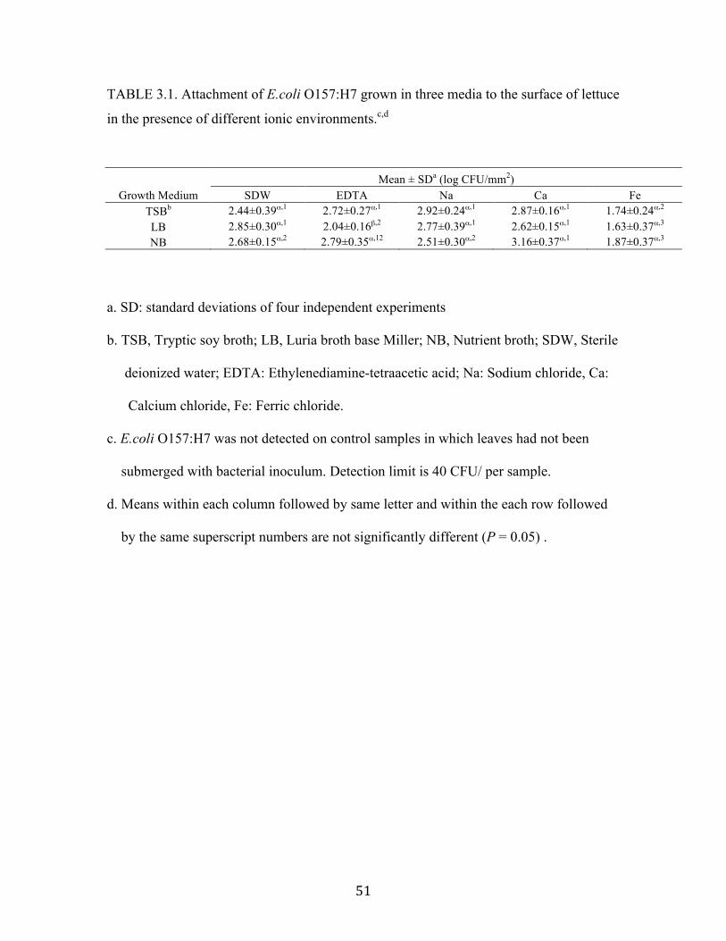
! &"!
TABLE 3.1. Attachment of E.coli O157:H7 grown in three media to the surface of lettuce
in the presence of different ionic environments.c,d
Mean ± SDa (log CFU/mm2)
Growth Medium SDW EDTA Na Ca Fe
TSBb 2.44±0.39",1 2.72±0.27",1 2.92±0.24",1 2.87±0.16",1 1.74±0.24",2
LB 2.85±0.30",1 2.04±0.16#,2 2.77±0.39",1 2.62±0.15",1 1.63±0.37",3
NB 2.68±0.15",2 2.79±0.35",12 2.51±0.30",2 3.16±0.37",1 1.87±0.37",3
a. SD: standard deviations of four independent experiments
b. TSB, Tryptic soy broth; LB, Luria broth base Miller; NB, Nutrient broth; SDW, Sterile
deionized water; EDTA: Ethylenediamine-tetraacetic acid; Na: Sodium chloride, Ca:
Calcium chloride, Fe: Ferric chloride.
c. E.coli O157:H7 was not detected on control samples in which leaves had not been
submerged with bacterial inoculum. Detection limit is 40 CFU/ per sample.
d. Means within each column followed by same letter and within the each row followed
by the same superscript numbers are not significantly different (P = 0.05) .
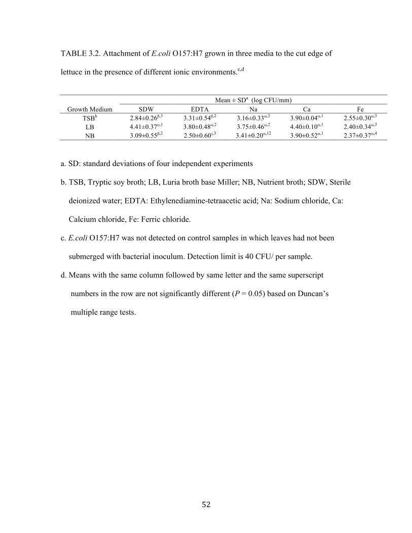
! &#!
TABLE 3.2. Attachment of E.coli O157:H7 grown in three media to the cut edge of
lettuce in the presence of different ionic environments.c,d
Mean ± SDa (log CFU/mm)
Growth Medium SDW EDTA Na Ca Fe
TSBb 2.84±0.26#,3 3.31±0.54#,2 3.16±0.33",2 3.90±0.04",1 2.55±0.30",3
LB 4.41±0.37",1 3.80±0.48",2 3.75±0.46",2 4.40±0.10",1 2.40±0.34",3
NB 3.09±0.55#,2 2.50±0.60$,3 3.41±0.20",12 3.90±0.52",1 2.37±0.37",4
a. SD: standard deviations of four independent experiments
b. TSB, Tryptic soy broth; LB, Luria broth base Miller; NB, Nutrient broth; SDW, Sterile
deionized water; EDTA: Ethylenediamine-tetraacetic acid; Na: Sodium chloride, Ca:
Calcium chloride, Fe: Ferric chloride.
c. E.coli O157:H7 was not detected on control samples in which leaves had not been
submerged with bacterial inoculum. Detection limit is 40 CFU/ per sample.
d. Means with the same column followed by same letter and the same superscript
numbers in the row are not significantly different (P = 0.05) based on Duncan’s
multiple range tests.

! &$!
TABLE 3.3. Attachment of E.coli O157:H7 grown in three media to the surface of
spinach in the presence of different ionic environments.c,d
Mean ± SDa (log CFU/mm2)
Growth Medium SDW EDTA Na Ca Fe
TSBb 3.43±0.22",1 3.19±0.20",2 3.05±0.26",2 3.24±0.07",2 2.85±0.35",3
LB 3.09±0.29",1 2.15±0.04#,2 2.80±0.38",1 3.21±0.17",1 2.75±0.30",2
NB 3.21±0.22",1 2.56±0.48"#,2 2.91±0.34",1 2.93±0.29",1 2.50±0.10",2
a. SD: standard deviations of four independent experiments
b. TSB, Tryptic soy broth; LB, Luria broth base Miller; NB, Nutrient broth; SDW, Sterile
deionized water; EDTA: Ethylenediamine-tetraacetic acid; Na: Sodium chloride, Ca:
Calcium chloride, Fe: Ferric chloride.
c. E.coli O157:H7 was not detected on control samples in which leaves had not been
submerged with bacterial inoculum. Detection limit is 40 CFU/ per sample.
d. Means with the same column followed by same letter and the same superscript
numbers in the row are not significantly different (P = 0.05) based on Duncan’s
multiple range tests.
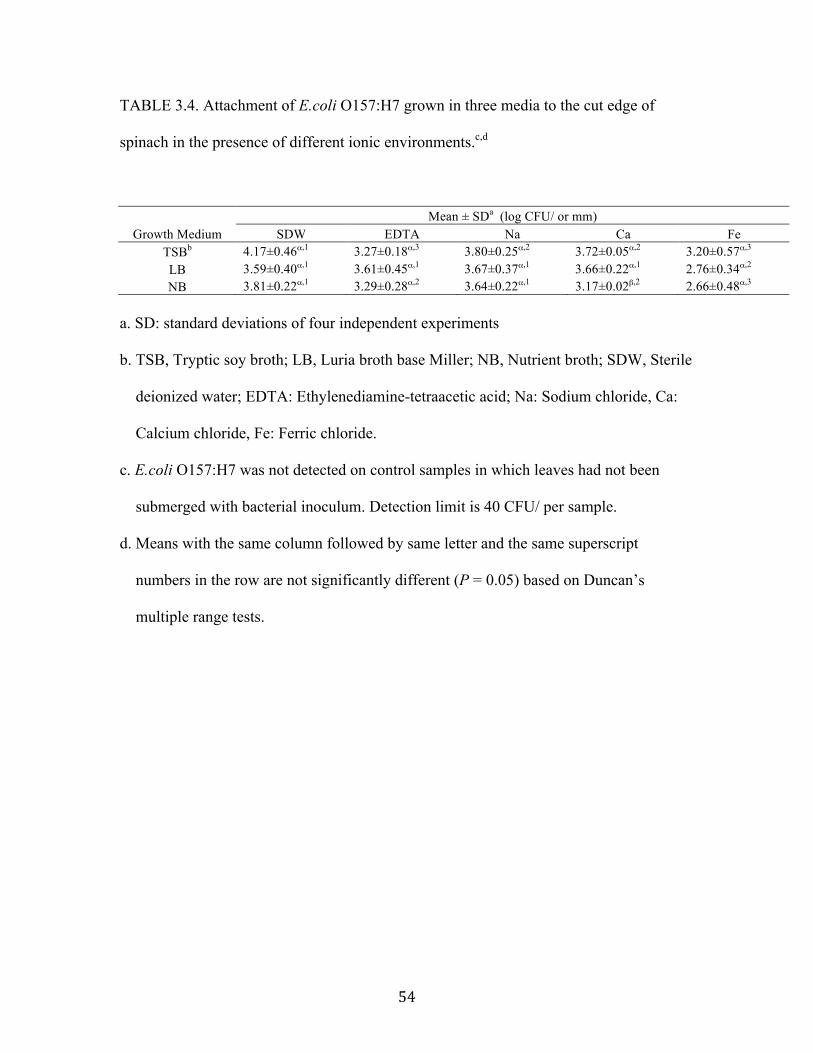
! &%!
TABLE 3.4. Attachment of E.coli O157:H7 grown in three media to the cut edge of
spinach in the presence of different ionic environments.c,d
Mean ± SDa (log CFU/ or mm)
Growth Medium SDW EDTA Na Ca Fe
TSBb 4.17±0.46",1 3.27±0.18",3 3.80±0.25",2 3.72±0.05",2 3.20±0.57",3
LB 3.59±0.40",1 3.61±0.45",1 3.67±0.37",1 3.66±0.22",1 2.76±0.34",2
NB 3.81±0.22",1 3.29±0.28",2 3.64±0.22",1 3.17±0.02#,2 2.66±0.48",3
a. SD: standard deviations of four independent experiments
b. TSB, Tryptic soy broth; LB, Luria broth base Miller; NB, Nutrient broth; SDW, Sterile
deionized water; EDTA: Ethylenediamine-tetraacetic acid; Na: Sodium chloride, Ca:
Calcium chloride, Fe: Ferric chloride.
c. E.coli O157:H7 was not detected on control samples in which leaves had not been
submerged with bacterial inoculum. Detection limit is 40 CFU/ per sample.
d. Means with the same column followed by same letter and the same superscript
numbers in the row are not significantly different (P = 0.05) based on Duncan’s
multiple range tests.
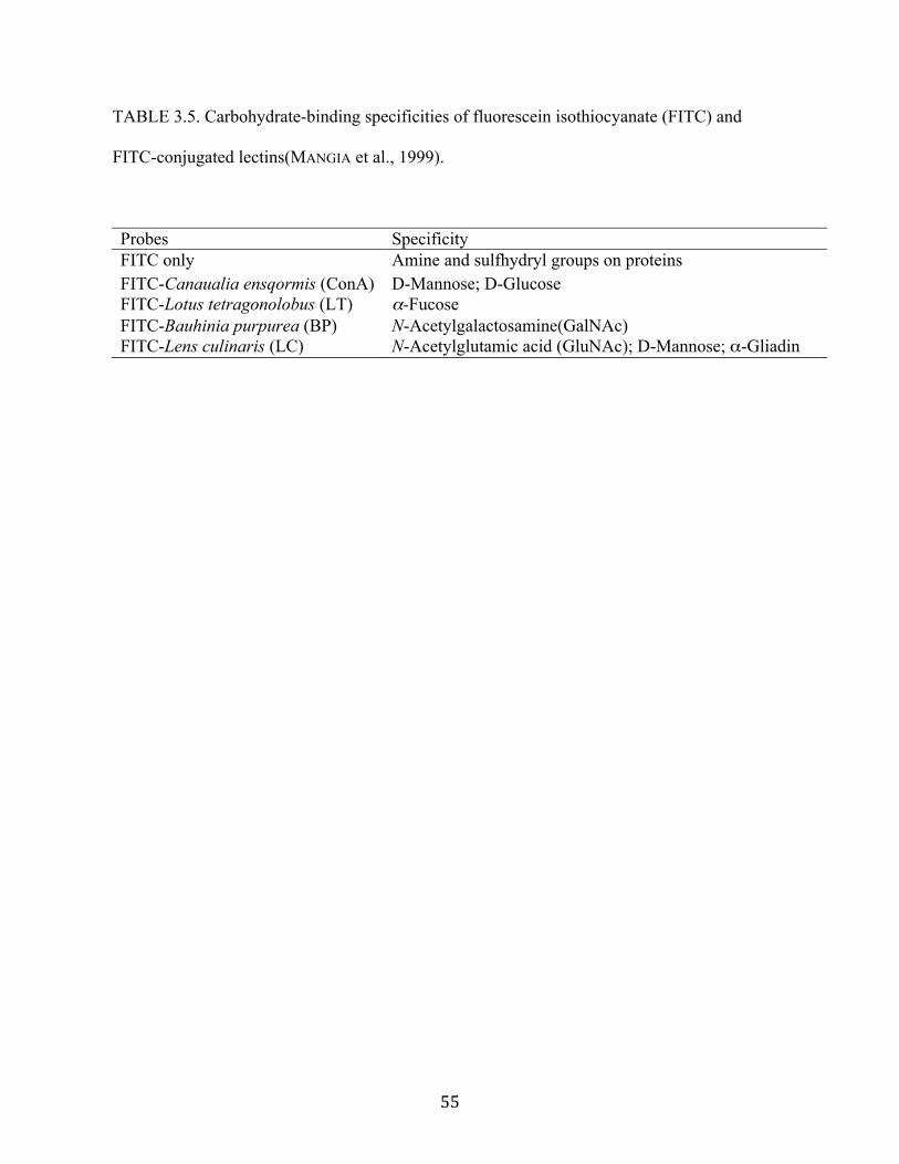
! &&!
TABLE 3.5. Carbohydrate-binding specificities of fluorescein isothiocyanate (FITC) and
FITC-conjugated lectins(MANGIA et al., 1999).
Probes Specificity FITC only Amine and sulfhydryl groups on proteins
FITC-Canaualia ensqormis (ConA) D-Mannose; D-Glucose FITC-Lotus tetragonolobus (LT) !-Fucose
FITC-Bauhinia purpurea (BP) N-Acetylgalactosamine(GalNAc) FITC-Lens culinaris (LC) N-Acetylglutamic acid (GluNAc); D-Mannose; "-Gliadin
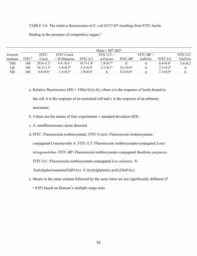
! &'!
TABLE 3.6. The relative fluorescence of E .coli O157:H7 resulting from FITC-lectin
binding in the presence of competitive sugars.e
a. Relative fluorescence (Rf) = 100(a-b)/(c-b), where a is the response of lectin bound to
the cell, b is the response of an unstained cell and c is the response of an arbitrary
maximum.
b. Values are the means of four experiments ± standard deviation (SD).
c. A: autofluorescence alone detected.
d. FITC: Fluoresecein isothiocyanate, FITC-ConA: Fluoresecein isothiocyanate-
conjugated Concanavalin A. FITC-LT: Fluoresecein isothiocyanate-conjugated Lotus
tetragonolobus. FITC-BP: Fluoresecein isothiocyanate-conjugated Bauhinia purpurea.
FITC-LC: Fluoresecein isothiocyanate-conjugated Len culinaris. N-
Acetylgalactosamine(GalNAc). N-Acetylglutamic acid (GluNAc).
e. Means in the same column followed by the same letter are not significantly different (P
= 0.05) based on Duncan’s multiple range tests.
Mean ± SDb (Rf)a Growth medium FITCd
FITC-ConA
FITC-ConA + D-Mannose FITC-LT
FITC-LT + #-Fucose FITC-BP
FITC-BP + GalNAc FITC-LC
FITC-LC+ GluNAc
TSB 100 29.6±5.2" 4.4 ±0.4 " 10.7±1.8" 1.9±0.7" Ac A 6.4±0.6" 3.6±0.2 LB 100 18.3±1.1# 1.4±0.5# 3.5±0.6# 3.1±0.1" 0.1±0.0" A 2.1±0.2# A NB 100 4.6±0.8$ 1.3±0.5# 1.9±0.6# A 0.2±0.0" A 1.3±0.4# A
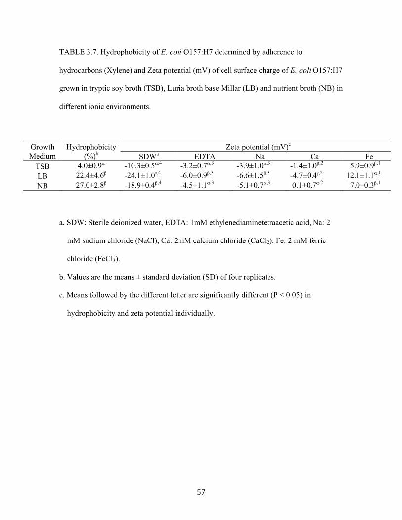
! &(!
TABLE 3.7. Hydrophobicity of E. coli O157:H7 determined by adherence to
hydrocarbons (Xylene) and Zeta potential (mV) of cell surface charge of E. coli O157:H7
grown in tryptic soy broth (TSB), Luria broth base Millar (LB) and nutrient broth (NB) in
different ionic environments.
a. SDW: Sterile deionized water, EDTA: 1mM ethylenediaminetetraacetic acid, Na: 2
mM sodium chloride (NaCl), Ca: 2mM calcium chloride (CaCl2). Fe: 2 mM ferric
chloride (FeCl3).
b. Values are the means ± standard deviation (SD) of four replicates.
c. Means followed by the different letter are significantly different (P < 0.05) in
hydrophobicity and zeta potential individually.
Zeta potential (mV)c Growth Medium
Hydrophobicity (%)b SDWa EDTA Na Ca Fe
TSB 4.0±0.9" -10.3±0.5",4 -3.2±0.7",3 -3.9±1.0",3 -1.4±1.0#,2 5.9±0.9#,1 LB 22.4±4.6# -24.1±1.0$,4 -6.0±0.9#,3 -6.6±1.5#,3 -4.7±0.4$,2 12.1±1.1",1
NB 27.0±2.8# -18.9±0.4#,4 -4.5±1.1",3 -5.1±0.7",3 0.1±0.7",2 7.0±0.3#,1
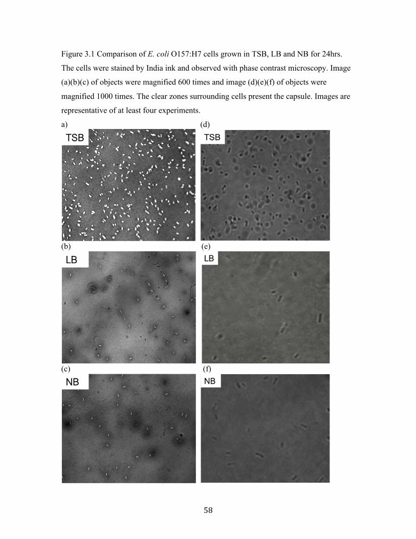
! &)!
Figure 3.1 Comparison of E. coli O157:H7 cells grown in TSB, LB and NB for 24hrs.
The cells were stained by India ink and observed with phase contrast microscopy. Image
(a)(b)(c) of objects were magnified 600 times and image (d)(e)(f) of objects were
magnified 1000 times. The clear zones surrounding cells present the capsule. Images are
representative of at least four experiments.
a) (d)
(b) (e)
(c) (f)
! &*!
Figure 3.2 Epifluorescence microscopic images of E.coli O157:H7 using fluorescein
isothiocyanate (FITC), FITC-labeled Concanavalin A agglutinin (ConA), FITC-labeled
lectin extracted from Lotus tetragonolobus agglutinin (LT), FITC- labeled lectin
extracted from Bauhinia purpurea agglutinin(BP) and FITC-labeled lectin extracted
from Lens culinaris (LC). E.coli O157:H7 was grown grown in tryptic soy (TSB),
nutrient broth (NB) and Luria broth base Miller (LB). Images are representative of at
least four experiments.
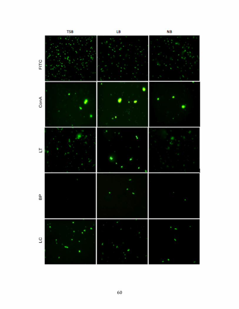
! '+!

! '"!
CHAPTER 4
THE ROLE OF CELLULOSE IN ATTACHMENT OF SHIGA TOXIN–PRODUCING
ESCHERICHIA COLI TO LETTUCE AND SPINACH IN DIFFERENT WATER
HARDNESS ENVIRONMENTS
Lee, C.C. and Frank, F. J. To be submitted to Journal of Food Protection
! '#!
ABSTRACT
Cellulose is a linear polysaccharide polymer composed of D-glucose with !
(1"4) glycosidic bond and usually exists in bacterial exopolysaccharide (EPS). The role
of cellulose produced by Shiga toxin–producing Escherichia coli (STEC) on attachment
to leafy greens is unclear. This study was undertaken to investigate the attachment of
cellulose-deficient derivatives of STEC to lettuce and spinach in different water hardness
environments. Two cellulose-producing wild-type STEC strains 19 and 49 and their
cellulose- deficient derivatives were used. Strain 49 also produces EPS comprised of
colanic acid. Viability of cells was determined by plate counts on the surface and cut
edge after leaves were inoculated with 108 CFU/ml cells at 4°C for 2 hours.
Hydrophobicity and surface charge were also determined. Strain 49 attached significantly
0.3 and 0.6 log greater to surface and 0.9 and 0.4 log greater cut edge of spinach
compared to strain 19 in wild-type and cellulose-deficient cells. In addition, there was
significantly greater attachment for cellulose-proficient cells on lettuce surface than
cellulose-deficient cells, but not on spinach surface. Contrary to the leaf surface results,
more cellulose-deficient cells attached (0.66 and 0.3 log greater) in strain 19 and 49 to cut
edge of lettuce than cellulose-proficient cells. Strain 19 was more hydrophobic than strain
49. In addition, there was a decreasing level of attachment for strain 49 when water
hardness increased from 200 to 1000pm on lettuce and spinach leaf surfaces. The zeta
potential of strain 49 was more negative than strain 19. Cells containing colanic
acid,which were less hydrophobic, had a greater potential to attach surface and cut edge
of spinach when compared to cellulose-producing cells which were more hydrophobic.

! '$!
Cell attachment to leafy green surfaces was also enhanced under higher water hardness
environments.
INTRODUCTION
Foodborne outbreaks of Shiga toxin-producing Escherichia coli (STEC) O157:H7
associated with consumption of leafy greens have been reported for decades
(ARUSCAVAGE et al., 2008; COOLEY et al., 2007; SODHA et al., 2011). In the United
States, STEC O157 is linked to 3,200 hospitalizations and more than 9,6000 episodes of
diarrheal illness every year (SCALLAN et al., 2011). In addition, outbreaks of STEC O157
are not only in US but also in European countries. In 2007, an international outbreak was
contaminated lettuce in the Netherlands and Iceland (FRIESEMA et al., 2008). In addition,
lettuce-linked outbreaks of pathogenic E. coli O157 were been reported in 2005 Sweden
and Enterotoxigenic E. coli in 2010 Denmark (ETHELBERG et al., 2010; SODERSTROM et
al., 2008).
Cell attachment is a complex process regulated by various characteristics of the cell
surface, growth medium and substratum (DONLAN, 2002). The hydrophobic and
electrokinetic properties of cells are crucial influences for cell attachment. The
hydrophobicity is affected by bacterial exopolysaccharides (EPS) components (GROSS
and LOGAN, 1995). The electrokinetic potential of cell surface charge can be linked to the
attachment of three E. coli strains on polymethacrylate surfaces (HARKES et al., 1991). In
addition, the components of bacterial EPS such as cellulose and colanic acid are
associated with cell attachment and biofilm formation. Cellulose was not required for E.
coli O157:H7 attachment to spinach leaves (MACARISIN et al., 2012) even though
! '%!
cellulose plays a crucial role in biofilm formation of E. coli 1094 on glass slides (DA RE
and GHIGO, 2006). Both cellulose and curli favor attachment of E. coli O127:H6 and
O157:H7 to cultured mammalian cells, cell colonization, biofilm development and
survival (SALDANA et al., 2009). The colanic acid production is significantly correlated
with adhesion forces of E. coli (RAZATOS et al., 1998) but biofilm formation of E. coli
BW25113 cells is inhibited due to colanic acid overproduction (ZHANG et al., 2008).
Hard water contains high amounts of minerals such as calcium bicarbonate and
magnesium bicarbonate. Water hardness of river and stream varies and is affected by
regional climate and geological system. The source of irrigation water in the field for
fresh produce is mainly from local stream and underground water. Therefore, we
hypothesized that the influence of water hardness would significantly affect attachment
of pathogenic E.coli to leafy greens. This study investigated the role of cellulose on
attachment of E .coli to iceberg lettuce and baby spinach leaves by using mutants lacking
cellulose production. The effect on cell attachment under different water hardness
environments, hydrophobicity and electronegativity of the cell surface was determined.
MATERIALS AND METHODS
Bacterial strains and culture conditions
E. coli O157:H7 994 (beef jerky isolate) was supplied by the Center for Food
Safety, University of Georgia, Griffin, GA. Cells were grown in tryptic soy broth (TSB)
at 35 °C for two successive 24 ± 2h transfers. The four STEC strains were obtained from
the laboratory of Dr. Jinru Chen, Department of Food Science and Technology,

! '&!
University of Georgia, Griffin. Two wild type cellulose-producing strains 19B (O5:H-)
and 49B (O103:H2) were used. In addition, two mutants 19D (O5:H-) and 49D
(O103:H2) which were their cellulose deficient derivates by spontaneous selection were
also used(YOO et al., 2010).The four strains were grown in Luria-Bertani no salt (LBNS)
broth at 28 °C by two successive 24 ± 2h transfers. Cultures were centrifuged at 4400g
(6,500 rpm) 4°C for 15 min. The supernatant fluid was discarded. Pellet was washed
twice with sterile de-ionized water and suspended in the appropriate amount of sterile de-
ionized water to contain approximately 8 log CFU/ml for sample inoculation.
Leafy green preparation
Iceberg lettuce (Lactuca sativa) and baby spinach (Spinacia oleracea) were
purchased in local grocery stores. Undamaged leaves were selected for use. Inner leaves
of iceberg lettuce were cut into designated sizes (square piece 2 ! 2 cm) after aseptically
removing outer leaves and core by using a sterile knife and surgical scalpel. In addition,
the petioles of the baby spinach leaves were removed and the leaves cut into 2 ! 2 cm
squares using a sterile surgical scalpel. All pieces of leaves were rinsed five times with
excess sterile de-ionized water (SDW).
Quantification of cell attachment
Synthetic hard water was prepared according to Association of Official Analytical
Chemists (AOAC) official methods 960.09(E). For attachment experiments, a square leaf
section was submerged in 10mL bacterial suspension with 0, 50, 100, 150, 200, 400, 700
and 1000ppm water hardness at 4°C for 2h. Leaves were taken from the cell suspension

! ''!
using sterile tweezers and rinsed twice with excess SDW. The outermost tissue in each
side of square leave piece was removed with a sterile surgical scalpel as cut edge 0.15cm.
The cut edge leave tissue and inner portions of the leaves (intact surface) were separately
combined with 10 ml of sterile 0.1% peptone water solution in polypropylene tubes and
the cells released from the tissue by disruption using a homogenizer (Omni mixer, model
17105) set at energy level 5 for 30 seconds, sufficient to disrupt the leaf tissue. Samples
were immersed in an ice bath during homogenization. Homogenized samples were
serially diluted in sterile 0.1% peptone water. The number of viable bacteria attached to
each leave surface and cut edge were determined by plating on sorbitol-MacConkey
(SMAC) agar with incubation at 35°C for 24h.
Hydrophobicity assay
Hydrophobicity was estimated semi-quantitatively using bacterial adhesion to
hydrocarbon as described by(LI and MCLANDSBOROUGH, 1999b). Cultures were grown
as previously described. Cells were suspended in phosphate buffered saline (PBS) after
centrifugation, and the pellet was washed twice as previously described. Bacterial
suspensions (4 ml) were added to each of four 10-mm diameter glass test tubes for each
bacterium tested, representing three tests and one control. One ml of xylene was added to
each bacterial assay tube except for the control. Tubes were vortex-mixed by using a
mini-vortexer (VWR Scientific products) for 40s and placed in 37°C water bath for 30
mins to allow the immiscible xylene and aqueous phases to separate. The aqueous lower
layer (3 ml portion) in each tube was transferred to clean tubes by using Pasteur pipettes.
Excess hydrocarbon was removed by bubbling air through longer vortex for 40s. The

! '(!
absorbance at 600 nm of the aqueous layer was determined using a Beckman DU-530
spectrophotometer. The percentage reduction in absorbance of the bacterial assay tubes
compared to the control (without xylene) was calculated.
Adhesion to Hydrocarbon (%) = 100!(Ac - Ab) / Ac.
Ab: absorbence of bacterial assay tubes,
Ac: absorbence of the control tubes
Cell surface charge measurement
Cells were prepared as described above and suspended individually in 0, 50, 100,
150, 200, 400, 700 and100ppm water hardness solution. Zeta potential in each suspension
was measured using Brookhaven’s Zeta Plus instrument (Brookhoven Instruments,
Holtsville, New York).
Statistical analysis
All data were analyzed with Statistical Analysis System (SAS) Software version
9.1.3 (SAS Institute Inc., Cary, NC) using the general liner model (GLM) procedure.
Analysis of variance (ANOVA) was calculated using the overall means of four
replications obtained from cells grown in three media using a significance level of P
value <0.05. Difference among sample means was determined using Duncan’s multiple-
range test (alpha = 5%).

! ')!
RESULTS
Attachment of STEC on the surface and cut edge of lettuce and spinach
The attachment of STEC population to lettuce and spinach leaves was
investigated by using wild-type cells and cellulose-deficient mutants in SDW. On the leaf
surface of iceberg lettuce, wild-type strains 19B and 49B attached at significantly greater
levels (0.5 and 0.9 log units) than cellulose-deficient strains 19D and 49D (Table 4.1).
These data indicate that cellulose on the cell surface enhances the ability of the cells to
attach to lettuce leaf. However, there is less attachment of wild type (19B and 49B) cells
to cut edge of lettuce than their cellulose-deficient mutant. More cellulose-deficient cells
attached (0.7 and 0.3 log greater) for strain 19 and 49 to the cut edge of lettuce than
cellulose-producing cells. These data indicate that cellulose-production may decrease cell
attachment to leaf cut edge.
For spinach leaves, strain 49B and 49D attached 0.3 and 0.6 log greater to surface
and 0.9 and 0.4 log greater to cut edge compared to strain 19B and 19D. There was no
significant difference in attachment between wild-type cell and cellulose-deficient
mutant. These data demonstrate that colanic acid in the cell envelope may play a more
important role than extra-cellular cellulose to increase attachment ability to spinach leave
surface and cut edge (Table 4.1).
The examination of bacterial hydrophobicity
Four strains grown in LBNS broth were evaluated for their bacterial
hydrophobicity towards hydrocarbons (xylene) on cell surface. Strain 19 cells were
significantly different with strain 49 cells. Strain 19B and 19D had relatively more
! '*!
affinity to xylene (55.57% and 46.20% respectively) followed by strain 49B and 49D for
which the percent hydrophobicity values were 20.22% and 28.82%, respectively. Strain
19B and 19D cells were significantly more hydrophobic than colanic acid–producing
strain 49B and 49D cells (Table 4.2). These results imply that the presence of colanic
acid may be associated with a more hydrophilic cell surface.
Attachment of STEC to lettuce in different water hardness environments
The influences of cellulose-production and a water hardness environment on
STEC attachment to lettuce and spinach leaves were determined. Data presented in
Figure 4.1 indicate that there was a progressive ordering with increasing attachment of
strain 49B/49D cells with water hardness levels of 50ppm/100ppm to 1000ppm on lettuce
leaves surfaces. However, this result was not observed in one wild type strain (19). In
addition, strain 49 (wild type) cells attached to lettuce more than strain 19 (wild type) in
hard water. This indicates that the influence of colanic acid to cell attachment in different
water hardness environments is more important than cellulose.
At the cut edge of lettuce leaves, the attachment population of strain 49 cells in
SDW is significantly higher than in hard water. In addition, there was no significantly
different attachment population of strain 19 and 49 (wild type) cells among all levels of
water hardness. These results indicate that the ability of cells to attach to cut edges is not
enhanced in the presence of hard water. The result when using strain 994 was similar to
that obtained with strain 49. These results demonstrate that the attachment ability of
E.coli O157 increases with increasing water hardness.
! (+!
Fewer 19D (cellulose-deficient, colonic deficient) cells attached to lettuce
leafsurfaces in SDW when compared to attachment in hard water. These results indicate
that water hardness may play an important role on cell attachment in the absence of
cellulose and colanic acid, likely due to increased electrostatic interactions. The influence
of cellulose to cell attachment was clearly observed only in 50ppm water hardness for
strain 19 and 49 cells on surface. The number of cellulose-deficient mutant was
significantly more than the wild-type parent cells. It indicated that the presence of
cellulose might reduce cell attachment in low water hardness environment. Therefore, it
appears that the presence of minerals in water may compensate for the lack of EPS
production in cell attachment.
Attachment of STEC to spinach in different water hardness environments
The influences of cellulose and water hardness for STEC attachment to spinach
leaves was also investigated (Figure 4.2). There was an ordering of decreasing attachment
of strain 49 with increase in water hardness from 0 to 150ppm, and afterwards from 200
to 1000 ppm in order progressively increasing attachment of cells on spinach leaves
surface. But this result of was not found for strain 19.
No influence of water hardness on cell attachment on cut edge was observed.
However, the result of 19D on spinach was similar to that obtained for lettuce, as cells in
hard water were able to attached more than in SDW. As with lettuce, the ions in hard
water may enhance cell attachment to spinach leaf surfaces and cut edges where there is
an absence of cellulose and colanic acid. There was no significant difference among all
! ("!
levels of water hardness and between wild-type parents 19 cell and mutant on leaf
surfaces.
The measurement of cell surface charge
The zeta potential of cell surface was measured in 0 ppm to 1000 ppm water
hardness environments. A difference of electrokinetic properties on cell surface was
observed. In Figure 4.3, the zeta potential of strain 49 was significantly more negative
than that of strain 19 at all water hardness levels. In addition, the surface charge of cells
in SDW was also significantly more negative than in any level of hard water.
DISCUSSION
The cellulose-deficient mutants had extensively lower attachment ability to
lettuce leave than wild-type parent cells. However, cellulose did not play a significant
role in attachment to spinach leave surface and cut edge; rather the role of colanic acid
was significantly more influential instead. In previous studies, the importance of
cellulose and colanic acid for attachment of E. coli to other surfaces has been addressed
in different results. Colanic acid and cellulose were associated with the biofilm
formation of E.coli K-12 on plastic surface but not required for E. coli O157:H7 attaching
to mammalian cells. In addition, there was reduction of E. coli O157:H7 cellulose
synthase deficient mutants in binding to alfalfa sprouts and colanic acid deficient mutants
also exhibited a large reduction in attachment (MATTHYSSE et al., 2008). On the other
hand, E. coli O157:H7 did not produce cellulose as a part of the extracellular matrix
! (#!
during its own biofilm formation on glass and polystyrene surface (UHLICH et al., 2006).
Cellulose was not found to be a necessary factor for developing strong attachment to
spinach leaves (MACARISIN et al., 2012). Colanic acid has been showed to be required for
developing biofilm of E. coli, but not for attachment (DANESE et al., 2000).
Leaf surfaces resist the attachment of microorganisms by the presence of a
hydrophobic waxy cuticle layer (ANDREWS and HIRANO, 1991). E. coli O157:H7 prefer
to attach on the cracked cuticle or damaged part of leaves which lacks the waxy tissue
because the surface of E. coli O157:H7 is highly hydrophilic (DEWANTI and WONG,
1995; MAFU et al., 1991) and wax is hydrophobic. Previous research found that viable E.
coli O157:H7 cells preferentially attached to the cut edge of lettuce leaves instead of to
intact leaf surface as visualized by confocal scanning laser microscopy (TAKEUCHI et al.,
2000b). Strain 49 with colanic acid production was less hydrophobic and its surface
charge was more negative than strain 19. This surface characteristic may explain why
strain 49 attached preferentially to spinach leaf surfaces and cut edges compared to strain
19. On the other hand, the influence of hydrophobic and electrostatic interactions was not
observed in cell attachment to lettuce surfaces. It seems that cellulose has a more crucial
role in E. coli attachment to lettuce leaf.
In this study, synthetic hard water was prepared based on Association of Official
Analytical Chemists official methods. The hard water contained calcium and magnesium
cations (Ca2+,Mg2+). In previous research, the influence of divalent cations on cell
attachment has not been consistent. Magnesium cations are more effective than calcium
cations in helping the attachment of chicken fibroblasts to protein-coated plastic surfaces.
However, increasing the concentration of divalent cations did not enhance cell
! ($!
attachment and the initiation of cell attachment seemed be inhibited by high calcium
concentration (TAKEICHI and OKADA, 1972). The attachment of Pseudomonas
fluorescens to glass surfaces was also affected by an increase in the concentration of
several cations such as calcium, ferric iron, lanthanum and sodium (FLETCHER, 1988).
When strain 49 (wild-type) cells were suspended in the higher water hardness,
attachment to lettuce and spinach leaves also increased. E. coli O157:H7 also gave
similar results when attaching to lettuce leave, except for strain 19. According to the U.S.
Department of the Interior and U.S. Geological Survey (USGS) in 2013, high hard waters
were measured in streams in Kansas, Texas, New Mexico, Arizona and southern
California. Irrigation water from streams in these areas may enhance cells attachment to
iceberg lettuce and baby spinach leaves,increasing the risk of E.coli O157:H7 outbreak
on leafy greens.
CONCLUSION
Cellulose-proficient cells exhibited a significantly greater level of attachment on
lettuce surfaces than cellulose-deficient cells, but the same was not found for spinach
surface. STEC strain 49B and 49D cells producing colanic acid were less hydrophobic
and had a higher negative zeta potential. These cells also demonstrated significantly
greater potentials to attach to the surfaces and cut edges of spinach in comparison with
the more hydrophobic and less negative charges of the cells of STEC strains 19B and
19D. In high water hardness environments, attachment of strain 49 (wild type) cells to
leafy green surfaces was also enhanced.

! (%!
ACKNOWLEDGEMENTS
We thank Dr. Jinru Chen for providing the strains, mutants and technical advice.
The research was funded by the state and Hatch funds allocated to the Georgia
Agricultural experiment stations.
REFERENCES
Andrews, J.H. and Hirano, S.S., 1991. Microbial ecology of leaves. Springer-Verlag, Inc.,
New York.
Aruscavage, D., Miller, S.A., Ivey, M.L., Lee, K., and LeJeune, J.T., 2008. Survival and
dissemination of Escherichia coli O157:H7 on physically and biologically
damaged lettuce plants. J Food Prot 71:2384-2388.
Cooley, M., Carychao, D., Crawford-Miksza, L., Jay, M.T., Myers, C., Rose, C., Keys,
C., Farrar, J., and Mandrell, R.E., 2007. Incidence and tracking of Escherichia
coli O157:H7 in a major produce production region in California. PLoS One
2:e1159.
Da Re, S. and Ghigo, J.M., 2006. A CsgD-independent pathway for cellulose production
and biofilm formation in Escherichia coli. J Bacteriol 188:3073-3087.
Danese, P.N., Pratt, L.A., and Kolter, R., 2000. Exopolysaccharide production is required
for development of Escherichia coli K-12 biofilm architecture. J Bacteriol
182:3593-3596.
Dewanti, R. and Wong, A.C., 1995. Influence of culture conditions on biofilm formation
by Escherichia coli O157:H7. Int J Food Microbiol 26:147-164.
Donlan, R.M., 2002. Biofilms: microbial life on surfaces. Emerg Infect Dis 8:881-890.
Ethelberg, S., Lisby, M., Bottiger, B., Schultz, A.C., Villif, A., Jensen, T., Olsen, K.E.,
Scheutz, F., Kjelso, C., and Muller, L., 2010. Outbreaks of gastroenteritis linked
to lettuce, Denmark, January 2010. Euro Surveill 15.
! (&!
Fletcher, M., 1988. Attachment of Pseudomonas fluorescens to glass and influence of
electrolytes on bacterium-substratum separation distance. J Bacteriol 170:2027-
2030.
Friesema, I., Sigmundsdottir, G., van der Zwaluw, K., Heuvelink, A., Schimmer, B., de
Jager, C., Rump, B., Briem, H., Hardardottir, H., Atladottir, A., Gudmundsdottir,
E., and van Pelt, W., 2008. An international outbreak of Shiga toxin-producing
Escherichia coli O157 infection due to lettuce, September-October 2007. Euro
Surveill 13.
Gross, M.J. and Logan, B.E., 1995. Influence of different chemical treatments on
transport of Alcaligenes paradoxus in porous media. Appl Environ Microbiol
61:1750-1756.
Harkes, G., Feijen, J., and Dankert, J., 1991. Adhesion of Escherichia coli on to a series
of poly(methacrylates) differing in charge and hydrophobicity. Biomaterials
12:853-860.
Li, J. and McLandsborough, L.A., 1999. The effects of the surface charge and
hydrophobicity of Escherichia coli on its adhesion to beef muscle. Int J Food
Microbiol 53:185-193.
Macarisin, D., Patel, J., Bauchan, G., Giron, J.A., and Sharma, V.K., 2012. Role of curli
and cellulose expression in adherence of Escherichia coli O157:H7 to spinach
leaves. Foodborne Pathog Dis 9:160-167.
Mafu, A.A., Roy, D., Goulet, J., and Savoie, L., 1991. Characterization of
physicochemical forces involved in adhesion of Listeria monocytogenes to
surfaces. Appl Environ Microbiol 57:1969-1973.
Matthysse, A.G., Deora, R., Mishra, M., and Torres, A.G., 2008. Polysaccharides
cellulose, poly-beta-1,6-n-acetyl-D-glucosamine, and colanic acid are required for
optimal binding of Escherichia coli O157:H7 strains to alfalfa sprouts and K-12
strains to plastic but not for binding to epithelial cells. Appl Environ Microbiol
74:2384-2390.
Razatos, A., Ong, Y.L., Sharma, M.M., and Georgiou, G., 1998. Molecular determinants
of bacterial adhesion monitored by atomic force microscopy. Proc Natl Acad Sci
U S A 95:11059-11064.

! ('!
Saldana, Z., Xicohtencatl-Cortes, J., Avelino, F., Phillips, A.D., Kaper, J.B., Puente, J.L.,
and Giron, J.A., 2009. Synergistic role of curli and cellulose in cell adherence and
biofilm formation of attaching and effacing Escherichia coli and identification of
Fis as a negative regulator of curli. Environ Microbiol 11:992-1006.
Scallan, E., Hoekstra, R.M., Angulo, F.J., Tauxe, R.V., Widdowson, M.A., Roy, S.L.,
Jones, J.L., and Griffin, P.M., 2011. Foodborne illness acquired in the United
States--major pathogens. Emerg Infect Dis 17:7-15.
Soderstrom, A., Osterberg, P., Lindqvist, A., Jonsson, B., Lindberg, A., Blide Ulander,
S., Welinder-Olsson, C., Lofdahl, S., Kaijser, B., De Jong, B., Kuhlmann-
Berenzon, S., Boqvist, S., Eriksson, E., Szanto, E., Andersson, S., Allestam, G.,
Hedenstrom, I., Ledet Muller, L., and Andersson, Y., 2008. A large Escherichia
coli O157 outbreak in Sweden associated with locally produced lettuce.
Foodborne Pathog Dis 5:339-349.
Sodha, S.V., Lynch, M., Wannemuehler, K., Leeper, M., Malavet, M., Schaffzin, J.,
Chen, T., Langer, A., Glenshaw, M., Hoefer, D., Dumas, N., Lind, L., Iwamoto,
M., Ayers, T., Nguyen, T., Biggerstaff, M., Olson, C., Sheth, A., and Braden, C.,
2011. Multistate outbreak of Escherichia coli O157:H7 infections associated with
a national fast-food chain, 2006: a study incorporating epidemiological and food
source traceback results. Epidemiol Infect 139:309-316.
Takeichi, M. and Okada, T.S., 1972. Roles of magnesium and calcium ions in cell-to-
substrate adhesion. Exp Cell Res 74:51-60.
Takeuchi, K., Matute, C.M., Hassan, A.N., and Frank, J.F., 2000. Comparison of the
attachment of Escherichia coli O157:H7, Listeria monocytogenes, Salmonella
typhimurium, and Pseudomonas fluorescens to lettuce leaves. J Food Prot
63:1433-1437.
Uhlich, G.A., Cooke, P.H., and Solomon, E.B., 2006. Analyses of the red-dry-rough
phenotype of an Escherichia coli O157:H7 strain and its role in biofilm formation
and resistance to antibacterial agents. Appl Environ Microbiol 72:2564-2572.
Yoo, B.K., Stewart, T., Guard-Bouldin, J., Musgrove, M., Gast, R., and Chen, J., 2010.
Selection and characterization of cellulose-deficient derivates of shiga toxin-
producing Escherichia coli. J Food Protect 73:1038-1046.
! ((!
Zhang, X.S., Garcia-Contreras, R., and Wood, T.K., 2008. Escherichia coli transcription
factor YncC (McbR) regulates colanic acid and biofilm formation by repressing
expression of periplasmic protein YbiM (McbA). ISME J 2:615-631.
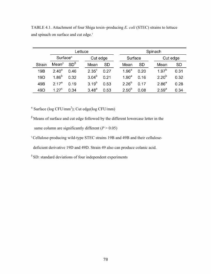
! ()!
TABLE 4.1. Attachment of four Shiga toxin–producing E. coli (STEC) strains to lettuce
and spinach on surface and cut edge.$
# Surface (log CFU/mm2); Cut edge(log CFU/mm)
! Means of surface and cut edge followed by the different lowercase letter in the
same column are significantly different (P > 0.05)
$ Cellulose-producing wild-type STEC strains 19B and 49B and their cellulose-
deficient derivative 19D and 49D. Strain 49 also can produce colanic acid.
$ SD: standard deviations of four independent experiments
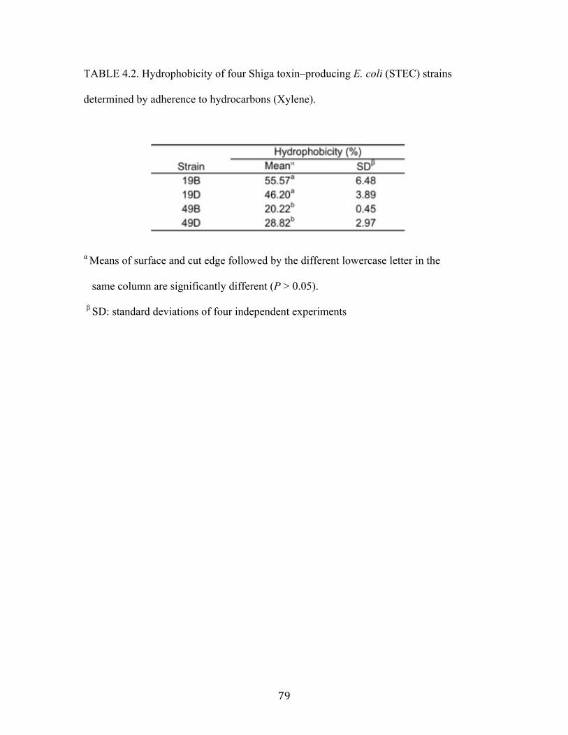
! (*!
TABLE 4.2. Hydrophobicity of four Shiga toxin–producing E. coli (STEC) strains
determined by adherence to hydrocarbons (Xylene).
# Means of surface and cut edge followed by the different lowercase letter in the
same column are significantly different (P > 0.05).
! SD: standard deviations of four independent experiments
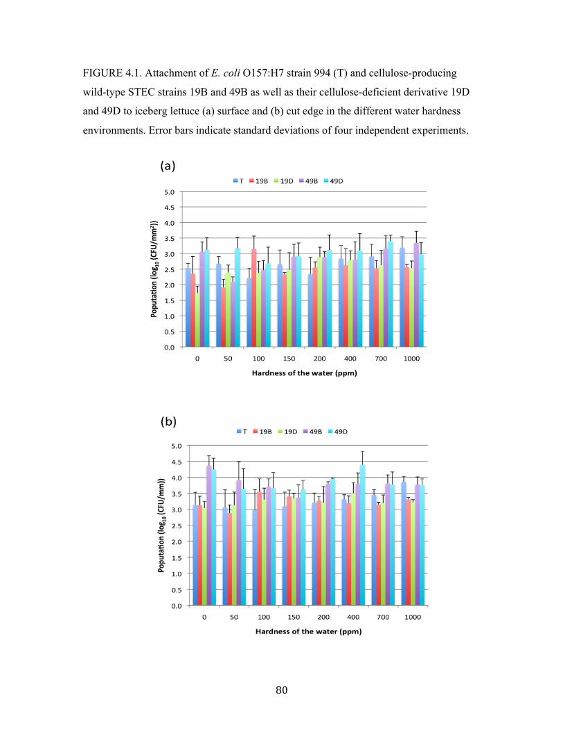
! )+!
FIGURE 4.1. Attachment of E. coli O157:H7 strain 994 (T) and cellulose-producing
wild-type STEC strains 19B and 49B as well as their cellulose-deficient derivative 19D
and 49D to iceberg lettuce (a) surface and (b) cut edge in the different water hardness
environments. Error bars indicate standard deviations of four independent experiments.
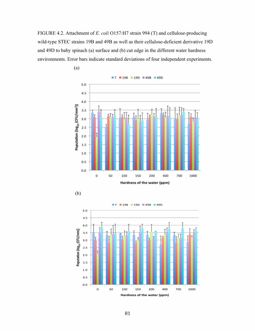
! )"!
FIGURE 4.2. Attachment of E. coli O157:H7 strain 994 (T) and cellulose-producing
wild-type STEC strains 19B and 49B as well as their cellulose-deficient derivative 19D
and 49D to baby spinach (a) surface and (b) cut edge in the different water hardness
environments. Error bars indicate standard deviations of four independent experiments.
(a)
(b)

! )#!
FIGURE 4.3. Zeta potential (mV) of cell surface charge of E. coli O157:H7 strain 994
(T) and cellulose-producing wild-type STEC strains 19B and 49B as well as their
cellulose-deficient derivative 19D and 19D in the different water hardness environments.

! )$!
CHAPTER 5
INFLUENCE OF EXTRA-CELLULAR CELLULOSE PRODUCTION ON THE
SURVIVAL OF SHIGA TOXIN-PRODUCING ESCHERICHIA COLI ON SPINACH
AND LETTUCE AFTER CHLORINE TREATMENT
Lee, C.C. and Frank, F. J. To be submitted to Journal of Food Protection
! )%!
ABSTRACT
Chlorinated water with the maximum allowable level 200 ppm free chlorine is widely
used to sanitize water used in the fresh-cut produce industry. Shiga toxin–producing
Escherichia coli (STEC) produce extra-cellular cellulose which may influence stress
tolerance. This study was undertaken to investigate the role of extra-cellular cellulose
produced by STEC on the survival of STEC after attachment to lettuce and spinach
followed by chlorine treatment. Four STEC strains, two wild-type cellulose-producing
and their cellulose-deficient derivatives, were used. One strain produced colanic acid in
addition to cellulose. Leaves of spinach and lettuce were treated with chlorinated water
(50 and 150 ppm) after cells were attached to the surfaces and cut edges of lettuce at 4 °C
for 2 hours. Chlorine treatment reduced populations of cellulose-deficient cells 1.2 log
CFU/ mm2 more than the wild type in 150ppm of chlorine on spinach leaf surfaces.
However, populations of cellulose-producing cells were reduced by 1.5 log CFU/mm2
more than its mutant counterparts when the cells can also produce colanic acid. There
were no significant differences in survival between four strains attached at the cut edge.
Extra-cellular cellulose production protects STEC cells attached to leafy greens from the
effects of chlorine on spinach leaf surfaces.
! )&!
INTRODUCTION
For decades, a variety of Shiga toxin-producing Escherichia coli (STEC)
serotypes have been associated with foodborne outbreaks in the United States (BROOKS et
al., 2005; PATON and PATON, 2003; RANGEL et al., 2005). They cause severe
gastroenteritis in human including mild to bloody diarrhea and hemolytic-uremic
syndrome (HUS) (GOULD et al., 2013; GRIFFIN and TAUXE, 1991). Multistate outbreaks
of STEC O157 and non-O157 infections linked to fresh produce have been reported by
Centers for Diseases Control and Prevention (CDC). These include STEC O26 in raw
clover sprouts at restaurants in 2012, STEC O157:H7 in organic spinach and spring mix
blend in 2012 and STEC O121 in frozen food products in 2013.
Microorganisms prefer to attach, aggregate, grow in community-based sedentary
microcolonies, and produce biofilms on surfaces in various environments including
freshwater, marine water, food, and medical and industrial systems (CARPENTIER and
CERF, 1993; GIBSON et al., 1999; MOSTELLER and BISHOP, 1993). For the food
industry, the formation of biofilms is of concern because they can be found on food
contact surfaces, equipment and water distribution systems and they contributes to
product contamination and foodborne illness outbreaks, especially those associated with
pathogenic and spoilage microorganisms (CHMIELEWSKI and FRANK, 2003). Therefore,
effective sanitizing in food industry and restaurants play an important role to decreasing
the risk of food contamination.
Attachment of pathogens to food contact surfaces has been investigated by many
researchers (FRANK and KOFFI, 1990; KIM and FRANK, 1994; TAKEUCHI et al., 2000a).
The efficiency of disinfectants against a variety of pathogenic biofilms has been

! )'!
determined (DHALIWAL et al., 1992; RONNER and WONG, 1993). Chlorine is the most
commonly applied sanitizer for water used in the food industry because of its low-cost,
accessibility and acceptable disinfecting effect. Its most potent bactericidal form is
hypochlorous acid (HOCl) from anion hypochlorite (OCl%). Although chlorine washing
is widely applied on fresh produce, it is not sufficient to inactivate and remove
microorganisms from vegetables and fruits (BEUCHAT, 1998; BRACKETT, 1992;
FISHBURN et al., 2012). Its main function is to prevent cross-contamination. Chlorine
solution at permitted concentration (< 200ppm) can typically reduce the population of
bacteria on fresh-cut produce surface by 1 to 2 log units (BEUCHAT et al., 1998;
KESKINEN et al., 2009; SAPERS et al., 1999; ZHUANG et al., 1995). Many viable E. coli
O157:H7 cells can be observed on the cut edges and stomata of lettuce leaves after
chlorine treatment (SEO and FRANK, 1999a).
Survival and growth of E. coli in foods is associated with numerous
environmental and food-related factors such as temperature, pH, and water activity.
Pathogenic and nonpathogenic E. coli have various physiological responses to sublethal
intrinsic and extrinsic stress produced by heat, acidity, osmosis, oxidation, and sanitizer
exposure (CHUNG et al., 2006). When microorganisms encounter environmental stress,
multiple defense mechanisms are triggered and cell envelope structure is modified. E.
coli is able to secrete the bacterial exopolysaccharides (EPS) including celloluse and
colanic acid that are associated with the cell surface. EPS consists of long chain polymers
and is involved with formation of biofilm as well as stress defenses. Colanic acid is
encoded by wca gene cluster and provides E. coli O157:H7 protection against acidic,
heat, osmotic and oxidative stresses (CHEN et al., 2004; MAO et al., 2001). Cellulose
! )(!
consists of a linear long chain of !(1"4) linked D-glucose units and may protect STEC
against oxidative and acidic treatments (YOO and CHEN, 2012). The hypothesis of this
study is that the protective effect of cellulose and calonic acid of STEC against chlorine
solution could be observed on leafy green surfaces and cut edges. The objective was to
determine the influence of cellulose on survival of E. coli O157 attached to iceberg
lettuce and baby spinach after chlorine treatment. Two cellulose-producing wild-type
STEC cells and their cellulose deficient derivatives were utilized in this study and two
levels of chlorine concentration were employed in the treatment.
MATERIALS AND METHODS
Spinach and Lettuce
Baby spinach(Spinacia oleracea) and iceberg lettuce (Lactuca sativa) were
purchased from retail grocery stores in Athens, Georgia. The petioles of the spinach
leaves were removed and the selected intact leaves were cut into 2 by 2 cm squares
without including midrib parts by using a sterile surgical scalpel. The core and outer
leaves of lettuce were discarded by using a sterile knife and surgical scalpel. Undamaged
inner leaves of lettuce were cut into designated pieces (2 by 2 mm). All pieces of leaves
were rinsed five times with sterile de-ionized water (SDW) to remove soil on the leaf
surfaces before following inoculation experiments.

! ))!
Preparation of STEC strains and their cellulose deficient derivates
The four STEC strains were obtained from the laboratory of Dr. Jinru Chen,
Department of Food Science and Technology, University of Georgia, Griffin. Two wild
type cellulose-producing strains 19B (O5:H-) and 49B (O103:H2) were used. In addition,
two mutants 19D (O5:H-) and 49D (O103:H2) which were their cellulose deficient
derivatives by spontaneous selection were also used (YOO et al., 2010). The four strains
were grown in Luria-Bertani no salt (LBNS) broth at 28 °C by two successive 24 ± 2h
transfers. E. coli O157:H7 994 (beef jerky isolate) was supplied from the Center for Food
Safety, University of Georgia, Griffin, GA. Cells were grown in tryptic soy broth (TSB)
at 35 °C for two successive 24 ± 2h transfers. Cells were collected by centrifuging at
4400g (6,500 rpm) 4°C for 15 min. After the supernatant fluid was discarded, the pellet
was washed twice with SDW and suspended in the appropriate amount of SDW to
contain approximately 8 log CFU/ml for sample inoculation.
Chlorine treatment after cell attachment
A square piece of spinach or lettuce leaf was submerged in 10mL bacterial inoculum at
4°C for 2hrs to allow cells to attach to surfaces and cut edges. Leaves were taken from
the cell suspension with sterile tweezers and rinsed twice with excess SDW to remove
unattached cells. Leaves were then submerged with 50 and 150ppm chlorinated water for
2 minutes. Leaves were taken from the chlorinated solution with sterile tweezers and
rinsed three times with excess SDW to remove chlorine solution.

! )*!
Determination of STEC Survival
To enumerate survival of STEC after treatment, the outermost leaf tissue in each
side of the square piece was removed by using a sterile surgical scalpel to provide a
1.5mm wide strip of the cut edge and total four cut edge strips from one square piece of
leaf. The cut edge tissue and inner portions of the leaves (intact surface) were separately
combined with 10 ml of sterile 0.1% peptone water solution and placed in homogenizer
blender bags. Cells were released by disrupting tissue using hand massaging to squeeze
the bag for 1min. The leaf juice was serially diluted with 0.1% sterile peptone water. The
population of viable cells on each leaf surface and cut edge were determined by plating
using tryptic soy agar (TSA) and sorbitol-MacConkey (SMAC) agar incubated at 35°C
for 24hrs.
Statistical analysis
Each experiment was utilized duplicate leaves and was repeated four times. Data
were obtained as CFU per sample of leaf or cut edge and were analyzed with Statistical
Analysis System (SAS) Software version 9.1.3 (SAS Institute Inc., Cary, NC) using the
general liner model (GLM) procedure. Analysis of variance (ANOVA) was calculated
with overall means of four replications based on a 95% confidence level. Multiple
comparisons of sample means was obtained by using Duncan’s multiple-range test (alpha
= 5%).

! *+!
RESULTS
Survival of STEC on lettuce surface and cut edge after chlorine treatment
Lettuce leaf pieces were treated with 50ppm and 150ppm chlorine solution after
cell attachment, and surviving cells determined. The data (Figure 5.1) indicated that there
were significantly less viable number of 49B and 49D strains remaining on the lettuce
surface cells after treatment at 50ppm and 150ppm chlorine leaf surface of iceberg lettuce
in comparison with the cell number before chlorine treatment. For cellulose-deficient
mutants, chlorine treatment reduced the population of colanic acid-producing cells at 1
log unit more than colanic acid-deficient cells in 50 and 150ppm of chlorine. This result
indicates that cells that produce colanic acid (but not cellulose) have decreased resistance
chlorine when on the lettuce surface. However, the inactivation of the cellulose-deficient
49D strain is 0.8 log greater than its wild type counterpart after 150 ppm chlorine
treatment. This result indicates that cellulose when combined with colonic acid may have
a role in protecting cells from chlorine stress in on lettuce surface.
The influence of colonic acid and cellulose production on survival on the cut edge
is similar to that observed on leaf surface. In addition, the population of all strains after
chlorine treatment is significantly lower on the cut edge than cell number before
treatment. However, this result was not observed on the leaf surface. Cellulose could also
provide cell protection in the presence of colanic acid. In addition, survival of strain 994
after treatment was greater than other STEC strains on both surface and cut edge,
indicating variation in strain responses to chlorine treatment.

! *"!
Survival of STEC on spinach surface and cut edge after chlorine treatment
Survival of cellulose and colanic acid deficient mutants of STEC was also
determined using cells attached to spinach followed by treatment with chlorine. Chlorine
treatment reduce the number of 19D and 49B cells more than 19B and 49D cells on
spinach surface and cut edge (Figure 5.2). Chlorine treatment reduced the number of
cellulose-deficient 19D cells 1 log unit more compared with wild-type 19B cells.
However, the chlorine treatment reduced the number of wild-type 49B cells 1.5 log units
more than the cellulose-deficient mutant 49D cells (colanic acid-producing) cells
Similar reductions were not observed for cells attached to the cut edge. These data
indicate that extra-cellular cellulose may protect STEC cells attached to spinach leave
surface from inactivation by chlorine. However, these cells were not be protected by the
cellulose when they produced colanic acid. The presence of colanic acid may influence
the protective effect of cellulose.
Sublethally-injured cells on surface and cut edge of lettuce and spinach after
chlorine treatment
The numbers of sublethally-injured cells remaining of the leaf surface and cut
edge after chlorine treatment were determined by subtracting the CFU obtained using
selective media (Sorbitol MacConkey agar) from those obtained using non-selective
media (tryptic soy agar).
There were no injured cells detected for strain 49D cells on spinach and only the
wild-type 19B cells and their mutant 19D cells were detected on spinach surface at 150
ppm chlorine treatment. There were significantly more injured cells for wild-type strain
! *#!
19B at 150ppm. This result indicates that cellulose production may reduce sublethal
damage resulting from the sanitizing ability of chlorine. Such sublethal injury may be
temporary, as it can lead to complete inactivation. In addition, significantly more injured
cells for strain 994 were observed than other four strains. However, more injured 19D
mutant cells were found on lettuce compared with wild-type. In addition, there were
significantly more injured cells on cut edge than leaf surface. These data also indicate
that chlorine is less effective on the cut edge than on surface and also that chlorine
inactivation being stronger on leaf surface possibly causes greater lethal damage to cells
thereby leaving fewer injured cells.
The spinach leaf surface and cut edge did not produce significantly different
numbers of injured cells. On the other hand, significantly more injured cells (strains 19B
and 49B) were produced on the lettuce cut edge as compared to the leaf surface when 50
ppm level chlorine was used. Similar results were obtained for the 19B cells when using
150ppm chlorine. Strains 19B and 49B are able to produce cellulose. This result
indicates that cellulose production may reduce sublethal damage resulting from the
sanitizing ability of chlorine. Such sublethal injury may be temporary, as it can lead to
complete inactivation. In addition, this observation was only for injured cells on the
lettuce cut edge, not on the leaf surface . These data also indicate that chlorine is less
effective on the cut edge than on surfaces or alternately, chlorine inactivation being
stronger on leaf surface causes lethal damage to cells thereby leaving fewer injured cells.
! *$!
DISCUSSION
A protective effect of colanic acid to chlorine was not observed in this study.
Rather, cells producing only colanic acid were more sensitive to chlorine treatment.
Colanic acid is an extracellular polyanionic acidic hereropolysaccharide which is
produced by Enterobacteriaceae, and is composed of L-fucose, D-glucose, D-galactose,
D-glucuronate sugars with attached O-acetyl and pyruvic acid side chains (GOEBEL,
1963; LAWSON et al., 1969; SUTHERLAND, 1969). It is a mucoid substance released by
cells and also known as the M antigen (GAREGG et al., 1971). In the food industry, the
efficacy of chlorine is enhanced by reducing pH to produce more hypochlorous acid. T-
128 (a formulation of washing aid associated with the manufacturer Smartwash
Solutions, LLC., Salinas, CA) was reported to enhance the reduction of Pseudomonas
and Salmonella population in biofilms on stainless steel when organic material increasing
in chlorinated wash solution (LEMONS, 2009; SHEN et al., 2012). The pH value in
sodium hypochlorite solution can be controlled at pH 6.5 by adding of T-128 as stabilizer
containing generally recognized as safe chemicals mainly composed of propylene glycol
and phosphoric acid. Organic material is highly reactive to free chlorine and reduces its
capacity of germicidal activity in washing solutions. Extensive vegetable exudates are
released from lesion tissues into washing solutions during fresh-cut processing (LUO,
2007). However, bactericidal activity of chlorine is not significantly influenced by sugars
and starches (LASMANIS and SPENCER, 1953). Therefore, it is possible that exocellular
colonic acid of cells may reduce the pH around the cell resulting in more free
hypochlorous acid and increasing bactericidal activity at the cell surface.

! *%!
Cells of E. coli O157:H7 that produce colanic acid were more resistant to
oxidative and osmotic stress than their wild-type parent cells in solution (CHEN et al.,
2004). Selected cellulose-producing STEC strains were protected from chlorine, NaCl
osmotic stress, oxidative treatment of hydrogen peroxide and glacial acetic treatment in
aquatic environments (YOO and CHEN, 2010; YOO and CHEN, 2012). Although
production of cellulose and colanic acid can provide protection for cells in suspension but
such protection may not occur for cells attached to the leaf surface and cut edge.
Planktonic bacteria are more susceptible to physical stress and antimicrobial chemicals
than attached cells or cells in microcolonies (CHUNG et al., 2006). The protective
mechanism of cellulose and colanic acid against environmental stress is still unclear. A
variety of cell envelope structures and gene regulators are involved in attachment of
bacteria. When cells attach to leaf surfaces, they may trigger a combination of
mechanisms to modify the protective effect of cellulose and colanic acid and even cause
cells to become more susceptible to chlorine inactivation.
When cellulose is in the presence of strong inorganic acid at the high-temperature
region, the acid-catalyzed hydrolysis with cleavage of "-1-4-glycosidic linkage occurs.
Although colanic acid is not highly acidic and the environmental temperature was not
high, it may still slightly break down the structure of cellulose and cause somehow
degradation of the glycosidic linkage. In this moment, cells may be more sensitive to
bactericidal ability of chlorine. Moreover, the degradation of both cellulose and colanic
acid may cause synergistic effect to enhance chlorine inactivation.
The depth of penetration of E. coli O157:H7 on lettuce leaves is related to cell
viability after chlorine treatment. Many viable cells were located in cut edge, trichomes,

! *&!
stomata and cracks of cuticle by observation using confocal scanning microscopy
following chlorine treatment (SEO and FRANK, 1999b). More injured cells were
observed on lettuce cut edge than leave surface. Chlorine inactivation is more effective
on cells exposed on the substratum surface so that significant difference of injured cells
could not be found. However, the bactericidal ability of chlorine may be reduced because
the internal leaf liquids at the cut edge may provide protection to the cells. Therefore,
these cells exhibited sublethal injury rather instead of death.
CONCLUSION
The results of this study showed that extra-cellular cellulose production protects
STEC cells attached to leafy greens from the effects of chlorine on spinach leaves surface.
While cells also produced colanic acid, cellulose production was not able to provide cells
protective effect against chlorine inactivation. Extracellular cellulose and attachment to
leaf structures provide cell protection from lethal damage under chlorine treatment.
ACKNOWLEDGEMENTS
We would like to thank Dr. Jinru Chen for providing the strains, mutants and
technical advice. The research was funded by the Georgia Agricultural Experiment
stations.
! *'!
REFERENCES
Beuchat, L.R., 1998. Surface Decontamination of Fruits and Vegetables Eaten Raw: A
Review. Food Safety Unit, World Health Organization.
Beuchat, L.R., Nail, B.V., Adler, B.B., and Clavero, M.R., 1998. Efficacy of spray
application of chlorinated water in killing pathogenic bacteria on raw apples,
tomatoes, and lettuce. J Food Prot 61:1305-1311.
Brackett, R.E., 1992. Shelf Stability and Safety of Fresh Produce as Influenced by
Sanitation and Disinfection. J Food Protect 55:808-814.
Brooks, J.T., Sowers, E.G., Wells, J.G., Greene, K.D., Griffin, P.M., Hoekstra, R.M., and
Strockbine, N.A., 2005. Non-O157 Shiga toxin-producing Escherichia coli
infections in the United States, 1983-2002. J Infect Dis 192:1422-1429.
Carpentier, B. and Cerf, O., 1993. Biofilms and Their Consequences, with Particular
Reference to Hygiene in the Food-Industry. Journal of Applied Bacteriology
75:499-511.
Chen, J.R., Lee, S.M., and Mao, Y., 2004. Protective effect of exopolysaccharide colanic
acid of Escherichia coli O157 : H7 to osmotic and oxidative stress. Int J Food
Microbiol 93:281-286.
Chmielewski, R.A.N. and Frank, J.F., 2003. Biofilm Formation and Control in Food
Processing Facilities. Comprehensive Reviews in Food Science and Food Safety
2:22-32.
Chung, H.J., Bang, W., and Drake, M.A., 2006. Stress Response of Escherichia coli.
Comprehensive Reviews in Food Science and Food Safety 5:52-64.
Dhaliwal, D.S., Cordier, J.L., and Cox, L.J., 1992. Impedimetric Evaluation of the
Efficiency of Disinfectants against Biofilms. Letters in Applied Microbiology
15:217-221.
Fishburn, J.D., Tang, Y.J., and Frank, J.F., 2012. Efficacy of various consumer-friendly
produce washing technologies in reducing pathogens on fresh produce. Food
Protection Trends 32.
Frank, J.F. and Koffi, R.A., 1990. Surface-Adherent Growth of Listeria-Monocytogenes
Is Associated with Increased Resistance to Surfactant Sanitizers and Heat. J Food
Protect 53:550-554.
! *(!
Garegg, P.J., Lindberg, B., Onn, T., and Sutherland, I.W., 1971. Comparative structural
studies on the M-antigen from Salmonella typhimurium. Escherichia coli and
Aerobacter cloacae. Acta Chem Scand 25:2103-2108.
Gibson, H., Taylor, J.H., Hall, K.E., and Holah, J.T., 1999. Effectiveness of cleaning
techniques used in the food industry in terms of the removal of bacterial biofilms.
J Appl Microbiol 87:41-48.
Goebel, W.F., 1963. Colanic Acid. P Natl Acad Sci USA 49:464-&.
Gould, L.H., Mody, R.K., Ong, K.L., Clogher, P., Cronquist, A.B., Garman, K.N.,
Lathrop, S., Medus, C., Spina, N.L., Webb, T.H., White, P.L., Wymore, K.,
Gierke, R.E., Mahon, B.E., and Griffin, P.M., 2013. Increased recognition of non-
O157 Shiga toxin-producing Escherichia coli infections in the United States
during 2000-2010: epidemiologic features and comparison with E. coli O157
infections. Foodborne Pathog Dis 10:453-460.
Griffin, P.M. and Tauxe, R.V., 1991. The epidemiology of infections caused by
Escherichia coli O157:H7, other enterohemorrhagic E. coli, and the associated
hemolytic uremic syndrome. Epidemiol Rev 13:60-98.
Keskinen, L.A., Burke, A., and Annous, B.A., 2009. Efficacy of chlorine, acidic
electrolyzed water and aqueous chlorine dioxide solutions to decontaminate
Escherichia coli O157:H7 from lettuce leaves. Int J Food Microbiol 132:134-140.
Kim, K.Y. and Frank, J.F., 1994. Effect of Growth Nutrients on Attachment of Listeria-
Monocytogenes to Stainless-Steel. J Food Protect 57:720-724.
Lasmanis, J. and Spencer, G.R., 1953. The Action of Hypochlorite and Other
Disinfectants on Micrococci with and without Milk. Am J Vet Res 14:514-516.
Lawson, C.J., Mccleary, C.W., Nakada, H.I., Rees, D.A., Sutherla.Iw, and Wilkinso.Jf,
1969. Structural Analysis of Colanic Acid from Escherichia coli by Using
Methylation and Base-Catalysed Fragmentation - Comparison with
Polysaccharides from Other Bacterial Sources. Biochem J 115:947-&.
Lemons, K., Eugene; (US), 2009. Antimicrobial compositions and methods of use
thereof. TAYLOR FRESH FOODS, INC, Salinas, CA, USA.
! *)!
Luo, Y.G., 2007. Fresh-cut produce wash water reuse affects water quality and packaged
product quality and microbial growth in Romaine lettuce. Hortscience 42:1413-
1419.
Mao, Y., Doyle, M.P., and Chen, J., 2001. Insertion mutagenesis of wca reduces acid and
heat tolerance of enterohemorrhagic Escherichia coli O157:H7. J Bacteriol
183:3811-3815.
Mosteller, T.M. and Bishop, J.R., 1993. Sanitizer Efficacy against Attached Bacteria in a
Milk Biofilm. J Food Protect 56:34-41.
Paton, J.C. and Paton, A.W., 2003. Methods for detection of STEC in humans. An
overview. Methods Mol Med 73:9-26.
Rangel, J.M., Sparling, P.H., Crowe, C., Griffin, P.M., and Swerdlow, D.L., 2005.
Epidemiology of Escherichia coli O157:H7 outbreaks, United States, 1982-2002.
Emerg Infect Dis 11:603-609.
Ronner, A.B. and Wong, A.C.L., 1993. Biofilm Development and Sanitizer Inactivation
of Listeria-Monocytogenes and Salmonella-Typhimurium on Stainless-Steel and
Buna-N Rubber. J Food Protect 56:750-758.
Sapers, G.M., Miller, R.L., and Mattrazzo, A.M., 1999. Effectiveness of sanitizing agents
in inactivating Escherichia coli in Golden Delicious apples. J Food Sci 64:734-
737.
Seo, K.H. and Frank, J.F., 1999a. Attachment of Escherichia coli O157 : H7 to lettuce
leaf surface and bacterial viability in response to chlorine treatment as
demonstrated by using confocal scanning laser microscopy. J Food Protect 62:3-
9.
Seo, K.H. and Frank, J.F., 1999b. Attachment of Escherichia coli O157:H7 to lettuce leaf
surface and bacterial viability in response to chlorine treatment as demonstrated
by using confocal scanning laser microscopy. J Food Prot 62:3-9.
Shen, C.L., Luo, Y.G., Nou, X.W., Bauchan, G., Zhou, B., Wang, Q., and Millner, P.,
2012. Enhanced Inactivation of Salmonella and Pseudomonas Biofilms on
Stainless Steel by Use of T-128, a Fresh-Produce Washing Aid, in Chlorinated
Wash Solutions. Applied and Environmental Microbiology 78:6789-6798.
Sutherland, I.W., 1969. The structure of colanic acid. J Gen Microbiol 57:18.
! **!
Takeuchi, K., Matute, C.M., Hassan, A.N., and Frank, J.F., 2000. Comparison of the
attachment of Escherichia coli O157 : H7, Listeria monocytogenes, Salmonella
typhimurium, and Pseudomonas fluorescens to lettuce leaves. J Food Protect
63:1433-1437.
Yoo, B.K. and Chen, J., 2010. Role of cellulose in protecting Shiga toxin-producing
Escherichia coli against osmotic and chlorine stress. J Food Prot 73:2084-2088.
Yoo, B.K. and Chen, J.R., 2012. Role of cellulose in protecting Shiga toxin producing
Escherichia coli against oxidative and acidic stress. Food Control 23:289-292.
Yoo, B.K., Stewart, T., Guard-Bouldin, J., Musgrove, M., Gast, R., and Chen, J., 2010.
Selection and characterization of cellulose-deficient derivates of shiga toxin-
producing Escherichia coli. J Food Protect 73:1038-1046.
Zhuang, R.Y., Beuchat, L.R., and Angulo, F.J., 1995. Fate of Salmonella-Montevideo on
and in Raw Tomatoes as Affected by Temperature and Treatment with Chlorine.
Applied and Environmental Microbiology 61:2127-2131.

! "++!
TABLE 5.1 The log CFU population of sublethally-injured cells" on surface and cut edge of spinach after chlorine treatment.#
50ppm 150ppm Strain Surface Cut edge Surface Cut edge
T 2.15a 4.66b N/A 5.29c 19B 2.11a 5.20c 2.52b 5.08b 19D 2.70b 4.02a 1.34a 4.85a 49B N/A% 4.85b N/A 4.81a 49D N/A N/A N/A 4.67a
". The population of sublethally-injured cells = Log [(CFU/ml of tryptic soy agar)–(
CFU/ml of sorbitol MacConkey agar)]
#. Means with the same column followed by same superscript letter are not significantly
different (P = 0.05) based on Duncan’s multiple range tests.
&. No sublethally-injured cells were detected.
! "+"!
TABLE 5.2. The log CFU population of sublethally-injured cells" on surface and cut
edge of lettuce after chlorine treatment.#
50ppm 150ppm
Strain Surface Cut edge Surface Cut edge T 2.89d 5.58d 2.61c 5.21b
19B 1.34a 4.30b 1.80b 4.13a 19D 2.28c 4.75c N/A& 4.28a 49B 1.82b 4.02a 0.93a 3.94a 49D 1.98b 4.41b 0.46a N/A
". The population of sublethally-injured cells = Log [(CFU/ml of tryptic soy agar) – (Log
CFU/ml of sorbitol MacConkey agar)]
#. Means with the same column followed by same superscript letter are not significantly
different (P = 0.05) based on Duncan’s multiple range tests.
&.No sublethally-injured cells were detected.
! "+#!
FIGURE 5.1. Survival of E. coli O157:H7 strain 994 (T) and cellulose-producing wild-
type STEC strains 19B and 49B as well as their cellulose-deficient derivative 19D and
49D to iceberg lettuce surface (a) and cut edge (b) after 50 and 150ppm chlorine
treatment. Error bars indicate standard deviations of four independent experiments.
(a)
(b)
! "+$!
FIGURE 5.2. Survival of E. coli O157:H7 strain 994 (T) and cellulose-producing wild-
type STEC strains 19B and 49B as well as their cellulose-deficient derivative 19D and
49D to baby spinach surface (a) and cut edge (b) after 50 and 150ppm chlorine treatment.
Error bars indicate standard deviations of four independent experiments.
(a)
(b)
! "+%!
CHAPTER 6
CONCLUSION
In this study, we demonstrated that the influence of capsule and cellulose on E. coli
attachment to leafy greens in ionic environments and different water hardnesses. The
protective role of cellulose to chlorine treatment was also determined. When cells
encounter different growth media and environments, their cell surface charge,
hydrophobicity and composition of cell envelop would be changed.
Attachment of E. coli O157:H7 to intact spinach leaf surface was enhanced in
presence of capsule and decreased hydrophobicity, but not to lettuce surface. However,
cell attachment to lettuce cut edges could be increased with addition of calcium and
decreased negative charge, but not to spinach cut edges. Cell attachment to lettuce leaf
surfaces decreased when divalent cations were chelated. In addition, the cell population
that is attached is more on spinach than on lettuce and also more on cut edges than intact
leaf surfaces. Hydrophobicity, electrostatic interactions and ions in the media play a
more important role in attachment of than capsule production of cells.
There was significantly greater attachment for cellulose-proficient cells on lettuce
surfaces than cellulose-deficient cells, but not on spinach surfaces. STEC strain 49B and
49D cells surface containing colanic acid, less hydrophobicity and more negative in zeta
potential have significantly more potential to attach surface and cut edge of spinach in
comparison with cellulose-producing, more hydrophobic and less negative on the surface
of STEC strain 19B and 19D cells without producing colanic acid. In high water hardness
! "+&!
environment, attachment of cells to leafy green surfaces also could be enhanced.
According to 2005 U.S. Geological Survey, high hard waters were measured in streams
in Kansas, Texas, New Mexico, Arizona and southern California. Irrigation water from
stream in these areas may enhance cells attachment to iceberg lettuce and baby spinach
leaves.
Extra-cellular cellulose production protects STEC cells attached to spinach leaves
surface from the effects of chlorine. However, the protective role of cellulose was not
observed when cells also produced colonic acid in addition to cellulose. Moreover, the
sanitizing effectiveness of chlorinated water is significantly stronger at the leaf surface,
with higher concentrations of chorine and for spinach leaves at 150ppm chlorine.
Extracellular cellulose provided cell protection from lethal damage under chlorine
treatment. These findings showed the mechanism of influential factors of cell
attachment to leafy greens has been clarified.