influence of hydrolysis on electrospinnability of chitosan/polyvinyl
TRANSCRIPT

Sains Malaysiana 45(1)(2016): 29–34
Influence of Hydrolysis on Electrospinnability of Chitosan/Polyvinyl Alcohol Blends Solution and Fiber Diameter Distribution
(Pengaruh Hidrolisis pada Keupayaan Elektropemusingan Kitosan/Larutan Campuran Alkohol Polivinil dan Pengedaran Diameter Serabut)
UMMA HABIBA, AMALINA M. AFIFI*, BEE CHIN ANG & SEPEHR TALEBIAN
ABSTRACT
In this study, the effect of hydrolysis on electrospinnability of chitosan/PVA blend solution has been investigated. Since crude chitosan (Mw=8.96105 g/mole, DD=40%) could not be dissolved even in concentrated acetic acid, it was hydrolyzed with 33.5 wt. % of NaOH at 90°C for 24 and 42 h. Hydrolyzed chitosan with two different time duration was analyzed using Fourier transform infrared (FTIR). Morphology of the product nanofiber was investigated by field emission scanning electron microscope (FESEM.). FTIR results showed that the hydrolysis did not destroy the molecular backbone of chitosan but increased the degree of deacetylation from 40 to 84% and 92% for 24 and 42 h hydrolysis time, respectively. FESEM image analysis was carried out and histogram was drawn to study on the distribution of fiber diameter. It showed that though the composition of chitosan/PVA remained the same, but mean fiber diameter, standard deviation and required applied voltage for electrospinning was smaller for the solution containing maximum time hydrolyzed chitosan. It indicated that longer hydrolysis time resulted in finer nanofiber which mostly attributed to lower required voltage for electrospinning. Threshold composition for defect free fiber is 50:50 and 60:40 of chitosan/PVA for 24 and 42 h hydrolysis, respectively. It meant that 42 h hydrolysis ensured the presence of more chitosan in the chitosan/PVA polymer blend because of smaller presence of amino group in chitosan molecule.
Keywords: Biodegradable polymers; chitosan; degree of deacetylation; electrospinning
ABSTRAK
Dalam kajian ini kesan hidrolisis ke atas larutan campuran keupayaan elektropemusingan kitosan /PVA telah dikaji. Kitosan (Mw=8.96 × 10⁵ g/mol, DD=40%) tidak boleh larut walaupun dalam asid asetik pekat, ia telah dihidrolisiskan dengan 33.5 bt. % NaOH pada 900°C selama 24 dan 42 jam. Kitosan yang dihidrolisis dengan dua tempoh masa yang berbeza dianalisis menggunakan transformasi Fourier inframerah (FTIR). Morfologi nanoserabut produk itu dikaji mengikut bidang pelepasan mikroskop imbasan elektron (FESEM.). Keputusan FTIR menunjukkan proses hidrolisis tidak menjejaskan tulang belakang molekul kitosan tetapi meningkatkan tahap deasitelasi daripada 40 kepada 84% dan 92% untuk 24 dan 42 jam masa hidrolisis. FESEM analisis imej telah dijalankan dan graf histogram disertakan bagi mengkaji mengenai pengagihan diameter serabut. Ia menunjukkan bahawa walaupun komposisi kitosan/PVA kekal sama, tetapi min diameter serabut, sisihan piawai dan voltan yang diperlukan untuk proses elektropemusingan adalah lebih kecil untuk larutan kitosan yang mengandungi masa hidrolisis yang maksimum. Ia menunjukkan bahawa lebih lama masa hidrolisis akan menghasilkan nanofiber yang lebih halus dengan hanya voltan yang lebih rendah diperlukan untuk proses elektropemusingan. Komposisi yang optimum untuk menghasilkan serat yang baik adalah 50:50 dan 60:40 kitosan/PVA masing-masing untuk 24 dan 42 jam hidrolisis. Ini bermakna bahawa 42 jam hidrolisis memastikan lebih banyak kitosan dalam kitosan/PVA polimer campuran kerana kehadiran kecil kumpulan amino dalam molekul kitosan.
Kata kunci: Biodegradasi polimer; kitosan elektropemusingan; tahap deasitelasi
INTRODUCTION
Electrospinning is the suitable process for nanofiber fabrication from vast selection of polymer solution or melts. The set up consist of a spinneret, a high voltage power supply and a grounded collector. Electric potential is applied between the spinneret and collector. After a certain value of electrostatic force, it can overcome the surface tension and a charge fluid jet is ejected from the tip of cone (Bognitzki et al. 2001; Fong & Reneker 1999; Huang et al. 2003; Zong et al. 2002).
Nanostructured material can play a significant role in resolving many problems. Electrospun chitosan a nanofiber with high surface area to volume ratio is attracting the researcher for application in filtration, tissue engineering, wound healing and drug delivery. Chitosan is a special biopolymer having good properties including biodegradability, biocompatibility and antibacterial activity (Borchard 2001; Fong et al. 1999; Suh & Matthew 2000). It can be considered a novel type of functional material to be electrospun for many applications. Chitosan is the

30
N-deacetylated derivative of chitin which is the second most abundant polysaccharide after cellulose. But deacetylation is not always complete. Usually degree of deacetylation needs to be higher than 70% to be dissolved in aqueous acidic medium. It can be dissolved in concentrated acetic acid, trihaloacetic acid, formic acid and lactic acids. Amino groups present in chitosan makes it positively charged and increase its solubility. But high crystallinity, capacity of making intermolecular hydrogen bond and polycationic behavior of chitosan in solution is an obstacle for electrospinning. Repulsive interactions among polycations prevent required chain entanglement for fiber formation and create beads only (Li & Hsieh 2006). Fabrication of pure electrospun chitosan fiber is difficult. Suitable additives and filler materials like polymer, dyer, particle, nanoparticle, protein, enzymes and or organic molecule are being mixed with chitosan to make it suitable for electrospinning (Shauer & Schiffman 2008). Normally, additional polymer used were PEO (Bin Duan et al. 2004; Kaco et al. 2014) and PVA (Li & Hsieh 2006). In this work, polyvinyl alcohol was used as additional polymer as it is nontoxic, biodegradable and soluble in acidic medium. PVA reduce the crystallinity in the chitosan network and make strong hydrogen bond with functional group –NH2 and NH-R present in blend solution (Pawlak & Mucha 2003; Qi et al. 2010). In this study raw chitosan was hydrolyzed with NaOH for 24 and 42 h to increase the degree of deacetylation. The effect of hydrolysis time on the electrospun nanofiber was observed by FESEM image. FTIR was done to know the chemical structure of chitosan after hydrolysis.
MATERIALS AND METHODS
MATERIALS
Chitosan (MW = 8.96105 g/mole, degree of deacetylation = 40%) was obtained from SE Chemical Co. Ltd and polyvinyl alcohol (Mw = 60000, degree of hydrolysis =
89%) was a commercial product purchased from kurray Co Ltd, Tokyo, Japan.
HYDROLYSIS OF CHITOSAN WITH NAOH
At first, 1:38 weight ratio of chitosan/33.5% of NaOH solution was stirred with magnetic stirrer at 90°C for 24 and 42 h for hydrolysis purpose. The samples were rinsed several times with distilled water and kept in the oven for 7 h at 60°C. Two types of chitosan was numbered as A and B, respectively.
SOLUTION PREPARATION
7 wt. % of chitosan A and B solutions were made in concentrated acetic acid. Then, both chitosan solution was mixed with aqueous PVA solution in a weight ratio of 50:50, 60:40, 70:30, 80:20 and 90:10.The details of the sample is shown in Table 1.
FTIR ANALYSIS OF CHITOSAN POWDER
FTIR spectroscopy was used to know the chemical structure of chitosan after hydrolysis. It was done using Nicolet iS10 FTIR spectrometer from Thermo Scientific. Spectral range is 600-3000 wavenumber with resolution 4 cm-1.
ELECTROSPINNING
Blend solution was taken in a 10 mL plastic syringe. Applied voltage and pump rate was adjusted according to the viscosity of the blend solution. Aluminum plate was used as collector. Distance between needle and collector was fixed at 10 cm.
MORPHOLOGY OBSERVATION
Morphology observation and measurement of fiber diameter was done by field emission scanning electron spectroscopy (ZEISS AURIGA). Mean diameter was determined from 100 fibers using Digimizer.
TABLE 1. Applied voltage, mean diameter of the blend solutions
Sample name
PVA Chitosan A Chitosan B Product name Voltage(kV) SD % MD(nm)
A1 50 50 Fibers 7.1 28.5 59A2 60 40 Fibers 9.1 52.94 84.92A3 70 30 Fibers 9.8 62.44 88A4 80 20 ParticlesA5 90 10 ParticlesB1 50 50 Fibers 7.0 15 50.35B2 60 40 Fibers 7.2 14.43 42.8B3 70 30 Fibers 7.5 11.5 34.22B4 80 20 ParticlesB5 90 10 Particles

31
RESULTS AND DISCUSSION
FTIR ANALYSIS
FTIR spectra of chitosan A and B are shown in Figure 1. The presence of saccharide group was ensured by absorption observed at 894, 1080 and 1150 cm-1 (Homayoni et al. 2009). The absorption band at 1590 and 1653 cm-1 indicated the presence of –NH2 and C=O-NHR, where 3363 cm-1
indicated N-H stretch (Homayoni et al. 2009). Therefore, it can be concluded that the chemical structure of chitosan remained unaffected after 24 and 42 h hydrolysis treatment. The degree of deacetylation calculated using the FTIR spectra is 84 and 92% for 24 and 42 h hydrolysis, respectively. Degree of deacetylation was calculated using the following equations:
DA% = 31.92 – 12.20, (1) DD% = 1 – DA%, (2)
where DD% is the degree of deacetylation; DA% is the degree of acetylation; and A is the absorption peak. This ratio of has been chosen for this calculation as it was sensitive to chemical composition of chitin and chitosan (Brugnerotto et al. 2001). Here, 1320 cm-1 for N-acetylglucosamine.
ELECTROSPINNING
Chitosan A and B did not give any nanofiber structure because of large repelling action between chitosan
molecules. This problem can be solved by blending the chitosan solution with PVA. Polymer blend of chitosan A and PVA numbered as A1, A2 and A3 were electrospun to nanofiber. However, polymer blend A4 and A5 formed particles only. Therefore, 70% was the maximum chitosan content in the blend solution for possible electrospinning to give fiber structure. It is because, for lower composition of PVA, hydrogen bond was built with CH2OH group of chitosan. CH2OH group of chitosan is more available for interaction, as it shows free rotation. As the PVA increased, it started to make bond with NH2 groups. This formation will help in getting a good distributed nanofiber. Similar trend goes to 42 h treated chitosan.
MORPHOLOGY OBSERVATION
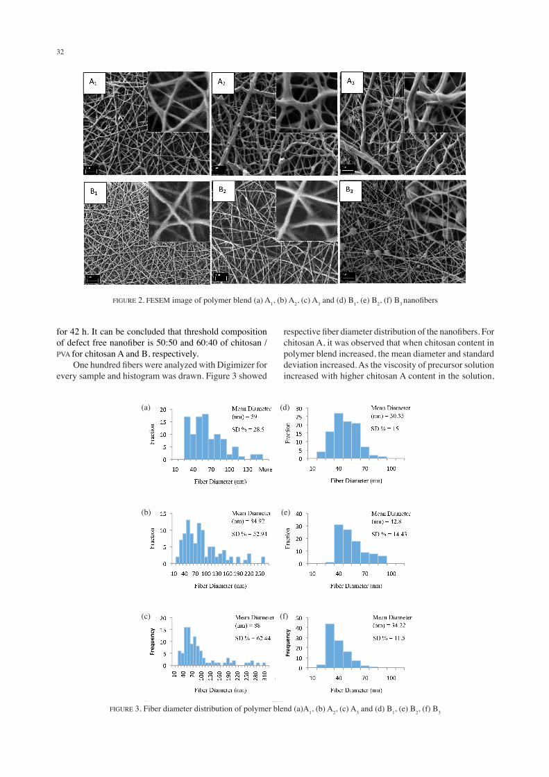
Figure 2 shows the FESEM images of nanofibers. The surface of nanofiber was comparatively smooth. But for chitosan A, some irregular shape and beads were observed in the nanofibrous mat of A2 and A3, respectively. Besides that, for chitosan B, FESEM image showed larger amount of beads for 70:30 wt. ratio of chitosan/PVA blend solution. Instability of jet, lower molecular weight, higher applied voltage, lower viscosity and higher surface tension are some reasons of bead formation in nanofiber (Homayoni et al. 2009; Nurhidayatullaili et al. 2011; Yarin 1993). Referring to Table 1, applied voltage for A2 and A3 are quite higher than A1. Applied voltage was larger for the precursor solution having higher viscosity. It caused uneven diameter of nanofiber. It was reported that hydrolysis reduces the molecular weight of chitosan (Homayoni et al. 2009). Therefore, in case of chitosan B, possible reason for bead formation is lower molecular weight as it was hydrolyzed
FIGURE 1. FTIR spectra of (a) Chitosan A and (b) Chitosan B
32
for 42 h. It can be concluded that threshold composition of defect free nanofiber is 50:50 and 60:40 of chitosan /PVA for chitosan A and B, respectively. One hundred fibers were analyzed with Digimizer for every sample and histogram was drawn. Figure 3 showed
respective fiber diameter distribution of the nanofibers. For chitosan A, it was observed that when chitosan content in polymer blend increased, the mean diameter and standard deviation increased. As the viscosity of precursor solution increased with higher chitosan A content in the solution,
FIGURE 2. FESEM image of polymer blend (a) A1, (b) A2, (c) A3 and (d) B1, (e) B2, (f) B3 nanofibers
(a) (d)
(e)(b)
(c) (f)
FIGURE 3. Fiber diameter distribution of polymer blend (a)A1, (b) A2, (c) A3 and (d) B1, (e) B2, (f) B3

33
homogeneity of fiber diameter could not be maintained. The value of standard deviation is maximum for the polymer blend containing higher amount of chitosan. It may be the result of uneven splaying of jet for higher viscosity of blend solution. On the other hand, applied voltage required for 70:30 weight ratio of Chitosan A/PVA polymer blend is 9.8 kV which is much higher than 7.1 kV, required for 50:50 weight ratio of Chitosan/PVA blend. This is because amino group of chitosan was protonated in the solution which made the solution viscous and higher voltage was required to get the stretched jet (Fong et al. 1999). As reported by Huang, higher voltage results in larger diameter (Klossner et al. 2008). Therefore, 50:50 of chitosan A/PVA is the best in mean diameter and standard deviation. It is believed that larger amount of PVA prevented repelling action between polycationic chitosan molecules and leaded to ensure good fiber diameter distribution. FESEM image and histogram for chitosan B and PVA polymer blend showed a different result. Smaller variation and mean diameter were observed which is shown in Figure 3 and Table 1. Table 1 shows the standard deviation and mean diameter decreased as chitosan weight percentage increased. The impact of chitosan content is less significant on voltage required for electrospinning as compared to chitosan A because the range was from 7-7.5 kV. It indicated that 42 h hydrolysis significantly decreased polycationic behavior of chitosan in acidic solution and protonation in water.
CONCLUSION
In this study, chitosan was hydrolyzed for 24 and 42 h at elevated temperature. This was due to the raw chitosan having degree of deacetylation of 40% could not be dissolved in concentrated acetic acid. FTIR results confirmed that chitosan molecular structure was not destroyed even after 42 h hydrolysis. The hydrolysis of chitosan eased the solubility and improved the degree of deacetylation. However, chitosan could not be electrospun without the addition of PVA. The results showed the possibility of electrospinning was up to 70% of chitosan in the polymer blend for both 24 and 42 h hydrolysis. Mean diameter and standard deviation were smaller for solution containing 42 h hydrolyzed chitosan than the solution of the same composition containing 24 h hydrolyzed chitosan. It was found that hydrolysis lowered the required voltage to stretch the droplet to conical shape. In addition, lower required voltage helped in the formation of finer diameter fiber as well as good fiber diameter distribution. For 24 h hydrolysis, defect free nanofiber was observed at 50:50 composition of chitosan/PVA and 60:40 of chitosan/PVA for 42 h hydrolysis.
ACKNOWLEDGEMENTS
The authors would like to thank University of Malaya for the financial support under Research Grant RP021-2012B and post graduate research fund PG048-2013B.
REFERENCES
Baiwen Qi, Aixi Yu, Shaobo Zhu, Biao Chen & Yan Li. 2010. The preparation and cytocompatibility of injectable thermosensitive chitosan/poly (vinyl alcohol) hydrogel. Journal of Huazhong University of Science and Technology (Medical Sciences) 30: 89-93.
Bin Duan, Dong Cunhai, Xiaoyan Yuan & Kangde Yao. 2004. Electrospinning of chitosan solutions in acetic acid with poly (ethylene oxide). Journal of Biomaterials Science, Polymer Edition 15(6): 797-811.
Bognitzki, M., Czado, W., Frese, T., Schaper, A., Hellwig, M., Steinhart, M., Greiner, A. & Wendorff, J.H. 2001. Nanostructured fibers via electrospinning. Advanced Materials 13(1): 70-72.
Borchard, G. 2001. Chitosans for gene delivery. Advanced Drug Delivery Reviews 52(2): 145-150.
Brugnerotto, J., Lizardi, J., Goycoolea, F.M., Argüelles-Monal, W., Desbrieres, J. & Rinaudo, M. 2001. An infrared investigation in relation with chitin and chitosan characterization. Polymer 42(8): 3569-3580.
Fong, H., Chun, I. & Reneker, D.H. 1999. Beaded nanofibers formed during electrospinning. Polymer 40(16): 4585-4592.
Fong, H. & Reneker, D.H. 1999. Elastomeric nanofibers of styrene-butadiene-styrene triblock copolymer. Journal of Polymer Science Part B Polymer Physics 37(24): 3488-3493.
Francis Suh, J-K. & Matthew, H.W.T. 2000. Application of chitosan-based polysaccharide biomaterials in cartilage tissue engineering: A review. Biomaterials 21(24): 2589-2598.
Homayoni, H., Ravandi, S.A.H. & Valizadeh, M. 2009. Electrospinning of chitosan nanofibers: Processing optimization. Carbohydrate Polymers 77(3): 656-661.
Huang, Z-M., Zhang, Y-Z., Kotaki, M. & Ramakrishna, S. 2003. A review on polymer nanofibers by electrospinning and their applications in nanocomposites. Composites Science and Technology 63(15): 2223-2253.
Kaco, H., Zakaria, S., Razali, N.F., Chia, C.H., Zhang, Li. & Jani, S.M. 2014. Properties of cellulose hydrogel from kenaf core prepared via pre-cooled dissolving method. Sains Malaysiana 43(8): 1221-1229.
Klossner, R.R., Queen, H.A., Coughlin, A.J. & Krause, W.E. 2008. Correlation of chitosan’s rheological properties and its ability to electrospin. Biomacromolecules 9(10): 2947-2953.
Li, L. & Hsieh, Y-L. 2006. Chitosan bicomponent nanofibers and nanoporous fibers. Carbohydrate Research 341(3): 374-381.
Nurhidayatullaili Muhd Julkapli, Hazizan Md Akil & Zulkifli Ahmad. 2011. Preparation, properties and applications of chitosan-based biocomposites/blend materials: A review. Composite Interfaces 18(6): 449-507.
Pawlak, A. & Mucha, M. 2003. Thermogravimetric and FTIR studies of chitosan blends. Thermochimica Acta 396(1): 153-166.
Shauer, C.L. & Schiffman, J.D. 2008. Fibrous mats containing chitosan nanofibers. Google Patents No. US 20100216211 A1.
Yarin, A.L. 1993. Free Liquid Jets and Films: Hydrodynamics and Rheology. England: Longman Publishing Group.
Zong, X., Kim, K-S., Fang D-F., Ran, S., Hsiao, B.S. & Chu, B. 2002. Structure and process relationship of electrospun bioabsorbable nanofiber membranes. Polymer 43(16): 4403-4412.

34
Materials Engineering Strategic UnitDepartment of Mechanical Engineering Faculty of Engineering, University of Malaya 50603 Kuala Lumpur, Federal TerritoryMalaysia
*Corresponding author; email: [email protected]
Received: 19 July 2014Accepted: 10 November 2014