influence of nanoprecipitation method parameters … · and prolonging precorneal drug residence...
TRANSCRIPT
IJASR International Journal of Academic Scientific Research ISSN: 2272-6446 Volume 3, Issue 1 (February - March), PP 01-12 www.ijasrjournal.org
Influence of nanoprecipitation method parameters on nanoparticles loaded with gatifloxacin for ocular drug delivery
A. Maaz1, W. Abdelwahed
2, I.A. Tekko
3, S. Trefi
1
1(Department of Pharmaceutical Chemistry & Quality Control, Faculty of Pharmacy, Aleppo University,
Syria) 2(Department of Pharmaceutical Technology, Faculty of Pharmacy, Aleppo University, Syria)
3(Department of biopharmaceutics and drug Formulation, Aleppo University, Syria)
ABSTRACT: Poor ocular bioavailability of drugs (<1%) from conventional eye drops (i.e., solution,
suspension, and ointments) is mainly due to the physiologic barriers of the eye. In general, ocular efficacy is
closely related to ocular drug bioavailability, which may be enhanced by increasing corneal drug penetration
and prolonging precorneal drug residence time. In our current work, we developed a colloidal system that is
polycaprolactone (PCL) nanoparticles for Gatifloxacin ophthalmic delivery, to improve precorneal residence
time and ocular penetration for enhanced drug bioavailability. In this research, we studied nanoparticles
preparation procedures and the effect of process variables on its characters. Nanoparticles were prepared by
nanoprecipitation technique and characterized for various properties such as particle size, polydispersity index
PdI, zeta potential and Entrapment efficiency EE%. The developed nanosuspension showed a mean particle size
in the range of 184 to 207nm, suitable for ophthalmic application with zeta potential range of -30 to -32 mV and
Entrapment Efficiency EE% of 40%. These results demonstrated that, the developed nanosuspension was found
to be applicable for sustained ocular drug delivery allowing minimizing dose repetition to reduce systemic side
effects and enhance patient compliance.
Keywords: Gatifloxacin, Nanoparticles, Nanoprecipitation, Ocular Delivery, Polycaprolacton
1. INTRODUCTION
Despite numerous scientific efforts in drug delivery field, efficient ocular drug delivery remains a
challenge for pharmaceutical scientists. The unique structure of the eye restricts the entry of drug molecules at
the required site of action. In ocular drug delivery system, ocular infections are treated by various topical drug
applications in the form of solutions, suspensions and ointment. These conventional dosage forms suffer from
the problems of poor ocular bioavailability due to the precorneal loss factors that include rapid tears turnover,
nonproductive absorption, transient residence time in the cel-de-sac, and relative impermeability of the drugs to
the corneal epithelial membrane [1, 2]. This poor ocular bioavailability imparts the need for frequent instillation
to achieve the therapeutic effect, which may leads sometimes to undesirable side effects caused by systemic
drug absorption. A polymeric nanoparticle formulation is one of the strategies currently used to improve drug
absorption across biological membranes [3].
Hence, in our current work we followed recent applications of nanoparticulate systems in the field of
ocular drug delivery, whereas utilizing nanoparticles for ocular disease treatment in a wide range of medical
research fields has become a popular strategy in recent years [4-6]. Much of the published data suggests that in
the case of ophthalmic drug delivery, an appropriate particle size, and a narrow size range, ensuring low
irritation, adequate bioavailability and compatibility with ocular tissues, should be controlled for every drug
loaded [7].
The preparation of Nanospheres (NS) by precipitation method has been studied extensively to produce
high qualified NS [8], using biodegradable polymer polycaprolactone (PCL) which is an anionic polymer and
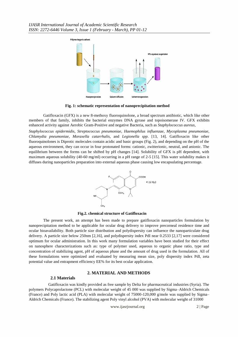
one of the most commonly used polymers in ophthalmic drug formulations [9]. Nanprecipitation method is also
called solvent displacement or interfacial deposition (Fig. 1), and it is one of the easiest preparation procedures
of nanospheres. Additionally to its simplicity, this procedure is reproducible, fast and economic [10] but the
encapsulation of water soluble compounds by this method is still providing challenges to the pharmaceutics
person [11, 12].
www.ijasrjournal.org 1 | Page
IJASR International Journal of Academic Scientific Research ISSN: 2272-6446 Volume 3, Issue 1 (February - March), PP 01-12
Fig. 1: schematic representation of nanoprecipitation method
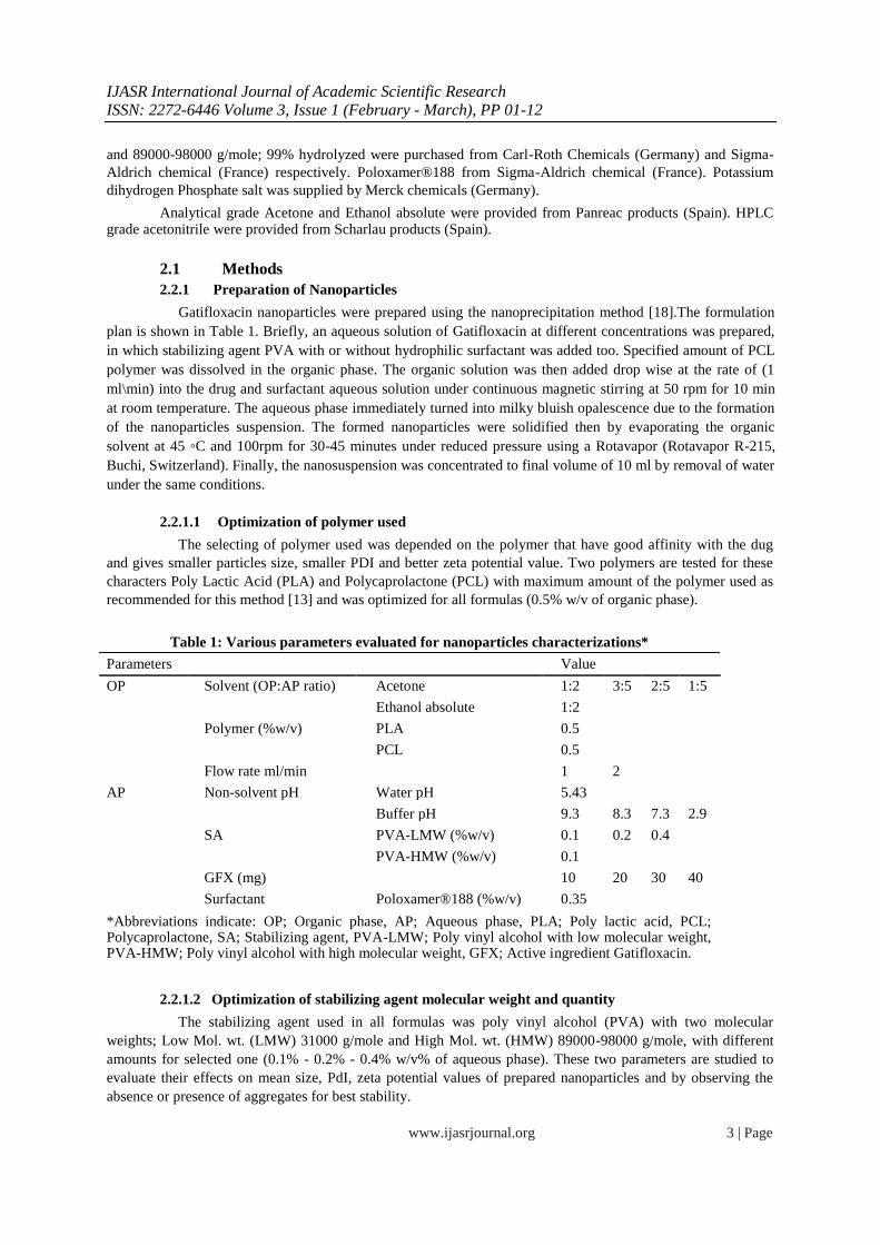
Gatifloxacin (GFX) is a new 8-methoxy fluoroquinolone, a broad spectrum antibiotic, which like other
members of that family, inhibits the bacterial enzymes DNA gyrase and topoisomerase IV. GFX exhibits
enhanced activity against Aerobic Gram-Positive and negative Bacteria, such as Staphylococcus aureus, Staphylococcus epidermidis, Streptococcus pneumoniae, Haemophilus influenzae, Mycoplasma pneumoniae,
Chlamydia pneumoniae, Moraxella catarrhalis, and Legionella spp. [13, 14]. Gatifloxacin like other
fluoroquinolones is Diprotic molecules contain acidic and basic groups (Fig. 2), and depending on the pH of the
aqueous environment, they can occur in four protonated forms: cationic, zwitterionic, neutral, and anionic. The
equilibrium between the forms can be shifted by pH changes [14]. Solubility of GFX is pH dependent, with
maximum aqueous solubility (40-60 mg/ml) occurring in a pH range of 2-5 [15]. This water solubility makes it
diffuses during nanoparticles preparation into external aqueous phase causing low encapsulating percentage.
Fig.2. chemical structure of Gatifloxacin
The present work, an attempt has been made to prepare gatifloxacin nanoparticles formulation by
nanoprecipitation method to be applicable for ocular drug delivery to improve precorneal residence time and
ocular bioavailability. Both particle size distribution and polydispersity can influence the nanoparticulate drug
delivery. A particle size below 250nm [2,16], and polydispersity index PdI near 0.2533 [2,17] were considered
optimum for ocular administration. In this work many formulation variables have been studied for their effect
on nanosphere characterizations such as: type of polymer used, aqueous to organic phase ratio, type and
concentration of stabilizing agent, pH of aqueous phase and the amount of drug used in the formulation. All of
these formulations were optimized and evaluated by measuring mean size, poly dispersity index PdI, zeta
potential value and entrapment efficiency EE% for its best ocular application.
2. MATERIAL AND METHODS
2.1 Materials
Gatifloxacin was kindly provided as free sample by Delta for pharmaceutical industries (Syria). The
polymers Polycaprolactone (PCL) with molecular weight of 45 000 was supplied by Sigma–Aldrich Chemicals
(France) and Poly lactic acid (PLA) with molecular weight of 75000-120,000 g/mole was supplied by Sigma–
Aldrich Chemicals (France). The stabilizing agent Poly vinyl alcohol (PVA) with molecular weight of 31000
www.ijasrjournal.org 2 | Page
IJASR International Journal of Academic Scientific Research ISSN: 2272-6446 Volume 3, Issue 1 (February - March), PP 01-12
and 89000-98000 g/mole; 99% hydrolyzed were purchased from Carl-Roth Chemicals (Germany) and Sigma-
Aldrich chemical (France) respectively. Poloxamer®188 from Sigma-Aldrich chemical (France). Potassium
dihydrogen Phosphate salt was supplied by Merck chemicals (Germany).
Analytical grade Acetone and Ethanol absolute were provided from Panreac products (Spain). HPLC grade acetonitrile were provided from Scharlau products (Spain).
2.1 Methods 2.2.1 Preparation of Nanoparticles
Gatifloxacin nanoparticles were prepared using the nanoprecipitation method [18].The formulation
plan is shown in Table 1. Briefly, an aqueous solution of Gatifloxacin at different concentrations was prepared,
in which stabilizing agent PVA with or without hydrophilic surfactant was added too. Specified amount of PCL
polymer was dissolved in the organic phase. The organic solution was then added drop wise at the rate of (1
ml\min) into the drug and surfactant aqueous solution under continuous magnetic stirring at 50 rpm for 10 min
at room temperature. The aqueous phase immediately turned into milky bluish opalescence due to the formation
of the nanoparticles suspension. The formed nanoparticles were solidified then by evaporating the organic
solvent at 45 ◦C and 100rpm for 30-45 minutes under reduced pressure using a Rotavapor (Rotavapor R-215,
Buchi, Switzerland). Finally, the nanosuspension was concentrated to final volume of 10 ml by removal of water
under the same conditions.
2.2.1.1 Optimization of polymer used
The selecting of polymer used was depended on the polymer that have good affinity with the dug
and gives smaller particles size, smaller PDI and better zeta potential value. Two polymers are tested for these
characters Poly Lactic Acid (PLA) and Polycaprolactone (PCL) with maximum amount of the polymer used as
recommended for this method [13] and was optimized for all formulas (0.5% w/v of organic phase).
Table 1: Various parameters evaluated for nanoparticles characterizations*
Parameters Value
OP Solvent (OP:AP ratio) Acetone 1:2 3:5 2:5 1:5
Ethanol absolute 1:2
Polymer (%w/v) PLA 0.5
PCL 0.5
Flow rate ml/min 1 2
AP Non-solvent pH Water pH 5.43
Buffer pH 9.3 8.3 7.3 2.9
SA PVA-LMW (%w/v) 0.1 0.2 0.4
PVA-HMW (%w/v) 0.1
GFX (mg) 10 20 30 40
Surfactant Poloxamer®188 (%w/v) 0.35
*Abbreviations indicate: OP; Organic phase, AP; Aqueous phase, PLA; Poly lactic acid, PCL; Polycaprolactone, SA; Stabilizing agent, PVA-LMW; Poly vinyl alcohol with low molecular weight, PVA-HMW; Poly vinyl alcohol with high molecular weight, GFX; Active ingredient Gatifloxacin.
2.2.1.2 Optimization of stabilizing agent molecular weight and quantity
The stabilizing agent used in all formulas was poly vinyl alcohol (PVA) with two molecular
weights; Low Mol. wt. (LMW) 31000 g/mole and High Mol. wt. (HMW) 89000-98000 g/mole, with different
amounts for selected one (0.1% - 0.2% - 0.4% w/v% of aqueous phase). These two parameters are studied to
evaluate their effects on mean size, PdI, zeta potential values of prepared nanoparticles and by observing the
absence or presence of aggregates for best stability.
www.ijasrjournal.org 3 | Page
IJASR International Journal of Academic Scientific Research ISSN: 2272-6446 Volume 3, Issue 1 (February - March), PP 01-12
2.2.1.3 Optimization of organic phase
Two types of organic phase as polymer solvent were tested: acetone and absolute ethanol. The
criteria for selecting the best solvent are high solubility of the polymer in the solvent and ease of evaporation
and removal.
2.2.1.4 Determination of organic to aqueous phase volume ratio effect Changing the organic to aqueous phase ratio was studied as follows: (3:5, 2:5 and 1:5). All other
constituents of the formulation were unchanged. This parameter is studied to evaluate its effect on particles
mean size and PdI.
2.2.1.5 Determination of organic phase flow rate effect
The addition of organic phase into aqueous one during procedure was completed by using two kinds
of syringes to apply two different flow rates; 5cc syringe (2ml/min) and 1cc syringe (1ml/min). This parameter's
effect was evaluated by measuring the mean particles size and PdI.
2.2.1.6 Determination of aqueous phase pH effect As long as Gatifloxacin solubility in aqueous phase is pH dependent, this parameter was studied
with different pH values; distilled water with pH 5.43 and phosphate buffer with pH 2.9, 7.3, 8.3 and 9.3. All
other constituents of the formulation were unchanged. The effect of pH value was evaluated by measuring the
mean size, PdI, zeta potential, and the entrapment efficiency.
2.2.1.7 Determination of Gatifloxacin amount effect
Four amounts of Gatifloxacin were tested (10, 20, 30, and 40 mg), all other constituents were
unchanged. The effect of Gatifloxacin quantity was evaluated by measuring the particles mean size, PdI, zeta
potential, and the entrapment efficiency.
2.2.1.8 Determination of hydrophilic surfactant addition effect Poloxamer® 188 was the surfactant which was tested with its recommended quantity 0.35% w/v of
aqueous phase in the optimized formula. The effect of Poloxamer® 188 addition was evaluated by measuring
the particles mean size, PdI, zeta potential, and the entrapment efficiency.
2.2.2 Evaluation of Gatifloxacin Nanoparticles 2.2.2.1 Particle mean size analysis and zeta-potential determination
The mean size (Z-average) of the GFX-loaded nanospheres and polydispersity index (PDI) was
determined by dynamic light-scattering particle size analyser (DLS), using a Malvern Zetasizer Nanoseries
(Nano-ZS, Malvern Instruments, Malvern, UK). The size distribution analysis was performed at a scattering
angle of 173º and at temperature of 25◦C. Zeta potential (mV) values are measured with the same instrument
with electrophoretic light scattering technique by calculating Smoluchowski’s equation from electrophoretic
mobility of nanoparticles. Nanoparticles samples were prepared by taking 1ml from nanosuspension, which was
diluted into 10mL of double distilled water, and sonicated for 1min by sonicator (Wise clean-ultrasonic cleaner-
wuc-ao6h, Korea). For each sample, the mean diameter/PDI/zeta potential ± standard deviation of three
determinations were calculated.
2.2.2.2 Encapsulation Efficiency Determination
The amount of drug loaded into the nanoparticles was evaluated through direct way by calculating
the amount of drug that was found inside the particles obtained and comparing it with the practical total amount
used to prepare the nanoparticles (T).Total Gatifloxacin concentration (T) was determined after dissolution of 1
ml of nanoparticles suspension in 4 ml acetone. After dissolving the polymer, 4 ml of Hydrochloric acid (HCL)
0.1N was then added, and the volume was completed to 10 ml with mobile phase used for high performance
www.ijasrjournal.org 4 | Page

IJASR International Journal of Academic Scientific Research ISSN: 2272-6446 Volume 3, Issue 1 (February - March), PP 01-12
liquid chromatography (HPLC) analysis method, and the mixture was mixed carefully for 30 min by magnetic
stirring and sonicated for 5min. thereafter the amount of drug in the water phase was detected applying HPLC
analysis. Drug-loaded nanoparticles were separated from the aqueous medium containing non-associated
Gatifloxacin by ultracentrifugation (CP 80WX Himac preparative ultracentrifuge, Hitachi, Japan). Samples were
centrifuged at 45 000 rpm for 30 min at 4◦C, and loaded Gatifloxacin in the sediment was determined under the
same condition of the total (T). The encapsulation efficiency of nanoparticles was determined according to
Lakshmana Rao et al. [19].
HPLC separation was performed with Agilent Liquid Chromatographer (LC-1260 Infinity, Agilent,
Germany) achieved on RP-C18 column (EC 150/4.5 Nucleodur 100-5 C18ec, Macherey-Nagel, Germany) with
mobile phase consisting of acetonitrile and 0.05M phosphate buffer in the ratio of 25:75 v/v in isocratic mode
was used. The HPLC system was operated at flow rate is 0.8ml/min and the detection wavelength is 293nm and
the. All the measurement were performed at 25◦C. The encapsulation efficiency was determined and calculated
as follows:
3. RESULTS AND DISCUSSION
3.1 Characterization of GFX-encapsulated nanoparticles; Particle Size and PDI 3.1.1 Optimization of polymer used
Based on literature data, the three most commonly used polymers in ophthalmic drug formulations
are poly (alkyl cyanoacrylates), polycaprolactone, and poly (lactic acid) /poly (lactic-co-glycolic acid) [2]. Two
kinds of polymers were tested in the optimization of unloaded nanospheres, poly lactic acid (PLA) and
polycaprolactone (PCL) Fig.3. The maximum amount of the polymer used as recommended for this method
[20], and was optimized for all formulas (0.5% w/v of organic phase).
Fig.3. Chemical structure of used polymers PCL and PLA
The results are presented in Table 2. Obtained particles size was 136-146 nm and PdI of 0.109-0.154
for PLA and PCL respectively. It was clear that no significant differences were observed on particle size and
PDI between both used polymers. Unloaded nanoparticles had a negative surface charge which can be attributed
with the presence of end carboxyl groups of the polymer on nanoparticles surface [21]. Both used polymers are
Aliphatic polyesters and have terminal carboxylic group which caused negative charge on the surface of
nanospheres and so negative zeta potential values were obtained, with no significant differences also between
both, -4.71 and -2.39 for PLA and PCL respectively.
www.ijasrjournal.org 5 | Page

IJASR International Journal of Academic Scientific Research ISSN: 2272-6446 Volume 3, Issue 1 (February - March), PP 01-12
Table 2: Effect of polymer type on the mean size of nanoparticles, PDI and zeta potential*
Formula Polymer type Mean size
PDI ± SD Zeta potential ± SD
(nm) ± SD
F1 PLA 136.7 ± 0.45 0.109 ± 0.003 -4.71 ± 0.19
F2 PCL 146.6 ± 1.442 0.154 ± 0.0179 -2.39 ± 0.1
*Data are the mean of three determinations ± SD.
With these results PCL was fixed in subsequent experiments because this polymer has a low commercial price that makes it a good candidate for large scale applications.
3.1.2 Optimization of stabilizing agent molecular weight and quantity
The stabilizing agent which used in all formulas is poly vinyl alcohol (PVA) with two molecular
weights; Low Mol. wt. (LMW) 31000 g/mole and High Mol. wt. (HMW) 89000 g/mole, and with different
amounts (0.1% - 0.2% and 0.4% w/v% of aqueous phase) for best stability. From the data obtained as presented
in Table 3, it was very clear that the particle size is strongly influenced by both parameters, with size range of
146 and 233 nm and PdI of 0.154 and 0.278 for LMW PVA and HMW PVA respectively. After choosing LMW
PVA due to its better results, three amounts 0.1% - 0.2% and 0.4% have been tested. The mean size for particles
was 146, 243 and 302 nm respectively and size distribution became wider, where PdI reached to 0.246 with
increasing particle size for highest amount of PVA. The results obtained here were found to be in accordance
with a previously published study [8,22]. PVA (Fig.4.) is a swellable, hydrophilic macromolecule, although it is
not required to ensure the formation of NP by nanoprecipitation, but the addition of PVA helps to preserve the
nanoparticle suspensions from agglomeration over long storage periods. The particle size is strongly influenced
with direct proportion by the PVA nature (molecular weight) and concentration [8], which could be due to high
viscosity and interfacial tension of aqueous phase. It has been reported that PVA grades with high degrees of
hydrolysis have low solubility in water. The solubility, viscosity, and surface tension of PVA depend on
temperature, concentration, % hydrolysis and molecular weight of the material [22]. Therefore high molecular
weight and high concentration of PVA led to larger particle size and wider size distribution. For this reason
PVA with lower molecular weight was retained for the following experiments for best particles uniformity and
greatest amount 0.4% (v/v) was selected for best stability.
Fig.4. chemical structure of Polyvinyl Alcohol
Table 3: Effect of PVA molecular weight and quantity on the mean size of nanoparticles, PdI and zeta potential (Z.P.)*
Formula PVA MW PVA (%w/v) Mean size (nm) ± SD PdI ± SD Z.P. ± SD
F3 High 0.1% 233.9 ± 0.737 0.278 ± 0.027 -8.14 ± 0.312
F2 Low 0.1% 146.6 ± 1.442 0.154 ±
-2.39 ± 0.1
0.0179
F5 Low 0.2% 243 ± 3.637 0.069 ± 0.008 -7.8 ± 0.142
F7 Low 0.4% 302.4 ± 1.504 0.246 ± 0.008 -14.7 ± 0.1
*Data are the mean of three determinations ± SD.
www.ijasrjournal.org 6 | Page

IJASR International Journal of Academic Scientific Research ISSN: 2272-6446 Volume 3, Issue 1 (February - March), PP 01-12
3.1.3 Optimization of organic phase
Acetone and absolute ethanol were examined as polymer solvent which both are miscible in water
and easy to remove by evaporation, but absolute ethanol was not suitable for dissolving PCL polymer with such
molecular weight (40,000) easily. Due to this reason, acetone was employed as organic phase for later
preparations.
3.1.4 Determination of organic to aqueous phase volume ratio effect We experimented three different Ratio of non-aqueous to aqueous phase (3:5 / 2:5 and 1:5) to obtain
low particle size. The results are shown in Table 4 nanoparticles of average size of 362, 243 and 214 nm were
obtained respectively. No significant variations were noticed between last two proportions, but mean particle
size increased with increasing organic phase portion. Over that size of distribution also became wider, whereas
polydispersity index PdI was 0.227, 0.069 and 0.099 respectively. As some researches pointed out, may
changing the solvent/non-solvent volume ratio was not a determinant factor for nanoparticle formation and their
final characteristics, provided that the final mixture itself did not become a solvent for the polymer [2]. But in
our experiments higher proportion of organic phase led to increase particle size which is corresponding with
other researches [21]. It could be attributed to an increase in the time required to evaporate the organic phase.
No significant differences were noticed on zeta potential values by this parameter as long as the amount of used
polymer and PVA were unchanged. From previous results we expected that 1:2 organic to aqueous phase ratio is
the best proportion which induced smaller particle size and narrower size distribution and it was retained for the
following experiments.
Table 4: Effect of organic to aqueous phase volume ratio on the mean size of nanoparticles, PdI and zeta potential*
Formula OP:AP
Mean size (nm) ± SD PdI ± SD Zeta potential ±
ratio** SD
F4 3:5 362.7±4.158 0.227±0.017 -13±0.854
F5 2:5 243±3.637 0.069±0.008 -7.8±0.142
F6 1:5 214±1.29 0.099±0.036 -11 ±0.854
*Data are the mean of three determinations ± SD. ** OP:AP ratio indicates organic to aqueous phase volume ratio.
3.1.5 Determination of organic phase flow rate effect At organic phase dropping into aqueous one step, we changed used injector syringe from 5cc into
1cc, in order to reduce the flow rate form 2ml/min to 1ml/min. This led to significantly decreasing in particles
size from 302 to 211nm and smaller size distribution from 0.246 to 0.084 as shown in Table 5. May these results
due to changing the rate of particle formation stages which determine the particle size as reported in previous
researches [20], a high nucleation rate and low growth rate is the key factor for smaller and uniform particles
formation. As for zeta potential values it has been noticed from all former results that smaller particles lead to
smaller numerical zeta potential value. The increase in nanoparticle size may possibly have influenced the
surface charge of the PCL nanoparticles [21]. Smaller flow rate was fixed for later experiments for smaller
particles size results.
www.ijasrjournal.org 7 | Page
IJASR International Journal of Academic Scientific Research ISSN: 2272-6446 Volume 3, Issue 1 (February - March), PP 01-12
Table 5: Effect of organic phase flow rate on the mean size of nanoparticles, PdI and zeta potential*
Formula Flow rate Mean size (nm) ± SD PdI ± SD Z.P. ± SD
F7 2 ml/min 302.4 ± 1.504 0.246 ± 0.008 -14.7 ± 0.1 F8 1 ml/min 211.4±0.2 0.084±0.032 -7.08±0.61 *Data are the mean of three determinations ± SD.
3.1.6 Determination aqueous phase pH effect
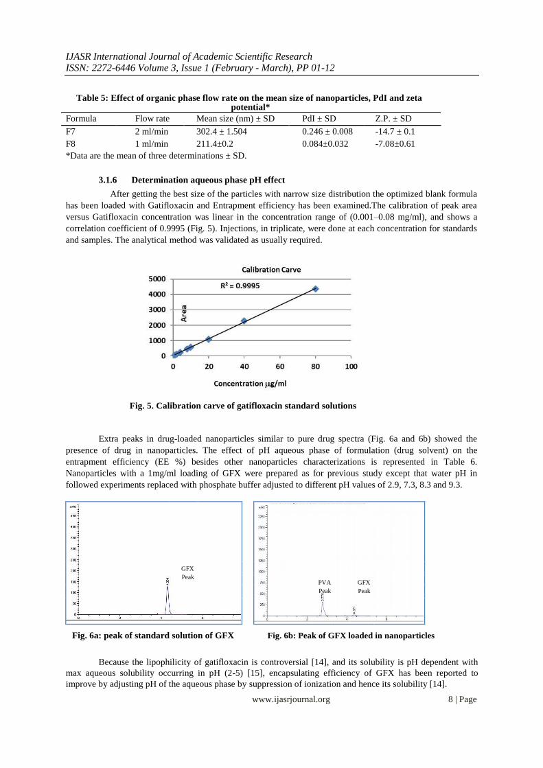
After getting the best size of the particles with narrow size distribution the optimized blank formula
has been loaded with Gatifloxacin and Entrapment efficiency has been examined.The calibration of peak area
versus Gatifloxacin concentration was linear in the concentration range of (0.001–0.08 mg/ml), and shows a
correlation coefficient of 0.9995 (Fig. 5). Injections, in triplicate, were done at each concentration for standards
and samples. The analytical method was validated as usually required.
Fig. 5. Calibration carve of gatifloxacin standard solutions
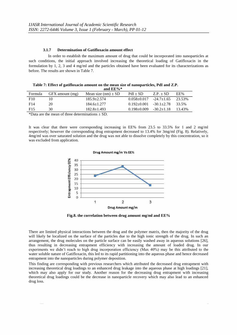
Extra peaks in drug-loaded nanoparticles similar to pure drug spectra (Fig. 6a and 6b) showed the
presence of drug in nanoparticles. The effect of pH aqueous phase of formulation (drug solvent) on the
entrapment efficiency (EE %) besides other nanoparticles characterizations is represented in Table 6.
Nanoparticles with a 1mg/ml loading of GFX were prepared as for previous study except that water pH in
followed experiments replaced with phosphate buffer adjusted to different pH values of 2.9, 7.3, 8.3 and 9.3.
GFX
Peak
GFX
PVA
Peak Peak
Fig. 6a: peak of standard solution of GFX Fig. 6b: Peak of GFX loaded in nanoparticles
Because the lipophilicity of gatifloxacin is controversial [14], and its solubility is pH dependent with
max aqueous solubility occurring in pH (2-5) [15], encapsulating efficiency of GFX has been reported to
improve by adjusting pH of the aqueous phase by suppression of ionization and hence its solubility [14].
www.ijasrjournal.org 8 | Page

IJASR International Journal of Academic Scientific Research ISSN: 2272-6446 Volume 3, Issue 1 (February - March), PP 01-12
Obviously as shown in Fig. 7, there was an increase in drug encapsulation with increasing aqueous phase pH
from 2.9 to 9.3 by 6.5 %, 12 %, 13.9 % and 23.5% respectively. It was therefore likely that Above GFX iso-
electric point, the solubility of GFX and hence its hydrophobic nature increases as the pH of solution increases
and this could enhance drug entrapment into nanoparticles [24]. The studies also illustrate that drug entrapment
was profoundly higher for particles prepared in phosphate buffer pH 9.3 than those prepared in water pH 5.43
(Table 6).
Fig.7. the correlation between pH aqueos phase and EE%
Whereas, the greater ionization degree of gatifloxacin in water pH 5.4 as compared to in phosphate buffer pH 9.3 at most probably contributed destabilization of particles hence decreasing Entrapment efficiency. The lower
of GFX solubility at the higher pH of 9.3 makes it adsorbed and/or dispersed into the polymeric matrix system
of PCL nanoparticles. It readily precipitated in aqueous medium and gets encapsulated by the PCL matrix preventing its diffusion in external phase. Some researches illustrated that drug adsorbed on the surface of
particles and may have also contributed to an improved nanosuspension stability [25]. It was interesting to note as shown in Table 6 that Particles size was affected by increasing drug encapsulating efficiency displayed from
154nm to 185nm, may this due to the increasing of drug content of the nanoparticles. Gatifloxacin loaded
nanoparticles have a higher surface charge (>-20) in comparison with blank formulas and this makes them discrete and prevents agglomeration. On the contrary, in case of drug absence, all formulas have lower surface
charge (>-15) and may this make them aggregate. Extremely positive or negative zeta potential values cause
larger repulsive forces, whereas repulsion between particles with similar electric charge prevents aggregation of the particles and thus ensures easy redispersion, so it is always favorable. High negative zeta potential values in
all drug-loaded formulations may be due to same explanation that has been mentioned before. Fluoroquinolone molecules including GFX have wide range of electrostatic potential from negative to positive demonstrates the
presence of dipole–dipole intermolecular interactions [14]. So may negative electrostatic potential on the surface
of a drug molecule adsorbed into PCL matrix resulted to displayed zeta potential values. May this also explains the increase in surface charge with increasing the amount of drug that is loaded within particles. From all of the
above the higher pH value 9.3 was fixed for followed formulas.
Table 6: Effect of aqueous phase pH on the mean size of nanoparticles, PdI and Z.P.
and EE%* Formula pH Mean size (nm) ± SD PdI ± SD Z.P. ± SD EE% F9 Water 5.43 154.8±1.79 0.138±0.018 -22.5±0.55 9.6%
F10 Buffer 9.3 185.9±2.574 0.058±0.017 -24.7±1.65 23.53%
F11 Buffer 8.3 187.8±1.153 0.073±0.01 -28.3±0.551 13.9%
F12 Buffer 7.3 169.7±0.757 0.122±0.032 -22.6±1.4 12%
F13 Buffer 2.9 188.8±1.153 0.236±0.005 -26.5±1.99 6.5%
*Data are the mean of three determinations ± SD
www.ijasrjournal.org 9 | Page
IJASR International Journal of Academic Scientific Research ISSN: 2272-6446 Volume 3, Issue 1 (February - March), PP 01-12
3.1.7 Determination of Gatifloxacin amount effect
In order to establish the maximum amount of drug that could be incorporated into nanoparticles at
such conditions, the initial approach involved increasing the theoretical loading of Gatifloxacin in the
formulation by 1, 2, 3 and 4 mg/ml and the particles obtained have been evaluated for its characterizations as
before. The results are shown in Table 7.
Table 7: Effect of gatifloxacin amount on the mean size of nanoparticles, PdI and Z.P. and EE%*
Formula GFX amount (mg) Mean size (nm) ± SD PdI ± SD Z.P. ± SD EE%
F10 10 185.9±2.574 0.058±0.017 -24.7±1.65 23.53% F14 20 184.6±1.277 0.192±0.001 -30.1±2.78 33.5% F15 30 182.8±1.493 0.198±0.009 -30.2±1.18 13.43% *Data are the mean of three determinations ± SD.
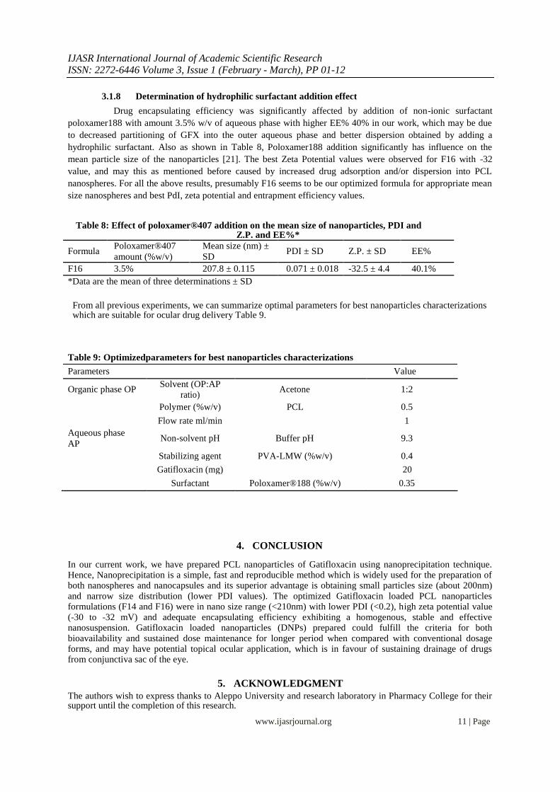
It was clear that there were corresponding increasing in EE% from 23.5 to 33.5% for 1 and 2 mg/ml respectively; however the corresponding drug entrapment decreased to 13.4% for 3mg/ml (Fig. 8). Relatively, 4mg/ml was over saturated solution and the drug was not able to dissolve completely by this concentration, so it was excluded from application.
Fig.8. the correlation between drug amount mg/ml and EE%
There are limited physical interactions between the drug and the polymer matrix, then the majority of the drug will likely be localized on the surface of the particles due to the high ionic strength of the drug. In such an arrangement, the drug molecules on the particle surface can be easily washed away in aqueous solutions [26], thus resulting in decreasing entrapment efficiency with increasing the amount of loaded drug. In our experiments we didn’t reach to high drug incorporation efficiency (Max 40%) may be this attributed to the water soluble nature of Gatifloxacin, this led to its rapid partitioning into the aqueous phase and hence decreased entrapment into the nanoparticles during polymer deposition. This finding are corresponding with previous researchers which attributed the decreased drug entrapment with increasing theoretical drug loadings to an enhanced drug leakage into the aqueous phase at high loadings [21], which may also apply for our study. Another reason for the decreasing drug entrapment with increasing theoretical drug loadings could be the decrease in nanoparticle recovery which may also lead to an enhanced drug loss.
www.ijasrjournal.org 10 | Page
IJASR International Journal of Academic Scientific Research ISSN: 2272-6446 Volume 3, Issue 1 (February - March), PP 01-12
3.1.8 Determination of hydrophilic surfactant addition effect
Drug encapsulating efficiency was significantly affected by addition of non-ionic surfactant
poloxamer188 with amount 3.5% w/v of aqueous phase with higher EE% 40% in our work, which may be due
to decreased partitioning of GFX into the outer aqueous phase and better dispersion obtained by adding a
hydrophilic surfactant. Also as shown in Table 8, Poloxamer188 addition significantly has influence on the
mean particle size of the nanoparticles [21]. The best Zeta Potential values were observed for F16 with -32
value, and may this as mentioned before caused by increased drug adsorption and/or dispersion into PCL
nanospheres. For all the above results, presumably F16 seems to be our optimized formula for appropriate mean
size nanospheres and best PdI, zeta potential and entrapment efficiency values.
Table 8: Effect of poloxamer®407 addition on the mean size of nanoparticles, PDI and
Z.P. and EE%*
Formula Poloxamer®407 Mean size (nm) ±
PDI ± SD Z.P. ± SD EE%
amount (%w/v) SD
F16 3.5% 207.8 ± 0.115 0.071 ± 0.018 -32.5 ± 4.4 40.1%
*Data are the mean of three determinations ± SD
From all previous experiments, we can summarize optimal parameters for best nanoparticles characterizations which are suitable for ocular drug delivery Table 9.
Table 9: Optimizedparameters for best nanoparticles characterizations Parameters Value
Organic phase OP
Solvent (OP:AP Acetone 1:2
ratio)
Polymer (%w/v) PCL 0.5
Flow rate ml/min 1
Aqueous phase Non-solvent pH Buffer pH 9.3
AP
Stabilizing agent PVA-LMW (%w/v) 0.4
Gatifloxacin (mg) 20
Surfactant Poloxamer®188 (%w/v) 0.35
4. CONCLUSION
In our current work, we have prepared PCL nanoparticles of Gatifloxacin using nanoprecipitation technique. Hence, Nanoprecipitation is a simple, fast and reproducible method which is widely used for the preparation of both nanospheres and nanocapsules and its superior advantage is obtaining small particles size (about 200nm) and narrow size distribution (lower PDI values). The optimized Gatifloxacin loaded PCL nanoparticles formulations (F14 and F16) were in nano size range (<210nm) with lower PDI (<0.2), high zeta potential value (-30 to -32 mV) and adequate encapsulating efficiency exhibiting a homogenous, stable and effective nanosuspension. Gatifloxacin loaded nanoparticles (DNPs) prepared could fulfill the criteria for both bioavailability and sustained dose maintenance for longer period when compared with conventional dosage forms, and may have potential topical ocular application, which is in favour of sustaining drainage of drugs from conjunctiva sac of the eye.
5. ACKNOWLEDGMENT
The authors wish to express thanks to Aleppo University and research laboratory in Pharmacy College for their support until the completion of this research.
www.ijasrjournal.org 11 | Page
IJASR International Journal of Academic Scientific Research ISSN: 2272-6446 Volume 3, Issue 1 (February - March), PP 01-12
REFERENCES
[1] T. Akanksha, S. Raj Kumar, Novel ocular drug delivery systems: An overview, Journal of Chemical and Pharmaceutical Research, 2(3), 2010, 348-355. [2] G. Himanshu, A. Mohammed, K. Roop K., A. Asgar, B. Aseem, M. Gaurav, Sparfloxacin-loaded PLGA nanoparticles for sustained ocular drug delivery, Nanomedicine: Nanotechnology, Biology, and Medicine, 6(2), 2010, 324–333. [3] M. Rubiana, U. Maria, C. Priscila, K. Najeh, C. Marco, E. Raul, G. Maria, Colloidal carriers for ophthalmic drug delivery, Current Drug Targets, 6(3), 2005, 363-371. [4] Z. Hong-Yan, H. Ji-Long, W. Shuang, Z. Yu, Z. Wen-Song, Nanoparticles in the ocular drug delivery, International Journal of Ophthalmology, 6(3), 2013, 390–396. [5] D. Yolanda, C. Margarita, Applications of nanoparticles in ophthalmology, Progress in Retinal and Eye Research, 29(6), 2010, 596-609. [6] M. Mudgil, N. Gupta, M. Nagpal, P. Pawar, Nanotechnology: a new approach for ocular drug delivery system, International Journal of Pharmacy and Pharmaceutical Sciences, 4(2), 2012, 105-112. [7] V.B. Patravale, A.D. Abhijit, R.M. Kulkarni, Nanosuspension: a promising drug delivery strategy, Journal of Pharmacy and Pharmacology, 56(7), 2004, 827-840. [8] R.J. Prasad, E.G. Kurt, Polymer nanoparticles: Preparation techniques and size-control parameters, Progress in Polymer Science, 36(7), 2011, 887–913. [9] W. Maria Ann, H. Dietmar Werner, The return of a forgotten polymer: Polycaprolactone in the 21st century, Progress in Polymer Science, 35(10), 2010, 1217-1256. [10] V. Christine, B. Kawthar, Methods for the Preparation and Manufacture of Polymeric Nanoparticles, Published in Pharmaceutical Research, 26(5), 2008, 1025-1058. [11] B.V.N. Nagavarma, K.S.Y. Hemant, A. Ayaz, L.S. Vasudha, H.G. Shivakumar, Different techniques for preparation of polymeric nanoparticles- a review, Asian Journal of Pharmaceutical and Clinical Research, 5(3), 2012, 16-23. [12] H. Samuli, Preparation and Characterization of Poly(Lactic Acid) Nanoparticles for Pharmaceutical Use, doctoral diss., Faculty of Pharmacy, University of Helsinki, Finland, 2008. [13] M.G. Dennis, Clinical Pharmacology of Gatifloxacin, a New Fluoroquinolone, Clinical Infectious Diseases, 31(2), 2000, S51–8. [14] E. Klosinska-Szmurlo, F.A. Plucinski, M. Grudzien, K. Betlejewska-Kielak, J. Biernacka, A.P. Mazurek, Experimental and theoretical studies on the molecular properties of ciprofloxacin, norfloxacin, pefloxacin, sparfloxacin, and gatifloxacin in determining bioavailability. Journal of biological physics, 40(4), 2014, 335–345. [15] B. Divya , P. Sabitha, R. Reddy, M. Kranthi Kumar Reddy, B.N. Rao, An Approach to Enhance Solubility of Gatifloxacin by Solid Dispersion Tecnique, Asian Journal of Research in Pharmaceutical Sciences, 2(2), 2012, 58-61. [16] J.V. Aukunuru, U.B. Kompella, In vitro delivery of nano- and microparticles to retinal pigment epithelial (RPE) cells, Drug Development and delivery, 2(2), 2002, 50-57. [17] D.S. Kohane, J.Y. Tse, Y. Yeo, R. Padera, M. Shubina, L. Robert., Biodegradable polymeric microspheres and nanospheres for drug delivery in the peritoneum, Journal of Biomedical Materials Research Part A, 77(2), 2006, 351-361. [18] H. Fessi, F. Puisieux, J.P. Devissaguet, N. Ammoury, S. Benita, Nanocapsule formation by interfacial polymer deposition following solvent displacement, International Journal of Pharmaceutics, 55(1), 1989, 25–28. [19] A. Lakshmana Rao, B.N.V. Ravi Kumar, G.Girija Sankar, Estimation of Gatifloxacin In Pharmaceutical Dosage Forms By High Performance Liquid Chromatography, Journal of pharmaceutical research and health care,3(3), 2011, 72-76. [20] C.E. Mora-Huertas, H. Fessi, A. Elaissari, Polymer-based nanocapsules for drug delivery, International Journal of Pharmaceutics, 385(1-2), 2010, 113–142. [21] G. Thirumala, S. Snjezana, C.G. Martin, I. Lisbeth, S.D. Stanley, PLGA nanoparticles prepared by nanoprecipitation: drug loading and release studies of a water soluble drug, Journal of Controlled Release, 57(2), 1999, 171–185. [22] G.K. Veeran, V.B. Guru, Water Soluble Polymers for Pharmaceutical Applications, Polymers, 3(4), 2011, 1972-2009. [23] R.C. Nagarwal, P.N. Singh, S. Kant, P. Maiti, J.K. Pandit. Chitosan coated PLA Nanoparticles for ophthalmic delivery: characterization, in-vitro and in-vivo study in rabbit eye, Journal of Biomedical Nanotechnology, 6(6), 2010, 648-657. [24] E. Alle´Mann, J.C. Leroux, R. Gurny, E. Doelker, In vitro extended release properties of drug-loaded poly(DL-lactic acid) nanoparticles produced by a salting out procedure, Pharmaceutical Research, 10(12), 1993, 1732–1737. [25] S. Stolnik, M.C. Garnett, M.C. Davies, L. Illum, M. Bousta, S.S. Davis, The colloidal properties of surfactant-free biodegradable nanospheres from poly (b-malic acid-co-benzyl malate)s and poly (lactic acid-co-glycolide), Colloids and surfaces. A, Physicochemical and engineering aspects, 97(3), 1995, 235–245. [26] K. Kwangsok, K.L. Yen, C. Charles, F. Dufei, S.H. Benjamin, C. Benjamin, H. Michael, Incorporation and controlled release of a hydrophilic antibiotic using poly(lactide-co-glycolide)-based electrospun nanofibrous scaffolds, Journal of Controlled Release, 98(1), 2004, 47– 56.
www.ijasrjournal.org 12 | Page