influence of non carious cervical lesions depth, …
TRANSCRIPT

648
Original Article
Biosci. J., Uberlândia, v. 31, n. 2, p. 648-656, Mar./Apr. 2015
INFLUENCE OF NON CARIOUS CERVICAL LESIONS DEPTH, LOADING POINT APPLICATION AND RESTORATION ON STRESS DISTRIBUTION PATTERN IN LOWER PREMOLARS: A 2D FINITE ELEMENT ANALYSIS
INFLUÊNCIA DA PROFUNDIDADE DA LESÃO CERVICAL NÃO CARIOSA, PONTO DE APLICAÇÃO DE CARGA E RESTAURAÇÃO NO PADRÃO DE
DISTRIBUIÇÃO DE TENSÕES EM PRÉ-MOLARES INFERIORES: 2D-ANÁLISE POR ELEMENTOS FINITOS
Livia Fávaro ZEOLA1; Fabrícia Araújo PEREIRA1; Alexia da Mata GALVÃO2; Tatiana Carvalho MONTES2; Sônia Cristina de SOUSA2;
Daniela Navarro Ribeiro TEIXEIRA2; Bruno Rodrigues REIS3; Paulo Vinícius SOARES4
1. Postgraduate Student and Member of Nucleus Extension, Research and Teaching at NCCL, Dentistry School, Federal University of
Uberlandia, Brazil; 2. Undergraduate Student and Member of Nucleus Extension, Research and Teaching at NCCL, Dentistry School,
Federal University of Uberlandia, Brazil; 3. Professor at Technical School of Health, and Member of Nucleus Extension, Research and
Teaching at NCCL, Dental School, Federal University of Uberlandia, Brazil; 4. Associate Professor at Operative Dentistry and Dental
Materials Department and Coordinator of Nucleus Extension, Research and Teaching at NCCL, Dentistry School, Federal University of
Uberlandia, Brazil. [email protected]
ABSTRACT: The aim of this study was to analyze the biomechanical behavior of lower premolars regarding
the non-carious cervical lesion (NCCL) depth, load type and restoration status, using finite element analysis. Two-
dimensional virtual model simulating a healthy lower premolar were created using the CAD software. Based on this
image, five models were generated: healthy (H), three types of NCCLs: small lesion (SL – 0.5 mm deep), medium lesion
(ML – 1.0 mm), deep lesion (DL – 1.5 mm), and restored lesion (RL). The models were export to a CAE software
(ANSYS Finite Element Analysis Software), the areas of all structures were plotted and each model was meshed using a
control mesh device. All of the virtual models were subjected to two occlusal load types, (100N each): occlusal load (OL)
and buccal load (BL) on buccal cusp. The magnitude and the stress distribution were obtained using the von Mises and
maximum principal stress criteria (σ1), in MPa. The quantitative analysis of stress (MPa) was identified at three points of
the NCCLs: enamel surface on its upper wall , dentin at the bottom wall and dentin on the lower wall. The results showed
a direct relation between sequential removal of cervical structure and higher stress concentration for any groups and for
both loads types. For OL the highest value of stress was 8.8 MPa for DL on upper wall of NCCLs.The BL exhibited higher
stress values in comparison to the OL for all models.In addtion, the BL was responsible for providing the highest stress
accumulation on the bottom wall, 38.2 MPa for DL. The restoration with composite resin was able to restore a stress
distribution close to the healthy model, for both load types. In conclusion, the extent of non-carious cervical lesion and
loading conditions influenced the stress distribution pattern of lower premolar. The outer load seems to be more critical in
affecting the biomechanical behavior of lower premolars, regardless of the lesion size. The restoration of NCCLs with
composite resin appears to recover the biomechanical behavior, similar to healthy model.
KEYWORDS: Finite element analysis. Non carious cervical lesion. Premolar.
INTRODUCTION
Non-carious cervical lesions (NCCLs) are a
routinely and increasingly found in dentistry clinical
practice 1. This kind of lesion is a pathological
process described by hard tissue dental loss at the
cement–enamel junction (CEJ) independent of
bacterial process (BORCIC et al,. 2004; REYES et
al., 2009). It is generally accepted that the formation
and progression of NCCLs are a multifactorial
process, involving the stress (abfraction),
biocorrosion (chemical, biochemical and
electrochemical degradation) and friction (wear)
(GRIPPO et al., 2012). The areas most affected by
cervical lesions are the buccal faces, with highest
prevalence in the upper and lower premolars
(SMITH et al., 2008).
The prevalence of NCCLs occur in up to 85
percent of individuals, increasing with age (SMITH
et al., 2008). This fact suggests a fatigue component
in NCCLs formation associated with occlusal
interferences (BRANDINI et al., 2012; WOOD et
al., 2009) or any event that changes the dental
occlusion, such as parafunction (BRANDINI et al.,
2012), bruxism (OMMERBORN et al., 2007) and
restorative procedures (BERNHARDT et al., 2006).
These events alter the stress distribution pattern in
the cervical region and can lead to weakening of the
Received: 29/09/14
Accepted: 12/12/14

649
Influence of non carious cervical… ZEOLA, L. F. et al.
Biosci. J., Uberlândia, v. 31, n. 2, p. 648-656, Mar./Apr. 2015
continuity between the hard tooth structures,
resulting in possible microcrack initiation and
rupture of rigid structures such as enamel (LEE W.
C.; EAKLE 1984; REES, 2002; REYES et al.,
2009). Studies reported that occlusal load position
modifies the intensity of stress concentration in
cervical regions (BENAZZI et al., 2014; REES,
2002; REES et al., 2003). The loads applied
obliquely to the long axis of the tooth may promote
an increase of stress in the cervical region and are
related to the formation and progression of NCCLs
(SOARES et al., 2014; REES, 2002; SOARES et
al., 2013).
The selection of appropriate direct material
to restore the NCCLs is an important step in the
treatment process. At this stage, should be evaluated
aesthetics, the presence of dentinal sensitivity
(MICHAEL et al., 2009), as well as the amount of
lost tooth structure. The literature reports different
restorative materials to restore NCCLs, such as
flowable composites, glass ionomer cement, resin-
modified glass ionomer, and composite resins
(MICHAEL et al., 2009; ONAL; PAMIR, 2005;
STEWARDSON et al., 2012).
Finite element analysis (FEA) has been
shown as a powerful tool in dental biomechanics for
measuring stress distribution pattern in teeth and
restorations in response to different occlusal
loads(BORCIC et al., 2005; POIATE et al., 2009;
VASUDEVA and BOGRA, 2008). This method
enables the simulation of commonly observed
clinical situations and the evaluation of different
factors in various dental tissues, based on their
biological mechanical properties (DEJAK and
MLOTKOWSKI, 2011; REES et al., 2003; SILVA
et al., 2009).
The treatment of non carious cervical
lesions is still a subject that generates discussion
among clinicians. Despite the amount of studies
related to non-carious cervical lesions, some issues
still need to be investigated. The literature reports
that lesions in early stage should not be restored
(ZUCCHELLI, et al.,2011). On the other hand, there
are reports that oblique loads associated with deep
lesions, provide large accumulation of tension in the
cervical region, tending to its evolution. For this
reason, the relationship between NCCLs sizes and
occusal loads for premolars teeth remains unknown.
In this context, the aim of this study was to analyze
the stress distribution pattern of mandibular
premolar teeth according to NCCLs deep,
restorative status and occlusal loading direction,
using finite element analysis.
MATERIALS AND METHODS
Two dimensional finite elements models
were created based on a scan image of a healthy
lower premolar. The scan data were exported to a
computer-aided design software (Autodesk
Mechanical Desktop 6; Autodesk Inc., San Rafael,
California) to generate the external contours of
enamel, dentine, pulp, polyether and polystyrene
cylinder. These contours were made by point and
line association using a polyline module of CAD
software. Five 2D FEA models were created
representing the study groups: healthy tooth-H,
three types of NCCLs size: small lesion-SL (0.5mm
depth), medium lesion-ML (1.0mm), deep lesion-
DL (1.5mm) and deep restored lesion with
composite resin-RL (Figure 1).
Figure 1. Description of five study groups. H) Healthy tooth; SL) Small lesion (0.5 mm deep); ML) Medium
lesion (1.0 mm deep); DL) Deep lesion (1.5 mm deep); and RL) Restored lesion.
The data obtained were exported to CAE
(Computer-aided engineering) software (ANSYS
12.0, ANSYS Inc., Houston, USA), using the
*.IGES format (Figure 2A), before they were
processed in three steps. The pre-processing step
was comprised of areas definition, insertion of tooth

650
Influence of non carious cervical… ZEOLA, L. F. et al.
Biosci. J., Uberlândia, v. 31, n. 2, p. 648-656, Mar./Apr. 2015
structure mechanical properties into the model,
mesh and designation of boundary conditions. The
processing step essentially involved calculation of
stress values at different parts of NCCLs. The post-
processing step involved stress distribution analysis
inside and around the NCCLs.
Figure 2. Two-dimensional finite element models generation. A) External and internal contours in CAD
software; B) Areas visualization of dental tissues and structures in ANSYS software; C) Mesh
representation of structures; D) Occlusal Load; E) Buccal Load.
Areas corresponding to each structure were
plotted (Figure 2B) by linear associations and
meshed with controlled and connected elements
(Figure 2C). The meshing process involved the
division of the system being studied into a set of
small discrete elements defined by nodes. Eight-
noded isoparametric plane elements (PLANE 183)
were used, in accordance to the mechanical
properties of each structure described in the
literature (Table 1).
Table 1. Mechanical properties used to construct two-dimensional finite element models of teeth.
The pulp tissue, polyether, polystyrene resin
and composite resin were modelled in the FEA as
elastic, isotropic and homogenous materials,
whereas enamel and dentin were assumed to be
orthotropic structures. A perfect bond was assumed
when creating meshes between the composite
restoration and the tooth structure in the deep lesion.
Two types of oblique loads (100N) were
applied to the buccal cusp of each model separately
on the internal surface (occlusal load-OL) (Figure
2D) and outer surface (buccal load –BL) (Figure
2E). All nodes of the side and base of the
polystyrene cylinder were restricted in all degrees of
liberty (Figures 2D and 2E). The intensity of
maximum principal stress (σ1) and von Mises
analysis were evaluate for all the models. Three
points in each NCCL were chosen for quantitative
analysis of stress (MPa), including the enamel
surface on its upper wall (UW), dentin at the bottom
wall (BW) and dentin on the lower wall (LW).
Structures Isotropic Structures
Elastic Modulus (MPa) Poisson Ratio (v)
Pulp (TOPARLI, 2003) 2 0.45
Polyether (SOARES et al., 2008) 68.9 0.45
Polystyrene Resin (SOARES et al.,
2008) 13700 0.30
Composite Resin (JOSHI,2011) 16600 0.27
651
Influence of non carious cervical… ZEOLA, L. F. et al.
Biosci. J., Uberlândia, v. 31, n. 2, p. 648-656, Mar./Apr. 2015
RESULTS
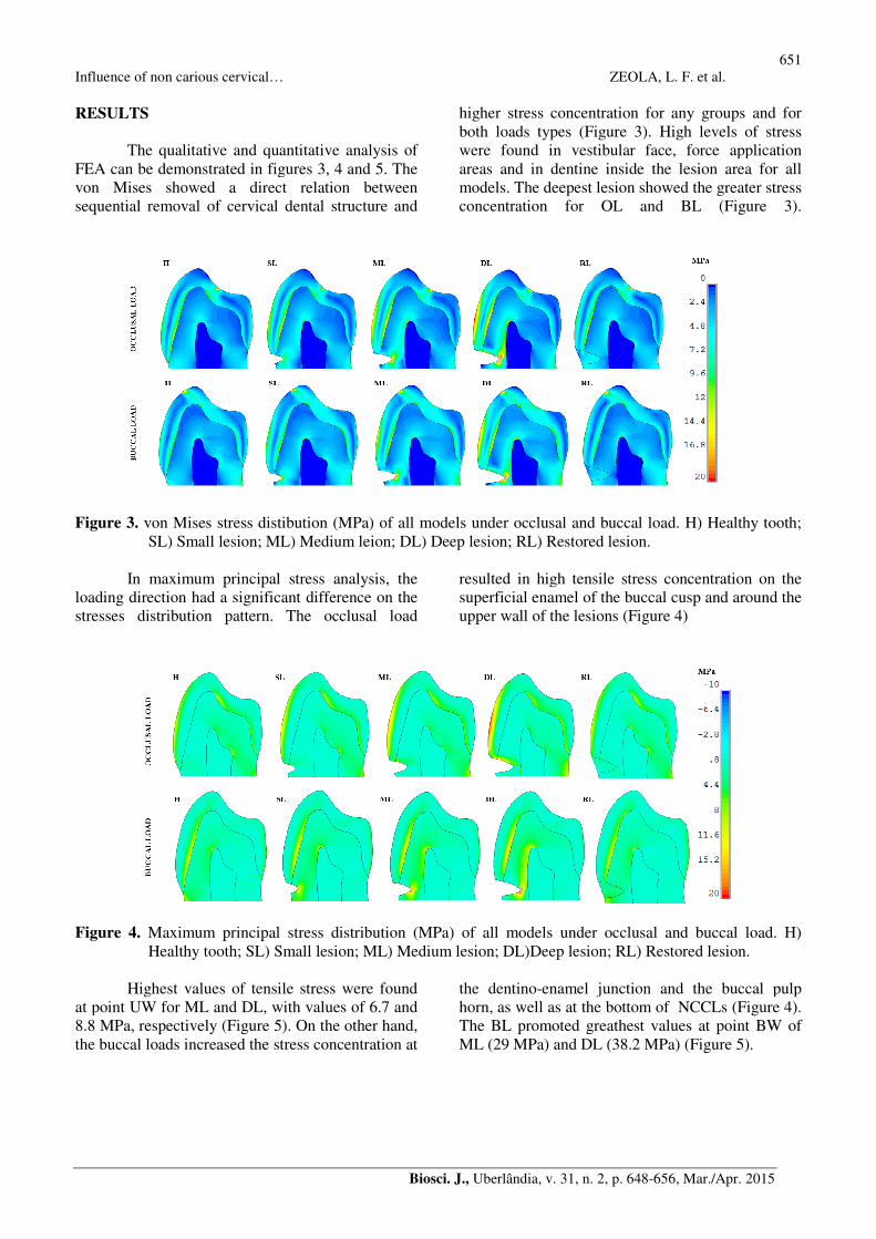
The qualitative and quantitative analysis of
FEA can be demonstrated in figures 3, 4 and 5. The
von Mises showed a direct relation between
sequential removal of cervical dental structure and
higher stress concentration for any groups and for
both loads types (Figure 3). High levels of stress
were found in vestibular face, force application
areas and in dentine inside the lesion area for all
models. The deepest lesion showed the greater stress
concentration for OL and BL (Figure 3).
Figure 3. von Mises stress distibution (MPa) of all models under occlusal and buccal load. H) Healthy tooth;
SL) Small lesion; ML) Medium leion; DL) Deep lesion; RL) Restored lesion.
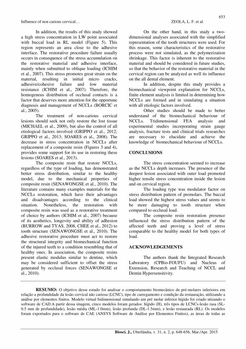
In maximum principal stress analysis, the
loading direction had a significant difference on the
stresses distribution pattern. The occlusal load
resulted in high tensile stress concentration on the
superficial enamel of the buccal cusp and around the
upper wall of the lesions (Figure 4)
Figure 4. Maximum principal stress distribution (MPa) of all models under occlusal and buccal load. H)
Healthy tooth; SL) Small lesion; ML) Medium lesion; DL)Deep lesion; RL) Restored lesion.
Highest values of tensile stress were found
at point UW for ML and DL, with values of 6.7 and
8.8 MPa, respectively (Figure 5). On the other hand,
the buccal loads increased the stress concentration at
the dentino-enamel junction and the buccal pulp
horn, as well as at the bottom of NCCLs (Figure 4).
The BL promoted greathest values at point BW of
ML (29 MPa) and DL (38.2 MPa) (Figure 5).

652
Influence of non carious cervical… ZEOLA, L. F. et al.
Biosci. J., Uberlândia, v. 31, n. 2, p. 648-656, Mar./Apr. 2015
Figure 5. Maximum principal stress values obtained from finite element analysis on three specific points within
NCCL subjected to the occlusal and buccal load (in MPa). UW) Upper wall of the lesion in enamel;
BW) Bottom wall of the lesion in dentine and LW) dentin on the lower wall.
Stress distribution for RL was similar to the
behavior of the healthy tooth model, for both
loading directions (Figures 3 and 4) . The restored
models presented low stress levels in the cervical
regions, with highest stress values 2.5 MPa and 5.6
MPa for OL and BL, respectively (Figure 5).
DISCUSSION
The findings of the present study showed
that the NCCLs size and load point application
plays an important role on stress distribution pattern
in lower premolars.
Finite element analysis in the present study
indicates that the presence of NCCLs promoted
changes on stress distribution pattern due to the loss
of dental structure (SOARES et al., 2008). NCCLs,
even as small (0.5mm,) generated high stress
concentration in the cervical region, with the stress
magnitude increasing with lesion size (Figure 3 and
4). This fact suggests that the stress generated by the
lesion presence may be a factor that leads to
progression of the NCCLs (SOARES et al., 2013).
For this reason, the treatment of shallow lesions
should be taken into consideration in dental clinical
practice, since only its monitoring, without the
control of the etiological factors and without the
restoration, can provide an acceleration of tooth
structure loss process.
The biomechanical behavior of the teeth
affected by NCCLs varies according to the loading
direction (REES, 2002; VASUDEVA and BOGRA,
2008; SOARES et al., 2014; BENAZZI et al.,
2014). High stress concentration is generated from
occlusal loading during tooth chewing and others
types of parafunctions (REES, 2002; WOOD et al.,
2009). The results of this study demonstrated that
each type of load generate stress concentration in
different regions. This finding ratifies that different
positions of occlusal contacts promote changes in
tensile stress magnitude in the cervical region, that
tend to be able to initiate failure (REES, 2002). This
load application promotes compressive and tensile
stress acting on the tooth structure, influencing on
disruption of chemical bonds between the enamel
crystals in cervical region (BORCIC et al., 2005;
LEE H. E. et al., 2002), accelerating the tooth
structure loss.
The presence of a traumatic dental occlusion
is considered an important factor for the occurrence
of NCCLs (BRANDINI et al., 2012). A better
understanding of the influence of the applied loads
may help a clinician select more biomechanically
satisfactory restorative treatment to reduce the
incidence and severity of NCCLs. Our findings
showed that occlusal loading produced tensile stress
in the upper wall of NCCLs, and the buccal loading
generated tensile stress in the bottom region of
NCCLs (Figure 4). Furthermore, the magnitude of
tensile stress was greater for outer loading than
inner loading (Figure 5). The stress acumulation in
UW point for OL suggests a progression of the
lesion towards the long axis of the tooth, which is
increasing in height. On the other hand, the presence
of stress at the bottom of the lesion (BW) generated
by BL, may tend to lead to increasing injury in the
horizontal direction, thus characterizing an
evolution in depth.
653
Influence of non carious cervical… ZEOLA, L. F. et al.
Biosci. J., Uberlândia, v. 31, n. 2, p. 648-656, Mar./Apr. 2015
In addition, the results of this study showed
a high stress concentration in LW point associated
with buccal load in RL model (Figure 5). This
region represents an area close to the adhesive
interface. The restorative procedure failure usually
occurs in consequence of the stress accumulation on
the restorative material and adhesive interface,
mainly when submitted to oblique loading (ICHIM
et al., 2007). This stress promotes great strain on the
material, resulting in initial micro cracks,
adhesive/cohesive failure and low material
resistance (ICHIM et al., 2007). Therefore, the
homogeneus distribution of occlusal contacts is a
factor that deserves more attention for the opportune
diagnosis and management of NCCLs (BORCIC et
al., 2005).
The treatment of non-carious cervical
lesions should seek not only restore the lost tissue
(MICHAEL et al., 2009), but also the control of all
etiological factors involved (GRIPPO et al., 2012;
GRIPPO et al., 2013; SOARES et al., 2008). The
decrease in stress concentration in NCCLs after
replacement of a composite resin (Figures 3 and 4),
provides some support for its use in restoring these
lesions (SOARES et al., 2013).
The composite resin that restore NCCLs,
regardless of the type of loading, has demonstrated
better stress distribution, similar to the healthy
model, due to the mechanical properties of
composite resin (SENAWONGSE et al., 2010). The
literature contains many examples materials for the
NCCLs restoration, which have their advantages
and disadvantages according to the clinical
situation. Nonetheless, the restoration with
composite resin was used as a restorative treatment
of choice by authors (ICHIM et al., 2007) because
of its aesthetics, longevity and ability of adhesion
(BURROW and TYAS, 2008, CHEE et al., 2012) to
tooth structure (SENAWONGSE et al., 2010). The
adhesive restorative procedure must act to restore
the structural integrity and biomechanical function
of the injured teeth to a condition resembling that of
healthy ones. In association, the composite resins
present elastic modulus similar to dentine, which
may be considered sufficient to offset the stress
generated by occlusal forces (SENAWONGSE et
al., 2010).
On the other hand, in this study a two-
dimensional analyses associated with the simplified
representation of the tooth structures were used. For
this reason, some characteristics of the restorative
process were not simulated, as the polymerization
shrinkage. This factor is inherent to the restorative
material and should be considered in future studies,
so that the behavior of the restorative material in the
cervical region can be analyzed as well its influence
on the all dental element.
In addition, despite this study provides a
biomechanical viewpoint explanation for NCCLs,
finite element analysis is limited in determining how
NCCLs are formed and in simulating a situation
with all etiologic factors involved.
Other studies should be made to better
understand of the biomechanical behaviour of
NCCLs. Tridimensional FEA analysis and
experimental studies incorporating strain gauge
analysis, fracture tests and clinical trials researches
are necessary to elucidate and achieve the
knowledge of biomechanical behaviour of NCCLs.
CONCLUSIONS
The stress concentration seemed to increase
as the NCCLs depth increases. The presence of the
deepest lesion associated with outer load promoted
higher tensile stress concentration inside the lesion
and on cervical region.
The loading type was modulator factor on
stress distribution pattern of premolars. The buccal
load showed the highest stress values and seems to
be more damaging to tooth structure when
compared to occlusal load.
The composite resin restoration presence
influenced the stress distribution pattern of the
affected teeth and proving a level of stress
comparable to the healthy model for both types of
load.
ACKNOWLEDGEMENTS
The authors thank the Integrated Research
Laboratory (CPBio-FOUFU) and Nucleus of
Extension, Research and Teaching of NCCL and
Dentin Hypersensitivity.
RESUMO: O objetivo desse estudo foi analisar o comportamento biomecânico de pré-molares inferiores em
relação a profundidade da lesão cervical não cariosa (LCNC), tipo de carregamento e condição da restauração, utilizando a
análise por elementos finitos. Modelo virtual bidimensional simulando um pré molar inferior hígido foi criado utizando o
software de CAD.A partir dessa imagem, cinco modelos foram gerados: hígido (H), três tipos de LCNCs-lesão rasa (SL-
0.5 mm de profundidade), lesão média (ML-1.0mm), lesão profunda (DL-1.5mm), e lesão restaurada (RL). Os modelos
foram exportados para o software de CAE (ANSYS Software de Análise por Elementos Finitos), as áreas de todas as
654
Influence of non carious cervical… ZEOLA, L. F. et al.
Biosci. J., Uberlândia, v. 31, n. 2, p. 648-656, Mar./Apr. 2015
estruturas foram plotadas e cada modelo foi malhado utilizando um dispositivo controle de malha.Todos os modelos
virtuais foram submetidos a dois tipos de carregamento oclusal (100N cada): carregamento oclusal (OL) e carregamento
externo (BL) na cúspide vestibular. A intensidade e a distribuição das tensões foram obtidas utilizando os critérios de von
Mises e tensão máxima principal (σ1),em Mpa. A análise quantitativa das tensões (MPa) foi identificada em três pontos
das LCNCs: parede superior em esmalte, parede de fundo em dentina e parede inferior em dentina. Os resultados
apresentaram uma relação direta entre a remoção sequencial de estrutura na região cervical e os maiores valores de
concentração de tensões para todos os grupos e para os dois tipos de carga. Para OL, o maior valor de tensão foi 8.8 MPa
para DL na parede superior da LCNCs. O BL exibiu maiores valores de tensão em comparação ao OL para todos os
modelos. Além disso, BL foi responsável por promover o maior acúmulo de tensão na parede de fundo, 38.2 MPa para
DL. A restauração com resina composta foi capaz de restaurar uma distribuição de tensões similar a do modelo hígido,
para ambos os tipos de carga. Em conclusão, a extensão da lesão e o tipo de carregamento influenciaram no padrão de
distribuição de tensões de pré-molares inferiores. A carga externa parece ser mais crítica para afetar o comportamento
biomecânico de pré-molares inferiores, independente do tamanho da lesão. A restauração das LCNCs com resina
composta, parece recuperar o comportamento biomecânico similar ao do modelo hígido.
PALAVRAS-CHAVES: Análise por elementos finitos. Lesão Cervical Não Cariosa. Pré-molar.
REFERENCES
BENAZZI, S.; GROSSE, I. R.; GRUPPIONI, G.; WEBER, G. W.; KULLMER, O. Comparison of occlusal
loading conditions in a lower second premolar using three-dimensional finite element analysis. Clin Oral Investig, Berlin, v. 18, n. 2, p. 369-75, 2014. http://dx.doi.org/10.1007/s00784-013-0973-8
BERNHARDT, O.; GESCH, D.; SCHWAHN, C.; MACK, F.; MEYER, G.; JOHN. U.; KOCHER,
T.Epidemiological evaluation of the multifactorial aetiology of abfractions. J Oral Rehabil, Oxford, v. 33, n.
1, p. 17-25, 2006. http://dx.doi.org/10.1111/j.1365-2842.2006.01532.x
BORCIC, J.; ANIC, I.; UREK, M.M.; FERRERI S. The prevalence of non-carious cervical lesions in
permanent dentition. J Oral Rehabil,Oxford, v. 31,n. 2, p. 117-23, 2004. http://dx.doi.org/10.1046/j.0305-
182X.2003.01223.x
BORCIC, J.; ANIC, I.; SMOJVER, I.; CATIC, A.; MILETIC, I.; RIBARIC, S.P. 3D finite element model and
cervical lesion formation in normal occlusion and in malocclusion. J Oral Rehabil, Oxford, v. 32, n. 7, p. 504-
10, 2005. http://dx.doi.org/10.1111/j.1365-2842.2005.01455.x
BRANDINI, D. A.; TREVISAN, C. L.; PANZARINI, S. R.; PEDRINI, D. Clinical evaluation of the
association between noncarious cervical lesions and occlusal forces. J Prosthet Dent, St. Louis, v. 108, n. 5,p.
298-303,2012.
BRANDINI, D. A.; PEDRINI, D.; PANZARINI, S. R.; BENETE, I. M.; TREVISAN, C. L. Clinical evaluation
of the association of noncarious cervical lesions, parafunctional habits, and TMD diagnosis. Quintessence Int, Berlin , v. 43, n. 3, p. 255-62, 2012.
BURROW, M. F.; TYAS, M. J. A clinical trial comparing two all-in-one adhesive systems used to restore non-
carious cervical lesions: results at one year. Aust Dent J, Sidney, v. 53, n. 3,p. 235-8, 2008.
CHEE, B., RICKMAN, L. J.; SATTERTHWAITE, J. D. Adhesives for the restoration of non-carious cervical
lesions: a systematic review. J Dent,Bristol, v. 40, n. 6,p. 443-52, 2012.
http://dx.doi.org/10.1016/j.jdent.2012.02.007
DEJAK, B.; MLOTKOWSKI, A. Finite element analysis of strength and adhesion of cast posts compared to
glass fiber-reinforced composite resin posts in anterior teeth. J Prosthet Dent, St. Louis, v. 105, n. 2, p. 115-
26, 2011.

655
Influence of non carious cervical… ZEOLA, L. F. et al.
Biosci. J., Uberlândia, v. 31, n. 2, p. 648-656, Mar./Apr. 2015
GRIPPO, J. O.; SIMRING, M.; COLEMAN, T. A. Abfraction, abrasion, biocorrosion, and the enigma of
noncarious cervical lesions: a 20-year perspective. J Esthet Restor Dent,London, v. 24, n. 1, p. 10-23, 2012.
http://dx.doi.org/10.1111/j.1708-8240.2011.00487.x
GRIPPO, J. O.; CHAIYABUTR, Y.; KOIS, J. C. Effects of cyclic fatigue stress-biocorrosion on noncarious
cervical lesions. J Esthet Restor Dent, London, v. 25, n. 4, p. 265-72, 2013.
http://dx.doi.org/10.1111/jerd.12024
ICHIM, I. P.; SCHMIDLIN,P. R.; LI, Q.; KIESER, J. A.; SWAIN, M. V. Restoration of non-carious cervical
lesions Part II. Restorative material selection to minimise fracture. Dent Mater,Kidlington, v. 23, n. 12,p.
1562-9, 2007.
JOSHI, S.; MUKHERJEE, A.; KHEUR,M.; MEHTA, A. Mechanical performance of endodontically treated
teeth.Finite Elem Anal Des, Amsterdam, v. 37, p. 587-601, 2001. http://dx.doi.org/10.1016/S0168-
874X(00)00059-7
LEE, H. E.; LIN, C. L.; WANG, C. H.; CHENG, C. H.; CHANG, C. H. Stresses at the cervical lesion of
maxillary premolar--a finite element investigation. J Dent, Bristol, v. 30, n. 7-8, p. 283-90, 2002.
http://dx.doi.org/10.1016/S0300-5712(02)00020-9
LEE, W. C.; EAKLE, W. S. Possible role of tensile stress in the etiology of cervical erosive lesions of teeth. J Prosthet Dent, St. Louis, v. 52, n. 3, p. 374-80,1984.
MICHAEL, J. A.; TOWNSEND, G. C.; GREENWOOD, L. F.; KAIDONIS, J. A. Abfraction: separating fact
from fiction. Aust Dent J,Sidney, v. 54, n. 1,p. 2-8, 2009.
MIURA, J.; MAEDA, Y.; NAKAI, H.; ZAKO, M. Multiscale analysis of stress distribution in teeth under
applied forces. Dent Mater, Kidlington, v. 25, n. 1, p. 67-73, 2009.
OMMERBORN, M. A.; SCHNEIDER, C.; GIRAKI, M.; SCHAFER, R.; SINGH, P.; FRANZ, M.; RAAB,
W.H. In vivo evaluation of noncarious cervical lesions in sleep bruxism subjects. J Prosthet Dent, St. Louis, v.
98, n. 2, p. 150-8, 2007.
ONAL, B.; PAMIR, T. The two-year clinical performance of esthetic restorative materials in noncarious
cervical lesions. J Am Dent Assoc,Chicago, v. 136, n. 11,p. 1547-55, 2005.
http://dx.doi.org/10.14219/jada.archive.2005.0085
POIATE, I. A.; VASCONCELLOS, A. B.; POIATE JUNIOR, E.; DIAS, K. R. Stress distribution in the
cervical region of an upper central incisor in a 3D finite element model. Braz Oral Res, São Paulo, v. 23, n. 2,
p. 161-8, 2009.
REES, J. S. The effect of variation in occlusal loading on the development of abfraction lesions: a finite
element study. J Oral Rehabil,Oxford , v. 29, n. 2, p. 188-93, 2002. http://dx.doi.org/10.1046/j.1365-
2842.2002.00836.x
REES, J. S.; HAMMADEH, M.; JAGGER, D. C. Abfraction lesion formation in maxillary incisors, canines
and premolars: a finite element study. Eur J Oral Sci, Chichester, v. 111,n. 2, p. 149-54, 2003.
http://dx.doi.org/10.1034/j.1600-0722.2003.00018.x
REYES, E.; HILDEBOLT, C.; LANGENWALTER, E.; MILEY, D. Abfractions and attachment loss in teeth
with premature contacts in centric relation: clinical observations. J Periodontol, Chicago, v. 80, n. 12, p. 1955-
62, 2009. http://dx.doi.org/10.1902/jop.2009.090149

656
Influence of non carious cervical… ZEOLA, L. F. et al.
Biosci. J., Uberlândia, v. 31, n. 2, p. 648-656, Mar./Apr. 2015
SENAWONGSE, P.; PONGPRUEKSA, P.; TAGAMI, J. The effect of the elastic modulus of low-viscosity
resins on the microleakage of Class V resin composite restorations under occlusal loading. Dent Mater J,
Tokyo, v. 29,n. 3,p. 324-9, 2010.
SILVA, N. R.; CASTRO, C. G.; SANTOS-FILHO, P. C.; SILVA, G. R.; CAMPOS, R. E.; SOARES, P. V.;
SOARES, C. J. Influence of different post design and composition on stress distribution in maxillary central
incisor: Finite element analysis. Indian J Dent Res, Ahmedabad,v. 20, n. 2,p. 153-8, 2009.
http://dx.doi.org/10.4103/0970-9290.52888
SMITH, W. A.; MARCHAN, S.; RAFEEK, R. N. The prevalence and severity of non-carious cervical lesions
in a group of patients attending a university hospital in Trinidad. J Oral Rehabil,Oxford, v. 35, n. 2,p. 128-34,
2008.
SOARES, P. V.; SANTOS-FILHO, P. C.; GOMIDE, H. A.; ARAUJO, C. A.; MARTINS, L. R.; SOARES, C.
J. Influence of restorative technique on the biomechanical behavior of endodontically treated maxillary
premolars. Part II: strain measurement and stress distribution. J Prosthet Dent, St. Louis, v. 99, n. 2, p. 114-22,
2008. http://dx.doi.org/10.1111/joor.12113
SOARES, P. V.; SOUZA, L. V.; VERISSIMO, C.; ZEOLA, L. F.; PEREIRA, A. G.; SANTOS-FILHO, P. C.;
FERNANDES-NETO, A. J.Effect of root morphology on biomechanical behaviour of premolars associated
with abfraction lesions and different loading types. J Oral Rehabil, Oxford, v. 41, n. 2, p. 108-14, 2014.
SOARES, P. V.; SANTOS-FILHO, P. C.; SOARES, C. J.; FARIA, V. L,; NAVES, M. F.; MICHAEL, J. A.;
KAIDONIS, J. A.; RANJITKAR, S.; TOWNSEND, G. C. Non-carious cervical lesions: influence of
morphology and load type on biomechanical behaviour of maxillary incisors. Aust Dent J, Sydney, v. 58, n. 3,
p. 306-14, 2013.
SOARES, P. V., MILITO, G. A.; PEREIRA,F. A.; ZEOLA, L. F.; NAVES, M. F. L.; FARIA, V. L. G.;
MACHADO, A. C.; SOUZA, P. G.; REIS, B. R. The effects of non carious cervical lesions - morphology,load
type and restoration - on the biomechanical behavior of maxillary premolars: a finite element analysis. Biosci J., Uberlândia, v. 29, n. 2, p. 526-535, 2013.
STEWARDSON, D.; CREANOR, S.; THORNLEY, P.; BIGG, T.; BROMAGE, C.; BROWNE, A.; COTTAM,
D.; DALBY, D.; GILMOUR, J.; HORTON, J.; ROBERTS, E,; WESTOBY, L.; BURKE, T. The survival of
Class V restorations in general dental practice: part 3, five-year survival. Braz Dent J., Ribeirão Preto, v. 212,
n. 9 ,p. E14, 2012.
TOPARLI, M. Stress analysis in a post-restored tooth utilizing the finite element method. J Oral Rehabil, Oxford , v. 30, n. 5, p. 470-6, May 2003. http://dx.doi.org/10.1046/j.1365-2842.2003.01090.x
VASUDEVA, G.; BOGRA, P. The effect of occlusal restoration and loading on the development of abfraction
lesions: A finite element study. J Conserv Dent, Mumbai, v. 11, n. 3,p. 117-20, Jul 2008.
http://dx.doi.org/10.4103/0972-0707.45250
ZUCCHELLI, G.; GORI, G.; MELE, M.; STEFANINI, M.; MAZZOTTI, C.; MARZADORI, M.;
MONTEBUGNOLI, L.; DE SANCTIS, M. Non-carious cervical lesions associated with gingival recessions: a
decision-making process. J Periodontol, Chicago, v. 82, n. 12, p. 1713-24.
WOOD, I. D.; KASSIR, A. S.; BRUNTON, P. A. Effect of lateral excursive movements on the progression of
abfraction lesions. Oper Dent, Seattle, v. 34, n. 3,p. 273-9, 2009. http://dx.doi.org/10.2341/08-100