influenza virus: a tiny moving target level: high school ...€¦ · influenza virus: a tiny moving...
TRANSCRIPT

Influenza Virus: A tiny moving target
Level: High School
Time: 2-3 50 minute periods
Overview:
The potential of a new flu pandemic is a frightening idea. This curriculum explains how
influenza viruses infect cells and replicate. It also has students explore where influenza
viruses come from, how viruses change, and why some become deadly.
Background for Teachers
Influenza viruses are always with us, constantly changing and causing misery each flu
season. Most of the time they knock people off their feet for a week or two but
sometimes the flu can be dangerous or even fatal. Immunocompromised people, those
who are already sick, or those whose immune systems are weak such as young children
and older people, are hit hardest by the flu. This version of the flu is with us every year
and we are accustomed to it, however the threat of a flu pandemic is a much larger
problem and rightfully causes a great deal of concern.
Flu pandemics emerge in a cycle and the world is overdue for the next one. If the avian
H5N1 virus doesn’t cause the next pandemic, another strain will. There were three
pandemics in the 1900’s: 1918, 1957, and 1968. In 1918, 30-40 million people died
world wide (For more information, see Reference 1). We know much more about
influenza now than we did in 1918, and the hope is that we can use this knowledge to
prevent or control a serious pandemic (Ref. 2). Pandemics occur when a virus acquires
the ability to infect a new host and spread rapidly through the population. How this
occurs is more easily understood when the normal replication cycle of the virus is
understood.
Influenza viruses come in three types: A, B, and C. All three can infect humans, but the
most common infection is by type A. Type A viruses are classified in subtypes based on
two proteins that stick out from the surface. These are hemagglutinin (H), which is
responsible for initiating entry into the host cell, and neuraminidase (N), which is
involved in release of new viral particles from the infected cell. There are 16 varieties of
hemagglutinin and 9 of neuraminidase. Subtypes are named by which version of
hemagglutinin and neuraminidase, such as H1N2 a common version in humans. The
virulence and pathogenicity of the virus depends on the combination of hemagglutinin
and neuraminidase.
Influenza viruses are normally found in wild waterfowl. The H5N1 avian version is
particularly deadly in its normal host, wild birds. It is easily transferred to domestic
birds, and from there has occasionally infected humans, particularly those who came in
close contact with infected birds. It appears to be very dangerous in humans as well, but
that may be due to reporting. Work in Turkey has revealed people who were infected

with H5N1, but did not get sick enough to require medical attention. At the same time,
other people in Turkey have died of the disease.
In order for H5N1 to develop into a pandemic, it must acquire the ability to transfer
rapidly between and effectively infect human hosts. Once that occurs, it will be difficult
to control the spread of the disease. Our global society ensures wide dispersal of diseases
via air travel, and the incubation period for the flu is short. This means that by the time
health officials recognize an outbreak, it will already have spread beyond the original
area. However, we understand influenza much better now than we have in previous
pandemics. We are better equipped to prevent transmission of the virus, have antiviral
medications to treat infections, and are working to develop effective vaccines. For more
information on influenza viruses and pandemics, see References 3, 4, and 5.
The control of influenza relies on basic evolutionary theory. The development of the
annual flu vaccine is based on our understanding of antigenic drift – or change over time.
We expect new strains to arise as viral polymerases make mistakes during replication.
We can predict the direction of change based on the trends observed in the virus genome
over the course of the flu season, and prepare our immune systems with a vaccine. The
vaccine is developed with the latest version of the flu so that when the “real thing” shows
up in our bodies, the immune system has seen it or something similar and can effectively
fight it off. Pandemics occur because our immune system cannot cope with invasion by a
completely new viral type and these viruses often infect more cell types than the
“normal” flu virus. Pandemic viruses avoid detection by the immune system, cause
massive damage in the host, and spread rapidly. Pandemics die down because any
pathogen that kills its host before replicating and spreading dies with the host. A milder
version of the disease is more likely to allow continued dispersal of the virus and so viral
strains causing less intense symptoms will have a selective advantage. In previous
pandemics, the virus became less dangerous after 18-24 months. However, incredible
damage was done during that brief time.
About the Lesson
This lesson is structured using the BSCS 5E model. In this model, students are first
Engaged, in this case by presenting them with a game modeling viral infection. Once the
students are interested, they move to combined Explore and Explain sections where they
have an opportunity to learn about viral structure and replication. Students then move to
an Elaborate section, in which they apply the information they have just learned at a
more advanced level to explore how viruses evolve. Evaluation can be conducted
throughout the inquiry based lesson, but a formal evaluation opportunity is suggested in
the form of presentations or written reports in which students are asked to use their
understanding of the virus to suggest ways in which to prevent or treat infection.
National Science Standards
Content
Standard A.
Abilities
Communicate and
Defend a scientific
argument
• Reviewing information, expressing
concepts, constructing a reasoned
argument.

Necessary to do
Scientific
Inquiry
C: Life Science
The Cell
Molecular Basis of
Heredity
Biological
Evolution
Interdependence of
Organisms
• Cells have particular structures that
underlie their functions. Every cell is
surrounded by a membrane that
separates it from the outside world.
Inside the cells in a concentrated mixture
of thousands of different molecules
which form a variety of specialized
structures that carry out such cell
functions as energy production, transport
of molecules, waste disposal, synthesis
of new molecules and the storage of
genetic material.
• In all organisms, the instructions for
specifying the characteristics of the
organism are carried DNA.
• Changes in DNA occur spontaneously at
low rates. Some of these changes make
no difference to the organism, whereas
others can changes cells and organisms.
• Species evolve over time. Evolution is
the consequence of the interactions of (1)
the potential for a species to increase its
numbers, (2) the genetic variability of
offspring due to mutation and
recombination of genes, (3) a finite
supply of the resources requires for life,
and (4) the ensuing selection by the
environment of those offspring better
able to survive and leave offspring.
• The millions of difference species of
plants, animals, and microorganisms that
live on earth today are related by descent
from common ancestors.
• Organisms both cooperate and compete
in ecosystems. The interrelationships
and interdependencies of these
organisms may generate ecosystems that

are stable for hundreds or thousands of
years.
• Human beings live within the world’s
ecosystems.
Content
Standard F:
Science in
Personal and
Social
Perspectives
Personal and
Community Health
• The severity of disease symptoms is
dependent on many factors, such as
human resistance and the virulence of
the disease-producing organisms. Many
diseases can be prevented, controlled, or
cured.
Content
Standard G:
History and
Nature of
Science
Science as a
Human Endeavor • Scientists are influenced by societal,
cultural, and personal beliefs and ways
of viewing the world. Science is not
separate from society but rather science
is a part of society.
• Occasionally, there are advances in
science and technology that have
important and long-lasting effects on
science and society. Examples of such
advances include the geologic time scale
and biological evolution.
Preparation
• Prepare the following for “Gotcha!”:
• 2 tokens per student
• 1 slip of paper per student stating cell type (virus, respiratory, lung,
immune, other organs as needed). Keep a ratio of 1:1:1:1:1, so in a class of
30, there will be 6 viruses, 6 respiratory cells, 6 lung cells, 6 immune cells,
and 6 other organs.
• Write a password on each slip. Give the viruses at least three different
passwords (ie. Blue, turtle, apple). Give all the respiratory cells one of the
viral passwords (ie. Blue). Give one lung cell a viral password, and give the
others completely different passwords. Give the immune cells two out of
three passwords in different combinations (ie. Blue and turtle, blue and
apple, turtle and apple). Give one of the other cell types a viral password
and the others completely different passwords.
• For the viral structure lesson, prepare one set of the following materials for each
group:
• 1 plastic egg
• 8 one inch strips of black yarn
• 8 half inch pieces of drinking straw
• 8 paper clips or buttons that can slide on the yarn
• 15-20 “dots” of Velcro (soft side), made with a hole punch
• 15-20 grains of rice.
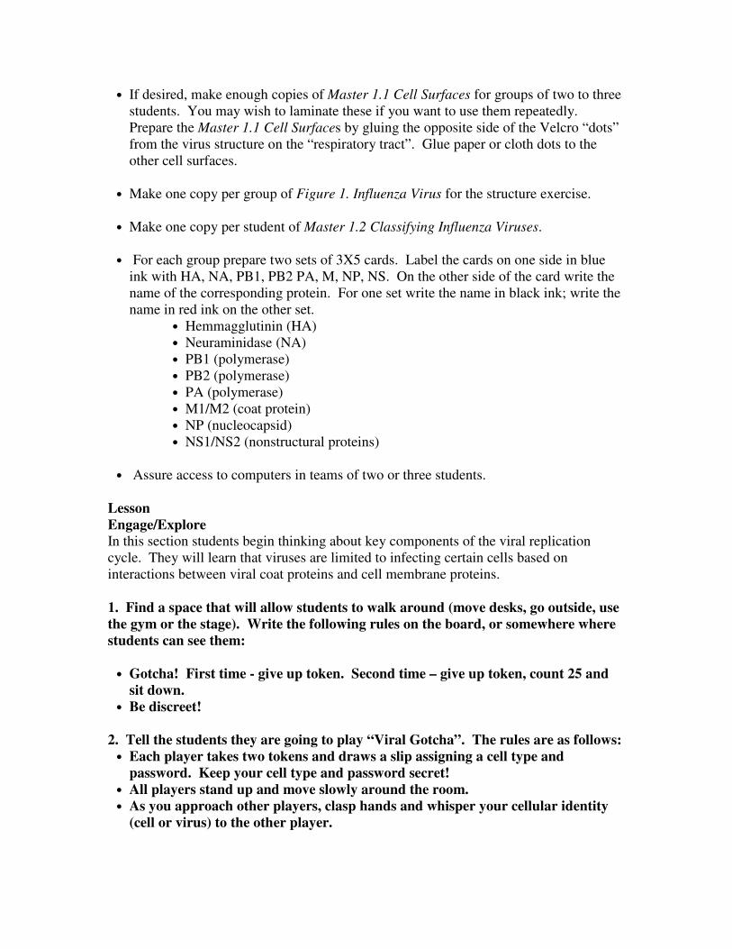
• If desired, make enough copies of Master 1.1 Cell Surfaces for groups of two to three
students. You may wish to laminate these if you want to use them repeatedly.
Prepare the Master 1.1 Cell Surfaces by gluing the opposite side of the Velcro “dots”
from the virus structure on the “respiratory tract”. Glue paper or cloth dots to the
other cell surfaces.
• Make one copy per group of Figure 1. Influenza Virus for the structure exercise.
• Make one copy per student of Master 1.2 Classifying Influenza Viruses.
• For each group prepare two sets of 3X5 cards. Label the cards on one side in blue
ink with HA, NA, PB1, PB2 PA, M, NP, NS. On the other side of the card write the
name of the corresponding protein. For one set write the name in black ink; write the
name in red ink on the other set.
• Hemmagglutinin (HA)
• Neuraminidase (NA)
• PB1 (polymerase)
• PB2 (polymerase)
• PA (polymerase)
• M1/M2 (coat protein)
• NP (nucleocapsid)
• NS1/NS2 (nonstructural proteins)
• Assure access to computers in teams of two or three students.
Lesson
Engage/Explore
In this section students begin thinking about key components of the viral replication
cycle. They will learn that viruses are limited to infecting certain cells based on
interactions between viral coat proteins and cell membrane proteins.
1. Find a space that will allow students to walk around (move desks, go outside, use
the gym or the stage). Write the following rules on the board, or somewhere where
students can see them:
• Gotcha! First time - give up token. Second time – give up token, count 25 and
sit down.
• Be discreet!
2. Tell the students they are going to play “Viral Gotcha”. The rules are as follows:
• Each player takes two tokens and draws a slip assigning a cell type and
password. Keep your cell type and password secret!
• All players stand up and move slowly around the room.
• As you approach other players, clasp hands and whisper your cellular identity
(cell or virus) to the other player.

• If you are a virus, and you find a cell, state your password. The “cell” player
will accept the password or say “no entry”. If you are a cell, any virus with a
matching password can “infect” you. Viruses with passwords that do not match
yours cannot infect you.
• If a “virus” finds a “cell” that accepts the password, the cell has to give the virus
a token – quietly! This means the “cell” has been infected.
• If a “cell” is “infected” twice, then after giving the second virus a token, the cell
player keeps moving around while counting to 25 (in his or her head) and then
sits down.
• Each “immune cell” player will have a list of passwords he or she “recognizes”.
When an “immune cell” meets a “virus” with a recognized password, the “virus”
player has to give his or her tokens to the “immune cell”. A “virus” that is
caught by an “immune cell” twice must count 25 in his or her head and sit down.
Allow students to play for 5-10 minutes, depending on how many are sitting down.
3. At the end, tally how many viruses and cells are left standing or “alive”. Ask
the “live” viruses how many tokens they each acquired. Group cells together by
type and password, and determine how many times each cell type was infected.
Ask the “immune cells” how many viruses each of them caught. Write the answers
on the board.
4. Ask the students what they notice about the results.
Guide the students to find answers to the following questions:
• Which did better, cells or viruses?
• Did a particular type of cell get infected more often?
• Did a particular type of virus infect more cells?
• Did a particular type of virus get caught by the immune cells more often?
Let students explore the results as independently as possible. The goal is for students to
come to understand that viruses do not infect cells randomly, and that it is possible for a
virus to avoid detection by the immune system.
One option, if time allows, is to run the game multiple times and see if the results vary.
5. Explain to the students that viruses cannot infect cells without the correct
“password” although in real life, the password is actually a coat protein on the virus
interacting with a membrane protein on the cell. Usually passwords are cell type
specific, for example, HIV targets T cells, but some viruses have a broader range of
host cells.
Explore/Explain
In this section, students explore viral structure and how the virus replicates.
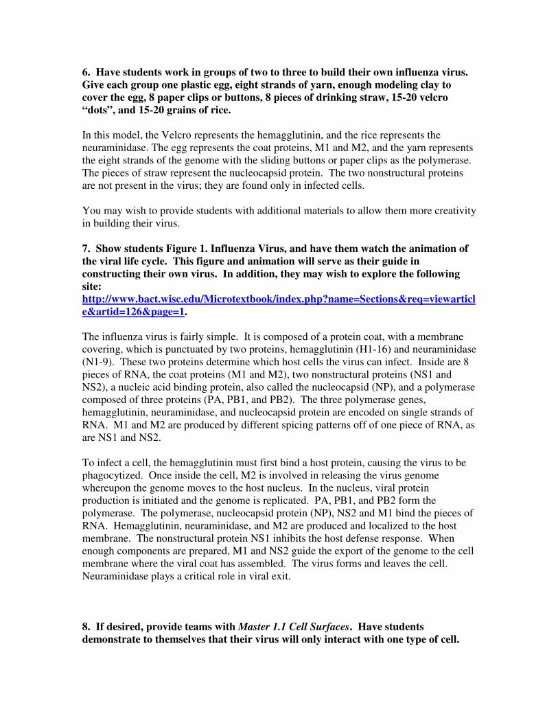
6. Have students work in groups of two to three to build their own influenza virus.
Give each group one plastic egg, eight strands of yarn, enough modeling clay to
cover the egg, 8 paper clips or buttons, 8 pieces of drinking straw, 15-20 velcro
“dots”, and 15-20 grains of rice.
In this model, the Velcro represents the hemagglutinin, and the rice represents the
neuraminidase. The egg represents the coat proteins, M1 and M2, and the yarn represents
the eight strands of the genome with the sliding buttons or paper clips as the polymerase.
The pieces of straw represent the nucleocapsid protein. The two nonstructural proteins
are not present in the virus; they are found only in infected cells.
You may wish to provide students with additional materials to allow them more creativity
in building their virus.
7. Show students Figure 1. Influenza Virus, and have them watch the animation of
the viral life cycle. This figure and animation will serve as their guide in
constructing their own virus. In addition, they may wish to explore the following
site:
http://www.bact.wisc.edu/Microtextbook/index.php?name=Sections&req=viewarticl
e&artid=126&page=1.
The influenza virus is fairly simple. It is composed of a protein coat, with a membrane
covering, which is punctuated by two proteins, hemagglutinin (H1-16) and neuraminidase
(N1-9). These two proteins determine which host cells the virus can infect. Inside are 8
pieces of RNA, the coat proteins (M1 and M2), two nonstructural proteins (NS1 and
NS2), a nucleic acid binding protein, also called the nucleocapsid (NP), and a polymerase
composed of three proteins (PA, PB1, and PB2). The three polymerase genes,
hemagglutinin, neuraminidase, and nucleocapsid protein are encoded on single strands of
RNA. M1 and M2 are produced by different spicing patterns off of one piece of RNA, as
are NS1 and NS2.
To infect a cell, the hemagglutinin must first bind a host protein, causing the virus to be
phagocytized. Once inside the cell, M2 is involved in releasing the virus genome
whereupon the genome moves to the host nucleus. In the nucleus, viral protein
production is initiated and the genome is replicated. PA, PB1, and PB2 form the
polymerase. The polymerase, nucleocapsid protein (NP), NS2 and M1 bind the pieces of
RNA. Hemagglutinin, neuraminidase, and M2 are produced and localized to the host
membrane. The nonstructural protein NS1 inhibits the host defense response. When
enough components are prepared, M1 and NS2 guide the export of the genome to the cell
membrane where the viral coat has assembled. The virus forms and leaves the cell.
Neuraminidase plays a critical role in viral exit.
8. If desired, provide teams with Master 1.1 Cell Surfaces. Have students
demonstrate to themselves that their virus will only interact with one type of cell.

Point out to students that this is very much not to scale. Another option is to make a
similar display, closer to scale, on poster board and have students stick their viruses on
the large display.
Explain/Elaborate
In this section, students will consider how a virus mutates, acquiring the ability to infect
different cells.
9. Have students fill out Master 1.2 Classifying Influenza Viruses using the CDC web
site: http://www.cdc.gov/flu/avian/gen-info/flu-viruses.htm. Have students go over
their results in teams or collect student responses and have a brief class discussion.
This is a Type A virus because only Type A is classified in subtypes. Subtypes of
influenza virus are named for the version of hemagglutinin and neuraminidase they carry.
H7N2 carries hemagglutinin version 7, and neuraminidase version 2. Usually, only
H1N1, H1N2, H3N2 influenza viruses are found in humans. H7 is a subtype that is
found in humans and wild birds. The most likely source of the H7N2 virus was the wild
geese at the pond.
Hemagglutinin and neuraminidase change by antigenic drift – changes due to mutations
caused by the influenza virus’ error prone polymerase. This generates different strains of
virus which the immune system may not recognize. A new vaccine must be developed
each year because of antigenic drift. The early sample was a similar strain to the one
used for the vaccine. By the end of the flu season in 2005, enough antigenic drift had
occurred to generate a new strain which was not included in the vaccine.
10. Tell students that a cell can be infected by more than one virus. Explain that in
this exercise they will model what could happen when two influenza viruses infect a
cell simultaneously.
11. Give each group a set of red viral genome cards and a set of black viral genome
cards. Have one student in each group shuffle the cards and hold them face down so
that the blue labels show. Another team member should choose 8 cards to assemble
a complete genome including one each of the cards labeled HA, NA, M, NP, PB1,
PB2, PA, and NS.
12. Have students flip the cards over and see if they were able to build a virus from
a single source. Ask students to figure out the probability of selecting a genome
from either the black set or the red set.
The odds of generating a genome that is completely red or completely black is 1/256.
There is a ½ chance each time a card is picked, since each protein must be represented, or
½ x ½ x ½ x ½ x ½ x ½ x ½ x ½ = 1/28
=1/256.

13. Ask the students what the outcome of this exercise is in terms of viral genomes.
If mixed viruses are generated, will they all be able to infect the same cell types?
What happens if they can infect fewer cell types? More? What are some other
possible outcomes?
The students should recognize that at many viruses produced under these circumstances
will have mixed genomes. In some cases these mixed genomes are not viable, but in
some cases they are. Sometimes the virus will be released, but be unable to replicate
because it lacks compatible proteins. The mixed viruses might also acquire new traits,
such as the ability to infect different cells or replicate faster. If the hemagglutinin or
neuraminidase changes, this is called “antigenic shift”. This is the scenario scientists fear
could lead to a pandemic – an H5N1 (avian flu) and some other influenza Type A virus
infecting a human cell, allowing the H5N1 subtype to acquire the ability to pass from
human to human. For more information go to: http://www.cdc.gov/flu/avian/gen-
info/transmission.htm.
14. Tell students one way for H5N1, the avian flu virus, to develop into a pandemic
is if an H5N1 virus infects a human cell simultaneously with a normal human Type
A virus. The two viruses might shuffle components creating a deadly virus with the
ability to spread rapidly in humans.
Sometimes this antigenic shift occurs in an intermediary host, such as pigs. It’s possible
that this chance event will never occur for H5N1, but it will occur sometime for some
other similar virus.
Evaluate Students should understand the basics of viral infection and replication. Evaluate their
understanding by asking them to apply this knowledge to design ways to prevent
infection or replication, or by asking them to determine how a virus could develop
resistance to anti-viral medications.
14. Tell students that they are members of the Centers for Disease Control Division
of Viral and Rickettsial Disease. Their task is to design two ways to prevent
influenza virus from causing disease in humans. Working in teams of three to four
students, they will prepare an oral report or poster that explains their methods for
prevention of disease. They must justify their methods based on what they know
about viral structure and replication cycle.
This question is deliberately left open-ended to allow students to design various methods.
All answers should be considered valid as long as they use the information they have
learned about viral structure and replication. For example, “Thoroughly wash your
hands” is valid if it is based on keeping the virus away from pathways into the body or
the concept that the virus capsule can be damaged by soap. More technical approaches
would include interfering with the viral-cell recognition step, inhibiting entry into the
cell, or interfering with viral replication.

You may wish to have students develop a public awareness campaign and put up posters
of their prevention and treatment suggestions. They may wish to create a flyer, a web
site, or an article for the school paper. Make sure student suggestions are valid before
allowing students to post them.
15. OR have students visit http://www.niaid.nih.gov/factsheets/fludrugs.htm where
they will learn about the four antiviral drugs currently available. This page
explains how each antiviral medication works. Have students explain in
evolutionary terms how a virus develops resistance to a medication. What is the
variation? What is the selective pressure? Do viruses evolve more or less quickly
than other species, vertebrates for example? Why?
This is an example of microevolution, in which small changes enhance the ability of the
organism to survive. Virus populations have high levels of variation first because the
RNA polymerase is not very accurate, and secondly because the replication cycle is fast
and third because each virus generates many offspring. The selective pressure is ability
to enter cells before the immune system detects and destroys the virus, survive in hostile
environments between hosts, and replicate quickly and effectively once inside the cell.
Successful viruses will replicate more often than unsuccessful viruses, increasing the
number of viruses in the population with the “successful” characteristics.
Evolution of viruses leads to some interesting questions as there is ongoing debate
regarding whether viruses are alive. Computer programs that model evolution may be
considered the equivalent of viruses, since they cannot survive outside the host (computer
program), and they evolve rapidly. For further exploration of this idea, try the Avida site:
http://dllab.caltech.edu/avida/ .
Transparencies/Handouts
Figure 1. Influenza Virus
Master 1.1 Cell Surfaces
Master 1.2 Classifying Influenza Viruses

Figure 1. Influenza Virus
Eight strands of RNA encoding:
Polymerase(PB2, PB1, PA)
Hemagglutinin (HA)
Nucleocapsid (NP)
Neuraminidase (NA)
Coat proteins (M1 and M2)
Nonstructural proteins (NS1 and NS2)
Arrows indicate location of proteins in virus. The polymerase, NS1 and NS2 are not
found in the virus particle. They are only produced in the host. NP coats the RNA
strands.

Master 1.1 Cell Surfaces/Nucleus
Cell Surfaces
Lung Tissue Respiratory Tissue
Muscle Tissue Epithelium

Master 1.2 Classifying Influenza Viruses
You are applying for a job as a lab technician at the Centers for Disease Control in the
Viral and Rickettsial Division. One task you must be able to complete is to gather
information from reliable sources and apply that information in your work. Demonstrate
your ability to do this by answering the following questions using the CDC web site as a
guide: http://www.cdc.gov/flu/avian/gen-info/flu-viruses.htm.
1) You are given a virus sample labeled “H7N2”. Answer the following questions about
this virus.
Is it Type A, B, or C?
What does “H7” indicate?
What does “N2” indicate?
This sample was isolated from a park caretaker in Turkey. The park has a golf course,
playground, duck pond, and petting zoo. The caretaker is responsible for maintaining
the lawn in the park, the playground equipment, and the pond area. There are many wild
geese, as well as a steady population of ducks at the pond. The petting zoo includes
goats, sheep, potbellied pigs, rabbits, and a pony. The man keeps a cat and an aquarium.
He has no children, but does work with groups visiting the petting zoo. He has not
traveled recently, but his sister and her family visited two weeks ago. What is the most
likely source of his infection and why?
2) You are given a set of 25 virus samples from a community. The first sample was
collected in December, 2004 and the last sample is from May, 2005. In lab tests, the
2004 influenza vaccine was found to be effective against the 2004 sample, but not as
effective against the 2005 sample. What could cause this difference?

References
1. American Experience “Influenza 1918”
The story of the 1918 pandemic.
http://www.pbs.org/wgbh/amex/influenza/
2. Secrets of the Dead “Case File: Killer Flu”
Modern scientists study the 1918 virus to understand the source of its virulence.
http://www.pbs.org/wnet/secrets/case_killerflu/
3. Centers for Disease Control
Basic Q&A, information for specific groups, and specific topics such as prevention and
outbreaks.
http://www.cdc.gov/flu/avian/
4. World Health Organization (WHO)
Concentrates on the potential pandemic, with information about how a pandemic will
affect the world.
http://www.who.int/csr/disease/avian_influenza/en/
5. BBC News In Depth Bird Flu
Includes an interactive site for tracking bird flu, video reports, Q&A on bird flu, and
various articles on “Background and Features” and “Fighting the Virus”.
http://news.bbc.co.uk/1/hi/in_depth/world/2005/bird_flu
6. National Institute on Allergies and Infectious Diseases
Information about flu drugs.
http://www.niaid.nih.gov/factsheets/fludrugs.htm
This material was developed by Kristin Jenkins for NESCent. For questions or
comments on this material, please contact the developer at [email protected].
The National Evolutionary Synthesis Center (NESCent) is funded by the National
Science Foundation (NSF), award number 0423641.