inhibition of bone miatrix formation, mineralization, - jci - welcome
TRANSCRIPT

Inhibition of Bone Miatrix Formation, Mineralization,
and Resorption in Thyroparathyroidectomized Rats
J. WERGEDAL,M. STAUFFER, D. BAYLINK, and C. RICH
From the University of Washington School of Medicine, Seattle, Washington98195, and the Veterans Administration Hospital, Seattle, Washington 98108
A B S T R A C T In previous work we found that vitaminD-deficiernt and also calcium-deficient rats developedhypocalcemia and an impairment of bone formation andmineralization. The present study of thyroparathyroid-ectomized (TPTX) rats was undertaken to determinethe effect of hypocalcemia without secondary hyperpara-thyroidism. TPTX rats fed a normal diet developed hy-pocalcemia and hyperphosphatemia in association withimpairment of osteoblastic bone matrix formation and ofmineralization of newly formed matrix. The serum cal-cium X phosphorus product was not decreased. The de-creased formation was largely due to a reduction inmatrix apposition indicating decreased synthetic activityof individual osteoblasts. In contrast to the above results,when TPTX rats were fed a high-calcium diet to pre-vent hypocalcemia, no impairment of either formation ormineralization was found. From the results of these twoexperiments, it is reasonably certain that hypocalcemiawas responsible for the inhibition of formation and min-eralization. Moreover, based on the magnitude of thechanges in serum calcium and bone parameters in TPTXrats, hypocalcemia could have accounted for the inhibi-tion of formation and mineralization in calcium-deficientas well as vitamin D-deficient rats.
In TPTX rats the mineralization defect was mani-fested by decreases in both the rate of osteoid matura-tion (indicating a delayed onset of mineralization) andthe rate of mineralization. A strong correlation (r=0.95, P < 0.001) was observed between these two ratessuggesting a tight coupling of these two aspects ofmineralization.
TPTX rats also had lower bone resorption rates andhigher serum phosphorus levels than sham-operated ani-
A preliminary report of this work was published in ab-.stract form in 1970 Clin. Res. 18: 143.
Dr. Baylink is recipient of Research Career DevelopmentAward DE 19108.
Received for publication 30 August 1972 and in revisedform 4 January 1973.
mals when the normal calcium diet was fed but not whenthe high-calcium diet was fed. Thus the inhibition of boneresorption in TPTX rats was at least partially preventedby correction of hyperphosphatemia. This is consistentwith previous work showing an inverse relationship be-tween serum phosphorus and bone resorption. Accord-ingly, the depression of bone resorption in TPTX ratswas probably due to hyperphosphatemia as well as tohypoparathyroidism.
INTRODUCTION
In a previous study we found that vitamin D deficiencyresulted in an inhibition of osteoblastic bone matrixformation and an impairment of matrix mineralization(1). The factor(s) responsible for these changes couldnot be established because in addition to having vitaminD deficiency, the animals were hypocalcemic and thuspresumably had secondary hyperparathyroidism.
The possibility that hypocalcemia was responsible forthe inhibition of formation and mineralization was sup-ported by the finding of strong correlations between therates of formation and mineralization and serum calciumbut not phosphorus (1), and also by the results of asubsequent study on calcium-deficient animals. Rats feda diet deficient in calcium but adequate in vitamin Ddeveloped hypocalcemia in association with an inhibitionof formation and mineralization of similar magnitudeto that observed in vitamin D-deficient animals (2).These results suggested that hypocalcemia, or hypocal-cemia and secondary hyperparathyroidism, were re-
sponsible for the inhibition of formation and minerali-zation.
If hypocalcemia were the cause of these changes, one
would expect to find similar changes in hypocalcemic,thyroparathyroidectomized (TPTX)' rats. Accordingly,
1 Abbreviations used in this paper: PTH, parathyroidhormone; TPTX, thyroparathyroidectomized.
1052 The Journal of Clinical Investigation Volume 52 May 1973 -1052-1058

the present study was undertaken to quantitate the ef-fects of hypocalcenmia without secondary hyperparathy-roidism on matrix formation and mineralization. In ad-dition, an attempt was made to assess the cause of theinhibition of osteoclastic bone resorption seen in TPTXrats.
METHODS
ProtocolMale weanling (22-day old) Sprague-Dawley rats were
randomly divided into two groups, both fed a semisyntheticdiet containing 0.6% calcium and 0.6% phosphorus (3).Prior to the experimental period, one group was thyropara-thyroidectomized (TPTX) by blunt dissection and the othergroup was sham-operated. The TPTX rats were injectedevery other day with 4 /Lg/lOOg body weight of L-thyroxin(Levoid; Nutritional Control Products, Hollywood, Fla.);and the sham-operated group was inj ected with an equalvolume of diluent. 3 days after TPTX, blood samples weredrawn from the tail for serum calcium analysis. Only thoseTPTX rats with serum calcium values of less than 9.0mg/100 ml were used for the study.
The rationale for using the protocol given below to makequantitative histological measurements of bone parametershas been described in detail elsewhere (1, 4, 5). When 27days of age both the sham-operated and TPTX rats weredivided into two groups of nine rats each (i.e., basal andfinal groups). The two basal groups were sacrificed at thestart of the experimental period, 2 h after i.p. injections of20 mg tetracycline per kg body weight. The final groupswere injected with 10 mg tetracycline per kg body weighti.p. daily for 7 days beginning at the time of sacrifice of thebasal groups. All rats were sacrificed 10 days after startingthe daily tetracycline injections and 2 h after a final injec-tion of tetracycline, 20 mg/kg body weight i.p., to label themineralizing front (1). Since a preliminary experiment indi-cated that food consumption was slightly decreased inTPTX rats, in this study the intact rats were pair fed withthe TPTX rats to avoid effects of differences in food con-sumption on bone parameters.
A second experiment was done using a protocol verysimilar to that described above, the major difference beingthat both the sham-operated group and the TPTX finalgroups received a diet containing 1.2% calcium and 0.55%phosphorus throughout the experimental period. We previ-ously demonstrated that this diet usually results in normalvalues of serum calcium and phosphorus in TPTX rats(5). In this second experiment, the rats were 28 days oldwhen tetracycline labeling was started and there were 10 ratseach in the basal and final control groups and 15 rats eachin the basal and final TPTX groups.
In a separate experiment the diurnal variation in serumcalcium and phosphorus was determined in 28-day oldTPTX rats fed a diet containing 1.2% calcium and 0.55%phosphorus. Serum measurements were made starting threedays after the animals were given a 1.2% calcium and0.55% phosphorus diet.
Serum chemistriesBlood was obtained by cardiac puncture at sacrifice or
from tail vein when a series of determinations was madeon the same animals. Total serum calcium and phosphorusdeterminations were made as previously described (1), andionized calcium was measured on freshly obtained serum by
means of an Orion Model 98-20 flow through electrodesystem (Orion Research, Inc., Cambridge, Mass.) (6).
Bone parametersAt sacrifice, the tibias were removed for analysis. Specific
sampling sites, section preparation, methods of analysis andcalculations have been treated in detail elsewhere (1, 4, 5).Provided below are definitions of the calculated bone param-eters, each of which is a measure of a discrete process.
Periosteal matrix formation rate (mm'/day). This in-cludes all periosteal matrix, mineralized matrix as well asosteoid, deposited during the experimental period. It isidentical to the bone formation rate, except when osteoidmaturation is impaired, in which case it exceeds the boneformation rate.
Periosteal mn-atrix apposition rate (Ain/day). This is thewidth of new periosteal matrix added per day and includesboth osteoid and mineralized matrix.
Periosteal osteoid maturation rate (%/h). This is ameasure of the onset of mineralization and is calculated byconsidering that osteoid is 0% mature when deposited and100% mature when mineralization is initiated. The term"maturation" is used because we have demonstrated thatchemical changes occur in osteoid prior to the onset ofmineralization (7). Osteoid maturation rate is probably abetter measurement of the onset of mineralization than isosteoid width since an increase in osteoid width may berelated to either an increase in matrix apposition or a delayin the onset of mineralization, whereas the osteoid matura-tion rate is independent of apposition (1).
Periosteal initial inineralivation, rate (% of maximtum/h).This is a measure of the rate at which mineral concentra-tion increases from 0 to 20% of maximum mineral concen-tration. This rate is expressed as percent of maximum min-eral concentration in mature bone but does not apply tomineral deposition between 21 to 100% of maximum mineralconcentration. This method, which is based on the distancethat tetracycline diffuses into low mineral content bone,gives results similar to those obtained from measurementsof the actual rates of calcium and phosphorus depositionmade by means of electron microprobe (8).
Enidosteal bone resorption rate (mm'l/day). Since re-sorption increases, whereas formation decreases medullaryarea, the resorption rate is equal to the mean daily changein medullary area plus the endosteal bone formation rate(1, 9). In our sampling site in the tibial diaphysis, theendosteal resorption rate represents essentially the total re-sorption rate because no resorption occurs at the periosteum,and that occurring at vascular canals is only 10% of thetotal and was ignored in this study (9).
Linear rate of endosteal bone resorption (pnm/day). Thisis the mean width of endosteal bone resorbed per day andis calculated by dividing the amount of bone resorbed bythe length of the endosteal surface involved in resorption.It is analogous to the periosteal matrix apposition rate.
RESULTS
TPTX rats fed our control diet containing 0.6% calciumand 0.6% phosphorus developed hypocalcemia and hyper-phosphatemia (Table I). Serum calcium in the basal andfinal TPTX groups were similar suggesting that the de-gree of hypocalcemia was sustained throughout the 10-clay experimental period (Table T). In these TPTX ratsas compared with shami-operated, intact control rats,
Bone Processes in TPTX Rats 1053

TABLE ISerum and Bone Parameters in Intact* and TPTX Rats
Fed a 0.6% Calcium and a 0.6% Phosphorus Diet
Intact TPTX
Body weight, g 92.0 ±3.0t 92.0±d 10.0Serum calcium, mg/100 ml (basal
group)§ 9.7 40.2 6.6±1.011Serum calcium, mg/100 ml 10.4±0.4 7.2±1.011Serum phosphorus, mg/100 ml 10.0±0.7 15.9 62.9IISerum calcium X phosphorus
(mg/100 mi)2 104.0±45.0 113.0±15.0Periosteal osteoid width, Am 6.1 40.7 8.3 1.5¶Periosteal mineral front width, jm 5.6 1:0.8 7.9 1.I 11Total area, mm2 2.704±0.12 2.56 ±O.12**Medullary area, mm2 1.05 ±0.07 0.87 40.0611Periosteal surface, mm 5.96 40.17 5.85 ±0.17Endosteal forming surface, mm 2.62±0.41 2.39±0.43Endosteal resorbing surface, mm 1.57 ±0.48 1.78 i0. 12Endosteal resorbing surface, % 37.2 ±9.8 43.1 ±4.9
* Sham-operated.t Mean ±SD.I All values are for final group with this exception.11 P < 0.001.¶ P <0.005.** P < 0.05.
osteoblastic matrix formation, the mineralization ofnewly formed matrix, and the osteoclastic bone re-sorption rate were all depressed (Table II). Because ofthe mineralization defect, bone formation was depressedmore than matrix formation.
The matrix formation rate is a function of two com-ponents, the forming surface and the matrix appositionrate; and the inhibition of matrix formation in the TPTXrats was almost exclusively due to a decreased matrix ap-position rate indicating decreased synthetic activity ofindividual osteoblasts (Table II).
In the TPTX rats there was a delayed onset of min-eralization, as indicated by the impairment of osteoidmaturation, and a decreased mineralization rate (TabltII). These changes are independent of matrix appositionand are typical of those seen in osteomalacia (1). Thus,despite adequate intake of vitamin D, calcium, and phos-
phorus, these TPTX rats developed a mineralizationdefect.
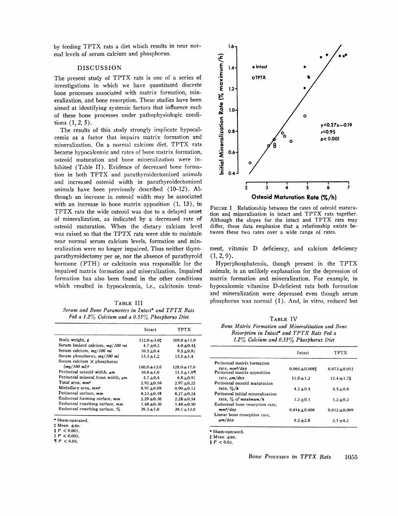
Of particular interest was the finding of a strong cor-relation between the rates of osteoid maturation andmineralization when the data from the sham-operatedcontrol rats and the TPTX rats were pooled (Fig. 1).Although the slopes for the sham-operated and TPTXgroups may differ, it is clear that a relationship existsover a wide range of rates. This relationship cannotbe explained by the manner in which these rates weredetermined since they represent independent measure-ments. These data suggest that the onset of mineraliza-tion and the rate of mineralization are tightly coupled,a conclusion consistent with previous observations(1-5).
In TPTX rats as compared with intact rats, there wasa 70% decrease in bone resorption (Table II). Boneresorption is a function of two components, the resorbingsurface and the linear rate of bone resorption. In theTPTX rats the decrease in resorption was largely dueto a decrease in the linear rate of bone resorption.
When TPTX rats were fed a 1.2% calcium and a0.55% phosphorus diet, rather than the 0.6% calciumand 0.6% phosphorus diet, only slight changes in serumcalcium and phosphorus in bone were found (Tables IIIand IV). In these TPTX rats as compared with sham-operated control rats fed the same diet, serum calciumwas decreased only about 10% and serum phosphoruswas essentially unchanged (Table III).
In sharp contrast to the first experiment, in theseTPTX rats neither matrix formation nor mineralizationwere decreased (Table IV). In addition, whereas in thefirst experiment bone resorption was markedly depressedin the TPTX rats, in this experiment bone resorptionwas similar in TPTX and sham-operated control ratsfed the same diet (Table IV). These two experimentsshow that the inhibition of matrix formation and min-eralization and bone resorption seen in hypocalcemichyperphosphatemic TPTX rats can be largely corrected
TABLE I IBone Matrix Formation and Mineralization and Bone Resorption in Intact* and TPTX Rats
Fed a 0.6% Calcium and a 0.6% Phosphorus Diet
Intact TPTX Change P
Periosteal matrix formation rate, mm3/day 0.05540.007t 0.045±0.009 -18 <0.05Periosteal matrix apposition rate, Mm/day 9.741.3 8.1±1.5 -16 <0.05Periosteal osteoid maturation rate, %//h 5.8±0.8 3.640.7 -38 <0.001Periosteal initial mineralization rate, %of maximum/h 1.440.1 0.7±0.1 -50 <0.001Endosteal bone resorption rate, mm3/day 0.020±0.008 0.006±0.006 -70 <0.001Linear bone resorption rate, Mm/day 10.4±4.9 2.5±2.5 -76 <0.001
* Sham-operated.Mean ±SD.
1054 J. Wergedal, M. Stauffer, D. Baylink, and C. Rich
by feeding TPTX rats a diet which results in near nor-mal levels of serum calcium and phosphorus.
DISCUSSION
The present study of TPTX rats is one of a series ofinvestigations in which we have quantitated discretebone processes associated with matrix formation, min-eralization, and bone resorption. These studies have beenaimed at identifying systemic factors that influence eachof these bone processes under pathophysiologic condi-tions (1, 2, 5).
The results of this study strongly implicate hypocal-cemia as a factor that impairs matrix formation andmineralization. On a normal calcium diet, TPTX ratsbecame hypocalcemic and rates of bone matrix formation,osteoid maturation and bone mineralization were in-hibited (Table II). Evidence of decreased bone forma-tion in both TPTX and parathyroidectomized animalsand increased osteoid width in parathyroidectomizedanimals have been previously described (10-12). Al-though an increase in osteoid width may be associatedwith an increase in bone matrix apposition (1, 13), inTPTX rats the wide osteoid was due to a delayed onsetof mineralization, as indicated by a decreased rate ofosteoid maturation. When the dietary calcium levelwas raised so that the TPTX rats were able to maintainnear normal serum calcium levels, formation and min-eralization were no longer impaired. Thus neither thyro-parathyroidectomy per se, nor the absence of parathyroidhormone (PTH) or calcitonin was responsible for theimpaired matrix formation and mineralization. Impairedformation has also been found in the other conditionswhich resulted in hypocalcemia, i.e., calcitonin treat-
TABLE IIISerum and Bone Parameters in Intact* and TPTX Rats
Fed a 1.2% Calcium and a 0.55%o Phosphorus Diet
Intact TPTX
Body weight, g 112.0 ±3.0 109.0±i 11.0Serum ionized calcium, mg/100 ml 4.7 ±0.2 4.0 10.4§Serum calcium, mg/100 ml 10.5 40.4 9.5±0.811Serum phosphorus, mg/100 ml 13.34±1.2 13.5±L1.4Serum calcium X phosphorus
(mg/100 mI)2 140.0 i 13.0 128.0 4 17.0Periosteal osteoid width, pim 10.6±1.0 11.5±1.0¶Periosteal mineral front width, pm 5.740.4 6.8±0.911Total area, mm2 2.92 ±0.16 2.97 ±0.22Medullary area, mm2 0.97±0.09 0.90±0.12Periosteal surface, mm 6.23 ±0.18 6.27 ±0.24Endosteal forming surface, mm 2.294±0.30 2.28 ±0.54Endosteal resorbing surface, mm 1.48±0.30 1.4640.50Endosteal resorhing surface, % 39.3 ±7.0 39.1 ±13.0
* Sham-operated.Mean ±SD.
§ P <0.001.11 P < 0.005.¶ P <0.05.
1.6
-C"-,E 1.4-ExaE 1.2-
00-a 1.0-
N 0.8-
0
s0.
E 0.4-
.,
.00
* Intact
oTPTX
0
S
0
0
y=0.27x-0.19r=0.95p( 0.001
r I I I 12 3 4 5 6
Osteoid Maturation Rate (%/h)7
FIGURE 1 Relationship between the rates of osteoid matura-tion and mineralization in intact and TPTX rats together.Although the slopes for the intact and TPTX rats maydiffer, these data emphasize that a relationship exists be-tween these two rates over a wide range of rates.
ment, vitamin D deficiency, and calcium deficiency(1,2,9).
Hyperphosphatemia, though present in the TPTXanimals, is an unlikely explanation for the depression ofmatrix formation and mineralization. For example, inhypocalcemic vitamine D-deficient rats both formationand mineralization were depressed even though serumphosphorus was normal (1). And, in vitro, reduced but
TABLE IVBone Matrix Formation and Mineralization and Bone
Resorption in Intact* and TPTX Rats Fed a1.2%o Calcium and 0.55% Phosphorus Diet
Intact TPTX
Periosteal matrix formationrate, mm'/day 0.065 0.008$ 0.073+0.012
Periosteal matrix appositionrate, pm/day 11.0±41.2 12.4 ±1.7§
Periosteal osteoid maturationrate, %/h 4.2 ±0.4 4.5 ±0.6
Periosteal initial mineralizationrate, %of maximum/h 1.2 ±0.1 1.2 ±0.2
Endosteal bone resorption rate,mm3/day 0.014±0.006 0.012 40.009
Linear bone resorption rate,pm/day 6.2 ±2.8 5.1 ±4.2
* Sham-operated.Mean 4SD.P <0.05.
Bone Processes in TPTX Rats 1055
I
i

TABLE VRelationship between Serum Calcium and Phosphorus and
Bone Processes in TPTX, Calcium-deficientand Vitamin D-deficient Rats*
Calcium Vitamin DTPTX deficient deficient
Serum calcium, %A -32$ -26 -31Serum phosphorus, %A +59 +16 +2Periosteal matrix apposition, %A -16 -37 -25Osteoid maturation rate, %A -38 -55 -50Mineralization rate, %A -50 -55 -52
* See references 1 and 2 for further data on vitamin D-deficient and calcium-deficient animals, respectively.I %Afrom control group, each test group having its own control group;data used to calculate %Awere mean values for about a 10 day experimentalperiod.
not elevated phosphorus in incubation medium inhibitedradioproline incorporation into bone matrix (14). Fromthese observations it is probable that hypocalcemia in-hibits matrix formation, osteoid maturation, and min-eral deposition.
The inhibition of the onset of mineralization in TPTXrats was associated with hypocalcemia but not a de-creased serum calcium phosphorus product; this productwas actually increased. The onset of mineralization ap-pears to be dependent upon osteoid maturation. Accord-ingly, matrix changes in lipid concentration, acid phos-phatase activity, and particularly proteinpolysaccharideconcentration are coordinated both spatially and tem-porally with the onset of mineralization (7, 15, 16). Be-cause osteoid maturation implies that the onset of min-eralization is at least partially under cellular control,it seems likely that hypocalcemia inhibits the onset ofmineralization by adversely affecting cell-mediated proc-esses.
The extent to which cellular activity is involved incalcium and phosphorus deposition once the process hasbeen initiated is uncertain, though there is evidence of a
functional membrane around bone, which is maintainedby bone cells and which influences the ionic compositionof bone water (17). In addition, the finding in animalstreated with large amounts of fluoride, of a decreasedmineralization rate despite normal levels of serum cal-cium and phosphorus, suggests that cellular activity mayalso be involved in this process (4). Thus, the inhibitionof the mineralization rate by hypocalcemia may also bethrough an effect on cellular activity.
The possibility that hypocalcemia inhibits osteoidmaturation and mineralization by a similar mechanismis consistent with the strong correlation between the rateof osteoid maturation and mineralization in the presentstudy. Furthermore, the magnitude of the changes in
these two rates was similar under a number of other ex-perimental conditions, viz., fluoride treatment, vitaminD deficiency, calcium deficiency, and phosphorus de-ficiency (1, 2, 4, 5). Although the reason for this rela-tionship is unknown, it is clear that these two rates aretightly coupled under a variety of experimental condi-tions.
It is generally held that the decrease in bone resorp-tion in hypoparathyroidism is entirely due to PTH de-ficiency whereas evidence is accumulating to indicatethat in TPTX rats hyperphosphatemia inhibits bone re-sorption. First, we previously found that in TPTX ratsthere is an inverse relationship between serum phos-phorus and percent endosteal resorbing surface over se-rum phosphorus values ranging from high to low (5).Second, in the present study bone resorption was lowin hyperphosphatemic but near normal in normophos-phatemic TPTX rats (Tables II and IV). Third, in tis-sue culture increasing phosphorus concentration in theincubation medium decreases bone resorption and alsodecreases the effect of PTH to stimulate bone resorp-tion (18). Thus, the decreased resorption in the TPTXrats was probably due to hyperphosphatemia as well ashypoparathyroidism. In view of this, it is doubtful thatPTHdetermines 75% of bone turnover since this conceptis based on the observation that turnover falls 75% afterremoval of the parathyroid glands (19).
Since the resorption rate was only slightly decreasedin the normophosphatemic TPTX rats (Table IV), onemight conclude that hyperphosphatemia, as opposed tohypoparathyroidism, was primarily responsible for theinhibition of bone resorption in the hyperphosphatemicTPTX rats (Table II). However, the control group forthe experiment in Table IV was fed a high calcium dietwhich may have partially inhibited PTH secretion andthus PTH-mediated bone resorption. Accordingly, itis possible that the resorption rate would have been lessin the normophosphatemic TPTX rats than in intact rats
fed a normal calcium diet. In any case, it is evident thatlowering the serum phosphorus was associated with an
increase in bone resorption.Although there is evidence that hypocalcemia per se
stimulates bone resorption (20, 21), in this study hypo-calcemia was associated with decreased, not increasedresorption. Also in tissue culture studies, decreasingcalcium concentration in the medium does not signifi-cantly enhance resorption (18).
Recent work suggests that the most potent vitamin Dmetabolite with respect to bone resorption is 1,25-dihy-droxycholecalciferol (1,25-OH2D,) (22), and that thesynthesis of this metabolite is PTH dependent (23).Accordingly, it is possible that some of the bone changes
1056 J. Wergedal, M. Stauffer, D. Baylink, and C. Rich

in TPTX rats results from an abnormality in vitamin Dmetabolism. If so, these changes were not evident inTPTX rats fed a diet which resulted in near normallevels of serum calcium and phosphorous (Tables IIIand IV).
A major reason for undertaking this study was to gainsome insight as to the factors responsible for the inhibi-tion of matrix formation and mineralization seen in vita-min D-deficient and also in calcium-deficient animals(1, 2). The relationships between serum and bone param-eters in TPTX, vitamin D-deficient, and calcium-de-ficient rats are compared in Table V. The changes inserum calcium and mineralization *were similar in thethree groups. However, on the basis of the degree ofhypocalcemia, the inhibition of formation in the TPTXrats was less than expected. That these three experi-ments were done at different times and under conditionswhich were not identical may not be an adequate explana-tion for this discrepancy.
The degree of hyperphosphatemia was greatest in theTPTX rats, and this may have accounted for the lesserinhibition of bone formation (24). Alternately, the pres-ence of secondary hyperparathyroidism as well as hypo-calcemia may have been responsible for the relativelygreater depression of matrix formation in the calcium-deficient and vitamin D-deficient rats (Table V). Thevalidity of this possibility is uncertain at the present time,because under some conditions hyperparathyroidism re-sults in decreased bone formation, whereas under otherconditions it results in increased bone formation (13,25-27).
This study emphasizes that changes in serum calciumand phosphorus have important effects on bone processes.It seems possible that the effects of the serum ionchanges are mediated through changes in the ionic con-centration of calcium and possibly phosphorus withinbone cells. If so, the results of this study are consistentwith the concept advanced by Rasmussen that intracel-lular calcium and phosphate ion concentration play animportant role in bone cell metabolism (28). In this con-text, since a number of factors influence the concentra-tion of calcium and phosphorus within cells, the simi-larities in Table V are more impressive than thedifferences.
ACKNOWLEDGMENTSWe are grateful to R. Haller for technical assistance, Mrs.K. Hashimoto for computer assistance, S. Baker for prepar-ing the figures, and Mrs. S. Grimes for typing the manu-script.
This study was supported in part by National Institutesof Health Grants AM09096 and HD-04872.
REFERENCES1. Baylink, D., M. Stauffer, J. Wergedal, and C. Rich.
1970. Formation, mineralization, and resorption of bonein vitamin D-deficient rats. J. Clin. Invest. 49: 1122.
2. Stauffer, M., D. Baylink, J. Wergedal, and C. Rich.1973. Decreased bone formation and mineralization, andenhanced resorption in calcium-deficient rats. Am. J.Physiol. In press.
3. Wergedal, J. 1969. Enzymes of protein and phosphatecatabolism in rat bone. I. Enzyme properties in normalrats. Calcif. Tissue Res. 3: 55.
4. Baylink, D., J. Wergedal, M. Stauffer, and C. Rich.1970. Effects of fluoride on bone formation, mineraliza-tion and resorption in the rat. In Fluoride in Medicine.T. L. Vischer, editor. Hans Huber, Bern, Switzerland.37.
5. Baylink, D., J. Wergedal, and M. Stauffer. 1971. Forma-tion, mineralization and resorption of bone in hypophos-phatemic rats. J. Clin. Invest. 50: 2519.
6. Moore, E. W. 1970. Ionized calcium in normal serumultrafiltrates, and whole blood determined by ion-ex-change electrodes. J. Clin. Invest. 49: 318.
7. Baylink, D., J. Wergedal, and E. Thompson. 1972. Lossof proteinpolysaccharides at sites where bone mineraliza-tion is initiated. J. Histochem. Cytochem. 20: 279.
8. Baylink, D., and J. Wergedal. 1971. Effect of vitaminD-deficiency on the bone mineralization rate. Clin. Res.19: 470.
9. Baylink, D., E. Morey, and C. Rich. 1969. Effect ofcalcitonin on the rates of bone formation and resorptionin the rat. Endocrinology. 84: 261.
10. Jowsey, J., R. E. Rowland, J. H. Marshall, and F. C.McLean. 1958. The effect of parathyroidectomy onhaversian remodeling of bone. Endocrinology. 63: 903.
11. Burkhart, J. M., and J. Jowsey. 1966. Morphologic evi-dence of osteomalacia in the parathyroidectomized dog.Mayo Clin. Proc. 41: 663.
12. Kelly, P. J. 1971. Effects of thyroid and parathyroiddeficiency on bone remodeling distal to a venous tourni-quet. J. Anat. 110: 349.
13. Sherrard, D. J., D. J. Baylink, J. E. Wergedal, andN. A. Maloney. 1972. Increased bone formation inuremic patients. Clin. Res. 20: 193.
14. Raisz, L. G. 1970. The pharmacology of bone. Fed. Proc.29: 1176.
15. Baylink, D. J., and J. E. Wergedal. 1971. Effets du floursur la mineralisation des os chez le rat. Med. Hyg. 29:1554.
16. Wergedal, J. E., and D. J. Baylink. 1969. Distributionof acid and alkaline phosphatase activity in undemineral-ized sections of the rat tibial diaphysis. J. Histochem.Cytochem. 17: 799.
17. Canas, F., A. R. Terepka, and W. F. Neuman. 1969.Potassium and milieu interieur of bone. Am. J. Physiol.217: 117.
18. Raisz, L. G., and I. Niemann. 1969. Effect of phosphate,calcium and magnesium on bone resorption and hor-monal responses in tissue culture. Endocrinology. 85:446.
19. Harris, William H., and Robert P. Heaney. 1969. Skele-tal renewal and metabolic bone disease (concluded). N.Engl. J. Med. 280: 303.
Bone Processes in TPTX Rats 105 7

20. Rasmussen, H., C. Anast, and C. Arnaud. 1967. Thyro-calcitonin, EGTA, and urinary electrolyte excretion.J. Clin. Invest. 46: 746.
21. Waron, M., and C. Rich. 1969. Rate of recovery fromacute hypocalcemia as a measure of calcium homeostaticefficiency in the dog. Endocrinology. 85: 1018.
22. Raisz, L. G., C. L. Trummel, M. F. Holick, and H. F.DeLuca. 1972. 1,25-dihydroxycholecalciferol: a potentstimulator of bone resorption in tissue culture. Science(Wash. D. C.). 175: 768.
23. Garabedian, M., M. F. Holick, H. F. DeLuca, and I.T. Boyle. 1972. Control of 25-hydroxycholecalciferolmetabolism by parathyroid glands. Proc. Natl. Acad.Sci. U. S. A. 69: 1673.
24. Rasmussen, H., J. Feinblatt, N. Nagata, and M. Pechet.1970. Effect of ions upon bone cell function. Fed. Proc.29: 1190.
25. Flanagan, B., and G. Nichols, Jr. 1969. Bone matrixturnover and balance in vitro. I. The effects of para-thyroid hormone and thyrocalcitonin. J. Clin. Invest.48: 595.
26. Kalu, D. N., J. Pennock, F. H. Doyle, and G. V.Foster. 1970. Parathyroid hormone and experimentalosteosclerosis. Lancet. 1: 1363.
27. Sherrard, D., D. Baylink, and J. Wergedal. 1972. Bonedisease in uremia. Transactions for the American Societyof Artificial Internal Organs. 18: 412.
28. Rasmussen, H. 1971. Ionic and hormonal control ofcalcium homeostasis. Am. J. Med. 50: 567.
1058 J. Wergedal, M. Stauffer, D. Baylink, and C. Rich