inhibition of cell surface export of group a streptococcal ... · solely by the glycolytic pathway,...
TRANSCRIPT

INFECTION AND IMMUNITY, Oct. 2005, p. 6237–6248 Vol. 73, No. 100019-9567/05/$08.00�0 doi:10.1128/IAI.73.10.6237–6248.2005Copyright © 2005, American Society for Microbiology. All Rights Reserved.
Inhibition of Cell Surface Export of Group A Streptococcal AnchorlessSurface Dehydrogenase Affects Bacterial Adherence and
Antiphagocytic PropertiesGregory Boel, Hong Jin, and Vijay Pancholi*
Laboratory of Bacterial Pathogenesis, Public Health Research Institute at The International Center for Public Health,225 Warren Street, Newark, New Jersey 07103-3535
Received 2 May 2005/Returned for modification 13 June 2005/Accepted 12 July 2005
Surface dehydrogenase (SDH) is an anchorless, multifunctional protein displayed on the surfaces of groupA Streptococcus (GAS) organisms. SDH is encoded by a single gene, sdh (gap or plr) that is essential for bacterialsurvival. Hence, the resulting nonfeasibility of creating a knockout mutant is a major limiting factor instudying its role in GAS pathogenesis. An insertion mutagenesis strategy was devised in which a nucleotidesequence encoding a hydrophobic tail of 12 amino acids (337IVLVGLVMLLLS348) was added at the 3� end ofthe sdh gene, successfully creating a viable mutant strain (M1-SDHHBtail). In this mutant strain, the SDHHBtailprotein was not secreted in the medium but was retained in the cytoplasm and to some extent trapped withinthe cell wall. Hence, SDHHBtail was not displayed on the GAS surface. The mutant strain, M1-SDHHBtail, grewat the same rate as the wild-type strain. The SDHHBtail protein displayed the same GAPDH activity as thewild-type SDH protein. Although the whole-cell extracts of the wild-type and mutant strains showed similarGAPDH activities, cell wall extracts of the mutant strain showed 5.5-fold less GAPDH activity than thewild-type strain. The mutant strain, M1-SDHHBtail, bound significantly less human plasminogen, adheredpoorly to human pharyngeal cells, and lost its innate antiphagocytic activity. These results indicate that theprevention of the cell surface export of SDH affects the virulence properties of GAS. The anchorless SDHprotein, thus, is an important virulence factor.
Surface proteins of pathogens play a variety of roles in vir-ulence. Group A Streptococcus (GAS) (Streptococcus pyo-genes), a human pathogen that is responsible for a variety ofmild to severe invasive diseases of the pharynx and skin, dis-plays an array of multifunctional proteins on its surface (5, 9,11). In gram-positive pathogens, including GAS, classical sur-face proteins with defined structural components, such as asignal sequence, a proline/glycine-rich cell wall-associated do-main, a hydrophobic membrane domain, and a cell wall-sortinghexapeptide motif (LPXTGX), have been characterized mostextensively (15, 30). More recently, complete genome se-quence analyses of six different GAS serotypes have revealedthat lipoproteins also contribute significantly to the surfacelayer of GAS and in disease pathogenesis (1, 2, 13, 29, 43).
In the last decade, after reports of the presence of theglycolytic enzymes glycerladehyde-3-phosphate dehydrogenase(GAPDH) or streptococcal surface dehydrogenase (SDH) (27,33) and streptococcal surface enolase (SEN) (37) on the sur-face of GAS, there has been an increasing awareness about thepresence of similar glycolytic enzymes on the surfaces of avariety of human pathogens (for a review, see reference 32).Since these surface-located metabolic enzymes are generallyfound in the cytoplasm and lack C-terminal hydrophobic tailsand N-terminal signal sequences required for their export to
the cell surface, they are called anchorless surface proteins(32).
In addition to innate glycolytic activities, both SDH and SENperform a variety of functions (27, 31, 33, 34, 36). Whereassome of the notable functions of SDH are its ability to bindvarious mammalian proteins, including plasminogen (Plg) (27,33), ADP ribosylation (34), and host cell-signaling regulation(36), the function of SEN seems to be to serve as a majorPlg-binding protein on the surface of GAS (31, 37). AlthoughSDH in comparison to SEN is a weak Plg-binding protein (33),the C-terminal lysine residues of both SDH and SEN seem toplay a crucial role in Plg binding (37). Like many otherGAPDH molecules (4, 17, 18, 44), the predictive three-dimen-sional structure of SDH has indicated that the SDH moleculepossesses two domains (24). The N-terminal NAD-binding do-main (amino acid residues [aa] 1 to 149) consists of a six-stranded parallel �-sheet flanked by helices. The C-terminalhalf of SDH contains the catalytic domain, which consists of anine-stranded antiparallel sheet followed by three approxi-mately parallel helices and a polypeptide chain that ends in along helix. The region spanning amino acid residues 179 and200, which forms an S-shaped antiparallel sheet, is also calledthe S-loop (18, 24, 44). Recently, we identified CD87/urokinaseplasminogen activator receptor (uPAR) as the SDH-specificreceptor on the surfaces of pharyngeal cells (24). The N-ter-minal domain of uPAR binds to the C-terminal alpha-helix andthe flanking regions of the S-loop of the SDH molecule (24).This specific binding also plays a crucial role in GAS adherenceto pharyngeal cells (24). Thus, the C-terminal portion of SDHis essential to perform a variety of biologically relevant func-tions. We also reported that ATP-producing glycolytic en-
* Corresponding author. Mailing address: Laboratory of BacterialPathogenesis, Public Health Research Institute at The InternationalCenter for Public Health, 225 Warren Street, Newark, NJ 07103-3535.Phone: (973) 854-3430. Fax: (973) 854-3431. E-mail: [email protected].
6237
on March 26, 2019 by guest
http://iai.asm.org/
Dow
nloaded from

zymes likely form a specialized complex on the surface of GAS(35). Although accumulated reports now indicate that anchor-less proteins are present on the surfaces of many pathogens,including gram-positive bacteria, fungi, and parasites (32), ourunderstanding of their biological functions and their roles inthe pathogenesis of GAS and other bacterial pathogens is farfrom complete. Likewise, how they are exported to the cellsurface is unknown.
To evaluate the role of virulence factors, the genetic ap-proach to create a knockout mutant that can be tested invarious in vitro and/or in vivo disease model systems as adistinct attenuated or nonpathogenic phenotype is highly de-sirable. However, the knockout mutagenesis approach is notachievable for a gene that is essential for microbial survival. Incertain organisms, such as Escherichia coli, Pseudomonasaeruginosa, and Saccharomyces cerevisiae, more than oneGAPDH and enolase isoenzymes are present; each of theseisoenzymes is encoded by a distinct gene or the organism isequipped with the Entner-Doudoroff alternate metabolic path-way (7, 20–22, 28, 46). Hence, it has been possible to createmutants defective in GAPDH, enolase, and other enzymes ofglycolysis (19, 22, 23, 28). In GAS, glucose is metabolizedsolely by the glycolytic pathway, as GAS lacks the tricarbolic-acetic acid (TCA)-metabolic cycle and the Entner-Doudoroffalternative metabolic pathway (1, 2, 13, 29, 43). The multifunc-tional SDH is encoded by a single essential gene (33, 45). Thegene knockout approach to prevent SDH expression in GAS,therefore, is not feasible. The site-directed mutagenesis strat-egies that allow alteration in the specific functional domainswithout disturbing the conformational structure responsiblefor the catalytic function have been successfully employed tocreate Plg-binding enolase- and SDH/Plr-specific deletion mu-tants (3, 12, 45). Since the putative transport system responsi-ble for exporting the anchorless metabolic surface enzymes isnot known, it has not been possible to create a GAS mutant inwhich the anchorless enzyme is selectively retained in the cy-toplasm and not expressed on the surface.
Since the SDH molecule does not possess the N-terminalsignal sequence and the C-terminal hydrophobic tail, we hy-pothesized that the introduction of only a hydrophobic tail atthe C-terminal end may result in its prevention of export to thesurface and the SDH may be retained in the cytoplasm and/orin the membrane. Accordingly, in the present investigation, wealtered the sdh gene by introducing a nucleotide sequenceencoding a de novo C-terminal hydrophobic tail. The resultingmutated SDH protein remained largely restricted to the cyto-plasm. This mutant strain behaved in a manner similar to thatof the wild-type strain in terms of growth characteristics andGAPDH enzyme activity, but it bound significantly less Plg,adhered poorly to human pharyngeal cells, and lost the typicalantiphagocytic activity associated with the wild-type strain. Ourresults also suggest that SDH may have a role in the regulationof the expression of a certain virulence factor-related gene(s),possibly at the transcriptional level.
MATERIALS AND METHODS
Bacterial strains, vectors, growth conditions, and DNA techniques. S. pyogeneswild-type strain M1-SF370 (M1-WT; ATCC 700294; American Type CultureCollection, Manassas, VA) was grown in Todd-Hewitt broth (Difco Laborato-ries) supplemented with 0.5% yeast extract or on proteose peptone-3 blood agar
plates supplemented with spectinomycin (up to 500 �g/ml), when required. E.coli strain XL1-Blue was used for the cloning experiments and grown in Luria-Bertani (LB) broth or on LB agar plates. Vector pFW5 containing the specti-nomycin resistance gene (aad-9) was used to transform the M1-SF370 strain (12,39).
Reagents. Unless otherwise stated, all chemicals were procured from Sigma(St. Louis, MO).
Human pharyngeal cell lines. Detroit 562 (ATCC CCL138) pharyngeal cellswere grown to confluence and maintained in a CO2 incubator as describedpreviously (36).
Determination of the sequence of the hydrophobic tail. The prediction ofhydrophobicity of a putative hydrophobic tail was determined based on the densealignment surface (DAS) score as described at the website http://www.sbc.su.se/�miklos/DAS/. The amino acid sequence of a hydrophobic tail to be inserted atthe C-terminal end of SDH was based on the translated amino acid sequence ofthe epf gene in the M1 genome sequence database (13) and was edited using theDAS method to get an optimum DAS score (�3.0), one indicative of a typicaltransmembrane location (8).
Construction of insertion and allelic-exchange sdhHBtail mutations in GAS. Tocreate streptococcal mutant strains expressing SDH with a hydrophobic tail atthe C-terminal end, the sdh gene (0.92-kb DNA fragment) and a 1.118-kb DNAfragment corresponding to the downstream region of the sdh gene in the M1-SF370 chromosome were PCR amplified using primer pairs SDH-SalI-F-/SDH-BamHI-R (5�-ACGCGTCGACATGGTAGTTAAAGT TGGTATTAACGG-3�and 5�-CGCGGATCCTTATTTAGCAATTTTTGCGAAGTACTCAAGAGTACG-3�) and SDHDWN-PstI-F/SDHDWN-NdeI-R (5�-TTTTCTGCAGCTTGGTTTATGCTG AGTTCTTTTTCG-3 and 5�-GGGAATTCCATATGTATTGGAAAATGCTATCGTGCG-3�), respectively. The PCR-amplified products werecloned in the multiple cloning sites located upstream and downstream of thespectinomycin resistance gene, aad-9, in the suicide plasmid pFW5 (2.7 kb) toconstruct pFW5-sdh. To insert a DNA sequence encoding the putative hydro-phobic tail at the 3� end of the sdh gene in the pFW5-sdh vector, a QuickChangeII site-directed mutagenesis kit was used according to the manufacturer’s in-structions (Stratagene) with the plasmid pFW5-sdh as the template and a pair ofcomplementary primers, SDHHBtail-F (5�-GTACTTCGCAAAAATTGCTAAAATTGTTCTTGTTGGCTTAGTTATGCTTCTTCTTTCTTAATAGGATCCTCGAGCTCTAG-3�) and SDHHBtail-R (5�-CTAGAGCTCGAGGATCCTATTAAGAAAGAAGAAGCATAACTAAGCCAACAAGAACAATTTTAGCAATTTTTGCGAAGTAC-3�). The resulting plasmid, pFW5-sdhHBtail, was introducedinto strain M1-WT by electroporation, using a Gene Pulser II electroporator (Bio-Rad) as described previously (12, 29). The mutant strain thus obtained was calledM1-SDHHBtail. The confirmation of mutations and insertions at various stages of theexperiments was obtained by one or more methods of PCR, DNA sequencing, andSouthern hybridization. Using a similar strategy, a mutant strain lacking the M1protein (M1Demm1) was created. The primers used to create this mutant wereemm1-F (5�-ACGCGTCGACTAGGTCAAAAAGGTGGC-3�), emm1-R (5�-CGCGGATCCGCATTCTCTAAT CTCGCTT-3�), emm1DWN-F (5�-AACTGCAGGACTTGGACGCATCACGTGAA-3�), and emm1DWN-R (5�-GATTCCATATGCTGTCTCTTAGTTTCCTTCATTGGTGC-3�). The italicized sequences in theseprimers represent SalI, BamHI, PstI and NdeI restriction sites, respectively.
Cell fractionation and whole-cell extraction. M1-WT and M1-SDHHBtail (mu-tant) strains were grown to late log phase/early stationary phase (optical densityat 600 nm [OD600], 0.8), and bacteria were harvested by centrifugation. Theculture supernatants from these strains were saved, and the proteins therein wereprecipitated with 25% trichloroacetic acid (TCA). The resulting precipitateswere separated by centrifugation (20,000 � g for 30 min at 4°C), washed twicewith acetone, and suspended in 1/50 of the original volume of 50 mM Tris-HCl,pH 7.0. The harvested bacteria were subjected to mutanolysin treatment in a cellwall-extracting buffer (50 mM Tris-HCl, pH 7.0, 30% raffinose) containing 250U/ml of mutanolysin (Sigma) as described previously to achieve nearly completewall extraction (33). The resulting protoplasts were lysed in a hypotonic buffer,followed by repeated freezing and thawing. Bacterial membrane and cytoplasmicfractions were separated from the lysed protoplasts by ultracentrifugation(100,000 � g for 45 min at 4°C) as described previously (33). Whole-cell extractsfrom these two strains were obtained using the same procedures described aboveexcept that the cell wall-extracting buffer was used without 30% raffinose.
SDH and SDHHBtail purification. Clear whole lysates obtained as describedabove from 500 ml THY broth culture with each of the M1-WT and M1-SDHHBtail strains were dialyzed against 0.05 M NH4HCO3 and precipitated at40%, 60%, and 80% ammonium sulfate saturation. The protein precipitatesobtained from 60% to 80% ammonium sulfate saturation, which contained themajor amount of SDH and SDHHBtail, were purified by Hi-trap Blue-HP column(1 ml) chromatography (Pharmacia) after equilibration with a starting buffer (50
6238 BOEL ET AL. INFECT. IMMUN.
on March 26, 2019 by guest
http://iai.asm.org/
Dow
nloaded from

mM Tris-HCl, pH 7.4). Proteins other than SDH and SDHHBtail were eluted firstby the five-column volume of starting buffers containing 0.5 M NaCl followed bythe five-column volume of the same buffer containing 1.0 M NaCl. The remainingcolumn-bound proteins were eluted with the five-column volume of the startingbuffer containing 5 mM NAD. The NAD-eluted fractions (1 ml each) containedonly SDH or SDHHBtail, as determined by sodium dodecyl sulfate-polyacrylam-ide gel electrophoresis (SDS-PAGE) and Western blotting. The pooled fractionscontaining SDH or SDHHBtail were dialyzed against the starting buffer andstored at �70°C until further use.
Preparation of monospecific anti-SDH antibody. Anti-SDH specific immuno-globulin G (IgG) was purified from the SDH-immunized rabbit serum using anSDH-linked diaminodipropylamine M-phase (Pierce) affinity column as de-scribed previously (33, 37). The purified IgG was then treated with immobilizedpapain enzyme (Pierce) at 4°C overnight to cleave them between their Fab� andFc regions. The latter was then removed by passing the cleaved products on aprotein A column (Pierce) per the manufacturer’s instructions. The unboundinitial fall-through was used as Fab�-specific anti-SDH IgG.
Electrophoresis on SDS-polyacrylamide gels and Western blotting. PurifiedSDH, SDHHBtail, and cell wall-associated proteins were separated by SDS-PAGE and Western blotted onto polyvinylidene difluoride (PVDF) membranes.Subsequently, the presence of SDH was detected by reacting PVDF membraneswith anti-SDH monoclonal antibody (clone 13D5) (16) or monospecific rabbitanti-SDH IgG followed by corresponding alkaline phosphatase-labeled conju-gate antibody (Fab� specific). SDH-specific protein bands on PVDF membraneswere visualized using a chromogenic or a chemiluminescence-based substrate asdescribed previously (16, 36, 37).
GAPDH activity. The ability of SDH and SDHHBtail to convert D-glyceralde-hyde-3-phosphate (G-3-P) to 1,3-diphosphoglycerate in the presence of NAD�
was measured spectrometrically at 340 nm essentially as described before withsome modifications (33). Briefly, the reaction was initiated by adding 3 �g ofpurified SDH or SDHHBtail proteins or 6 �g of each cell wall extract (strainsM1-SF370 and M1-SDHHBtail) in a reaction buffer at a final volume of 600 �lcontaining 50 mM Tris-HCl, pH 7.4, 4 mM NaH2PO4, 4 mM NAD, 4 mM G-3-P,and 1 mM dithiothreitol. The conversion of NAD to NADH was monitored every10 s spectrometrically (Beckman Coulter model DU640). To measure theGAPDH activity of intact bacteria, instead of a kinetic measurement as describedabove, only an endpoint OD340 was measured. M1-WT and M1HBtail strains weregrown until late log phase (OD600 � 0.8) and washed twice in phosphate-buffered saline (PBS) to finally adjust the OD600 of the bacterial suspension to1.0 (�5 � 108 CFU). One milliliter of washed bacterial suspension was centri-fuged, and the 600 �l of complete enzyme reaction was resuspended as describedabove for 5 min. At the end of incubation, the bacteria were removed bycentrifugation and the absorbance of the supernatant was measured at 340 nm asdescribed above.
Real-time quantitative PCR. Total RNA was extracted from M1-SF370 andM1-SDHHBtail strains grown to an OD of 0.8 using a commercially availableRNA isolation kit (RNAwiz; Ambion). The total RNA (2 �g) isolated from thewild-type and mutant strains were converted to first-strand cDNA using anSYBR green iScript cDNA synthesis kit (Bio-Rad) in a final volume of 40 �l.Real-time PCR was performed in a reaction mixture (25 �l) containing 2.5 mMMgCl2, 250 �M deoxynucleoside triphosphate mix, 1.25 U of AmpliTaqGoldDNA polymerase, 500 pM of each primer, and 1 �g of template. Gene-specificprimer pairs used in this study were as follows: sdh (sdh-F, 5�-ATTAACGGTTTCGGTCGTATCGGACG-3�; sdh-R, 5�-GGATCACGTTCAGCAGAAACTTTG-3�), gyrA (gyrA-F, 5�-TTCGTATGGCTCAGTGGTTTAGTT-3�; gyrA-R,5�-CTGGTTCTCTTTCGCTTCCATCGT-3�), and proS (proS-F, 5�-GGGTGGTTCTTGACAAGTCTATTGCG-3�; proS-R, 5�-TTCTGCCAAGGCATCTTCAGCA-3�). The mRNA copies specific to sdh were normalized with those spe-cific to the housekeeping genes gyrA and proS for the M1-WT and M1-SDHHBtail
strains, and data were evaluated as relative mRNA copy number ratios.Different concentrations of genomic DNA were used to obtain a standard
curve for each gene. The reaction mixtures were incubated for 10 min at 95°C toactivate DNA polymerase, followed by 40 cycles each of denaturing at 95°C for30 s, annealing at 58°C for 50 s, and extension at 72°C for 30 s. Fluorescence wasmeasured in every well during each annealing step throughout the course of eachreaction. Data were automatically analyzed by software of the real-time PCRinstrument (Stratagene Mx-4000).
Immunofluorescence microscopy. The M1-WT and M1-SDHHBtail strains (106
CFU/ml of PBS) were attached to polylysine-treated eight-well glass slides andblocked with DAKO blocking buffer. Adherent bacteria were incubated withaffinity-purified anti-SDH IgG (Fab� fragment, diluted 1:10 in DAKO dilutionbuffer) at 4°C for 8 h. The wells were washed several times with PBS and reactedwith fluorescein isothiocyanate-conjugated anti-rabbit IgG antibody (1:50 dilu-
tion) and incubated at room temperature for 1 h. The slides were washed severaltimes with PBS, and the bacteria were stained with DAPI (4�,6�-diamidino-2-phenylindole, 1 �g/ml) in SlowFade equilibration buffer (Molecular Probes) for5 min. The stained bacteria were kept in SlowFade buffer containing glycerol andobserved under a Nikon Eclipse C600 fluorescence microscope coupled with aDiagnostic RT color camera and the Spot software (v3.5.9). Bacterial cellsstained with only conjugated antibody were treated as a control.
Fluorescence labeling of plasminogen. Human Plg was labeled with Alex-aFluor-488–carboxylic acid succinimidyl ester (antibodies Em 495 and Em 519;Molecular Probes) using EDC (1-ethyl-3-[3-dimethylamino-propyl]carbodiim-ide) as per the manufacturer’s instructions. Fluorescence of the labeled plasmin-ogen was measured with a POLARstar Galaxy fluorimeter (BMG Labtechnolo-gies GmbH, Offenburg, Germany). The values of fluorescence units versusdifferent amounts of labeled plasminogen (1 to 5 �g) were plotted to obtain astandard curve.
Plasminogen-binding assay. Plg-binding activities of purified SDH proteins(SDH and SDHHBtail) were determined using a solid-phase ligand-binding assayand a slot blot apparatus (Bio-Rad) essentially as described before with somemodifications (12). The binding of Plg to serially diluted SDH proteins wasdetected by rabbit anti-human Plg antibody (DAKO, Denmark), followed bycorresponding alkaline-phosphatase-labeled conjugate. To determine Plg-bind-ing activities of the intact M1-WT and M1-SDHHBtail strains, late-log-phaseTHY broth cultures (OD600, 0.8) were adjusted to an OD600 of 1.0 after twowashes in 50 mM Tris, pH 8.0, buffer. The cultures were then centrifuged, andthe resulting bacterial pellets were resuspended in TBST (Tris-buffered salinecontaining 0.5% Tween 20, 0.5% gelatin, and 0.5% bovine serum albumin)blocking buffer in a final volume that was 1/10 of the original volume. GASstrains (100 �l, �2 �109 CFU) were then mixed with twofold serially dilutedlabeled Plg in a deep-well 96-well plate for 2 h under constant mixing at roomtemperature. At the end of incubation, unbound labeled Plg was removed andthe amount of bound Plg was determined with a fluorimeter as described above.Each dilution of Plg was tested in triplicate wells. Based on the standard curve,the exact amount of Plg bound to GAS strains was calculated. Data were statis-tically evaluated by unpaired t test with Welch’s correction.
Phagocytosis/bactericidal assays. The ability of M1-WT and M1-SDHHBtail toresist phagocytosis and survive in heparinized human blood was determined by abactericidal test as described previously (26). An overnight culture of eachbacterial strain was diluted 1:50 in THY broth and grown without agitation to anOD600 of 0.15 at 37°C in a CO2 incubator. The bacterial suspension was dilutedin THY broth (1:10,000) to obtain �4 � 103 CFU/ml for the initial inoculum ofthe assay. The exact number of bacteria added to the blood was determined byplating the bacterial culture and counting CFU on sheep blood agar plates. Fiftymicroliters of the suspension containing approximately 200 CFU was added to 1ml of freshly heparinized blood in sterile glass tubes (100 by 10 mm) and wasincubated with end-over-end rotation at 37°C for 3 h. The number of CFU in theinoculum used and those obtained after growth in human blood were quantifiedby the pour plate method using sheep blood agar plates. Blood for these exper-iments was drawn from healthy volunteers in 10-ml Vacutainer tubes (BectonDickinson, Franklin Lakes, NJ) containing 143 U of freeze-dried heparin. Op-sonophagocytosis assays were performed essentially in the same way as thebactericidal assays except that the former assays were carried out in the presenceof increasing amounts of purified anti-SDH antibody (SDH-specific IgG). Priorto these experiments, the necessary approval of the Institutional Review Boardof UMDNJ, New Jersey, for human subject use was obtained.
Estimation of the hyaluronic acid of the bacterial capsule. The hyaluronic acidcapsule produced by wild-type, mutant, and complemented mutant strains wasmeasured essentially as described previously (40). Briefly, a standard curve wasgenerated by the addition of 2 ml of a chromogenic reagent {20 mg of 1-ethyl-2-[3-(1-ethylnaphtho-[1,2-d]thiazolin-2-ylidene)-2-methylpropenyl]naptho-[1,2-d]thiazolium bromide (Kodak) and 60 �l of glacial acetic acid in 100 ml of 50%formamide} into different concentrations of hyaluronic acid (0 to 30 �g/ml) in a50-�l volume (ICN/MP Biomedicals LLC) and measuring absorbance at 640 nm.Bacterial cells from a 10-ml late-log-phase culture were washed with water andresuspended in 0.5 ml water. Capsule was released by shaking with 1 ml ofchloroform and centrifuged. The aqueous phase (50 �l) was then processed asdescribed above, and the hyaluronic acid content was determined by comparingthe absorbance values with the standard curve as described above.
Streptococcal adherence assays. Streptococcal adherence to Detroit pharyn-geal cells was carried out in 24-well tissue culture plates essentially as previouslydescribed (36, 38). Briefly, overnight cultures of M1-WT and M1-SDHHBtail werewashed and adjusted to an OD600 of 1.0 in minimal essential medium withoutfetal bovine serum. Cells were infected with these strains (multiplicity of infec-tion, 50 bacteria to 1 cell, �4 � 107 to 5 � 107 CFU/well) and incubated in a
VOL. 73, 2005 PREVENTION OF CELL SURFACE EXPORT OF ANCHORLESS SDH 6239
on March 26, 2019 by guest
http://iai.asm.org/
Dow
nloaded from

humidified CO2 incubator at 37°C for 3 h. Nonadherent bacteria were removedby repeated washing, Detroit cells with adherent bacteria were lysed, and theresulting cell lysates containing cell-associated bacteria were counted as CFU onsheep blood agar plates as described previously (24).
RESULTS
Fusion of a hydrophobic tail to SDH. Gram-positive bacte-rial surface proteins contain a signal sequence, an LPXTGXhexapeptide motif, and a hydrophobic tail for anchoring in themembrane lipid bilayer and subsequently for surface export(30). We, therefore, hypothesized that the addition of only ahydrophobic tail at the C-terminal end of SDH would allowSDH to anchor to the membrane without being exported to thecell surface, as the resulting protein would not have either asorting signal for cell wall anchoring or a signal sequence forsecretion. To create such a construct, we designed a putativehydrophobic tail based on the amino acid sequence of thehydrophobic tail region of the epf gene (spy0737) product inthe M1-SF370 genome database (13). The translated aminoacid sequence of the epf gene (2,045 aa, 221,959 Da) containsan LPXTGX motif and a hydrophobic tail with 18 amino acids(2020IVLVGLG2026VMS2029LLLG2033MVLY2037) with a pre-dictable hydrophobicity score of 3.5 according to the DASmethod (http://www.sbc.su.se/�miklos/DAS/) (8). This score iswell above the recommended optimum score (2.2) required fora transmembrane protein (8, 13). We edited this sequence andshortened the size of the de novo hydrophobic tail by removingGly2026, Ser2029, and the last 5 amino acid residues(2033GMVLY2037). The resulting sequence (IVLVGLVMLLL) gave a DAS score of 3.1. The removal of the last 5residues and only the Gly2026 residue gave DAS scores of 2.8and 3.2, respectively. However, the deletion of the serine res-idue (Ser2029) of the sequence IVLVGLVMS2029LLL and itsaddition at the end of a resulting sequence, i.e., IVLVGLVMLLLS, increased the DAS value from 3.2 to 3.5. Thus, theamino acid sequence IVLVGLVMLLLS with a DAS score of3.5 was considered the putative hydrophobic transmembraneregion for SDH to be inserted at its C-terminal end (Fig. 1Aand B).
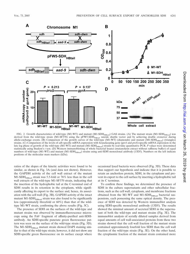
Phenotypic characteristics of M1-SDHHBtail. PCR, DNA se-quencing, and Southern hybridization assays with the mutantstrain M1-SDHHBtail confirmed that the two-crossover allelic-exchange event between the M1-SF370 genome and pFW5-sdhHBtail occurred only at the desired locus without any polareffect (Fig. 2A).
Growth characteristics. M1-WT and M1-SDHHBtail strainsgrew similarly in THY broth throughout the lag and log phasesof the growth period and until the stationary phase (Fig. 2B).Thus, the hydrophobic tail at the C-terminal end of SDH hadno effect on bacterial growth.
SDH-specific mRNA and protein expression. To determinethe effect of the insertion of a hydrophobic tail at the C-terminal end of SDH on protein expression, first SDH-specificmRNA expression was measured in wild-type and mutantstrains grown to the late log phase (OD600 of 0.8) by real-timequantitative PCR. The expression of SDH-specific mRNAswith respect to housekeeping gene (gyrA and proS)-specificmRNAs in both the wild-type and mutant strains was found tobe essentially the same without a statistically significant differ-
ence (gyrA, P � 0.137; proS, P � 0.213) (Fig. 2C). Westernimmunoblot analysis of whole-cell extract using anti-SDH an-tibody also indicated the presence of SDH and the SDHHBtail
protein in their respective strains (Fig. 2D). We concluded thatthe introduction of hydrophobic residues at the C terminus ofSDH does not affect metabolic functions or the growth of theresultant M1-SDHHBtail strain. The SDHHBtail protein, how-ever, migrated faster than SDH during gel electrophoresis. Thefaster migration of the SDHHBtail protein could be attributedto the insertion of additional hydrophobic residues at the Cterminus.
GAPDH activity of purified SDH and SDHHBtail. To deter-mine the impact of the insertion of a hydrophobic tail in SDHon its enzymatic activity, the wild-type SDH and mutantSDHHBtail proteins were purified as described under Materialsand Methods from whole-cell extracts and their GAPDH ac-tivities were determined using the same amount of protein(Fig. 3A). Both SDH and SDHHBtail demonstrated similarkinetics for the conversion of G-3-P to 1,3-diphosphoglyceratein the presence of NAD, indicating that the hydrophobic tailfused to the C-terminal part of SDH does not affect or inter-fere with the catalytic GAPDH activity of the SDHHBtail pro-tein (Fig. 3A).
Surface localization of SDH and SDHHBtail. To demonstratethe effect of insertion mutagenesis in the sdh gene on thesurface export of SDH, initially GAPDH activities per unitamount of total protein of the whole-cell extract of the wild-type and mutant strains were determined. In both cases, the
FIG. 1. Strategy to insert a hydrophobic tail at the C-terminal endof SDH. (A) Amino acid sequences of the SDH protein (only residues1 to 20 and 301 to 336 are noted for brevity) and an inserted hydro-phobic tail (337IVLVGLVMLLLS348; in bold) at the C-terminal end(sdh/plr in the M1 genome is annotated as SPy0274 [13]). The sequenceof the hydrophobic tail for SDH was derived from that of the hydro-phobic tail portion (2020IVLVGLGVMSLLLGMVLY2037) of theproduct of the epf gene (SPy0737) of the M1-SF370 genome (13).(B) DAS hydrophobicity index of the putative hydrophobic tail se-quence (IVLVGLVMLLLS) to be inserted at the C-terminal end ofthe SDH molecule.
6240 BOEL ET AL. INFECT. IMMUN.
on March 26, 2019 by guest
http://iai.asm.org/
Dow
nloaded from
ratios of the slopes of the kinetic activities were found to besimilar, as shown in Fig. 3A (and data not shown). However,the GAPDH activity of the cell wall extract of the mutantM1-SDHHBtail strain was 5.5-fold or 76% less than in the cellwall extracts of the wild-type M1-SF370 strain, indicating thatthe insertion of the hydrophobic tail at the C-terminal end ofSDH results in its retention in the cytoplasm, while signifi-cantly affecting its export to the surface and, hence, its associ-ation with the cell wall (Fig. 3B). GAPDH activity of the intactmutant M1-SDHHBtail strain was also found to be significantlyless (approximately threefold or 60%) than that of the wild-type M1-WT strain, confirming the above results (Fig. 3C).
The presence of SDH on the surfaces of the wild-type andmutant strains was observed by immunofluorescence micros-copy using the Fab� fragment of affinity-purified anti-SDH-antibody. An SDH-specific punctate green fluorescence pat-tern was seen on the surface of the wild-type strain (Fig. 3D).The M1-SDHHBtail mutant strain showed DAPI staining sim-ilar to that of the wild-type strain; however, it did not show anySDH-specific green fluorescence on its surface except where
occasional lysed bacteria were observed (Fig. 3D). These datathus support our hypothesis and indicate that it is possible toretain an anchorless protein, SDH, in the cytoplasm and pre-vent its export to the cell surface by inserting a hydrophobic tailat its C terminus.
To confirm these findings, we determined the presence ofSDH in the culture supernatants and other subcellular frac-tions, such as the cell wall, cytoplasm, and membrane fractionsobtained from the M1-WT and M1-SDHHBtail bacterial sus-pensions, each possessing the same optical density. The pres-ence of SDH was detected by Western immunoblot analysisusing SDH-specific monoclonal antibody (13D5). The resultsshowed the minimal amount of secreted SDH in the superna-tant of both the wild-type and mutant strains (Fig. 3E). Theimmunoblot analysis of serially diluted samples derived fromequal amounts of cell wall-associated proteins from these twostrains showed that the cell wall fraction of the mutant straincontained approximately fourfold less SDH than the cell wallfraction of the wild-type strain (Fig. 3E). On the other hand,the cytoplasmic fraction of the mutant strain contained more
FIG. 2. Growth characteristics of wild-type (M1-WT) and mutant (M1-SDHHBtail) GAS strains. (A) The mutant strain (M1-SDHHBtail) wasderived from the wild-type strain (M1-SF370) using the pFW5-SDHHBtail suicide shuttle vector and by achieving double crossover duringallelic-exchange events. (B) Comparison of the growth curves of the wild-type (M1-WT) (diamonds) and mutant (M1-SDHHBtail) (triangles)strains. (C) Comparison of the levels of sdh-specific mRNA expression with housekeeping gene (gyrA and proS)-specific mRNA expression in thelate log phase of growth of the wild-type (M1-WT) and mutant (M1-SDHHBtail) strains by real-time quantitative PCR. P values were determinedstatistically using Student’s t test. (D) Western immunoblotting of whole bacterial lysates (mutanolysin extract without raffinose buffer) of equalnumbers of wild-type (M1-WT) and mutant (M1-SDHHBtail) strains using anti-SDH monoclonal antibody (13D5). Numbers on the left indicatepositions of the molecular mass markers (kDa).
VOL. 73, 2005 PREVENTION OF CELL SURFACE EXPORT OF ANCHORLESS SDH 6241
on March 26, 2019 by guest
http://iai.asm.org/
Dow
nloaded from

FIG. 3. Surface and cell wall-associated GAPDH activities of the M1-WT and M1-SDHHBtail strains. The GAPDH activity of purified SDH,cell wall extract, or intact bacteria was measured by monitoring the conversion of G-3-P to 1,3-diphosphoglycerate spectrometrically (OD340) atdifferent time intervals or as an endpoint reading after 5 min of incubation. (A) Comparison of GAPDH enzymatic activities of purified wild-typeSDH (diamonds) and SDHHBtail (triangles) (3 �g of each). (B) Comparison of GAPDH enzyme activities of the cell wall extracts (CW) of thewild-type (M1-WT) (diamonds) and the mutant (M1-SDHHBtail) (triangles) strain (6 �g of each cell wall extract). (C) Comparison of GAPDH
6242 BOEL ET AL. INFECT. IMMUN.
on March 26, 2019 by guest
http://iai.asm.org/
Dow
nloaded from

SDH than that in the cytoplasmic fraction of the wild-typestrain. These results corroborated those obtained by enzymekinetic assay (Fig. 3B) and confirmed that the insertion of ahydrophobic tail at the C-terminal end of SDH results in theretention of SDH primarily in the bacterial cytoplasm and tosome extent also within the bacterial cell walls. These resultsalso showed that, contrary to our expectation, the SDHHBtail
protein was not retained or trapped in the membrane, suggest-ing that the hydrophobic tail may interfere with the secretionof SDH.
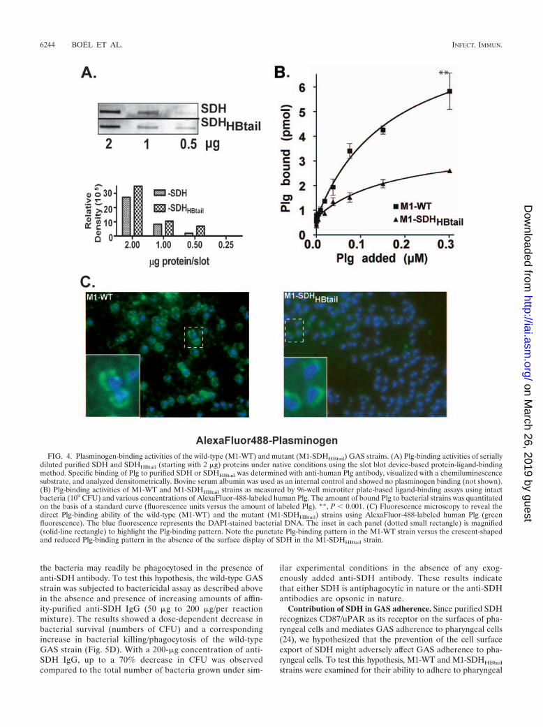
Role of SDH in plasminogen binding. Both SDH/Plr (27, 33)and SEN (12, 37, 38) directly bind Plg; however, their relativecontributions to the overall Plg-binding activity of GAS are notknown. To determine the contribution of SDH to the Plg-binding activity of GAS, serially diluted purified SDH andSDHHBtail proteins (starting concentration, 2 �g each) weresubjected to Plg binding and then detection of Plg binding byanti-Plg antibody in a slot blot-based direct ligand-bindingWestern blot assay. Plg-binding activities of serially dilutedSDH and SDHHBtail revealed that both proteins bind to Plgequally (Fig. 4A), indicating that the insertion of hydrophobictail at the C-terminal end of SDH does not interfere with thePlg-binding activity of the SDH molecule. To determine Plg-binding activities of the wild-type and mutant strains, eachstrain (109 CFU) was mixed with various concentrations ofAlexaFluor-488-labeled Plg, and the amount of bound Plg wasestimated based on the standard curve (labeled Plg versusfluorescence units) incorporated in each assay. At the highestconcentration (300 nM) of exogenously added labeled Plg, themutant strain acquired almost 60% less Plg than the wild-type(M1-WT) strain (�2.4 pmol in the mutant strain versus �6.0pmol in the wild type; P 0.001), indicating that surface-expressed SDH plays a significant role in plasminogen-bindingactivity (Fig. 4B). Immunofluorescence microscopy showed thereduced Plg-binding activity of the mutant strain (Fig. 4C) andalso confirmed the results obtained with the solid-phase ligand-binding assay (Fig. 4B). The residual plasminogen-binding ac-tivity of the mutant strain (Fig. 4C) is likely due to the presenceof SEN, which is the major Plg-binding protein on the surfaceof GAS (12, 37, 38). The unique crescent-shaped Plg-bindingpattern at the bacterial poles displayed by M1-SDHHBtail (Fig.4C, right panel inset) indicates that in the absence of the
surface display of SDH, the distribution of other plasminogen-binding proteins, such as SEN (37), is likely affected.
Contribution of SDH to the antiphagocytic activity of GAS.Antiphagocytic properties of the wild-type and mutant strainswere determined by measuring their abilities to multiply infresh human blood during a 3-h incubation period as describedin Materials and Methods. The numbers of live bacteriapresent before and after incubation were determined by count-ing CFU on blood agar plates. The results expressed in theform of multiplication factors (MF) (the number of CFU at 3 hover the number of CFU at time zero) revealed that, whereasthe wild-type (M1-WT) strain survived in the blood (MF �59.63 6) as expected, the mutant strain (M1-SDHHBtail)were readily killed (MF � 0.58 0.36), indicating that theantiphagocytic activity of the mutant strain was completelyinhibited (99% inhibition; P 0.0001) as was found for theM1�emm1 strain, which lacks expression of a major virulencefactor, the Emm1 protein (Fig. 5A). SDH, therefore, eitherdirectly or indirectly is involved in the antiphagocytic processof GAS. Since the M protein and the bacterial capsular poly-saccharide are the major surface components responsible forthe antiphagocytic activity of GAS, we questioned whether theloss of antiphagocytic activity in the mutant strain was due tothe loss of the expression of the M1 protein and/or the poly-saccharide capsule. To determine this, Western immunoblotanalysis of the cell wall as well as cytoplasm-associated proteinsof the wild-type and mutant strains was carried out using theclass I M protein-specific 10B6 mouse monoclonal antibody(raised against the M6 protein [25]). The results demonstratedthe absence of the expression of M1 protein in both fractionsof the M1-SDHHBtail mutant strain (Fig. 5B). In a parallelstudy of capsular polysaccharide estimation, we observed nosignificant change (P � 0.05) in the capsule polysaccharideassociated with the mutant M1-SDHHBtail strain (22.6 0.1�g/ml) from that in the wild-type strain (20.3 0.6 �g/ml).Together, these results demonstrate that the insertion of thehydrophobic tail at the C-terminal end of SDH affects M1protein expression but not capsule formation in GAS.
Alternatively, it is also conceivable that SDH also contrib-utes to the antiphagocytic activity of GAS since in the absenceof a surface display of SDH, the bacteria are readily phagocy-tosed and, as in the presence of anti-M protein antibody (14),
activities of intact M1-WT and M1-SDHHBtail GAS strains (200 �l of a bacterial suspension with an OD600 of 1.0, i.e.,108 CFU) as an endpointreading of the supernatant obtained after 5 min of incubation in 600 �l of the reaction buffer followed by pelleting of the bacteria. Each readingpoint in panels A, B, and C represents an average from three independent experiments standard errors. (D) Indirect immunofluorescencemicroscopy of the wild-type (M1-WT) and mutant (M1-SDHHBtail) strains using affinity-purified monospecific rabbit anti-SDH IgG (Fab�fragment) as the primary antibody followed by fluorescein isothiocyanate-labeled anti-rabbit Fab�-specific IgG (green fluorescence) as thesecondary antibody. In parallel control experiments, both bacterial strains did not stain with the secondary conjugate antibody (not shown). Theinset in each panel (dotted-line rectangle) is magnified (solid-line rectangle) to highlight bacterial-surface-associated SDH-specific punctatefluorescence. Note the absence of green fluorescence in the M1-SDHHBtail mutant strain. The blue fluorescence represents the DAPI-stainedbacterial DNA. The background fluorescence in M1-SDHHBtail (inset) is due to lysed bacteria as revealed by the absence of DAPI stain.(E) Western blot analysis of the presence of SDH in culture supernatant (Sup) and subcellular fractions (CW, cell wall; Cyt, cytoplasm; and Mem,membrane) of the wild-type (M1-WT) and mutant (M1-SDHHBtail) strains using SDH-specific monoclonal antibody (13D5) (16). Both strains weregrown to an OD of 0.8 in THY broth, and the supernatants were separated by centrifugation. The resulting bacterial pellets obtained from eachstrain were subjected to subcellular fractionation. Twenty-five percent-TCA-precipitated proteins obtained from 125 �l of the supernatant fromeach strain were tested for the presence of SDH. (F) Semiquantitative analysis of the presence of SDH in the cytoplasm and cell wall fractionsof equal numbers of M1-WT and M1-SDHHBtail cells. Serial twofold dilutions of 4 �g of total proteins of the cytoplasmic and cell wall fractionsobtained from both strains were resolved on PVDF membranes by SDS-PAGE and electroblotting. The presence of SDH was determined byimmunoblotting using anti-SDH monoclonal antibody (13D5) and visualized with a chemiluminescence substrate.
VOL. 73, 2005 PREVENTION OF CELL SURFACE EXPORT OF ANCHORLESS SDH 6243
on March 26, 2019 by guest
http://iai.asm.org/
Dow
nloaded from
the bacteria may readily be phagocytosed in the presence ofanti-SDH antibody. To test this hypothesis, the wild-type GASstrain was subjected to bactericidal assay as described abovein the absence and presence of increasing amounts of affin-ity-purified anti-SDH IgG (50 �g to 200 �g/per reactionmixture). The results showed a dose-dependent decrease inbacterial survival (numbers of CFU) and a correspondingincrease in bacterial killing/phagocytosis of the wild-typeGAS strain (Fig. 5D). With a 200-�g concentration of anti-SDH IgG, up to a 70% decrease in CFU was observedcompared to the total number of bacteria grown under sim-
ilar experimental conditions in the absence of any exog-enously added anti-SDH antibody. These results indicatethat either SDH is antiphagocytic in nature or the anti-SDHantibodies are opsonic in nature.
Contribution of SDH in GAS adherence. Since purified SDHrecognizes CD87/uPAR as its receptor on the surfaces of pha-ryngeal cells and mediates GAS adherence to pharyngeal cells(24), we hypothesized that the prevention of the cell surfaceexport of SDH might adversely affect GAS adherence to pha-ryngeal cells. To test this hypothesis, M1-WT and M1-SDHHBtail
strains were examined for their ability to adhere to pharyngeal
FIG. 4. Plasminogen-binding activities of the wild-type (M1-WT) and mutant (M1-SDHHBtail) GAS strains. (A) Plg-binding activities of seriallydiluted purified SDH and SDHHBtail (starting with 2 �g) proteins under native conditions using the slot blot device-based protein-ligand-bindingmethod. Specific binding of Plg to purified SDH or SDHHBtail was determined with anti-human Plg antibody, visualized with a chemiluminescencesubstrate, and analyzed densitometrically. Bovine serum albumin was used as an internal control and showed no plasminogen binding (not shown).(B) Plg-binding activities of M1-WT and M1-SDHHBtail strains as measured by 96-well microtiter plate-based ligand-binding assays using intactbacteria (109 CFU) and various concentrations of AlexaFluor-488-labeled human Plg. The amount of bound Plg to bacterial strains was quantitatedon the basis of a standard curve (fluorescence units versus the amount of labeled Plg). **, P 0.001. (C) Fluorescence microscopy to reveal thedirect Plg-binding ability of the wild-type (M1-WT) and the mutant (M1-SDHHBtail) strains using AlexaFluor-488-labeled human Plg (greenfluorescence). The blue fluorescence represents the DAPI-stained bacterial DNA. The inset in each panel (dotted small rectangle) is magnified(solid-line rectangle) to highlight the Plg-binding pattern. Note the punctate Plg-binding pattern in the M1-WT strain versus the crescent-shapedand reduced Plg-binding pattern in the absence of the surface display of SDH in the M1-SDHHBtail strain.
6244 BOEL ET AL. INFECT. IMMUN.
on March 26, 2019 by guest
http://iai.asm.org/
Dow
nloaded from

cells. The GAS adherence assay revealed that the mutantstrain, M1-SDHHBtail, adhered to pharyngeal cells threefoldless (�70% less) than the wild-type strain (Fig. 6). Since theM1-SDHHBtail mutant strain does not express the M1 protein(Fig. 5B), we also included another mutant strain lacking theexpression of the M1 protein (M1Demm1) in the bacterial ad-herence assay as an internal control. The results showed thatthe M1Demm1 mutant strain adhered in a way similar to that ofthe wild-type strain (P � 0.268), indicating that SDH and notthe M protein plays an important role in GAS adherence topharyngeal cells (Fig. 5).
DISCUSSION
SDH belongs to a novel class of anchorless cell wall-associ-ated surface proteins (32). Several proteins, including SDH,that belong to this class of surface proteins are not only mul-
tifunctional but also essential for bacterial survival. Hence, theresulting nonfeasibility of the “gene knockout” strategy hasbeen a major limiting factor in our progress in understandingtheir roles in disease pathogenesis. In the present investigation,we have addressed this limiting factor and devised a mutagen-esis strategy that allows an insertion of a hydrophobic tail atthe C-terminal end of SDH and prevents the export of SDH tothe cell surface. This strategy is important for two main rea-sons. First, it does not affect the innate GAPDH activity ofGAS. Second, this strategy also allows retaining this modifiedSDH in the cytoplasm and does not affect growth characteris-tics of bacteria.
SDH and other anchorless surface glycolytic enzymes fromGAS and other microorganisms are exported to the cell sur-face in the absence of a signal sequence and hydrophobicdomain. The precise mechanism of their export to the cell
FIG. 5. Phagocytosis/bactericidal assays. (A) Growth of the wild-type (M1-WT) and mutant (M1-SDHHBtail) strains in human blood isexpressed as a multiplication factor. The MF is the number of CFU obtained at the end of the incubation period over the number of CFU obtainedat time zero. The M1�emm1 strain was used as a negative control for antiphagocytic activity. Bars and error bars represent the means standarderrors obtained from two independent experiments, each with triplicate samples. ***, P 0.0001. (B) Western blot analysis of cell wall- andcytoplasm-associated proteins of the wild-type (M1-WT) and mutant (M1-SDHHBtail) strains using M1 protein-reacting 10B6 monoclonal antibody(mAb). Proteins were visualized with a chromogenic substrate. Each lane received a total of 5 �g of total cell wall-associated or cytoplasmicproteins. (C) Estimation of the hyaluronic capsule associated with the wild-type and mutant (M1-SDHHBtail) strains grown until late log phase(OD
600of 0.8). N.S., not significant (P � 0.05). (D) Opsonophagocytosis or inhibition of the antiphagocytic activity of the wild-type GAS strain in
the presence of various concentrations of affinity-purified anti-SDH antibodies. The bactericidal assay was performed as described for panel A. Theleft y axis denotes the numbers of bacteria that survived at different concentrations of anti-SDH antibodies in the assay mixture at the end of theincubation period. The right y axis denotes percentages of bacteria being killed/phagocytosed at different concentrations of anti-SDH antibodies.Stat, stationary; Rot, rotation.
VOL. 73, 2005 PREVENTION OF CELL SURFACE EXPORT OF ANCHORLESS SDH 6245
on March 26, 2019 by guest
http://iai.asm.org/
Dow
nloaded from

surface and subsequent secretion into the medium is notknown. It is, therefore, interpreted that the surface associationof this enzyme is possibly due to the reassociation of leakedcytoplasmic proteins as a result of the uncontrolled naturallysis of a few bacteria. We monitored the secretion of SDH inculture supernatant and its retention within bacteria by immu-nofluorescence, enzyme kinetics, and immunoblot methods.Whereas the immunofluorescence provided a visual proof ofthe location of wild-type and mutant SDH, the remaining twomethods provided quantitative analysis of their associationwith the GAS cell wall, cytoplasm, and membranes. All meth-ods indicated primarily that the mutated SDH is not secretedand is not displayed on the GAS surface. Further, in the wild-type strain, surface-displayed SDH is not derived from lysedbacteria. Based on the design of the experiment, we had ex-pected a large amount of SDH to be trapped in the membrane.However, our results showed that the large amount of SDHwas restricted to the cytoplasm, to some extent in the mem-brane, and also in the cell wall. The detection of some amountof SDHHBtail in the cell wall fraction of the intact mutantbacteria, albeit three to five times less than that in the wildtype, suggest that while the hydrophobic tail primarily inter-feres with the secretion of SDH and helps to retain the SDH-HBtail molecule within the cytoplasm, some molecules may es-cape during cell division and be retained and trapped withinthe cell wall while they transgress the cell envelope. TheseSDH molecules are likely accessible to the glyceraldehydes-3-phosphate substrate during enzyme kinetic assay of the intactbacteria but not accessible to anti-SDH antibody during theimmunofluorescence assay. Thus, the presence of SDHHBtail
buried within the GAS cell walls was detected in immunoblot-ting assays, and only the surface-located SDH was detected by the
immunofluorescence method. Based on the latter method, morethan 95% of the mutant bacterial population (M1-SDHHBtail)showed the absence of SDH on the surface.
SDH and many of the other anchorless proteins are multi-functional. Because of their essential nature, previous studiesof mutational analyses of this and other anchorless proteinswere restricted to functional regions, such as Plg-binding do-mains, that do not participate in essential catalytic activities (3,12, 45). Further, the functional analyses of such domains thatoverlap catalytic domains were based on in vitro ligand-bindingassays, i.e., measuring the Plg-binding activity of internal bind-ing sites of SEN of GAS or a similar protein of S. pneumoniae,using truncated recombinant proteins or short relevant syn-thetic peptides bound to a solid phase (3, 12). In SDH/Plr, thesecond Plg-binding site is proposed to be located in the N-terminal region (45). The nature of this site is unknown. It is,however, likely that the putative second plasminogen-bindingsite contributes to the catalytic domain, since several N-termi-nally truncated recombinant SDH proteins representing dele-tions of the entire region in the first N-terminal half werecatalytically inactive (24; V. Pancholi, unpublished data). Itmay be emphasized that although the C-terminal lysine residueplays an important role in Plg binding (45), the insertion of anadditional hydrophobic tail did not interfere with the Plg-binding activity of the SDH molecule (Fig. 4). The mutagenesisapproach described in the present study thus successfully al-lowed us to study the Plg-binding activity of GAS mediated bythe SDH molecule as a whole. By using a similar mutagenesisapproach, we propose that secretion of SEN and other anchor-less surface molecules of GAS can also be prevented and thatthey can be localized or restrained to the cytoplasm withoutaffecting their innate enzyme activities and other functions.The successful creation of such mutants may then help under-stand the contribution of anchorless surface proteins in GASpathogenesis.
In the present study, the unexpected finding that the isogenicM1-SDHHBtail mutant strain lost the antiphagocytic activity ofthe wild-type strain raises an important question, namely,whether SDH is indeed antiphagocytic in nature. To under-stand the contribution of SDH to GAS antiphagocytic activity,we determined the antiphagocytic activity of the wild-typestrain in the presence of anti-SDH polyclonal antibody. Theincreased phagocytosis of the wild-type GAS strain in the pres-ence of anti-SDH antibody is likely due to its opsonic activity,as was also found for the antibody against another well-studiedanchorless protein, SEN (16, 37). On the other hand, theabsence of antiphagocytic activity in M1-SDHHBtail indeedseems due to the absence of the M1 protein as the expressionof another antiphagocytic molecule, the polysaccharide cap-sule (14), was not affected in the mutant strain. Despite thefacts that the sdh gene (SPy274) is monocistronic in the M1genome as well as in other described GAS genomes (1, 2, 13,29, 43) and hence that the insertion of a hydrophobic tail andthe spectinomycin resistance gene aad-9 does not create anypolar effect in the M1-SDHHBtail mutant, the expression of theemm1 gene (SPy 2018) product (M1 protein) is down regu-lated. Eukaryotic GAPDH has been shown to bind DNA andRNA, and this binding has been shown to play a role in thetranscription regulation of eukaryotic genes (for reviews, seereferences 41 and 42). It is likely that SDH and its C-terminal
FIG. 6. Bacterial adherence assays. Confluent human pharyngealcells (Detroit 562) grown in 24-well tissue culture plates were incu-bated with the wild-type (M1-WT) and mutant (M1-SDHHBtail) strains(multiplicity of infection, 1:50, cells to bacteria) for 3 h. The mutantstrain lacking the expression of the M1 protein (M1�emm1) was used asan internal control in this assay. At the end of incubation, cell-associ-ated bacteria were counted as numbers of CFU on sheep blood agarplates. The adherence index for each preparation was calculated as apercentage of the initial inoculum (4 � 107 to 5 � 107/well, i.e., 100 �lof an OD 1.0 bacterial suspension). Each error bar represents anaverage of results from three independent experiments, each fromtriplicate wells standard errors. **, P 0.001. N.S., not significant.
6246 BOEL ET AL. INFECT. IMMUN.
on March 26, 2019 by guest
http://iai.asm.org/
Dow
nloaded from

portion in some way may have an important role in M1 ex-pression, possibly at the transcriptional level.
We recently reported that SDH recognizes uPAR/CD87 asits receptor on the surfaces of pharyngeal cell and mediatesGAS adherence (24). In that publication, we showed that themutant GAS strain, displaying SDH with the C-terminal lysineresidue (Lys336) deleted or with a C-terminal leucine residueinstead of the lysine residue (Lys336Leu), adhered poorly topharyngeal cells (24). It was, however, not possible to studythose mutants to understand the contribution of other domainsof the SDH molecule in the adherence process, as other do-mains contribute to the essential GAPDH activity of SDH(24). In the present study, this shortcoming was overcome bypreventing the display of the whole SDH protein on the GASsurface. Our results showing the adherence ability of theM1�emm1 mutant strain to pharyngeal cells in a way similar tothat of the wild-type strain concur with the previously pub-lished reports, which indicated that the expression of the Mprotein has little effect on GAS adherence to human tonsillar/pharyngeal cells and keratinocytes (6, 10). In conjunction withthis finding, the poor adherence ability of the M1-SDHHBtail
strain, which does not display SDH, as well as of the M1protein on its surface underscores the important contributionof SDH in GAS adherence to pharyngeal cells.
In summary, the mutagenesis approach used in the presentstudy revealed new functions of SDH and its role in GASpathogenesis, such as GAS adherence and antiphagocytic ac-tivity. It is not clear at present whether SDH is itself antiph-agocytic or whether, in addition, it plays a role in the transcrip-tional or posttranscriptional control of GAS virulence factors.As the latter is true for the eukaryotic GAPDH (41, 42), it is animportant question for future research. Evidence provided inthe present study concludes that SDH is an important surface-expressed virulence factor and plays an important role in GASpathogenesis.
ACKNOWLEDGMENTS
We thank Vincent A. Fischetti (The Rockefeller University, NewYork, NY) for providing 10B6 monoclonal antibody. We thank CarlDrlica and Issar Smith for critically reading the manuscript and JanetHahn and Hedia Maamar for their initial help in immunofluorescencemicroscopy.
This research was supported by grant RO1-42827 (to V.P.) from theNational Institutes of Health.
REFERENCES
1. Banks, D. J., S. F. Porcella, K. D. Barbian, S. B. Beres, L. E. Philips, J. M.Voyich, F. R. DeLeo, J. M. Martin, G. A. Somerville, and J. M. Musser. 2004.Progress toward characterization of the group A Streptococcus metagenome:complete genome sequence of a macrolide-resistant serotype M6 strain.J. Infect. Dis. 190:727–738.
2. Beres, S. B., G. L. Sylva, K. D. Barbian, B. Lei, J. S. Hoff, N. D. Mammarella,M.-Y. Liu, J. C. Smoot, S. F. Porcella, L. D. Parkins, D. S. Campbell, T. M.Smith, J. K. McCormick, D. Y. M. Leung, P. Schlievert, and J. M. Musser.2002. Genome sequence of a serotype M3 strain of group A Streptococcus:phage-encoded toxins, the high-virulence phenotype, and clone emergence.Proc. Natl. Acad. Sci. USA 99:10078–10083.
3. Bergmann, S., M. Rohde, G. S. Chhatwal, and S. Hammerschmidt. 2001.Alpha-enolase of Streptococcus pneumoniae is a plasmin(ogen)-binding pro-tein displayed on the bacterial cell surface. Mol. Microbiol. 40:1273–1287.
4. Biesecker, G., J. I. Harris, J. C. Thierry, J. E. Walker, and A. J. Wonacott.1977. Sequence and structure of D-glyceraldehyde-3-phosphate dehydroge-nase from Bacillus stearothermophilus. Nature 266:328–333.
5. Bisno, A. L., and D. L. Stevens. 1996. Streptococcal infections of skin andsoft tissues. N. Engl. J. Med. 334:240–245.
6. Caparon, M. G., D. S. Stephens, A. Olsen, and J. R. Scott. 1991. Role of Mprotein in adherence of group A streptococci. Infect. Immun. 59:1811–1817.
7. Conway, T. 1992. The Entner-Doudoroff pathway: history, physiology andmolecular biology. FEMS Microbiol. Rev. 103:1–28.
8. Cserzo, M., E. Wallin, I. Simon, G. Von Heijne, and A. Elofsson. 1997.Prediction of transmembrane alpha-helices in prokaryotic membrane pro-teins: the dense alignment surface method. Protein Eng. 10:673–676.
9. Cunningham, M. W. 2000. Pathogenesis of group A streptococcal infections.Clin. Microbiol. Rev. 13:470–511.
10. Darmstadt, G. L., L. Mentele, A. Podbielski, and C. Rubens. 2000. Role ofgroup A streptococcal virulence factors in adherence to keratinocytes. Infect.Immun. 68:1215–1221.
11. Davies, H. D., A. McGeer, B. Schwartz, K. Green, D. Cann, A. E. Simor, andD. E. Low. 1996. Invasive group A streptococcal infections in Ontario, Can-ada. N. Engl. J. Med. 335:547–554.
12. Derbise, A., Y. P. Song, S. Parikh, V. A. Fischetti, and V. Pancholi. 2004.Role of the C-terminal lysine residues of streptococcal surface enolase inGlu- and Lys-plasminogen-binding activities of group A streptococci. Infect.Immun. 72:94–105.
13. Ferretti, J. J., W. M. McShan, D. Ajdic, D. J. Savic, G. Savic, K. Lyon, C.Primeaux, S. Sezate, A. N. Suvorov, S. Kenton, H. S. Lai, S. P. Lin, Y. Qian,H. G. Jia, F. Z. Najar, Q. Ren, H. Zhu, L. Song, J. White, X. Yuan, S. W.Clifton, B. A. Roe, and R. McLaughlin. 2001. Complete genome sequence ofan M1 strain of Streptococcus pyogenes. Proc. Natl. Acad. Sci. USA 98:4658–4663.
14. Fischetti, V. A. 1989. Streptococcal M protein: molecular design and biolog-ical behavior. Clin. Microbiol. Rev. 2:285–314.
15. Fischetti, V. A. 2000. Surface proteins on gram-positive bacteria, p. 11–26. InV. A. Fischetti, R. P. Novick, J. J. Ferretti, D. A. Portnoy, and J. I. Rood(ed.), Gram-positive pathogens. ASM Press, Washington, D.C.
16. Fontan, P. A., V. Pancholi, M. M. Nociari, and V. A. Fischetti. 2000. Anti-bodies to streptococcal surface enolase react with human -enolase: impli-cations in poststreptococcal sequelae. J. Infect. Dis. 182:1712–1721.
17. Fothergill-Gilmore, L. A., and P. A. M. Michels. 1993. Evolution of glycol-ysis. Prog. Biophys. Mol. Biol. 59:105–235.
18. Harris, J. I., and M. Waters. 1976. Glyceraldehyde-3-phosphate dehydroge-nase, p. 1–49. In P. D. Boyers (ed.), The enzymes. Academic Press, NewYork, N.Y.
19. Hillman, J. D., and D. G. Fraenkel. 1975. Glyceraldehyde 3-phosphate de-hydrogenase mutants of Escherichia coli. J. Bacteriol. 122:1175–1179.
20. Holland, J. P., L. Labieniec, C. Swimmer, and M. J. Holland. 1983. Homol-ogous nucleotide sequences at the 5� termini of messenger RNAs synthe-sized from the yeast enolase and glyceraldehyde-3-phosphate dehydrogenasegene families. J. Biol. Chem. 258:5291–5299.
21. Holland, M. J., J. P. Holland, G. P. Thill, and K. A. Jackson. 1981. Theprimary structures of two yeast enolase genes. Homology between the 5�noncoding flanking regions of yeast enolase and glyceraldehyde-3-phosphatedehydrogenase genes. J. Biol. Chem. 256:1385–1395.
22. Irani, M. H., and P. K. Maitra. 1976. Glyceraldehyde 3-P dehydrogenase,glycerate 3-P kinase and enolase mutants of Escherichia coli: genetic studies.Mol. Gen. Genet. 145:65–71.
23. Irani, M. H., and P. K. Maitra. 1977. Properties of Escherichia coli mutantsdeficient in enzymes of glycolysis. J. Bacteriol. 132:398–410.
24. Jin, H., Y. P. Song, G. Boel, J. Kochar, and V. Pancholi. 2005. Group Astreptococcal surface GAPDH, SDH, recognizes uPAR/CD87 as its receptoron the human pharyngeal cell and mediates bacterial adherence to host cells.J. Mol. Biol. 350:27–41.
25. Jones, K. F., B. N. Manjula, K. H. Johnston, S. K. Hollingshead, J. R. Scott,and V. A. Fischetti. 1985. Location of variable and conserved epitopes amongthe multiple serotypes of streptococcal M protein. J. Exp. Med. 161:623–628.
26. Lancefield, R. 1957. Differentiation of group A streptococci with a commonR antigen into three serological types, with special reference to the bacteri-cidal test. J. Exp. Med. 106:525–544.
27. Lottenberg, R., C. C. Broder, M. D. P. Boyle, S. J. Kain, B. L. Schroeder, andR. Curtiss III. 1992. Cloning, sequence analysis, and expression in Esche-richia coli of a streptococcal plasmin receptor. J. Bacteriol. 174:5204–5210.
28. McAlister, L., and M. J. Holland. 1985. Isolation and characterization ofyeast strains carrying mutations in the glyceraldehyde-3-phosphate dehydro-genase genes. J. Biol. Chem. 260:15013–15018.
29. Nakagawa, I., K. Kurokawa, A. Yamashita, M. Nakata, Y. Tomiyasu, N.Okahashi, S. Kawabata, K. Yamazaki, T. Shiba, T. Yasunaga, H. Hayashi,M. Hattori, and S. Hamada. 2003. Genome sequence of an M3 strain ofStreptococcus pyogenes reveals a large-scale genomic rearrangement in inva-sive strains and new insights into phage evolution. Genome Res. 13:1042–1055.
30. Navarre, W. W., and O. Schneewind. 1999. Surface proteins of gram-positivebacteria and mechanisms of their targeting to the cell wall envelope. Micro-biol. Mol. Biol. Rev. 63:174–229.
31. Pancholi, V. 2001. Multifunctional -enolase: its role in diseases. Cell. Mol.Life Sci. 58:902–920.
32. Pancholi, V., and G. S. Chhatwal. 2003. Housekeeping enzymes as virulencefactors for pathogens. Int. J. Med. Microbiol. 293:1–11.
33. Pancholi, V., and V. A. Fischetti. 1992. A major surface protein on group A
VOL. 73, 2005 PREVENTION OF CELL SURFACE EXPORT OF ANCHORLESS SDH 6247
on March 26, 2019 by guest
http://iai.asm.org/
Dow
nloaded from

streptococci is a glyceraldehyde-3-phosphate dehydrogenase with multiplebinding activity. J. Exp. Med. 176:415–426.
34. Pancholi, V., and V. A. Fischetti. 1993. Glyceraldehyde-3-phosphate dehy-drogenase on the surface of group A streptococci is also an ADP-ribosylatingenzyme. Proc. Natl. Acad. Sci. USA 90:8154–8158.
35. Pancholi, V., and V. A. Fischetti. 1997. Identification of a glycolytic enzymecomplex on the surface of group A streptococci, p. 35. In Abstr. 97th Gen. Meet.Am. Soc. Microbiol. American Society for Microbiology, Washington, D.C.
36. Pancholi, V., and V. A. Fischetti. 1997. Regulation of the phosphorylation ofhuman pharyngeal cell proteins by group A streptococcal surface dehydro-genase (SDH): signal transduction between streptococci and pharyngealcells. J. Exp. Med. 186:1633–1643.
37. Pancholi, V., and V. A. Fischetti. 1998. -Enolase, a novel strong plasmin-(ogen) binding protein on the surface of pathogenic streptococci. J. Biol.Chem. 273:14503–14515.
38. Pancholi, V., P. A. Fontan, and H. Jin. 2003. Plasminogen-mediated group Astreptococcal adherence to and pericellular invasion of human pharyngealcells. Microb. Pathog. 35:293–303.
39. Podbielski, A., B. Spellerberg, M. Woischnik, B. Pohl, and R. Lutticken.1996. Novel series of plasmid vectors for gene inactivation and expressionanalysis in group A streptococci (GAS). Gene 177:137–147.
40. Schrager, H. M., J. G. Rheinwald, and M. R. Wessels. 1996. Hyaluronic acidcapsule and the role of streptococcal entry into keratinocytes in invasive skininfection. J. Clin. Investig. 98:1954–1958.
41. Sirover, M. A. 1999. New insights into an old protein: the functional diversityof mammalian glyceraldehyde-3-phosphate dehydrogenase. Biochim. Bio-phys. Acta 1432:159–184.
42. Sirover, M. A. 2005. New nuclear functions of the glycolytic protein, glycer-ladehyde-3-phosphate dehydrogenase, in mammalian cells. J. Cell. Biochem.95:45–52.
43. Smoot, J. C., K. D. Barbian, J. J. Van Gompel, L. M. Smoot, M. S. Chaussee,G. L. Sylva, D. E. Sturdevant, S. M. Ricklefs, S. F. Porcella, L. D. Parkins,S. B. Beres, D. S. Campbell, T. M. Smith, Q. Zhang, V. Kapur, J. A. Daly,L. G. Veasey, and J. M. Musser. 2002. Genome sequence and comparativemicroarray analysis of serotype M18 group A Streptococcus strains associatedwith acute rheumatic fever outbreaks. Proc. Natl. Acad. Sci. USA 99:4668–4673.
44. Walker, J. E., A. F. Carne, M. J. Runswick, J. Bridgen, and J. I. Harris. 1992.D-Glyceraldehyde-3-phosphate dehydrogenase. Eur. J. Biochem. 108:549–565.
45. Winram, S. B., and R. Lottenberg. 1998. Site-directed mutagenesis of strep-tococcal plasmin receptor protein (Plr) identifies the C-terminal Lys334 asessential for plasmin binding, but mutation of the plr gene does not reduceplasmin-binding to group A streptococi. Microbiology 144:2025–2035.
46. Zablotny, R., and D. G. Fraenkel. 1967. Glucose and gluconate metabolismin a mutant of Escherichia coli lacking gluconate-6-phosphate dehydrase. J.Bacteriol. 93:1579–1581.
Editor: J. N. Weiser
6248 BOEL ET AL. INFECT. IMMUN.
on March 26, 2019 by guest
http://iai.asm.org/
Dow
nloaded from