inhibition of cholinergic signaling causes apoptosis in...
TRANSCRIPT
Inhibition of cholinergic signaling causes apoptosis in human bronchioalveolar
carcinoma
Jamie K. Lau1$, Kathleen C. Brown1$, Brent A. Thornhill1$, Clayton M. Crabtree1, Aaron M.
Dom1, Theodore R. Witte2, W. Elaine Hardman2, Christopher A. McNees1, Cody A. Stover1, A.
Betts Carpenter3, Haitao Luo4, Yi C. Chen4, Brandon S. Shiflett1, and Piyali Dasgupta1*
1Department of Pharmacology, Physiology, and Toxicology, Joan C. Edwards School of
Medicine, Marshall University, Huntington, WV 25755
2Department of Biochemistry and Microbiology, Joan C. Edwards School of Medicine, Marshall
University, Huntington, WV 25755
3Department of Anatomy and Pathology, Joan C. Edwards School of Medicine, Marshall
University, Huntington, WV 25755
4Department of Biology, Alderson-Broaddus College, Philippi, WV 26416
*Correspondence to: Piyali Dasgupta, Department of Pharmacology, Physiology, and
Toxicology, Joan C. Edwards School of Medicine, Marshall University, 1700 3rd Avenue,
Huntington, WV 25755. E-mail: [email protected].
$These authors contributed equally to the work presented here and should therefore be
regarded as equivalent authors.
The authors declare that they have no conflict of interest.
on June 25, 2018. © 2012 American Association for Cancer Research. cancerres.aacrjournals.org Downloaded from
Author manuscripts have been peer reviewed and accepted for publication but have not yet been edited. Author Manuscript Published OnlineFirst on December 7, 2012; DOI: 10.1158/0008-5472.CAN-12-3190

2
Abstract
Recent case-controlled clinical studies show that bronchioalveolar carcinomas (BACs) are
correlated with smoking. Nicotine, the addictive component of cigarettes, accelerates cell
proliferation through nicotinic acetylcholine receptors (nAChRs). In this study, we show that
human BACs produce acetylcholine (ACh) and contain several cholinergic factors including
acetylcholinesterase (AChE), choline acetyltransferase (ChAT), choline transporter 1 (CHT1,
SLC5A7), vesicular acetylcholine transporter (VAChT, SLC18A3) and nACh receptors (AChRs,
CHRNAs). Nicotine increased the production of ACh in human BACs and ACh acts as a growth
factor for these cells. Nicotine-induced ACh production was mediated by α7-, α3β2-, and β3-
nAChRs, ChAT and VAChT pathways. We observed that nicotine upregulated ChAT and
VAChT. Therefore, we conjectured that VAChT antagonists, such as vesamicol, may suppress
the growth of human BACs. Vesamicol induced potent apoptosis of human BACs
in cell culture and nude mice models. Vesamicol did not have any effect on EGF or IGF-II-
induced growth of human BAC's. siRNA-mediated attenuation of VAChT reversed the apoptotic
activity of vesamicol. We also observed that vesamicol inhibited Akt phosphorylation during cell
death, and overexpression of constitutively active Akt reversed the apoptotic activity of
vesamicol. Taken together, our results suggested that disruption of nicotine-induced cholinergic
signaling by agents such as vesamicol may have applications in BAC therapy.
KEY WORDS: Bronchioalveolar carcinoma, Acetylcholine, α7/β3 nAChR, VAChT, CHT1,
Vesamicol, Akt
on June 25, 2018. © 2012 American Association for Cancer Research. cancerres.aacrjournals.org Downloaded from
Author manuscripts have been peer reviewed and accepted for publication but have not yet been edited. Author Manuscript Published OnlineFirst on December 7, 2012; DOI: 10.1158/0008-5472.CAN-12-3190

3
Précis
Findings that cholinergic signaling are critical to the survival of bronchioalveolar carcinoma cells
should prompt immediate clinical testing of approved drugs that disrupt this pathway to
determine if they can improve the efficacy of treatments for this form of lung cancer.
on June 25, 2018. © 2012 American Association for Cancer Research. cancerres.aacrjournals.org Downloaded from
Author manuscripts have been peer reviewed and accepted for publication but have not yet been edited. Author Manuscript Published OnlineFirst on December 7, 2012; DOI: 10.1158/0008-5472.CAN-12-3190
4
Introduction
Bronchioalveolar carcinoma (BAC) is a subtype of lung adenocarcinoma arising from type II
pneumonocytes in the lung. The World Health Organization (WHO) revised its classification of
BAC’s in 2004 to include lung adenocarcinomas, which grow in a lepidic fashion along the
alveolar septa without invasion into the stroma, pleura, blood vessels or lymphatics (1-3). The
incidence of pure BAC is about 4%; however, mixed subtypes, including BACs with stromal
invasion and pulmonary adenocarcinoma with BAC-like morphological features, account for
almost 20% of all non-small cell lung cancers (NSCLC) (4). Cigarette smoking is the leading risk
factor for the development of lung cancer. It is estimated that smoking is associated with 80-
90% of lung cancer cases throughout the world (5). Smoking has a stronger association with
small cell lung cancer (SCLC) than with adenocarcinoma (6). BAC is a relatively rare type of
adenocarcinoma; therefore, only a few epidemiological studies have investigated the
relationship between BAC and smoking (7-9).
Traditionally, the role of tobacco smoking in the etiology of human BAC originates from a
series of early studies involving human lung cancer patients, most of which were non-smokers
(1). These non-smoking lung cancer patients had higher incidence of BAC than other types of
lung cancer. The resulting impression was that smoking is unimportant in the etiology of human
BAC (1). However, these reports did not contain any cohort or case control studies that formally
determined the relationship of human BACs to cigarette smoking.
Epidemiological data have demonstrated an association between human BACs and
smoking (7, 8, 10). The risk of developing BACs is greater for people who started smoking at a
younger age, smoked for a longer time or smoked more cigarettes per day. Conversely, the risk
decreases in proportion to the duration of smoking cessation. Smoking habits have been
correlated to both the mucinous and non-mucinous form of human BACs (11). Rolen et al.,
(2003) performed a case-control study and compared smoking status of 198 BAC patients to an
equal number of controls. They observed that the risk of BAC is strongly related to the smoking
on June 25, 2018. © 2012 American Association for Cancer Research. cancerres.aacrjournals.org Downloaded from
Author manuscripts have been peer reviewed and accepted for publication but have not yet been edited. Author Manuscript Published OnlineFirst on December 7, 2012; DOI: 10.1158/0008-5472.CAN-12-3190

5
history of patients. They also found that both current and former smokers were at risk of
developing BAC (9). Recently, Boffeta et al., (2011) analyzed seven case controlled studies in
the USA comprising of 799 cases of BACs and 15,859 controls. They found that ever smokers
are at a two-fold greater risk of developing lung BAC as compared to never smokers. They also
observed a positive correlation between the duration and amount of smoking and the
development of lung BAC (8).
Although cigarette smoke is comprised of a mixture of many compounds, nicotine is the
addictive component of cigarette smoke (12). Several convergent studies have shown that
nicotine promotes the progression of human BACs and confers resistance against
chemotherapy (12, 13). All of these observations suggest that nicotine-induced mitogenic and
pro-survival pathways contribute to the pathophysiology of BACs.
The proliferative activity of nicotine is mediated by nicotinic acetylcholine receptors
(nAChRs) (12). The endogenous ligand for nAChRs is acetylcholine (ACh) (14). Recent studies
have shown that nAChRs are present in non-neuronal tissues, including lung cancer cells, lung
epithelial cells, endothelial cells and keratinocytes (12). Small cell lung cancers (SCLCs) and
squamous cell carcinoma of the lung (SCC-L) express all components of the ACh autocrine
loop, including acetylcholinesterase (AChE), choline acetyltransferase (ChAT), vesicular
acetylcholine transporter (VAChT), choline transporter1 (CHT1), nAChRs and muscarinic
acetylcholine receptors. SCLC and SCC-L cells secrete ACh, which promotes their proliferation
(14-17). Song et al., (2008) found that the muscarinic receptor antagonist Darifenacin displayed
anti-tumor activity in human SCC-L in both cell culture and nude mice models (15, 18). Such
observations suggest that the cholinergic network may be a viable molecular target in the
therapy of human lung cancer.
The present manuscript investigates whether the cholinergic loop exists in human BACs.
We show that human BAC cell lines produce ACh and express cholinergic proteins. We also
show for the first time that nicotine can amplify the components of the cholinergic loop in human
on June 25, 2018. © 2012 American Association for Cancer Research. cancerres.aacrjournals.org Downloaded from
Author manuscripts have been peer reviewed and accepted for publication but have not yet been edited. Author Manuscript Published OnlineFirst on December 7, 2012; DOI: 10.1158/0008-5472.CAN-12-3190
6
BACs. Nicotine increased the production of ACh in human BAC cell lines in a time- and dose-
dependent manner. ACh acted as a growth factor for human BAC cells. Nicotine upregulated
VAChT and ChAT expression in human BAC cells. We conjectured that nicotine-induced
increase of VAChT levels may provide a viable molecular target in human BACs. The VAChT
antagonist vesamicol induced robust apoptosis of human BAC cells in both cell culture and in
vivo models. Vesamicol did not affect EGF or IGF-II-induced growth pathways in human BACs.
This finding suggests that the pro-apoptotic activity of vesamicol is specific to the acetylcholine-
signaling pathway in human BACs. The pro-apoptotic activity of vesamicol was mediated via
suppression of Akt activation. The data presented in this paper characterizes the cholinergic
system in human BACs and offers novel avenues for BAC therapy. The results of our
experiments are relevant to BAC patients who are exposed to secondhand smoke or use
nicotine-based cessation devices (e.g. patches and gums) to quit smoking.
Methods
Ethical use and care of laboratory animals.
Nude mice (Charles River Laboratories International, Inc., Wilmington, MA, USA) were
acclimatized for one week. They were housed in autoclaved cages with ad libitum access to
food and water in HEPA-filtered racks and closely monitored by animal facility staff. All
procedures involving nude mice were conducted according to the Animal Care and Use
Guidelines in a facility accredited by the Association for Assessment and Accreditation of
Laboratory Animal Care (AAALAC) International and were approved by the Institutional Animal
Care and Use Committee (IACUC) of the Joan C. Edwards School of Medicine, Marshall
University (Protocol # 421).
Authentication of Cell lines
on June 25, 2018. © 2012 American Association for Cancer Research. cancerres.aacrjournals.org Downloaded from
Author manuscripts have been peer reviewed and accepted for publication but have not yet been edited. Author Manuscript Published OnlineFirst on December 7, 2012; DOI: 10.1158/0008-5472.CAN-12-3190

7
The human bronchioalveolar carcinoma (BAC) cell lines, A549, NCI-H358, and NCI-
H650 (hereinafter referred to as A549, H358 and H650), and the human SCC-L cell line H520
were obtained from ATCC (Manassas, VA). The A549, H358 and H520 cells were authenticated
by the ATCC Cell Authentication Service in October 2012. They used Short Tandem Repeat
(STR) profiling for authentication of these cells, and the results are summarized in
Supplementary Figure S1. The human BAC cell line H650 was passaged for less than six
months and therefore did not require authentication. This cell line was obtained from ATCC
which used STR profiling for its characterization. Primary human pulmonary alveolar epithelial
cells (HPAEpiCs) were obtained from ScienCell (Carlsbad, CA). These cells were characterized
by ScienCell using immunostaining for specific markers. A certificate of analysis was provided.
Cell Culture
A549 and H358 were cultured in RPMI-1640, supplemented with 2.0 mM glutamine, 100
units/mL penicillin, 50.0 µg/mL streptomycin, 1.0 mg/mL BSA, 1X insulin, transferrin, sodium
selenite (ITS) supplement (Invitrogen Corp, Carlsbad, CA), 50 nM hydrocortisone and 1.0 µg/mL
human EGF (15, 16). This medium will be referred to hereafter as serum-free RPMI (SF-RPMI).
For a few experiments, the cells were rendered quiescent by incubating them in SF-RPMI
containing ¼ ITS supplement, 12.5 nM hydrocortisone, 0.25 µg/mL EGF. This media, reduced
serum-free RPMI, will be referred to hereafter as SF-RPMI-R. H650 was grown in a 1:1 mixture
of DMEM and Ham’s F-12K, supplemented with 2.0 mM L-glutamine, 100 units/mL penicillin,
50.0 µg/mL streptomycin, 0.02 mg/mL bovine insulin, 1X ITS, 50 nM hydrocortisone, 100 mM
ethanolamine, 100 mM O-phosphorylethanolamine, 100 mM 3,3`,5-triiodo-L-thyronine, 5% (w/v)
BSA, 0.5 mM sodium pyruvate, 10 mM HEPES, and 100 µg/mL EGF. The culture conditions for
H520 and HPAEpiCs are described in Supplementary Methods.
Measurement of ACh production
on June 25, 2018. © 2012 American Association for Cancer Research. cancerres.aacrjournals.org Downloaded from
Author manuscripts have been peer reviewed and accepted for publication but have not yet been edited. Author Manuscript Published OnlineFirst on December 7, 2012; DOI: 10.1158/0008-5472.CAN-12-3190

8
A549 cells were grown in SF-RPMI. On the day of the assay, 100 µM neostigmine (an
acetylcholinesterase inhibitor) was added to each plate (16). Four hours after the addition of
neostigmine, the indicated doses of nicotine were added and the cells were incubated at 37°C
for 36 hours. The supernatant was collected and spun at 800g. Subsequently, the supernatant
was lyophilized, reconstituted with 1/5 volume autoclaved water, and stored at -80°C until
further analysis. The amount of ACh in the sample was measured using the
Choline/acetylcholine Quantification Kit (Biovision, Milpitas, CA, USA) (15-17). Each sample
was assayed in triplicate, and the whole experiment was performed two independent times for
each cell line.
Immunohistochemical staining of VAChT, ChAT in human BAC tissue microarray (TMA)
and normal lung TMA
Human BAC tissue microarray slides (Abnova, Walnut, CA, USA) were deparaffinized
and rehydrated as described previously (19, 20). The immunostaining was performed using
Vectastain ABC Kit (Vector Laboratories, Burlingame, CA, USA) following the manufacturer’s
protocol. The dilutions of primary antibodies used were 1:50 (polyclonal VAChT antibody) and
1:25 (monoclonal ChAT antibody). The images were captured by phase contrast microscopy
(Leica Microsystems, Wetzlar, Germany) at a magnification of 400X. The normal lung TMA (US
Biomax Inc, Rockville, MD) was stained for VAChT and ChAT as described above.
VAChT, ChAT and nAChR ELISA assays
The concentration of VAChT and ChAT was measured using the VAChT ELISA Kit
(Antibodies Online Inc., Atlanta, GA, USA) and the ChAT ELISA Kit (NovaTein Biosciences,
Cambridge, MA), according to manufacturer’s instructions. The expression of nAChR subunits
in human BAC cell lines was analyzed by using α7-, α3-, β2- and β3- nAChR ELISA Kits
on June 25, 2018. © 2012 American Association for Cancer Research. cancerres.aacrjournals.org Downloaded from
Author manuscripts have been peer reviewed and accepted for publication but have not yet been edited. Author Manuscript Published OnlineFirst on December 7, 2012; DOI: 10.1158/0008-5472.CAN-12-3190

9
(Antibodies Online Inc.). Each of these assays was completed in duplicate, and the whole
experiment was performed two independent times for each cell line.
TUNEL assays
A549 or H358 cells (10,000 cells/well) were seeded into an 8-well chamber slide in SF-
RPMI and incubated overnight at 37°C (13, 19). Subsequently, the medium was replaced with
SF-RPMI-R for 24 hours. After 24 hours, cells were treated with 100 nM nicotine in the presence
or absence of 50 μM vesamicol. Cells were incubated for 48 hours at 37°C. Apoptosis was
measured by the colorimetric TUNEL Assay (Promega Corporation, Madison, WI, USA),
according to manufacturer’s protocol. The magnitude of TUNEL-positive cells in the untreated
control wells were considered to be equal to 1, and the TUNEL-positive cells in the remaining
wells were calculated as fold-increase relative to the control. The experiment was performed
two independent times with two replicates in each experiment.
Caspase-3 activity assay
Human BAC cells were incubated in SF-RPMI-R for 24 hours. Subsequently, cells were
treated with 100 nM nicotine in the presence or absence of 50 μM vesamicol for 48 hours at
37°C. Lysates were made using the Caspase-3 Activity Kit (EMD Millipore Corporation, Billerica,
MA, USA). Caspase-3 activity in untreated lysates was considered to be equal to 1, and the
activity observed in treated lysates was calculated as fold-increase relative to the control cells.
The experiment was performed two independent times with two replicates in each experiment.
Antitumor studies in nude mice
Four week-old male nude mice were acclimatized for one week and housed in
autoclaved cages with ad libitum access to food and water in HEPA-filtered racks. A549 cells
on June 25, 2018. © 2012 American Association for Cancer Research. cancerres.aacrjournals.org Downloaded from
Author manuscripts have been peer reviewed and accepted for publication but have not yet been edited. Author Manuscript Published OnlineFirst on December 7, 2012; DOI: 10.1158/0008-5472.CAN-12-3190

10
were re-suspended in a 1:1 (v/v) solution of serum-free media and Matrigel matrix (BD
Biosciences, San Jose, CA). One million cells were injected subcutaneously between the
scapulae of each mouse (15, 21). After the tumors reached approximately 100 mm3, the mice
were randomized into two groups, a control (N=8) and a treatment (N=8) group. The control
group was fed an AIN-76A based diet containing 10% corn oil. The treatment group was fed a
diet containing 50 mg vesamicol/kg food, (approximately 10 mg vesamicol/kg body weight) per
day. Both groups were administered nicotine in their drinking water (200 μg/mL in 2% saccharin
sodium) (20, 21). The drug treatment was continued until tumors of the control group reached
approximately 1500 mm3. Mice were weighed once per week. Their food consumption and
water consumption was monitored daily. Tumor lengths (l), widths (w) and height (h) were
measured daily (6 days a week) for each mouse. Tumor volumes were calculated as (l x w x
h)/2 (22, 23). After euthanizing the mice, the tumors were excised. Half of the tumor was snap
frozen in liquid nitrogen. Tumor lysates were prepared using T-Per lysis buffer (Pierce
Biotechnology, Rockford, IL), according to manufacturer’s protocol (24). The other half of the
tumor was fixed in 10% formalin buffered saline and used for immunohistochemistry.
Statistical analysis
All data was plotted using GraphPad Prism 5 Software, Inc (La Jolla, CA, USA), and was
represented as the mean ± standard error of the mean (SEM). Results from the control and
treated samples were compared using an analysis of variance followed by a Neumann-Keuls
multiple comparison test. All analyses were completed using a 95% confidence interval. Data
was considered significant when p < 0.05.
Results
Cholinergic proteins are expressed on bronchioalveolar carcinomas.
on June 25, 2018. © 2012 American Association for Cancer Research. cancerres.aacrjournals.org Downloaded from
Author manuscripts have been peer reviewed and accepted for publication but have not yet been edited. Author Manuscript Published OnlineFirst on December 7, 2012; DOI: 10.1158/0008-5472.CAN-12-3190
11
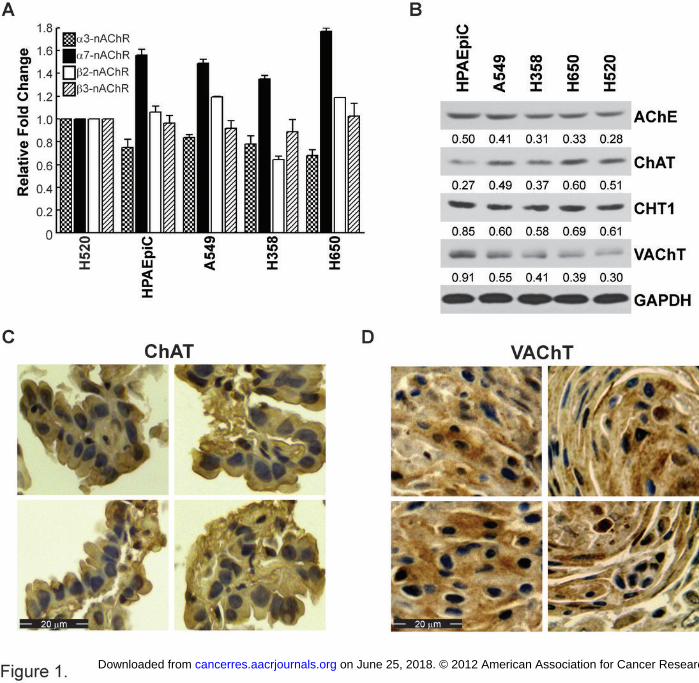
The cholinergic pathway proteins have been traditionally found in neuromuscular
junctions and in neuronal cells (14). However, studies have shown that genes for these proteins
are found in SCC-L and SCLC cells (14). ELISA experiments were performed to examine
whether cholinergic proteins were expressed in human BAC cell lines and in HPAEpiCs. Figure
1A shows that multiple nAChR subunits are expressed on A549, H358 and H650 human BAC
cells. Similarly, HPAEpiCs also expressed a diverse array of nAChR subunits. Immunoblotting
experiments were performed to examine the presence of AChE, ChAT, CHT1 and VAChT
(Figure 1B) in human BAC cell lines and in HPAEpiCs. H520 human SCC-L cells were used as
the positive control for both of the experiments. We observed that human BAC cell lines, as well
HPAEpiCs normal lung cells, express VAChT, ChAT, AChE and CHT1. The antibodies to
VAChT, ChAT, CHT1 and AChE were found to be specific and showed only a single band at the
correct molecular weight in full screen western blots (Supplementary Figure S2A-D). The
expression of ChAT and VAChT was examined in human BAC tumors isolated from patients
using BAC tissue microarray (TMA). The specific VAChT and ChAT antibodies described above
were used for the immunostaining. Each TMA contained eighty-one samples of human BAC
tumors from patients. Human BAC tumors displayed robust expression of both ChAT (Figure
1C; left panel) and VAChT (Figure 1D; right panel). Both VAChT and ChAT were found to be
expressed in the cytoplasm. This observation is in agreement with previous data from several
research groups showing that VAChT and ChAT are localized in the cytoplasm of cells (25-28).
We also analyzed the expression of VAChT and ChAT in normal lung tissues using normal lung
TMA (Supplementary Figure S3A-B). This TMA contained 20 samples of normal lung tissue.
We found that normal lung tissues also express VAChT and ChAT, and these proteins are
localized in the cytoplasm.
Nicotine induces acetylcholine production in bronchioalveolar carcinomas.
on June 25, 2018. © 2012 American Association for Cancer Research. cancerres.aacrjournals.org Downloaded from
Author manuscripts have been peer reviewed and accepted for publication but have not yet been edited. Author Manuscript Published OnlineFirst on December 7, 2012; DOI: 10.1158/0008-5472.CAN-12-3190
12
Next we wanted to assess the effect of nicotine on acetylcholine production in human
BACs. We observed that nicotine increased ACh production from A549 cells in a concentration-
dependent manner (Figure 2A). The maximum ACh production (2 μM) was observed at 100 nM
nicotine and remained constant thereafter. A similar pattern was observed in H358 and H650
human BAC cells (Figure 2A). We also performed a time-kinetics experiment with nicotine on
A549 cells and found that the maximal ACh production (2 μM) occurred at 36 hours and
remained relatively constant thereafter (Figure 2B). Therefore, we selected a concentration of
100 nM nicotine and a 36 hour time point for all of our ACh production experiments. A point to
note here is that 100 nM nicotine is within the concentration range found in the plasma of an
average smoker (1 nM - 1 μM) (29).
Studies by Song et al., (2003) have demonstrated that ACh acts as an autocrine growth
factor for SCLC cells (16, 17). We analyzed the mitogenic activity of ACh in human A549 BAC
cells and found that ACh stimulated the proliferation of A549 cells in a concentration-dependent
manner; the maximal proliferation observed at 2 μM ACh (Figure 2C: white bars). This finding is
significant because the maximal mitogenic activity of ACh is similar to the amount of ACh
secreted by human BACs. We repeated the BrdU cell proliferation assays in H358 human BAC
cells and obtained similar results (Figure 2C: black bars). Our observations raise the possibility
that nicotine induces the production of ACh in human BACs, which in turn, promotes the growth
of human BACs in an autocrine manner.
Next, we wanted to determine the role of nAChRs in nicotine-induced ACh production.
The treatment of A549 cells with the generalized nAChR antagonist mecamylamine (MCA)
suppressed nicotinic-induced ACh production, whereas atropine, an antagonist to the closely
related muscarinic receptor, had little to no effect on nicotine-induced ACh levels. We obtained
similar results in another human BAC cell line H358. Our results show that nicotine promotes
ACh levels in human BACs in an nAChR dependent manner (Supplementary Figure S4A).
on June 25, 2018. © 2012 American Association for Cancer Research. cancerres.aacrjournals.org Downloaded from
Author manuscripts have been peer reviewed and accepted for publication but have not yet been edited. Author Manuscript Published OnlineFirst on December 7, 2012; DOI: 10.1158/0008-5472.CAN-12-3190
13
The next series of experiments aimed to investigate which specific nAChR subunits were
responsible for nicotine-induced ACh production. Our data showed that the treatment of A549
and H358 human BAC cells with α7-nAChR subunit antagonists, methyllycaconitine (MLA) and
α-bungarotoxin (α-BT), ablated nicotine-induced ACh production. Additionally, 1 μM of α-
conotoxin MII (α-CT; α3β2 and β3 subunit antagonist) reversed the pro-secretory effect of
nicotine, whereas DHβE (α3β2 and α4β2 nAChR antagonist) had little to no effect
(Supplementary Figure S4B). Taken together, these results suggest that nicotine-induced ACh
secretion is mediated via nAChRs, specifically through the α7-, α3β2- and β3-containing-nAChR
subunits.
Finally, we wanted to examine the effect of choline/acetylcholine transporters on
nicotine-induced ACh production. The treatment of A549 and H358 cells with hemicholinium (an
antagonist of CHT1) suppressed nicotine-induced ACh secretion. Similarly, vesamicol (an
antagonist of VAChT) potently abrogated nicotine-induced ACh production (Supplementary
Figure S4C). Taken together, this shows that CHT1 and VAChT function are vital for nicotine-
induced ACh production.
Nicotine increases VAChT and ChAT levels in human BAC cells.
We wanted to investigate whether nicotine promoted ACh secretion via the
VAChT/ChAT pathway in BAC cells. We used an ELISA kit to measure VAChT and ChAT
levels, which allowed us to quantitate these levels in an accurate and sensitive manner. We
observed that the VAChT ELISA kit detected VAChT levels in asynchronous A549, H358, H650
BAC cells and HPAEpic normal lung cells (Supplementary Figure S5). The relative pattern of
VAChT expression in the ELISA correlated well with the results obtained from western blotting
(Figure 1B; fourth panel). ELISA The treatment of A549 cells with 100 nM nicotine caused a 4-
fold increase in VAChT levels (Figure 3A) and a 1.5-fold increases in ChAT levels (Figure 3B).
on June 25, 2018. © 2012 American Association for Cancer Research. cancerres.aacrjournals.org Downloaded from
Author manuscripts have been peer reviewed and accepted for publication but have not yet been edited. Author Manuscript Published OnlineFirst on December 7, 2012; DOI: 10.1158/0008-5472.CAN-12-3190

14
Similarly, nicotine increased VAChT levels in H358 by 5.5-fold and ChAT levels by 1.4-fold
(Figure 3A and 3B). We also tested the effect of nicotine on AChE and CHT1 in human BACs.
We found that nicotine decreased AChE levels in H358 and A549 human BACs (Figure 3C).
The levels of CHT1 were relatively unaffected by nicotine in both the cell lines (Figure 3C).
The VAChT antagonist vesamicol causes apoptosis in human BACs.
Our results showed that nicotine-induced ACh production was blocked by the VAChT
inhibitor vesamicol (Supplementary Figure S4C). It may be envisaged that vesamicol will
suppress nicotine-induced ACh production and thereby block ACh-induced growth of human
BACs. Additionally, we conjectured that nicotine-induced upregulation of VAChT should provide
a viable molecular target for vesamicol therapy in human BACs. MTT assays (Supplementary
Methods online) demonstrated that vesamicol decreased the viability of nicotine-treated A549
and H358 human BAC lines in a concentration-dependent manner. The maximum reduction in
cell viability was observed at 50 μM vesamicol (Figure 4A). Therefore, we used 50 μM
vesamicol for all further experiments.
Next, we wanted to examine whether vesamicol induced apoptosis in human BAC cell
lines. Quiescent A549 and H358 cells were treated with 100 nM nicotine in the presence or
absence of 50 μM vesamicol. Apoptosis was measured by TUNEL assays. Figure 4B shows
that vesamicol caused robust apoptosis in both A549 and H358 human BACs. The results of the
TUNEL assays were verified by caspase-3 activity assay. We observed that vesamicol caused
2.5- to 3-fold increase in apoptosis relative to nicotine-treated human BAC cells (Figure 4C).
We also wanted to assess the effect of vesamicol on other mitogenic signaling pathways
in human BACs (30-35). Several convergent studies indicate that EGF and IGF-II are potent
growth factors for human BAC cells (30-35). We performed a BrdU assay (Supplementary
Methods online) to test the effect of vesamicol on EGF-induced proliferation of human BAC cell
on June 25, 2018. © 2012 American Association for Cancer Research. cancerres.aacrjournals.org Downloaded from
Author manuscripts have been peer reviewed and accepted for publication but have not yet been edited. Author Manuscript Published OnlineFirst on December 7, 2012; DOI: 10.1158/0008-5472.CAN-12-3190

15
lines. We found that vesamicol has no effect on EGF-induced proliferation of A549 and H358
human BAC cell lines (Figure 4D). Similarly, vesamicol did not affect IGF-II-induced
proliferation of human A549 and H358 cells (Figure 4E). Our findings seem to suggest that
vesamicol specifically inhibits the acetylcholine-proliferative pathway in human BACs.
Vesamicol induces apoptosis in human BAC cells by specifically targeting VAChT
Vesamicol is a well characterized antagonist of VAChT (36). However, previous studies
indicate that vesamicol also binds to the sigma-receptor in several types of human cancer cells
(37, 38). We wanted to examine whether vesamicol induced apoptosis in human BAC cells by
specifically targeting VAChT. For this purpose, we used siRNA methodology to suppress the
expression of VAChT or sigma receptor in A549 cells. A549 cells were transfected with VAChT-
siRNA or sigma-receptor-siRNA (Supplementary Methods Online). Eighteen hours post
transfection, the cells were rendered quiescent by incubating them in SF-RPMI-R medium for 24
hours (19). Subsequently, the cells were treated with 100nM nicotine in the presence or
absence of 50 μM vesamicol for 48 hours at 37°C. A549 cells transfected with a non-targeting
control-siRNA was used as the negative control for the experiment. After 48 hours cell lysates
were made and vesamicol-induced apoptosis was measured by the caspase-3 activity assay.
We observed that vesamicol-induced apoptosis was decreased upon transfection of
VAChT-siRNA and unaffected by sigma-receptor-siRNA. The apoptotic activity of vesamicol
was also unaffected by the control non-targeting siRNA (Figure 5A). We repeated these
experiments in H358 human BAC cells and obtained similar results (Figure 5B). These
experiments were also repeated with a second independent VAChT-siRNA (Ambion Inc., Grand
Island, NY) and similar results were obtained (Figure 5C and D).
Parallel transfection experiments were performed to test the efficacy of VAChT-siRNA
and sigma-receptor-siRNA in A549 cells. ELISA assays show that the levels of VAChT are
robustly suppressed upon transfection with both sets of VAChT-siRNA (Supplementary Figure
on June 25, 2018. © 2012 American Association for Cancer Research. cancerres.aacrjournals.org Downloaded from
Author manuscripts have been peer reviewed and accepted for publication but have not yet been edited. Author Manuscript Published OnlineFirst on December 7, 2012; DOI: 10.1158/0008-5472.CAN-12-3190

16
S6A and S6B) in A549 and H358 human BAC cells. Western blotting experiments show that the
levels of sigma receptor are ablated upon transfection of sigma-receptor-siRNA (Supplementary
Figure S6C and S6D) in both A549 and H358 cells.
We also examined whether vesamicol was targeting the VAChT pathway at
concentrations lower than 50µM. We chose 10µM and 25µM vesamicol for our experiments.
Caspase-3 activity assays show that the cellular apoptosis induced by 10µM vesamicol was
suppressed by two independent sets of VAChT-siRNA in both A549 and H358 cells
(Supplementary Figure S7A and S7B). Similarly, the transfection of VAChT-siRNA efficiently
abrogated the apoptotic activity of 25µM vesamicol in nicotine-treated A549 and H358 cells
(Supplementary Figure S7C and S7D). ELISA assays indicate that both the VAChT-siRNA
decreased the expression of VAChT in A549 and H358 cells (Supplementary Figure S7E).
Taken together, our data show that vesamicol caused apoptosis in human BAC cells by
specifically targeting the VAChT pathway.
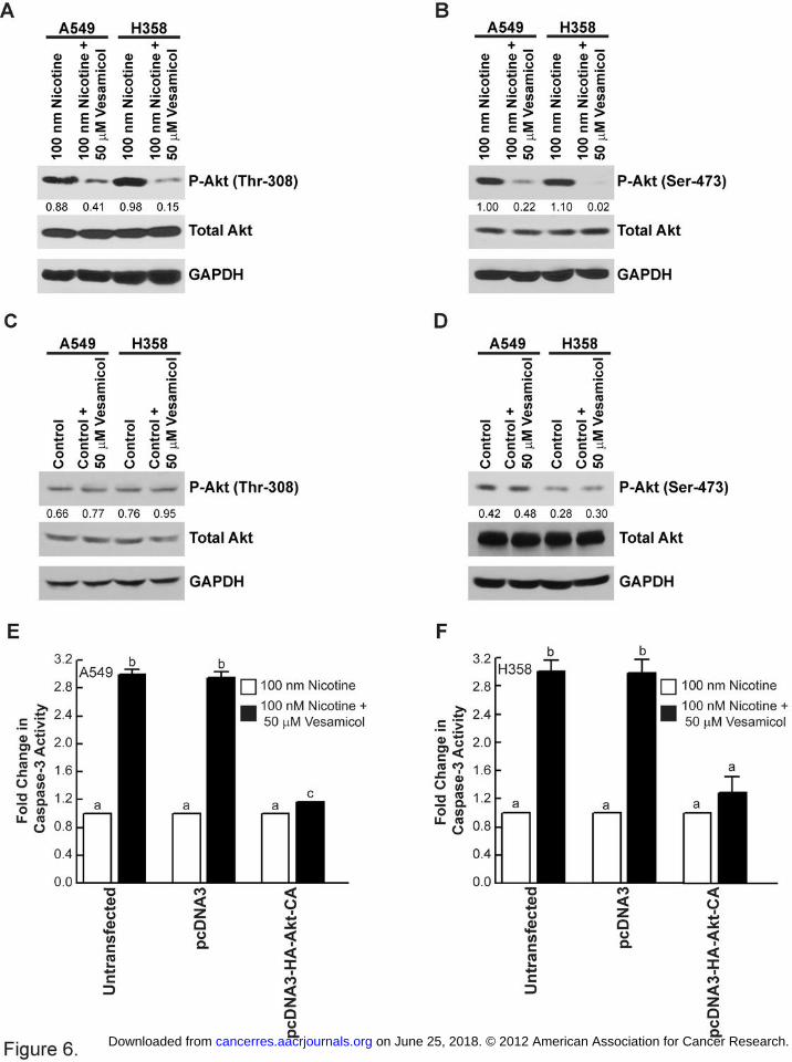
Vesamicol-induced apoptosis is mediated by the Akt pathway in human BACs.
The Akt signaling pathway plays a vital role in nAChR signaling in normal lung cells and
lung cancer cells (12). We observed that the treatment of A549 and H358 human BAC cells with
nicotine and vesamicol caused potent decreases in phosphorylated-Akt levels (Figure 6A and
B). The levels of both phosphorylated-Akt (Thr308) and phosphorylated-Akt (Ser473) are
robustly suppressed upon vesamicol treatment, whereas total Akt levels remain constant
(Figure 6A and 6B, respectively). Western blotting analysis also showed that vesamicol had little
to no effect on the expression of phosphorylated-Akt in untreated A549 (or H358) cells (Figure
6C and 6D). The transfection of constitutively active Akt (pcDNA3-HA-Akt-CA; Supplementary
Methods Online) reversed the apoptotic effect of vesamicol, demonstrating that vesamicol
induces cell death by suppressing Akt activation (Figure 6E and 6F). Western blotting analysis
on June 25, 2018. © 2012 American Association for Cancer Research. cancerres.aacrjournals.org Downloaded from
Author manuscripts have been peer reviewed and accepted for publication but have not yet been edited. Author Manuscript Published OnlineFirst on December 7, 2012; DOI: 10.1158/0008-5472.CAN-12-3190

17
confirms the over expression of HA-tagged Akt in A549 and H358 human BAC lines upon
transfection (Supplementary Fig. S8A and S8B).
Vesamicol inhibited the growth of human A549 cells in vivo.
The anti-tumor activity of vesamicol was examined in vivo using a nude mice model (15,
21). A549 human BAC cells were injected between the scapulae of nude mice. The tumors were
allowed to grow until approximately 100 mm3, after which they were randomized into two
groups. The control group was administered nicotine in the drinking water. The “vesamicol
group” was administered 50 mg vesamicol/kg food along with nicotine. Our results showed that
the administration of vesamicol decreased the tumor growth rate of A549 human BAC tumors
(Figure 7A). The administration of 50 mg vesamicol/kg food in the diet was well tolerated and
caused no discomfort or weight loss in mice (mean control = 25.6 ± 0.6 g; mean vesamicol
treated = 25.2 ± 0.5 g). Additionally, food intake (mean control = 6.2 ± 0.09 g/day; mean
vesamicol-treated = 6.4 ± 0.09 g/day) and water consumption (mean control = 11.0 ± 0.7
mL/day; mean vesamicol-treated = 11.2 ± 0.8 mL/day) was similar between groups.
H and E staining of the tumors revealed the presence of apoptotic bodies in the tumors
belonging to the vesamicol-treated mice (Figure 7B). Caspase-3 activity assays indicate that
tumor lysates from vesamicol-treated mice displayed about 2.5-fold greater apoptosis relative to
control nicotine-treated mice (Figure 7C). Our data from cell culture suggested that vesamicol
induced apoptosis by suppression of Akt activation. We wanted to investigate whether
vesamicol-treated tumors had lower levels of phosphorylated-Akt. Western blotting analysis
revealed that vesamicol-treated tumors had substantially lower levels of phosphorylated-Akt
(Thr308 and Ser 473) relative to nicotine-treated A549 tumors (Figure 7D; top two panels). The
total Akt levels were similar in all four pairs of tumors (Figure 7D; third panel from the top).
Taken together, these data indicate that vesamicol decreases tumor growth rates of A549
human BAC tumors in vivo by inducing robust apoptosis via an Akt-dependent pathway.
on June 25, 2018. © 2012 American Association for Cancer Research. cancerres.aacrjournals.org Downloaded from
Author manuscripts have been peer reviewed and accepted for publication but have not yet been edited. Author Manuscript Published OnlineFirst on December 7, 2012; DOI: 10.1158/0008-5472.CAN-12-3190

18
Discussion
Our study shows for the first time that human BACs produce ACh and contain a
functional acetylcholine-signaling system. A functional cholinergic loop has also been detected
in SCLCs, SCC-Ls and normal bronchial epithelial cells (14). Most importantly, nicotine
regulates the cholinergic machinery and increases ACh levels by about 10-fold in human BACs.
Nicotine upregulates the levels of cholinergic proteins, namely VAChT and ChAT, and
concomitantly downregulates AChE in human BACs. Nicotine-induced increases of VAChT and
ChAT promote ACh content and its transport into the extracellular environment. On the other
hand, nicotine decreases AChE levels, which in turn suppress ACh degradation. Thus, the
enhanced levels of ACh provide proliferative stimuli to human BACs. The amplification of the
cholinergic network by nicotine offers novel therapeutic strategies for BAC therapy. These
phenomena are highly significant since about 30% of lung cancer patients continue to smoke,
use nicotine-based cessation patches or gum, or are exposed to environmental tobacco smoke
after their diagnosis (39-41).
We observed that nicotine caused a 3-fold increase in VAChT and ChAT levels in
human BAC cells. This led us to hypothesize that antagonists of VAChT should attenuate
nicotine-induced ACh production and thereby suppress the growth of human BACs. As a “proof-
of-principle” we decided to use the well-characterized VAChT antagonist, vesamicol, for our
experiments. Our hypothesis was supported by the results of Song et al., (2003) who showed
that vesamicol suppressed the growth of H82 human SCLC cells in vitro. We believe that our
results are the first to report characterizing the anti-cancer activity of VAChT antagonists in
human BAC using both cell culture and in vivo model systems. We found that vesamicol
induced robust apoptosis in human BAC cells in both cell culture and in vivo systems.
A survey of literature shows that only very few studies have evaluated the anti-tumor
activity of vesamicol. Ogawa et al., (2009) evaluated radioiodinated vesamicol analogs for tumor
on June 25, 2018. © 2012 American Association for Cancer Research. cancerres.aacrjournals.org Downloaded from
Author manuscripts have been peer reviewed and accepted for publication but have not yet been edited. Author Manuscript Published OnlineFirst on December 7, 2012; DOI: 10.1158/0008-5472.CAN-12-3190

19
imaging and anti-tumor activity (42, 43). They found that radioiodinated-vesamicol-analogs
suppressed the growth of human prostate cancer cells in mice model. However, their results
showed that vesamicol exerted its anti-tumor activity by binding to the sigma receptor on DU145
prostate cancer cells (42). Several convergent studies have shown that vesamicol is also a
ligand for the sigma-receptor. However, our experiments involving VAChT-siRNA and sigma-
receptor-siRNA showed that the anti-cancer activity of vesamicol was specifically mediated by
VAChT. It is possible that sigma-receptors do not play a vital role in nicotine-induced
proliferative signaling in human BAC cells.
The present manuscript shows that apoptotic effects of vesamicol are mediated via
suppression of the Akt pathway. Clinical studies have shown that the activation of Akt is highly
prevalent in human BAC tumors. Nicotine causes rapid activation of Akt and its downstream
substrates. Studies by West et al., (2001) have speculated that inhibition of the Akt pathway
may be a viable strategy for treatment of tobacco-related lung cancers (44, 45). The EGFR
inhibitors Gefitinib and Erlotinib have been shown to suppress Akt phosphorylation in human
lung cancer cells (46, 47). Similarly, PI-3kinase/Akt inhibitors, i.e. LY294002 and deregulin,
suppress proliferation of human BAC cells in vitro and in mouse models of tobacco
carcinogenesis (48). Our results suggest that VAChT-antagonists such as vesamicol are
another class of therapeutic agents capable of inhibiting activated Akt in human BACs.
The acetylcholine-signaling pathway has been found to regulate multiple cellular
functions, such as proliferation, cell-to-cell contact, differentiation and cytoskeletal integrity (14).
Apart from lung cancer cells, endothelial cells, mesothelial cells, immune cells and keratinocytes
have been found to synthesize, transport and degrade acetylcholine (14). The pharmacological
manipulation of this non-neuronal cholinergic system by agents, such as vesamicol, could lead
to the development of novel therapies for multiple tobacco-related diseases including lung
cancer.
on June 25, 2018. © 2012 American Association for Cancer Research. cancerres.aacrjournals.org Downloaded from
Author manuscripts have been peer reviewed and accepted for publication but have not yet been edited. Author Manuscript Published OnlineFirst on December 7, 2012; DOI: 10.1158/0008-5472.CAN-12-3190

20
Acknowledgements
We thank Dr. Srikumar Chellappan and his laboratory for continuous support. We are grateful to
Adam W. Buckley and Jarrod C. Harman for technical assistance. We also acknowledge Dr.
Woodgett providing us the constructs used in this study. This work was supported by the grants
Young Clinical Scientist Award (#82115) from the Flight Attendant Medical Association, Miami,
FL and 1R15CA161491-01A1 from NIH to PDG. YCC is funded by NIH Grant 5P20RR016477
and 8P20GM103434 (PI: Gary O. Rankin). AMD and KCB are recipients of graduate fellowships
from the WVSGC. CMC is the recipient of a GIAR undergraduate research grant from the
Sigma-Xi Society. We thank Luke Damron for editorial support and suggestions.
on June 25, 2018. © 2012 American Association for Cancer Research. cancerres.aacrjournals.org Downloaded from
Author manuscripts have been peer reviewed and accepted for publication but have not yet been edited. Author Manuscript Published OnlineFirst on December 7, 2012; DOI: 10.1158/0008-5472.CAN-12-3190

21
References
1. Levy BP, Drilon A, Makarian L, Patel AA, Grossbard ML. Systemic approaches for
multifocal bronchioloalveolar carcinoma: is there an appropriate target? Oncology
(Williston Park). 2010;24:888-98, 900.
2. Saintigny P, Wistuba, II, Kim ES. Bronchioloalveolar carcinoma: a translational
perspective. Oncology (Williston Park). 2010;24:907-8, 14.
3. Colby TV, Noguchi M, Henschke C, Vazquez MF, Geisinger K, Yokose T.et al.
Adenocarcinoma. In: Travis WD, Brambilla E, Muller-Hermelink HK, Harris CC et al.,
editors. World health classification of tumours pathology and genetics of tumours of the
lung, pleura, thymus and heart. Lyon, France: IARC Press; 2004. p. 35-44.
4. Ebbert JO, Chhatwani L, Aubry MC, Wampfler J, Stoddard S, Zhang F, et al. Clinical
features of bronchioloalveolar carcinoma with new histologic and staging definitions. J
Thorac Oncol. 2010;5:1213-20.
5. The Health Consequences of Smoking. A report of the Surgeon General's Office on
Smoking and Health, DHHS, Washington DC. 2004.
6. Cancer IARC. IARC Monographs on the Evaluation of Carcinogenic Risks to Humans.
Tobacco Smoke and Involuntary Smoking: IARC, Lyon, France; 2004. p. 51-1187.
7. Garfield D. Mucinous and nonmucinous bronchioloalveolar carcinoma and smoking. Am
J Clin Pathol. 2010;133:341-2.
8. Boffetta P, Jayaprakash V, Yang P, Asomaning K, Muscat JE, Schwartz AG, et al.
Tobacco smoking as a risk factor of bronchioloalveolar carcinoma of the lung: pooled
analysis of seven case-control studies in the International Lung Cancer Consortium
(ILCCO). Cancer Causes Control. 2011;22:73-9.
9. Rolen KA, Fulton JPT, D. J., Strauss GM. Bronchoalveolar carcinoma (BAC) of the lung
is related to cigarette smoking: A case-control study from Rhode Island (RI). 2003
ASCO Annual Meeting; 2003.
on June 25, 2018. © 2012 American Association for Cancer Research. cancerres.aacrjournals.org Downloaded from
Author manuscripts have been peer reviewed and accepted for publication but have not yet been edited. Author Manuscript Published OnlineFirst on December 7, 2012; DOI: 10.1158/0008-5472.CAN-12-3190

22
10. Morabia A, Wynder EL. Relation of bronchioloalveolar carcinoma to tobacco. BMJ.
1992;304:541-3.
11. Sartori G, Cavazza A, Sgambato A, Marchioni A, Barbieri F, Longo L, et al. EGFR and
K-ras mutations along the spectrum of pulmonary epithelial tumors of the lung and
elaboration of a combined clinicopathologic and molecular scoring system to predict
clinical responsiveness to EGFR inhibitors. Am J Clin Pathol. 2009;131:478-89.
12. Singh S, Pillai S, Chellappan S. Nicotinic acetylcholine receptor signaling in tumor
growth and metastasis. J Oncol. 2011;2011:456743.
13. Dasgupta P, Kinkade R, Joshi B, Decook C, Haura E, Chellappan S. Nicotine inhibits
apoptosis induced by chemotherapeutic drugs by up-regulating XIAP and survivin. Proc
Natl Acad Sci U S A. 2006;103:6332-7.
14. Song P, Spindel ER. Basic and clinical aspects of non-neuronal acetylcholine:
expression of non-neuronal acetylcholine in lung cancer provides a new target for cancer
therapy. J Pharmacol Sci. 2008;106:180-5.
15. Song P, Sekhon HS, Fu XW, Maier M, Jia Y, Duan J, et al. Activated cholinergic
signaling provides a target in squamous cell lung carcinoma. Cancer Res.
2008;68:4693-700.
16. Song P, Sekhon HS, Jia Y, Keller JA, Blusztajn JK, Mark GP, et al. Acetylcholine is
synthesized by and acts as an autocrine growth factor for small cell lung carcinoma.
Cancer Res. 2003;63:214-21.
17. Song P, Sekhon HS, Proskocil B, Blusztajn JK, Mark GP, Spindel ER. Synthesis of
acetylcholine by lung cancer. Life Sci. 2003;72:2159-68.
18. Song P, Sekhon HS, Lu A, Arredondo J, Sauer D, Gravett C, et al. M3 muscarinic
receptor antagonists inhibit small cell lung carcinoma growth and mitogen-activated
protein kinase phosphorylation induced by acetylcholine secretion. Cancer Res.
2007;67:3936-44.
on June 25, 2018. © 2012 American Association for Cancer Research. cancerres.aacrjournals.org Downloaded from
Author manuscripts have been peer reviewed and accepted for publication but have not yet been edited. Author Manuscript Published OnlineFirst on December 7, 2012; DOI: 10.1158/0008-5472.CAN-12-3190
23
19. Dasgupta P, Rizwani W, Pillai S, Davis R, Banerjee S, Hug K, et al. ARRB1-mediated
regulation of E2F target genes in nicotine-induced growth of lung tumors. J Natl Cancer
Inst. 2011;103:317-33.
20. Brown KC, Lau JK, Dom AM, Witte TR, Luo H, Crabtree CM, et al. MG624, an alpha7-
nAChR antagonist, inhibits angiogenesis via the Egr-1/FGF2 pathway. Angiogenesis.
2012;15:99-114.
21. Brown KC, Witte TR, Hardman WE, Luo H, Chen YC, Carpenter AB, et al. Capsaicin
displays anti-proliferative activity against human small cell lung cancer in cell culture and
nude mice models via the E2F pathway. PLoS One. 2010;5:e10243.
22. Tomayko MM, Reynolds CP. Determination of subcutaneous tumor size in athymic
(nude) mice. Cancer Chemother Pharmacol. 1989;24:148-54.
23. Shabbir M, Thompson CS, Mikhailidis D, Morgan RJ, Burnstock G. Extracellular ATP
attenuates the growth of hormone refractory prostate cancer in vivo. Eur Urology
Supplements. 2003;2:24-7.
24. Kinkade R, Dasgupta P, Carie A, Pernazza D, Carless M, Pillai S, et al. A small
molecule disruptor of Rb/Raf-1 interaction inhibits cell proliferation, angiogenesis, and
growth of human tumor xenografts in nude mice. Cancer Res. 2008;68:3810-8.
25. Schafer MK, Weihe E, Erickson JD, Eiden LE. Human and monkey cholinergic neurons
visualized in paraffin-embedded tissues by immunoreactivity for VAChT, the vesicular
acetylcholine transporter. J Mol Neurosci. 1995;6:225-35.
26. Tayebati SK, Tomassoni D, Di Stefano A, Sozio P, Cerasa LS, Amenta F. Effect of
choline-containing phospholipids on brain cholinergic transporters in the rat. Journal of
the neurological sciences. 2011;302:49-57.
27. Papka RE, Traurig HH, Schemann M, Collins J, Copelin T, Wilson K. Cholinergic
neurons of the pelvic autonomic ganglia and uterus of the female rat: distribution of
axons and presence of muscarinic receptors. Cell Tissue Res. 1999;296:293-305.
on June 25, 2018. © 2012 American Association for Cancer Research. cancerres.aacrjournals.org Downloaded from
Author manuscripts have been peer reviewed and accepted for publication but have not yet been edited. Author Manuscript Published OnlineFirst on December 7, 2012; DOI: 10.1158/0008-5472.CAN-12-3190

24
28. Takahara Y, Maeda M, Nakatani T, Kiyama H. Transient suppression of the vesicular
acetylcholine transporter in urinary bladder pathways following spinal cord injury. Brain
Res. 2007;1137:20-8.
29. Heeschen C, Jang JJ, Weis M, Pathak A, Kaji S, Hu RS, et al. Nicotine stimulates
angiogenesis and promotes tumor growth and atherosclerosis. Nature Medicine.
2001;7:833-9.
30. Wilson KJ, Gilmore JL, Foley J, Lemmon MA, Riese DJ, 2nd. Functional selectivity of
EGF family peptide growth factors: implications for cancer. Pharmacology &
therapeutics. 2009;122:1-8.
31. Siegfried JM. Detection of human lung epithelial cell growth factors produced by a lung
carcinoma cell line: use in culture of primary solid lung tumors. Cancer Res.
1987;47:2903-10.
32. Domingo G, Perez CA, Velez M, Cudris J, Raez LE, Santos ES. EGF receptor in lung
cancer: a successful story of targeted therapy. Expert Rev Anticancer Ther.
2010;10:1577-87.
33. Linnerth NM, Baldwin M, Campbell C, Brown M, McGowan H, Moorehead RA. IGF-II
induces CREB phosphorylation and cell survival in human lung cancer cells. Oncogene.
2005;24:7310-9.
34. Camidge DR, Dziadziuszko R, Hirsch FR. The rationale and development of therapeutic
insulin-like growth factor axis inhibition for lung and other cancers. Clin Lung Cancer.
2009;10:262-72.
35. Dziadziuszko R, Camidge DR, Hirsch FR. The insulin-like growth factor pathway in lung
cancer. J Thorac Oncol. 2008;3:815-8.
36. Bahr BA, Parsons SM. Acetylcholine transport and drug inhibition kinetics in Torpedo
synaptic vesicles. J Neurochem. 1986;46:1214-8.
on June 25, 2018. © 2012 American Association for Cancer Research. cancerres.aacrjournals.org Downloaded from
Author manuscripts have been peer reviewed and accepted for publication but have not yet been edited. Author Manuscript Published OnlineFirst on December 7, 2012; DOI: 10.1158/0008-5472.CAN-12-3190

25
37. Wilke RA, Mehta RP, Lupardus PJ, Chen Y, Ruoho AE, Jackson MB. Sigma receptor
photolabeling and sigma receptor-mediated modulation of potassium channels in tumor
cells. J Biol Chem. 1999;274:18387-92.
38. Efange SM, Mach RH, Smith CR, Khare AB, Foulon C, Akella SK, et al. Vesamicol
analogues as sigma ligands. Molecular determinants of selectivity at the vesamicol
receptor. Biochem Pharmacol. 1995;49:791-7.
39. Johnson BE. Tobacco and lung cancer. Prim Care. 1998;25:279-91.
40. Johnson BE, Cortazar P, Chute JP. Second lung cancers in patients successfully treated
for lung cancer. Semin Oncol. 1997;24:492-9.
41. Johnston-Early A, Cohen MH, Minna JD, Paxton LM, Fossieck BE, Jr., Ihde DC, et al.
Smoking abstinence and small cell lung cancer survival. An association. Jama.
1980;244:2175-9.
42. Ogawa K, Shiba K, Akhter N, Yoshimoto M, Washiyama K, Kinuya S, et al. Evaluation of
radioiodinated vesamicol analogs for sigma receptor imaging in tumor and radionuclide
receptor therapy. Cancer Sci. 2009;100:2188-92.
43. Ogawa K, Kanbara H, Shiba K, Kitamura Y, Kozaka T, Kiwada T, et al. Development
and evaluation of a novel radioiodinated vesamicol analog as a sigma receptor imaging
agent. EJNMMI research. 2012;2:54.
44. West KA, Brognard J, Clark AS, Linnoila IR, Yang X, Swain SM, et al. Rapid Akt
activation by nicotine and a tobacco carcinogen modulates the phenotype of normal
human airway epithelial cells. J Clin Invest. 2003;111:81-90.
45. West KA, Linnoila IR, Belinsky SA, Harris CC, Dennis PA. Tobacco carcinogen-induced
cellular transformation increases activation of the phosphatidylinositol 3'-kinase/Akt
pathway in vitro and in vivo. Cancer Res. 2004;64:446-51.
on June 25, 2018. © 2012 American Association for Cancer Research. cancerres.aacrjournals.org Downloaded from
Author manuscripts have been peer reviewed and accepted for publication but have not yet been edited. Author Manuscript Published OnlineFirst on December 7, 2012; DOI: 10.1158/0008-5472.CAN-12-3190
26
46. Kitazaki T, Soda H, Doi S, Nakano H, Nakamura Y, Kohno S. Gefitinib inhibits MUC5AC
synthesis in mucin-secreting non-small cell lung cancer cells. Lung Cancer. 2005;50:19-
24.
47. Ko JC, Ciou SC, Jhan JY, Cheng CM, Su YJ, Chuang SM, et al. Roles of MKK1/2-
ERK1/2 and phosphoinositide 3-kinase-AKT signaling pathways in erlotinib-induced
Rad51 suppression and cytotoxicity in human non-small cell lung cancer cells. Mol
Cancer Res. 2009;7:1378-89.
48. Denlinger CE, Rundall BK, Jones DR. Inhibition of phosphatidylinositol 3-kinase/Akt and
histone deacetylase activity induces apoptosis in non-small cell lung cancer in vitro and
in vivo. J Thorac Cardiovasc Surg. 2005;130:1422-9.
on June 25, 2018. © 2012 American Association for Cancer Research. cancerres.aacrjournals.org Downloaded from
Author manuscripts have been peer reviewed and accepted for publication but have not yet been edited. Author Manuscript Published OnlineFirst on December 7, 2012; DOI: 10.1158/0008-5472.CAN-12-3190
27
Figure Legends
Figure 1. Human BACs and normal human alveolar epithelial cells (HPAEpiCs) express
cholinergic proteins. (A) ELISA assays show that human BAC cell lines and HPAEpiCs express
multiple nAChR subunits. The assay was completed in duplicate, and the whole experiment was
performed two independent times. Results indicated by a different letter are significantly
different (P < 0.05). (B) HPAEpiCs and human BACs express AChE, ChAT, CHT1 and VAChT.
H520 human SCC-L cells were used as the positive control for the experiments outlined in (A)
and (B). GAPDH was used as the loading control, and the results were quantitated by
densitometric analysis. The experiment was repeated twice, and the representative data is
shown. (C) and (D) Immunohistochemistry of the human BAC microarray showed that BAC
tumors (isolated from patients) express ChAT and VAChT in the cytoplasm, adjacent to the
hemotoxylin-counterstained dark nuclei in the tissue samples. A tumor microarray containing 81
cores of human BACs was used for these experiments and four panels of representative photos
are shown. Scale bar = 20 µm.
Figure 2. Nicotine induces the production of the growth factor ACh in human BACs. (A) Nicotine
caused a concentration-dependent increase in ACh production in A549, H358 and H650 human
BAC cell lines. (B) Time kinetics of nicotine-induced ACh production in A549 human BAC cells.
The maximal ACh production was observed at 100 nM nicotine at 36 hours. (C) ACh stimulated
proliferation of A549 and H358 cells. BrdU assays show that 2 µM of ACh (which is
approximately the amount of ACh produced in nicotine-treated BAC cells) caused a 4- to 4.5-
fold increase in proliferation of A549 and H358 human BACs. Each sample was analyzed in
triplicate. Data represent mean + SEM from two independent experiments. Results indicated by
a different letter are significantly different (P < 0.05).
on June 25, 2018. © 2012 American Association for Cancer Research. cancerres.aacrjournals.org Downloaded from
Author manuscripts have been peer reviewed and accepted for publication but have not yet been edited. Author Manuscript Published OnlineFirst on December 7, 2012; DOI: 10.1158/0008-5472.CAN-12-3190

28
Figure 3. Effect of nicotine on cholinergic proteins in human BACs. (A) The treatment of A549
and H358 cells with 100 nM nicotine caused 4- to 5-fold increase in VAChT levels. (B) Nicotine
produces a modest increase in ChAT levels. (C) Nicotine decreases AChE levels in both A549
and H358 cells. The levels of CHT1 are relatively unaffected by nicotine in human BAC cells.
Data represent mean + SEM from two independent experiments. Each sample was analyzed in
triplicate. Results indicated by a different letter are significantly different (P < 0.05).
Figure 4. Vesamicol caused apoptosis in human BAC cells. (A) MTT assays showed that
vesamicol decreases the viability of A549 and H358 cells in a concentration-dependent manner.
Each sample was analyzed in triplicate. (B) The treatment of human BAC cell lines with 50 µM
vesamicol caused 2.5- to 3-fold apoptosis in nicotine-treated A549 and H358 cells. (C) The
apoptotic activity of vesamicol was confirmed with caspase-3 activity assay and similar results
were obtained. (D) Vesamicol had no effect on EGF-induced proliferation of A549 and H358
cells. BrdU assays show that the treatment of A549 and H358 human BAC cells with 10 ng/ml
EGF causes robust proliferation. The addition of 50 µM vesamicol along with EGF produced no
effect on EGF-induced proliferation of human BACs. (E) BrdU assays demonstrate that
vesamicol does not suppress IGF-II-induced proliferation of human BAC cell lines. Each
sample was analyzed in duplicate. Data represent mean + SEM from two independent
experiments. Results indicated by a different letter are significantly different (P < 0.05).
Figure 5. Vesamicol-induced apoptosis is specifically mediated by VAChT in human BAC cells.
(A) Caspase-3 activity assays showed that the transfection of VAChT-siRNA reversed
vesamicol-induced apoptosis in nicotine-treated A549 cells, whereas transfection of sigma-
receptor-siRNA (SigmaR-siRNA) or control-siRNA has no effect. (B) The experiment was
repeated in H358 human BAC cells, and similar results were obtained. (C) The suppression of
VAChT, with another independent VAChT-siRNA (VAChT-siRNA-2), abrogated vesamicol-
on June 25, 2018. © 2012 American Association for Cancer Research. cancerres.aacrjournals.org Downloaded from
Author manuscripts have been peer reviewed and accepted for publication but have not yet been edited. Author Manuscript Published OnlineFirst on December 7, 2012; DOI: 10.1158/0008-5472.CAN-12-3190
29
induced apoptosis in A549 human BAC cells. (D) The experiment was repeated in H358 cells,
and similar results were obtained. Each sample was analyzed in duplicate. Data represent
mean + SEM from two independent experiments. Results indicated by a different letter are
significantly different (P < 0.05).
Figure 6. The apoptotic activity of vesamicol is mediated by the Akt pathway. (A) The treatment
of A549 and H358 human BAC cells with nicotine and vesamicol inhibited Akt phosphorylation
at Thr-308. (B) Vesamicol decreased the levels of phospho-Akt (Ser-473) in nicotine-treated
A549 and H358 cells. (C) Vesamicol had no effect on the levels of phospho-Akt (Thr-308) in
untreated A549 and H358 cells. (D) The treatment of quiescent A549 and H358 cells 50 µM
vesamicol does not affect phospho-Akt (Ser-473) levels. (E) The transfection of pcDNA3-HA-
Akt-CA (T308D, S473D) reverses the apoptotic activity of vesamicol, whereas transfection of
the empty vector had no effect on vesamicol-induced apoptosis in A549 human BAC cells. (F)
The transfection experiment was repeated in H358 cells and similar results were obtained. Data
represent mean + SEM from two independent experiments. Each sample was analyzed in
duplicate. Results indicated by a different letter are significantly different (P < 0.05).
Figure 7. Vesamicol induces apoptosis of human BAC tumors in vivo. (A) The administration of
vesamicol (50 mg vesamicol/kg food) decreased the growth rate of A549 human BAC tumors
xenografted in nude mice. The nude mice experiments comprised of eight mice per group. (B)
The tumors from the mice were excised and stained with H & E. The black arrow shows the
presence of apoptotic bodies in the tissue sections. The vesamicol-treated tumors (right panel)
showed a greater number of apoptotic bodies relative to the mice administered vehicle (left
panel). Scale bar = 500 µm. (C) An aliquot of the tumors from nude mice were frozen and
lysates were made. Caspase-3 activity of these lysates was measured. The vesamicol-treated
tumors had higher caspase-3 activity (indicating higher apoptosis) than the tumors in the control
on June 25, 2018. © 2012 American Association for Cancer Research. cancerres.aacrjournals.org Downloaded from
Author manuscripts have been peer reviewed and accepted for publication but have not yet been edited. Author Manuscript Published OnlineFirst on December 7, 2012; DOI: 10.1158/0008-5472.CAN-12-3190

30
group. (D) Western blotting analysis showed that vesamicol-treated tumors showed lower levels
of phospho-Akt (Thr308) and phospho-Akt (Ser473). The western blot was quantitated using
NIH ImageJ 1.46p. Data represent mean + SEM. Results indicated by a different letter or an
asterisk are significantly different (P < 0.05).
on June 25, 2018. © 2012 American Association for Cancer Research. cancerres.aacrjournals.org Downloaded from
Author manuscripts have been peer reviewed and accepted for publication but have not yet been edited. Author Manuscript Published OnlineFirst on December 7, 2012; DOI: 10.1158/0008-5472.CAN-12-3190
Published OnlineFirst December 7, 2012.Cancer Res Jamie K. Lau, Kathleen C. Brown, Brent A. Thornhill, et al. bronchioalveolar carcinomaInhibition of cholinergic signaling causes apoptosis in human
Updated version
10.1158/0008-5472.CAN-12-3190doi:
Access the most recent version of this article at:
Material
Supplementary
http://cancerres.aacrjournals.org/content/suppl/2012/12/06/0008-5472.CAN-12-3190.DC1
Access the most recent supplemental material at:
Manuscript
Authoredited. Author manuscripts have been peer reviewed and accepted for publication but have not yet been
E-mail alerts related to this article or journal.Sign up to receive free email-alerts
Subscriptions
Reprints and
To order reprints of this article or to subscribe to the journal, contact the AACR Publications
Permissions
Rightslink site. Click on "Request Permissions" which will take you to the Copyright Clearance Center's (CCC)
.http://cancerres.aacrjournals.org/content/early/2012/12/06/0008-5472.CAN-12-3190To request permission to re-use all or part of this article, use this link
on June 25, 2018. © 2012 American Association for Cancer Research. cancerres.aacrjournals.org Downloaded from
Author manuscripts have been peer reviewed and accepted for publication but have not yet been edited. Author Manuscript Published OnlineFirst on December 7, 2012; DOI: 10.1158/0008-5472.CAN-12-3190