inhibition of human tumor xenograft growth in nude … · decided to study the effect of mab1e4-1c2...
TRANSCRIPT

Abstract. Heparan sulfate proteoglycans (HSPGs) wereisolated from normal human liver and monoclonal antibody(MAb) was raised against them. Preliminary studies showedthat MAb clone 1E4-1C2 was able to react with many celllines tested, including hematopoietic cells and solid tumors.MAb1E4-1C2 was used to study whether HSPG was involvedin growth and proliferation of human liver cancer usinghepatocellular carcinoma (HCC) cell line (HepG2) as amodel. Inhibition by MAb1E4-1C2 of HepG2 cellproliferation was studied in vitro by MTT assay. For in vivoassay, xenograft induction in athymic mice was performed.The results showed that MAb1E4-1C2 inhibited proliferationof HepG2 cells significantly compared to isotype andmedium control. MAb1E4-1C2 also retarded the growth oftumor, resulting in smaller tumor size and weight. Theinvestigation also showed that MAb1E4-1C2 inhibitedproliferation and retarded tumor growth through theinduction of apoptosis. The results suggest that HSPG mightbe involved in liver cancer cell proliferation. Therefore, aspecific MAb that was raised against liver HSPG might bean alternative therapeutic agent for the treatment of humanliver cancer.
Interactions of cells with extracellular materials are mediatedby cell surface matrix receptors (1). Many molecular processesassociated with tumor growth, angiogenesis and metastasis are
influenced by specific interactions between cells and theextracellular matrix (ECM). Heparan sulfate proteoglycans(HSPGs), with one or more HS glycosaminoglycan (GAG)chains linked to a protein core are found at or near the surfaceof all adherent cells (2) and are among those molecules (3, 4)found to be key components of such interactions.
HSPGs bind to and regulate the activities of numeroussignaling molecules such as growth factors and cytokines (5,6). There are two forms of cell surface HSPGs, cell surfacebound through glycosylphosphatidyl inositol (GPI) anchor (7)or through an integral membrane protein (4, 7, 8). HSPGs arealso found in the ECM in both parenchymal cell basementmembrane (9) and pericellular matrix of fibroblasts (10).HSPGs appear to have multiple functions such as regulation ofcell adhesion (11-13), cell migration and differentiation (14,15), and basement membrane permeability (16). HS GAGchains are extracellular, suggesting that they play a role asreceptors or co-receptors for various kinds of heparin-bindingproteins (17). Liver is known to be a rich source of GAGs.HSPG in liver serves as a receptor for many molecules (18,19), is involved in some diseases (20), and is a target ofpathogens (21-25). A recent study, focused on the structuralcharacterization of HS isolated from human liver, demonstratedthat the isolated GAG was HS, not heparin (26).
Monoclonal antibodies (MAb) were raised against humanliver HSPGs and characterized. One of the clones obtained,1E4-1C2, afforded MAb that specifically reacts withmembrane molecules of various malignant cells lines,including hematopoietic cells in erythromyeloid series,various kinds of solid tumor cell lines (unpublished data),and peripheral blood leukocytes isolated from patients withacute myeloid leukemia (27). These data suggest that cellsurface HS is involved in and influences tumor growth andmetastasis (28-32). Based on the results showing thatMAb1E4-1C2 specifically reacted with HepG2, humanhepatocellular carcinoma (HCC) cell line (HepG2), we
Inhibition of Human Tumor Xenograft Growth in Nude Mice bya Novel Monoclonal Anti-HSPG Isolated from Human Liver
PREEYANAT VONGCHAN1, SUCHART KOTHAN2, YUPANAN WUTTI-IN1 and ROBERT J. LINHARDT3
1Department of Medical Technology, Faculty of Associated Medical Sciences, Chiang Mai University, Chiang Mai, Thailand;
2Laboratory of Physical Chemistry, Molecular and Cellular Biology, Center of Excellence for Molecular Imaging,and Department of Radiologic Technology, Faculty of Associated Medical Sciences,
Chiang Mai University, Chiang Mai, Thailand;3Rensselaer Polytechnic Institute, NY, U.S.A.
0250-7005/2011 $2.00+.40
decided to study the effect of MAb1E4-1C2 on growth ofthis cell type both in vitro and in vivo to address whetherMAb1E4-1C2 might play a role in the progression of livercancer cells.
In this study, we examined the potential of this novel MAbspecific to carcinoma cells for use as an anticancer agent andpotentially for the high specificity delivery of cytotoxic drugsto solid tumors.
Materials and Methods
Monoclonal anti-human liver HSPG (MAb1E4-1C2) production andpurification. BALB/c mice (8 weeks old, weighing 26-28 g) wereintraperitoneal primed with pristane (500 μl). Hybrid clone 1E4-1C2(28), which produced monoclonal anti-human liver HSPG (1×10⁶
cells/500 μl/dose), was injected 1 week later. Monoclonal antibodyfrom ascites was purified using Protein G affinity agarose beads(PIERCE, Rockford, IL, USA) according to the manufacturer’sdirections. Briefly, ascites was centrifuged at 14,000 × g for 5 minto eliminate cell debris. Supernatant was diluted 1:1 with bindingbuffer. Diluted sample was applied and allowed to flow completelyinto the resin. Column was washed with binding buffer beforeeluting with 5 ml of elution buffer. Fractions of 1.0 ml werecollected and adjusted to physiologic pH with 100 μl ofneutralization buffer. Fractions were then measured for absorbanceat 280 nm. Purified monoclonal antibody was dialyzed againstphosphate buffered saline (PBS) (pH 7.2) and concentrated. Aliquotswere analyzed for purity by sodium dodecyl sulfate-polyacrylamidegel electrophoresis (SDS-PAGE) and specificity by indirectimmunofluorescence staining with HepG2 cell lines.
Expression of molecules on HepG2 cells specifically bindingMAb1E4-1C2. Indirect immunofluorescence staining was performedto determine whether HepG2 cells expressed membrane moleculesspecific to MAb1E4-1C2. Fifty microliters of HepG2 cells (1×10⁷
cells/ml) (CLS Cell Lines Service, Eppelheim, Germany) wasincubated with heat-inactivated normal human AB serum (BloodBank Unit, Maharaj Hospital, Chiang Mai University, Chiang Mai,Thailand) on ice at a final concentration of 10% for 30 min to blockFc receptor. MAb1E4-1C2 (28) 50 μl was added to a finalconcentration of 20 μg/ml and cells were incubated on ice foranother 30 min. MAb anti-HIV p24 (IgG1, in-house production,Department of Associated Medical Science, Chiang Mai University,Chiang Mai, Thailand) and 1% bovine serum albumin (BSA)(Sigma-Aldrich, St Louis, MO, USA) in PBS (pH 7.2), with 0.02%sodium azide were used as IgG1 isotype matched control andnegative control, respectively. After three washes with 1% BSA-PBS(pH 7.2) and 0.02% sodium azide, cells were re-suspended in 50 μlwith 1% BSA-phosphate buffered saline and 50 μl of fluoresceinisothiocyanate (FITC)-conjugated rabbit anti-mouse Igs, 1:25(DakoCytomation, Glostrup, Denmark) was added. The reactionswere extended by incubation on ice for 30 min and washed out withfour changes of 1% BSA-PBS (pH 7.2), with 0.02% sodium azide.Finally, stained cells were suspended with 500 μl of sheath fluid(Becton Dickinson, CA, USA) and analyzed by flow cytometry(Coulter, Danvers, MA, USA).
Isotype matched control using MAb anti-HIV p24. Isotype matchedcontrol experiments were performed to demonstrate that target
molecule interaction was an antigen-antibody specific reaction andnot the result of MAb isotype. An isotype matched MAb should notstain target cells. MAb 1E4-1C2 was isotyped as IgG1, so MAbanti-HIV p24, which had been produced and characterized asisotype IgG1 in our laboratory, was selected as the isotype control.
Growth rate and growth curve analysis. HepG2 cells (1×10⁴
cells/ml) were seeded in a 25-ml tissue culture flask withDulbecco’s Modified Eagle’s Medium (DMEM) (Gibco, Invitrogen,NY, USA) supplemented with 10% fetal calf serum. Cells weretrypsinized and counted every day. Dead cells determined by trypanblue exclusion assay, were not counted. The cell populationdoubling time was estimated by analysis of cell counts obtained inthe first week of cell proliferation during the log phase of cellreplication. Saturation density was determined by counting cellnumber at day 7 of the experiment. Experiments were carried outin triplicate.
Inhibition of HepG2 cell growth and proliferation by MAb1E4-1C2.Cytotoxicity assay of HepG2 cells was performed to determine thehalf maximal inhibitory concentration (IC50) of MAb1E4-1C2.HepG2 cells were trypsinized and washed with Ca2+/Mg2+-free PBS(pH 7.2). Cells were adjusted to 4×10⁴ cells/ml with DMEMsupplemented with 10% fetal calf serum (Hyclone, Logan, UT,USA) and plated (50 μl/well) in 96-well cell culture plate (Corning,Corning, NY, USA) overnight at 37˚C with 5% CO2 and 95%humidity. Fifty microliters of serial 2-fold diluted sterile MAb1E4-1C2 or MAb anti-HIV p24 (isotype control) were added to finalconcentrations of 0-0.50 μg/ml. Culture medium was used asnegative control. Cultures were incubated for another 2 days.Supernatants were discarded, added to 20 μl/well ofmethylthiazolyldiphenyl-tetrazolium bromide (MTT) reagent(Promega, Madison, WI, USA) and incubated for 4 h at 37˚C with5% CO2. Sterile SDS (10% v/v in PBS) was added at 25 μl/welland the plate was kept at room temperature for 18 h beforemeasuring optical density at 490 nm. Each assay was performed intriplicate, and DMEM was used as a blank control. The percentageof viable cells was calculated as [(O.D. of cell control–O.D. oftreated cells)/(O.D. of cell control–O.D. of initiated cell)] ×100. TheIC50 values were defined as the concentration resulting in 50% cellsurvival. The dose–response curve was plotted between viable cells(%) (Y-axis) and final concentration of monoclonal antibody tested(X-axis) and the IC50 was determined.
Induction of apoptosis by monoclonal antibody 1E4-1C2. Cell surfacechanges and intracellular caspase-8 activity were analyzed in parallelto explore whether MAb1E4-1C2 induces apoptosis of HepG2 cells.Briefly, HepG2 cells (1×10⁵ cells/ml) were plated in a 24-well cultureplate (Corning) for 24 h at 37˚C with 5% CO2 and 95% humidity.Different concentrations (0-31.2 μg/ml) of MAb1E4-1C2 or MAbanti-HIV p24 (IgG1, isotype matched control) were added. Cells wereincubated for another 24 h in the absence or presence antibodies ofinterest. Cells were trypsinized with 0.25% trypsin in Ca2+/Mg2+ free2% BSA-PBS (Sigma, St. Louis, MO, USA) and washed twice withCa2+/Mg2+-free PBS. Cells were adjusted to 1×10⁶ cells/ml withannexin V incubation reagent (TACS™ Annexin V-FITC; R&DSystems, Minneapolis, MN, USA). The reaction was allowed toproceed in the dark for 30 min at room temperature. Finally, 400 μl of1× binding buffer was added and flow cytometry carried out within 1h. Intracellular caspase-8 activity was also quantified (ApoStat; R&D
ANTICANCER RESEARCH 31: xxx-xxx (2011)
Systems). ApoStat (10 μl) was added to the culture wells for the last30 min of the culture period. Cells were harvested, washed andanalyzed by flow cytometry.
Western blot analysis. Cells treated under all conditions mentionedabove were subjected to Western blot analysis to confirm theexpression of caspase-8. Briefly, treated cells were washed andextracted in extraction buffer (1% NP-40, 50 mM Tris HCl pH 8.0,100 mM NaCl, 2 mM EDTA, 0.02% sodium azide) with proteaseinhibitors (1 mM phenylonethanesulfonyl fluoride (PMSF), 5 mMiodoacetamide, 10 μg/ml aprotinin). Cell extract was run under non-reducing conditions in 10% SDS-PAGE and blotted ontopolyvinylidene fluoride (PVDF) membrane. Non-specific sites onmembrane were blocked with 5% skimmed milk in Tris-bufferedsaline (TBS) (0.1 M Tris HCl, 0.15 M NaCl, pH 6.5) for 1 h at roomtemperature before adding anti-caspase-8 antibody (R&D Systems)overnight at room temperature. The membrane was washed four-times with washing buffer (0.5% Tween-20, TBS) and horseradishperoxide (HRP) conjugated rabbit anti-mouse Igs, 1:1000 (DAKO)was applied. The reaction was performed for another 1 h at roomtemperature and washed out. Finally, the reaction was observed bychemiluminescence (Pierce Biotechnology, Rockford, IL, USA)followed by autoradiography.
Cell cycle analysis. Cells were seeded into 6-well plates and treatedwith MAb1E4-1C2 or MAb anti-HIV p24, IgG1, isotype matchedcontrol (0 μg/ml, 3.9 μg/ml and 7.8 μg/ml) for 24 h and 48 h. Cellswere then trypsinized, washed twice with ice-cold PBS and fixedwith 70% ethanol overnight at 4˚C. After a further PBS wash, cellswere treated with Triton X-100 (1%), stained with 0.1 mg/mlpropidium iodide and 2 mg/ml RNase at 37˚C for 30 min. DNAcontent was detected by flow cytometry. Cellular DNA histogramswere analyzed by FCS Express V3 software (De Novo Software,Los Angeles, CA, USA).
Animal experiments. The animal study was approved by theExperimental Animal Committee of Faculty of Medicine, Chiang MaiUniversity. All animal experiments met the Animal Welfareguidelines. Male BALB/c nude mice (6 weeks old) were purchasedfrom The Institute of Experimental Animal, Mahidol University,Bangkok, Thailand. Mice were housed in laminar-flow cabinets underspecific pathogen-free conditions at room temperature with a 24-hnight-day cycle and fed with pellets and water ad libitum. Loggrowth-phase of HepG2 cells (1×10⁵ cells in 0.1 ml PBS) wereinjected to subcutaneously into the right flank of athymic nude mice(n=4) to establish a model of tumor-bearing mice. On day 4 afterimplanting, MAb1E4-1C2 was injected subcutaneously (10 μg/50 μl)daily for 4 doses. Monoclonal anti-platelet antibody (PY-13) andsterile PBS were used as isotype and negative control, respectively.Tumor growth was observed every 3 days by measuring its diameterwith Vernier calipers. Tumor weight (TW) was calculated by TW(g)=tumor volume (cm3)=d2 × D/2, where d is the shortest and D isthe longest diameter, respectively. Mice were sacrificed when thetumor size reached 2.0 cm in diameter, and samples were harvested.
Immunohistochemistry. Tumor specimens were taken from the mice,embedded in paraffin and stained with hematoxylin-eosin. Paraffin-embedded, 5-μm sections were deparaffinized, rehydrated, andsubjected to hematoxylin and eosin staining to visualize vessels,arteries and endothelial cells.
Results
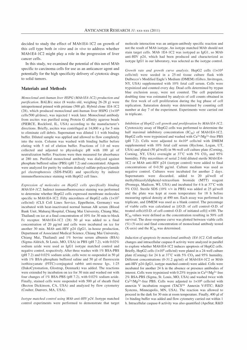
Expression of MAb1E4-1C2 reactive molecules on HepG2cells. Prior to studying the effect of 1E4-1C2, monoclonalantibody specific to HSPG isolated from human liver on theinhibition of HepG2 cells growth, indirect immunofluorescencewas performed to verify the expression of 1E4-1C2 on specificcells. The results showed that 1E4-1C2 specifically reactedwith HepG2 cells (Figure 1).
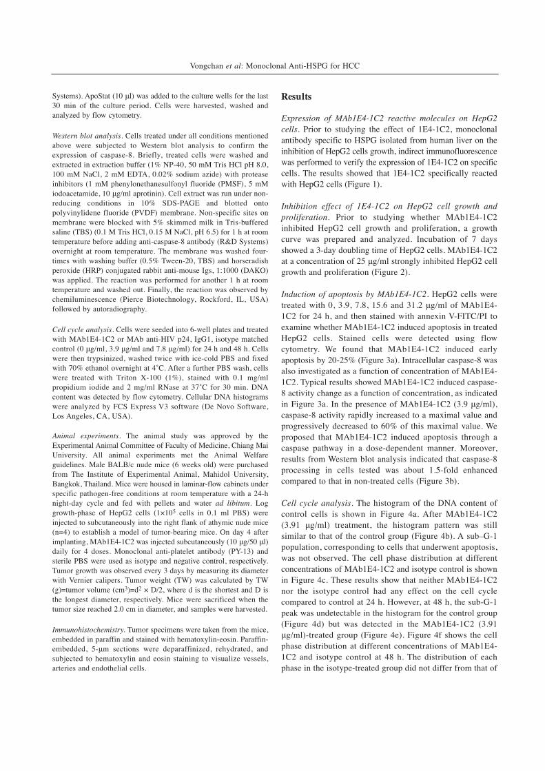
Inhibition effect of 1E4-1C2 on HepG2 cell growth andproliferation. Prior to studying whether MAb1E4-1C2inhibited HepG2 cell growth and proliferation, a growthcurve was prepared and analyzed. Incubation of 7 daysshowed a 3-day doubling time of HepG2 cells. MAb1E4-1C2at a concentration of 25 μg/ml strongly inhibited HepG2 cellgrowth and proliferation (Figure 2).
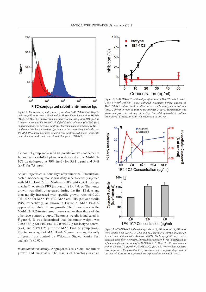
Induction of apoptosis by MAb1E4-1C2. HepG2 cells weretreated with 0, 3.9, 7.8, 15.6 and 31.2 μg/ml of MAb1E4-1C2 for 24 h, and then stained with annexin V-FITC/PI toexamine whether MAb1E4-1C2 induced apoptosis in treatedHepG2 cells. Stained cells were detected using flowcytometry. We found that MAb1E4-1C2 induced earlyapoptosis by 20-25% (Figure 3a). Intracellular caspase-8 wasalso investigated as a function of concentration of MAb1E4-1C2. Typical results showed MAb1E4-1C2 induced caspase-8 activity change as a function of concentration, as indicatedin Figure 3a. In the presence of MAb1E4-1C2 (3.9 μg/ml),caspase-8 activity rapidly increased to a maximal value andprogressively decreased to 60% of this maximal value. Weproposed that MAb1E4-1C2 induced apoptosis through acaspase pathway in a dose-dependent manner. Moreover,results from Western blot analysis indicated that caspase-8processing in cells tested was about 1.5-fold enhancedcompared to that in non-treated cells (Figure 3b).
Cell cycle analysis. The histogram of the DNA content ofcontrol cells is shown in Figure 4a. After MAb1E4-1C2(3.91 μg/ml) treatment, the histogram pattern was stillsimilar to that of the control group (Figure 4b). A sub–G-1population, corresponding to cells that underwent apoptosis,was not observed. The cell phase distribution at differentconcentrations of MAb1E4-1C2 and isotype control is shownin Figure 4c. These results show that neither MAb1E4-1C2nor the isotype control had any effect on the cell cyclecompared to control at 24 h. However, at 48 h, the sub-G-1peak was undetectable in the histogram for the control group(Figure 4d) but was detected in the MAb1E4-1C2 (3.91μg/ml)-treated group (Figure 4e). Figure 4f shows the cellphase distribution at different concentrations of MAb1E4-1C2 and isotype control at 48 h. The distribution of eachphase in the isotype-treated group did not differ from that of
Vongchan et al: Monoclonal Anti-HSPG for HCC
the control group and a sub-G-1 population was not detected.In contrast, a sub-G-1 phase was detected in the MAb1E4-1C2 treated-group at 39% (n=3) for 3.91 μg/ml and 54%(n=3) for 7.8 μg/ml.
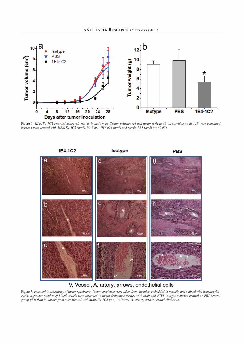
Animal experiments. Four days after tumor cell inoculation,each tumor-bearing mouse was daily subcutaneously injectedwith MAb1E4-1C2, or MAb anti-HIV p24 (IgG1, isotypematched), or sterile PBS (as controls) for 4 days. The tumorgrowth was slightly increased during the first 18 days andthen rapidly increased with specific growth rates of 0.37,0.61, 0.56 for MAb1E4-1C2, MAb anti-HIV p24 and sterilePBS, respectively, as shown in Figure 5. MAb1E4-1C2appeared to inhibit tumor growth. The tumor sizes in theMAb1E4-1C2-treated group were smaller than those of theother two control groups. The tumor weight is indicated inFigure 6. It was determined that the tumor weight was9.88±2.43 g for PBS (n=3), 9.09±0.79 g for isotype control(n=4) and 5.39±1.28 g for the MAb1E4-1C2 group (n=4).The tumor weight of MAb1E4-1C2 group was significantlydifferent from control by Wilcoxon Signed Ranks Testanalysis (p<0.05).
Immunohistochemistry. Angiogenesis is crucial for tumorgrowth and metastasis. The results of hematoxylin-eosin
ANTICANCER RESEARCH 31: xxx-xxx (2011)
Figure 1. Expression of antigen recognized by MAb1E4-1C2 on HepG2cells. HepG2 cells were stained with MAb specific to human liver HSPGs(MAb1E4-1C2) by indirect immunofluorescence using anti-HIV p24 asisotype control and Dulbecco’s Modified Eagle’s Medium (DMEM) (cellculture medium) as negative control. Fluorescien isothiocyanate (FITC)-conjugated rabbit anti-mouse Igs was used as secondary antibody and1% BSA-PBS azide was used as conjugate control. Red peak: Conjugatecontrol, clear peak: cell control and blue peak: 1E4-1C2.
Figure 2. MAb1E4-1C2 inhibited proliferation of HepG2 cells in vitro.Cells (4×10⁴ cells/ml) were cultured overnight before adding ofMAb1E4-1C2 (black line) or MAb anti-HIV p24 (isotype control, redline). Cultivation was continued for another 2 days. Supernatant wasdiscarded prior to adding of methyl thiazolyldiphenyl-tetrazoliumbromide(MTT) reagent. O.D was measured at 490 nm.
Figure 3. MBb1E4-1C2 induced apoptosis in HepG2 cells. a: HepG2 cellswere treated with 0, 3.9, 7.8, 15.6 and 31.2 μg/ml of MAb1E4-1C2 for 24h, and then stained with Annexin V-(PI). Early apoptotic cells weredetected using flow cytometry. Intracellular caspase-8 was investigated asa function of concentration of MAb1E4-1C2. b: HepG2 cells were treatedwith 0, 3.9 and 7.8 μg/ml of MAb1E4-1C2 for 24 h. Western blot analysiswas performed. Caspase-8 activity was assessed as a percentage that ofthe control. Results are expressed are expressed as mean±SE (n=3).

Vongchan et al: Monoclonal Anti-HSPG for HCC
Figure 4. Flow cytometric analysis of the cell cycle distribution in MAb1E4-1C2 treated HepG2. a-e: HepG2 cells were incubated for 24 h and d-f: for 48 h. In b and e, MAb1E4-1C2 (3.9 μg/ml) was present for 24 h and 48 h, respectively. The percentages of cells residing in sub-G-1, G0/G1,S- and G2/M phases were counted. In c and f, the cell phase distribution was determined at various concentrations of MAb1E4-1C2 and isotypecontrol (MAb anti-HIV p24) for 24 h and 48 h, respectively. Results are expressed as mean±SE (n=3).
Figure 5. Inhibition of tumor growth by MAb1E4-1C2 in nude mice xenografted with human HCC HepG2. On day 4 after implanting tumor cells,MAb1E4-1C2, MAb anti-HIV p24 (isotype control group) or sterile PBS (vehicle control group) were injected subcutaneously (10 μg/50 μl) dailyfor 4 doses. Tumor growth was observed every 3 days by measuring its diameter with Vernier caliper. Tumor weight (TW) was calculated by TW(g)=tumor volume (cm3)=d2 × D/2, where d is the shortest and D is the longest diameter, respectively.
ANTICANCER RESEARCH 31: xxx-xxx (2011)
Figure 6. MAb1E4-1C2 retarded xenograft growth in nude mice. Tumor volumes (a) and tumor weights (b) at sacrifice on day 28 were comparedbetween mice treated with MAb1E4-1C2 (n=4), MAb anti-HIV p24 (n=4) and sterile PBS (n=3) (*p<0.05).
Figure 7. Immunohistochemistry of tumor specimens. Tumor specimens were taken from the mice, embedded in paraffin and stained with hematoxylin-eosin. A greater number of blood vessels were observed in tumor from mice treated with MAb anti-HIV1, isotype matched control or PBS controlgroup (d-i) than in tumors from mice treated with MAb1E4-1C2 (a-c). V: Vessel, A: artery, arrows: endothelial cells.
staining showed that the number of blood vessels wasincreased in tumor in mice treated with isotype or PBS(Figure 7d-i). Endothelial cells were observed. In contrast,there was a smaller number of blood vessels in the MAb1E4-1C2 treated group (Figure 7a-c).
Discussion
HSPGs interact with a multitude of proteins and have beenimplicated in the regulation of a broad range of biologicalprocesses including cell division, migration, adhesion, tissueorganization and viral entry (33). MAb1E4-1C2 had beenprepared in our laboratory against a human liver HSPG (26)and showed some activity against acute myeloid leukemia(27). Since HepG2 human HCCs were shown to react withMAb1E4-1C2, we hypothesized that the specific moleculebeing recognized may be involved in tumor cell growth andproliferation. Both in vitro and in vivo experiments in ananimal model, clearly demonstrate the activity of this MAbagainst human HCC.
Flow cytometry with PI staining was used to evaluate theapoptosis of HepG2 cells after treatment with MAb1E4-1C2for 48 h. The resulting apoptotic cells showed a reduction ofDNA content and, therefore, could be recognized, followingstaining of cellular DNA, as cells with low DNA stainability(sub-G-1) that were lower than that of G-1 cells (34). TheMAb1E4-1C2 treated groups showed a sub-G-1 apoptosispeak. The apoptotic rate progressively increased from 39%to 54% in a dose dependent manner. The results correspondto an enhancement of intracellular caspase-8 on exposure toMAb1E4-1C2. Caspase-8 is the typical apoptosis initiatorcaspase downstream of TNF super-family of death receptors.Indeed, caspase-8 deficient cells are resistant to deathreceptor-mediated cell death both in vitro and in vivo (35).
Angiogenesis plays an important role in cancer, sincevessels are needed to supply nutrients and oxygen to sustaintumor growth, and to excrete metabolic waste. Theneovessels provide access for tumor cells to penetrate intothe circulation. Tumor cells stimulate the formation of newblood vessels by means of enhanced production of the majorangiogenic growth factors. Angiogenesis is controlled byvarious receptor tyrosine kinases and their ligands. Vascularendothelial growth factor (VEGF) receptors play animportant role in angiogenesis. VEGF, secreted by hepatomacells and hepatic stellate cells, is up-regulated during tumordifferentiation and vascular development of HCC (36, 37).VEGF is expressed at high levels in tumor blood vessels(38). Hypervascularity is one of the main characteristics oflarge and moderately or poorly differentiated HCC. Tissuefactor, a plasma membrane glycoprotein expressed byendothelial cells and smooth muscle cells, and known toinitiate blood coagulation, can also play a role in tumorprogression and angiogenesis (39, 40). An improved
knowledge of molecular mechanisms regulating angiogenesisin cancer provides insight into molecular targets for anti-angiogenic treatment. Anti-angiogenic treatment can preventneovascularization by inhibiting proliferation, migration anddifferentiation of endothelial cells. The endothelial cellstaining by H&E showed both decreased vasculature anddistorted vascular morphology in MAb1E4-1C2-treatedxenografts and the resulting endothelial vessels wereundersupplied. These data suggest that the tumor size in thecontrol group, which was higher than that for MAb1E4-1C2,might result from tumor vascularization (41).
In conclusion, our results reveal that the human HCC cellline HepG2 express HSPG. HSPGs are reported to beinvolved in cell growth and proliferation, thus, wehypothesized that HSPG isolated from human liver may takepart in cell proliferation both in vitro and in vivo. Using MAbraised against human liver HSPGs as a tool, we demonstratedthat this specific MAb inhibited proliferation of HepG2 cellsand retarded tumor growth in an animal model. This is thefirst report of using this specific MAb to inhibit proliferationand growth of HCC.
Acknowledgements
This work was supported by Thailand Research Fund (TRF, RMU5080018), and National Research Council of Thailand (NRCT,2009), Bangkok, Thailand.
References
1 Yamada KM: Cell surface interactions with extracellularmaterials. Annu Rev Biochem 52: 761-799, 1983.
2 Hook M, Kjellen L and Johansson S: Cell-surfaceglycosaminoglycans. Annu Rev Biochem 53: 847-869, 1984.
3 Laterra J, Silbert JE and Culp LA: Cell surface heparan sulfatemediates some adhesive responses to glycosaminoglycan-bindingmatrices, including fibronectin. J Cell Biol 96: 112-123, 1983.
4 Rapraeger AC and Bernfield M: Heparan sulfate proteoglycansfrom mouse mammary epithelial cells. A putative membraneproteoglycan associates quantitatively with lipid vesicles. J BiolChem 258: 3632-3636, 1983.
5 Sasisekharan R and Venkataraman G: Heparin and heparansulfate: biosynthesis, structure and function. Curr Opin ChemBiol 4: 626-631, 2000.
6 Esko JD and Lindahl U: Molecular diversity of heparan sulfate.J Clin Invest 108: 169-173, 2001.
7 Kjellen L, Oldberg A and Hook M: Cell-surface heparan sulfate.Mechanisms of proteoglycan-cell association. J Biol Chem 255:10407-10413, 1980.
8 Norling B, Glimelius B and Wasteson A: Heparan sulfateproteoglycan of cultured cells: demonstration of a lipid- and amatrix-associated form. Biochem Biophys Res Commun 103:1265-1272, 1981.
9 Hassell JR, Robey PG, Barrach HJ, Wilczek J, Rennard SI andMartin GR: Isolation of a heparan sulfate-containing proteoglycanfrom basement membrane. Proc Natl Acad Sci USA 77: 4494-4498, 1980.
Vongchan et al: Monoclonal Anti-HSPG for HCC
10 Hedman K, Johansson S, Vartio T, Kjellen L, Vaheri A and HookM: Structure of the pericellular matrix: association of heparanand chondroitin sulfates with fibronectin-procollagen fibers. Cell28: 663-671, 1982.
11 LeBaron RG, Esko JD, Woods A, Johansson S and Hook M:Adhesion of glycosaminoglycan-deficient chinese hamster ovarycell mutants to fibronectin substrata. J Cell Biol 106: 945-952,1988.
12 Rollins BJ and Culp LA: Preliminary characterization of theproteoglycans in the substrate adhesion sites of normal andvirus-transformed murine cells. Biochemistry 18: 5621-5629,1979.
13 Saunders S and Bernfield M: Cell surface proteoglycan bindsmouse mammary epithelial cells to fibronectin and behaves as areceptor for interstitial matrix. J Cell Biol 106: 423-430, 1988.
14 Cohn RH, Cassiman JJ and Bernfield MR: Relationship oftransformation, cell density, and growth control to the cellulardistribution of newly synthesized glycosaminoglycan. J Cell Biol71: 280-294, 1976.
15 Lander AD, Fujii DK, Gospodarowicz D and Reichardt LF:Characterization of a factor that promotes neurite outgrowth:evidence linking activity to a heparan sulfate proteoglycan. JCell Biol 94: 574-585, 1982.
16 Kanwar YS, Linker A and Farquhar MG: Increased permeabilityof the glomerular basement membrane to ferritin after removalof glycosaminoglycans (heparan sulfate) by enzyme digestion. JCell Biol 86: 688-693, 1980.
17 Park PW, Reizes O and Bernfield M: Cell surface heparansulfate proteoglycans: selective regulators of ligand-receptorencounters. J Biol Chem 275: 29923-29926, 2000.
18 Dong J, Peters-Libeu CA, Weisgraber KH, Segelke BW, RuppB, Capila I, Hernaiz MJ, LeBrun LA and Linhardt RJ:Interaction of the N-terminal domain of apolipoprotein E4 withheparin. Biochemistry 40: 2826-2834, 2001.
19 Libeu CP, Lund-Katz S, Phillips MC, Wehrli S, Hernaiz MJ,Capila I, Linhardt RJ, Raffai RL, Newhouse YM, Zhou F andWeisgraber KH: New insights into the heparan sulfateproteoglycan-binding activity of apolipoprotein E. J Biol Chem276: 39138-39144, 2001.
20 Bazin HG, Marques MA, Owens AP, 3rd, Linhardt RJ andCrutcher KA: Inhibition of apolipoprotein E-relatedneurotoxicity by glycosaminoglycans and their oligosaccharides.Biochemistry 41: 8203-8211, 2002.
21 Lin YL, Lei HY, Lin YS, Yeh TM, Chen SH and Liu HS:Heparin inhibits dengue-2 virus infection of five human liver celllines. Antiviral Res 56: 93-96, 2002.
22 Pradel G, Garapaty S and Frevert U: Proteoglycans mediatemalaria sporozoite targeting to the liver. Mol Microbiol 45: 637-651, 2002.
23 Barth H, Schafer C, Adah MI, Zhang F, Linhardt RJ, Toyoda H,Kinoshita-Toyoda A, Toida T, Van Kuppevelt TH, Depla E, VonWeizsacker F, Blum HE and Baumert TF: Cellular binding ofhepatitis C virus envelope glycoprotein E2 requires cell surfaceheparan sulfate. J Biol Chem 278: 41003-41012, 2003.
24 Rathore D, McCutchan TF, Garboczi DN, Toida T, Hernaiz MJ,LeBrun LA, Lang SC and Linhardt RJ: Direct measurement ofthe interactions of glycosaminoglycans and a heparindecasaccharide with the malaria circumsporozoite protein.Biochemistry 40: 11518-11524, 2001.
25 Chen Y, Maguire T, Hileman RE, Fromm JR, Esko JD, LinhardtRJ and Marks RM: Dengue virus infectivity depends onenvelope protein binding to target cell heparan sulfate. Nat Med3: 866-871, 1997.
26 Vongchan P, Warda M, Toyoda H, Toida T, Marks RM andLinhardt RJ: Structural characterization of human liver heparansulfate. Biochim Biophys Acta 1721: 1-8, 2005.
27 Vongchan P and Linhardt RJ: Expression of human liver HSPGson acute myeloid leukemia. Clin Immunol 122: 194-206, 2007.
28 Hulett MD, Freeman C, Hamdorf BJ, Baker RT, Harris MJ andParish CR: Cloning of mammalian heparanase, an importantenzyme in tumor invasion and metastasis. Nat Med 5: 803-809,1999.
29 Liu D, Shriver Z, Venkataraman G, El Shabrawi Y andSasisekharan R: Tumor cell surface heparan sulfate as crypticpromoters or inhibitors of tumor growth and metastasis. ProcNatl Acad Sci USA 99: 568-573, 2002.
30 Sanderson RD: Heparan sulfate proteoglycans in invasion andmetastasis. Semin Cell Dev Biol 12: 89-98, 2001.
31 Vlodavsky I and Friedmann Y: Molecular properties andinvolvement of heparanase in cancer metastasis andangiogenesis. J Clin Invest 108: 341-347, 2001.
32 Borsig L, Wong R, Feramisco J, Nadeau DR, Varki NM andVarki A: Heparin and cancer revisited: mechanistic connectionsinvolving platelets, P-selectin, carcinoma mucins, and tumormetastasis. Proc Natl Acad Sci USA 98: 3352-3357, 2001.
33 Lindahl U, Kusche-Gullberg M and Kjellen L: Regulateddiversity of heparan sulfate. J Biol Chem 273: 24979-24982,1998.
34 Darzynkiewicz Z, Juan G, Li X, Gorczyca W, Murakami T, andTraganos F: Cytometry in cell necrobiology: analysis ofapoptosis and accidental cell death (necrosis). Cytometry 27: 1-20, 1997.
35 Chen L, Zhuang G, Li W, Liu Y, Zhang J and Tian X: RGD-FasLinduces apoptosis of pituitary adenoma cells. Cell Mol Immunol5: 61-68, 2008.
36 Torimura T, Ueno T, Kin M, Harada R, Taniguchi E, NakamuraT, Sakata R, Hashimoto O, Sakamoto M, Kumashiro R, Sata M,Nakashima O, Yano H and Kojiro M: Overexpression ofangiopoietin-1 and angiopoietin-2 in hepatocellular carcinoma.J Hepatol 40: 799-807, 2004.
37 Pang RW, Joh JW, Johnson PJ, Monden M, Pawlik TM and PoonRT: Biology of hepatocellular carcinoma. Ann Surg Oncol 15:962-971, 2008.
38 Ruoslahti E and Rajotte D: An address system in the vasculature ofnormal tissues and tumors. Annu Rev Immunol 18: 813-827, 2000.
39 De S, Razorenova O, McCabe NP, O'Toole T, Qin J and ByzovaTV: VEGF-integrin interplay controls tumor growth andvascularization. Proc Natl Acad Sci USA 102: 7589-7594, 2005.
40 Dupuy E, Hainaud P, Villemain A, Bodevin-Phedre E, BroulandJP, Briand P and Tobelem G: Tumoral angiogenesis and tissuefactor expression during hepatocellular carcinoma progressionin a transgenic mouse model. J Hepatol 38: 793-802, 2003.
41 Pang R and Poon RT: Angiogenesis and antiangiogenic therapyin hepatocellular carcinoma. Cancer Lett 242: 151-167, 2006.
Received August 16, 2011Revised October 24, 2011
Accepted October 25, 2011
ANTICANCER RESEARCH 31: xxx-xxx (2011)