inhibition of lncrna mir31hg promotes osteogenic ... · inhibition of lncrna mir31hg promotes...
TRANSCRIPT

Inhibition of lncRNA MIR31HG Promotes
Osteogenic Differentiation of Human
Adipose-Derived Stem Cells
CHANYUAN JIN,a,e
LINGFEI JIA,b,c
YIPING HUANG,d
YUNFEI ZHENG,d
NING DU,c
YUNSONG LIU,a
YONGSHENG ZHOUa,e
Key Words. lncRNA MIR31HG • NF-jB • Human adipose-derived stem cells • Osteogenic differenti-ation
ABSTRACT
Osteogenic differentiation and bone formation is suppressed under condition of inflammation
induced by proinflammation cytokines. A number of studies indicate miRNAs play a significant
role in tumor necrosis factor-a-induced inhibition of bone formation, but whether long non-
coding RNAs are also involved in this process remains unknown. In this study, we evaluated the
role of MIR31HG in osteogenesis of human adipose-derived stem cells (hASCs) in vitro and in
vivo. The results suggested that knockdown of MIR31HG not only significantly promoted osteo-
genic differentiation, but also dramatically overcame the inflammation-induced inhibition of
osteogenesis in hASCs. Mechanistically, we found MIR31HG regulated bone formation and
inflammation via interacting with NF-jB. The p65 subunit bound to the MIR31HG promoter and
promoted MIR31HG expression. In turn, MIR31HG directly interacted with IjBa and participated
in NF-jB activation, which builds a regulatory circuitry with NF-jB. Targeting this MIR31HG–NF-
jB regulatory loop may be helpful to improve the osteogenic capacity of hASCs under inflam-
matory microenvironment in bone tissue engineering. STEM CELLS 2016; 00:000–000
SIGNIFICANCE STATEMENT
In the condition of bone loss, the defective or injured tissues are often associated with inflam-mation. We identify that knockdown of MIR31HG not only promoted osteogenic differentiationbut also reversed the inflammation-induced inhibition of bone formation. And we find the nov-el regulatory circuitry between MIR31HG and the NF-jB. MIR31HG is induced by nuclear trans-location of NF-jB, and MIR31HG, in turn, directly binds to IjBa and contributes to IjBa
phosphorylation and NF-jB activation. Thus, knockdown of MIR31HG expression can be anapproach to improve bone regeneration and may have a broad future in clinical applications.
INTRODUCTION
Human adipose-derived stem cells (hASCs) area type of adult mesenchymal stem cell (MSC)capable of bone regeneration and repair. Asthey can be obtained from adipose tissues bya more abundant and less invasive procedure,hASCs have become an attractive source ofstem cells in bone tissue engineering [1, 2]. Anumber of studies have shown the ability ofASCs to repair bone defects in animal models[3, 4], but most of these experiments wereperformed under minimal inflammation. How-ever, in therapeutic bone regeneration, thedefective or injured tissues are frequently inan inflamed condition [5]. Moreover, growingevidence suggests that MSC differentiation isprecisely regulated by the molecular signals fromthe extracellular environment [6, 7], and that the
proinflammatory cytokines in the cell microenvi-ronment inhibit osteogenic differentiation andbone formation [8]. Thus, it is necessary to over-come the inflammation-induced inhibition ofosteogenesis in bone tissue regeneration.
Bone remodeling is a continuous processthat depends on the balance of osteoclast-mediated bone resorption and osteoblast-mediated bone formation. The inflammatorycytokines disrupt normal bone homeostasis,impair bone formation, and induce bone loss ininflammatory diseases. Several molecular path-ways related to osteoblast differentiation aredysregulated under inflammatory environment,such as the mitogen-activated protein kinase[9], nuclear factor-jB (NF-jB) [10], bone mor-phogenetic protein (BMP) [11], and Wnt signal-ing pathway [12]. Among these, NF-jB is a key
aDepartment ofProsthodontics, bDepartmentof Oral and MaxillofacialSurgery, cCentral Laboratory,dDepartment ofOrthodontics, eNationalEngineering Lab for Digitaland Material Technology ofStomatology, PekingUniversity School andHospital of Stomatology,Beijing Key Laboratory ofDigital Stomatology, Beijing,China
Correspondence: YongshengZhou, D.D.S., Ph.D., Departmentof Prosthodontics, PekingUniversity School and Hospitalof Stomatology, 22Zhongguancun South Avenue,Haidian District, Beijing100081, China. Telephone:186-10-82195370;Fax: 186-10-62173402; e-mail:[email protected]
Received January 26, 2016;accepted for publication May30, 2016; first published onlinein STEM CELLS EXPRESS June 22,2016.
VC AlphaMed Press1066-5099/2016/$30.00/0
http://dx.doi.org/10.1002/stem.2439
STEM CELLS 2016;00:00–00 www.StemCells.com VC AlphaMed Press 2016
EPIGENETICS, GENOMICS, PROTEOMICS
AND METABONOMICS

regulator of inflammation, and is activated by tumor necrosisfactor-a (TNF-a), interleukin-17 (IL-17), and lipopolysaccharides.Growing evidence has showed that NF-jB signaling inhibitsosteoblast (OB) differentiation and bone formation [13, 14],while specific inhibition of NF-jB in differentiated osteoblastssignificantly enhanced bone matrix formation and mineral den-sity [15]. Recently, non-protein-coding RNAs (ncRNAs) haveemerged as important regulatory transcripts in biological con-trol and pathology [16]. Small ncRNAs, including miR-17 [17],miR-21 [18], miR-23a [19], and miR-155 [20], have been impli-cated in the TNF-a-induced inhibition of bone formation. Bycontrast, the function of long non-coding RNAs (lncRNAs), tenta-tively defined as ncRNAs >200 nt in length [21, 22], are largelyunknown in MSC osteogenic differentiation under inflammatoryconditions. Recent studies have shown that the lncRNAs Lethe
[23] and NKILA [24] directly interact with the NF-jB complex ininflammatory diseases, implying that lncRNAs are important forthe NF-jB signaling pathway.
In this study, we aimed to investigate the role of lncRNA(MIR31HG) in inflammation-mediated bone metabolism.MIR31HG (previously known as LOC554202), located on chromo-some 9 (9p21.3), is demonstrated to affect cell proliferation, dif-ferentiation, and motility in tumor development [25, 26].However, the role of MIR31HG in inflammation or bone forma-tion has not been reported. Since a previous study has shownthat the MIR31HG promoter contains putative NF-jB bindingsites [27], we hypothesized that MIR31HG may be involved in theNF-jB-mediated inhibition of osteogenesis. In this study, we iden-tified that the lncRNA MIR31HG, induced by NF-jB, is alsoinvolved in an NF-jB regulatory feedback loop. Importantly,knockdown of MIR31HG not only significantly promotesosteogenic differentiation of hASCs, but also reverses theinflammation-induced inhibition of osteogenesis and boneformation.
MATERIALS AND METHODS
Cell Culture
Primary hASCs were obtained from ScienCell (San Diego, CA)and cultured in proliferation media (PM) consisting of DMEMwith 10% fetal bovine serum and 1% antibiotics. Stem cellsfrom three donors were used for the in vitro and in vivoexperiments. All cell-based in vitro experiments were repeat-ed in triplicate. For the osteogenic differentiation experiment,cells were induced in osteogenic media (OM) containing stan-dard PM supplemented with 100 nM dexamethasone,0.2 mM ascorbic acid, and 10 mM b-glycerophosphate. The293T cells were obtained from American Type Culture Collec-tion (Manassas, VA) and cultured in DMEM supplementedwith 10% fetal bovine serum and 1% antibiotics.
Lentivirus Infection
Recombinant lentiviruses containing full-length MIR31HG andthe scramble control (NC) were obtained from Integrated BiotechSolutions Co. (Ibsbio Co., Shanghai, China). Recombinant lentivi-ruses targeting MIR31HG (shMIR31HG21 and shMIR31HG22)and the scramble control (shNC) were obtained from Gene-Pharma Co. (Shanghai, China). Transfection of the hASCs wasperformed by exposing them to dilutions of the viral supernatantin the presence of polybrene (5 lg/ml) for 72 h.
Alkaline Phosphatase Staining and Activity
Cells cultured for 7 days under PM or OM were assayed foralkaline phosphatase (ALP) staining and activity, as describedpreviously [28, 29]. ALP staining was performed according tothe protocol of the NBT/BCIP staining kit (CoWin Biotech, Bei-jing, China). ALP activity was analyzed using an ALP activity col-orimetric assay kit (Biovision, Milpitas, CA). Total protein wasdetermined in the same sample with the BCA method usingthe Pierce protein assay kit (Thermo Fisher Scientific, Rockford,IL). ALP activity relative to the control treatment was calculatedafter normalization to the total protein content.
Alizarin Red S Staining and Quantification
Cells cultured for 14 days under PM or OM were assayed asdescribed previously [28, 29]. The cultured cells were fixed with4% paraformaldehyde and then stained with 0.1% Alizarin red SpH 4.2 (Sigma-Aldrich, Saint Louis, MO) for 20 min. For quantita-tive assessment of the degree of mineralization, the stain waseluted by 100 mM cetylpyridinium chloride (Sigma-Aldrich) for1 hour and quantified by spectrophotometric absorbance at570 nm. Alizarin red S intensity relative to the control treatmentwas calculated after normalization to the total protein content.
Von Kossa Staining
Cells cultured for 14 days under PM or OM were assessed usingthe von Kossa staining method as described previously [30].Briefly, cells were fixed with 4% paraformaldehyde and thenincubated with 5% silver nitrate solution for 30 minutes in thedark, followed by exposure to UV light. Unincorporated silvernitrate was removed using 5% sodium thiosulfate, followed byseveral washes with water.
RNA Oligoribonucleotide, Chemicals,
and Transient Transfection
Small-interfering RNAs (siRNA) targeting the MIR31HG, p65,p50, IjBa, and the scramble control (si-NC) were purchasedfrom Integrated Biotech Solutions Co. The sequences are listedin Supporting Information Table 1. For transient transfection,cells were cultured and grown to 70–80% confluence, thentransfected with plasmids or siRNA using Lipofectamine 2000(Invitrogen, Carlsbad, CA) according to the manufacturer’sprocedure.
The recombinant human TNF-a and IL-17 were purchasedfrom PeproTech Inc. (Rocky Hill, NJ). The specific NF-jB inhibitor,BAY117082, was obtained from Sigma-Aldrich to inhibit IjBa
phosphorylation. Lithium chloride (LiCl), a GSK-3b inhibitor, wasobtained from Sigma-Aldrich to block b-catenin degradation. Inthe treatment group, TNF-a or IL-17 (10 ng/ml) was added tothe culture media every 24 hours with or without BAY117082(5 lmol/l) or LiCl (5 mmol/l). For RNA and protein analyses,the cells were harvested within 24 hours. For osteogenic differ-entiation, the cells were harvested after 7 or 14 days.
Vectors
A 2,198 bp fragment of human MIR31HG promoter (21,994 bpto 1204 bp relative to the transcription start site) was amplifiedfrom the genomic DNA of HeLa cells and cloned into the PGL3-Enhancer vector (Promega, Madison, WI). The primers used toclone the MIR31HG promoter were as follows: sense, 50-GCTAGC ACA AAG TCT TGC CTA GTG GGA TT-30 and antisense,
2 Inhibition of lncRNA MIR31HG Promotes Osteogenesis
VC AlphaMed Press 2016 STEM CELLS

50-CTC GAG GTC CGA GTA GGA GGA CAG AAG C-30. A series ofdeletion of the MIR31HG promoter were constructed by Inte-grated Biotech Solutions Co. based on the full-length promoterreporter construct. The plasmid containing 53 NF-jB consen-sus-binding sites with luciferase reporter gene was a gift fromProfessor Zhong Chen (National Institutes of Health, Bethesda,MD) [31]. All constructs were confirmed by DNA sequencing.
Dual Luciferase Reporter Assay
Luciferase assays were performed as described previously[32]. For each transfection with subsequent reporter assay,the total quantity of transfected plasmid was kept constantby the addition of pRL-TK, a plasmid expressing renilla lucifer-ase (Promega, Madison, WI). The renilla and firefly luciferaseactivities were measured 24 hours after transfection using theDual Luciferase Reporter Assay System (Promega). All lucifer-ase values were normalized by renilla luciferase and expressedrelative to the basal activity as fold induction.
RNA Isolation and Quantitative Reverse
Transcription-Polymerase Chain Reaction (qRT-PCR)
Total RNA was extracted using TRIzol reagent (Invitrogen) accordingto the manufacturer’s procedure, and then reverse-transcribedinto cDNA using a cDNA Reverse Transcription Kit (Applied Biosys-tems, Foster City, CA). Quantitative polymerase chain reaction wasconducted with SYBR Green Master Mix (Life Technologies) asdescribed on the ABI Prism 7500 real-time PCR System (AppliedBiosystems) using GAPDH for normalization [33]. The primers usedare listed in Supporting Information Table 1.
Immunofluorescence Staining
Immunofluorescence staining was performed as described previ-ously [34]. Cells grown on sterile glass coverslips were fixed with4% paraformaldehyde for 30 minutes, permeabilized with 0.1%Triton X-100 for 15 minutes, and then blocked with 5% normalgoat serum for 30 minutes. Thereafter, cells were incubated withprimary antibodies against osteocalcin (OCN, Abcam, Cambridge,UK) at 48C overnight, and then incubated with the specified sec-ondary antibodies for 1 hour. Nuclei were counterstained withDAPI, and the coverslips were mounted on a glass slide. Imageswere captured with a LSM 5 EXCITER confocal imaging system(Carl Zeiss, Oberkochen, Germany).
Western Blot Analysis
Western blot analysis was performed as described previously[33]. Briefly, cells were harvested, washed with PBS, and lysed inRIPA buffer. Proteins were separated by 10% sodium dodecylsulfate–polyacrylamide gel electrophoresis and transferred tonitrocellulose membranes. Primary antibodies against p-IjBa
(Abcam), IjBa (Abcam), p65 (Cell Signaling Technology, Beverly,MA), p-p65 (Cell Signaling Technology), b-catenin (Cell SignalingTechnology), OCN (Abcam), and GAPDH (Abcam) were diluted1:1,000. The intensities of the bands were quantified usingImageJ software (http://rsb.info.nih.gov/ij/). The background wassubtracted, and the signal of each target band was normalized tothat of the GAPDH band.
Cell Fractionation
For the fractionation assay, cytoplasmic and nuclear RNAswere performed using a Nuclei Isolation Kit (KeyGEN, Nanjing,China). Briefly, cells were harvested and resuspended in lysis
buffer. Then, the lysate was treated with Reagent A, incubatedon ice for 15 minutes, followed by centrifugation at 48C. Thepellet was then resuspended in lysis buffer followed by centri-fugation. The supernatant was transferred to a new tube asthe cytoplasmic fraction; the pellet was resuspended in Medi-um Buffer A and then added to a new tube with MediumBuffer B, followed by centrifugation at 48C. The supernatantwas saved as the cytoplasmic fraction. The pellet was used asthe nuclear fraction. RNA was extracted from both fractionsusing Trizol.
Chromatin Immunoprecipitation
Chromatin immunoprecipitation (ChIP) assay was performedusing an EZ-Magna ChIP assay kit (Merck Millipore, Darmstadt,Germany) according to manufacturer’s instructions. Briefly,cells were cross-linked with 1% formaldehyde, collected, lysed,and sonicated to shear DNA. Then the DNA-protein complexeswere isolated with antibodies against p65 (Abcam), or isotypeIgG (Santa Cruz Biotechnology, Santa Cruz, CA). The protein/DNA complexes were then eluted and reverse cross-linked.Spin columns were used to purify the DNA. The precipitatedDNA was quantified by polymerase chain reaction. Relativeenrichment is calculated as the amount of amplified DNA nor-malized to input and relative to values obtained after normalIgG immunoprecipitation. The primers used are listed in Sup-porting Information Table 1.
RNA Immunoprecipitation
RNA immunoprecipitation (RIP) was performed with RNA-Binding Protein Immunoprecipitation Kit (Merck Millipore)according to the manufacturer’s instructions. Briefly, magneticbeads were incubated with antibodies against p65 (Abcam),p50 (Abcam), IjBa (Abcam), and IgG control (from the kit). A50 ll sample from the 500 ll beads suspension for each anti-body was used to test the efficiency of immunoprecipitationby Western blot. The cells were collected for lysis in RIP Lysisbuffer. The lysate was immunoprecipitated with differentbead-antibody complexes. RNA was recovered with TRIzol andanalyzed by qRT-PCR. The results were normalized relative tothe input control.
RNA pull-down Assay
RNA pull-down and deletion mapping were performed as pre-viously described [35, 36]. A series of deletion mutants of theMIR31HG were constructed by bioBAY Co. (Suzhou, China).Briefly, biotinylated RNAs were prepared with T4 RNA poly-merase (Roche, Basel, Switzerland) and Biotin RNA labelingMix (Roche). The labeled RNA was captured with streptavidinmagnetic beads. Cell lysates or human recombinant p65 pro-tein (Sino Biological, Beijing, China), IjBa protein (Sino Biolog-ical), and p50 protein (Enzo Life Sciences, Farmingdale, NY)were prepared and incubated with the labeled RNA at 48C.Then, RNA-binding protein complexes were washed and elut-ed. A magnetic stand was used to collect the beads and thesupernatant was collected for Western blot analysis.
Fluorescent In Situ Hybridization
In situ hybridization was performed with a Fluorescent In SituHybridization (FISH) Kit (RiboBio, Guangzhou, China). Cells werebriefly rinsed in PBS and fixed in 4% formaldehyde for 10 minutes.Then the cells were permeabilized in PBS containing 0.5% Triton X-
Jin, Jia, Huang et al. 3
www.StemCells.com VC AlphaMed Press 2016

100 at 48C for 5 minutes, washed with PBS three times for 5minutes, and prehybridizated at 378C for 30 minutes before hybrid-ization. Then an anti-MIR31HG, anti-U6, or anti-18S oligodeoxynu-cleotide probe was used in the hybridization solution at 378Covernight in the dark. The next day, the cells were counterstainedwith DAPI and imaged using a confocal laser-scanning microscope(Carl Zeiss).
Colocalization of lncRNA and Protein
Colocalization analysis of MIR31HG and IjBa was performedas described previously [24]. The cells were hybridized withan anti-MIR31HG probe (RiboBio). Then the cells were rinsedbriefly in PBS and incubated with an IjBa antibody (Abcam)at 48C overnight. The next day, the cells were incubated inthe specified secondary antibodies and counterstained withDAPI. Images were obtained with a confocal laser-scanningmicroscope (Carl Zeiss).
Microarray and Bioinformatics
Total RNA was isolated from hASCs transfected withshMIR31HG and shNC. Biotinylated cDNA was preparedaccording to the standard Affymetrix protocol from 250 ngtotal RNA by using the Ambion WT Expression Kit (Life Tech-nologies). Following labeling, 5.5 lg of cDNA was hybridizedfor 16 hour at 458C on a GeneChip Human TranscriptomeArray 2.0. GeneChips were washed and stained in the Affyme-trix Fluidics Station 450. GeneChips were scanned by usingthe Affymetrix GeneChip Command Console installed in theGeneChip Scanner 3000 7G. Data were analyzed with robustmultichip analysis (RMA) algorithm using Affymetrix defaultanalysis settings. Values presented are log2 RMA signal inten-sity. For calling of differentially expressed genes, the fold dif-ferences (�1.5), FDR (<0.001) and average expression wereused. For the Gene Ontology enrichment and KEGG pathwayanalysis, the DAVID webserver (http://david.ncifcrf.gov/) wasused.
In Vivo Heterotopic Bone Formation Assay
The hASCs were induced under OM for 1 week before the invivo study. The cells were resuspended and incubated with7 mm 3 5 mm 3 2 mm Bio-Oss Collagen (Geistlich, GEWOGmbH, Baden-Baden, Germany) scaffolds for 1 hour at 378Cfollowed by centrifugation at 150g for 5 minutes, and thenimplanted subcutaneously on the back of 5-week-old BALB/chomozygous nude (nu/nu) mice (five mice per group), asdescribed previously [28, 29, 37, 38]. Implants were harvestedafter 8 weeks and fixed in 4% paraformaldehyde. All animalexperiments were approved by the Peking University AnimalCare and Use Committee.
Hematoxylin and Eosin Staining, Masson’s Trichrome,
and Immunohistochemical Analysis
The specimens were decalcified in 10% ethylene diamine tetra-acetic acid (pH 7.4) for 1 month, followed by dehydration andembedding in paraffin. Sections (5 lm) were cut and stainedwith hematoxylin and eosin (H&E) and Masson’s trichrome.Meanwhile, sections were evaluated by immunohistochemicalanalysis, as described previously [34]. The specimens wereblocked with 5% normal goat serum for 30 minutes and
then incubated with primary antibody against OCN (Santa CruzBiotechnology) at 48C overnight. Then, sections were processedusing the ABC detection kit (Vector Laboratories, Burlingame,CA) and visualized under an Olympus microscope (Olympus Co.,Tokyo, Japan).
Statistical Analysis
Statistical analyses were performed using SPSS 16.0 (SPSS,Chicago, IL). All data are expressed as the mean6 standarddeviation (SD). Differences between groups were analyzedusing Student’s t test. In cases of multiple-group testing, one-way analysis of variance was conducted. A two-tailed value ofp< .05 was considered statistically significant.
RESULTS
TNF-a and IL-17 Inhibit Osteoblast Differentiation
by Activating NF-jB
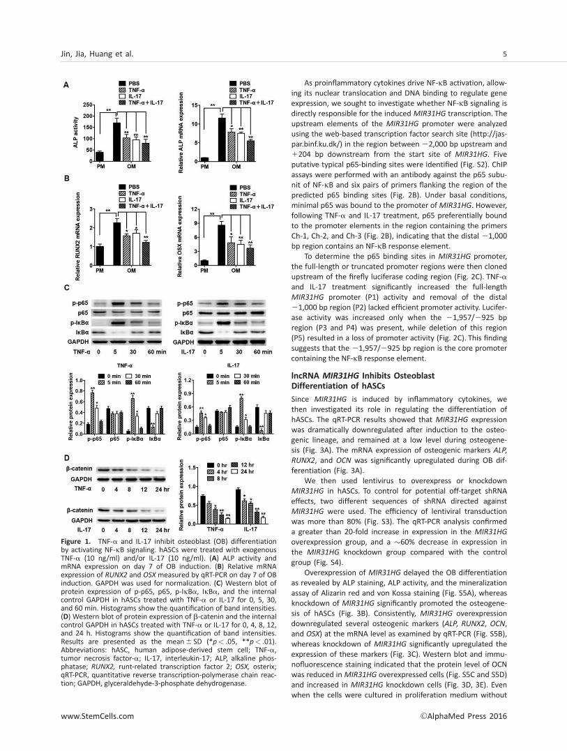
To confirm the effect of inflammatory cytokines on the OB dif-ferentiation of hASCs, we treated the cells with exogenousTNF-a (10 ng/ml) or IL-17 (10 ng/ml) under osteogenic induc-tion. TNF-a and IL-17 significantly inhibited ALP expression andactivity (Fig. 1A). The mRNA expression of osteogenic markers,runt-related transcription factor 2 (RUNX2) and osterix (OSX),was also significantly reduced by TNF-a and IL-17 (Fig. 1B).When cells were treated with TNF-a and IL-17, the ALP expres-sion and activity and the osteogenic gene expression were fur-ther suppressed (Fig. 1A, 1B).
Further, we detected the NF-jB signaling by Western blotanalysis. NF-jB dimers (predominantly p65/p50 dimers) arepresent in the cytoplasm in a transcriptionally inactive formdue to their interactions with the IjB [39]. TNF-a and IL-17rapidly activated the phosphorylation of IjBa in 5 minutes,and its level returned to normal after 60 minutes, while totalIjBa was rapidly reduced in 5 minutes, and returned to nor-mal level after 30 minutes (Fig. 1C). Once dissociated fromIjB, p65 undergoes phosphorylation, enters the nucleus, andinitiates transcription [40]. Activated NF-jB signaling pathwayhas been found to promote the degradation of b-catenin[15], which is a component of the Wnt signaling and playsessential roles in bone formation. Consistently, after TNF-aand IL-17 treatment, we found that p65 was rapidly phos-phorylated (Fig. 1C) and the total b-catenin expression wasgradually reduced (Fig. 1D).
To further confirm the inflammatory cytokines inhibitosteogenic differentiation of hASCs via NF-jB and b-cateninpathway, we used BAY117082 (BAY) to block the activation ofNF-jB, and Lithium chloride (LiCl) to block the degradation ofb-catenin. The inhibition of osteogenesis induced by TNF-aand IL-17 was reverted when BAY or LiCl was used (Fig. S1Aand S1B).
lncRNA MIR31HG Is Upregulated by Inflammatory
Cytokines via NF-jB Signaling
To investigate whether lncRNA MIR31HG is affected underinflammatory environment, we assessed its expression in hASCstreated with TNF-a or IL-17. The qRT-PCR results showed thatMIR31HG was significantly upregulated and displayed an oscilla-tion pattern consistent with TNF-a and IL-17 stimulation pat-terns (Fig. 2A).
4 Inhibition of lncRNA MIR31HG Promotes Osteogenesis
VC AlphaMed Press 2016 STEM CELLS
As proinflammatory cytokines drive NF-jB activation, allow-ing its nuclear translocation and DNA binding to regulate geneexpression, we sought to investigate whether NF-jB signaling isdirectly responsible for the induced MIR31HG transcription. Theupstream elements of the MIR31HG promoter were analyzedusing the web-based transcription factor search site (http://jas-par.binf.ku.dk/) in the region between 22,000 bp upstream and1204 bp downstream from the start site of MIR31HG. Fiveputative typical p65-binding sites were identified (Fig. S2). ChIPassays were performed with an antibody against the p65 subu-nit of NF-jB and six pairs of primers flanking the region of thepredicted p65 binding sites (Fig. 2B). Under basal conditions,minimal p65 was bound to the promoter of MIR31HG. However,following TNF-a and IL-17 treatment, p65 preferentially boundto the promoter elements in the region containing the primersCh-1, Ch-2, and Ch-3 (Fig. 2B), indicating that the distal 21,000bp region contains an NF-jB response element.
To determine the p65 binding sites in MIR31HG promoter,the full-length or truncated promoter regions were then clonedupstream of the firefly luciferase coding region (Fig. 2C). TNF-aand IL-17 treatment significantly increased the full-lengthMIR31HG promoter (P1) activity and removal of the distal21,000 bp region (P2) lacked efficient promoter activity. Lucifer-ase activity was increased only when the 21,957/2925 bpregion (P3 and P4) was present, while deletion of this region(P5) resulted in a loss of promoter activity (Fig. 2C). This findingsuggests that the 21,957/2925 bp region is the core promotercontaining the NF-jB response element.
lncRNA MIR31HG Inhibits Osteoblast
Differentiation of hASCs
Since MIR31HG is induced by inflammatory cytokines, wethen investigated its role in regulating the differentiation ofhASCs. The qRT-PCR results showed that MIR31HG expressionwas dramatically downregulated after induction to the osteo-genic lineage, and remained at a low level during osteogene-sis (Fig. 3A). The mRNA expression of osteogenic markers ALP,RUNX2, and OCN was significantly upregulated during OB dif-ferentiation (Fig. 3A).
We then used lentivirus to overexpress or knockdownMIR31HG in hASCs. To control for potential off-target shRNAeffects, two different sequences of shRNA directed againstMIR31HG were used. The efficiency of lentiviral transductionwas more than 80% (Fig. S3). The qRT-PCR analysis confirmeda greater than 20-fold increase in expression in the MIR31HG
overexpression group, and a �60% decrease in expression inthe MIR31HG knockdown group compared with the controlgroup (Fig. S4).
Overexpression of MIR31HG delayed the OB differentiationas revealed by ALP staining, ALP activity, and the mineralizationassay of Alizarin red and von Kossa staining (Fig. S5A), whereasknockdown of MIR31HG significantly promoted the osteogene-sis of hASCs (Fig. 3B). Consistently, MIR31HG overexpressiondownregulated several osteogenic markers (ALP, RUNX2, OCN,and OSX) at the mRNA level as examined by qRT-PCR (Fig. S5B),whereas knockdown of MIR31HG significantly upregulated theexpression of these markers (Fig. 3C). Western blot and immu-nofluorescence staining indicated that the protein level of OCNwas reduced in MIR31HG overexpressed cells (Fig. S5C and S5D)and increased in MIR31HG knockdown cells (Fig. 3D, 3E). Evenwhen the cells were cultured in proliferation medium without
Figure 1. TNF-a and IL-17 inhibit osteoblast (OB) differentiationby activating NF-jB signaling. hASCs were treated with exogenousTNF-a (10 ng/ml) and/or IL-17 (10 ng/ml). (A) ALP activity andmRNA expression on day 7 of OB induction. (B) Relative mRNAexpression of RUNX2 and OSX measured by qRT-PCR on day 7 of OBinduction. GAPDH was used for normalization. (C) Western blot ofprotein expression of p-p65, p65, p-IjBa, IjBa, and the internalcontrol GAPDH in hASCs treated with TNF-a or IL-17 for 0, 5, 30,and 60 min. Histograms show the quantification of band intensities.(D) Western blot of protein expression of b-catenin and the internalcontrol GAPDH in hASCs treated with TNF-a or IL-17 for 0, 4, 8, 12,and 24 h. Histograms show the quantification of band intensities.Results are presented as the mean6 SD (*p< .05, **p< .01).Abbreviations: hASC, human adipose-derived stem cell; TNF-a,tumor necrosis factor-a; IL-17, interleukin-17; ALP, alkaline phos-phatase; RUNX2, runt-related transcription factor 2; OSX, osterix;qRT-PCR, quantitative reverse transcription-polymerase chain reac-tion; GAPDH, glyceraldehyde-3-phosphate dehydrogenase.
Jin, Jia, Huang et al. 5
www.StemCells.com VC AlphaMed Press 2016

osteogenic supplements, knockdown of MIR31HG demonstrat-ed a similar, but weaker, pro-osteogenic effect (Fig. 3B, 3C).
NF-jB Signaling Is Inhibited by MIR31HG Knockdown
To understand the molecular mechanisms by which lncRNAMIR31HG regulates OB differentiation, we established MIR31HG
knockdown hASCs and conducted a transcriptome microarray
analysis. Knockdown of MIR31HG resulted in the upregulationof 364 genes and the downregulation of 495 genes (Fig. 4A).Interestingly, MIR31HG knockdown led to the downregulationof genes within important signaling pathways, including the NF-jB signaling pathway (Fig. 4B; Fig. S6). The expression of NF-jBtargets, such as IL-6, IL-7, IL-11, and TNF-a were significantlyreduced (Fig. 4C; Fig. S6), as confirmed by qRT-PCR (Fig. 4D).Moreover, we found that the NF-jB activity induced by TNF-aand IL-17 was reduced by �60% in cells with MIR31HG knock-down (Fig. 4E). Consistently, confocal microscopy showed thatp65 nuclear translocation induced by TNF-a and IL-17 stimula-tion was suppressed in MIR31HG knockdown cells (Fig. 4F).Finally, downstream b-catenin degradation induced by TNF-aand IL-17 was eliminated in MIR31HG knockdown hASCs(Fig. 4G).
lncRNA MIR31HG Directly Binds to IjBa and
Participates in Its Phosphorylation and NF-jB
Activation
To further study the underlying mechanism by which MIR31HG
regulates NF-jB signaling, we analyzed the distribution ofMIR31HG by confocal microscopy using FISH under physiologicaland inflammatory conditions. In control cells (physiological con-ditions), MIR31HG was found to be located in both the nucleusand the cytoplasm (Fig. 5A). However, following TNF-a and IL-17stimulation, MIR31HG was almost exclusively cytoplasmic(Fig. 5A), which was also confirmed by nuclear/cytoplasm frac-tionation (Fig. 5B).
The increased cytoplasmic localization of MIR31HG byTNF-a and IL-17 implies that the lncRNA may function in thecytoplasm. Therefore, we determined the effect of MIR31HG
on the activation of NF-jB complex in cytoplasm. Westernblot analysis showed that IjBa and p65 phosphorylationinduced by TNF-a and IL-17 stimulation was eliminated inMIR31HG knockdown hASCs (Fig. 5C).
The function of most lncRNAs depends on their ability to inter-act with proteins [21, 22]. As MIR31HG may function by interactingwith the NF-jB:IjB complex, we performed an RIP assay usingantibodies against p65, p50, or IjBa. All three antibodies retrievedsignificant amounts of MIR31HG RNA and NF-jB:IjB complex.However, successful knockdown of IjBa (Fig. S7) eliminatedMIR31HG enrichment in p65 or p50 immunoprecipitates, but
Figure 2. NF-jB induces MIR31HG expression. (A) Relative expres-sion of MIR31HG in hASCs stimulated with TNF-a or IL-17 for theconcentration indicated (by qRT-PCR; normalized by GAPDH;relative to untreated groups). (B) Upper: Diagram of the MIR31HGpromoter and location of the primers. Positions marked are relativeto the transcriptional start site (TSS). Lower: ChIP-qPCR showing theinteraction between the p65 subunit and the MIR31HG promoter inhASCs untreated or treated with TNF-a or IL-17 for 4 h. IgG wasused for normalization. (C) Upper: Schematic maps representing thefive potential binding sites of p65 in the MIR31HG promoter andthe four deletion mutants of the MIR31HG promoter. The arrowindicates the position of the TSS 11. Lower: Luciferase activity of293T cells transfected with luciferase constructs containing full-length (P1) or truncated forms (P2–P5) of the MIR31HG promoter.The ratio of firefly luciferase to renilla luciferase activity was calcu-lated and normalized to that of cells treated with PBS. Results arepresented as the mean6 SD (*p< .05, **p< .01). Abbreviations:TNF-a, tumor necrosis factor-a; IL-17, interleukin-17; qRT-PCR,quantitative reverse transcription-polymerase chain reaction;GAPDH, glyceraldehyde-3-phosphate dehydrogenase; ChIP, Chroma-tin immunoprecipitation; hASC, human adipose-derived stem cell.
6 Inhibition of lncRNA MIR31HG Promotes Osteogenesis
VC AlphaMed Press 2016 STEM CELLS

Figure 3. MIR31HG knockdown promotes osteoblast (OB) differentiation of hASCs. (A) Relative expression of MIR31HG, ALP, RUNX2, andOCN during OB differentiation of hASCs (by qRT-PCR; normalized by GAPDH; relative to day 0 groups). (B) Images of ALP, Alizarin red (AZR),and von Kossa (VK) staining in shNC, shMIR31HG21, and shMIR31HG22 groups. Cells were cultured in proliferation medium (PM) or osteo-genic medium (OM). Histograms show ALP activity and quantification of AZR staining by spectrophotometry. (C) Relative mRNA expressionof ALP, RUNX2, OCN, and OSX measured by qRT-PCR on day 14 of OB induction. GAPDH was used for normalization. (D) Confocal microscopyof OCN with DAPI counterstaining in shNC, shMIR31HG21, and shMIR31HG22 groups after induction to the osteogenic lineage on day 14.Scale bars: 50 lm. (E) Western blot of protein expression of OCN and the internal control GAPDH on day 14 of OB induction. Histogramsshow the quantification of band intensities. Results are presented as the mean6 SD (*p< .05, **p< .01). Abbreviations: hASC, humanadipose-derived stem cell; ALP, alkaline phosphatase; RUNX2, runt-related transcription factor 2; OCN, osteocalcin; qRT-PCR, quantitativereverse transcription-polymerase chain reaction; GAPDH, glyceraldehyde-3-phosphate dehydrogenase; shNC, scramble control; OSX, osterix.
Jin, Jia, Huang et al. 7
www.StemCells.com VC AlphaMed Press 2016

Figure 4. NF-jB signaling is inhibited by MIR31HG knockdown. (A) Volcano plot showed that a total of 364 genes were upregulated and495 genes were downregulated in MIR31HG knockdown hASCs. (B) Kyoto Encyclopedia of Genes and Genomes analysis showed that the dif-ferentially expressed genes were enriched in the NF-jB signaling pathway. (C) Heat map from microarray data showing the differentiallyexpressed genes within the NF-jB signaling pathway. (D) Relative mRNA expression of IL-6, IL-7, IL-11, and TNF-a measured by qRT-PCR.GAPDH was used for normalization. (E) The 293T cells were co-transfected with scramble siRNA (si-NC) or MIR31HG siRNA (si-MIR31HG21and si-MIR31HG22) and NF-jB luciferase reporter, and stimulated with TNF-a or IL-17 for 24 h. Luciferase activity was measured andnormalized by renilla luciferase activity. (F) Immunofluorescent confocal microscopy of p65 nuclear translocation in hACSs expressing shNC,shMIR31HG21, or shMIR31HG22, treated with TNF-a or IL-17 for 24 h. Scale bars: 20 lm (G) Western blot of protein expression ofb-catenin and the internal control GAPDH in hASCs expressing shNC, shMIR31HG21, or shMIR31HG22 treated with TNF-a or IL-17 for 24 h.Histograms show the quantification of band intensities. Results are presented as the mean6 SD (*p< .05, **p< .01). Abbreviations: hASC,human adipose-derived stem cell; IL-6, interleukin-6; IL-7, interleukin-7; IL-11, interleukin-11; qRT-PCR, quantitative reverse transcription-polymerase chain reaction; TNF-a, tumor necrosis factor-a; IL-17, interleukin-17; shNC, scramble control; GAPDH, glyceraldehyde-3-phos-phate dehydrogenase
8 Inhibition of lncRNA MIR31HG Promotes Osteogenesis
VC AlphaMed Press 2016 STEM CELLS

Figure 5. MIR31HG directly binds to IjBa and participates in IjBa phosphorylation. (A) Confocal FISH images showing localization ofMIR31HG in hASCs unstimulated or stimulated with TNF-a and IL-17 for 24 h. 18S, probe for 18S rRNA; U6, probe for U6 snRNA. Scalebars: 20 lm (B) Percentage of nuclear and cytoplasmic RNA levels of MIR31HG, MALAT1, and GAPDH measured by qRT-PCR after subcel-lular fractionation in hASCs untreated or treated with TNF-a and IL-17 for 24 h. (C) Western blot of protein expression of p-p65, p65, p-IjBa, IjBa, and the internal control GAPDH in hASCs expressing shNC, shMIR31HG21, or shMIR31HG22 treated with TNF-a or IL-17for 30 min. Histograms show the quantification of band intensities. (D) Left: RIP using p65, p50, or IjBa antibody followed by qRT-PCRfor MIR31HG in hASCs transfected with scramble siRNA (si-NC), p65 siRNA (si-p65), p50 siRNA (si-p50), or IjBa siRNA (si-IjBa). Right:Western blot showing p65, p50, or IjBa antibody retrieved p65:p50:IjBa complex in hASCs. (E) RNA pull-down showing MIR31HG bind-ing capacity to IjBa in hASCs. AS, antisense. (F) Confocal FISH images of colocalization of MIR31HG and IjBa in hASCs with or withoutTNF-a or IL-17 treatment. Scale bars: 50 lm. (G) Upper: Schematic maps representing four deleted MIR31HG mutants (F1–F4). Lower:RNA pull-down showing deleted MIR31HG mutants binding capacity to IjBa. PC, positive control; NC, negative control; FL, full-length.(H) Prediction of MIR31HG structure of 1–1,000 nt region by RNA folding analyzes software (Mfold, http://unafold.rna.albany.edu/?q5mfold). Results are presented as the mean6 SD (*p< .05, **p< .01). Abbereviations: FISH, Fluorescent In Situ Hybridization; hASC,human adipose-derived stem cell; TNF-a, tumor necrosis factor-a; IL-17, interleukin-17; GAPDH, glyceraldehyde-3-phosphate dehydroge-nase; qRT-PCR, quantitative reverse transcription-polymerase chain reaction.
Jin, Jia, Huang et al. 9
www.StemCells.com VC AlphaMed Press 2016

successful knockdown of p65 or p50 (Fig. S7) did not affectMIR31HG enrichment in IjBa immunoprecipitates (Fig. 5D). Wefurther performed an RNA pull-down assay using in vitro-generated biotinylated full-length MIR31HG transcripts. Consistent-ly, MIR31HG transcripts pulled down substantial amounts of p65,p50, and IjBa (Fig. 5E). However, MIR31HG transcripts cannotretrieve p65 and p50 in IjBa knockdown cells (Fig. S8). Further-more, confocal microscopy for MIR31HG FISH and IjBa immunos-taining showed colocalization of MIR31HG and IjBa in thecytoplasm (Fig. 5F).
To further map the binding domain, a series of deletionmutants of MIR31HG were generated. According to the RNAfolding analyzes software (Mfold, http://unafold.rna.alba-ny.edu/?q5mfold) [41], MIR31HG contains several functionaldomains (Fig. S9). We generated four MIR31HG deletion
mutants and tested their binding capacity with IjBa. Interest-ingly, mutants containing 1–500 nt and 501–1,000 nt (F1 andF2) were sufficient to bind IjBa, while other deletion mutantswithout this fragment (F3 and F4) lost the binding capacity(Fig. 5G). The Mfold software [41] indicated stable stem-loopstructures within 1–1,000 nt (Fig. 5H), which might provide thenecessary spatial conformation for the interaction.
Knockdown of MIR31HG Reverses the Inhibition
of OB Differentiation Induced by TNF-a and IL-17
Since lncRNA MIR31HG builds a regulation circuitry with NF-jB, we investigated the effect of MIR31HG knockdown onosteogenesis under stimulated inflammatory conditions. Theinhibition of OB differentiation induced by TNF-a and IL-17was reversed by knockdown of MIR31HG, as shown by ALP
Figure 6. MIR31HG knockdown reverses the inhibition of osteogenic differentiation induced by TNF-a and IL-17. Cells were treatedwith TNF-a (10 ng/ml) or IL-17 (10 ng/ml) during osteogenic differentiation. (A) Images of ALP, Alizarin red (AZR), and von Kossa (VK)staining in shNC, shMIR31HG21, and shMIR31HG22 groups treated with TNF-a or IL-17. Histograms show ALP activity and quantifica-tion of AZR staining by spectrophotometry. (B) Relative mRNA expression of ALP, RUNX2, OCN, and OSX measured by qRT-PCR in shNC,shMIR31HG21, and shMIR31HG22 groups with or without TNF-a or IL-17 treatment on day 14 of OB induction. GAPDH was used fornormalization. (C) Western blot of protein expression of OCN and the internal control GAPDH in shNC, shMIR31HG21, andshMIR31HG22 groups with or without TNF-a or IL-17 treatment on day 14 of OB induction. Histograms show the quantification ofband intensities. Results are presented as the mean6 SD (*p< .05, **p< .01). Abbereviations: TNF-a, tumor necrosis factor-a; IL-17,interleukin-17; shNC, scramble control; ALP, alkaline phosphatase; RUNX2, runt-related transcription factor 2; OCN, osteocalcin; OSX,osterix; qRT-PCR, quantitative reverse transcription-polymerase chain reaction; GAPDH, glyceraldehyde-3-phosphate dehydrogenase.
10 Inhibition of lncRNA MIR31HG Promotes Osteogenesis
VC AlphaMed Press 2016 STEM CELLS

staining, ALP activity, and mineralized matrix staining (Fig. 6A).The downregulated mRNA expression of ALP, RUNX2, OCN,and OSX by TNF-a and IL-17 treatment was rescued byMIR31HG knockdown (Fig. 6B). The inhibition of OCN proteinexpression induced by TNF-a and IL-17 treatment wasreversed in MIR31HG knockdown cells (Fig. 6C).
Knockdown of MIR31HG Promotes Bone
Formation In Vivo
To clarify whether downregulation of MIR31HG can enhancebone formation in vivo, hASCs expressing shMIR31HG,MIR31HG and the control were loaded on Bio-Oss Collagenscaffolds, and implanted in the subcutaneous tissue of nude
Figure 7. MIR31HG knockdown promoted heterotopic bone formation in vivo. (A) Schematic diagram illustrating the experimental set-up. (B) H&E staining, Masson’s trichrome staining, and immunohistochemical staining of OCN in shNC, shMIR31HG21, shMIR31HG22,NC, and MIR31HG groups. Scale bar: 50 lm. Abbreviations: OCN, osteocalcin; shNC, scramble control.
Jin, Jia, Huang et al. 11
www.StemCells.com VC AlphaMed Press 2016

mice (five mice per group). The study plan is given in Figure7A. After 8 weeks, the implantation samples were harvestedand analyzed. The amount of bone tissue in H&E staining andcollagen organization with blue color in Masson’s trichromestaining was significantly higher in implants containingMIR31HG knockdown cells and thinner in the MIR31HG
group. Meanwhile, the osteoblasts and bone trabeculae werepositive for OCN, as observed in immunohistochemical stain-ing. The size and intensity of staining were increased in theMIR31HG knockdown group and decreased in the MIR31HG
group (Fig. 7B).
DISCUSSION
In this study, we demonstrated that knockdown of MIR31HG
not only enhanced the osteogenic differentiation, but also suffi-ciently reversed the inhibition of osteogenesis in inflammatoryenvironment. Numerous genes and several signaling pathwaysare involved in the lineage commitment of hASCs to osteoblasts,and some lncRNAs have been shown to regulate osteogenesis ofMSCs [42–45], but none of these lncRNAs are demonstrated ininflammatory environment. In therapeutic bone regeneration,the defective or injured tissues are frequently associated withabnormal inflammatory and protein mediators [5]. Bone forma-tion is compromised under pathogenic conditions of chronicinflammation [13]. Thus, knockdown of MIR31HG may serve asa promising approach to improve osteogenesis under aninflamed microenvironment in bone repair and regeneration.
Mechanistically, we found that the cytoplasmic lncRNAMIR31HG directly bound to IjBa and participated in its phos-phorylation and NF-jB activation. MIR31HG was physically asso-ciated with IjBa as determined by RIP and RNA pull-downassay, and silencing IjBa eliminated the affinity of MIR31HG forthe NF-jB:IjB complex. The phosphorylation of IjBa and activa-tion of NF-jB pathway relied on the function of lncRNAMIR31HG. The complex secondary and higher-order structuresof lncRNA extend their capability to interact with protein com-plexes [21, 22, 46, 47]. According to the RNA folding analysis,the 1–500 nt fragment and 501–1,000 nt fragment of MIR31HG
contained stable stem-loop structures, which were the coreIjBa binding domains. Consistently, previous study have identi-fied a lncRNA function in the similar way, lncRNA NKILA binds top65 and directly inhibits IKK-induced IjB phosphorylation bymasking the phosphorylation sites of IjB from IKK [24]. Howev-er, the precise molecular mechanism by which the MIR31HG
domain acts on IjBa phosphorylation is not fully understood,but the result of this study does imply the role of MIR31HG inactivation of NF-jB pathway. Additionally, according to themicroarray analysis, several important signaling pathways werealso regulated by MIR31HG. Therefore, we could not excludethe possibility that MIR31HG interact with other signaling path-ways to regulate bone formation.
Interestingly, we also found that the expression of MIR31HG
was upregulated after NF-jB activation. There existed positivebi-directional interaction between MIR31HG and NF-jB. Thep65 subunit bound to MIR31HG promoter and promoted itsexpression. We identified a p65 binding site located at the distal21,000 bp upstream of MIR31HG transcript start site, consis-tent with a previous study [27]. Usually, the regulatory functionof p65 depends on the manner in which it is induced and the
interacted transcription factors [48]. Although our studyrevealed that p65 directly bound to the MIR31HG promoter,previous studies have shown that the expression of MIR31HG
can also be mediated by the activation of C/EBP-b [49], DNAmethylation and chromatin acetylation [25]. Coincidently, NF-jB p65 and C/EBP-b form a complex by direct protein/proteininteraction to synergistically mediate the expression of someother genes [50], and TNF-a can increase the expression andactivation of C/EBP-b [51]. Thus, other transcription factors mayalso be involved in upregulation of MIR31HG under inflamedconditions.
Even though majority of the lncRNAs remain in the nucle-us and localize with their associated transcriptional regulatorto DNA [52], there is evidence that some lncRNAs operate inthe cytoplasm, such as NKLIA [24] and lnc-DC [53]. Interest-ingly, we found that MIR31HG was normally diffusely distrib-uted in nucleus, but was exported to the cytoplasm oninflammatory stimulation. Consistently, MIR31HG has beenshowed to be transported to the cytoplasm during oncogene-induced senescence [26]. Indeed, several regulatory RNAsshow similar export from nucleus to cytoplasm on specific sig-nals or stimuli, such as antisense ubiquitin carboxy terminalhydrolase L1 (AS Uchl1) lncRNA [54] and the mouse-specificCTN-RNA [55]. The transportation of RNAs on specific signals/stimuli is defined as “quick response” mechanism [56] andthe transportation of MIR31HG may be associated with thegeneral RNA export factor Aly [26].
In summary, we identified a lncRNA, MIR31HG, involvedin inflammation-mediated inhibition of bone formation.Ourresults highlight the bi-directional interaction between lncRNAMIR31HG and NF-jB pathways that may attenuate osteogenicdifferentiation of hASCs. The growing knowledge of MIR31HG
is pointing toward the potential use as RNA based targets fornovel therapeutic approaches to inhibit inflammation andenhance bone formation in bone tissue engineering. To reachthis goal, a further understanding of the functional versatilityof lncRNAs in physiological conditions is required.
CONCLUSIONS
Knockdown of MIR31HG not only promoted osteogenic differ-entiation but also reversed the inflammation-induced inhibi-tion of bone formation. MIR31HG is induced by nucleartranslocation of NF-jB, and MIR31HG, in turn, directly bindsto IjBa and contributes to IjBa phosphorylation and NF-jBactivation. Inhibition of the MIR31HG–NF-jB regulatory cir-cuitry may have benefits in enhancing bone regeneration andinhibiting inflammation in bone tissue engineering.
ACKNOWLEDGEMENTS
This study was financially supported by grants from theNational Natural Science Foundation of China (No. 81371118,No. 81402235), the Program for New Century Excellent Tal-ents in University from Ministry of Education of China (NCET-11-0026), the Ph.D. Programs Foundation of Ministry of Edu-cation of China (No. 20130001110101), and grants from thePKU School of Stomatology for Talented Young Investigators(PKUSS20140104).
12 Inhibition of lncRNA MIR31HG Promotes Osteogenesis
VC AlphaMed Press 2016 STEM CELLS

AUTHOR CONTRIBUTIONS
C.J., L.J., Y.H.: contributed equally to this work.
DISCLOSURE OF POTENTIAL CONFLICTS OF INTEREST
The authors have no conflicts of interest to disclose.
REFERENCES
1 Tapp H, Hanley EN, Jr., Patt JC et al. Adi-pose-derived stem cells: characterization andcurrent application in orthopaedic tissuerepair. Exp Biol Med 2009;234:1–9.
2 Rada T, Reis RL Gomes ME. Adiposetissue-derived stem cells and their applica-tion in bone and cartilage tissue engineering.Tissue Eng Part B Rev 2009;15:113–125.
3 Levi B Longaker MT. Concise review:Adipose-derived stromal cells for skeletalregenerative medicine. STEM CELLS 2011;29:576–582.
4 Levi B, James AW, Nelson ER et al.Human adipose derived stromal cells healcritical size mouse calvarial defects. PloS One2010;5:e11177.
5 Bastian O, Pillay J, Alblas J et al. System-ic inflammation and fracture healing. Journalof Leukocyte Biol 2011;89:669–673.
6 Huebsch N, Arany PR, Mao AS et al.Harnessing traction-mediated manipulationof the cell/matrix interface to control stem-cell fate. Nat Mater 2010;9:518–526.
7 Sacchetti B, Funari A, Michienzi S et al.Self-renewing osteoprogenitors in bone mar-row sinusoids can organize a hematopoieticmicroenvironment. Cell 2007;131:324–336.
8 Lacey DC, Simmons PJ, Graves SE et al.Proinflammatory cytokines inhibit osteogenicdifferentiation from stem cells: Implicationsfor bone repair during inflammation. Osteo-arthritis Cartilage/OARS, Osteoarthritis ResSoc 2009;17:735–742.
9 Huang RL, Yuan Y, Tu J et al. OpposingTNF-alpha/IL-1beta- and BMP-2-activatedMAPK signaling pathways converge on Runx2to regulate BMP-2-induced osteoblastic dif-ferentiation. Cell Death Dis 2014;5:e1187.10 Novack DV. Role of NF-kappaB in theskeleton. Cell Res 2011;21:169–182.11 Matzelle MM, Shaw AT, Baum R et al.Inflammation in arthritis induces expressionof BMP3, an inhibitor of bone formation.Scandinavian J Rheumatol 2016;1–5.12 Shaw AT Gravallese EM. Mediators ofinflammation and bone remodeling in rheu-matic disease. Seminars Cell Dev Biol 2016;49:2–10.13 Krum SA, Chang J, Miranda-Carboni Get al. Novel functions for NFkappaB: Inhibi-tion of bone formation. Nat Rev Rheumatol2010;6:607–611.14 Chang J, Wang Z, Tang E et al. Inhibitionof osteoblastic bone formation by nuclearfactor-kappaB. Nat Med 2009;15:682–689.15 Chang J, Liu F, Lee M et al. NF-kappaBinhibits osteogenic differentiation of mesen-chymal stem cells by promoting beta-catenindegradation. Proc Nat Acad Sci U S A 2013;110:9469–9474.16 Mattick JS. RNA regulation: A newgenetics? Nat Rev Genet 2004;5:316–323.17 Liu Y, Liu W, Hu C et al. MiR-17 modu-lates osteogenic differentiation through acoherent feed-forward loop in mesenchymalstem cells isolated from periodontal
ligaments of patients with periodontitis. STEMCELLS 2011;29:1804–1816.18 Yang N, Wang G, Hu C et al. Tumornecrosis factor alpha suppresses the mesen-chymal stem cell osteogenesis promoter miR-21 in estrogen deficiency-induced osteoporo-sis. J Bone Mineral Res 2013;28:559–573.19 Dong J, Cui X, Jiang Z et al. MicroRNA-23amodulates tumor necrosis factor-alpha-inducedosteoblasts apoptosis by directly targeting Fas.J Cell Biochem 2013;114:2738–2745.20 Wu T, Xie M, Wang X et al. miR-155modulates TNF-alpha-inhibited osteogenicdifferentiation by targeting SOCS1 expression.Bone 2012;51:498–505.21 Hung T, Chang HY. Long noncoding RNAin genome regulation: Prospects and mecha-nisms. RNA Biol 2010;7:582–585.22 Li X, Wu Z, Fu X et al. lncRNAs: Insightsinto their function and mechanics in underly-ing disorders. Mutat Res Rev Mutat Res2014;762:1–21.23 Rapicavoli NA, Qu K, Zhang J et al. Amammalian pseudogene lncRNA at the inter-face of inflammation and anti-inflammatorytherapeutics. eLife 2013;2:e00762.24 Liu B, Sun L, Liu Q et al. A cytoplasmicNF-kappaB interacting long noncoding RNAblocks IkappaB phosphorylation and sup-presses breast cancer metastasis. Cancer Cell2015;27:370–381.25 Augoff K, McCue B, Plow EF et al. miR-31 and its host gene lncRNA LOC554202 areregulated by promoter hypermethylation intriple-negative breast cancer. Mol Cancer2012;11:5.26 Montes M, Nielsen MM, Maglieri Get al. The lncRNA MIR31HG regulatesp16(INK4A) expression to modulate senes-cence. Nat Commun 2015;6:6967.27 Rajbhandari R, McFarland BC, Patel Aet al. Loss of tumor suppressive microRNA-31enhances TRADD/NF-kappaB signaling in glio-blastoma. Oncotarget 2015;6:17805–17816.28 Ge W, Shi L, Zhou Y et al. Inhibition ofosteogenic differentiation of human adipose-derived stromal cells by retinoblastoma bind-ing protein 2 repression of RUNX2-activatedtranscription. STEM CELLS (DAYTON, OHIO) 2011;29:1112–1125.29 Ge W, Liu Y, Chen T et al. The epigeneticpromotion of osteogenic differentiation ofhuman adipose-derived stem cells by thegenetic and chemical blockade of histonedemethylase LSD1. Biomaterials 2014;35:6015–6025.30 Bhargava U, Bar-Lev M, Bellows CGet al. Ultrastructural analysis of bone nodulesformed in vitro by isolated fetal rat calvariacells. Bone 1988;9:155–163.31 Yang X, Lu H, Yan B et al. DeltaNp63 ver-satilely regulates a broad NF-kappaB geneprogram and promotes squamous epithelialproliferation, migration, and inflammation.Cancer Res 2011;71:3688–3700.32 Zhou H, Rigoutsos I. MiR-103a-3p tar-gets the 5’ UTR of GPRC5A in pancreaticcells. RNA 2014;20:1431–1439.
33 Jia LF, Wei SB, Gan YH et al. Expression,regulation and roles of miR-26a and MEG3 intongue squamous cell carcinoma. Int J Can-cer 2014;135:2282–2293.34 Wei J, Li H, Wang S et al. let-7 enhancesosteogenesis and bone formation whilerepressing adipogenesis of human stromal/mesenchymal stem cells by regulatingHMGA2. STEM CELLS DEV 2014;23:1452–1463.35 Wang L, Zhao Y, Bao X et al. LncRNADum interacts with Dnmts to regulate Dppa2expression during myogenic differentiationand muscle regeneration. Cell Res 2015;25:335–350.36 Tsai MC, Manor O, Wan Y et al. Longnoncoding RNA as modular scaffold of his-tone modification complexes. Science 2010;329:689–693.37 Yu BH, Zhou Q Wang ZL. Periodontal lig-ament versus bone marrow mesenchymalstem cells in combination with Bio-Oss scaf-folds for ectopic and in situ bone formation:a comparative study in the rat. J BiomaterAppl 2014;29:243–253.38 Lai RF, Li ZJ, Zhou ZY et al. Effect ofrhBMP-2 sustained-release nanocapsules onthe ectopic osteogenesis process in Sprague-Dawley rats. Asian Pacific J Trop Med 2013;6:884–888.39 Beg AA, Ruben SM, Scheinman RI et al.I kappa B interacts with the nuclear localiza-tion sequences of the subunits of NF-kappaB: A mechanism for cytoplasmic retention.Genes Dev 1992;6:1899–1913.40 Pahl HL. Activators and target genes ofrel/NF-kappaB transcription factors. Onco-gene 1999;18:6853–6866.41 Zuker M. Mfold web server for nucleicacid folding and hybridization prediction.Nucl Acid Res 2003;31:3406–3415.42 Zhu L, Xu PC. Downregulated LncRNA-ANCR promotes osteoblast differentiation bytargeting EZH2 and regulating Runx2 expres-sion. Biochem Biophys Res Commun 2013;432:612–617.43 Zhang JF, Fu WM, He ML et al. MiR-637maintains the balance between adipocytesand osteoblasts by directly targeting Osterix.Mol Biol Cell 2011;22:3955–3961.44 Zhuang W, Ge X, Yang S et al. Upregula-tion of lncRNA MEG3 promotes osteogenicdifferentiation of mesenchymal stem cellsfrom multiple myeloma patients by targetingBMP4 Transcription. STEM CELLS 2015;33:1985–1997.45 Huang Y, Zheng Y, Jia L et al. Long non-coding RNA H19 promotes osteoblast differ-entiation via TGF-beta1/Smad3/HDACsignaling pathway by deriving miR-675. STEMCELLS 2015;33:3481–3492.46 Batista PJ, Chang HY. Long noncodingRNAs: Cellular address codes in developmentand disease. Cell 2013;152:1298–1307.47 Ulitsky I, Bartel DP. lincRNAs: Genomics,evolution, and mechanisms. Cell 2013;154:26–46.48 Campbell KJ, Rocha S, Perkins ND. Activerepression of antiapoptotic gene expression
Jin, Jia, Huang et al. 13
www.StemCells.com VC AlphaMed Press 2016

by RelA(p65) NF-kappa B. Mol Cell 2004;13:853–865.49 Xi S, Yang M, Tao Y et al. Cigarettesmoke induces C/EBP-beta-mediated activa-tion of miR-31 in normal human respiratoryepithelia and lung cancer cells. PloS One2010;5:e13764.50 Diehl AM, Yang SQ, Yin M et al. Tumornecrosis factor-alpha modulates CCAAT/enhancer binding proteins-DNA binding activ-ities and promotes hepatocyte-specific geneexpression during liver regeneration. Hepatol-ogy 1995;22:252–261.
51 Xia C, Cheshire JK, Patel H et al. Cross-talk between transcription factors NF-kappaB and C/EBP in the transcriptional regulationof genes. Int J Biochem Cell Biol 1997;29:1525–1539.52 Sharma S, Findlay GM, Bandukwala HSet al. Dephosphorylation of the nuclear fac-tor of activated T cells (NFAT) transcriptionfactor is regulated by an RNA-protein scaffoldcomplex. Proc Nat Acad Sci U S A 2011;108:11381–11386.53 Wang P, Xue Y, Han Y et al. The STAT3-binding long noncoding RNA lnc-DC controls
human dendritic cell differentiation. Science2014;344:310–313.54 Carrieri C, Cimatti L, Biagioli M et al.Long non-coding antisense RNA controlsUchl1 translation through an embeddedSINEB2 repeat. Nature 2012;491:454–457.55 Prasanth KV, Prasanth SG, Xuan Z et al.Regulating gene expression through RNAnuclear retention. Cell 2005;123:249–263.56 Singh DK, Prasanth KV. Functional insightsinto the role of nuclear-retained long noncodingRNAs in gene expression control in mammaliancells. Chromosome Res 2013;21:695–711.
See www.StemCells.com for supporting information available online.
14 Inhibition of lncRNA MIR31HG Promotes Osteogenesis
VC AlphaMed Press 2016 STEM CELLS