inhibition of the intestinal glucose transporter glut2 by flavonoids
TRANSCRIPT
The FASEB Journal • Research Communication
Inhibition of the intestinal glucose transporter GLUT2by flavonoids
Oran Kwon,* Peter Eck,* Shenglin Chen,* Christopher P. Corpe,* Je-Hyuk Lee,*Michael Kruhlak,† and Mark Levine,*,1
*Molecular and Clinical Nutrition Section, Digestive Diseases Branch, Intramural Research Programof the National Institute of Diabetes and Digestive and Kidney Diseases, National Institutes of Health,Bethesda, Maryland, USA; and †Experimental Immunology Branch, Intramural Research Program ofthe National Cancer Institute, National Institutes of Health, Bethesda, Maryland, USA
ABSTRACT We tested whether the dominant intesti-nal sugar transporter GLUT2 was inhibited by intestinalluminal compounds that are inefficiently absorbed andnaturally present in foods. Because of their abundancein fruits and vegetables, flavonoids were selected asmodel compounds. Robust inhibition of glucose andfructose transport by GLUT2 expressed in Xenopuslaevis oocytes was produced by the flavonols myricetin,fisetin, the widely consumed flavonoid quercetin, andits glucoside precursor isoquercitrin. IC50s for querce-tin, myricetin, and isoquercitrin were �200- to 1000-fold less than glucose or fructose concentrations, andnoncompetitive inhibition was observed. The two othermajor intestinal sugar transporters, GLUT5 andSGLT1, were unaffected by flavonoids. Sugar transportby GLUT2 overexpressed in pituitary cells and natu-rally present in Caco-2E intestinal cells was similarlyinhibited by quercetin. GLUT2 was detected on theapical side of Caco-2E cells, indicating that GLUT2 wasin the correct orientation to be inhibited by luminalcompounds. Quercetin itself was not transported by thethree major intestinal glucose transporters. Because theflavonoid quercetin, a food component with an excel-lent pharmacology safety profile, might act as a potentluminal inhibitor of sugar absorption independent ofits own transport, flavonols show promise as new phar-macologic agents in the obesity epidemic.—Kwon, O.,Eck, P., Chen, S., Corpe, C. P., Lee, J-H., Kruhlak, M.,Levine, M. Inhibition of the intestinal glucose trans-porter GLUT2 by flavonoids. FASEB J. 21, 366–377(2007)
Key Words: intestinal sugar transporter � polyphenols � in-traluminal flavonoids � Xenopus laevis
Diabetes and obesity are emerging worldwidehealth problems (1, 2). New prevention and treatmentoptions for both conditions could be based on strate-gies to dampen or inhibit nutrient absorption. Similarstrategies are the basis of agents currently used clini-cally to inhibit fat absorption, cholesterol absorption,and intestinal catabolism of complex carbohydrates(3–5). A new class of agents that delayed or inhibitedglucose absorption could have substantial impact in
managing diabetes and obesity. Emerging evidenceindicates that apical, or luminal, facing-facilitated glu-cose transporter 2 (GLUT2) is a major pathway of sugarabsorption, and therefore an attractive target of suchpotential agents (6, 7).
Flavonoids are polyphenols that are widely distrib-uted in foods, especially fruits and vegetables (8).Nutritive functions of flavonoids are unknown (9, 10).Quercetin is a commonly ingested flavonoid, and 20–100 mg daily is ingested by dietary intake (8). Peakplasma concentrations of flavonoids such as quercetindo not exceed 1–2 �M after ingestion, but intestinalluminal concentrations are believed to be �50-foldhigher (11–14). Based on these high intraluminalconcentrations, we proposed that a novel action ofintraluminal flavonoids may be to dampen, redistrib-ute, or frankly inhibit intestinal absorption of candidatenutrients (15, 16). Flavonoids either as food compo-nents or coadministered with foods could potentiallyhave these actions, and such actions would not requireflavonoids themselves to be absorbed. Partial supportfor this proposal was provided by data showing thatsome flavonoids found in foods inhibited vitamin C andglucose intestinal transport and absorption (16). Thefindings suggested that quercetin, the dominant fla-vonoid ingested by humans, may modulate glucoseintestinal absorption by the sodium-independent facil-itative glucose transporter GLUT2.
Although these findings are promising, uncertaintiesremain. Some investigators suggested that flavonoidsdecreased glucose uptake by a sodium-dependent path-way via the sodium-dependent glucose transporter 1SGLT1. This conclusion was based on experimentsusing intestinal cells, brush border membrane vesicles,or Xenopus laevis oocytes expressing SGLT1 (17–24). Incell and vesicle preparations it is difficult to distinguishwhich transporter(s) are inhibited, and the concentra-
1 Correspondence: Molecular and Clinical Nutrition Sec-tion, Digestive Diseases Branch, Intramural Research Pro-gram of the National Institute of Diabetes and Digestive andKidney Diseases, NIH, Bethesda, MD 20892, USA. E-mail:[email protected]
doi: 10.1096/fj.06-6620com
366 0892-6638/07/0021-0366 © FASEB
tions of substrates and flavonoids used in all theseexperiments were not relevant to in vivo conditions. Insystems where SGLT1 was either overexpressed in cellsor in Xenopus oocytes, flavonoid effects on glucosetransport were either modest or not directly tested(21–23). Other investigators suggested that flavonoidscould be nonspecific inhibitors based on their behaviorin isolated enzyme systems (25). It is uncertain whichintestinal glucose transporters are inhibited by differ-ent flavonoids, which flavonoids are the most potentinhibitors of glucose transport, whether there is selec-tivity for inhibition of transport of different substrates,whether flavonoids must first be deglycosylated ortransported for inhibition to occur, and whether theappropriate transporters are in the same location as theinhibitory concentrations of flavonoids.
Addressing these issues would provide a clear data-base of flavonoid action that could serve as the foun-dation of a pilot clinical study of flavonoid effects onsugar absorption. To test transporter specificity, westudied oocytes that were injected with cRNAs to ex-press specific intestinal sugar transporters and incu-bated these oocytes with a variety of flavonoids todetermine potency. To learn whether flavonoid inhibi-tion of sugar transport was specific for glucose trans-porters expressed only in oocytes, cell systems were alsostudied. Flavonoids, flavonoid concentrations, andsugar substrate concentrations were all selected to havein vivo relevance. We show that several flavonoids werepotent inhibitors of GLUT2-mediated glucose and fruc-tose transport but had no effect on other major intes-tinal sugar transporters; that flavonoid structure andglycosylation affected inhibition; that GLUT2 overex-pressed in cells and present in an intestinal cell modelwas inhibited by the relevant flavonoids; that GLUT2was present in the proper location to be inhibited byluminal flavonoids; and that the potent inhibitor, quer-cetin, was not itself transported by GLUT2.
MATERIALS AND METHODS
Materials
[3H] 2-Deoxyglucose (25.5 Ci/mmol), [14C] fructose (300mCi/mmol), and [14C] glucose (265 mCi/mmol) were pur-chased from NEN Life Science Products (Boston, MA, USA)and [14C] quercetin(53 mCi/mmol) from ChemSyn Labora-tories (Lenexa, KS, USA). Quercetin, fisetin, myricetin, rutin,gossypin, apigenin, naringenin, naringen, hesperetin,genistein, luteolin, daidzein, epicatechin, catechin, phloretin,and phloridzin were purchased from Sigma (St. Louis, MO,USA). Isoquercitrin, spiraeoside, gossypetin, cyanidin, anddelphinidin were purchased from Indofine Chemicals (Som-erville, NJ, USA). Restriction enzymes and SP6/T7 transcrip-tion materials were obtained from Ambion (Austin, TX,USA). cDNAs encoding human GLUT2, human GLUT5, andrabbit SGLT1 were used and plasmid constructs were de-scribed previously (26). Dulbecco’s modified Eagle medium(DMEM) (25 mM glucose) was obtained from Biofluids(Rockville, MD, USA); all other media supplements werefrom GIBCO Life Technologies (Gaithersburg, MD, USA).
AtT20/D16v-F2 (mouse pituitary adenoma cells) andAtT20ins/CGT-6 (GLUT2-overexpressing mouse pituitary ad-enoma cells) were purchased from American Type CultureCollection (Rockville, MD, USA). Caco-2E cells were a gener-ous gift from Drs. David Fitzgerald and Marian McKee(National Cancer Institute, NIH, Bethesda, MD, USA). Xeno-pus laevis was purchased from Xenopus One (Ann Arbor, MI,USA).
Preparation and injection of Xenopus laevis oocytes
Oocytes were isolated from Xenopus laevis and injected withcRNAs as described (16, 26). Briefly, opened ovarian lobeswere incubated with two changes of collagenase (2 mg/ml,Sigma) in OR-2 medium without calcium (5 mM HEPES, 82.5mM NaCl, 2.5 mM KCl, 1 mM MgCl2, 1 mM Na2HPO4, 1 mMCaCl2, 100 �g/ml gentamicin, pH 7.8) for 30 min each anddefolliculated mature oocytes (Stages V and VI) were isolated.After a 24 h recovery period at 18°C in OR-2 medium, 36nanograms of cRNAs coding for intestinal monosaccharidetransporters GLUT2, GLUT5, or SGLT1 in 36.8 nl wereinjected (Nanoject II injector, Drummond Scientific,Broomall, PA, USA). After injection, oocytes were incubatedat 18°C in OR-2 containing 1 mM pyruvate with daily mediachanges until experiments were performed.
Glucose and fructose transport in injected oocytes
Two days postinjection, oocytes were equilibrated at roomtemperature in OR-2. Transport was initiated by addingflavonoids and [3H] 2-deoxyglucose, [14C] glucose, or [14C]fructose together at the indicated concentrations for thetimes specified at room temperature. Stock solutions offlavonoids in dimethyl sulfoxide (DMSO) were diluted withOR-2 before transport experiments. The resulting final con-centration of 1% DMSO did not affect the transport of2-deoxyglucose, glucose, or fructose (data not shown). Con-trol oocytes were incubated in substrate with 1% DMSOwithout flavonoids. Transport was terminated by addition ofexcess ice-cold PBS, followed by three washes in that solution.Individual oocytes were dissolved in 100 �l of sodium dodecylsulfate (SDS) 1% before addition of 5 ml Cytoscint scintilla-tion cocktail (ICN Biomedicals, Aurora, OH, USA), andinternalized radioactivity was quantified by scintillation spec-trometry as pmol/oocyte. [3H] 2-Deoxyglucose was used inglucose transport experiments because it is phosphorylatedrapidly and completely; it is not effluxed after phosphoryla-tion, and therefore is trapped, and is not otherwise metabo-lized (27, 28). In experiments comparing sugar transport bySGLT1 and GLUT2, [14C] glucose was used because SGLT1does not transport 2-deoxyglucose. Rabbit SGLT1 was used inexperiments because rabbit SGLT1 and human SGLT1 are87% identical in amino acid sequence, their predicted sec-ondary structures are identical, and their transport propertiesare virtually the same (29).
Quercetin uptake via intestinal sugar transporters inoocytes
Oocytes were injected to express intestinal monosaccharidetransporters GLUT2, GLUT5, or SGLT1, or were sham in-jected. Two days later oocytes were incubated in OR-2 with250 �M [14C] glucose (specific activity, 4.68 mCi/mM) or 250�M [14C] quercetin (specific activity, 52.9 mCi/mM) for 10min. Transport was terminated by four washings with excessice-cold PBS. Five or more oocytes were used to determinetotal radioactivity. Remaining oocytes (always �5) were trans-ferred individually to a wired Petri dish that contained PBS.
367FLAVONOID INHIBITION OF GLUT2
The oocyte injector (Nanoject II injector system, DrummondScientific) was set to maximal withdrawal (69.8 nl) andcytosolic fractions of constant volume were withdrawn fromeach oocyte using a capillary needle filled with mineral oil.Each capillary containing cytosolic fractions was transferredto a scintillation vial and radioactivity was determined byscintillation spectrometry.
Cell cultures
Cells were cultured in a humidified incubator (Forma Scien-tific, Marietta, OH, USA) in an atmosphere of 5% CO2-95%air (v/v; O2 partial pressure of 150 Torr) at 37°C. Untrans-fected mouse pituitary adenoma cells (AtT20/D16v-F2) andGLUT2-overexpressing mouse pituitary adenoma cells(AtT2ins/CGT-6; stably transfected with GLUT2-cDNAcloned into the vector pCB-7 immediately downstream of itscytomegalovirus promoter) (30) were grown in DMEM (25mM glucose) supplemented with 10% heat-inactivated FBS, 4mM l-glutamine, 1% none-essential amino acids, 1.5 g so-dium bicarbonate, and antibiotics (50 U/ml penicillin and 50�g/ml streptomycin). Caco-2E cells were grown in DMEM(25 mM glucose), supplemented with 10% heat-inactivatedFBS, 2 mM l-glutamine, 1% nonessential amino acids, andantibiotics (100 U/ml penicillin and 100 �g/ml streptomy-cin). All cells were subcultured at confluency by trypsintreatment.
For Caco-2E cell experiments using semipermeable mem-branes for uptake, throughput, and confocal microscopy,Caco-2E stock cell cultures were maintained in 75-cm2 plasticflasks and cultured in a 95% air, 5% CO2 atmosphere inDulbecco’s modified Eagle’s minimal essential medium con-taining 15 mM glucose supplemented with 10% heat-inacti-vated FBS, 0.1 mM non essential amino acids, and 0.1 mMglutamine. All experiments were carried out on cells ofpassage number 48.
Transport measurements in AtT-20 cells and Caco-2E cells
For AtT-20 and Caco-2E cell transport experiments on 12-wellplates (Corning Costar, Cambridge, MA, USA), cells wereseeded and grown to confluence. Cells were washed twicewith PBS and preincubated with Krebs buffer (glucose 5 mM;HEPES, 30 mM; NaCl, 130 mM; KH2PO4, 4 mM; MgSO4, 1mM; CaCl2, 1 mM; pH 7.4) for 30 min at 37°C. Transportmeasurements were initiated by replacing the medium with300 �l of prewarmed Krebs buffer without glucose supple-mented with [14C] fructose or [3H] 2-deoxyglucose andflavonoids together during the required time at 37°C. Fla-vonoids were diluted 1:100 from concentrated stock solutionsprepared fresh by dissolving flavonoids in DMSO. Controlexperiments demonstrated that 1% DMSO had no effect on2-deoxyglucose and fructose transport (data not shown).Transport was terminated by adding 1 ml of ice-cold PBS andcells were washed three times with the same solution beforelysis with 300 �l of NaOH (0.1 M)/CHAPS (10 g/L; J.T. BakerInc, Phillipsburg, NJ, USA) solution. Aliquots of 100 �l wereadded to 0.5 ml of scintillation cocktail for radioactivitydetermination and the remainder used for protein measure-ment by bichinchonic acid (BCA protein assay; Pierce, Rock-ford, IL, USA).
For Caco-2E cell transport experiments on semipermeablemembranes (Polyester Transwell® inserts, Costar 3460, poresize 0.4 �m), cells were seeded at a density of 1 � 104
cells/cm2 onto semipermeable membranes and used 30 dayslater. Differentiation of the monolayer was assessed by lightmicroscopy and confluence by electrical resistance of theepithelial cells in culture using a Millicell-ERS Voltohmmeter.
Only Transwell inserts with a resistance exceeding a blankmembrane by 400 � were utilized in the experiments.Caco-2E cells were fully differentiated at the time of experi-ments and demonstrated a small intestinal phenotype, asdescribed previously (31). Medium was changed a day priorto experiments. For experiments, medium was discarded andcells were washed once with Krebs buffer. To the uppercompartments was added 200 �l Krebs buffer modified asfollows: without glucose and containing the indicated concen-trations of [14C] fructose, and 1% DMSO with or withoutquercetin. Krebs buffer unmodified 600 �l was added to thelower compartment. After cells were incubated for the indi-cated times at 37°C, 200 �l buffer was removed from thelower compartment, each membrane was washed with ice-cold PBS three times, and 1 ml NaOH (0.1 mol/L)/CHAPS(10 g/L) solution was added to lyse cells. Aliquots were takenfor scintillation spectrometry (200 �l) and protein measure-ment (25 �l), and sugar uptake was expressed per microgramof cell protein.
Caco-2E fixation, antibody (Ab) labeling, and confocalmicroscopy
Caco-2E cells grown on semipermeable insert supports werefixed for 10 min with freshly prepared 2% paraformaldehydesolution in PBS. After blocking with Blocking Reagent(Chemicon, Temecula CA, USA) for 60 min, monolayerswere incubated overnight at 4°C with GLUT2 (1:200) orSGLT1 (1:200) antibodies in PBS (all antibodies were rabbit-derived from Chemicon). An Alexa-fluor546 labeled goatanti-rabbit secondary Ab (Invitrogen/Molecular Probes,Carlsbad, CA, USA) was used in appropriate concentration(usually 1:500) determined for each experiment to visualizeprimary Ab distribution.
For confocal microscopy, Transwell® inserts were cut outand mounted on microscope slides with #1.5 coverslips(Fisher Scientific, Hampton, NH, USA) for microscopy. Im-ages were collected with a Zeiss 510 META laser scanningconfocal microscope using a 63� Plan-Apochromat (N.A.1.4) lens, 100 nm/pixel xy sampling, and a pinhole diameterset to provide an optical slice thickness of 1.0 �. Image Z-axisseries or stacks were collected through the depth of the cellmonolayer using 400 nm step size. Differential interferencecontrast images were collected using the 543 nm laser line.Orthogonal views of each stack were exported as Tiff filesusing Zeiss LSM 510 software v 3.2 and organized into figuresusing Adobe Photoshop V 6.0 (Adobe Systems, Inc. San Jose,CA, USA).
Statistics
All data shown are representative of at least three experi-ments that yielded similar results, and all error bars indicatesd. For all experiments describing glucose and fructosetransport in injected Xenopus oocytes, each data point repre-sents the mean value of 10�15 oocytes � sd. For Eadie-Hofstee transformations of GLUT2 transport activity in in-jected oocytes, GLUT 2 is a single component low-affinity,high-capacity transporter, so that Eadie-Hofstee transformeddata of GLUT2 are linear (16, 28). Negative slopes of linesfrom Eadie-Hofstee transformations represent Km values, andy intercepts represent Vmax values. Comparisons of linearregressions of Eadie-Hofstee transformed data may distortexperimental error. Given this limitation, slopes of lineswithout and with flavonoids were calculated (Sigmaplot,Systat Software, Richmond, CA, USA). Standard deviations ofslope values were �10% for quercetin and �5% for isoquer-citrin. Similar parallel slopes with Eadie-Hofstee transforma-
368 Vol. 21 February 2007 KWON ET AL.The FASEB Journal
tions indicate constant Km and variable Vmax, characteristic ofnoncompetitive inhibition. For both [3H] 2-deoxyglucoseand [14C] fructose transport in oocytes, IC50s were calculatedfor each flavonoid. Data points (each representing the meanvalue of 10–15 oocytes as above) were analyzed by nonlinearregression analysis by fitting to a monoexponential equation(Sigmaplot). IC50 indicates flavonoid concentration at whichsugar uptake was decreased by half compared with controlwithout flavonoid. For experiments describing quercetin up-take by intestinal sugar transporters in injected oocytes, fiveor more oocytes were used for each experimental condition.Transport measurements in AtT-20 and Caco-2E cells repre-sent mean values � sd of 3 replicates.
RESULTS
Flavonoid inhibition of GLUT2 expressed in cRNA-injected Xenopus laevis oocytes
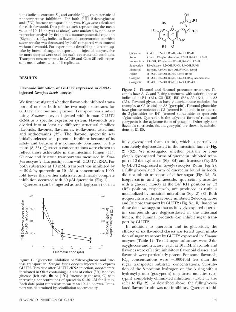
We first investigated whether flavonoids inhibited trans-port of one or both of the two major substrates forGLUT2: fructose and glucose. Transport was studiedusing Xenopus oocytes injected with human GLUT2cRNA as a specific expression system. Flavonoids aredivided into at least six different structural families:flavonols, flavones, flavanones, isoflavones, catechins,and anthocyanins (32). The flavonol quercetin wasinitially selected as a potential inhibitor because of itssafety and because it is commonly consumed by hu-mans (8, 33). Quercetin concentrations were chosen toreflect those achievable in the intestinal lumen (11).Glucose and fructose transport was measured in Xeno-pus oocytes 2 days postinjection with GLUT2 cRNA. Forboth substrates at 10 mM, transport was inhibited by� 50% by quercetin at 10 �M, a concentration 1000-fold lower than either substrate, and nearly completeinhibition occurred with 50 �M quercetin (Fig. 1).
Quercetin can be ingested as such (aglycone) or in a
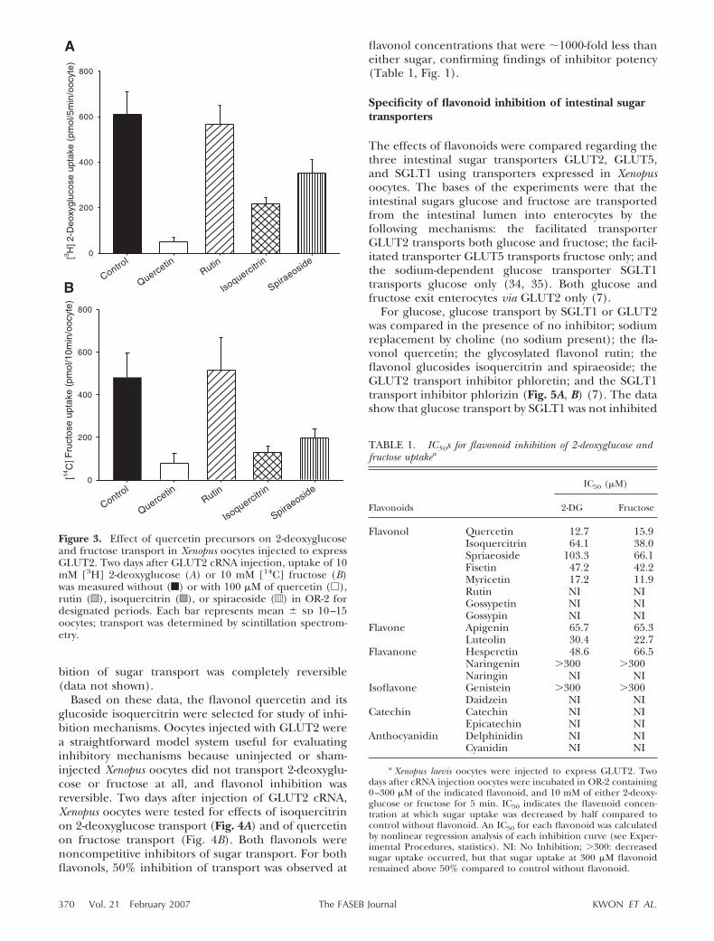
fully glycosylated form (rutin), which is partially orcompletely deglycosylated in the intestinal lumen (Fig.2) (32). We investigated whether partially or com-pletely glycosylated forms of quercetin inhibited trans-port of 2-deoxyglucose (Fig. 3A) and fructose (Fig. 3B)by GLUT2 expressed in Xenopus oocytes. Rutin (Fig. 2),a fully glycosylated form of quercetin found in foods,did not inhibit transport of either sugar (Fig. 3A, B).Isoquercitrin and spiraeoside, quercetin glucosideswith a glucose moiety at the B4�(R1) position or C3(R2) position, respectively, are produced as rutin ismetabolized by intestinal microflora (Fig. 2) (8). Bothisoquercitrin and spiraeoside inhibited 2-deoxyglucoseand fructose transport by GLUT2 (Fig. 3A, B). Based onthese data, we suggest that as fully glycosylated querce-tin compounds are deglycosylated in the intestinallumen, the luminal products can inhibit sugar trans-port by GLUT2.
In addition to quercetin and its glucosides, theefficacy of six flavonoid classes was tested upon inhibi-tion of sugar transport by GLUT2 expressed in Xenopusoocytes (Table 1). Tested sugar substrates were 2-de-oxyglucose and fructose, each at 10 mM. Flavonols andflavones were effective inhibitory flavonoid classes, andflavonols were particularly potent. For some flavonols,IC50 concentrations were �1000-fold less than thesugar transporter substrate concentrations. Substitu-tion of the 8 position hydrogen on the A ring with ahydroxyl group (gossypetin) or glucose moieties (gos-sypin) completely eliminated inhibition (Table 1; alsorefer to Fig. 2). As described above, the fully glycosy-lated flavonol rutin was not inhibitory. Quercetin inhi-
Figure 1. Quercetin inhibition of 2-deoxyglucose and fruc-tose transport in Xenopus laevis oocytes injected to expressGLUT2. Two days after GLUT2 cRNA injection, oocytes wereincubated in OR-2 containing 10 mM of either [3H] 2-deoxy-glucose (left axis, ● ) or [14C] fructose (right axis, E) withincreasing concentrations of quercetin 0–50 �M for 5 min.Each data point represents mean � sd 10–15 oocytes. Trans-port was determined by scintillation spectrometry.
Figure 2. Flavonol and flavonol precursor structures. Fla-vonols have A, C, and B ring structures, with substitutions asindicated at B4� (R1), C3 (R2), B5� (R3), A5 (R4), and A8(R5). Flavonol glycosides have glucorhamnose moieties, forexample, at C3 (rutin) or A8 (gossypin). Flavonol glucosideshave glucose moieties at C3 (termed isoquercitrin or querce-tin 3-glucoside) or B4� (termed spiraeoside or quercetin4�glucoside). Quercetin is the aglycone form of rutin, andgossypetin is the aglycone form of gossypin. Other aglyconeflavonols (myricetin, fisetin, gossypin) are shown by substitu-tions at R1-R5.
369FLAVONOID INHIBITION OF GLUT2
bition of sugar transport was completely reversible(data not shown).
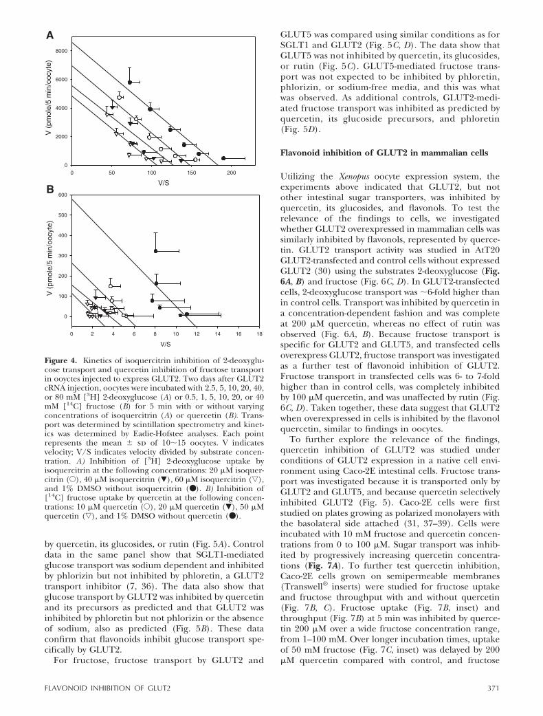
Based on these data, the flavonol quercetin and itsglucoside isoquercitrin were selected for study of inhi-bition mechanisms. Oocytes injected with GLUT2 werea straightforward model system useful for evaluatinginhibitory mechanisms because uninjected or sham-injected Xenopus oocytes did not transport 2-deoxyglu-cose or fructose at all, and flavonol inhibition wasreversible. Two days after injection of GLUT2 cRNA,Xenopus oocytes were tested for effects of isoquercitrinon 2-deoxyglucose transport (Fig. 4A) and of quercetinon fructose transport (Fig. 4B). Both flavonols werenoncompetitive inhibitors of sugar transport. For bothflavonols, 50% inhibition of transport was observed at
flavonol concentrations that were �1000-fold less thaneither sugar, confirming findings of inhibitor potency(Table 1, Fig. 1).
Specificity of flavonoid inhibition of intestinal sugartransporters
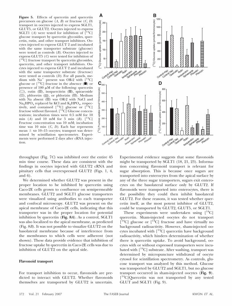
The effects of flavonoids were compared regarding thethree intestinal sugar transporters GLUT2, GLUT5,and SGLT1 using transporters expressed in Xenopusoocytes. The bases of the experiments were that theintestinal sugars glucose and fructose are transportedfrom the intestinal lumen into enterocytes by thefollowing mechanisms: the facilitated transporterGLUT2 transports both glucose and fructose; the facil-itated transporter GLUT5 transports fructose only; andthe sodium-dependent glucose transporter SGLT1transports glucose only (34, 35). Both glucose andfructose exit enterocytes via GLUT2 only (7).
For glucose, glucose transport by SGLT1 or GLUT2was compared in the presence of no inhibitor; sodiumreplacement by choline (no sodium present); the fla-vonol quercetin; the glycosylated flavonol rutin; theflavonol glucosides isoquercitrin and spiraeoside; theGLUT2 transport inhibitor phloretin; and the SGLT1transport inhibitor phlorizin (Fig. 5A, B) (7). The datashow that glucose transport by SGLT1 was not inhibited
Figure 3. Effect of quercetin precursors on 2-deoxyglucoseand fructose transport in Xenopus oocytes injected to expressGLUT2. Two days after GLUT2 cRNA injection, uptake of 10mM [3H] 2-deoxyglucose (A) or 10 mM [14C] fructose (B)was measured without (f) or with 100 �M of quercetin (�),rutin (o), isoquercitrin (s), or spiraeoside (d) in OR-2 fordesignated periods. Each bar represents mean � sd 10–15oocytes; transport was determined by scintillation spectrom-etry.
TABLE 1. IC50s for flavonoid inhibition of 2-deoxyglucose andfructose uptakea
Flavonoids
IC50 (�M)
2-DG Fructose
Flavonol Quercetin 12.7 15.9Isoquercitrin 64.1 38.0Spriaeoside 103.3 66.1Fisetin 47.2 42.2Myricetin 17.2 11.9Rutin NI NIGossypetin NI NIGossypin NI NI
Flavone Apigenin 65.7 65.3Luteolin 30.4 22.7
Flavanone Hesperetin 48.6 66.5Naringenin �300 �300Naringin NI NI
Isoflavone Genistein �300 �300Daidzein NI NI
Catechin Catechin NI NIEpicatechin NI NI
Anthocyanidin Delphinidin NI NICyanidin NI NI
a Xenopus laevis oocytes were injected to express GLUT2. Twodays after cRNA injection oocytes were incubated in OR-2 containing0–300 �M of the indicated flavonoid, and 10 mM of either 2-deoxy-glucose or fructose for 5 min. IC50 indicates the flavenoid concen-tration at which sugar uptake was decreased by half compared tocontrol without flavonoid. An IC50 for each flavonoid was calculatedby nonlinear regression analysis of each inhibition curve (see Exper-imental Procedures, statistics). NI: No Inhibition; �300: decreasedsugar uptake occurred, but that sugar uptake at 300 �M flavonoidremained above 50% compared to control without flavonoid.
370 Vol. 21 February 2007 KWON ET AL.The FASEB Journal
by quercetin, its glucosides, or rutin (Fig. 5A). Controldata in the same panel show that SGLT1-mediatedglucose transport was sodium dependent and inhibitedby phlorizin but not inhibited by phloretin, a GLUT2transport inhibitor (7, 36). The data also show thatglucose transport by GLUT2 was inhibited by quercetinand its precursors as predicted and that GLUT2 wasinhibited by phloretin but not phlorizin or the absenceof sodium, also as predicted (Fig. 5B). These dataconfirm that flavonoids inhibit glucose transport spe-cifically by GLUT2.
For fructose, fructose transport by GLUT2 and
GLUT5 was compared using similar conditions as forSGLT1 and GLUT2 (Fig. 5C, D). The data show thatGLUT5 was not inhibited by quercetin, its glucosides,or rutin (Fig. 5C). GLUT5-mediated fructose trans-port was not expected to be inhibited by phloretin,phlorizin, or sodium-free media, and this was whatwas observed. As additional controls, GLUT2-medi-ated fructose transport was inhibited as predicted byquercetin, its glucoside precursors, and phloretin(Fig. 5D).
Flavonoid inhibition of GLUT2 in mammalian cells
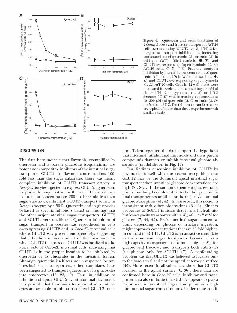
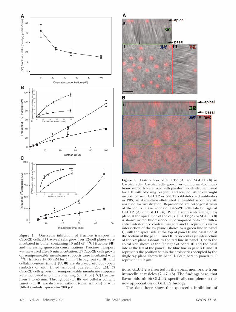
Utilizing the Xenopus oocyte expression system, theexperiments above indicated that GLUT2, but notother intestinal sugar transporters, was inhibited byquercetin, its glucosides, and flavonols. To test therelevance of the findings to cells, we investigatedwhether GLUT2 overexpressed in mammalian cells wassimilarly inhibited by flavonols, represented by querce-tin. GLUT2 transport activity was studied in AtT20GLUT2-transfected and control cells without expressedGLUT2 (30) using the substrates 2-deoxyglucose (Fig.6A, B) and fructose (Fig. 6C, D). In GLUT2-transfectedcells, 2-deoxyglucose transport was �6-fold higher thanin control cells. Transport was inhibited by quercetin ina concentration-dependent fashion and was completeat 200 �M quercetin, whereas no effect of rutin wasobserved (Fig. 6A, B). Because fructose transport isspecific for GLUT2 and GLUT5, and transfected cellsoverexpress GLUT2, fructose transport was investigatedas a further test of flavonoid inhibition of GLUT2.Fructose transport in transfected cells was 6- to 7-foldhigher than in control cells, was completely inhibitedby 100 �M quercetin, and was unaffected by rutin (Fig.6C, D). Taken together, these data suggest that GLUT2when overexpressed in cells is inhibited by the flavonolquercetin, similar to findings in oocytes.
To further explore the relevance of the findings,quercetin inhibition of GLUT2 was studied underconditions of GLUT2 expression in a native cell envi-ronment using Caco-2E intestinal cells. Fructose trans-port was investigated because it is transported only byGLUT2 and GLUT5, and because quercetin selectivelyinhibited GLUT2 (Fig. 5). Caco-2E cells were firststudied on plates growing as polarized monolayers withthe basolateral side attached (31, 37–39). Cells wereincubated with 10 mM fructose and quercetin concen-trations from 0 to 100 �M. Sugar transport was inhib-ited by progressively increasing quercetin concentra-tions (Fig. 7A). To further test quercetin inhibition,Caco-2E cells grown on semipermeable membranes(Transwell® inserts) were studied for fructose uptakeand fructose throughput with and without quercetin(Fig. 7B, C). Fructose uptake (Fig. 7B, inset) andthroughput (Fig. 7B) at 5 min was inhibited by querce-tin 200 �M over a wide fructose concentration range,from 1–100 mM. Over longer incubation times, uptakeof 50 mM fructose (Fig. 7C, inset) was delayed by 200�M quercetin compared with control, and fructose
Figure 4. Kinetics of isoquercitrin inhibition of 2-deoxyglu-cose transport and quercetin inhibition of fructose transportin ooyctes injected to express GLUT2. Two days after GLUT2cRNA injection, oocytes were incubated with 2.5, 5, 10, 20, 40,or 80 mM [3H] 2-deoxyglucose (A) or 0.5, 1, 5, 10, 20, or 40mM [14C] fructose (B) for 5 min with or without varyingconcentrations of isoquercitrin (A) or quercetin (B). Trans-port was determined by scintillation spectrometry and kinet-ics was determined by Eadie-Hofstee analyses. Each pointrepresents the mean � sd of 10�15 oocytes. V indicatesvelocity; V/S indicates velocity divided by substrate concen-tration. A) Inhibition of [3H] 2-deoxyglucose uptake byisoquercitrin at the following concentrations: 20 �M isoquer-citrin (E), 40 �M isoquercitrin (�), 60 �M isoquercitrin (ƒ),and 1% DMSO without isoquercitrin (● ). B) Inhibition of[14C] fructose uptake by quercetin at the following concen-trations: 10 �M quercetin (E), 20 �M quercetin (�), 50 �Mquercetin (ƒ), and 1% DMSO without quercetin (● ).
371FLAVONOID INHIBITION OF GLUT2
throughput (Fig. 7C) was inhibited over the entire 45min time course. These data are consistent with thefindings in oocytes injected with GLUT2 cRNA andpituitary cells that overexpressed GLUT2 (Figs. 1, 4,and 6).
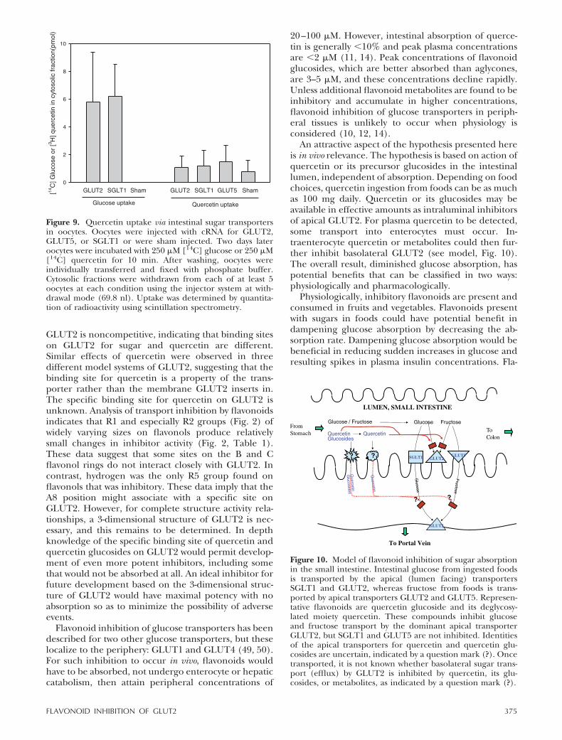
We determined whether GLUT2 was present in theproper location to be inhibited by quercetin usingCaco-2E cells grown to confluence on semipermeablemembranes. GLUT2 and SGLT1 glucose transporterswere visualized using antibodies to each transporterand confocal microscopy. GLUT2 was present on theapical membrane of Caco-2E cells, indicating that thistransporter was in the proper location for potentialinhibition by quercetin (Fig. 8A). As a control, SGLT1was also localized to the apical membrane, as predicted(Fig. 8B). It was not possible to visualize GLUT2 on thebasolateral membrane because of interference fromthe membranes to which cells were adherent (notshown). These data provide evidence that inhibition offructose uptake by quercetin in Caco-2E cells was due toinhibition of GLUT2 on the apical side.
Flavonoid transport
For transport inhibition to occur, flavonoids are pre-dicted to interact with GLUT2. Whether flavonoidsthemselves are transported by GLUT2 is uncertain.
Experimental evidence suggests that some flavonoidsmight be transported by SGLT1 (18, 21, 23). Informa-tion concerning flavonoid transport is relevant forsugar absorption. This is because once sugars aretransported into enterocytes from the apical surface byany of the three sugar transporters, sugars exit entero-cytes on the basolateral surface only by GLUT2. Ifflavonoids were transported into enterocytes, there isthe possibility they could then inhibit basolateralGLUT2. For these reasons, it was tested whether quer-cetin itself, as the most potent inhibitor of GLUT2,could be transported by GLUT2, GLUT5, or SGLT1.
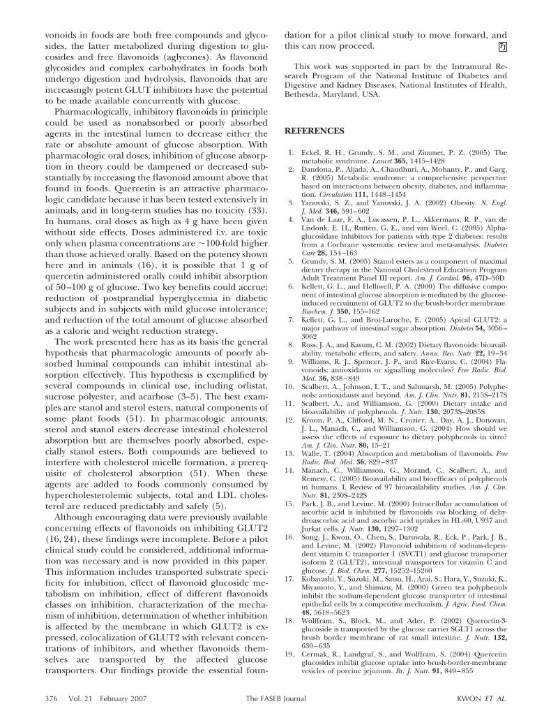
These experiments were undertaken using [14C]quercetin. Sham-injected oocytes do not transport[14C] glucose or [14C] fructose and have virtually nobackground radioactivity. However, sham-injected oo-cytes incubated with [14C] quercetin have backgroundradioactivity, which hinders determination of whetherthere is quercetin uptake. To avoid background, oo-cytes with or without expressed transporters were incu-bated with [14C] substrate. After washing, transport wasdetermined by micropuncture withdrawal of oocytecytosol for scintillation spectrometry. As controls, glu-cose transport was analyzed by this method. Glucosewas transported by GLUT2 and SGLT1, but no glucosetransport occurred in sham-injected oocytes (Fig. 9).[14C]Quercetin was not transported by any testedGLUT and SGLT1 (Fig. 9).
Figure 5. Effects of quercetin and quercetinprecursors on glucose (A, B) or fructose (C, D)transport in oocytes injected to express SGLT1,GLUT5, or GLUT2. Oocytes injected to expressSGLT1 (A) were tested for inhibition of [14C]glucose transport by quercetin glycosides, quer-cetin, rutin, and other transport inhibitors. Oo-cytes injected to express GLUT 2 and incubatedwith the same transporter substrate (glucose)were tested as controls (B). Oocytes injected toexpress GLUT5 (C) were tested for inhibition of[14C] fructose transport by quercetin glycosides,quercetin, and other transport inhibitors. Oo-cytes injected to express GLUT 2 and incubatedwith the same transporter substrate (fructose)were tested as controls (D). For all panels, me-dium with Na present was OR-2 with [14C]glucose or [14C] fructose in the absence (f) orpresence of 100 �M of the following: quercetin(�), rutin (o), isoquercitrin (s), spiraeoside(d), phloretin ([O), or phlorizin (z). Mediumwith Na absent (p) was OR-2 with NaCl andNa2HPO4 replaced by KCl and K2HPO4, respec-tively, and contained [14C] glucose or [14C]fructose without flavonol. [14C] Glucose concen-trations; incubation times were 0.5 mM for 10min (A) and 10 mM for 5 min (B). [14C]Fructose concentration was 10 mM; incubationtime was 10 min (C, D). Each bar representsmean � sd 10–15 oocytes; transport was deter-mined by scintillation spectrometry. Experi-ments were performed 2 days after cRNA injec-tion.
372 Vol. 21 February 2007 KWON ET AL.The FASEB Journal
DISCUSSION
The data here indicate that flavonols, exemplified byquercetin and a parent glucoside isoquercitrin, arepotent noncompetitive inhibitors of the intestinal sugartransporter GLUT2. At flavonol concentrations 100-fold less than the sugar substrates, there was nearlycomplete inhibition of GLUT2 transport activity inXenopus oocytes injected to express GLUT2. Quercetin,its glucoside isoquercitrin, or the related flavonol myr-icetin, all at concentrations 200- to 1000-fold less thansugar substrates, inhibited GLUT2 transport activity inXenopus oocytes by �50%. Quercetin and its glucosidesbehaved as specific inhibitors based on findings thatthe other major intestinal sugar transporters, GLUT5and SGLT1, were unaffected. Quercetin inhibition ofsugar transport in oocytes was reproduced in cellsoverexpressing GLUT2 and in Caco-2E intestinal cellswhere GLUT2 was present endogenously, suggestingthat inhibition is independent of the membrane inwhich GLUT2 is expressed. GLUT2 was localized to theapical side of Caco-2E intestinal cells, indicating thatGLUT2 is in the proper location to be inhibited byquercetin or its glucosides in the intestinal lumen.Although quercetin itself was not transported by anyintestinal sugar transporters, other candidates havebeen suggested to transport quercetin or its glucosidesinto enterocytes (13, 23, 40). Thus, in addition toinhibition of apical GLUT2 by intraluminal flavonoids,it is possible that flavonoids transported into entero-cytes are available to inhibit basolateral GLUT2 trans-
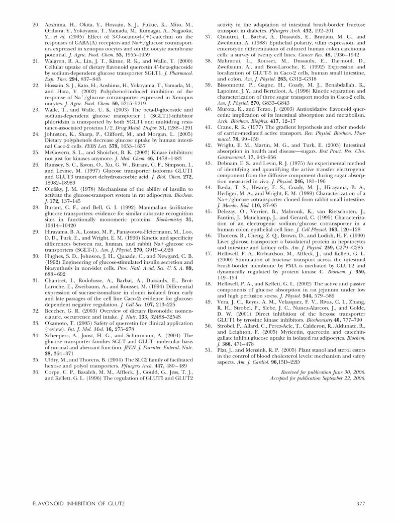
port. Taken together, the data support the hypothesisthat intestinal intraluminal flavonoids and their parentcompounds dampen or inhibit intestinal glucose ab-sorption (model shown in Fig. 10)
Our findings describing inhibition of GLUT2 byflavonoids fit well with the recent recognition thatGLUT2 may be the dominant apical intestinal sugartransporter when intestinal glucose concentrations arehigh (7). SGLT1, the sodium-dependent glucose trans-porter, has long been described to be the apical intes-tinal transporter responsible for the majority of luminalglucose absorption (41, 42). In retrospect, this notion isinconsistent with other observations (6, 43). Kineticsproperties of SGLT1 indicate that it is a high-affinitybut low-capacity transporter with a Km of � � 2 mM forglucose (7, 44, 45). Peak intestinal sugar concentra-tions, depending on glucose or fructose ingestion,might approach concentrations that are 50-fold higher.In contrast to SGLT1, GLUT2 is an attractive candidateas the dominant sugar transporter because it is ahigh-capacity transporter, has a much higher Km forglucose and fructose, and transports both substrates(vs. glucose only for SGLT1) (7). A confoundingproblem was that GLUT2 was believed to localize onlyto the basolateral and not the apical enterocyte surface(46). More recent localization data show that GLUT2localizes to the apical surface (6, 36); these data areconfirmed here in Caco-2E cells. Inhibitor and trans-porter data also indicate that GLUT2 appears to play amajor role in intestinal sugar absorption with highintraluminal sugar concentrations. Under these condi-
Figure 6. Quercetin and rutin inhibition of2-deoxyglucose and fructose transport in AtT-20cells overexpressing GLUT2. A, B) [3H] 2-De-oxyglucose transport inhibition by increasingconcentrations of quercetin (A) or rutin (B) inwild-type (WT) (filled symbols: ● , �) andGLUT2-overexpressing (open symbols: E, ƒ)AtT-20 cells. C, D) [14C] Fructose transportinhibition by increasing concentrations of quer-cetin (C) or rutin (D) in WT (filled symbols: �,Œ) and GLUT2-overexpressing (open symbols:�, ‚) AtT-20 cells. Cells in 12-well plates wereincubated in Krebs buffer containing 10 mM ofeither [3H] 2-deoxyglucose (A, B) or [14C]fructose (C, D) with increasing concentrations(0–200 �M) of quercetin (A, C) or rutin (B, D)for 5 min at 37°C. Data shown (mean�sd, n3)are typical of more than three experiments withsimilar results.
373FLAVONOID INHIBITION OF GLUT2
tions, GLUT 2 is inserted in the apical membrane fromintracellular vesicles (7, 47, 48). The findings here, thatflavonoids inhibit GLUT2, specifically complement thisnew appreciation of GLUT2 biology.
The data here show that quercetin inhibition of
Figure 7. Quercetin inhibition of fructose transport inCaco-2E cells. A) Caco-2E cells grown on 12-well plates wereincubated in buffer containing 10 mM of [14C] fructose (● )and increasing quercetin concentrations. Fructose transportwas measured after 5 min incubation. B) Caco-2E cells grownon semipermeable membrane supports were incubated with[14C] fructose 1–100 mM for 5 min. Throughput (�, f) andcellular content (inset) (E, F) are displayed without (opensymbols) or with (filled symbols) quercetin 200 �M. C)Caco-2E cells grown on semipermeable membrane supportswere incubated in buffer containing 50 mM of [14C] fructosefrom 5 to 45 min. Throughput (�, f) and cellular content(inset) (E, F) are displayed without (open symbols) or with(filled symbols) quercetin 200 �M.
Figure 8. Distribution of GLUT2 (A) and SGLT1 (B) inCaco-2E cells. Caco-2E cells grown on semipermeable mem-brane supports were fixed with paraformaldehyde, incubatedfor 1 h with blocking reagent, and washed. After overnightincubation with GLUT2 or SGLT1 rabbit-derived antibodiesin PBS, an Alexa-fluor546-labeled anti-rabbit secondary Abwas used for visualization. Represented are orthogonal viewsof the entire z axis series of Caco-2E cells labeled againstGLUT2 (A) or SGLT1 (B). Panel I represents a single x-yplane at the apical side of the cells. GLUT2 (A) or SGLT1 (B)is shown in red fluorescence superimposed onto the differ-ential interference contrast image. Panel II represents an x-zintersection of the x-y plane (shown by a green line in panelI), with the apical side at the top of panel II and basal side atthe bottom of the panel. Panel III represents a y-z intersectionof the x-y plane (shown by the red line in panel I), with theapical side shown at the far right of panel III and the basalside at the left of the panel. The blue line in panels II and IIIrepresents the position within the z axis series occupied by thesingle x-y plane shown in panel I. Scale bars in panels A, Brepresent �10 �m.
374 Vol. 21 February 2007 KWON ET AL.The FASEB Journal
GLUT2 is noncompetitive, indicating that binding siteson GLUT2 for sugar and quercetin are different.Similar effects of quercetin were observed in threedifferent model systems of GLUT2, suggesting that thebinding site for quercetin is a property of the trans-porter rather than the membrane GLUT2 inserts in.The specific binding site for quercetin on GLUT2 isunknown. Analysis of transport inhibition by flavonoidsindicates that R1 and especially R2 groups (Fig. 2) ofwidely varying sizes on flavonols produce relativelysmall changes in inhibitor activity (Fig. 2, Table 1).These data suggest that some sites on the B and Cflavonol rings do not interact closely with GLUT2. Incontrast, hydrogen was the only R5 group found onflavonols that was inhibitory. These data imply that theA8 position might associate with a specific site onGLUT2. However, for complete structure activity rela-tionships, a 3-dimensional structure of GLUT2 is nec-essary, and this remains to be determined. In depthknowledge of the specific binding site of quercetin andquercetin glucosides on GLUT2 would permit develop-ment of even more potent inhibitors, including somethat would not be absorbed at all. An ideal inhibitor forfuture development based on the 3-dimensional struc-ture of GLUT2 would have maximal potency with noabsorption so as to minimize the possibility of adverseevents.
Flavonoid inhibition of glucose transporters has beendescribed for two other glucose transporters, but theselocalize to the periphery: GLUT1 and GLUT4 (49, 50).For such inhibition to occur in vivo, flavonoids wouldhave to be absorbed, not undergo enterocyte or hepaticcatabolism, then attain peripheral concentrations of
20–100 �M. However, intestinal absorption of querce-tin is generally �10% and peak plasma concentrationsare �2 �M (11, 14). Peak concentrations of flavonoidglucosides, which are better absorbed than aglycones,are 3–5 �M, and these concentrations decline rapidly.Unless additional flavonoid metabolites are found to beinhibitory and accumulate in higher concentrations,flavonoid inhibition of glucose transporters in periph-eral tissues is unlikely to occur when physiology isconsidered (10, 12, 14).
An attractive aspect of the hypothesis presented hereis in vivo relevance. The hypothesis is based on action ofquercetin or its precursor glucosides in the intestinallumen, independent of absorption. Depending on foodchoices, quercetin ingestion from foods can be as muchas 100 mg daily. Quercetin or its glucosides may beavailable in effective amounts as intraluminal inhibitorsof apical GLUT2. For plasma quercetin to be detected,some transport into enterocytes must occur. In-traenterocyte quercetin or metabolites could then fur-ther inhibit basolateral GLUT2 (see model, Fig. 10).The overall result, diminished glucose absorption, haspotential benefits that can be classified in two ways:physiologically and pharmacologically.
Physiologically, inhibitory flavonoids are present andconsumed in fruits and vegetables. Flavonoids presentwith sugars in foods could have potential benefit indampening glucose absorption by decreasing the ab-sorption rate. Dampening glucose absorption would bebeneficial in reducing sudden increases in glucose andresulting spikes in plasma insulin concentrations. Fla-
Figure 10. Model of flavonoid inhibition of sugar absorptionin the small intestine. Intestinal glucose from ingested foodsis transported by the apical (lumen facing) transportersSGLT1 and GLUT2, whereas fructose from foods is trans-ported by apical transporters GLUT2 and GLUT5. Represen-tative flavonoids are quercetin glucoside and its deglycosy-lated moiety quercetin. These compounds inhibit glucoseand fructose transport by the dominant apical transporterGLUT2, but SGLT1 and GLUT5 are not inhibited. Identitiesof the apical transporters for quercetin and quercetin glu-cosides are uncertain, indicated by a question mark (?). Oncetransported, it is not known whether basolateral sugar trans-port (efflux) by GLUT2 is inhibited by quercetin, its glu-cosides, or metabolites, as indicated by a question mark (?).
Figure 9. Quercetin uptake via intestinal sugar transportersin oocytes. Oocytes were injected with cRNA for GLUT2,GLUT5, or SGLT1 or were sham injected. Two days lateroocytes were incubated with 250 �M [14C] glucose or 250 �M[14C] quercetin for 10 min. After washing, oocytes wereindividually transferred and fixed with phosphate buffer.Cytosolic fractions were withdrawn from each of at least 5oocytes at each condition using the injector system at with-drawal mode (69.8 nl). Uptake was determined by quantita-tion of radioactivity using scintillation spectrometry.
375FLAVONOID INHIBITION OF GLUT2
vonoids in foods are both free compounds and glyco-sides, the latter metabolized during digestion to glu-cosides and free flavonoids (aglycones). As flavonoidglycosides and complex carbohydrates in foods bothundergo digestion and hydrolysis, flavonoids that areincreasingly potent GLUT inhibitors have the potentialto be made available concurrently with glucose.
Pharmacologically, inhibitory flavonoids in principlecould be used as nonabsorbed or poorly absorbedagents in the intestinal lumen to decrease either therate or absolute amount of glucose absorption. Withpharmacologic oral doses, inhibition of glucose absorp-tion in theory could be dampened or decreased sub-stantially by increasing the flavonoid amount above thatfound in foods. Quercetin is an attractive pharmaco-logic candidate because it has been tested extensively inanimals, and in long-term studies has no toxicity (33).In humans, oral doses as high as 4 g have been givenwithout side effects. Doses administered i.v. are toxiconly when plasma concentrations are �100-fold higherthan those achieved orally. Based on the potency shownhere and in animals (16), it is possible that 1 g ofquercetin administered orally could inhibit absorptionof 50–100 g of glucose. Two key benefits could accrue:reduction of postprandial hyperglycemia in diabeticsubjects and in subjects with mild glucose intolerance;and reduction of the total amount of glucose absorbedas a caloric and weight reduction strategy.
The work presented here has as its basis the generalhypothesis that pharmacologic amounts of poorly ab-sorbed luminal compounds can inhibit intestinal ab-sorption effectively. This hypothesis is exemplified byseveral compounds in clinical use, including orlistat,sucrose polyester, and acarbose (3–5). The best exam-ples are stanol and sterol esters, natural components ofsome plant foods (51). In pharmacologic amounts,sterol and stanol esters decrease intestinal cholesterolabsorption but are themselves poorly absorbed, espe-cially stanol esters. Both compounds are believed tointerfere with cholesterol micelle formation, a prereq-uisite of cholesterol absorption (51). When theseagents are added to foods commonly consumed byhypercholesterolemic subjects, total and LDL choles-terol are reduced predictably and safely (5).
Although encouraging data were previously availableconcerning effects of flavonoids on inhibiting GLUT2(16, 24), these findings were incomplete. Before a pilotclinical study could be considered, additional informa-tion was necessary and is now provided in this paper.This information includes transported substrate speci-ficity for inhibition, effect of flavonoid glucoside me-tabolism on inhibition, effect of different flavonoidsclasses on inhibition, characterization of the mecha-nism of inhibition, determination of whether inhibitionis affected by the membrane in which GLUT2 is ex-pressed, colocalization of GLUT2 with relevant concen-trations of inhibitors, and whether flavonoids them-selves are transported by the affected glucosetransporters. Our findings provide the essential foun-
dation for a pilot clinical study to move forward, andthis can now proceed.
This work was supported in part by the Intramural Re-search Program of the National Institute of Diabetes andDigestive and Kidney Diseases, National Institutes of Health,Bethesda, Maryland, USA.
REFERENCES
1. Eckel, R. H., Grundy, S. M., and Zimmet, P. Z. (2005) Themetabolic syndrome. Lancet 365, 1415–1428
2. Dandona, P., Aljada, A., Chaudhuri, A., Mohanty, P., and Garg,R. (2005) Metabolic syndrome: a comprehensive perspectivebased on interactions between obesity, diabetes, and inflamma-tion. Circulation 111, 1448–1454
3. Yanovski, S. Z., and Yanovski, J. A. (2002) Obesity. N. Engl.J. Med. 346, 591–602
4. Van de Laar, F. A., Lucassen, P. L., Akkermans, R. P., van deLisdonk, E. H., Rutten, G. E., and van Weel, C. (2005) Alpha-glucosidase inhibitors for patients with type 2 diabetes: resultsfrom a Cochrane systematic review and meta-analysis. DiabetesCare 28, 154–163
5. Grundy, S. M. (2005) Stanol esters as a component of maximaldietary therapy in the National Cholesterol Education ProgramAdult Treatment Panel III report. Am. J. Cardiol. 96, 47D–50D
6. Kellett, G. L., and Helliwell, P. A. (2000) The diffusive compo-nent of intestinal glucose absorption is mediated by the glucose-induced recruitment of GLUT2 to the brush-border membrane.Biochem. J. 350, 155–162
7. Kellett, G. L., and Brot-Laroche, E. (2005) Apical GLUT2: amajor pathway of intestinal sugar absorption. Diabetes 54, 3056–3062
8. Ross, J. A., and Kasum, C. M. (2002) Dietary flavonoids: bioavail-ability, metabolic effects, and safety. Annu. Rev. Nutr. 22, 19–34
9. Williams, R. J., Spencer, J. P., and Rice-Evans, C. (2004) Fla-vonoids: antioxidants or signalling molecules? Free Radic. Biol.Med. 36, 838–849
10. Scalbert, A., Johnson, I. T., and Saltmarsh, M. (2005) Polyphe-nols: antioxidants and beyond. Am. J. Clin. Nutr. 81, 215S–217S
11. Scalbert, A., and Williamson, G. (2000) Dietary intake andbioavailability of polyphenols. J. Nutr. 130, 2073S–2085S
12. Kroon, P. A., Clifford, M. N., Crozier, A., Day, A. J., Donovan,J. L., Manach, C., and Williamson, G. (2004) How should weassess the effects of exposure to dietary polyphenols in vitro?Am. J. Clin. Nutr. 80, 15–21
13. Walle, T. (2004) Absorption and metabolism of flavonoids. FreeRadic. Biol. Med. 36, 829–837
14. Manach, C., Williamson, G., Morand, C., Scalbert, A., andRemesy, C. (2005) Bioavailability and bioefficacy of polyphenolsin humans. I. Review of 97 bioavailability studies. Am. J. Clin.Nutr. 81, 230S–242S
15. Park, J. B., and Levine, M. (2000) Intracellular accumulation ofascorbic acid is inhibited by flavonoids via blocking of dehy-droascorbic acid and ascorbic acid uptakes in HL-60, U937 andJurkat cells. J. Nutr. 130, 1297–1302
16. Song, J., Kwon, O., Chen, S., Daruwala, R., Eck, P., Park, J. B.,and Levine, M. (2002) Flavonoid inhibition of sodium-depen-dent vitamin C transporter 1 (SVCT1) and glucose transporterisoform 2 (GLUT2), intestinal transporters for vitamin C andglucose. J. Biol. Chem. 277, 15252–15260
17. Kobayashi, Y., Suzuki, M., Satsu, H., Arai, S., Hara, Y., Suzuki, K.,Miyamoto, Y., and Shimizu, M. (2000) Green tea polyphenolsinhibit the sodium-dependent glucose transporter of intestinalepithelial cells by a competitive mechanism. J. Agric. Food. Chem.48, 5618–5623
18. Wolffram, S., Block, M., and Ader, P. (2002) Quercetin-3-glucoside is transported by the glucose carrier SGLT1 across thebrush border membrane of rat small intestine. J. Nutr. 132,630–635
19. Cermak, R., Landgraf, S., and Wolffram, S. (2004) Quercetinglucosides inhibit glucose uptake into brush-border-membranevesicles of porcine jejunum. Br. J. Nutr. 91, 849–855
376 Vol. 21 February 2007 KWON ET AL.The FASEB Journal
20. Aoshima, H., Okita, Y., Hossain, S. J., Fukue, K., Mito, M.,Orihara, Y., Yokoyama, T., Yamada, M., Kumagai, A., Nagaoka,Y., et al. (2005) Effect of 3-O-octanoyl-()-catechin on theresponses of GABA(A) receptors and Na/glucose cotransport-ers expressed in xenopus oocytes and on the oocyte membranepotential. J. Agric. Food. Chem. 53, 1955–1959
21. Walgren, R. A., Lin, J. T., Kinne, R. K., and Walle, T. (2000)Cellular uptake of dietary flavonoid quercetin 4�-beta-glucosideby sodium-dependent glucose transporter SGLT1. J. Pharmacol.Exp. Ther. 294, 837–843
22. Hossain, S. J., Kato, H., Aoshima, H., Yokoyama, T., Yamada, M.,and Hara, Y. (2002) Polyphenol-induced inhibition of theresponse of Na/glucose cotransporter expressed in Xenopusoocytes. J. Agric. Food. Chem. 50, 5215–5219
23. Walle, T., and Walle, U. K. (2003) The beta-D-glucoside andsodium-dependent glucose transporter 1 (SGLT1)-inhibitorphloridzin is transported by both SGLT1 and multidrug resis-tance-associated proteins 1/2. Drug Metab. Dispos. 31, 1288–1291
24. Johnston, K., Sharp, P., Clifford, M., and Morgan, L. (2005)Dietary polyphenols decrease glucose uptake by human intesti-nal Caco-2 cells. FEBS Lett. 579, 1653–1657
25. McGovern, S. L., and Shoichet, B. K. (2003) Kinase inhibitors:not just for kinases anymore. J. Med. Chem. 46, 1478–1483
26. Rumsey, S. C., Kwon, O., Xu, G. W., Burant, C. F., Simpson, I.,and Levine, M. (1997) Glucose transporter isoforms GLUT1and GLUT3 transport dehydroascorbic acid. J. Biol. Chem. 272,18982–18989
27. Olefsky, J. M. (1978) Mechanisms of the ability of insulin toactivate the glucose-transport system in rat adipocytes. Biochem.J. 172, 137–145
28. Burant, C. F., and Bell, G. I. (1992) Mammalian facilitativeglucose transporters: evidence for similar substrate recognitionsites in functionally monomeric proteins. Biochemistry 31,10414–10420
29. Hirayama, B. A., Lostao, M. P., Panayotova-Heiermann, M., Loo,D. D., Turk, E., and Wright, E. M. (1996) Kinetic and specificitydifferences between rat, human, and rabbit Na-glucose co-transporters (SGLT-1). Am. J. Physiol. 270, G919–G926
30. Hughes, S. D., Johnson, J. H., Quaade, C., and Newgard, C. B.(1992) Engineering of glucose-stimulated insulin secretion andbiosynthesis in non-islet cells. Proc. Natl. Acad. Sci. U. S. A. 89,688–692
31. Chantret, I., Rodolosse, A., Barbat, A., Dussaulx, E., Brot-Laroche, E., Zweibaum, A., and Rousset, M. (1994) Differentialexpression of sucrase-isomaltase in clones isolated from earlyand late passages of the cell line Caco-2: evidence for glucose-dependent negative regulation. J. Cell Sci. 107, 213–225
32. Beecher, G. R. (2003) Overview of dietary flavonoids: nomen-clature, occurrence and intake. J. Nutr. 133, 3248S–3254S
33. Okamoto, T. (2005) Safety of quercetin for clinical application(review). Int. J. Mol. Med. 16, 275–278
34. Scheepers, A., Joost, H. G., and Schurmann, A. (2004) Theglucose transporter families SGLT and GLUT: molecular basisof normal and aberrant function. JPEN. J. Parenter. Enteral. Nutr.28, 364–371
35. Uldry, M., and Thorens, B. (2004) The SLC2 family of facilitatedhexose and polyol transporters. Pfluegers Arch. 447, 480–489
36. Corpe, C. P., Basaleh, M. M., Affleck, J., Gould, G., Jess, T. J.,and Kellett, G. L. (1996) The regulation of GLUT5 and GLUT2
activity in the adaptation of intestinal brush-border fructosetransport in diabetes. Pfluegers Arch. 432, 192–201
37. Chantret, I., Barbat, A., Dussaulx, E., Brattain, M. G., andZweibaum, A. (1988) Epithelial polarity, villin expression, andenterocytic differentiation of cultured human colon carcinomacells: a survey of twenty cell lines. Cancer Res. 48, 1936–1942
38. Mahraoui, L., Rousset, M., Dussaulx, E., Darmoul, D.,Zweibaum, A., and Brot-Laroche, E. (1992) Expression andlocalization of GLUT-5 in Caco-2 cells, human small intestine,and colon. Am. J. Physiol. 263, G312–G318
39. Bissonnette, P., Gagne, H., Coady, M. J., Benabdallah, K.,Lapointe, J. Y., and Berteloot, A. (1996) Kinetic separation andcharacterization of three sugar transport modes in Caco-2 cells.Am. J. Physiol. 270, G833–G843
40. Murota, K., and Terao, J. (2003) Antioxidative flavonoid quer-cetin: implication of its intestinal absorption and metabolism.Arch. Biochem. Biophys. 417, 12–17
41. Crane, R. K. (1977) The gradient hypothesis and other modelsof carrier-mediated active transport. Rev. Physiol. Biochem. Phar-macol. 78, 99–159
42. Wright, E. M., Martin, M. G., and Turk, E. (2003) Intestinalabsorption in health and disease—sugars. Best Pract. Res. Clin.Gastroenterol. 17, 943–956
43. Debnam, E. S., and Levin, R. J. (1975) An experimental methodof identifying and quantifying the active transfer electrogeniccomponent from the diffusive component during sugar absorp-tion measured in vivo. J. Physiol. 246, 181–196
44. Ikeda, T. S., Hwang, E. S., Coady, M. J., Hirayama, B. A.,Hediger, M. A., and Wright, E. M. (1989) Characterization of aNa/glucose cotransporter cloned from rabbit small intestine.J. Membr. Biol. 110, 87–95
45. Delezay, O., Verrier, B., Mabrouk, K., van Rietschoten, J.,Fantini, J., Mauchamp, J., and Gerard, C. (1995) Characteriza-tion of an electrogenic sodium/glucose cotransporter in ahuman colon epithelial cell line. J. Cell Physiol. 163, 120–128
46. Thorens, B., Cheng, Z. Q., Brown, D., and Lodish, H. F. (1990)Liver glucose transporter: a basolateral protein in hepatocytesand intestine and kidney cells. Am. J. Physiol. 259, C279–C285
47. Helliwell, P. A., Richardson, M., Affleck, J., and Kellett, G. L.(2000) Stimulation of fructose transport across the intestinalbrush-border membrane by PMA is mediated by GLUT2 anddynamically regulated by protein kinase C. Biochem. J. 350,149–154
48. Helliwell, P. A., and Kellett, G. L. (2002) The active and passivecomponents of glucose absorption in rat jejunum under lowand high perfusion stress. J. Physiol. 544, 579–589
49. Vera, J. C., Reyes, A. M., Velasquez, F. V., Rivas, C. I., Zhang,R. H., Strobel, P., Slebe, J. C., Nunez-Alarcon, J., and Golde,D. W. (2001) Direct inhibition of the hexose transporterGLUT1 by tyrosine kinase inhibitors. Biochemistry 40, 777–790
50. Strobel, P., Allard, C., Perez-Acle, T., Calderon, R., Aldunate, R.,and Leighton, F. (2005) Myricetin, quercetin and catechin-gallate inhibit glucose uptake in isolated rat adipocytes. Biochem.J. 386, 471–478
51. Plat, J., and Mensink, R. P. (2005) Plant stanol and sterol estersin the control of blood cholesterol levels: mechanism and safetyaspects. Am. J. Cardiol. 96,15D–22D
Received for publication June 30, 2006.Accepted for publication September 22, 2006.
377FLAVONOID INHIBITION OF GLUT2