insects jump to provide new rubber-like material: biomaterials
TRANSCRIPT
Improving the diagnosis of genetic
disease requires new methods for the
rapid and highly sensitive detection of
DNA.
Tza-Huei Wang and colleagues at The
Johns Hopkins University have
developed a quantum dot (QD)-based
sensor that produces a distinct signal
on binding just 50 copies of DNA or
less [Zhang et al., Nat. Mater. (2005)
doi: 10.1038/nmat1508].
The sensor consists of a CdSe-ZnS
core-shell QD decorated with
streptavidin molecules and two short,
single-stranded DNA probes, one with
a fluorescent dye attached and the
other labeled with biotin. The probes
are designed to bind to opposite ends
of a target DNA molecule. When the
target DNA is present, the two probes
assemble on the target and bind the
QD through biotin-streptavidin links.
This brings the fluorescent dye into
proximity with the QD.
The team uses a laser that excites the
fluorescence of the QDs but not the
dye on the free DNA probes. Without
any target DNA, only QD fluorescence
is observed. When target DNA is
present, the proximity of the dye and
QD in the QD-DNA assembly allows
fluorescence resonance energy transfer
(FRET) to occur and emission from the
dye is observed.
Many target DNA molecules are
concentrated at the surface of the QD,
giving an increased signal. As a result,
the current detection limit is already
~100 times better than conventional
FRET-based assays.
The Johns Hopkins researchers have
demonstrated the potential of their
QD sensor system for diagnosis. In
combination with a standard assay,
they used the sensor to detect DNA
point mutations in clinical samples
from patients with a type of ovarian
cancer.
“The ultrahigh sensitivity and accuracy
of this QD nanosensor makes it an
ideal tool for diagnosis of disease at an
early stage,” says Tza-Huei Wang.
Jonathan Wood
Single QDsdetect DNANANOTECHNOLOGY
Resilin is an elastic protein related to elastin, gluten, and
spider silks that is characterized by low stiffness, high strain,
and efficient energy storage. It enables insects to fly and fleas
to jump up to 200 times the length of their body.
Australian researchers have now transferred the first portion
of the resilin gene from a fruit fly into Escherichia coli
bacteria, enabling a soluble protein to be produced in the
laboratory [Elvin et al., Nature (2005) 437, 999]. The team
from the Commonwealth Scientific and Industrial Research
Organisation (CSIRO), the University of Queensland, and the
Australian National University is then able to cast the protein
into a rubber-like, high molecular weight biomaterial by rapid
Ru(II)-mediated photocrosslinking.
“We believe that our work will greatly facilitate structural
investigations into the functional properties of resilin and shed
light on more general aspects of the structure of elastomeric
proteins,” say the researchers.
The resiliency of the samples (the ability to recover after
deformation under an applied stresss) was compared to their
biological counterparts. A tendon from a dragonfly wing
shows a resiliency of ~92% with negligible hysteresis upon
compression. In comparison, the resiliency of lab-formed
resilin samples varies between 90% and 92%, which is about
10% better than other high-resiliency synthetic polymers.
Patrick Cain
Insects jump to provide new rubber-like materialBIOMATERIALS
Tissue-like constructs made in minutesBIOMATERIALS
Tissue engineering typically involves seeding a scaffold
material with cells, then allowing the cells to grow in the
laboratory and remodel the construct into a biologically
functional tissue. This is a slow process that is expensive
and difficult to control, and it has found limited success.
Rather than rely on cells, UK researchers at University
College London and Eastman Dental Institute have
simply and rapidly engineered tissue-like constructs
without waiting for cell participation [Brown et al., Adv.
Funct. Mater. (2005) 15 (11), 1762].
The starting point is hyperhydrated collagen gels seeded
with human dermal fibroblasts. Collagen is a protein
that acts as a structural support in a wide range of
tissues including skin, bone, tendons, ligaments,
cartilage, blood vessels, and nerves. The team discovered
that a compressive load could rapidly expel liquid from
the gels. Placing the gels on blotting paper and using a
load of 50 g for 5 minutes reduces a 3.6 mm high gel
into a sheet of ~30 µm thickness. Since the thin sheets
are difficult to handle, the researchers rolled them up
into tubes. This process can be extended to construct
complex, heterogeneous three-dimensional structures
with dimensions of 10-100 µm from the collagen sheets.
Fine collagen lamellae are found throughout the
compressed sheets and the process does not affect cell
viability. In fact, the cells tend to line up along the
collagen lamellae, assuming a tissue-like distribution and
morphology. The break stress of the collagen sheets is
0.55 MPa, which is greater than conventional cell-seeded
collagen gels even after being cultured for weeks. “The
construct is shrunk to give micro- and nanoscale
features with a biomimetic collagen fibrillar architecture,
density, and mechanical strength in minutes,” says
Robert A. Brown.
The researchers believe that fabrication of some
collagen-based skin equivalents could become
>200 times faster using their technique. They also
suggest that drug release from native collagen gels
would be improved through greater control of pore size
and increased mechanical strength.
“This process is not necessarily limited to collagen,
though this is by far the most important and useful
scaffold material as it means the completed construct is
close to being a natural biological tissue,” explains
Brown. “The speed and engineering control which this
provides suggests that we can now work toward the
bedside fabrication of customized implants for patients,”
he claims.
Jonathan Wood
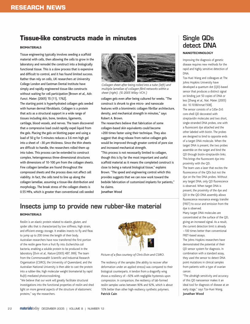
Collagen sheet after being rolled into a tube (left) and
multiple lamellae of collagen fibril networks within a
sheet (right). (© 2005 Wiley-VCH.)
DECEMBER 2005 | VOLUME 8 | NUMBER 12 22
RESEARCH NEWS
Picture of a flea courtesy of Chris Elvin and CSIRO.