insight into sucnr1 (gpr91) structure and function et al 2016.pdf · insight into sucnr1 (gpr91)...
TRANSCRIPT
Published in: Pharmacology & Therapeutics (2016) Status: Postprint (Author’s version)
Insight into SUCNR1 (GPR91) structure and function Julie Gilissen a,b, François Jouret c,d, Bernard Pirotte b, Julien Hanson a,b a Laboratory of Molecular Pharmacology, GIGA-Molecular Biology of Diseases, University of Liège, Liège, Belgium b Laboratory of Medicinal Chemistry, Centre for Interdisciplinary Research on Medicines (CIRM), University of Liège, Belgium c Laboratory of Experimental Surgery, GIGA-Cardiovascular Sciences, University of Liège, Liège, Belgium d Division of Nephrology, University of Liège Hospital (ULg CHU), Liège, Belgium
ABSTRACT SUCNR1 (or GPR91) belongs to the family of G protein-coupled receptors (GPCR), which represents the largest group of membrane proteins in human genome. The majority of marketed drugs targets GPCRs, directly or indirectly. SUCNR1 has been classified as an orphan receptor until a landmark study paired it with succinate, a citric acid cycle intermediate.
According to the current paradigm, succinate triggers SUCNR1 signaling pathways to indicate local stress that may affect cellular metabolism. SUCNR1 implication has been well documented in renin-induced hypertension, ischemia/reperfusion injury, inflammation and immune response, platelet aggregation and retinal angiogenesis. In addition, the SUCNR1-induced increase of blood pressure may contribute to diabetic nephropathy or cardiac hypertrophy.
The understanding of SUCNR1 activation, signaling pathways and functions remains largely elusive, which calls for deeper investigations. SUCNR1 shows a high potential as an innovative drug target and is probably an important regulator of basic physiology. In order to achieve the full characterization of this receptor, more specific pharmacological tools such as small-molecules modulators will represent an important asset. In this review, we describe the structural features of SUCNR1, its current ligands and putative binding pocket. We give an exhaustive overview of the known and hypothetical signaling partners of the receptor in different in vitro and in vivo systems. The link between SUCNR1 intracellular pathways and its pathophysiological roles are also extensively discussed.
KEYWORDS SUCNR1 ; GPR91 ; Succinate ; Ischemia-reperfusion injury ; Hypertension ; Diabetes
ABBREVIATIONS (GPCRs), G protein-coupled receptors family; (SUCNR.1), succinate receptor 1 ; (AA), amino acids; (7TM), seven transmembrane domains; (ECLs), extracellular loops; (ICLs), intracellular loops; (cAMP), cyclic adenosine monophosphate; (AC), adenylate cyclase; (GRKs), G protein-coupled receptor kinases; (CHO) cells, Chinese hamster ovary; (HEK293) cells, human embryonic kidney; (MDCK) cells, madin darby canin kidney; (iDC), immature dendritic cells; (PTX), pertussis toxin; (ERK1/2), extracellular signal-regulated kinases 1 and 2; (RGC), retina ganglion cells; (RAS), renin-angiotensin system; (JGA), juxtaglomerular apparatus; (MD), macula densa; (JG) cells, juxtaglomerular; (COX-2), cyclooxygenase 2; E2 (PGE2), prostaglandin; (PGI2), prostaglandin 12; (AA), arachidonic acid; (GENCs), juxtaglomerular endothelium cells; (NO), nitric oxide; (CH), cardiac hypertrophy; (CaMKIIδ), calcium/ calmodulin-dependent protein kinase IIδ; (HDAC5), histone deacetylase 5; (PKA), protein kinase A; (PLN), phospholamban; (RyR2), ryanodine receptor 2; (PDE), phosphodiesterase; (RVH), right ventricular hypertrophy; (LVH), left ventricle hypertrophy; (PI3K), phosphatidylinositol-4,5-bisphosphate 3-kinase; (Akt), protein kinase B; (WAT), white adipose tissue; (MS), metabolic syndrome; (ASA), acetylsalicylic acid; (ADP), adenosine diphosphate; (TXA2), thromboxane A2; (DR), diabetic retinopathy; (VEGF), vascular endothelial growth factor; (JNK), c-Jun N-terminal kinases; (ROP), retinopathy of prematurity ; (CHI), cerebral hypoxic-ischemic; (EP4), prostaglandin E receptor 4; (AMD), age-related macular degeneration; (RPE), retinal pigment epithelium; (FSK), forskolin; (HSC), hepatic stellate cells; (α-SMA),α-smooth muscle actin; (HPC), hematopoietic progenitor cells; (IP), inositol phosphate; (iDC), immature dendritic cells; (TNF-α), tumor necrosis factor α; (IL-1β), pro-inflammatory cytokine interleukin-1 beta; (IFN--γ), interferon-γ; (IRI), ischemia-reperfusion injury.

1. SUCCINATE RECEPTOR 1 STRUCTURE AND LIGANDS SUCNR1 was first spotted in a megacaryocytic cell line in 1995 and called "P2U2", a name coined for its homology with the purinergic receptor P2Y2, known as P2u at that time (Gonzalez et al., 2004). SUCNR1 gene was later re-discovered as GPR91 in 2001 on human chromosome 3q24-3q25 using an expressed sequence tag data mining strategy (Wittenberger et al., 2001). Of important note is the possibility of two open-reading frames (ORF) for SUCNR1, one giving a protein of 330 amino acids (AA) and the other one 334 AA. Wittenberger et al. noted that the 330-AA protein was more likely to be expressed given the Kozak sequence surrounding the second ATG (Wittenberger et al., 2001). In the present article we will use the AA numbering according to a 330-AA protein, although the current databases sometimes report SUCNR1 as being 334-AA long. There is a high degree of homology between man and mouse (68%) with the exception of the C-terminal tail, which is 12 AA shorter in rodents (Ariza et al., 2012; Wittenberger et al., 2001). In humans, other genes have been found on the same locus and consist in a cluster of P2Y1, P2Y12, P2Y13, H963 (GPR171) and GPR87 (Abbracchio et al., 2006). In their seminal paper, Wittenberger et al. speculated that all these receptors originated from a common ancestor, presumably a nucleotide receptor (Wittenberger et al., 2001). Several comprehensive reviews have been published on the receptor or succinate role in metabolic/oxidative stress conditions (Ariza et al., 2012; Peti-Peterdi et al., 2013). The present work, in addition to discussing the most recent developments, considers extensively the mechanisms linking SUCNR1-activated signaling pathways and all (patho)physiological states where the receptor might play a role. SUCNR1 belongs to G protein-coupled receptors (GPCRs) family. In the human genome, it is the largest group of proteins involved in signal transduction across biological membranes (Fredriksson et al., 2003). GPCRs are currently the direct or indirect target for ~60% of marketed drugs and thus the most successful receptor family for treating human diseases (Davenport et al., 2013). GPCRs are classified into different families according to sequence homology and to their various types of ligands. Rhodopsin-like or class A GPCRs can be sub-divided in four groups: α, β, γ and δ. SUCNRl belongs to the latter (Fredriksson et al., 2003) and possesses a number of highly conserved residues and short-sequence motifs (Venkatakrishnan et al., 2013; Wittenberger et al., 2001). SUCNRl was initially viewed as a purinergic receptor due to its high sequence homology with P2Y receptors (29% with P2Y1 (Wittenberger et al., 2001)) and predicted to bind purinergic ligands (Fredriksson et al., 2003; Joost & Methner, 2002; Wittenberger et al., 2002). However, it has been paired by He et al. with a molecule not even remotely similar to purines: succinate (He et al., 2004). Receptors responding to purines and derivatives are classified between ionic channels P2X and metabotropic receptors (GPCRs) P2Y In 2006, the P2Y family was divided between P2Y1-like receptors and P2Y12-like receptors based on three criteria (Abbracchio et al., 2006). First, phylogenetic similarity, second the presence of AA motifs proposed to be important for ligand binding, and, third, primary G protein coupling. Regarding the second criterion, SUCNRl has some interesting similarities with P2Yi-like receptors in the TM6 (see below) such as the H6.52XXR/K6.55 and a slightly modified Q/KXXR (SUCNR1 has IVTR7.38) motifs (superscript indicates residue numbering using Ballesteros-Weinstein nomenclature (Ballesteros & Weinstein, 1995)). They might be important for agonist activity. For the third criterion, SUCNRl, just like P2Y12, 13, 14 receptors, almost exclusively couples to Gi/0 (see below) in contrast with P2Y1-like receptors that preferentially activate Gq signaling and induce calcium release (Abbracchio et al., 2006). Therefore, with regard to purinergic receptor classification, SUCNR1 cannot be related to either class, although it has striking similarities with this family. A better understanding of SUCNR1 structure will be a key step towards development of potent small-molecules modulators. Little information is currently available concerning the receptor tridimensional structure. Recently, many crystallographic data for class A GPCRs bound to different ligands, in different crystal forms or using different approaches to receptor stabilization and crystallization have been disclosed (a complete listing is outside the scope of this article but readers may find complete information in recent reviews (Katritch et al., 2013; Zhang et al., 2015)). Although SUCNR1 has not been crystallized yet, the information on its structure can be hypothesized by comparison with closely related proteins. Two representative purinergic receptors (P2Y1 and P2Y12) have been crystallized recently (Zhang et al., 2015; Zhang J. et al., 2014; Zhang K. et al., 2014) and some careful inferences can be made on SUCNR1 structure. 1.1. Extracellular domains SUCNR1 shares with GPCRs the general extracellular structure where transmembrane domains are connected by three hydrophilic extracellular loops (ECLs). A disulfide bond highly conserved among GPCRs links the top of the TM3 (at the end of ECL1) to the middle of ECL2 and is present in many GPCRs. The presence of two conserved cysteines in ECL2 (C168) and top of TM3 (C913.25) suggests that SUCNR1 possesses this canonical disulfide bridge (Fig. 1). A second conserved disulfide bridge is often observed between the N-terminus of the receptors and the top of TM7 (or at the end of ECL3). It stabilizes the extracellular structures and forms a pseudo-loop that has recently
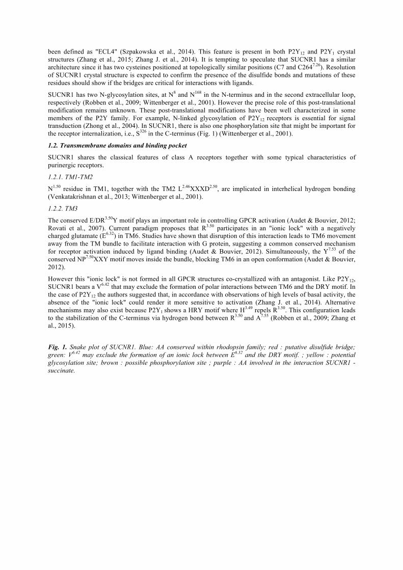
been defined as "ECL4" (Szpakowska et al., 2014). This feature is present in both P2Y12 and P2Y1 crystal structures (Zhang et al., 2015; Zhang J. et al., 2014). It is tempting to speculate that SUCNR1 has a similar architecture since it has two cysteines positioned at topologically similar positions (C7 and C2647.26). Resolution of SUCNR1 crystal structure is expected to confirm the presence of the disulfide bonds and mutations of these residues should show if the bridges are critical for interactions with ligands. SUCNR1 has two N-glycosylation sites, at N8 and N168 in the N-terminus and in the second extracellular loop, respectively (Robben et al., 2009; Wittenberger et al., 2001). However the precise role of this post-translational modification remains unknown. These post-translational modifications have been well characterized in some members of the P2Y family. For example, N-linked glycosylation of P2Y12 receptors is essential for signal transduction (Zhong et al., 2004). In SUCNR1, there is also one phosphorylation site that might be important for the receptor internalization, i.e., S326 in the C-terminus (Fig. 1) (Wittenberger et al., 2001). 1.2. Transmembrane domains and binding pocket SUCNR1 shares the classical features of class A receptors together with some typical characteristics of purinergic receptors. 1.2.1. TM1-TM2 N1.50 residue in TM1, together with the TM2 L2.46XXXD2.50, are implicated in interhelical hydrogen bonding (Venkatakrishnan et al., 2013; Wittenberger et al., 2001). 1.2.2. TM3 The conserved E/DR3.50Y motif plays an important role in controlling GPCR activation (Audet & Bouvier, 2012; Rovati et al., 2007). Current paradigm proposes that R3.50 participates in an "ionic lock" with a negatively charged glutamate (E6.32) in TM6. Studies have shown that disruption of this interaction leads to TM6 movement away from the TM bundle to facilitate interaction with G protein, suggesting a common conserved mechanism for receptor activation induced by ligand binding (Audet & Bouvier, 2012). Simultaneously, the Y7.53 of the conserved NP7.50XXY motif moves inside the bundle, blocking TM6 in an open conformation (Audet & Bouvier, 2012). However this "ionic lock" is not formed in all GPCR structures co-crystallized with an antagonist. Like P2Y12, SUCNR1 bears a V6.42 that may exclude the formation of polar interactions between TM6 and the DRY motif. In the case of P2Y12 the authors suggested that, in accordance with observations of high levels of basal activity, the absence of the "ionic lock" could render it more sensitive to activation (Zhang J. et al., 2014). Alternative mechanisms may also exist because P2Y1 shows a HRY motif where H3.49 repels R3.50. This configuration leads to the stabilization of the C-terminus via hydrogen bond between R3.50 and A7.55 (Robben et al., 2009; Zhang et al., 2015).
Fig. 1. Snake plot of SUCNR1. Blue: AA conserved within rhodopsin family; red : putative disulfide bridge; green: V6.42 may exclude the formation of an ionic lock between E6.32 and the DRY motif. ; yellow : potential glycosylation site; brown : possible phosphorylation site ; purple : AA involved in the interaction SUCNR1 -succinate.

1.2.3. TM4-TM5-TM6-TM7 SUCNR1 possesses the conserved W4.50 and a TM5 that may be bulged and bent at a highly conserved P5.50 called proline kink (Audet & Bouvier, 2012). This feature is present in P2Y1 and distinct from P2Y12 (bearing N instead of P at position 5.50), P2Y13 and P2Y14 (V5.50). In P2Y12, it results into a TM5 with straight conformation and tilted orientation (Zhang J. et al., 2014; Zhang K. et al., 2014). SUCNR1 shows a FTP motif instead of the conserved WXP6.50 (Fig. 1). The replacement of tryptophan (W) by phenylalanine (F) seems to be a common feature in the δ group of rhodopsin family and may be involved in ligand binding (Zhang J. et al., 2014; Zhang K. et al., 2014). 1.2.4. Binding pocket Site-directed mutagenesis showed the importance of several positively charged AA for succinate binding (R993.29, R2817.39, R2526.55 and H1033.33) (He et al., 2004). R7.39 and R6.55 are topologically shared with crystallized P2Y receptors where they seem to neutralize the phosphate negative charges of nucleotides (Zhang et al., 2015; Zhang et al., 2014a). A similar mechanism can be hypothesized for the carboxylate negative charges of succinate (Gonzalez et al., 2004; He et al., 2004). In contradiction with a shared mechanism for activation, the mutation of the highly conserved H6.52 that contributes to ligand recognition in P2Y1, P2Y2 and P2Y12 receptors does not abolish the interaction between SUCNR1 and succinate whereas a mutant of H3.33 does (Fig. 1) (He et al., 2004). 1.3. Ligands Initially, succinate was detected in pig kidney extracts with [Ca2+]i transients measured on Chinese hamster ovary (CHO) cells heterologously transfected with SUCNR1 (He et al., 2004). In mouse, succinate intravenous infusion induces an increase in blood pressure (He et al., 2004). This effect is abolished in SUCNR1-deficient animals, although angiotensin II induced hypertension was similar in both genotypes (He et al., 2004). Succinate response seems to be highly specific because 800 pharmacologically active compounds as well as 200 carboxylic acids and structurally related analogues, including other citric acid cycle intermediates, were not able to fully activate the receptor (He et al., 2004). Only maleate and methylmalonate were able to induce a response with 5-

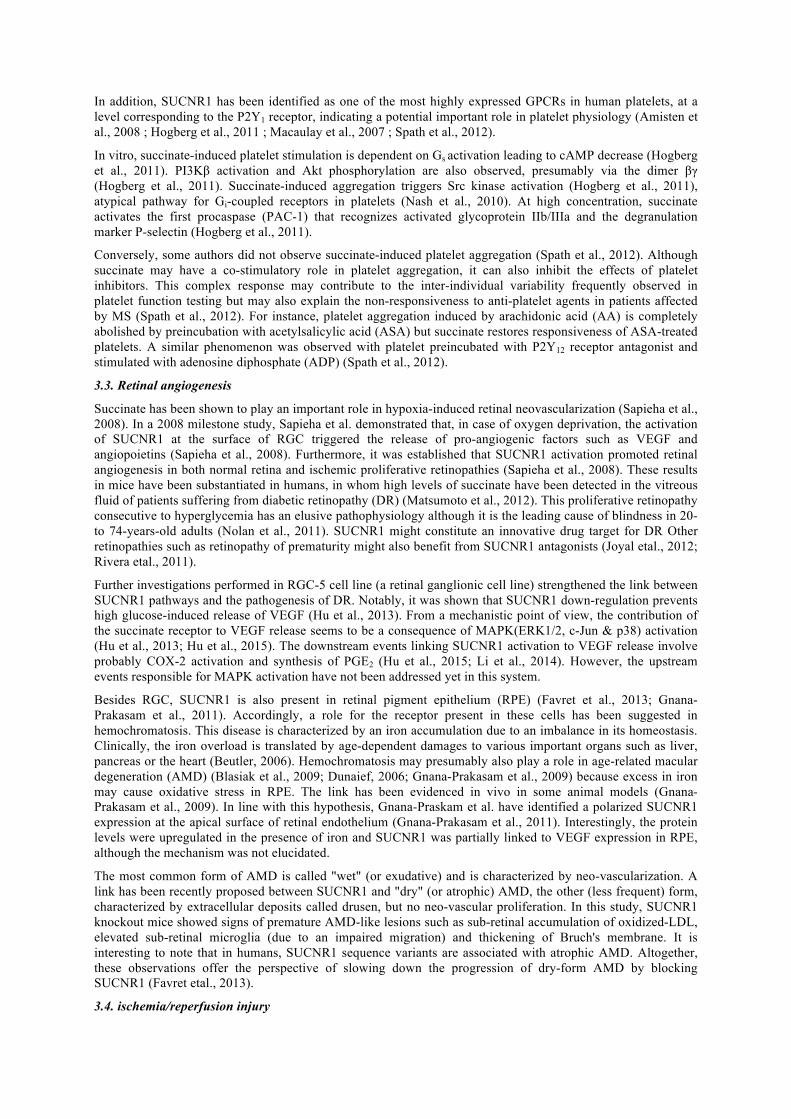
to 10-fold lower potency compared to succinate (He et al., 2004). The partial agonist nature of maleate on SUCNR1 was independently confirmed recently (Gilissen et al., 2015). In 2011 Bhuniya et al. reported a screening hit with antagonist profile following high-throughput screening. This hit compound was reported as able to inhibit succinate mediated [Ca2+]i mobilization (IC50 = 0.8 µM) in CHO-K1 cells overexpressing human SUCNR1. A structure-activity relationships study provided potent and selective (with respect to GPR99) antagonists with IC50 in the nanomolar range. 2c and 4c antagonists (Fig. 2) were demonstrated to inhibit succinate-induced blood pressure in rat (Bhuniya et al., 2011). Recently, this family of compounds gave rise to (99 m)Tc and (18)F radiotracers that may be useful for competition or labeling studies (Klenc et al., 2015). Interestingly, these antagonists showed no obvious structural relationship to succinate and no negative charges at physiological pH. The binding site for these compounds remains elusive because no competitive binding with succinate was performed. Given the absence of negative charges, it is tempting to speculate that the compounds might actually bind to a remote site compared to succinate and act as allosteric antagonists. These modulators represent reliable starting points for the validation of the receptor as drug target, but require deeper pharmacological characterization.
Fig. 2. Known SUCNR1 ligands. Succinate and maleate are the active species at physiological pH.
2. SUCCINATE RECEPTOR 1 SIGNALING PATHWAYS GPCR activation induces a conformational rearrangement that promotes the recruitment of heterotrimeric G proteins and activates its dissociation into α-subunit and a βγ dimer. These subunits are able to modulate the activity of effectors and mediate cellular responses. There are four main families of G proteins which differ in the signaling pathways they couple to: Gi/o and Gs regulate cyclic adenosine monophosphate (cAMP) through adenylate cyclase (AC) modulation, Gq/11 induces [Ca2+]i release from intracellular stores and G12/13 is involved in migration, growth and cell division (Wettschureck & Offermanns, 2005). Many GPCRs are desensitized and internalized upon activation. For most of them G protein-coupled receptor kinases (GRKs) phosphorylate the receptors at intracellular sites. The phosphorylated C-tail of an activated receptor promotes the binding of arrestins (Lefkowitz & Shenoy, 2005). These adapter proteins bring the receptors to clathrin-coated pits and may mediate further signaling. However, there are many exceptions to this central paradigm because several GPCRs do not require arrestins to get internalized. Many examples have been described such as the serotonin receptor 5-HT2A (Gray et al., 2001), protease-activated receptor PAR1 (Paing et al., 2002), prostacyclin receptor IP (Smyth et al., 2000), leukotriene B4 receptor BLT1 (Chen et al., 2004), formyl-peptide receptor FPR (Vines et al., 2003) or muscarinic receptor M2 (Vogler et al., 1999). SUCNR1 was originally described as being coupled to both Gi and Gq proteins in human embryonic kidney (HEK293) cells (He et al., 2004). The initial results were later confirmed by Robben et al., in polarized Madin Darby Canine Kidney (MDCK) cells (Robben et al., 2009). Several authors repeatedly confirmed Gi activation by demonstrating a decrease of cAMP levels upon succinate binding to SUCNR1 (Fig. 3), in both heterologous

and native systems (Gilissen et al., 2015; Gnana-Prakasam et al., 2011 ; Hakak et al., 2009; Hogberg et al., 2011 ; Sundström et al., 2013). However, the view of SUCNR1 being coupled to Gq has been challenged and it was proposed by several authors that the observed [Ca2+]i mobilization was a consequence of PLC-β activation by the βγ dimer (Fig. 3) (Gilissen et al., 2015; Sundström et al., 2013). The discrepancy between results may reflect distinct G protein partners among different cell types or artifacts induced by overexpression of the receptor. The existence of Gq coupling with SUCNR1 requires additional investigations, especially in native and physiologically relevant systems. Succinate binding to SUCNR1 triggers the activation of mitogen-activated pathway (MAP) kinases, especially extracellular signal-regulated kinases 1 and 2 (ERK1/2) in HEK293 cell line (Gilissen etal., 2015; He et al., 2004), MDCK (Robben et al., 2009), immature dendritic cells (iDC) (Rubic et al., 2008), retinal ganglion neuronal cell line (RGC) (Hu et al., 2013), TF-1 (human erythroleukaemia cell line) and cardiomyocytes (Aguiar et al., 2010) (Fig. 3). Some results indicate that ERK activation is Gi-dependent and should probably be mediated through the dimer βγ (Gilissen et al., 2015; Hakak et al., 2009). SUCNR1 is internalized into vesicular structures upon succinate exposure in HEK293 cells (He et al., 2004). In contrast, Robben et al., showed that SUCNR1 is rapidly desensitized and re-sensitized but not internalized in polarized MDCK cells (Robben et al., 2009). Hogberg et al. also showed desensitization of SUCNR1 in platelets (Hogberg et al., 2011). The coupling of activated receptor to arrestins 2 and 3 is very weak (Gilissen et al., 2015; Southern et al., 2013). Therefore, it is likely that homologous desensitization and internalization occur by a mechanism that does not involve arrestins. However, these questions have never been addressed directly. 3. IMPLICATION IN (PATHO)PHYSIOLOGY Succinate is a citric acid cycle intermediate that accumulates in mitochondria, cytosol and outside the cell in case of oxygen deprivation due to the imbalance between energy demand and oxygen supply (Feldkamp et al., 2004; Hébert, 2004; Peti-Peterdi et al., 2013; Toma et al., 2008). It activates SUCNR1 signaling pathways for the detection of local stress, including ischemia, hypoxia, toxicity, and hyperglycemia. The succinate-SUCNR1 response affects cellular metabolism and pathophysiology of diseases in multiple organs (Table 1 and Fig. 3). 3.1. Regulation of blood pressure The observation that succinate may impact cardiovascular functions in humans dates back to the 50s when succinate infusions were tested as a treatment of barbiturate intoxications (Zuckerbrod & Graef 1950). In 1976, succinate was shown to induce renin release in isolated rat glomeruli while evaluating its role as an energy substrate (Baumbach et al., 1976). The mechanism for this effect was partially elucidated via the identification of its membrane receptor. The roles of succinate in blood pressure regulation have been well documented in several in vivo and in vitro studies and are strongly linked to the renin-angiotensin system (RAS) activation in the kidney (He et al., 2004; Pluznick & Caplan, 2015; Sadagopan et al., 2007; Toma et al., 2008). In addition, SUCNR1 mRNA has been detected in different nephron segments such as proximal tubule, distal nephron (He et al., 2004), afferent arteriole, glomerulus (Robben et al., 2009; Toma et al., 2008) and the juxtaglomerular apparatus (JGA), the most important regulatory site of systemic blood pressure (He et al., 2004). SUCNR1 protein is also expressed in cortical collecting duct (CD), inner medullary CD principal cell and in macula densa (MD) cells (Robben et al., 2009; Vargas et al., 2009). MD cells are known to control renal blood flow, glomerular filtration, and release of renin (Peti-Peterdi & Harris, 2010; Robben et al., 2009; Vargas et al., 2009). The current model remains largely elusive but states that succinate binding to SUCNR1 results in production and release of prostaglandins I2 and E2 together with NO, which are vasodilators and crucial paracrine mediators of renin release by the juxtaglomerular apparatus (Robben et al., 2009; Toma et al., 2008). The production of these mediators is thought to be the consequence of [Ca2+]i increase and ERK1/2 phosphorylation (Fig. 3). Since renin is a key component of the renal ability to increase blood pressure in the face of low blood volume (Pluznick, 2013), it is tempting to speculate that succinate may be acting via SUCNR1 to support increases in blood volume (and therefore blood pressure). Contradictions in this model include the reported increase of [Ca2+]i in glomerular endothelial cell line (GENC) by succinate that usually results in an inhibition of renin release (Amaral et al., 2012; Toma et al., 2008). Also, Sucnr1-/- mice have normal baseline blood pressures, likely due to long-term blood pressure counter-regulatory mechanisms. The pathological activation of the RAS contributes to or occurs with several pathological states such as diabetes, obesity or hypertension. Succinate and its receptor might play a key role in these conditions and metabolic syndrome. The current observations supporting such role are discussed below.

Fig. 3. Signaling pathways following succinate binding on its receptor. Dashed lines represent hypothetical links and/or discrepancies in the literature (see text for details).
3.1.1. Hypertension Circulating levels of succinate are elevated in rodent models of hypertension, including the spontaneously hypertensive rats. This aspect of succinate pathophysiology has not been confirmed in man (Sadagopan et al., 2007). However, this does not preclude a local signaling role of succinate, which may not be reflected by the circulating levels. Hence, as an indirect example of chronic hypertension, serum succinate levels are increased in patients suffering from cardiac hypertrophy (CH) (Aguiar et al., 2014). Activated SUCNR1 may indirectly contribute to CH by inducing hypertension but might also have a direct action because it is expressed at protein level in ventricular cardiomyocytes (in sarcolemmal membrane and T-tubules) (Aguiar et al., 2010; Aguiar et al., 2014). In these cells, SUCNR1 activation triggers hypertrophic gene expression via ERK1/2 phosphorylation, increased expression of calcium/calmodulin-dependent protein kinase IIδ (CaMKIIδ) and the translocation of histone deacetylase 5 (HDAC5) into the cytoplasm (Aguiar et al., 2014). Moreover, it was shown in another study by the same group as for cardiomyocytes, SUCNR1 alters Ca2+ transient via AC and protein kinase A (PKA) activation that phosphorylate phospholamban (PLN) and ryanodine receptor 2 (RyR2), two well-known Ca2+ handling proteins (Aguiar et al., 2010). These results suggest a Gs-mediated effect. However, the data are difficult to interpret because the concentration of succinate used was very high (10 mM) and the direct interaction of the receptor with Gs was not demonstrated (Aguiar et al., 2010). Considering that right ventricular hypertrophy (RVH) has a close relationship with left ventricle hypertrophy (LVH) (Yang et al., 2014), SUCNR1 may also be involved in RVH caused by pulmonary pressure overload via phosphatidylinositol-4, 5-bisphosphate 3-kinase (PI3K)/ protein kinase B (Akt) signaling pathway (Yang et al., 2014). Interestingly this pathway is known to be activated by Gi in cardiac myocytes and its inhibition to convert survival to apoptotic signaling (Zhu et al., 2001).

Table 1. Signaling pathways of SUCNR1 in specific cell sub-types. Tissue Cellular sub-types Cell lines used in
vitro Signaling pathways and effectors
Potential pathophysiological effect
Kidney Proximal tubule, distal nephron, JGA, afferent arteriole and glomerulus
Kidney and macula densa cells (Vargas et al., 2009)
p38 and ERK1/2 phosphorylation, renin release via COX-2 and PGE2 (Vargas et al., 2009)
Hypertension
Diabetic nephropathy
Macula densa
Renal complications (Robben et al., 2009; Vargas et al., 2009)
MDCK (Robben et al., 2009)
Gq and Gi, ERK1/2 phosphorylation PGE2 and PGI2 via AA (Vargas et al., 2009)
Hypertension (Vargas et al., 2009)
GENC (Toma et al., 2008)
↑Ca2+ renin release via NO and PGE2 (Toma et al., 2008)
Hypertension
Liver Hepatic stellate cells ↑ α-SMA ↑ transdifferentiation (Correa et al., 2007; Li et al., 2015)
Hepatic fibrosis (Correa et al., 2007; Li et al., 2015)
White adipose
Adipocytes (Regard et al., 2008)
Gi (Regard et al., 2008) Inhibition of lipolysis (Regard et al., 2008)
Retina RGC-5 (Deen & Robben, 2011 ;
ERK1/2 and JNK phosphorylation,
Diabetic retinopathy
RPE and microglial cells (Favretet al.,2013)
Sapiehaet al., 2008) ↑ VEGF/cell proliferation via COX-2 and PGE2 (Deen & Robben, 2011 ; Hu et al., 2015; Jianyan Hu, Wu, Li, Chen, & Wang, 2013 ; Sapieha et al., 2008)
(Huet al.,2013) Blindness caused by ROP Dry-form AMD (Favret et al., 2013 )
Brain Neurons and astrocytes (Hamel et al., 2014)
RGC-5 and RPA ↑ VEGF via PGE2 and EP4 (Hamel et al., 2014)
Brain revascularization (Hamel et al., 2014)
Heart Sarcolemmal membrane and
↑ Ca2+ via PKA and AC activation, PLN and RyR2 phosphorylation (Aguiar et al., 2010)
Cardiac hypertrophy (Carla J. Aguiaret al.,2010)
T-tubules of cardiomyocytes (Aguiaret al.,2010)
ERK1/2 phosphorylation, CaMKIIδ expression and the translocation of HDAC5 (Aguiar et al., 2014) PKA activation (Aguiar et al., 2010) PI3K/Akt signaling (Yang et al., 2014)
Cardiac hypertrophy (Aguiar et al., 2014) Myocytes death (Aguiar et al., 2010) RVH (Yang et al., 2014)
Bone marrow
Hematopoietic progenitor cells
Erythroids, megakaryocytes,
Gi Stimulation of immunity, protection from apoptosis (Hakak et al., 2009)
(Hakak et al., 2009) TF-1 (Hakak et al., 2009)
↑ IP, ERK1/2 phosphorylation (Hakak et al., 2009)
Blood Immature dendritic cells (iDC)
iDCandU937 ↑Ca2+ Enhancement of immunity transplant rejection (Rubic et al., 2008)
(Rubic et aL, 2008) ERK1/2 phosphorylation ↑ TNF-α, IL-1β and cytokines (Rubic et al., 2008) iDC migration

Platelet (Macaulay et al., 2007)
↓ cAMP and Src kinase activation via Gi PI3K activation, Akt phosphorylation via βγ P-selectin and glycoprotein (GP)IIb-IIIa activation (Hogberg et al., 2011 ) Cross-talk with other GPCRs (ADP, TXA2) (Spath et al., 2012) ↓ platelet aggregation (Macaulay et al., 2007; Spath et al., 2012)
(Potentiation of (Spath et al., 2012)) platelet aggregation (Hogberg et al., 2011) Inhibition of anti-platelets
Monocytes T and B (Rubic et al., 2008)
↑ TNFα and IFN--γ (Rubic et al., 2008)
Immunity (Rubic et al., 2008)
3.1.2. Diabetes and obesity Succinate accumulates locally in diabetic mouse kidneys (Toma et al., 2008). The increased levels associated with this condition are compatible with SUCNR1 receptor activation. The resulting renin release by the juxtaglomerular apparatus is absent in SUCNR1-deficient mice (Toma et al., 2008). Therefore, SUCNR1 may contribute to diabetes-induced hypertension by increasing renin release in conditions of chronic hyperglycemia, when filtrated succinate exceeds the reabsorbed levels (Robben et al., 2009). Since the urinary increase in succinate is one- to twofold in diabetic mice, there is an interest in urinary succinate as a potential early biomarker for diabetic nephropathy (Toma et al., 2008). One major complication of diabetes is diabetic nephropathy, a condition characterized by damage to the capillaries of renal glomeruli. Although RAS activation may contribute to the progression of diabetic nephropathy, the mechanism of this pathology is incompletely understood (Van Buren & Toto, 2011). Interestingly, ERK1/2 phosphorylation observed in diabetic nephropathy could be explained by succinate-activated SUCNR1 in the renal tubules of diabetic mice (Robben et al., 2009). Thus, SUCNR1 may contribute to the development of tubulo-interstitial fibrosis and could become a target to prevent diabetic nephropathy (Peti-Peterdi & Harris, 2010). Furthermore, rodent models of metabolic diseases, including ob/ob mice, and db/db mice, are characterized by increased circulating succinate levels which are sufficient to trigger the activation of SUCNR1 (Sadagopan et al., 2007). SUCNR1 is expressed at the surface of adipocytes of the white adipose tissue (WAT) and stimulation of G; by succinate in these cells inhibits lipolysis and prevents the release of free fatty acids (Fig. 3) (McCreath et al., 2015; Regard, Sato, & Coughlin, 2008). Deep metabolic studies have recently demonstrated that Sucnr1-/-mice have a modified weight gain and glucose homeostasis (McCreath et al., 2015). Mice fed with standard diet displayed a mild lean pheno-type. However, KO mice under high-fat diet showed a significant reduction in weight gain (between 4 and 12 weeks), although this effect was lost at later time (>16 weeks). In KO mice, the overall content of WAT was increased and concomitant with a hyperglycemic profile, characterized by a reduced insulin secretion. According to McCreath et al., these results point to a role for SUCNR1 in metabolism and metabolic disease in the context of obesity (McCreath et al., 2015). Collectively, these data highlight the putative role of SUCNR1 in metabolic syndrome (MS), a pathological state characterized by obesity, diabetes and hypertension. People suffering from MS are at increased risk of developing cardiovascular disease. 3.2. Platelet physiology Succinate was identified as a platelet stimulant more than 30 years ago (Huang et al., 1984). The precise mechanism of this effect remains unknown, but has been rediscovered and investigated more recently (Hogberg et al., 2011; Macaulay et al., 2007; Spath, Hansen, Bokemeyer, & Langer, 2012). SUCNR1 is expressed in megakaryocytes at both protein and mRNA levels (Hakak et al., 2009; Macaulay et al., 2007). SUCNR1 is expressed in hematopoietic progenitor cells (HPC) of the bone marrow. In vitro, succinate stimulates the proliferation of erythroid and megakaryocyte progenitor cells as well as erythroid-like cell lines such as TF-1 cells. The proliferative effect seems to be mediated through Gi pathway that leads to the activation of ERK1/2 and inositol phosphate (IP) increase. These pathways are relevant for HPC proliferation, differentiation, and survival (Hakak et al., 2009). SUCNR1 also stimulates blood cell development by increasing the levels of hemoglobin, platelets, and neutrophils in a mouse model of chemotherapy-induced myelosuppression (Hakak et al., 2009).
In addition, SUCNR1 has been identified as one of the most highly expressed GPCRs in human platelets, at a level corresponding to the P2Y1 receptor, indicating a potential important role in platelet physiology (Amisten et al., 2008 ; Hogberg et al., 2011 ; Macaulay et al., 2007 ; Spath et al., 2012). In vitro, succinate-induced platelet stimulation is dependent on Gs activation leading to cAMP decrease (Hogberg et al., 2011). PI3Kβ activation and Akt phosphorylation are also observed, presumably via the dimer βγ (Hogberg et al., 2011). Succinate-induced aggregation triggers Src kinase activation (Hogberg et al., 2011), atypical pathway for Gi-coupled receptors in platelets (Nash et al., 2010). At high concentration, succinate activates the first procaspase (PAC-1) that recognizes activated glycoprotein IIb/IIIa and the degranulation marker P-selectin (Hogberg et al., 2011). Conversely, some authors did not observe succinate-induced platelet aggregation (Spath et al., 2012). Although succinate may have a co-stimulatory role in platelet aggregation, it can also inhibit the effects of platelet inhibitors. This complex response may contribute to the inter-individual variability frequently observed in platelet function testing but may also explain the non-responsiveness to anti-platelet agents in patients affected by MS (Spath et al., 2012). For instance, platelet aggregation induced by arachidonic acid (AA) is completely abolished by preincubation with acetylsalicylic acid (ASA) but succinate restores responsiveness of ASA-treated platelets. A similar phenomenon was observed with platelet preincubated with P2Y12 receptor antagonist and stimulated with adenosine diphosphate (ADP) (Spath et al., 2012). 3.3. Retinal angiogenesis Succinate has been shown to play an important role in hypoxia-induced retinal neovascularization (Sapieha et al., 2008). In a 2008 milestone study, Sapieha et al. demonstrated that, in case of oxygen deprivation, the activation of SUCNR1 at the surface of RGC triggered the release of pro-angiogenic factors such as VEGF and angiopoietins (Sapieha et al., 2008). Furthermore, it was established that SUCNR1 activation promoted retinal angiogenesis in both normal retina and ischemic proliferative retinopathies (Sapieha et al., 2008). These results in mice have been substantiated in humans, in whom high levels of succinate have been detected in the vitreous fluid of patients suffering from diabetic retinopathy (DR) (Matsumoto et al., 2012). This proliferative retinopathy consecutive to hyperglycemia has an elusive pathophysiology although it is the leading cause of blindness in 20- to 74-years-old adults (Nolan et al., 2011). SUCNR1 might constitute an innovative drug target for DR Other retinopathies such as retinopathy of prematurity might also benefit from SUCNR1 antagonists (Joyal etal., 2012; Rivera etal., 2011).
Further investigations performed in RGC-5 cell line (a retinal ganglionic cell line) strengthened the link between SUCNR1 pathways and the pathogenesis of DR. Notably, it was shown that SUCNR1 down-regulation prevents high glucose-induced release of VEGF (Hu et al., 2013). From a mechanistic point of view, the contribution of the succinate receptor to VEGF release seems to be a consequence of MAPK(ERK1/2, c-Jun & p38) activation (Hu et al., 2013; Hu et al., 2015). The downstream events linking SUCNR1 activation to VEGF release involve probably COX-2 activation and synthesis of PGE2 (Hu et al., 2015; Li et al., 2014). However, the upstream events responsible for MAPK activation have not been addressed yet in this system. Besides RGC, SUCNR1 is also present in retinal pigment epithelium (RPE) (Favret et al., 2013; Gnana-Prakasam et al., 2011). Accordingly, a role for the receptor present in these cells has been suggested in hemochromatosis. This disease is characterized by an iron accumulation due to an imbalance in its homeostasis. Clinically, the iron overload is translated by age-dependent damages to various important organs such as liver, pancreas or the heart (Beutler, 2006). Hemochromatosis may presumably also play a role in age-related macular degeneration (AMD) (Blasiak et al., 2009; Dunaief, 2006; Gnana-Prakasam et al., 2009) because excess in iron may cause oxidative stress in RPE. The link has been evidenced in vivo in some animal models (Gnana-Prakasam et al., 2009). In line with this hypothesis, Gnana-Praskam et al. have identified a polarized SUCNR1 expression at the apical surface of retinal endothelium (Gnana-Prakasam et al., 2011). Interestingly, the protein levels were upregulated in the presence of iron and SUCNR1 was partially linked to VEGF expression in RPE, although the mechanism was not elucidated. The most common form of AMD is called "wet" (or exudative) and is characterized by neo-vascularization. A link has been recently proposed between SUCNR1 and "dry" (or atrophic) AMD, the other (less frequent) form, characterized by extracellular deposits called drusen, but no neo-vascular proliferation. In this study, SUCNR1 knockout mice showed signs of premature AMD-like lesions such as sub-retinal accumulation of oxidized-LDL, elevated sub-retinal microglia (due to an impaired migration) and thickening of Bruch's membrane. It is interesting to note that in humans, SUCNR1 sequence variants are associated with atrophic AMD. Altogether, these observations offer the perspective of slowing down the progression of dry-form AMD by blocking SUCNR1 (Favret etal., 2013). 3.4. ischemia/reperfusion injury

Ischemia/reperfusion injury (IRI) represents a worldwide public health issue of increasing incidence. IRI may virtually affect all organs and tissues, and is associated with a significant morbidity and mortality. Particularly, the duration of blood supply deprivation has been recognized as a critical factor in stroke, myocardial infarction or acute kidney injury, as well as in solid organ transplantation or cardiac surgery (Eltzschig & Collard, 2004; Erpicum et al., 2014; Lagny et al., 2015). IRI causes multiple cellular and tissular metabolic and architectural changes (Carden & Granger, 2000). Furthermore, the reperfusion of ischemic tissues induces both local and systemic inflammation (Rowart et al., 2015). Various GPCRs have been implicated in the cascade of IRI (Weekers et al., 2015; Yap & Lee, 2012). Interestingly, tissue succinate levels have been shown to increase during ischemic hypoxia (Goldberg et al., 1966; He et al., 2004), which may reflect oxidative stress or energy deprivation. More recently, it has been demonstrated that succinate was responsible for mitochondrial reactive oxygen species (ROS) generation, a typical feature of IRI (Chouchani et al., 2014). In case of renal IRI, it has been suggested that accumulation of succinate could contribute to stenosis-associated hypertension (He et al., 2004; Pluznick, 2013). Thus, preventing succinate accumulation may represent innovative therapeutic targets to protect against IRI-associated damage, notably in kidney or following heart attack (Chouchani et al., 2014; O'Neill, 2014). The specific role of SUCNR1 has been investigated in some animal models of IRI. In the liver, although succinate levels significantly increase following ischemia, neither succinate nor glucose are able to increase hepatic perfusion pressure when infused into the portal vein (Correa et al., 2007). Interestingly, succinate does not inhibit the production of cAMP induced by forskolin neither increase Ca2+ in quiescent hepatic stellate cells (HSC), where SUCNR1 is present, both at the protein and mRNA level (Correa et al., 2007). However succinate accelerates activation of ischemic HSC, through upregulation of α-smooth muscle actin (α-SMA), a marker of myofibroblastic trans-differentiation (Fig. 3). Stimulation of SUCNR1 probably potentiates the effects of growth factors and hormones, and by non-traditional signals such as matrix stiffness or oxidant stress that are known to induce stellate cell activation (Correa et al., 2007). Upon activation of HSC, the expression of SUCNR1 in these cells decreases rapidly, suggesting that SUCNR1 serves as an early detector of hepatic stress or damage. SUCNR1 signaling plays an enhancing role in HSC activation to restore damaged tissues in the ischemic liver, but may also contribute to the formation of fibrosis (Correa et al., 2007; Y.H. Li, Woo, Choi, & Cho, 2015). In contrast, high levels of succinate after cerebral 1R1 enhance brain vascularization and recovery by inducing expression of VEGF through SUCNR1, which is mainly expressed in neurons and astrocytes (Hamel et al., 2014). In RGC and rat cerebral astrocyte neuronal cell line (RPA), SUCNR1 upregulates VEGF via PGE2 and prostaglandin E receptor 4 (EP4), already known to promote angiogenesis (Hamel et al., 2014). Since succinate only transiently accumulates in case of brain 1R1, long-acting agonists of SUCNR1 might potentiate the recovery process (Hamel et al., 2014). 3.5. Immune system and inflammation In humans, SUCNR1 protein was identified at the surface of T (CD4+, CD8+) and B (CD19+) cells as well as monocytes (CD14+) (Macaulay et al., 2007). At the mRNA level, SUCNR1 was also detected in macrophages and iDC (Rubic et al., 2008). Accumulating evidence points to succinate as a prominent signal molecule in inflammation, through the stabilization of the hypoxia-inducible factor (HIF-α), induction of the pro-inflammatory cytokine interleukin-1 beta (IL-1β) and generation of mitochondrial ROS (Chouchani et al., 2014; Mills & O'Neill, 2014; Tannahill et al., 2013). The elevation of succinate levels is described as a consequence of hypoxia at sites of inflammation, with a switch to glycolysis by macrophages driven by HIF-α (Mills & O'Neill, 2014). The role of SUCNR1 in these processes remains unknown but its function has been investigated in some specific inflammatory and immune response processes such as dendritic cells activation. Dendritic cells are an important population of antigen-presenting cells implicated in the initiation of immune response. They derive from monocytes and become iDC in peripheral tissue. Once in contact with antigens and various pathogens' stimuli they secrete proinflammatory cytokines and undergo maturation to dendritic cells. They thus migrate to the lymph node where they can activate T cells. In vitro, SUCNR1 is present at the surface of iDC and can elicit direct stimulation by [Ca2+]i mobilization (Rubic et al., 2008). Succinate also directly triggers migration of dendritic cells and this effect may also be indirect, by facilitating leukocytes trafficking through blood flow acceleration (Rubic et al., 2008). In synergy with toll-like receptors (TLR3 and TLR7), succinate bound SUCNR1 potentiates the expression of tumor necrosis factor α (TNF-α) and IL-1β and potentiate cytokines production via ERKl/2 phosphorylation. Succinate can be seen as an endogenous "warning" signal, regulating cytokines expression by iDC during their migration to the lymph nodes (Rubic et al., 2008). Of important note, SUCNR1 may contribute to transplant rejection because skin allografts have a longer survival time in immunocompetent SUCNR1-!- mice (Rubic et al., 2008). 4. CONCLUSION On the basis of the existing observations mostly acquired from in vitro and in vivo rodent models, SUCNR1

represents a promising drug target in various common human diseases, like hypertension, diabetes and IRI. However, the current paucity of specific pharmacological tools delays its validation as a drug target. Up to now, no synthetic agonists and very few ligands have been described. More widely available tools should rapidly prompt further investigations on the putative binding pocket to facilitate the design of new modulators. Such ligands might be used to address more deeply the role of the receptor, particularly in systems more relevant to human physiology. CONFLICT OF INTEREST The authors declare no conflicts of interest. ACKNOWLEDGMENTS This work was supported by the "Fonds pour la Recherche Scientifique" (F.R.S.-FNRS) Incentive Grant for Scientific Research (F.4510.14) and Research Credit (#3309), University of Liège (Crédit de Démarrage FSRD-12/11 and Fonds Spéciaux FSRC-14/107) and Léon Fredericq Foundation. JH and FJ are F.R.S.-FNRS research associate and MD/PhD post-doctoral fellow, respectively. REFERENCES Abbracchio, M.P, Burnstock, G., Boeynaems, J.M., Barnard, E.A., Boyer, J.L, Kennedy, C, Knight, G.E, Fumagalli, M., Cachet, C, Jacobson, KA, & Weisman, G A (2006). International Union of Pharmacology LVΠI: update on the P2Y G protein-coupled nucleotide receptors: from molecular mechanisms and pathophysiology to therapy. Pharmacol Rev 58, 281-341.
Aguiar, C.J., Andrade, V.L, Gomes, E.R, Alves, M.N., Ladeira, M.S., Pinheiro, A.C., Gomes, DA, Almeida, A.P., Goes, A.M., Resende, R.R., Guatimosim, S., & Leite, M.F. (2010). Succinate modulates Ca(2+) transient and cardiomyocyte viability through PKA-dependent pathway. Cell Calcium 47,37-46.
Aguiar, C.J., Rocha-Franco, J.A., Sousa, PA, Santos, A.K., Ladeira, M., Rocha-Resende, C, Ladeira, L.O., Resende, R.R, Botoni, FA, Barrouin Melo, M., Lima, C.X., Carballido, J.M, Cunha, T.M., Menezes, G.B., Guatimosim, S, & Leite, M.F. (2014). Succinate causes pathological cardiomyocyte hypertrophy through GPR91 activation. Cell Commun Signal 12, 78.
Amaral, A.U, Cecatto, C, Busanello, E.N., Ribeiro, CA, Melo, D.R, Leipnitz, G., Castilho, RE, 6 Wajner, M. (2012). Ethylmalonic acid impairs brain mitochondrial succinate and mala te transport Mol Genet Metab 105,84-90.
Amisten, S, Braun, O.O, Bengtsson, A, & Erlinge, D. (2008). Gene expression profiling for the identification of G-protein coupled receptors in human platelets. Thromb Res 122, 47-57.
Ariza, A.C, Deen, P.M., & Robben, J.H. (2012). The succinate receptor as a novel therapeutic target for oxidative and metabolic stress-related conditions. Front Endocrinol (Lausanne) 3,22.
Audet, M, & Bouvier, M. (2012). Restructuring G-protein-coupled receptor activation. Cell 151, 14-23.
Ballesteros, J, & Weinstein, H. (1995). Integrated methods for the construction of three-dimensional models and computational probing of structure-function relations in G protein-coupled receptors. Methods Neurosci 25, 366-428.
Baumbach, L, Leyssac, PP., & Skinner, S.L. (1976). Studies on renin release from isolated superfused glomeruli: effects of temperature, urea, ouabain and ethacrynic acid. 7 Physiol 258, 243-256.
Beutler, E. (2006). Hemochromatosis: genetics and pathophysiology. Annu Rev Med 57, 331-347.
Bhuniya, D, Umrani, D, Dave, B, Salunke, D, Kukreja, G, Gundu, J, Naykodi, M, Shaikh, N.S, Shitole, P, Kurhade, S, De, S, Majumdar, S, Reddy, S.B, Tambe, S, Shejul, Y., Chugh, A, Palle, V.P, Mookhtiar, KA, Cully, D, Vacca, J, Chakravarty, P.K, Nargund, RP, Wright, S.D, Graziano, MP, Singh, S.B, Roy, S, & Cai, T.Q, (2011). Discovery of a potent and selective small molecule hGPR91 antagonist. BioorgMed Chem Lett 21, 3596-3602.
Blasiak, J, Sklodowska, A, Ulinska, M, & Szaflik, JP. (2009). Iron and age-related macular degeneration.Klin Oczna 111, 174-177.
Carden, D.L, & Granger, D.N. (2000). Pathophysiology of ischaemia-reperfusion injury. J Pathol 190, 255-266.
Chen, Z, Gaudreau, R, Le Gouill, C, Rola-Pleszczynski, M, & Stankova, J. (2004). Agonist-induced internalization of leukotriene B(4) receptor 1 requires G-protein-coupled receptor kinase 2 but not arrestins. Mol Pharmacol 66, 377-386.
Chouchani, E.T, Pell, V.R, Gaude, E, Aksentijevic, D, Sundier, S.Y, Robb, E.L, Logan, A, Nadtochiy, S.M., Ord, E.N., Smith, A.C., Eyassu, F, Shirley, R, Hu, C.H., Dare, A.J, James, A.M, Rogatti, S, Hartley, RC, Eaton, S, Costa, A.S., Brookes, P.S, Davidson, S.M, Duchen, M.R, Saeb-Parsy, K, Shattock, M.J, Robinson, AJ, Work, LM, Frezza, C, Krieg, T, & Murphy, MP. (2014). Ischaemic accumulation of succinate controls re-perfusion injury through mitochondrial ROS. Nature 515,431-435.
Correa, P.R, Kruglov, EA, Thompson, M, Leite, M.F, Dranoff, JA, & Nathanson, M.H. (2007). Succinate is a paracrine signal for liver damage.J Hepatol 47, 262-269.

Davenport, A.P, Alexander, S.P.H, Sharman, J.L, Pawson, AJ, Benson, H.E, Monaghan, A.E, Liew, W.C, Mpamhanga, CP, Bonner, T.I, Neubig, RR, Pin, JP, Spedding, M, & Harmar, AJ. (2013). International Union of Basic and Clinical Pharmacology. LXXXVIII. G protein-coupled receptor list: recommendations for new pairings with cognate ligands. Pharmacol Rev 65,967-986.
Deen, P. M., SRobben, J. H. (2011). Succinate receptors in the kidney.J Am Soc Nephrol 22(8), 1416-1422.
Dunaief, J.L. (2006). Iron induced oxidative damage as a potential factor in age-related macular degeneration: the Cogan Lecture. Invest Ophthalmol Vis Sci 47, 4660-4664.
Eltzschig, H.K, & Collard, CD. (2004). Vascular ischaemia and reperfusion injury. Br Med Bull 70, 71-86.
Erpicum, P., Detry, O., Weekers, L, Bonvoisin, C, Lechanteur, C, Briquet, A., Béguin, Y., Krzesinski, J.M, & Jouret, F. (2014). Mesenchymal stromal cell therapy in conditions of renal ischaemia/reperfusion. Nephrol Dial Transplant 29,1487-1493.
Favret, S., Binet, F., Lapalme, E., Leboeuf, D, Carbadillo, J, Rubic, T, Picard E, Mawambo, G, Tetreault, N, Joyal, J.S., Chemtob, S., Sennlaub, F., SanGiovanni, J.P., Guimond, M., & Sapieha, P. (2013). Deficiency in the metabolite receptor SUCNR1 (GPR91) leads to outer retinal lesions. Aging 5, 427-444.
Feldkamp, T., Kribben, A., Roeser, N.F., Senter, R.A., Kemner, S., Venkatachalam, M.A., Nissim, I., & Weinberg, J.M. (2004). Preservation of complex I function during hypox-ia-reoxygenation-induced mitochondrial injury in proximal tubules. Am J Physiol Renal Physiol 286, F749-F759.
Fredriksson, R., Lagerstrom, M.C., Lundin, L -G., & Schiöth, H.B. (2003). The G-protein-coupled receptors in the human genome form five main families. Phylogenetic analysis, paralogon groups, and fingerprints. Mol Pharmacol 63,1256-1272.
Gilissen, J, Geubelle, P., Dupuis, N, Laschet, C, Pirotte, B., & Hanson, J. (2015). Forskolin-free cAMP assay for Gi-coupled receptors. Biochem Pharmacol 98,381-391.
Gnana-Prakasam, J.P., Ananth, S., Prasad, P.D., Zhang, M., Atherton, S.S., Martin, P.M., Smith, S.B., & Ganapathy, V. (2011). Expression and iron-dependent regulation of succinate receptor GPR91 in retinal pigment epithelium. Invest Ophthalmol Vis Sci 52, 3751-3758.
Gnana-Prakasam, J.P, Thangaraju, M, Liu, K., Ha, Y., Martin, P.M., Smith, S.B., & Ganapathy, V. (2009). Absence of iron-regulatory protein Hfe results in hyperproliferation of retinal pigment epithelium: role of cystine/glutamate exchanger. Biochem J 424,243-252.
Goldberg, N.D., Passonneau, J.V., & Lowry, O.H. (1966). Effects of changes in brain metabolism on the levels of citric acid cycle intermediates. J Biol Chem 241, 3997-4003.
Gonzalez, N.S., Communi, D, Hannedouche, S., & Boeynaems, J.M. (2004). The fate of P2Y-related orphan receptors: GPR80/99 and GPR91 are receptors of dicarboxylic acids. Purinergic Signal 1, 17-20.
Gray, J.A., Sheffler, D.J., Bhatnagar, A., Woods, J.A., Hufeisen, S.J., Benovic, J.L., & Roth, B.L. (2001). Cell-type specific effects of endocytosis inhibitors on 5-hydroxytryptamine(2A) receptor desensitization and resensitization reveal an arrestin-, GRK2-, and GRK5-independent mode of regulation in human embryonic kidney 293 cells. Mol Pharmacol 60,1020-1030.
Hakak, Y, Lehmann-Bruinsma, K., Phillips, S, Le, T., Liaw, C, Connolly, D.T., & Behan, D.P (2009). The role of the GPR91 ligand succinate in hematopoiesis. J Leukoc Biol 85, 837-843.
Hamel, D., Sanchez, M, Duhamel, F, Roy, O, Honore, J.C, Noueihed, B., Zhou, T, Nadeau-Vallee, M., Hou, X, Lavoie, J.C, Mitchell, G, Mamer, OA, & Chemtob, S. (2014). G-protein-coupled receptor 91 and succinate are key contributors in neonatal postcerebral hypoxia-ischemia recovery. Arterioscler Thromb Vase Biol 34, 285-293.
He, W., Miao, F.J. -P., Lin, DC -H., Schwandner, RT, Wang, Z, Gao.J., Chen, J. -L, Tian, H., & Ling, L (2004). Citric acid cycle intermediates as ligands for orphan G-protein-coupled receptors. Nature 429,188-193.
Hébert, S.C (2004). Physiology: orphan detectors of metabolism. Nature 429,143-145.
Hogberg, C, Gidlof, O., Tan, C, Svensson, S., Nilsson-Ohman, J., Erlinge, D., & Olde, B. (2011). Succinate independently stimulates full platelet activation via cAMP and phosphoinositide 3-kinase-beta signaling J Thromb Haemost 9, 361-372.
Hu, J., Li, T., Du, S., Chen, Y., Wang, S., Xiong, F., & Wu, Ci (2015). The MAPK signaling pathway mediates the GPR91 -dependent release of VEGF from RGC-5 cells. Int J Mol Med 36,130-138.
Hu, J, Wu, Q, Li, T., Chen, Y, & Wang, S. (2013). Inhibition of high glucose-induced VEGF release in retinal ganglion cells by RNA interference targeting G protein-coupled receptor 91. Exp Eye Res 109, 31-39.
Huang, E.M, McGowan, E.B, & Detwiler, T.C (1984). Succinate potentiates the action of platelet agonists. Thromb Res 36,1-8.
Joost, P, & Methner, A. (2002). Phylogenetic analysis of 277 human G-protein-coupled receptors as a tool for the prediction of orphan receptor ligands. Genome Biol 3 (RESEARCH0063).
Joyal, J.S, Omri, S, Sitaras, N, Rivera, J.C, Sapieha, P., & Chemtob, S. (2012). Neovascularization in retinopathy of prematurity: opposing actions of neuronal factors GPR91 and semaphorins 3A Acta Paediatr 101, 819-826.

Katritch, V, Cherezov, V., & Stevens, R.C. (2013). Structure-function of the G protein-coupled receptor superfamily. Annu Rev Pharmacol Toxicol 53, 531-556.
Klenc, J., Lipowska, M., & Taylor, A.T. (2015). Identification of lead compounds for (99m)Tc and (18)F GPR91 radiotracers. BioorgMed Chem Lett25,2335-2339.
Lagny, M.G, Jouret, E, Koch, J.N, Blaffart, F, Donneau, AF, Albert, A, Roediger, L, Krzesinski, J.M, & Defraigne, J.O. (2015). Incidence and outcomes of acute kidney injury after cardiac surgery using either criteria of the RIFLE classification. BMC Nephrol 16,76.
Lefkowitz, RJ, & Shenoy, S.K. (2005). Transduction of receptor signals by beta-arrestins. Science 308,512-517.
Li, T., Hu, J, Du, S, Chen, Y, Wang, S, & Wu, Ci (2014). ERK1/2/COX-2/PGE2 signaling pathway mediates GPR91-dependent VEGF release in streptozotocin-induced diabetes. Mol Vis 20,1109-1121.
Li, Y.H, Woo, S.H, Choi, D.H, & Cho, E.H. (2015). Succinate causes alpha-SMA production through GPR91 activation in hepatic stellate cells. Biochem Biophys Res Commun 463, 853-858.
Macaulay, I.C, Tijssen, M.R, Thijssen-Timmer, D.C, Gusnanto, A, Steward, M, Burns, P, Langford, CF, Ellis, P.D, Dudbridge, F, Zwaginga, J.J, Watkins, N. a. Van Der Schoot, CE, & Ouwehand, W.H. (2007). Comparative gene expression profiling of in vitro differentiated megakaryocytes and erythroblasts identifies novel activatory and inhibitory platelet membrane proteins. Blood 109, 3260-3269.
Matsumoto, M, Suzuma, K, Maki, T, Kinoshita, H, Tsuiki, E, Fujikawa, A, & Kitaoka, T. (2012). Succinate increases in the vitreous fluid of patients with active proliferative diabetic retinopathy. Am J Ophthalmol 153(896-902), e891.
McCreath, K.J, Espada, S, Galvez, B.G, Benito, M, de Molina, A, Sepulveda, P, & Cervera, AM. (2015). Targeted disruption of the SUCNR1 metabolic receptor leads to dichot-omous effects on obesity. Diabetes 64,1154-1167.
Mills, E, & O'Neill, LA (2014). Succinate: a metabolic signal in inflammation. Trends Cell Biol 24, 313-320.
Nash, CA, Séverin, S, Dawood, B.B, Makris, M, Mumford, A, Wilde, J, Senis, Y.A, & Watson, S.P. (2010). Src family kinases are essential for primary aggregation by G(i)-coupled receptors. J Thromb Haemost 8, 2273-2282.
Nolan, C.J, Damm, P, & Prentki, M. (2011 ). Type 2 diabetes across generations: from pathophysiology to prevention and management. Lancet 378,169-181.
O'Neill, LA (2014). Biochemistry: succinate strikes. Nature 515,350-351.
Paing, M.M, Stutts, AB, Kohout, TA, Lefkowitz, RJ, & Trejo, J. (2002). Beta-arrestins regulate protease-activated receptor-1 desensitization but not internalization or down-regulation J Biol Chem 277,1292-1300.
Peti-Peterdi, J, Gevorgyan, H, Lam, L, & Riquier-Brison, A (2013). Metabolic control of renin secretion. Pflugers Arch 465,53-58.
Peti-Peterdi, J, & Harris, R.C. (2010). Macula densa sensing and signaling mechanisms of renin releaseJ/lmSocNep/iro/2!, 1093-1096.
Pluznick, J.L. (2013). Renal and cardiovascular sensory receptors and blood pressure regulation. Am] Physiol Renal Physiol 305, F439-F444.
Pluznick, J.L, & Caplan, M.J. (2015). Chemical and physical sensors in the regulation of renal function. Clin J Am Soc Nephrol 10,1626-1635.
Regard, J.B, Sato, I.T, & Coughlin, S.R (2008). Anatomical profiling of G protein-coupled receptor expression. Cell 135, 561-571.
Rivera, J.C, Sapieha, P, Joyal, J.S, Duhamel, F, Shao, Z, Sitaras, N, Picard, E, Zhou, E, Lachapelle, P, & Chemtob, S. (2011). Understanding retinopathy of prematurity: update on pathogenesis. Neonatology 100, 343-353.
Robben, J.H, Fenton, RA, Vargas, S.L, Schweer, H, Peti-Peterdi, J, Deen, P.M., & Milligan, G. (2009). Localization of the succinate receptor in the distal nephron and its signaling in polarized MDCK cells. Kidney Int 76,1258-1267.
Rovati, G, Capra, V, & Neubig, R. (2007). The highly conserved DRY motif of class AG protein-coupled receptors: beyond the ground state. Mol Pharmacol 71, 959-964.
Rowart, P, Erpicum, P, Detry, O, Weekers, L, Grégoire, C, Lechanteur, C, Briquet, A, Béguin, Y, Krzesinski, J.M, & Jouret, F. (2015). Mesenchymal stromal cell therapy in ischemia/reperfusion injury J Immunol Res 2015, 602597.
Rubic, T., Lametschwandtner, G, Jost, S, Hinteregger, S, Kund, J, Carballido-Perrig, N, Schwarzler, C, Junt, T, Voshol, H, Meingassner, J.G, Mao, X., Werner, G, Rot, A, & Carballido, J.M. (2008). Triggering the succinate receptor GPR91 on dendritic cells enhances immunity. Nat Immunol 9,1261-1269.
Sadagopan, N, Li, W, Roberds, S.L, Major, T, Preston, G.M, Yu, Y, & Tones, MA (2007). Circulating succinate is elevated in rodent models of hypertension and metabolic disease. Am J Hypertens 20,1209-1215.
Sapieha, P, Sirinyan, M, Hamel, D, Zaniolo, K, Joyal, J.S, Cho, J.H, Honore, J.C, Kermorvant-Duchemin, E, Varma, D.R, Tremblay, S, Leduc, M, Rihakova, L, Hardy, P, Klein, W.H, Mu, X, Mamer, O, Lachapelle, P, Di Polo, A, Beausejour, C,

Andelfinger, G, Mitchell, G, Sennlaub, F, & Chemtob, S. (2008). The succinate receptor GPR91 in neurons has a major role in retinal angiogenesis. Nat Med 14, 1067-1076.
Smyth, E.M, Austin, S.C, Reilly, M.P, & FitzGerald, GA (2000). Internalization and sequestration of the human prostacyclin receptor J Biol Chem 275,32037-32045.
Southern, C, Cook, J.M, Neetoo-Isseljee, Z, Taylor, D.L, Kettleborough, CA, Merritt, A, Bassoni, D.L, Raab, W.J, Cminn, E, Wehrman, T.S, Davenport, A.P, Brown, A.J, Green, A, Wigglesworth, M.J, & Rees, S. (2013). Screening beta-arrestin recruitment for the identification of natural ligands for orphan G-protein-coupled receptors. J Biomol Screen 18, 599-609.
Spath, B, Hansen, A, Bokemeyer, C, & Langer, F. (2012). platelet inhibition by acetylsalicylic acid and P2Y receptor antagonists. Platelets 23,60-68.
Sundström, L, Greasley, P.J, Engberg, S, Wallander, M, & Ryberg, E. (2013). Succinate receptor GPR91, a Gαi coupled receptor that increases intracellular calcium concentrations through PLCβ. FEBS Lett 587, 2399-2404.
Szpakowska, M, Perez Bercoff, D, & Chevigne, A (2014). Closing the ring: a fourth extracellular loop in chemokine receptors. Sci Signal 7, pe21.
Tannahill, G.M, Curtis, A.M, Adamik, J, Palsson-McDermott, E.M, McGettrick, AF, Goel, G, Frezza, C, Bernard, N.J, Kelly, B, Foley, N.H, Zheng, L, Gardet, A, Tong, Z, Jany, S.S, Corr, S.C, Haneklaus, M, Caffrey, B.E, Pierce, K, Walmsley, S, Beasley, F.C, Cummins, E, Nizet, V, Whyte, M, Taylor, C.T, Lin, H, Masters, S.L, Gottlieb, E, Kelly, V.P., Clish, C, Auron, P.E, Xavier, RJ, & O'Neill, LA. (2013). Succinate is an inflammatory signal that induces IL-1beta through HIF-1 alpha. Nature 496, 238-242.
Toma, I, Kang, J.J, Sipos, A, Vargas, S, Bansal, E, Hanner, F, Meer, E, & Peti-Peterdi, J. (2008). Succinate receptor GPR91 provides a direct link between high glucose levels and renin release in murine and rabbit kidney. J Clin Invest 118, 2526-2534.
Van Buren, P.N, & Toto, R (2011). Hypertension in diabetic nephropathy: epidemiology, mechanisms, and management. Adv Chronic Kidney Dis 18, 28-41.
Vargas, S.L, Toma, I, Kang, J.J, Meer, E.J, & Peti-Peterdi, J. (2009). Activation of the succinate receptor GPR91 in macula densa cells causes renin release. J Am Soc Nephrol 20, 1002-1011.
Venkatakrishnan, A.J, Deupi, X, Lebon, G, Tate, C.G, Schertler, G.F, & Babu, M.M. (2013). Molecular signatures of G-protein-coupled receptors. Nature 494, 185-194.
Vines, CM., Revankar, CM., Maestas, D.C, LaRusch, LL, Cimino, D.F., Kohout, TA, Lefkowitz, R.J., & Prossnitz, E.R. (2003). N-Formyl peptide receptors internalize but do not recycle in the absence of arrestins J Biol Chem278,41581-41584. Vogler, O., Nolte, B., Voss, M., Schmidt, M., jakobs, K.H., & van Koppen, C.J. (1999).
Regulation of muscarinic acetylcholine receptor sequestration and function by beta-arrestin J Biol Chem274,12333-12338. Weekers, L, de Tullio, P., Bovy, C, Poma, L, Maree, R., Bonvoisin, C, Defraigne, J.O.,
Krzesinski, J.M, & Jouret, F. (2015). Activation of the calcium-sensing receptor before renal ischemia/reperfusion exacerbates kidney injury. Am] Transi Res 7,128-138. Wettschureck, N, & Offermanns, S. (2005). Mammalian G proteins and their cell type specific functions. Physiol Rev 85,1159-1204.
Wittenberger, T., Hellebrand, S, Munck, A, Kreienkamp, H.J, Schaller, H.C, & Hampe, W. (2002). GPR99, a new G protein-coupled receptor with homology to a new subgroup of nucleotide receptors. BMC Genomics 3,17.
Wittenberger, T, Schaller, H.C, & Hellebrand, S. (2001 ). An expressed sequence tag (EST) data mining strategy succeeding in the discovery of new G-protein coupled receptors. J Mol Biol 307, 799-813.
Yang, L, Yu, D., Fan, H.H., Feng, Y., Hu, L, Zhang, W.Y., Zhou, K., & Mo, X.M. (2014). Triggering the succinate receptor GPR91 enhances pressure overload-induced right ventricular hypertrophy. Int] Clin Exp Pathol 7, 5415-5428.
Yap, S.C, & Lee, H.T. (2012). Adenosine and protection from acute kidney injury. CurrOpin Nephrol Hypertens 21, 24-32.
Zhang, D, Gao, Z.G, Zhang, K, Kiselev, E, Crane, S, Wang, J, Paoletta, S, Yi, C, Ma, L, Zhang, W, Han, G.W, Liu, H, Cherezov, V, Katritch, V, Jiang, H., Stevens, R.C., Jacobson, K.A, Zhao, Q, & Wu, B. (2015). Two disparate ligand-binding sites in the human P2Y1 receptor. Nature 520,317-321.
Zhang, X, Stevens, RC, & Xu, F. (2015). The importance of ligands for G protein-coupled receptor stability. Trends Biochem Sci 40, 79-87.
Zhang, J, Zhang, K, Gao, Z.G., Paoletta, S, Zhang, D, Han, G.W, Li, T, Ma, L, Zhang, W, Muller, CE, Yang, H, Jiang, H, Cherezov, V, Katritch, V, Jacobson, K.A, Stevens, RC, Wu, B, & Zhao, Q, (2014). Agonist-bound structure of the human P2Y12 receptor. Nature 509,119-122.
Zhang, K, Zhang, J, Gao, Z.G, Zhang, D, Zhu, L, Han, G.W, Moss, S.M, Paoletta, S, Kiselev, E, Lu, W, Fenalti, G, Zhang, W, Muller, CE, Yang, H, Jiang, H, Cherezov, V, Katritch, V, Jacobson, K.A, Stevens, RC, Wu, B, & Zhao, Q, (2014). Structure of the human P2Y12 receptor in complex with an antithrombotic drug. Nature 509,115-118.
Zhong, X, Kriz, R, Seehra, J, & Kumar, R (2004). N-linked glycosylation of platelet P2Y12 ADP receptor is essential for

signal transduction but not for ligand binding or cell surface expression. FEBS Lett 562,111-117.
Zhu, W.Z, Zheng, M, Koch, W.J, Lefkowitz, RJ, Kobilka, B.K, & Xiao, RP. (2001). Dual modulation of cell survival and cell death by beta(2)-adrenergic signaling in adult mouse cardiac myocytes. Proc Natl Acad Sci USA98,Λ 607-1612.
Zuckerbrod, M, & Graef, I. (1950). Clinical evaluation of disodium succinate, including a report on its ineffectiveness in two cases of severe barbiturate poisoning and some toxicologic notes on other succinate salts. Ann Intern Med 32,905-916.