insights from data: ai for medical...
TRANSCRIPT
Insights from Data:AI for Medical Imaging Artificial Intelligence series

Insights from Data: AI for Medical Imaging | Konica Minolta Inc. 01
Digital transformation, affecting all major industries, holds particular promise for healthcare systems throughout the world. The digitalisation of medical data management allows for easy recording, exchange and accessibility, whilst improving interoperability between healthcare providers. The levels of detail with which vast volumes of data can now be examined fuels the expectation of significant improvements in both quality and efficiency.
However, the increasing amount of digital information, including patient records, lab reports and medical images, does not directly lead to an improvement in patient care. This creates a challenge: how do we obtain meaningful insights from the information that is available? How do we make sure the patient history is understood in its entirety?
In particular, in medical imaging, the expectation of obtaining a more accurate diagnosis from detailed anatomical and functional images forces radiologists to deal with an ever-growing amount of heterogeneous data, utilising a plethora of available modalities. For example, in breast imaging, in the 1950s, palpation was the only diagnostic method available, while today x-ray mammography, (3D) ultrasound, digital breast tomosynthesis, contrast-enhanced magnetic imaging and positron-emission mammography are all at the doctor’s disposal. To reduce the cognitive burden on radiologists and improve the quality of diagnosis, computer support and in particular, Artificial Intelligence (AI), will play a key role.
The promise of AI and early successesFrom speech recognition and driver assistance systems in cars, to smart chat-bots for helpdesks, artificial intelligence is making its way into countless routine applications. Notably impressive results have been achieved when AI is used to correctly classify images into several hundred categories1,employing deep neural networks that significantly surpass human performance of the task. Showcase demonstrations of AI systems defeating human competitors in quiz shows2 and at complex board games3 have fuelled the prospect of intelligent computer systems supporting and automating many areas of cognitive-intensive work.
1 A. Krizhevsky, I. Sutskever and G. E. Hinton, “Imagenet Classification with Deep Convolutional Neural Networks”, in Advances in Neural Information Processing Systems (2012), pp. 1097–105; Y. LeCun, Bengio and G. Hinton, “Deep learning”, Nature 521: 7553 (2015), pp. 436–44.2 A. Kalyanpur, J. W. Murdock, J. Fan and C. Welty, “Leveraging Community-Built Knowledge for Type Coercion in Question Answering”, in International Semantic Web Conference (Berlin/Heidelberg: Springer, 2011), pp. 144–56.
3 D. Silver et al. “Mastering the Game of Go with Deep Neural Networks and Tree Search”, Nature 529: 7587 (2016), pp. 484–9. 4 Yun Liu et al., “Detecting Cancer Metastases on Gigapixel Pathology Images” (Mountain View, CA: Google Inc/Verily Life Sciences, 2017).5 A. Esteva et al., “Dermatologist-Level Classification of Skin Cancer with Deep Neural Networks”, Nature, 542: 7639 (2017), pp. 115–186 Dennis Curry, “Cognitive Hub: the OS for the workplace of the future”, September 2017
Problems around medical imaging seem ideally suited for this kind of computer support. Several recent successful case-studies highlight the potential of deep learning in this field. For example, pathologists face the task of identifying and quantifying cancerous regions by investigating tissue samples on microscopy slides. In a recent study, an AI-based algorithm achieved a detection rate surpassing that of a trained pathologist4. In the case of skin cancer detection, a team of researchers have successfully trained a computer to identify melanoma from images as accurately as a human dermatologist 5. So it is no surprise that medical IT conferences are brimming with presentations of AI breakthroughs. Recently, the first machine-learning and even deep-learning algorithms have been FDA-cleared in the US for clinical use.
With these advances in mind, it may appear that a widespread application of AI algorithms in healthcare is just around the corner. However, both now, and in the near future, with each application of AI technology the specific problem-definition of each task requires individual consideration. Only careful analysis of the problem and the context allow systems to integrate into the workflow and support decision-making.
Within healthcare, the hurdles to adoption are particularly challenging – in particular due to the regulatory requirements for medical devices. Medical decision-making is often based on complex, heterogeneous information that is further complicated by the uniqueness of each medical case. Therefore an understanding of how to deploy AI-based algorithms in routine clinical settings including the interaction of humans with computers is just as important as the algorithmic development itself.
The case for artificial intelligence in medical imaging

Insights from Data: AI for Medical Imaging | Konica Minolta Inc. 02
Towards ubiquitous AI in medical imagingThe first applications of AI systems in medical imaging require the human doctor to oversee every decision with diagnostic or therapeutic impact. Instead of replacing human responsibility in its entirety, AI-based smart assistants are built into the workflow to augment and support clinicians. This eases the cognitive strain on doctors and frees up time to handle more complex tasks.
The challenge therefore lies in understanding the ways in which the clinician can retain their authority while taking advantage of AI. Considering radiotherapy treatment planning as an example, the aim is to kill cancer cells while avoiding damaging healthy tissue within the patient’s body. The task is therefore to distinguish precisely between regions targeted by the X-ray beam and those areas that must be spared. This can require many hours of outlining both pathological regions and healthy organs, in a process labelled as ‘segmentation’, to ensure successful and safe treatment. The radiation therapy workflow could be streamlined if AI was used to automate the segmentation. All the doctor would then need to perform is a relatively fast review of the results produced by the computer. In case of any errors, correcting such a segmentation would be much faster as this would only occur in a minority of cases.
Similarly, non-critical applications of AI could be worklist prioritization or the computer acting as a second-reader to increase the confidence of an assessment. For many other use-cases, integration of AI is far more complex. For example, the review of a case can be just as time-consuming as the original decision-making process, in particular if for regulatory reasons the doctor has to discard many false positive. In the short term, this necessitates the careful investigation of the workflows where AI-based augmentation might effectively support clinical decision-making.
Looking further ahead and advancing into more widespread use, tasks including fully automated screening of patients for early detection of cancer, decision support, or even the prediction of diseases based on the history of previous cases would certainly be breakthrough applications. To unlock the full potential of AI and realise these use cases, Konica Minolta is investigating areas of research that will enhance the applicability of AI-based algorithms.
In the first instance, we need to increase the transparency around AI-based decision-making, and determine the relevant features used to reach a valid conclusion. This would not only allow a detailed review of cases where the computer was wrong, but also help to establish greater trust. Users would receive a detailed explanation similar to that from a human assistant. This stream of research includes novel visualization techniques as well as approaches to train machine learning algorithms to utilise features that are understandable to doctors.
Secondly, AI algorithms need to be enhanced to more reliably assess their own degree of confidence. Trained professionals are proficient at knowing when a problem is beyond their capabilities,
whereas the same awareness in machine-learning algorithms is not sufficiently robust. Tests are required against deliberately constructed deceiving counter-examples – so-called ‘adversarial attacks’, which are not easy to construct in the medical setting. Also, new methods for assessing confidence must be developed.
The focus in previous years has primarily been on achieving higher accuracy, whereas going forward, the most pressing challenge will be improving the robustness of AI-based methods. Only then, can they become more generally applicable to healthcare settings.
Our vision: a hub for cognitive computing in healthcareUltimately, however, the goal is not to provide a single algorithm for a single application, but to bring together intuitive interfaces, smart assistants, analytics components and computer-controlled or robotic therapeutic systems into one integrated streamlined clinical workflow.
Konica Minolta is focussing on the development of Cognitive Hub6, an AI-based operating system combined with cognitive capabilities to enable organisations to seamlessly manage the ever-growing quantity and complexity of data. The federated architecture of Cognitive Hub connects people, spaces and devices to provide a holistic approach to complex decision-making scenarios and automatically derive actionable insights.
As a system integrator for AI-based solutions, Konica Minolta develops customer-centred solutions for various industries, providing intuitive user interfaces, analytical insights, decision support and task automation.
In healthcare, the Cognitive Hub architecture integrates all medical information - from clinical data, images, -omics or wearables - and orchestrates the intelligent systems that provide insight and decision support based on this data. By tailoring solutions to medical workflows and serving as an interoperability system, Cognitive Hub creates an ecosystem of intelligent agents to build the future of digital healthcare.
“AI-based algorithms will play a key role in medical imaging. But for widespread adoption, the outcome of such algorithms needs to become more transparent and therefore accessible to humans” Stefan Braunewell – Manager for Digital Healthcare, Konica Minolta Laboratory Europe
Insights from Data: AI for Medical Imaging | Konica Minolta Inc. 03 — 04
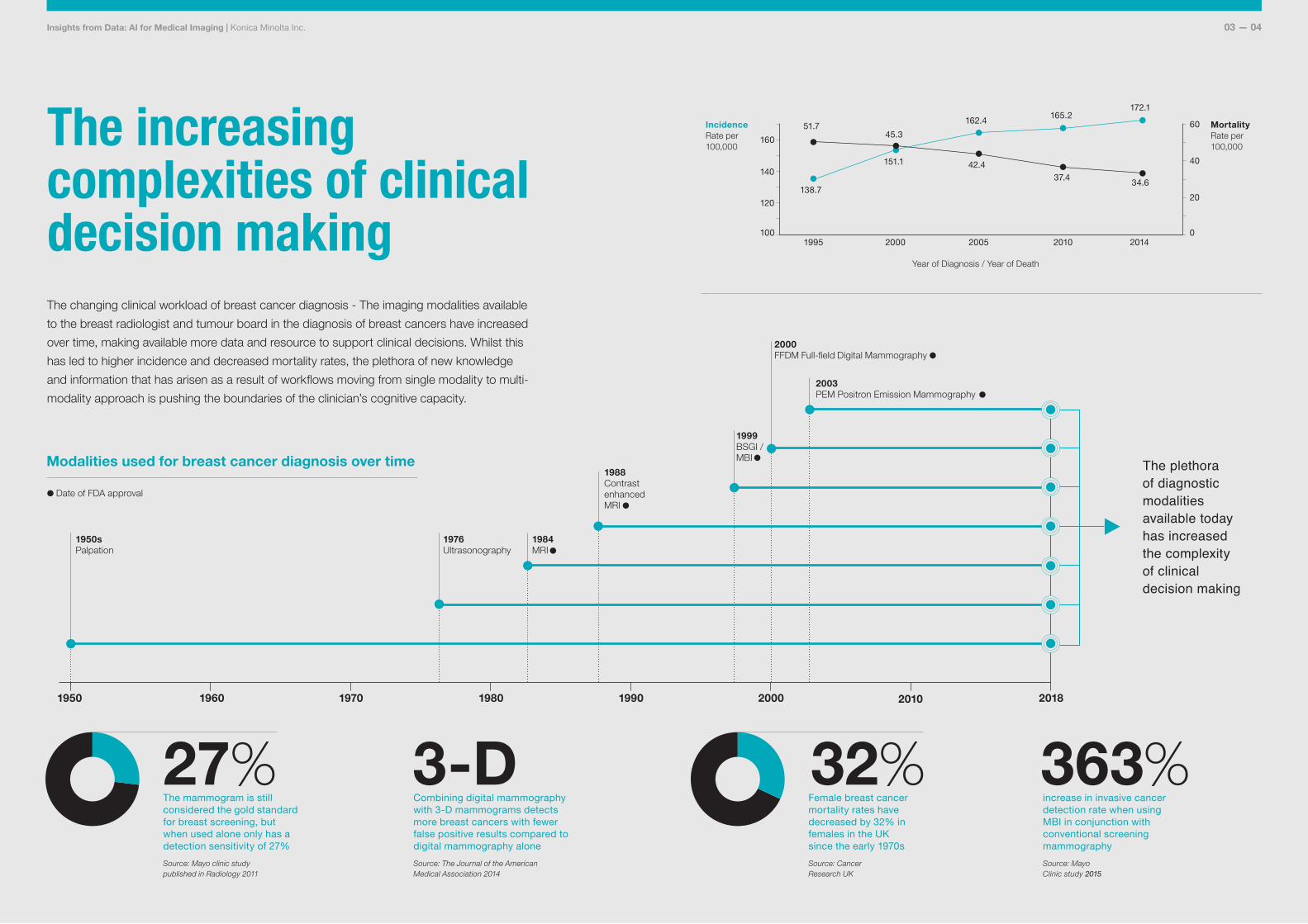
The increasing complexities of clinical decision making
1950 1960 1970 1980 1990 2000 2010 2018
1950sPalpation
1976Ultrasonography
The changing clinical workload of breast cancer diagnosis - The imaging modalities available to the breast radiologist and tumour board in the diagnosis of breast cancers have increased over time, making available more data and resource to support clinical decisions. Whilst this has led to higher incidence and decreased mortality rates, the plethora of new knowledge and information that has arisen as a result of workflows moving from single modality to multi-modality approach is pushing the boundaries of the clinician’s cognitive capacity.
The mammogram is still considered the gold standard for breast screening, but when used alone only has a detection sensitivity of 27%Source: Mayo clinic study published in Radiology 2011
27%Female breast cancer mortality rates have decreased by 32% in females in the UK since the early 1970sSource: Cancer Research UK
32%Combining digital mammography with 3-D mammograms detects more breast cancers with fewer false positive results compared to digital mammography aloneSource: The Journal of the American Medical Association 2014
3-Dincrease in invasive cancer detection rate when using MBI in conjunction with conventional screening mammographySource: Mayo Clinic study 2015
363%
Modalities used for breast cancer diagnosis over time The plethora of diagnostic modalities available today has increased the complexity of clinical decision making
138.7
151.1
162.4165.2
172.1
1995 2000 2005 2010 2014
MortalityRate per 100,000
IncidenceRate per 100,000
Year of Diagnosis / Year of Death
51.745.3
42.437.4
34.6
0100
20120
40140
60
160
1984MRI
1988Contrast enhanced MRI
1999BSGI / MBI
2000FFDM Full-field Digital Mammography
2003PEM Positron Emission Mammography
Date of FDA approval
AI has the ability to have a profound impact on patient outcomes and clinician workflows. We’re committed towards this goal.
Konica Minolta’s research in our laboratories in Europe, Japan and the United States brings together the various platforms that will form Cognitive Hub. Get in touch if you are interested in further discussions around developing and using AI for healthcare.
http://research.konicaminolta.euhttp://artificial-intelligence.konicaminolta.eu [email protected]
© Konica Minolta Inc. Konica Minolta Laboratory Europe – Konica Minolta Inc.90 Chancery Lane, London, WC2A 1EU,United Kingdom All rights reserved.
Talk to us