insights into pneumococcal pathogenesis from the crystal structure of the modular teichoic acid...
TRANSCRIPT
NATURE STRUCTURAL & MOLECULAR BIOLOGY VOLUME 12 NUMBER 6 JUNE 2005 533
Insights into pneumococcal pathogenesis from the crystal structure of the modular teichoic acid phosphorylcholine esterase PceJuan A Hermoso1, Laura Lagartera2, Ana González3, Meike Stelter4, Pedro García3, Martín Martínez-Ripoll1, José L García3 & Margarita Menéndez2
Phosphorylcholine, a specific component of the pneumococcal cell wall, is crucial in pathogenesis. It directly binds to the human platelet-activating factor (PAF) receptor and acts as a docking station for the family of surface-located choline-binding proteins (CBP). The first structure of a complete pneumococcal CBP, Pce (or CbpE), has been solved in complex with the reaction product and choline analogs. Pce has a novel modular structure, with a globular N-terminal module containing a binuclear Zn2+ catalytic center, and an elongated choline-binding module. Residues involved in substrate binding and catalysis are described and modular configuration of the active center accounts for in vivo features of teichoic acid hydrolysis. The hydrolysis of PAF by Pce and its regulatory role in phosphorylcholine decoration of the bacterial surface provide new insights into the critical function of Pce in pneumococcal adherence and invasiveness.
Streptococcus pneumoniae is a Gram-positive pathogen and a major cause of pneumonia, sepsis and meningitis. Many interactions of this bacterium with its host seem to be mediated by components of the bacterial cell wall, specifically by phosphorylcholine (PC) residues of pneumococcal teichoic and lipoteichoic acids. PC, which serves as an anchor for surface-located CBPs1, is essential for the optimal activity of murein hydrolases2, andseems to be involved in many other physiological functions of S. pneumoniae2,3. PC is also recognized by components of the host response, such as human C-reactive protein4 (CRP) and the PAF receptor5.
CBP family members share an N- or C-terminal choline-binding module made up of homologous repeats of ~20 residues6, which facilitates the anchorage of these proteins to the cell envelope through noncovalent interaction with choline residues. In addition to cell wall lytic enzymes7, the CBP family includes virulence factors involved in cellular adhesion8,9 and colonization10–13. The cell wall hydrolase most recently characterized at the molecular level is the teichoic acid phosphorylcholine esterase, Pce (or CbpE). This protein has a cleav-able signal peptide that renders, after its cleavage, a mature protein of 69,426 Da (refs. 14,15). The enzymatic activity of Pce was first described in 1974 (ref. 16), showing that the enzyme is capable of removing a limited number of PC residues from pneumococcal cell walls. The molecular architecture of Pce includes a catalytic mod-ule localized at the N-terminal part of the protein (312 residues),
a C-terminal choline-binding module with ten homologous repeat-ing units (205 residues), and a long C-terminal tail of 85 residues. Notably, a pce mutant showed substantially reduced colonization of the rat nasopharynx, which was attributed to a decreased ability to adhere to human cells17. As a prelude to invasive disease, pneumococci enter the host via the nasopharynx, where they attach to epithelial cells and, in some instances, persist for several months18. Therefore, characterization of the structure of Pce can lead to new therapeutic strategies for the treatment of pneumococcal diseases.
The first crystal structure of a choline-binding domain showed that the C-terminal module of the major pneumococcal autolysin, LytA, adopts a peculiar β-solenoid structure19. Notably, the complete struc-ture of the Cpl-1 lysozyme revealed that the choline-binding module can fold in two different structural motifs20. Recently, the solution structure of the adhesion R2 domain of the principal pneumococcal adhesin (CbpA) has been solved21. To obtain further insights into the structure and function of CBPs, we determined, at a resolution of 1.9 Å, the three-dimensional structure of the first pneumococcal-encoded CBP carrying both the catalytic and the choline-binding modules. The structure of Pce reveals that the specific arrangement of protein modules in each CBP is critical for its function on the bacterial surface. Elucidation of the Pce structure provides new insights into its mechanism of teichoic acid degradation as well as its putative role in bacterial pathogenicity.
1Grupo de Cristalografía Macromolecular y Biología Estructural and 2Departamento de Química-Física de Macromoléculas Biológicas, Instituto Química-Física Rocasolano, CSIC, Serrano 119, 28006 Madrid, Spain. 3Departamento de Microbiología Molecular, Centro de Investigaciones Biológicas, CSIC, Ramiro de Maeztu 9, 28040 Madrid, Spain. 4Institut de Biologie Structurale J.-P. Ebel CEA-CNRS-UJF, 41 rue Jules Horowitz, 38027 Grenoble Cedex 1, France. Correspondence should be addressed to J.A.H. ([email protected]).
Published online 15 May 2005; doi:10.1038/nsmb940
A R T I C L E S©
2005
Nat
ure
Pub
lishi
ng G
roup
ht
tp://
ww
w.n
atur
e.co
m/n
smb
534 VOLUME 12 NUMBER 6 JUNE 2005 NATURE STRUCTURAL & MOLECULAR BIOLOGY
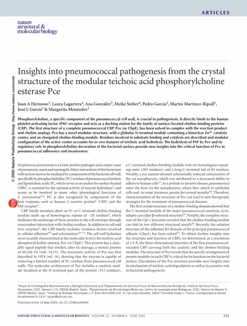
RESULTSOverall structure of Pce–phosphorylcholine complexThe crystal structure (Fig. 1a) comprises two structural modules, the catalytic module (residues 1–300) and the choline-binding module (resi-dues 313–540), which are joined by a small linker comprising residues 301–312. The catalytic module is formed by a single globular domain of 55 × 53 × 45 Å that folds into an αβ-βα sandwich, following a metallo-β-lactamase-like fold despite its low (≤13%) sequence identity with other family members and its larger size. The structure of Pce catalytic module can be divided into two near-equivalent regions, each formed by an antiparallel β-sheet (five and four β-strands, respectively) followed by three αβ-motifs (Supplementary Fig. 1 online). The active site is located within a shallow groove at the interface between the two β-sheets and is surrounded by αβ-loops (Fig. 1a). Comparison of the Pce structure with those of the β-lactamase fold family22 shows that the closest structures are those from the Stenotrophomonas maltophilia β-lactamase23 (13% identity; r.m.s. deviation of 3.1 Å for 176 Cα atoms), human glyoxalase II24 (15% identity; r.m.s. deviation of 3.4 Å for 173 Cα atoms) and rubredoxin-oxygen oxidoreduc-tase from Desulfovibrio gigas25 (13% identity; r.m.s. deviation of 7.9 Å for 183 Cα atoms). To the best of our knowledge, the Pce catalytic module exhibits a new fold among phospho-ester-hydrolyzing enzymes.
The choline-binding module is formed by ten repeats (p1–p10), with the consensus motif
(GWXK-X4–5-WYY-Φ-X3–5-GXMX2–3, where X is any residue and Φ is a hydrophobic residue, and a C-terminal tail (residues 517–540). Each repeat comprises a symmetrical β-hairpin followed by a loop and a coiled region (Supplementary Fig. 1). The overall shape of the Pce choline-binding module is approximately a triangular prism ~110 Å high with sides of 26 Å, with the choline-binding sites placed along the three lateral faces. The repeats are strictly arranged following a left-handed superhelical fold, similar to that observed in C-LytA19 and in the CI domain of Cpl-1 (ref. 20). The hairpins extend perpendicularly to the axis of the superhelix and each repeat is located at a 120° counter-clockwise rotation. Notably, despite the lack of sequence identity, the first 21 amino acids of the Pce C-terminal tail (residues 517–534) follow the same superhelical fold.
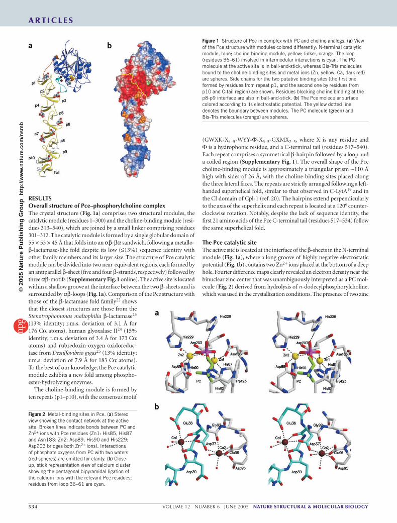
The Pce catalytic siteThe active site is located at the interface of the β-sheets in the N-terminal module (Fig. 1a), where a long groove of highly negative electrostatic potential (Fig. 1b) contains two Zn2+ ions placed at the bottom of a deep hole. Fourier difference maps clearly revealed an electron density near the binuclear zinc center that was unambiguously interpreted as a PC mol-ecule (Fig. 2) derived from hydrolysis of n-dodecylphosphorylcholine, which was used in the crystallization conditions. The presence of two zinc
Figure 1 Structure of Pce in complex with PC and choline analogs. (a) View of the Pce structure with modules colored differently: N-terminal catalytic module, blue; choline-binding module, yellow; linker, orange. The loop (residues 36–61) involved in intermodular interactions is cyan. The PC molecule at the active site is in ball-and-stick, whereas Bis-Tris molecules bound to the choline-binding sites and metal ions (Zn, yellow; Ca, dark red) are spheres. Side chains for the two putative binding sites (the first one formed by residues from repeat p1, and the second one by residues from p10 and C-tail region) are shown. Residues blocking choline binding at the p8-p9 interface are also in ball-and-stick. (b) The Pce molecular surface colored according to its electrostatic potential. The yellow dotted line denotes the boundary between modules. The PC molecule (green) and Bis-Tris molecules (orange) are spheres.
Figure 2 Metal-binding sites in Pce. (a) Stereo view showing the contact network at the active site. Broken lines indicate bonds between PC and Zn2+ ions with Pce residues (Zn1: His85, His87 and Asn183; Zn2: Asp89, His90 and His229; Asp203 bridges both Zn2+ ions). Interactions of phosphate oxygens from PC with two waters (red spheres) are omitted for clarity. (b) Close-up, stick representation view of calcium cluster showing the pentagonal bipyramidal ligation of the calcium ions with the relevant Pce residues; residues from loop 36–61 are cyan.
A R T I C L E S©
2005
Nat
ure
Pub
lishi
ng G
roup
ht
tp://
ww
w.n
atur
e.co
m/n
smb

NATURE STRUCTURAL & MOLECULAR BIOLOGY VOLUME 12 NUMBER 6 JUNE 2005 535
atoms was confirmed by ICP-OE spectrometry (see Methods). The Zn2+ cations are 3.6 Å apart; their coordination geometry can be described as distorted square pyramidal, with the apical positions being occupied by Oδ1 of Asn183 (Zn1) and by Nε2 of His229 (Zn2); Zn2 is shifted 0.6 Å out of the basal plane. The mean Zn-ligand distance is 2.04 Å for both cations. Pce residues involved in metal coordination (Fig. 2) differ from Zn ligands in other hydrolases of the metallo-β-lactamase fold family23–26 but almost overlap with them in their three-dimensional structures.
In contrast with data reported for unligated binuclear enzymes of the metallo-β-lactamase family fold26, no water or hydroxide molecule shared by Zn2+ ions was observed in the Pce–PC complex, where two oxygens of PC complete the pentavalent coordination of Zn2+ ions (Fig. 2a). However, displacement of the metal-bridging hydroxide has been previously described in phospholipase C upon formation of com-plex with phosphatidylcholine analogs27,28.
The structure of the Pce–PC complex provides the basis for Pce sub-strate recognition, with both metal ions involved in binding and posi-tioning of the substrate through direct coordination of the phosphate moiety. Phosphate recognition is also mediated by polar interactions with His228 and two water molecules (Fig. 2a), while the trimethyl-ammonium group is stabilized by interaction with Asp89 and Trp123 (cation-π interaction). Notably, a very similar PC recognition pattern (two coordinated ions, Ca2+, an acidic Glu81, and an aromatic residue, Phe66) has been found in human CRP29.
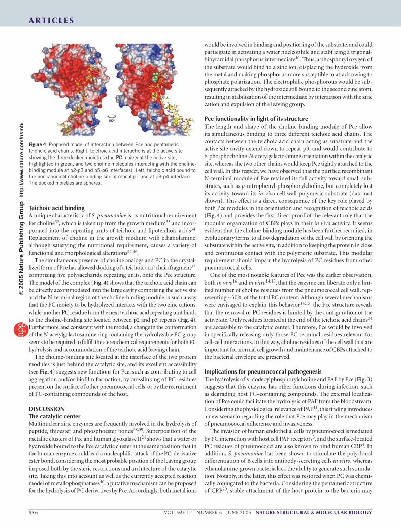
Hydrolysis of phosphorylcholine-containing compoundsThe ability of Pce to hydrolyze the n-dodecylphosphorylcholine (Fig. 3a) included in the crystallization conditions was verified by mass spectrometry. Detergent samples incubated in either the absence or presence of Pce were analyzed and showed complete degradation by the enzyme (Fig. 3b). This result suggests other potential roles for Pce during infection by degrading host PC–containing compounds. Indeed, mass spectrometry analysis showed that PAF is cleaved as efficiently as the structurally related n-dodecylphosphorylcholine; that is, ~80% of this potent lipidic first messenger is degraded after 5 h (Fig. 3).
Choline-binding sitesCholine-binding sites are located at the interface of two consecutive repeats, where three structurally conserved aromatic residues (two tryp-
tophans from the first repeat and one tyrosine from the following one) form a cavity in which the choline trimethyl-ammonium group is stabi-lized primarily by cation-π interactions30. The cavity walls are completed by a hydrophobic residue (methionine or leucine) placed at its bottom.
The crystal structure of Pce reveals several notable features. First, the sequence and structure of repeat p8 (residues 457–476) preclude choline binding at the p8-p9 interface, as the two critical tryptophans are replaced by glutamate and histidine, respectively. In addition, sub-stitution of the conserved methionine by arginine markedly changes the nature and geometry of the choline-binding site (Fig. 1a). The electron density map was consistent with the presence of six Bis-Tris molecules bound to choline-binding sites. This finding can be explained by the ability of choline-binding sites to recognize tertiary amines as choline analogs31, and by the high concentration of Bis-Tris in the crys-tallization buffer. The other two available sites (Fig. 1a) showed poor residual electron density that could not be unambiguously assigned. Notably, two additional noncanonical sites can be considered as putative choline-binding sites. The first one is located at the interface of the two protein modules, at the back side of the active site, where the aromatic residues forming the cavity are provided only by amino acids from repeat p1 (Tyr319, Trp323 and Tyr333), which are not conserved in the other repeats (Fig. 1a and Supplementary Fig. 1). At this locus, a well-defined Bis-Tris molecule can be modeled into the electron density map. The second noncanonical choline-binding site is formed by four aromatic residues (Fig. 1a) provided by p10 (Trp498 and Trp505) and the C-terminal tail (Tyr524 and Trp532). At this site, a residual electron density was also detected but could not be unambiguously assigned. Taken together, the data are consistent with the presence of ten choline-binding sites in the entire Pce structure.
The modular organization of PcePce shows a novel structural arrangement of its constituent modules. Despite strong differences in the sizes and shapes of the modules, the overall structure seems to be quite rigid. The Pce structural framework is determined by three main factors: (i) a short linker on the surface of the catalytic module; (ii) the presence of a very long loop in the cata-lytic module (residues 36–61) that strongly interacts with the first three repeating units of the choline-binding module; and (iii) two structural Ca2+ ions reinforcing the 36–61 loop conformation. The major interac-tions mediated by this loop comprise a zipper-like system in which aro-matic residues from both modules alternate. Intermodular interactions are reinforced by two salt bridges (Asp42-Lys359 and Lys59-Asp328). The structural calciums exhibit the typical pentagonal bipyramidal ligation of this metal (Fig. 2b). Ca1 binding to Glu36 (Oε2) and Asp37 (Oδ1) produces a tight conformation at the beginning of loop 36–61, whereas Ca2 coordination (Gly92, Asp95 and Glu96) strengths the inter-action between the loop and the rest of the catalytic module, with Asp37 acting as a bidentated ligand of the two Ca2+ ions. The loop contacts account for most of the 1,985 Å2 of the Pce surface that is occluded between modules, and may have important consequences regarding the activity of Pce in vivo. The loop enlarges the surface of the catalytic site toward the third choline-binding site (located 22 Å apart), maintaining its strongly acidic character (Fig. 1b).
Figure 3 Hydrolysis of PAF and n-dodecylphosphorylcholine by Pce. (a) Schematics comparing PAF (1-O-hexadecyl-2-acetoyl-glycero-3-phosphorylcholine) and n-dodecylphosphorylcholine molecules. (b) Time course of PC-derivative hydrolysis followed by liquid chromatography and mass spectrometry. The hydrolysis of n-dodecylphosphorylcholine (1.5 mM) and PAF (1.5 mM) by Pce (80 µM) was monitored at 20 °C.
A R T I C L E S©
2005
Nat
ure
Pub
lishi
ng G
roup
ht
tp://
ww
w.n
atur
e.co
m/n
smb
536 VOLUME 12 NUMBER 6 JUNE 2005 NATURE STRUCTURAL & MOLECULAR BIOLOGY
Teichoic acid bindingA unique characteristic of S. pneumoniae is its nutritional requirement for choline32, which is taken up from the growth medium33 and incor-porated into the repeating units of teichoic and lipoteichoic acids34. Replacement of choline in the growth medium with ethanolamine, although satisfying the nutritional requirement, causes a variety of functional and morphological alterations35,36.
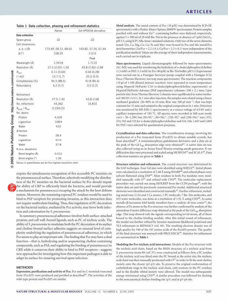
The simultaneous presence of choline analogs and PC in the crystal-lized form of Pce has allowed docking of a teichoic acid chain fragment37, comprising five polysaccharide repeating units, onto the Pce structure. The model of the complex (Fig. 4) shows that the teichoic acid chain can be directly accommodated into the large cavity comprising the active site and the N-terminal region of the choline-binding module in such a way that the PC moiety to be hydrolyzed interacts with the two zinc cations, while another PC residue from the next teichoic acid repeating unit binds to the choline-binding site located between p2 and p3 repeats (Fig. 4). Furthermore, and consistent with the model, a change in the conformation of the N-acetylgalactosamine ring containing the hydrolyzable PC group seems to be required to fulfill the stereochemical requirements for both PC hydrolysis and accommodation of the teichoic acid leaving chain.
The choline-binding site located at the interface of the two protein modules is just behind the catalytic site, and its excellent accessibility (see Fig. 4) suggests new functions for Pce, such as contributing to cell aggregation and/or biofilm formation, by crosslinking of PC residues present on the surface of other pneumococcal cells, or by the recruitment of PC-containing compounds of the host.
DISCUSSIONThe catalytic centerMultinuclear zinc enzymes are frequently involved in the hydrolysis of peptide, thioester and phosphoester bonds38,39. Superposition of the metallic clusters of Pce and human glyoxalase II24 shows that a water or hydroxide bound to the Pce catalytic cluster at the same position that in the human enzyme could lead a nucleophilic attack of the PC-derivative ester bond, considering the most probable position of the leaving group imposed both by the steric restrictions and architecture of the catalytic site. Taking this into account as well as the currently accepted reaction model of metallophosphatases40, a putative mechanism can be proposed for the hydrolysis of PC derivatives by Pce. Accordingly, both metal ions
would be involved in binding and positioning of the substrate, and could participate in activating a water nucleophile and stabilizing a trigonal-bipyramidal phosphorus intermediate40. Thus, a phosphoryl oxygen of the substrate would bind to a zinc ion, displacing the hydroxide from the metal and making phosphorus more susceptible to attack owing to phosphate polarization. The electrophilic phosphorous would be sub-sequently attacked by the hydroxide still bound to the second zinc atom, resulting in stabilization of the intermediate by interaction with the zinc cation and expulsion of the leaving group.
Pce functionality in light of its structureThe length and shape of the choline-binding module of Pce allow its simultaneous binding to three different teichoic acid chains. The contacts between the teichoic acid chain acting as substrate and the active site cavity extend down to repeat p3, and would contribute to 6-phospho choline-N-acetylgalactosamine orientation within the catalytic site, whereas the two other chains would keep Pce tightly attached to the cell wall. In this respect, we have observed that the purified recombinant N-terminal module of Pce retained its full activity toward small sub-strates, such as p-nitrophenyl-phosphorylcholine, but completely lost its activity toward its in vivo cell wall polymeric substrate (data not shown). This effect is a direct consequence of the key role played by both Pce modules in the orientation and recognition of teichoic acids (Fig. 4) and provides the first direct proof of the relevant role that the modular organization of CBPs plays in their in vivo activity. It seems evident that the choline-binding module has been further recruited, in evolutionary terms, to allow degradation of the cell wall by orienting the substrate within the active site, in addition to keeping the protein in close and continuous contact with the polymeric substrate. This modular requirement should impair the hydrolysis of PC residues from other pneumococcal cells.
One of the most notable features of Pce was the earlier observation, both in vivo16 and in vitro14,15, that the enzyme can liberate only a lim-ited number of choline residues from the pneumococcal cell wall, rep-resenting ~30% of the total PC content. Although several mechanisms were envisaged to explain this behavior14,15, the Pce structure reveals that the removal of PC residues is limited by the configuration of the active site. Only residues located at the end of the teichoic acid chains34 are accessible to the catalytic center. Therefore, Pce would be involved in specifically releasing only those PC terminal residues relevant for cell-cell interactions. In this way, choline residues of the cell wall that are important for normal cell growth and maintenance of CBPs attached to the bacterial envelope are preserved.
Implications for pneumococcal pathogenesisThe hydrolysis of n-dodecylphosphorylcholine and PAF by Pce (Fig. 3) suggests that this enzyme has other functions during infection, such as degrading host PC–containing compounds. The external localiza-tion of Pce could facilitate the hydrolysis of PAF from the bloodstream. Considering the physiological relevance of PAF41, this finding introduces a new scenario regarding the role that Pce may play in the mechanism of pneumococcal adherence and invasiveness.
The invasion of human endothelial cells by pneumococci is mediated by PC interaction with host cell PAF receptors5, and the surface-located PC residues of pneumococci are also known to bind human CRP4. In addition, S. pneumoniae has been shown to stimulate the polyclonal differentiation of B cells into antibody-secreting cells in vitro, whereas ethanolamine-grown bacteria lack the ability to generate such stimula-tion. Notably, in the latter, this effect was restored when PC was chemi-cally conjugated to the bacteria. Considering the pentameric structure of CRP29, stable attachment of the host protein to the bacteria may
Figure 4 Proposed model of interaction between Pce and pentameric teichoic acid chains. Right, teichoic acid interactions at the active site showing the three docked moieties (the PC moiety at the active site, highlighted in green, and two choline molecules interacting with the choline-binding module at p2-p3 and p5-p6 interfaces). Left, teichoic acid bound to the noncanonical choline-binding site at repeat p1 and at p3-p4 interface. The docked moieties are spheres.
A R T I C L E S©
2005
Nat
ure
Pub
lishi
ng G
roup
ht
tp://
ww
w.n
atur
e.co
m/n
smb
NATURE STRUCTURAL & MOLECULAR BIOLOGY VOLUME 12 NUMBER 6 JUNE 2005 537
require the simultaneous recognition of five accessible PC moieties on the pneumococcal surface. Therefore, selectively modifying the distribu-tion of the PC moieties on the bacterial surface by Pce should impair the ability of CRP to efficiently bind the bacteria, and would provide a mechanism for pneumococci escaping the attack by the host defense system. Moreover, the remaining exposed PC residues would be used to bind to PAF receptors for promoting invasion, as this interaction does not require multivalent binding. Thus, fine regulation of PC decoration on the bacterial surface, mediated by Pce activity, may favor both infec-tion and colonization by S. pneumoniae.
In summary, pneumococcal adherence involves both surface-attached proteins and cell wall–bound ligands, such as PC of teichoic acids. The ability of S. pneumoniae to modulate both the PC decoration of its surface and choline-bound surface adhesins suggests an unusual level of com-plexity underlying the regulation of pneumococcal adherence, in which Pce seems to play an important role. The finding that Pce may have a dual function—that is, hydrolyzing and/or sequestering choline-containing compounds, such as PAF, and regulating the binding of pneumococci to CRP, while it conserves their ability to bind to PAF receptors—suggests new approaches for investigating how this important pathogen is able to adapt its surface for ensuring survival upon infection.
METHODSExpression, purification and activity of Pce. Pce and its C-terminal truncated form (Pce∆55) were produced and purified as described42. The activities of the wild-type protein and Pce∆55 are similar.
Metal analysis. The metal content of Pce (10 µM) was determined by ICP-OE spectrometry with a Perkin-Elmer Optima 2000DV spectrometer. Protein samples purified with and without Zn2+-containing buffers were dialyzed, respectively, against 5 × 500 ml of 20 mM Bis-Tris in the presence or absence of 3 µM ZnCO3, pH 5.5, using ICP (Alfa Aesar) standard solutions. Only two of the seven elements tested (Zn, Ca, Mg, Co, Cu, Fe and Mn) were bound to Pce and the metal/Pce stoichiometries (Zn/Pce = 2.2 ± 0.1; Ca/Pce = 2.3 ± 0.1) were independent of the purification method. Values are the average of three independent measurements, each carried out in triplicate.
Mass spectrometry. Liquid chromatography followed by mass-spectrometry (LC-MS) was used for monitoring the hydrolysis of n-dodecylphosphorylcholine (1.5 mM) or PAF (1.5 mM) by Pce (80 µM) in Bis-Tris buffer, pH 5.5. Experiments were carried out in a Finnigan Surveyor pump coupled with a Finnigan LCQ Deca (Thermo Electron) ion trap mass spectrometer. The reaction components (10 µl of 1:100 diluted mixture reaction) were separated at room temperature using Hypersil HyPurity C18 (n-dodecylphosphorylcholine experiments) or Hypersil HyPurity Advance (PAF experiments) columns (100 × 2.1 mm; 3 µm particle size) from Thermo Electron. Columns were equilibrated in water/metha-nol (80/20÷(v/v)). At 1 min after injection, the samples were eluted using a linear methanol gradient (20–90% in 10 min; flow rate 100 µl min–1) that was kept constant for 15 min and ramped to the original composition in 1 min. Detection was monitored by MS-ESI(+) spectrometry at a source voltage of 4.0 kV and a capillary temperature of 185 °C. All spectra were recorded in full-scan mode (m/z = 50–1,200) but [M+H]+, [M+Na]+, [2M+H]+ and [2M+Na]+ ions (352, 374, 703 and 725 for n-dodecylphosphorylcholine and 524, 546, 1,047 and 1,069 for PAF) were selected for quantization purposes.
Crystallization and data collection. The crystallization strategy, involving the production of a Pce truncated form (Pce∆55) to obtain suitable crystals, has been described42. A nonisomorphous gadolinium derivative and a data set at the peak of the Gd LIII absorption edge were obtained42. A native data set was also collected using an in-house Enraf Nonius rotating anode generator. X-ray diffraction data were processed and scaled using MOSFLM43 and SCALA44. Data collection statistics are given in Table 1.
Structure solution and refinement. The crystal structure was determined by the SAD technique. Four Gd sites were identified using SHELX45. Initial phases were calculated at a resolution of 2.68 Å using SHARP46 and refined phases were solvent-flattened using DM44. Most residues in both Pce modules were mod-eled manually with O47 and refined with CNS48. The molecular replacement procedure was carried out using MOLREP from the CCP4 package44, with the native data set and the previously constructed Pce model. Additional structural elements were identified and constructed manually47. Further refinement, includ-ing metal ions (2 Zn and 2 Ca atoms), 1 PC molecule, 7 Bis-Tris molecules, and 433 water molecules, was done at a resolution of 1.92 Å using CNS48. As some metallo-β-lactamase fold family members have a catalytic di-iron center25, the absence of Fe atoms in the Pce structure was further confirmed by analysis of the anomalous Fourier difference map obtained at the peak of the Gd LIII absorption edge. This map showed only the signals corresponding to Gd atoms, all of them bound to the choline-binding module. After this initial round of refinement, the model was further refined by iterative maximum likelihood positional and TLS refinement in REFMAC5 (ref. 49). The resulting electron density was of high quality for 540 of the 547 amino acids of the Pce∆55 protein. The quality of the final structure was assessed with PROCHECK50. Statistics for refinement are summarized in Table 1.
Modeling the Pce-teichoic acid interactions. Models of the Pce structure with
the teichoic acid chain, based on the RMN structure of a teichoic acid from S. pneumoniae strain R6 (ref. 37), were constructed as follows: first, a PC residue of the teichoic acid was fitted onto the PC bound at the active site; the teichoic acid chain was then manually positioned with O47 in order to fit the next choline moiety onto the closest (p2-p3) site. To preserve the original conformation of carbohydrate rings in the teichoic acid chain, only torsions in the PC residues and in the flexible ribitol moiety were allowed. The model was subsequently energy-minimized using CNS48. A similar procedure was followed for docking to the noncanonical choline-binding site (p1) and at p3-p4 site.
Table 1 Data collection, phasing and refinement statistics Native Gd-HPDO3A derivative
Data collection
Space group C2 C2
Cell dimensions
a, b, c (Å) 173.49, 58.12, 68.65 169.82, 57.26, 67.44
β (°) 108.29 112.6
Peak
Wavelength (Å) 1.5418 1.7110
Resolution (Å) 27.5 (2.02)–1.92 43.8 (2.82)–2.68
Rsym 0.11 (0.60) 0.04 (0.28)
I / σ(I) 13.7 (1.7) 20.2 (5.0)
Completeness (%) 95.5 (88.5) 97.8 (85.4)
Redundancy 6.2 (5.7) 3.5 (2.2)
Refinement
Resolution (Å) 27.5–1.92 43.8–2.68
No. reflections 44,362 16,810
Rwork / Rfree 0.19/0.23
No. atoms
Protein 4,439
Ligand/ion 108/4
Water 433
B-factors
Protein 28.4
Ligand/ion 57.0
Water 37.8
R.m.s. deviations
Bond lengths (Å) 0.012
Bond angles (°) 1.36
Values in parentheses are for the highest-resolution shell.
A R T I C L E S©
2005
Nat
ure
Pub
lishi
ng G
roup
ht
tp://
ww
w.n
atur
e.co
m/n
smb
538 VOLUME 12 NUMBER 6 JUNE 2005 NATURE STRUCTURAL & MOLECULAR BIOLOGY
Accession codes. Atomic coordinates and structure factors for the Pce–phophoryl-choline complex have been deposited in the Protein Data Bank (accession code 2BIB). BIND identifiers (http://bind.ca/): 262672, 262685.
Note: Supplementary information is available on the Nature Structural & Molecular Biology website.
ACKNOWLEDGMENTSThe authors thank L. Serre for helpful suggestions, R. Kahn for help in data processing and analysis, Bracco Imaging (Milan) for providing a sample of Gd–HPDO3A, the staff of the BM30A beamline at the European Synchrotron Radiation Facility (Grenoble) for support and R.A. Klein for providing the atomic coordinates of the pentameric lipoteichoic acid. We thank R. López and E. García for critically reading the manuscript, W. Ran for correcting the English version and A. Torrecillas and SUIC staff for help in the ICP-OE measures. This work was supported by grants BIO2000-1307, BIO2002-02887, BIO2003-01952 and BMC2003-00074 from Dirección General de Investigación and by grant Contrato-Programa de Grupos Estratégicos (BMC2000-1002) de la Comunidad Autónoma de Madrid. L.L. and A.G. hold a fellowship from the Spanish Ministry of Science and Technology.
COMPETING INTERESTS STATEMENTThe authors declare that they have no competing financial interests.
Received 1 February; accepted 4 April 2005Published online at http://www.nature.com/nsmb/
1. Swiatlo, E., McDaniel, L.S. & Briles, D.E. Choline-binding proteins. In The Pneumococcus (eds. Tuomanen, E.I., Mitchell, T.J., Morrison, D.A. & Spratt, B.G.) 49–60 (ASM Press, Washington, DC, 2004).
2. López, R., García, E., García, P. & García, J.L. Cell wall hydrolases. In The Pneumococcus (eds. Tuomanen, E.I., Mitchell, T.J., Morrison, D.A. & Spratt, B.G.) 75– 88 (ASM Press, Washington, DC, 2004).
3. López, R., García, E., García, P., Ronda, C. & Tomasz, A. Choline-containing bac-teriophage receptors in Streptococcus pneumoniae. J. Bacteriol. 151, 1581–1590 (1982).
4. Pepys, M.B. & Hirschfield, G.M. C-reactive protein: a critical update. J. Clin. Invest. 111, 1805–1812 (2003).
5. Cundell, D.R., Gerard, N.P., Gerard, C., Idanpaan-Heikkila, I. & Tuomanen, E.I. Streptococcus pneumoniae anchors to activated human cells by the receptor for plate-let-activating factor. Nature 377, 435–438 (1995).
6. García, E. et al. Molecular evolution of lytic enzymes of Streptococcus pneumoniae and its bacteriophages. Proc. Natl. Acad. Sci. USA 85, 914–918 (1988).
7. López, R. & García, E. Recent trends on the molecular biology of pneumococcal cap-sules, lytic enzymes, and bacteriophage. FEMS Microbiol. Rev 28, 553–580 (2004).
8. Sánchez-Beato, A.R., López, R. & García, J.L. Molecular characterization of PcpA: a novel choline-binding protein of Streptococcus pneumoniae. FEMS Microbiol. Lett. 164, 207–214 (1998).
9. Rosenow, C. et al. Contribution of novel choline-binding proteins to adherence, coloniza-tion and immunogenicity of Streptococcus pneumoniae. Mol. Microbiol. 25, 819–829 (1997).
10. Crain, M.J. et al. Pneumococcal surface protein A (PspA) is serologically highly vari-able and is expressed by all clinically important capsular serotypes of Streptococcus pneumoniae. Infect. Immun. 58, 3293–3299 (1990).
11. Hammerschmidt, S., Talay, S.R., Brandtzaeg, P. & Chhatwal, G.S. SpsA, a novel pneu-mococcal surface protein with specific binding to secretory immunoglobulin A and secretory component. Mol. Microbiol. 25, 1113–1124 (1997).
12. Brooks-Walter, A., Briles, D.E. & Hollingshead, S.K. The pspC gene of Streptococcus pneumoniae encodes a polymorphic protein, PspC, with elicits cross-reactive anti bodies to PspA and provides immunity to pneumococcal bacteremia. Infect. Immun. 67, 6533–6542 (1999).
13. Tu, A.H., Fulgham, R.L., McCroy, M.A., Briles, D.E. & Szalai, A.J. Pneumococcal sur-face protein A inhibits complement activation by Streptococcus pneumoniae. Infect. Immun. 67, 4720–4724 (1999).
14. De las Rivas, B., García, J.L., López, R. & García, P. Molecular characterization of the pneumococcal teichoic acid phosphorylcholine esterase. Microb. Drug Resist. 7, 213–222 (2001).
15. Vollmer, W. & Tomasz, A. Identification of the teichoic acid phosphorylcholine esterase in Streptococcus pneumoniae. Mol. Microbiol. 39, 1610–1622 (2001).
16. Höltje, J.V. & Tomasz, A. Teichoic acid phophorylcholine esterase. A novel enzyme activity in pneumococcus. J. Biol. Chem. 249, 7032–7034 (1974).
17. Gosink, K.K., Mann, E.R., Guglielmo, C., Tuomanen, E.I. & Masure, H.R. Role of novel choline binding proteins in virulence of Streptococcus pneumoniae. Infect. Immun. 68, 5690–5695 (2000).
18. Austrian, R. Some aspects of the pneumococcal carrier state. J. Antimicrob. Chemother. 18 suppl. suppl. A, 35–45 (1986).
19. Fernández-Tornero, C., López, R., García, E., Giménez-Gallego, G. & Romero, A. A novel solenoid fold in the cell wall anchoring domain of the pneumococcal virulence factor LytA. Nat. Struct. Biol. 8, 1020–1024 (2001).
20. Hermoso, J.A. et al. Structural basis for selective recognition of pneumococcal cell wall by modular endolysin from phage Cp-1. Structure 11, 1239–1249 (2003).
21. Luo, R. et al. Solution structure of choline binding protein A, the major adhesin of Streptococcus pneumoniae. EMBO J. 24, 34–43 (2005).
22. Holm, L. & Sander, C. Mapping the protein universe. Science 273, 595–603 (1996).
23. Ullah, J.H. et al. The crystal structure of the L1 metallo-β-lactamase from Stenotrophomonas maltophilia at 1.7 Å resolution. J. Mol. Biol. 284, 125–136 (1998).
24. Cameron, A.D., Ridderström, M., Olin, B. & Mannervik, B. Crystal structure of human glyoxalase II and its complex with a glutathione thiolester substrate analogue. Structure 7, 1067–1078 (1999).
25. Frazao, C. et al. Structure of a dioxygen reduction enzyme from Desulfovibrio gigas. Nat. Struct. Biol. 7, 1041–1045 (2000).
26. Rasia, R.M., Ceolín, M. & Vila, A.J. Grafting a new metal ligand in the cocatalytic site of B. cereus metallo-β-lactamase: Structural flexibility without loss of activity. Protein Sci. 12, 1538–1546 (2003).
27. Hansen, S., Hansen, L.K. & Hough, E. Crystal structures of phosphate, iodide and iodate-inhibited phospholipase C from Bacillus cereus and structural investigations of the binding of reaction products and a substrate analogue. J. Mol. Biol. 225, 543–549 (1992).
28. Hansen, S., Hough, E., Svensson, L.A., Wong, Y-L. & Martin, S.F. Crystal structure of phospholipase C from Bacillus cereus complexed with a substrate analog. J. Mol. Biol. 234, 179–187 (1993).
29. Thompson, D., Pepys, M.B. & Wood, S.P. The physiological structure of human C-reactive protein and its complex with phosphocholine. Structure 7, 169–177 (1999).
30. Dougherty, D.A. & Stauffer, D.A. Acetylcholine binding by a synthetic receptor: implica-tions for biological recognition. Science 250, 1558–1560 (1990).
31. Sanz, J.M., López, R. & García, J.L. Structural requirements of choline derivatives for “conversion” of pneumococcal amidase. FEBS Lett. 232, 308–312 (1988).
32. Tomasz, A. Choline in the cell wall of a bacterium: novel type of polymer-linked choline in pneumococcus. Science 157, 694–697 (1967).
33. Bean, B. & Tomasz, A. Choline metabolism in pneumococci. J. Bacteriol. 130, 571–574 (1977).
34. Behr, T., Fischer, W., Peter-Katalinic, J. & Edge, H. The structure of pneumococcal lipoteichoic acid. Improved preparation, chemical and mass spectrometric studies. Eur. J. Biochem. 207, 1063–1075 (1992).
35. Tomasz, A. Biological consequences of the replacement of choline by ethanolamine in the cell wall of pneumococcus: chain formation, loss of transformability, and loss of autolysin. Proc. Natl. Acad. Sci. USA 59, 86–93 (1968).
36. Yother, J., Handsome, G.L. & Briles, D.E. Truncated forms of PspA that are secreted from Streptococcus pneumoniae and their use in functional studies and cloning of the pspA gene. J. Bacteriol. 174, 610–618 (1992).
37. Klein, R.A., Hartmann, R., Egge, H., Behr, T. & Fischer, W. The aqueous solution structure of a lipoteichoic acid from Streptococcus pneumoniae strain R6 contain-ing 2,4-diamino-2,4,6-trideoxy-galactose: evidence for conformational mobility of the galactopyranose ring. Carbohydr. Res. 281, 79–98 (1996).
38. Wilcox, D.E. Binuclear metallohydrolases. Chem. Rev. 96, 2435–2458 (1996).39. Lipscomb, W.N. & Sträter, N. Recent advances in zinc enzymology. Chem. Rev. 96,
2375–2434 (1996).40. Knowles, J.R. Enzyme-catalyzed phosphoryl transfer reactions. Annu. Rev. Biochem.
49, 877–919 (1980).41. O´Flaherty. J.T. & Wykle, R.L. In Platelet Activating Factor and Human Disease
(eds. Barnes, P.J., Page, C.P. & Henson, P.) 117–137 (Blackwell Scientific, Oxford, 1989).
42. Lagartera, L. et al. Crystallization and preliminary X-ray diffraction studies of the pneumococcal teichoic acid phosphorylcholine esterase Pce. Acta Crystallogr. F 61, 221–224 (2005).
43. Leslie, A.G.W. Profile fitting. In Proceedings of the CCP4 Study Weekend (eds. Machin, J.R. & Papiz, M.Z.) 39–50 (SERC Daresbury Laboratory, Warrington, UK, 1987).
44. Collaborative Computational Project, Number 4. The CCP4 suite: programs for protein crystallography. Acta Crystallogr. D 50, 760–763 (1994).
45. Sheldrick, G.M. Direct Methods for Solving Macromolecular Structures 401–411 (Kluwer Academic, Dordrecht, The Netherlands, 1998).
46. De la Fortelle, E. & Bricogne, G. Maximum-likelihood heavy atom parameter refinement in the MIR and MAD methods. Methods Enzymol. 276, 472–494 (1997).
47. Jones, T.A., Zou, J.Y., Cowan, S.W. & Kjeldgaard, M. Improved methods for building protein models in electron density maps and the location of errors in these models. Acta Crystallogr. A 47, 110–119 (1991).
48. Brunger, A.T. et al. Crystallography & NMR system: A new software suite for macro-molecular structure determination. Acta Crystallogr. D 54, 905–921 (1998).
49. Murshudov, G.N. Refinement of macromolecular structures by the maximum-likelihood method. Acta Crystallogr. D 53, 240–255 (1997).
50. Laskowski, R.A., MacArthur, M.W., Moss, D.S. & Thornton, J.M. PROCHECK: a program to check the stereochemical quality of protein structures. J. Appl. Crystallogr. 26, 283–291 (1993).
A R T I C L E S©
2005
Nat
ure
Pub
lishi
ng G
roup
ht
tp://
ww
w.n
atur
e.co
m/n
smb