insightsintothespecificityoflysineacetyltransferases · je16236 mete205 ara-9 pat3 je16237 mete205...
TRANSCRIPT
Insights into the Specificity of Lysine Acetyltransferases*
Received for publication, September 23, 2014, and in revised form, November 5, 2014 Published, JBC Papers in Press, November 7, 2014, DOI 10.1074/jbc.M114.613901
Alex C. Tucker‡1, Keenan C. Taylor§, Katherine C. Rank§2, Ivan Rayment§, and Jorge C. Escalante-Semerena‡3
From the ‡Department of Microbiology, University of Georgia, Athens, Georgia 30602 and the §Department of Biochemistry,University of Wisconsin, Madison, Wisconsin 53706
Background: Gcn5-related N-acetyltransferases (GNATs) modify proteins in all domains of life.Results: The structure of a GNAT was determined in complex with a protein substrate.Conclusion: Specificity of the GNAT-protein interaction is dictated by an extensive interaction surface compared with GNAT-peptide structures.Significance: This is the first structure of a GNAT-protein acetylation complex, and it may enable structure-based identifica-tion and engineering of GNAT substrates.
Reversible lysine acetylation by protein acetyltransferases is aconserved regulatory mechanism that controls diverse cellularpathways. Gcn5-related N-acetyltransferases (GNATs), namedafter their founding member, are found in all domains of life.GNATs are known for their role as histone acetyltransferases,but non-histone bacterial protein acetytransferases have beenidentified. Only structures of GNAT complexes with short his-tone peptide substrates are available in databases. Given the bio-logical importance of this modification and the abundance oflysine in polypeptides, how specificity is attained for larger pro-tein substrates is central to understanding acetyl-lysine-regu-lated networks. Here we report the structure of a GNAT in com-plex with a globular protein substrate solved to 1.9 Å. GNATbinds the protein substrate with extensive surface interactionsdistinct from those reported for GNAT-peptide complexes. Ourdata reveal determinants needed for the recognition of a proteinsubstrate and provide insight into the specificity of GNATs.
Organisms from all domains of life acetylate proteins todynamically regulate processes such as chromatin maintenance(1), transcriptional regulation (2), protein stability (3), primarymetabolism (4), and cell structure (5). The enzymes responsiblefor this posttranslational modification are classified into fivedifferent groups that include the Gcn5-related-N-acetyltrans-ferases (GNATs)4 (6). The GNAT superfamily is enormously
versatile. Although important members are histone acetyl-transferases, the domain was first identified in aminoglycosideacetyltransferases and more recently in non-histone bacterialprotein acetyltransferase (Pat) enzymes (7). To date, only struc-tures of GNAT complexes with short peptide substrates areavailable in databases. GNATs are now known to acetylate adiversity of protein substrates. Thus, a detailed characterizationof the GNAT-substrate interface is critical for understandingGNAT specificity for protein targets, given the prevalence oflysine in biological proteomes.
In bacteria, many members of the multidomain AMP-form-ing acyl-CoA synthetase family are regulated by N�-lysineacetylation, as was first identified in Salmonella enterica foracetyl-CoA synthetase (SeAcsWT) (8). Acetylation of residueLys609 of SeAcsWT by the S. enterica protein acetyltransferasePat (SePatWT) leads to enzyme inactivation that can be reversedby the CobB sirtuin-type NAD�-dependent deacetylase (Fig.1A) (9). SePatWT consists of a GNAT catalytic domain and alarger domain of unknown function (Fig. 1B) (10). Studies ofSePatWT homologues in other bacteria show that these acetyl-transferases can regulate a wide range of acyl-CoA synthetasesthat all share a conserved PX4GK motif near the C-terminusof these proteins. The lysine in this motif is catalytic and is thetarget of acetylation. The roles of the proline and glycine resi-dues are unknown and are not critical for acyl-CoA synthetaseactivity (11). Interestingly, the presence of the PX4GK motif isnecessary but not always sufficient for acetylation of theseenzymes, suggesting that additional determinants lie outside ofthe signature sequence (12).
To gain insight into how specificity governs recognition andmodification in a reversible lysine acetyl-lysine signaling path-way, we characterized the interaction interface between aGNAT and a protein substrate. We determined the structureof the GNAT domain of the Streptomyces lividans PatA(SlPatAWT) enzyme (Fig. 1, C and D) and used the ClusProversion 2.0 server to identify potential interacting surfacesbetween the GNAT domain of SlPatA (hereafter referred to asSlPatAGNAT) and the C-terminal domain of SeAcs (hereafterreferred to as SeAcsCTD), a substrate of SlPatAWT for which athree-dimensional crystal structure was available (PDB entries1PG3 and 1PG4) (13). We demonstrated that SlPatAGNAT
acetylated SeAcsCTD in vivo and in vitro and that reversing
* This work was supported, in whole or in part, by National Institutes of HealthGrants R01 GM062203 (to J. C. E.-S.) and R01 GM083987 (to I. R.).
The atomic coordinates and structure factors (codes 4NXY and 4U5Y) have beendeposited in the Protein Data Bank (http://wwpdb.org/).
1 Present address: Dept. of Biological Engineering, MIT, Cambridge, MA 02139.2 Supported by National Institutes of Health Grant T32-GM07215. Present
address: Pharmaceutical Product Development, LLC, Middleton, WI 53562.3 To whom correspondence should be addressed: Dept. of Microbiology, Uni-
versity of Georgia, 212C Biological Sciences Bldg., 120 Cedar St., Athens, GA30602-2605. Tel.: 706-542-2651; E-mail: [email protected].
4 The abbreviations used are: GNAT, Gcn5-related acetyltransferase; Pat, proteinacetyltransferase; SePat, S. enterica Pat; SlPatA, S. lividans PatA; SlPatAGNAT,GNAT domain of the SlPatA enzyme; SeAcs, S. enterica (AMP-forming) acetyl-coenzyme A synthetase; CTD, C-terminal domain; RpPat, R. palustris Patenzyme; SsPat, Sulfolobus sulfataricus Pat enzyme; MtPat, M. tuberculosis Patenzyme; LB, lysogeny broth; rTEV, recombinant tobacco etch virus protease;MBP, maltose-binding protein; IPTG, isopropyl �-D-1-thiogalactopyranoside;BisTris, 2-[bis(2-hydroxyethyl)amino]-2-(hydroxymethyl)propane-1,3-diol; PDB,Protein Data Bank.
THE JOURNAL OF BIOLOGICAL CHEMISTRY VOL. 289, NO. 52, pp. 36249 –36262, December 26, 2014© 2014 by The American Society for Biochemistry and Molecular Biology, Inc. Published in the U.S.A.
DECEMBER 26, 2014 • VOLUME 289 • NUMBER 52 JOURNAL OF BIOLOGICAL CHEMISTRY 36249
at University of W
isconsin-Madison on A
ugust 10, 2016http://w
ww
.jbc.org/D
ownloaded from
charges near a predicted interface prevented interaction andSeAcsCTD acetylation. To test the biological relevance of theinteraction model, we tethered the SlPatAGNAT domain toSeAcsCTD in multiple orientations with cross-linkers thatresulted in spacing between the proteins ranging between 2 and20 Å. We identified a single orientation that resulted in acety-lation of SeAcsCTD when SlPatAGNAT was linked to SeAcsCTD
by a direct disulfide bond, demonstrating catalysis in an enzyme-substrate complex with limited movement. We report the struc-ture of the catalytic complex composed of the SlPatAGNAT domainand SeAcsCTD at 1.9 Å resolution. The structure revealed a con-stellation of determinants needed for recognition of a protein sub-strate by the SlPatGNAT domain.
EXPERIMENTAL PROCEDURES
Bacterial Strains and Growth Conditions—All strains, plas-mids, and oligonucleotides used in this study are listed in
Tables 1–3. Escherichia coli and S. enterica strains were grownat 37 °C in lysogeny broth (LB; Difco) (14) or no-carbon essen-tial minimal medium (15) supplemented with sodium acetate(10 mM), MgSO4 (1 mM), and ampicillin (100 �g/ml). Whennecessary, antibiotics were used at the following concentra-tions: ampicillin, 100 �g/ml; carbenicillin, 100 �g/ml; tetracy-cline, 10 �g/ml; kanamycin, 50 �g/ml. L-(�)-arabinose wasadded at varying concentrations (0 –25 �M) to induce theexpression of SlPatAGNAT cloned into the expression vectorpBAD30 (16). Growth experiments were performed at 37 °Cusing a microtiter plate and a microtiter plate reader (Bio-TekInstruments).
Molecular Techniques—DNA manipulations were performedusing standard techniques (17). Restriction endonucleases werepurchased from Fermentas. DNA was amplified using Pfu UltraII Fusion DNA polymerase (Agilent) or Herculase II FusionDNA polymerase (Agilent). Site-directed mutagenesis was per-
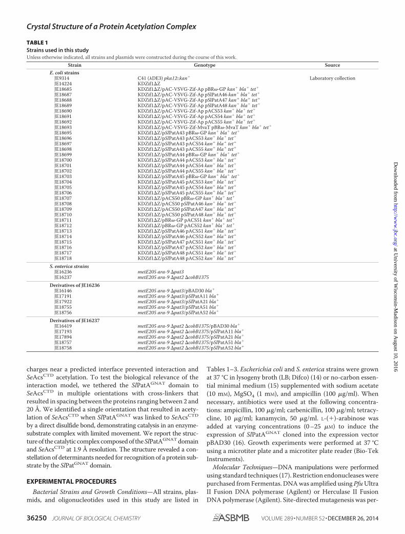
TABLE 1Strains used in this studyUnless otherwise indicated, all strains and plasmids were constructed during the course of this work.
Strain Genotype SourceE. coli strains
JE9314 C41 (�DE3) pka12::kan� Laboratory collectionJE14224 KDZif1�ZJE18685 KDZif1�Z/pAC-VSVG-Zif-Ap pBR�-GP kan� bla� tet�
JE18687 KDZif1�Z/pAC-VSVG-Zif-Ap pSlPatA46 kan� bla� tet�
JE18688 KDZif1�Z/pAC-VSVG-Zif-Ap pSlPatA47 kan� bla� tet�
JE18689 KDZif1�Z/pAC-VSVG-Zif-Ap pSlPatA48 kan� bla� tet�
JE18690 KDZif1�Z/pAC-VSVG-Zif-Ap pACS53 kan� bla� tet�
JE18691 KDZif1�Z/pAC-VSVG-Zif-Ap pACS54 kan� bla� tet�
JE18692 KDZif1�Z/pAC-VSVG-Zif-Ap pACS55 kan� bla� tet�
JE18693 KDZif1�Z/pAC-VSVG-Zif-MvaT pBR�-MvaT kan� bla� tet�
JE18695 KDZif1�Z/pSlPatA43 pBR�-GP kan� bla� tet�
JE18696 KDZif1�Z/pSlPatA43 pACS53 kan� bla� tet�
JE18697 KDZif1�Z/pSlPatA43 pACS54 kan� bla� tet�
JE18698 KDZif1�Z/pSlPatA43 pACS55 kan� bla� tet�
JE18699 KDZif1�Z/pSlPatA44 pBR�-GP kan� bla� tet�
JE18700 KDZif1�Z/pSlPatA44 pACS53 kan� bla� tet�
JE18701 KDZif1�Z/pSlPatA44 pACS54 kan� bla� tet�
JE18702 KDZif1�Z/pSlPatA44 pACS55 kan� bla� tet�
JE18703 KDZif1�Z/pSlPatA45 pBR�-GP kan� bla� tet�
JE18704 KDZif1�Z/pSlPatA45 pACS53 kan� bla� tet�
JE18705 KDZif1�Z/pSlPatA45 pACS54 kan� bla� tet�
JE18706 KDZif1�Z/pSlPatA45 pACS55 kan� bla� tet�
JE18707 KDZif1�Z/pACS50 pBR�-GP kan� bla� tet�
JE18708 KDZif1�Z/pACS50 pSlPatA46 kan� bla� tet�
JE18709 KDZif1�Z/pACS50 pSlPatA47 kan� bla� tet�
JE18710 KDZif1�Z/pACS50 pSlPatA48 kan� bla� tet�
JE18711 KDZif1�Z/pBR�-GP pACS51 kan� bla� tet�
JE18712 KDZif1�Z/pBR�-GP pACS52 kan� bla� tet�
JE18713 KDZif1�Z/pSlPatA46 pACS51 kan� bla� tet�
JE18714 KDZif1�Z/pSlPatA46 pACS52 kan� bla� tet�
JE18715 KDZif1�Z/pSlPatA47 pACS51 kan� bla� tet�
JE18716 KDZif1�Z/pSlPatA47 pACS52 kan� bla� tet�
JE18717 KDZif1�Z/pSlPatA48 pACS51 kan� bla� tet�
JE18718 KDZif1�Z/pSlPatA48 pACS52 kan� bla� tet�
S. enterica strainsJE16236 metE205 ara-9 �pat3JE16237 metE205 ara-9 �pat2 �cobB1375
Derivatives of JE16236JE16146 metE205 ara-9 �pat3/pBAD30 bla�
JE17191 metE205 ara-9 �pat3/pSlPatA11 bla�
JE17922 metE205 ara-9 �pat3/pSlPatA21 bla�
JE18755 metE205 ara-9 �pat3/pSlPatA51 bla�
JE18756 metE205 ara-9 �pat3/pSlPatA52 bla�
Derivatives of JE16237JE16419 metE205 ara-9 �pat2 �cobB1375/pBAD30 bla�
JE17193 metE205 ara-9 �pat2 �cobB1375/pSlPatA11 bla�
JE17894 metE205 ara-9 �pat2 �cobB1375/pSlPatA21 bla�
JE18757 metE205 ara-9 �pat2 �cobB1375/pSlPatA51 bla�
JE18758 metE205 ara-9 �pat2 �cobB1375/pSlPatA52 bla�
Crystal Structure of a Protein Acetylation Complex
36250 JOURNAL OF BIOLOGICAL CHEMISTRY VOLUME 289 • NUMBER 52 • DECEMBER 26, 2014
at University of W
isconsin-Madison on A
ugust 10, 2016http://w
ww
.jbc.org/D
ownloaded from

formed using the QuikChangeTM site-directed mutagenesis kit(Agilent). Plasmids were isolated using the Wizard Plus SVMiniprep kit (Promega), and PCR products were purified usingthe Wizard SV Gel and PCR Clean-Up System (Promega). DNAsequencing was performed using BigDye� (ABI PRISM) proto-cols, and dye-terminator sequencing reactions were resolvedand analyzed by capillary electrophoresis at the University ofGeorgia Genomics Facility. Oligonucleotide primer sequencesare listed in Table 3.
Construction of Complementation Plasmids—The allele encod-ing the GNAT domain of wild-type SlPatA (residues Met1–Leu194) was amplified from S. lividans TK24 genomic DNAusing primers listed in Table 3. Primers used in the amplifica-tion changed the first codon of SlPatA from TTG to the morecommon ATG start codon. The amplified fragment was cutwith EcoRI and KpnI and ligated into pBAD30 cut with thesame enzymes. The resulting plasmid, pSlPatA11, directedthe synthesis of SlPatGNAT in response to L-(�)-arabinose. Theplasmid directing the synthesis of variant SlPatAGNAT E123Qwas generated by site-directed mutagenesis using primers listedin Table 3.
Plasmids for in Vivo Two-hybrid System Assays—Alleles encod-ing the wild-type SlPatAGNAT domain, variant SlPatAGNAT
E121R, and variant SlPatAGNAT D185R were amplified fromplasmids pSlPatA27, pSlPatA33, and pSlPatA35, respectively,using primers listed in Table 3. Alleles encoding wild-typeSeAcsCTD (residues Asp518–Ser652) and variants SeAcsCTD
R606E and SeAcsCTD R613D were amplified from plasmidspACS38, pACS42, and pACS44, respectively, using primerslisted in Table 3. DNA fragments were cut with NotI andBamHI and then ligated into plasmids pACTR-V-Zif-AP andpBR�GP, which had been cut with NotI and BamHI. Theresulting plasmids are listed in Table 2.
SlPatAGNAT Overproduction Plasmids—The allele encodingthe GNAT domain of SlPatA (residues Met1–Leu194) wasamplified from S. lividans TK24 genomic DNA with primerslisted in Table 3. The primers used were designed to change thefirst codon of SlPatA from TTG to the more common ATGstart codon. The amplified fragment was cut with KpnI and SalIand ligated into pKLD66 (18) cut with the same enzymes. Theresulting plasmid pSlPatA14 directed the synthesis of wild-typeSlPatAGNAT fused at its N terminus to a His6-maltose-bindingprotein (MBP) tag cleavable by recombinant tobacco etch virus(rTEV) protease using protocols described previously (19).Plasmids directing the synthesis of variants SlPatAGNAT S73C,SlPatAGNAT A110C, and SlPatAGNAT A164C were generated
TABLE 2Plasmids used in this studyUnless noted, all plasmids were constructed during the course of this study.
Plasmid Genotype Reference
pTEV5 N-terminal, rTEV-cleavable His6 tag overexpression vector, bla� Ref. 18pBAD30 ParaBAD expression vector, bla�
pKLD66 N-terminal, rTEV-cleavable His6-MBP-tag overexpression vector, bla�
pBR�GP N-terminal RNAP � fusion vector for bacterial 2-hybrid assay, bla� Ref. 46pACTR-V-Zif-AP N-terminal VSV-G-Zif268 fusion vector for bacterial 2-hybrid assay, tet� Ref. 46pTEV5 derivatives
pACS38 Encodes the wild-type SeAcs CTD (SeAcsCTD)pACS42 Encodes variant SeAcsCTD R606EpACS44 Encodes variant SeAcsCTD R613DpACS56 Encodes variant SeAcsCTD A538CpACS57 Encodes variant SeAcsCTD H567CpACS58 Encodes variant SeAcsCTD D600CpACS60 Encodes variant SeAcsCTD R606EpACS61 Encodes variant SeAcsCTD R613DpSlPatA27 Encodes the wild-type GNAT domain of SlPatApSlPatA33 Encodes variant SlPatAGNAT E121RpSlPatA35 Encodes variant SlPatAGNAT D185R
pKLD66 derivativespSlPatA14 Encodes the wild-type GNAT domain of SlPatA (SlPatAGNAT)pSlPatA53 Encodes variant SlPatAGNAT S73CpSlPatA54 Encodes variant SlPatAGNAT A110CpSlPatA56 Encodes variant SlPatAGNAT A164C
pBAD30 derivativespSlPatA11 Encodes the wild-type GNAT of SlPatA (SlPatAGNAT)pSlPatA21 Encodes variant SlPatAGNAT E123QpSlPatA51 Encodes variant SlPatAGNAT E121RpSlPatA52 Encodes variant SlPatAGNAT D185R
pBR�GP derivativespSlPatA46 Encodes the wild-type SlPatA GNAT domain (SlPatAGNAT)pSlPatA47 Encodes variant SlPatAGNAT E121RpSlPatA48 Encodes variant SlPatAGNAT D185RpACS53 Encodes the wild-type CTD of SeAcspACS54 Encodes the SeAcsR606E CTD variantpACS55 Encodes the SeAcsR613D CTD variant
pACTR-V-Zif-AP derivativespSlPatA43 Encodes the wild-type GNAT domain of SlPatA (SlPatAGNAT)pSlPatA44 Encodes variant SlPatAGNAT E121RpSlPatA45 Encodes variant SlPatAGNAT D185RpACS50 Encodes the wild-type CTD of SeAcs (SeAcsCTD)pACS51 Encodes variant SeAcsCTD R606EpACS52 Encodes variant SeAcsCTD R613D
Crystal Structure of a Protein Acetylation Complex
DECEMBER 26, 2014 • VOLUME 289 • NUMBER 52 JOURNAL OF BIOLOGICAL CHEMISTRY 36251
at University of W
isconsin-Madison on A
ugust 10, 2016http://w
ww
.jbc.org/D
ownloaded from
from plasmid pSlPatA14 using site-directed mutagenesis usingprimers listed in Table 3.
SeAcs C-terminal Domain (SeAcsCTD) Overexpression Plasmids—The allele encoding SeAcsCTD (residues Asp518–Ser652) wasamplified from S. enterica LT2 DNA using primers listed inTable 3. The amplified fragment was cut with NheI and EcoRIand ligated into pTEV5 cut with the same enzymes. The result-ing plasmid pACS38 directed the synthesis of wild-typeSeAcsCTD fused at its N terminus to a hexahistidine tagcleavable by rTEV protease. Plasmids directing synthesis ofvariants SeAcsCTD R606E, SeAcsCTD R613D, SeAcsCTD A238C,SeAcsCTD H567C, and SeAcsCTD D600C were generated fromplasmid pACS38 by site-directed mutagenesis using primerslisted in Table 3.
Full-length SeAcs Overproduction Plasmids—Plasmids direct-ing the synthesis of variants SeAcsCTD R606E and SeAcsCTD
R613D were generated from plasmid pACS33 (Table 2) usingsite-directed mutagenesis. The resulting plasmids pACS60 andpACS61 directed the synthesis of variants SeAcsCTD R606E andSeAcsCTD R613D, respectively, each fused to an N-terminalHis6 tag cleavable by rTEV protease.
SlPatAGNAT Overproduction and Purification—PlasmidspSlPatA14, pSlPatA53, pSlPatA54, and pSlPatA56 were trans-formed into the �pka derivative of E. coli C41 (�DE3) (strainJE9314). The resulting strains were grown overnight and sub-cultured 1:100 (v/v) into 2 liters of LB containing ampicillin(100 �g/ml). The cultures were grown with shaking at 25 °C to
A600 �0.7, and His6-MBP-SlPatAGNAT synthesis was inducedwith isopropyl �-D-1-thiogalactopyranoside (IPTG) (0.5 mM).Upon induction, the cultures were grown overnight at 25 °C.Cells were harvested at 6000 � g for 10 min at 4 °C in an AvantiJ-2 XPI centrifuge fitted with rotor JLA-8.1000 (BeckmanCoulter). Cell pellets were resuspended in 30 ml of cold His-Bind buffer A (Tris-HCl buffer (50 mM, pH 8), NaCl (500 mM),and imidazole (5 mM)) containing phenylmethanesulfonyl fluo-ride (PMSF; 1 mM). Cells were placed on ice and lysed by soni-cation for 1 min (2-s pulse followed by 4 s of cooling) at level 7in a model 550 sonic dismembrator (Fisher). The extract wascleared by centrifugation at 4 °C for 30 min at 43,367 � g. His6-tagged SlPatAGNAT was purified from the soluble fraction bynickel affinity purification using a 1-ml bed volume of His-Purnickel-nitrilotriacetic acid resin (Thermo). Unbound proteinswere eluted off of the column by extensive washing with bufferA. The column was then washed with buffer B1 (Tris-HClbuffer (50 mM, pH 8), NaCl (500 mM), and imidazole (15 mM)),and SlPatAGNAT was eluted from the column with buffer C(Tris-HCl buffer (50 mM, pH 8), NaCl (500 mM), and imidazole(250 mM)). All fractions containing His6-MBP-SlPatAGNAT
were combined. rTEV protease was added to His6-MBP-SlPatAGNAT, and the SlPatAGNAT/rTEV mixture was incubatedat room temperature for 3 h. The SlPatAGNAT/rTEV mixturewas dialyzed at 4 °C against buffer C (Tris-HCl (50 mM, pH 8),NaCl (500 mM)) twice for 3 h and again against buffer C con-taining imidazole (5 mM) for 12 h. After cleavage and dialysis,
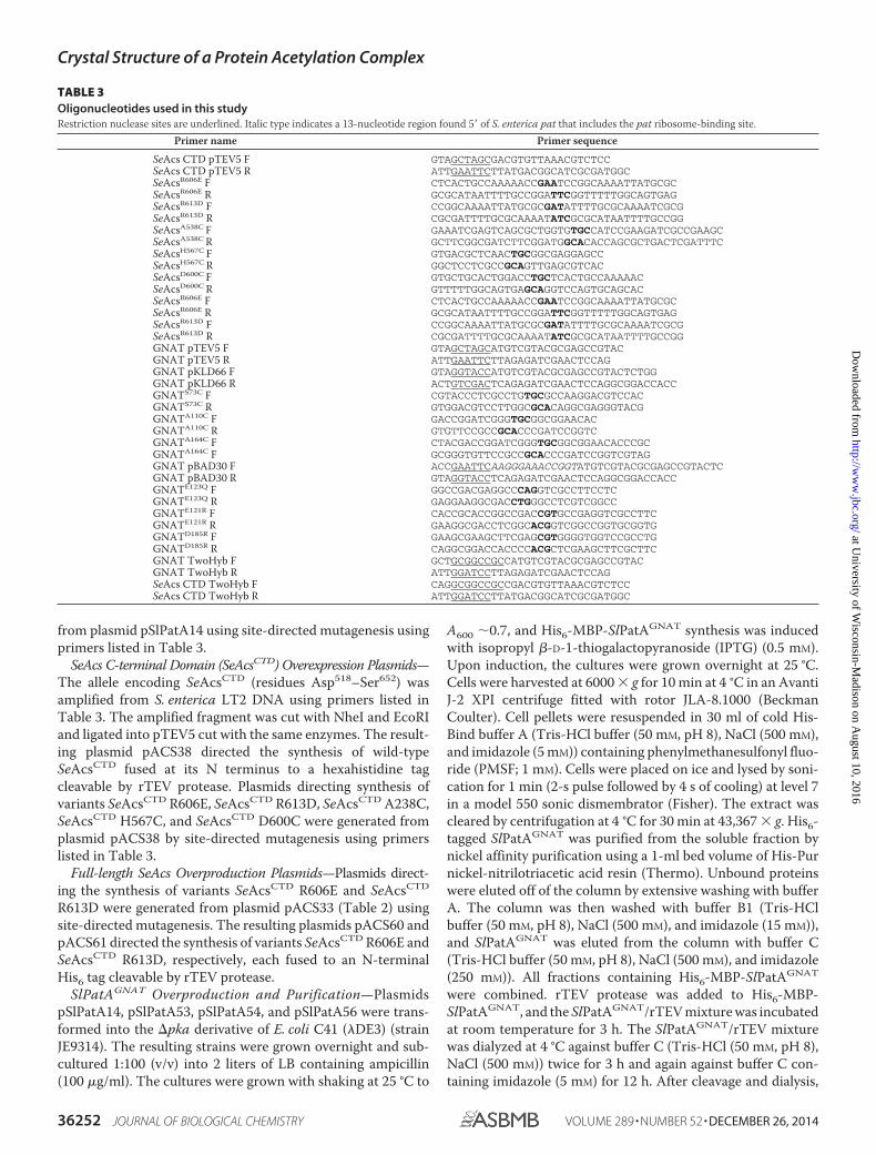
TABLE 3Oligonucleotides used in this studyRestriction nuclease sites are underlined. Italic type indicates a 13-nucleotide region found 5� of S. enterica pat that includes the pat ribosome-binding site.
Primer name Primer sequence
SeAcs CTD pTEV5 F GTAGCTAGCGACGTGTTAAACGTCTCCSeAcs CTD pTEV5 R ATTGAATTCTTATGACGGCATCGCGATGGCSeAcsR606E F CTCACTGCCAAAAACCGAATCCGGCAAAATTATGCGCSeAcsR606E R GCGCATAATTTTGCCGGATTCGGTTTTTGGCAGTGAGSeAcsR613D F CCGGCAAAATTATGCGCGATATTTTGCGCAAAATCGCGSeAcsR613D R CGCGATTTTGCGCAAAATATCGCGCATAATTTTGCCGGSeAcsA538C F GAAATCGAGTCAGCGCTGGTGTGCCATCCGAAGATCGCCGAAGCSeAcsA538C R GCTTCGGCGATCTTCGGATGGCACACCAGCGCTGACTCGATTTCSeAcsH567C F GTGACGCTCAACTGCGGCGAGGAGCCSeAcsH567C R GGCTCCTCGCCGCAGTTGAGCGTCACSeAcsD600C F GTGCTGCACTGGACCTGCTCACTGCCAAAAACSeAcsD600C R GTTTTTGGCAGTGAGCAGGTCCAGTGCAGCACSeAcsR606E F CTCACTGCCAAAAACCGAATCCGGCAAAATTATGCGCSeAcsR606E R GCGCATAATTTTGCCGGATTCGGTTTTTGGCAGTGAGSeAcsR613D F CCGGCAAAATTATGCGCGATATTTTGCGCAAAATCGCGSeAcsR613D R CGCGATTTTGCGCAAAATATCGCGCATAATTTTGCCGGGNAT pTEV5 F GTAGCTAGCATGTCGTACGCGAGCCGTACGNAT pTEV5 R ATTGAATTCTTAGAGATCGAACTCCAGGNAT pKLD66 F GTAGGTACCATGTCGTACGCGAGCCGTACTCTGGGNAT pKLD66 R ACTGTCGACTCAGAGATCGAACTCCAGGCGGACCACCGNATS73C F CGTACCCTCGCCTGTGCGCCAAGGACGTCCACGNATS73C R GTGGACGTCCTTGGCGCACAGGCGAGGGTACGGNATA110C F GACCGGATCGGGTGCGGCGGAACACGNATA110C R GTGTTCCGCCGCACCCGATCCGGTCGNATA164C F CTACGACCGGATCGGGTGCGGCGGAACACCCGCGNATA164C F GCGGGTGTTCCGCCGCACCCGATCCGGTCGTAGGNAT pBAD30 F ACCGAATTCAAGGGAAACCGGTATGTCGTACGCGAGCCGTACTCGNAT pBAD30 R GTAGGTACCTCAGAGATCGAACTCCAGGCGGACCACCGNATE123Q F GGCCGACGAGGCCCAGGTCGCCTTCCTCGNATE123Q R GAGGAAGGCGACCTGGGCCTCGTCGGCCGNATE121R F CACCGCACCGGCCGACCGTGCCGAGGTCGCCTTCGNATE121R R GAAGGCGACCTCGGCACGGTCGGCCGGTGCGGTGGNATD185R F GAAGCGAAGCTTCGAGCGTGGGGTGGTCCGCCTGGNATD185R R CAGGCGGACCACCCCACGCTCGAAGCTTCGCTTCGNAT TwoHyb F GCTGCGGCCGCCATGTCGTACGCGAGCCGTACGNAT TwoHyb R ATTGGATCCTTAGAGATCGAACTCCAGSeAcs CTD TwoHyb F CAGGCGGCCGCCGACGTGTTAAACGTCTCCSeAcs CTD TwoHyb R ATTGGATCCTTATGACGGCATCGCGATGGC
Crystal Structure of a Protein Acetylation Complex
36252 JOURNAL OF BIOLOGICAL CHEMISTRY VOLUME 289 • NUMBER 52 • DECEMBER 26, 2014
at University of W
isconsin-Madison on A
ugust 10, 2016http://w
ww
.jbc.org/D
ownloaded from

protein mixtures were passed over the 1-ml HisTrap columnusing the buffers described above, and the untagged proteinswere eluted in the flow-through and buffer A wash. PurifiedSlPatAGNAT was analyzed by SDS-PAGE. Fractions containingSlPatAGNAT were pooled together. SlPatAGNAT was stored inTris-HCl buffer (50 mM, pH 8.0) containing NaCl (100 mM) andglycerol (10%, v/v). Tris(2-carboxyethyl)phosphine (0.3 mM)and EDTA (0.5 mM) were included in the storage buffer forSlPatAGNAT cysteine variants. SlPatAGNAT concentration wasdetermined by measuring absorbance at 280 nm. The molarextinction coefficient used to calculate SlPatAGNAT concentra-tion was 17,420 M�1 cm�1. The purification protocol forSlPatAGNAT for crystallography was similar to the onedescribed above except that the purified, untagged protein wasdialyzed into Tris-HCl buffer (10 mM, pH 8.0) and concentratedto 11 mg/ml before flash freezing into liquid nitrogen.
Selenomethionine-labeled SlPatAGNAT was overproduced asfollows.PlasmidpSlPatA14wastransformedintostrainJE9314,and a1-liter culture of the resulting strain was grown overnight in M9glucose medium. The culture was used to reinoculate 2 � 2liters of fresh M9 glucose medium at a 1:100 inoculum, and theculture was grown to an A600 of �1. The culture was cooled onice to 16 °C for 10 min, and a defined amino acid mixture con-taining lysine (100 mg/liter), threonine (100 mg/liter), phenyl-alanine (100 mg/liter), leucine (50 mg/liter), isoleucine (50mg/liter), valine (50 mg/liter), and selenomethionine (50 mg/li-ter) was added to suppress methionine biosynthesis. The cul-ture was grown at 37 °C for 30 min before the addition of IPTG(1 mM) to induce SlPatAGNAT expression. The culture wasgrown overnight at 37 °C, and the protein was purified andstored for crystallography as described above.
Purification of SeAcsWT and SeAcsCTD—Plasmids pACS38,pACS44, pACS56, pACS57, pACS58, pACS60, and pACS61were transformed into a �pka derivative of E. coli C41 (�DE3)(JE9314) to prevent acetylation prior to overproduction. Theresulting strains were grown overnight and subcultured 1:100(v/v) into 2 liters of LB containing ampicillin (100 �g/ml). Thecultures were grown with shaking at 25 °C to A600 �0.7, andprotein synthesis was induced with IPTG (0.25 mM). Uponinduction, the cultures were grown overnight at 25 °C.SeAcsWT and SeAcsCTD proteins were purified and stored asdescribed above for SlPatAGNAT with modifications. Duringthe first purification step, the His6-SeAcs-bound resins werewashed with buffer B (Tris-HCl buffer (50 mM, pH 8), NaCl (500mM), and imidazole (20 mM)) before His6-SeAcs proteins wereeluted with buffer C. His6-SeAcsCTD-bound resins were washedwith buffer B3 (Tris-HCl buffer (50 mM, pH 8), NaCl (500 mM),and imidazole (40 mM)) before elution with buffer C. In thesecond purification step, SeAcsWT proteins and SeAcsCTD pro-teins did not adsorb to the column and were present in theflow-through fractions. Proteins were stored in Tris-HCl buffer(50 mM, pH 8.0) containing NaCl (100 mM) and glycerol (10%,v/v). Tris(2-carboxyethyl)phosphine (0.3 mM) and EDTA (0.5mM) were included in the storage buffer for SlPatAGNAT cys-teine variants. The molar extinction coefficients used to calcu-late protein concentrations were 72,152 M�1 cm�1 for SeAcsWT
and 15,470 M�1 cm�1 for SeAcsCTD.
Two-hybrid System Assay and �-Galactosidase ActivityMeasurement—E. coli strain KDZif1�Z harboring compatibleplasmids were grown overnight in 1 ml of nutrient brothsupplemented with kanamycin and carbenicillin. The fol-lowing day, strains were subcultured 1:100 into 200 �l ofnutrient broth supplemented with kanamycin, carbenicillin,and IPTG (200 �M) to induce expression from two-hybridscreen plasmids. Strains were grown for 3.5 h at 37 °C withshaking at medium intensity in a BioTek plate reader(BioTek Instruments, Inc.). Absorbance values of the cul-tures were measured in the microtiter plate at a 650 nmwavelength using a Spectramax Plus 384 spectrophotometer(Molecular Devices).
The �-galactosidase activity of the cultures was measured asdescribed (20, 21), with modifications. Cultures were diluted 20�l into 80 �l of permeabilization solution (Na2HPO4 (100 mM),KCl (20 mM), MgCl2 (2 mM), hexadecyltrimethylammoniumbromide (0.06%), sodium deoxycholate (0.04%), 2-mercapto-ethanol (38 mM)). Samples were shaken for 5 s in a SpectramaxPlus 384 spectrophotometer (Molecular Devices) to mix. Per-meabilized cells were then diluted 25 �l into 150 �l of substratesolution (Na2HPO4 (60 mM), NaH2PO4 (40 mM), O-nitrophe-nyl-�-galactoside (1 mg/ml), 2-mercaptoethanol (38 mM)). Theabsorbance at 420 nm was monitored in a 96-well plate usingthe Spectramax Plus 384 spectrophotometer. A unit of activ-ity was defined as the amount of enzyme required to catalyzethe hydrolysis of 1 nmol of O-nitrophenyl-�-galactoside permin.
AMP-forming CoA Ligase Assays—SeAcsWT activity wasmeasured using an NADH-consuming assay (22, 23). Reactions(100-�l total volume) contained HEPES buffer (50 mM, pH 7.5),tris(2-chloroethyl) phosphate (tris(2-carboxyethyl)phosphine;1 mM), ATP (2.5 mM), CoA (0.5 mM), MgCl2 (5 mM), KCl (1 mM),phosphoenolpyruvate (3 mM), NADH (0.1 mM), pyruvatekinase (1 unit), myokinase (5 unit), lactate dehydrogenase (1.5units), and acetate (0.2 mM). Reactions were started by the addi-tion of SeAcsWT (15 nM). The absorbance at 340 nm was mon-itored in a 96-well plate using the Spectramax Plus UV-visiblespectrophotometer (Molecular Devices). Enzyme specificactivities were determined to be in the linear range for the assayand were calculated as described (22). Values are reported asthe mean � S.D.
In Vitro Acetylation—Protein acetylation was monitoredusing radiolabeled [1-14C]Ac-CoA (50 �M; 56.8 mCi mmol�1)as described (23–25). Briefly, reactions contained SeAcsWT orSeAcsCTD (3 �M) and either SePatWT or SlPatAWT (1 �M). Sam-ples (10 �l) were removed and quenched with 2 �l of loadingbuffer (glycerol (50%), EDTA (1 mM), bromphenol blue (0.25%),xylene cyanol (0.25%)) before being resolved and visualizedwith SDS-PAGE (26) and Coomassie Blue staining (27), respec-tively. Labeled proteins were visualized using a Typhoon FLA9000 variable mode imager (GE Healthcare) equipped withImageQuant TL software (GE Healthcare). Acetylation wasquantified as a measure of the radiolabel signal and reported asdigital light units. Gels and phosphor images were cropped tothe SeAcsWT, SeAcsCTD, or SeAcs variant bands.
The effect of acetylation on activities of SeAcsWT, SeAcsR606E, andSeAcsR613D was determined as described (23) with modifica-
Crystal Structure of a Protein Acetylation Complex
DECEMBER 26, 2014 • VOLUME 289 • NUMBER 52 JOURNAL OF BIOLOGICAL CHEMISTRY 36253
at University of W
isconsin-Madison on A
ugust 10, 2016http://w
ww
.jbc.org/D
ownloaded from
tions. SeAcsWT, SeAcsR606E, or SeAcsR613D (3 �M) was incu-bated with SlPatA (1 �M) or SePat (1 �M) and 50 �M Ac-CoA for90 min at 30 °C (SlPatAWT) or 37 °C (SePatAWT) using thebuffer system described above. At 90 min, reactions werediluted 1:20 into HEPES buffer (50 mM, pH 7.5) at 4 °C. SeAcsactivity was measured as described above.
SlPatAGNAT-SeAcsCTD Cross-linking and Acetylation Assays—Cysteine variants of wild-type SeAcsCTD were exchanged intocross-linking buffer (20 mM HEPES, pH 7.5, 100 mM NaCl,sparged with N2) using Zeba Spin desalting columns (Pierce).SeAcsCTD proteins were immediately diluted into a 10-foldmolar excess of aldriothiol-2 (AT-2, 2.05-Å cross-linker;Sigma), bis(maleimido)ethane (8.0-Å cross-linker; Thermo),1,4-bis(maleimido)butane (10.9 Å cross-linker; Thermo),bis(maleimido)hexane (16.0-Å cross-linker; Thermo), or 1,4-di-(3�-(2�-pyridyldithio)-propionamido)butane (19.9-Å cross-linker; bioWorld) and allowed to react for 1 h at room temper-ature. Activated SeAcsCTD variants and SlPatAGNAT cysteinevariants were desalted into cross-linking buffer as describedabove. SlPatAGNAT variants were diluted into a 5-fold molarexcess of SeAcsCTD and allowed to react for 2 h at 4 °C. AT-2and 1,4-di-(3�-[2�-pyridyldithio]-propionamido)butane cross-linking reactions were quenched with iodoacetamide (10 mM).Bis(maleimido)ethane, 1,4-bis(maleimido)butane, and bis(ma-leimido)hexane cross-linking reactions were quenched withDTT (10 mM). Cross-linking reactions were analyzed by non-reducing SDS-PAGE to verify cross-linking and to quantifycross-linked species.
Cross-linked species were incubated with BisTris (100 mM,pH 6.0) and [1-14C]Ac-CoA (20 �M; 56.8 mCi mmol�1) at 30 °Cfor 1 h. Reactions were resolved by non-reducing SDS-PAGEand analyzed as described above. Gels and phosphor imageswere cropped to the wild-type SeAcsCTD-SlPatAGNAT complexbands.
Crystallization of SlPatAGNAT and SlPatA-SeAcsCTD Complex—SlPatAGNAT crystals were grown by small scale batch crystalli-zation, where 4 �l of concentrated protein at 10.7 mg/ml inTris-HCl buffer (10 mM, pH 8.0) containing ethyl-CoA (2 mM)was mixed with 5 �l of PEG solution (22% (w/v) monomethylpolyethylene glycol 5000 in MOPS buffer (100 mM, pH 7.0)containing MgCl2 (25 mM), 2,2,2-trifluoroethanol (2% v/v), andglycerol (6%, v/v) (28). Drops were immediately streak-seededand then allowed to grow at room temperature for 1 week.Crystals grew to maximal dimensions of 300 � 100 � 100 �m.For freezing, crystals were transferred to PEG solution contain-ing ethyl-CoA (1 mM) and then transferred stepwise to a solu-tion of monomethyl polyethylene glycol 5000 (22%, w/v), glyc-erol (20%, v/v), MOPS buffer (100 mM, pH 7.0) containingMgCl2 (25 mM), 2,2,2-trifluoroethanol (2%, v/v), and ethyl-CoA(2 mM). Crystals were flash-frozen in liquid nitrogen. Sel-enomethionine protein crystals were grown under identicalconditions except that all solutions lacked ethyl-CoA.
SlPatAGNAT-SeAcsCTD complex crystals were grown byhanging drop diffusion, where 1 �l of concentrated protein at10.0 mg/ml in Tris-HCl buffer (10 mM, pH 8.0) containing CoA(2 mM) was mixed with 1 �l of 11.2% (w/v) polyethylene glycol8000, 100 mM 1,4-piperazinediethanesulfonic acid, pH 6.5, and120 mM Li2SO4. Crystals formed spontaneously after 2 months
and grew to a maximum dimension of 400 � 50 � 50 �m. Forfreezing, crystals were transferred to paratone-N and thenflash-frozen in liquid nitrogen.
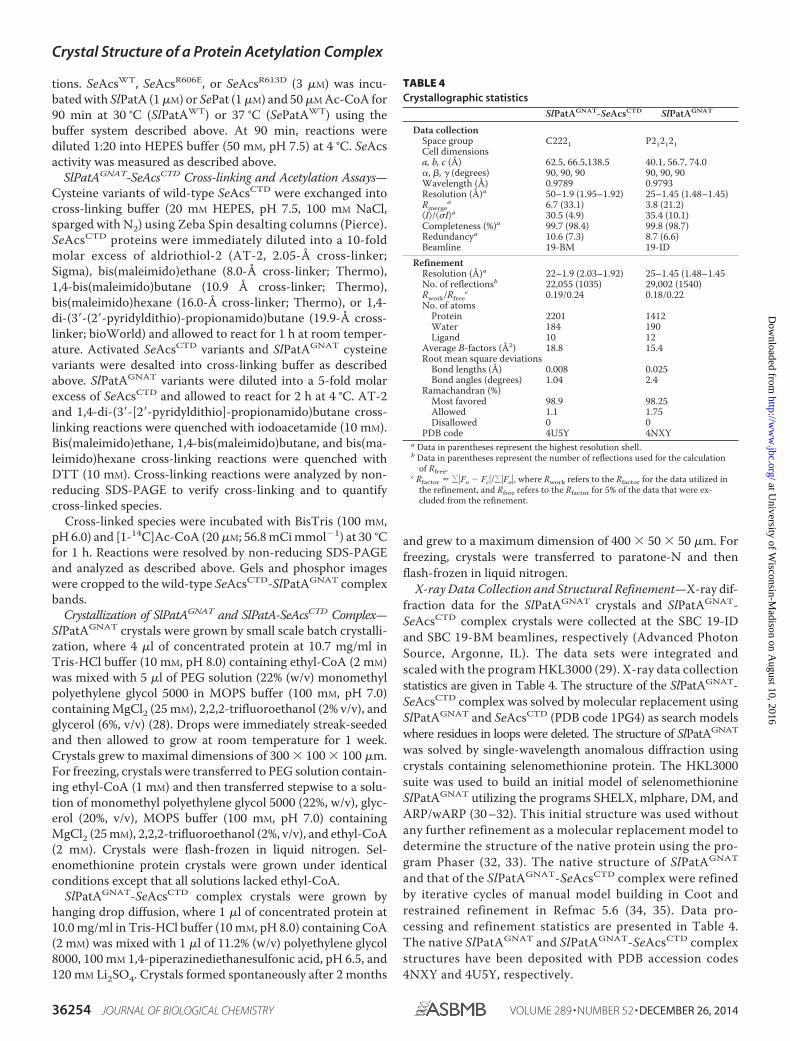
X-ray Data Collection and Structural Refinement—X-ray dif-fraction data for the SlPatAGNAT crystals and SlPatAGNAT-SeAcsCTD complex crystals were collected at the SBC 19-IDand SBC 19-BM beamlines, respectively (Advanced PhotonSource, Argonne, IL). The data sets were integrated andscaled with the program HKL3000 (29). X-ray data collectionstatistics are given in Table 4. The structure of the SlPatAGNAT-SeAcsCTD complex was solved by molecular replacement usingSlPatAGNAT and SeAcsCTD (PDB code 1PG4) as search modelswhere residues in loops were deleted. The structure of SlPatAGNAT
was solved by single-wavelength anomalous diffraction usingcrystals containing selenomethionine protein. The HKL3000suite was used to build an initial model of selenomethionineSlPatAGNAT utilizing the programs SHELX, mlphare, DM, andARP/wARP (30 –32). This initial structure was used withoutany further refinement as a molecular replacement model todetermine the structure of the native protein using the pro-gram Phaser (32, 33). The native structure of SlPatAGNAT
and that of the SlPatAGNAT-SeAcsCTD complex were refinedby iterative cycles of manual model building in Coot andrestrained refinement in Refmac 5.6 (34, 35). Data pro-cessing and refinement statistics are presented in Table 4.The native SlPatAGNAT and SlPatAGNAT-SeAcsCTD complexstructures have been deposited with PDB accession codes4NXY and 4U5Y, respectively.
TABLE 4Crystallographic statistics
SlPatAGNAT-SeAcsCTD SlPatAGNAT
Data collectionSpace group C2221 P212121Cell dimensionsa, b, c (Å) 62.5, 66.5,138.5 40.1, 56.7, 74.0�, �, � (degrees) 90, 90, 90 90, 90, 90Wavelength (Å) 0.9789 0.9793Resolution (Å)a 50–1.9 (1.95–1.92) 25–1.45 (1.48–1.45)Rmerge
a 6.7 (33.1) 3.8 (21.2)I/Ia 30.5 (4.9) 35.4 (10.1)Completeness (%)a 99.7 (98.4) 99.8 (98.7)Redundancya 10.6 (7.3) 8.7 (6.6)Beamline 19-BM 19-ID
RefinementResolution (Å)a 22–1.9 (2.03–1.92) 25–1.45 (1.48–1.45No. of reflectionsb 22,055 (1035) 29,002 (1540)Rwork/Rfree
c 0.19/0.24 0.18/0.22No. of atoms
Protein 2201 1412Water 184 190Ligand 10 12
Average B-factors (Å2) 18.8 15.4Root mean square deviations
Bond lengths (Å) 0.008 0.025Bond angles (degrees) 1.04 2.4
Ramachandran (%)Most favored 98.9 98.25Allowed 1.1 1.75Disallowed 0 0
PDB code 4U5Y 4NXYa Data in parentheses represent the highest resolution shell.b Data in parentheses represent the number of reflections used for the calculation
of Rfree.c Rfactor � ��Fo � Fc�/��Fo�, where Rwork refers to the Rfactor for the data utilized in
the refinement, and Rfree refers to the Rfactor for 5% of the data that were ex-cluded from the refinement.
Crystal Structure of a Protein Acetylation Complex
36254 JOURNAL OF BIOLOGICAL CHEMISTRY VOLUME 289 • NUMBER 52 • DECEMBER 26, 2014
at University of W
isconsin-Madison on A
ugust 10, 2016http://w
ww
.jbc.org/D
ownloaded from

RESULTS
SlPatAGNAT Interacts with SeAcsCTD—To address the ques-tion of how GNAT specificity is achieved, the interactionbetween SlPatAGNAT and SeAcsCTD was investigated. Ourattempts to crystallize SePat were not met with success, so theprotein acetyltransferase from S. lividans (SlPatAGNAT) waschosen. SlPatAGNAT proved amenable to structural studies, andprevious studies showed that SlPatAWT acetylates SeAcsWT invitro (10), although the domain organization of SePatWT andSlPatAWT is reversed (Fig. 1B). SlPatAGNAT was sufficient forfunctionality in vivo through its ability to substitute forSePatWT during growth on 10 mM acetate. As expected, expres-sion of SlPatAGNAT inhibited growth of an S. enterica �pat�cobB strain but allowed growth of an S. enterica �pat cobB�
strain, which retained the ability to deacetylate acetyllysine(Fig. 1, A and E).
Interaction Model for SlPatAGNAT and SeAcsCTD—Initialattempts to co-crystallize SlPatAGNAT with SeAcsCTD (whichcontains the target Lys609) were unsuccessful. This problemwas overcome by introducing a covalent linkage between theSlPatAGNAT and SeAcsCTD domains at a position that affectedneither the enzymatic activity of SlPatAGNAT nor the formationof the SlPatAGNAT-SeAcsCTD binary complex (describedbelow). The location for the linkage was identified by first cre-ating a computational model for the complex from the highresolution structures of the individual domains.
The three-dimensional structure of SlPatAGNAT was deter-mined to 1.5 Å (Fig. 1, C and D, and Table 4). The structure ofSlPatAGNAT revealed a characteristic mixed �/� GNAT foldthat contained the conserved Ac-CoA binding site, includingthe catalytic residue Glu123. SlPatAGNAT residues Phe126 andMet168 overlapped with the modeled Ac-CoA structure andthus are likely to undergo a shift upon Ac-CoA binding,as observed for the analogous residues of the Ac-CoA-boundstructure of MtPatA (Phe238 and Met280). The most similarstructure to SlPatAGNAT was that of the GNAT domain fromSulfolobus solfataricus Pat (PDB code 3F8K), with a root meansquare deviation of 1.32 Å over 131 residues. The major second-ary structure differences between the SsPat GNAT andSlPatAGNAT exist along the protein substrate-binding surface.
The structure of SlPatAGNAT determined here was combinedwith the previously reported structures of SeAcsWT (PDB codes1PG3 and 1PG4) (13) to generate computer models of the inter-action interface using the ClusPro 2.0 server (36 –39). Themodels were evaluated by requiring that the distance betweenthe SlPatAGNAT catalytic residue Glu123 and the �-amino groupof the target lysine in the PX4GK (where K is SeAcs residueLys609) motif be similar to that of 8 Å observed in the crystalstructure of the Tetrahymena Gcn5 bound to the H3 peptidesubstrate (PDB code 1QSN). The best computational modelsplaced residue Glu123 within 15 Å of the �-carbon of the targetlysine (the Lys609 side chain was not resolved in the SeAcsWT
structure) (40). The best model for SeAcsWT-SlPatAGNAT
interactions is shown in Fig. 1F. Notably, SlPatAGNAT was pre-dicted to interact predominantly with SeAcsCTD. Fig. 2A showsthe best model for the interaction between the SlPatAGNAT
domain and the SeAcsCTD (residues Asp518–Ser652).
FIGURE 1. SlPatAGNAT domain and in vivo function. A, regulation of SeAcsWT
activity by SePatWT or SlPatAGNAT in S. enterica. B, domain arrangements ofcharacterized wild-type protein acetyltransferases are listed for S. lividans(SlPatA), S. enterica (SePat), and Mycobacterium tuberculosis (MtPatA). cNMP,cyclic mononucleotide monophosphate-binding domain. C, ribbon represen-tation of SlPatAGNAT with its catalytic residue Glu123 shown in purple (PBDcode 4NXY). Ac-CoA was modeled into the SlPatAGNAT structure using theclosely related MtPatAWT structure as a guide (44) (PDB code 4AVB). D, align-ment of SlPatAGNAT (beige) with MtPatA (teal) shows the location of SlPatAGNAT
residues Phe126 and Met168, which overlap with the modeled Ac-CoA. Homol-ogous residues of MtPatA (Phe238 and Met280) are shown and accommodatethe bound Ac-CoA. SlPatAGNAT active site residue Glu123 is colored purple, andputative active site MtPatA Glu235 is shown in dark teal. E, growth of S. enterica�pat cobB� (open symbols) and �pat �cobB strains (filled symbols), producingSlPatAGNAT (squares), SlPatAGNAT active site variant E123Q (triangles), or anempty vector (circles) on acetate. F, ClusPro interaction model of SlPatAGNAT
(beige) with SeAcs (N-terminal domain in green, C-terminal domain in blue)with active site residue Glu123 (red) and SeAcsWT target residue Lys609 (blue)shown as spheres.
Crystal Structure of a Protein Acetylation Complex
DECEMBER 26, 2014 • VOLUME 289 • NUMBER 52 JOURNAL OF BIOLOGICAL CHEMISTRY 36255
at University of W
isconsin-Madison on A
ugust 10, 2016http://w
ww
.jbc.org/D
ownloaded from
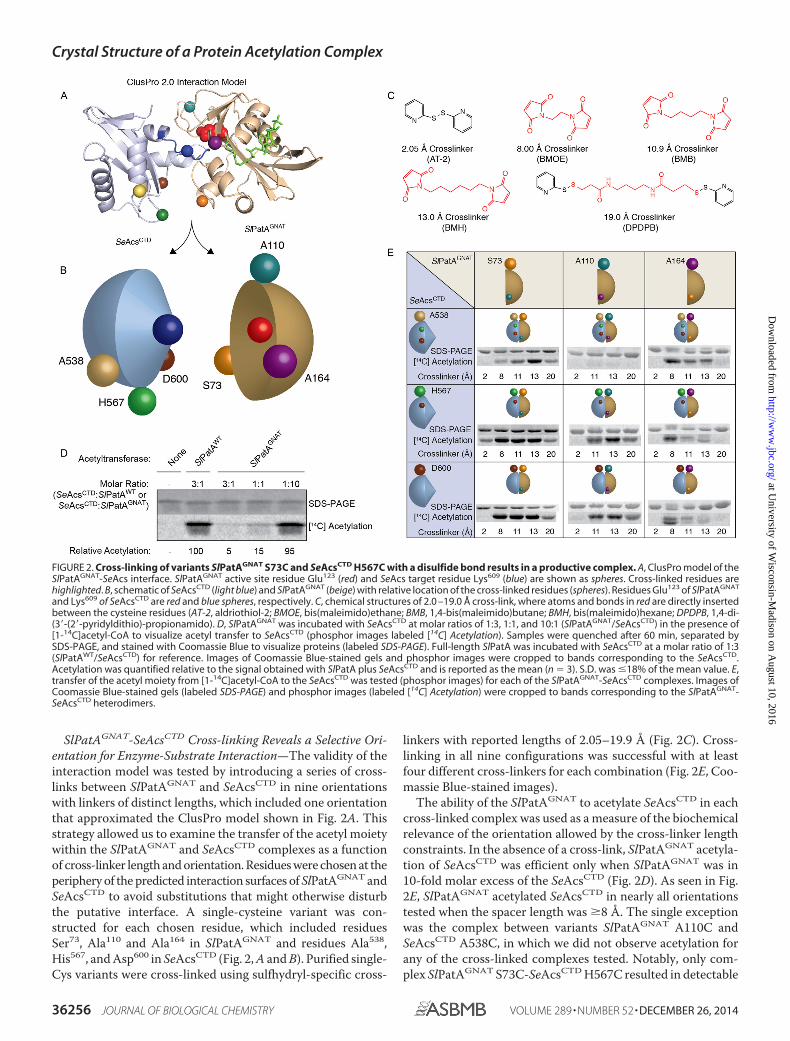
SlPatAGNAT-SeAcsCTD Cross-linking Reveals a Selective Ori-entation for Enzyme-Substrate Interaction—The validity of theinteraction model was tested by introducing a series of cross-links between SlPatAGNAT and SeAcsCTD in nine orientationswith linkers of distinct lengths, which included one orientationthat approximated the ClusPro model shown in Fig. 2A. Thisstrategy allowed us to examine the transfer of the acetyl moietywithin the SlPatAGNAT and SeAcsCTD complexes as a functionof cross-linker length and orientation. Residues were chosen at theperiphery of the predicted interaction surfaces of SlPatAGNAT andSeAcsCTD to avoid substitutions that might otherwise disturbthe putative interface. A single-cysteine variant was con-structed for each chosen residue, which included residuesSer73, Ala110 and Ala164 in SlPatAGNAT and residues Ala538,His567, and Asp600 in SeAcsCTD (Fig. 2, A and B). Purified single-Cys variants were cross-linked using sulfhydryl-specific cross-
linkers with reported lengths of 2.05–19.9 Å (Fig. 2C). Cross-linking in all nine configurations was successful with at leastfour different cross-linkers for each combination (Fig. 2E, Coo-massie Blue-stained images).
The ability of the SlPatAGNAT to acetylate SeAcsCTD in eachcross-linked complex was used as a measure of the biochemicalrelevance of the orientation allowed by the cross-linker lengthconstraints. In the absence of a cross-link, SlPatAGNAT acetyla-tion of SeAcsCTD was efficient only when SlPatAGNAT was in10-fold molar excess of the SeAcsCTD (Fig. 2D). As seen in Fig.2E, SlPatAGNAT acetylated SeAcsCTD in nearly all orientationstested when the spacer length was 8 Å. The single exceptionwas the complex between variants SlPatAGNAT A110C andSeAcsCTD A538C, in which we did not observe acetylation forany of the cross-linked complexes tested. Notably, only com-plex SlPatAGNAT S73C-SeAcsCTD H567C resulted in detectable
FIGURE 2. Cross-linking of variants SlPatAGNAT S73C and SeAcsCTD H567C with a disulfide bond results in a productive complex. A, ClusPro model of theSlPatAGNAT-SeAcs interface. SlPatAGNAT active site residue Glu123 (red) and SeAcs target residue Lys609 (blue) are shown as spheres. Cross-linked residues arehighlighted. B, schematic of SeAcsCTD (light blue) and SlPatAGNAT (beige) with relative location of the cross-linked residues (spheres). Residues Glu123 of SlPatAGNAT
and Lys609 of SeAcsCTD are red and blue spheres, respectively. C, chemical structures of 2.0 –19.0 Å cross-link, where atoms and bonds in red are directly insertedbetween the cysteine residues (AT-2, aldriothiol-2; BMOE, bis(maleimido)ethane; BMB, 1,4-bis(maleimido)butane; BMH, bis(maleimido)hexane; DPDPB, 1,4-di-(3�-(2�-pyridyldithio)-propionamido). D, SlPatAGNAT was incubated with SeAcsCTD at molar ratios of 1:3, 1:1, and 10:1 (SlPatAGNAT/SeAcsCTD) in the presence of[1-14C]acetyl-CoA to visualize acetyl transfer to SeAcsCTD (phosphor images labeled [14C] Acetylation). Samples were quenched after 60 min, separated bySDS-PAGE, and stained with Coomassie Blue to visualize proteins (labeled SDS-PAGE). Full-length SlPatA was incubated with SeAcsCTD at a molar ratio of 1:3(SlPatAWT/SeAcsCTD) for reference. Images of Coomassie Blue-stained gels and phosphor images were cropped to bands corresponding to the SeAcsCTD.Acetylation was quantified relative to the signal obtained with SlPatA plus SeAcsCTD and is reported as the mean (n � 3). S.D. was �18% of the mean value. E,transfer of the acetyl moiety from [1-14C]acetyl-CoA to the SeAcsCTD was tested (phosphor images) for each of the SlPatAGNAT-SeAcsCTD complexes. Images ofCoomassie Blue-stained gels (labeled SDS-PAGE) and phosphor images (labeled [14C] Acetylation) were cropped to bands corresponding to the SlPatAGNAT-SeAcsCTD heterodimers.
Crystal Structure of a Protein Acetylation Complex
36256 JOURNAL OF BIOLOGICAL CHEMISTRY VOLUME 289 • NUMBER 52 • DECEMBER 26, 2014
at University of W
isconsin-Madison on A
ugust 10, 2016http://w
ww
.jbc.org/D
ownloaded from
acetylation when a direct disulfide bond between the cysteineresidues of each protein held the complex together. This directdisulfide bond severely restricted the interactions between
SlPatAGNAT and SeAcsCTD, yet the acetylation signal wasstrong (Fig. 2E). Thus, we hypothesized that the protein orien-tations in the SlPatAS73C-SeAcsH567C complex reflected the
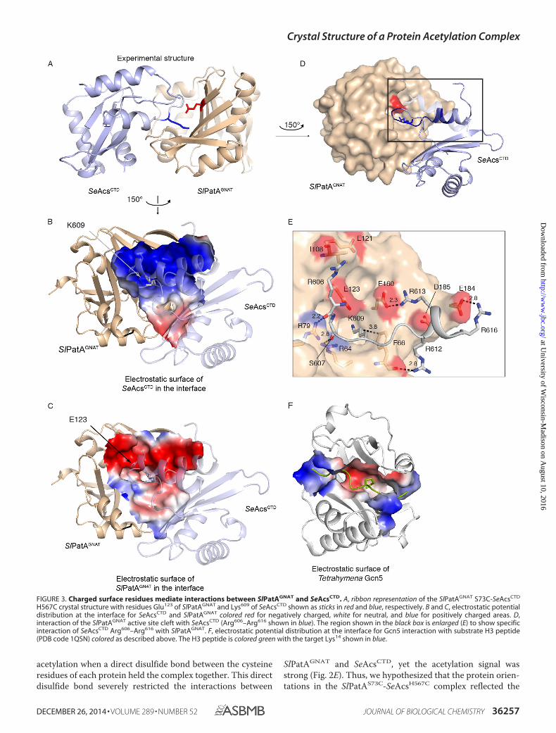
FIGURE 3. Charged surface residues mediate interactions between SlPatAGNAT and SeAcsCTD. A, ribbon representation of the SlPatAGNAT S73C-SeAcsCTD
H567C crystal structure with residues Glu123 of SlPatAGNAT and Lys609 of SeAcsCTD shown as sticks in red and blue, respectively. B and C, electrostatic potentialdistribution at the interface for SeAcsCTD and SlPatAGNAT colored red for negatively charged, white for neutral, and blue for positively charged areas. D,interaction of the SlPatAGNAT active site cleft with SeAcsCTD (Arg606–Arg616 shown in blue). The region shown in the black box is enlarged (E) to show specificinteraction of SeAcsCTD Arg606–Arg616 with SlPatAGNAT. F, electrostatic potential distribution at the interface for Gcn5 interaction with substrate H3 peptide(PDB code 1QSN) colored as described above. The H3 peptide is colored green with the target Lys14 shown in blue.
Crystal Structure of a Protein Acetylation Complex
DECEMBER 26, 2014 • VOLUME 289 • NUMBER 52 JOURNAL OF BIOLOGICAL CHEMISTRY 36257
at University of W
isconsin-Madison on A
ugust 10, 2016http://w
ww
.jbc.org/D
ownloaded from

interactions of these proteins in vivo. Significantly, residuesSer73 of SlPatAGNAT and His567 of SeAcsCTD were positionednear one another in the ClusPro interaction model (Fig. 2A).
Crystal Structure of the SlPatAGNAT-SeAcsCTD Complex at1.9 Å Resolution—To visualize how variants SlPatAGNAT S73Cand SeAcsCTD H567C interacted, we crystallized the SlPatAGNAT
S73C-SeAcsCTD H567C complex formed with an 8-Å linker,and the structure was determined to 1.9 Å (Fig. 3, A–D, andTable 4). In the of SlPatAGNAT S73C-SeAcsCTD H567C struc-ture, the catalytic Glu123 residue of SlPatAGNAT was 4.7 Å awayfrom the target Lys609 of SeAcsCTD (Fig. 3, D and E), a distancewithin the range observed for Tetrahymena Gcn5 bound to alysine-containing peptide substrate (40, 41).
The interface between SlPatAGNAT and SeAcsCTD includedmore than just the interactions between the Lys609-containingSeAcsWT loop and the primary active site of SlPatAGNAT; forexample, it included interactions that are well separated in theprimary sequence. The interface shows good shape comple-mentarity with a shape correlation statistic (Sc) of 0.54, which issimilar to the 0.60 observed in the Gcn5 H3 peptide complex(PDB code 1QSN) (42). These values fall within the expected
range for this type of complex, where an antibody-antigen com-plex results in an Sc value from 0.64 to 0.68, whereas an aberrantinterface will result in an Sc of around 0.35. The SlPatAGNAT-SeAcsCTD interaction surface was distinct and larger than thatof the Gcn5-H3 peptide complex (Fig. 3, B, C, and F). The inter-face between SlPatAGNAT and SeAcsCTD buried a total surfacearea of 2150 Å2, where this was 48% polar and 52% non-polar,which is typical for recognition surfaces. The distribution ofhydrophobicity in the interface is roughly the same as across thetotal surface area of either protein. This is consistent with atransient interface in which specificity is driven by charge-charge interactions with minimal hydrophobic contributions.The size and disposition of the residues in the binding interfacewas consistent with the hypothesis that Pat substrate specificityinvolves elements outside the simple PX4GK loop motif (12).However, the PX4GK loop does play a structural role in posi-tioning Lys609 into the active site cleft of SlPatAGNAT. The car-bonyl oxygen of the preceding glycine residue hydrogenbonded to residue Arg64 of SlPatAGNAT and facilitated a bend inthe backbone loop conformation (� 97°, � 9°). Also locatedwithin the PX4GK loop, the carbonyl oxygens of SeAcsCTD
FIGURE 4. SlPatAGNAT and SeAcsCTD residues at the interaction interface are conserved. A, alignment of sequences in and around the AcsCTD PX4GK motif(black box) from S. enterica (SeAcs, accession number NP_463140), Saccharomyces cerevisiae (Acs2p, accession number NP_013254), Halobacterium salinarum(HsAcs, accession number WP_0109027), and S. lividans (SlAcs, accession number EFD68454). Blue shaded boxes indicate conserved positively charged resi-dues. *, fully conserved residue; :, residues with high similarity; ., residues with low similarity. B, alignment of GNAT domain from homologues of SlPatA(accession number EFD66247) from S. enterica (SePat, accession number XNP_461586), R. palustris (RpPat, accession number NP_494576), and M. tuberculosis(MtPat, accession number WP_003906490). Notation is described as above. Red and green shaded boxes indicate negatively charged and hydrophobic residues,respectively, observed at the SlPatAGNAT-SeAcsCTD interaction interface. Sequence alignment generated in ClustalW2 (45).
Crystal Structure of a Protein Acetylation Complex
36258 JOURNAL OF BIOLOGICAL CHEMISTRY VOLUME 289 • NUMBER 52 • DECEMBER 26, 2014
at University of W
isconsin-Madison on A
ugust 10, 2016http://w
ww
.jbc.org/D
ownloaded from

Arg606 and Ser607 hydrogen-bond with the positively chargedside chains of SlPatAGNAT residues Arg79 and Arg64, respec-tively (Fig. 3E). Similar to the Tetrahymena Gcn5 H3 complex,hydrophobic interactions were also involved in positioning thetarget lysine (40). SlPatAGNAT residue Phe66 packed against themethylene groups of SeAcsCTD residue Lys609 (Fig. 3E).
In addition to the interactions with the PX4GK motif, therewere complementary ionic interactions between the proteindomains, where a large negatively charged surface patch onSlPatAGNAT interacted with a complementary positive patchon SeAcsCTD (Fig. 3, B and C). A prominent group of arginineresidues in SeAcsCTD (Arg612, Arg613, and Arg616) lay on thesurface of a short �-helix that followed the PX4GK motif (Fig.3B), where these are conserved in Acs homologues from bacte-ria, archaea, and eukaryotes (Fig. 4A) (43). These argininesinteracted with a negative patch on SlPatAGNAT that includedresidues Phe66, Glu160, and Glu184 (Fig. 3C). These interactionsmost likely contribute to the specificity of the SlPatAGNAT
domain for its substrate. SlPatAGNAT homologues shown toacetylate the cognate Acs from the same organism exhibitamino acid sequence conservation at several of the residuesnoted above (Fig. 4B).
In Vitro and in Vivo Evidence That Amino Acid Charge Rever-sals at the SlPatAGNAT-SeAcsCTD Interface Disrupt Interactions—The protein-protein interactions observed in the crystal struc-ture were tested with a bacterial two-hybrid assay in vivo bymutating charged surface residues (Fig. 5). Introduction of anopposing charge into the interacting surface of the SlPatAD185R
or SeAcsCTD (e.g. SeAcsR606E or SeAcsR613D) significantly reducedinteractions of those proteins with SeAcsCTD or SlPatAGNAT,respectively. Conversely, as a control, substitution of a residuenear but outside of the interaction interface (SlPatAGNAT
E121R) did not significantly affect the SlPatAGNAT-SeAcsCTD
interaction, supporting the orientation of the domains in thex-ray structural model.
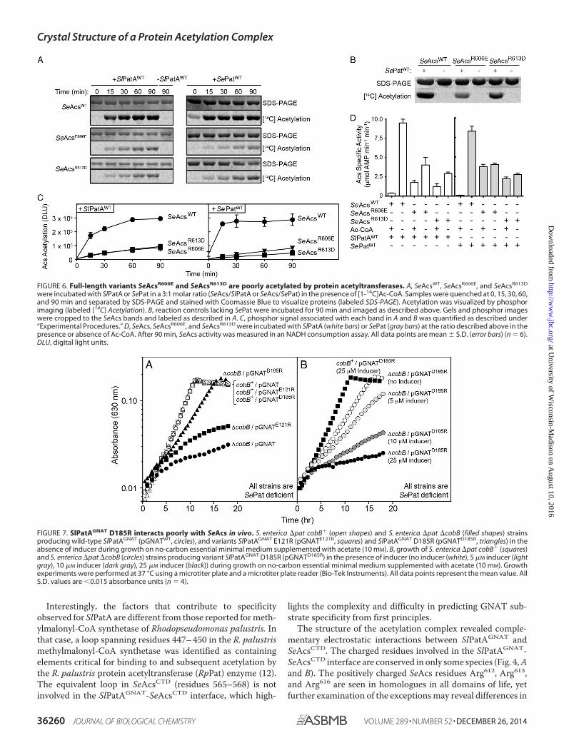
Importantly, the bacterial two-hybrid system results werereproduced both in vitro and in vivo when non-truncated formsof SeAcs and SlPatA were used and when the SlPatA homo-logue from S. enterica (SePat) was used (Figs. 6 and 7). Full-length SeAcsWT variants SeAcsR606E and SeAcsR613D retainedactivity in vitro despite amino acid substitutions near the cata-lytic Lys609 (SeAcsWT, 8.3 � 0.5 �mol AMP min�1 mg�1;SeAcsR606E, 5.1 � 0.2 �mol AMP min�1 mg�1; and SeAcsR613D,2.8 � 0.2 �mol AMP min�1 mg�1; mean � S.D., n � 9); how-ever, SlPatAWT and its S. enterica homologue SePatWT acety-lated these proteins less efficiently (Fig. 6). Amino acid substi-tutions near the active site lysine of AMP-forming CoA ligaseshave been shown to affect activity (11).
We also demonstrated that variant SlPatGNAT E121Rinteracted with SeAcsWT in vivo and inhibited growth of anS. enterica �pat �cobB strain but not the growth of anS. enterica �pat cobB� strain. In contrast, variant SlPatGNAT
D185R only slightly inhibited growth of the S. enterica �pat�cobB strain (Fig. 7). These data were consistent with theobservation that the SlPatD185R variant exhibited significantlyweaker interactions with SeAcsCTD in the bacterial two-hybridassay (Fig. 5). When higher levels of variant SlPatGNAT D185Rwere present, the growth of S. enterica on 10 mM acetate wasinhibited (Fig. 7). These results were consistent with the ideathat variant SlPatGNAT D185R and SeAcsCTD interactions wereweakened but not abolished.
DISCUSSION
Mapping the interaction surface between the SlPatAGNAT
and the globular protein substrate SeAcsCTD is a significantadvance in our understanding of how protein lysine acetyl-transferases recognize, with specificity, large globular proteintargets. To generate an interaction model of a GNAT with aprotein substrate, the crystal structure of SlPatAGNAT was firstsolved. The structure of SlPatAGNAT and SsPatWT, the mostclosely related structure, differed significantly at the predictedsubstrate interface. SsPatWT was crystallized after limited pro-teolysis, and the absence of amino acids (specifically residues42–52 that link helix �2 to strand �2) may distort the active sitecleft. SsPatWT acetylates the DNA-binding protein Alba, but itis unknown whether it can acetylate AMP-forming CoA ligases.Thus, we cannot rule out the possibility that the structural dif-ferences surrounding the SsPatWT substrate-binding cleft mayrepresent distinct substrate specificity.
FIGURE 5. Reversing charges at the interaction surface disrupts interac-tions between SlPatAGNAT and SeAcsCTD. A, effect of different Zif-SlPatAGNAT
variants and empty vector on transcription in vivo from promoter PlacZif1– 61 with �-SeAcsCTD variants or empty vector. B, data from the reciprocalbait-prey experiment (Zif-SeAcsCTD and �-SlPatAGNAT). “� only” refers toempty prey plasmid expressing only the � subunit of the RNA polymerase �subunit. Zif only, empty bait plasmid expressing only the zinc finger protein. *,p 0.0001; ^, interactions were detected in bait-prey reciprocal experiments.Error bars, S.D.
Crystal Structure of a Protein Acetylation Complex
DECEMBER 26, 2014 • VOLUME 289 • NUMBER 52 JOURNAL OF BIOLOGICAL CHEMISTRY 36259
at University of W
isconsin-Madison on A
ugust 10, 2016http://w
ww
.jbc.org/D
ownloaded from
Interestingly, the factors that contribute to specificityobserved for SlPatA are different from those reported for meth-ylmalonyl-CoA synthetase of Rhodopseudomonas palustris. Inthat case, a loop spanning residues 447– 450 in the R. palustrismethylmalonyl-CoA synthetase was identified as containingelements critical for binding to and subsequent acetylation bythe R. palustris protein acetyltransferase (RpPat) enzyme (12).The equivalent loop in SeAcsCTD (residues 565–568) is notinvolved in the SlPatAGNAT-SeAcsCTD interface, which high-
lights the complexity and difficulty in predicting GNAT sub-strate specificity from first principles.
The structure of the acetylation complex revealed comple-mentary electrostatic interactions between SlPatAGNAT andSeAcsCTD. The charged residues involved in the SlPatAGNAT-SeAcsCTD interface are conserved in only some species (Fig. 4, Aand B). The positively charged SeAcs residues Arg612, Arg613,and Arg616 are seen in homologues in all domains of life, yetfurther examination of the exceptions may reveal differences in
FIGURE 6. Full-length variants SeAcsR606E and SeAcsR613D are poorly acetylated by protein acetyltransferases. A, SeAcsWT, SeAcsR606E, and SeAcsR613D
were incubated with SlPatA or SePat in a 3:1 molar ratio (SeAcs/SlPatA or SeAcs/SePat) in the presence of [1-14C]Ac-CoA. Samples were quenched at 0, 15, 30, 60,and 90 min and separated by SDS-PAGE and stained with Coomassie Blue to visualize proteins (labeled SDS-PAGE). Acetylation was visualized by phosphorimaging (labeled [14C] Acetylation). B, reaction controls lacking SePat were incubated for 90 min and imaged as described above. Gels and phosphor imageswere cropped to the SeAcs bands and labeled as described in A. C, phosphor signal associated with each band in A and B was quantified as described under“Experimental Procedures.” D, SeAcs, SeAcsR606E, and SeAcsR613D were incubated with SlPatA (white bars) or SePat (gray bars) at the ratio described above in thepresence or absence of Ac-CoA. After 90 min, SeAcs activity was measured in an NADH consumption assay. All data points are mean � S.D. (error bars) (n � 6).DLU, digital light units.
FIGURE 7. SlPatAGNAT D185R interacts poorly with SeAcs in vivo. S. enterica �pat cobB� (open shapes) and S. enterica �pat �cobB (filled shapes) strainsproducing wild-type SlPatAGNAT (pGNATWT, circles), and variants SlPatAGNAT E121R (pGNATE121R, squares) and SlPatAGNAT D185R (pGNATD185R, triangles) in theabsence of inducer during growth on no-carbon essential minimal medium supplemented with acetate (10 mM). B, growth of S. enterica �pat cobB� (squares)and S. enterica �pat �cobB (circles) strains producing variant SlPatAGNAT D185R (pGNATD185R) in the presence of inducer (no inducer (white), 5 �M inducer (lightgray), 10 �M inducer (dark gray), 25 �M inducer (black)) during growth on no-carbon essential minimal medium supplemented with acetate (10 mM). Growthexperiments were performed at 37 °C using a microtiter plate and a microtiter plate reader (Bio-Tek Instruments). All data points represent the mean value. AllS.D. values are 0.015 absorbance units (n � 4).
Crystal Structure of a Protein Acetylation Complex
36260 JOURNAL OF BIOLOGICAL CHEMISTRY VOLUME 289 • NUMBER 52 • DECEMBER 26, 2014
at University of W
isconsin-Madison on A
ugust 10, 2016http://w
ww
.jbc.org/D
ownloaded from

PatA-Acs interactions among species. Likewise, few of the PatAresidues involved in the PatA-Acs interaction are conserved.Considering that the GNAT family of acetyltransferases isnoted for its lack of primary sequence conservation (7), predict-ing interacting residues of GNATs in the absence of structuraldata remains challenging. The extensive interaction surfaceobserved at the SlPatAGNAT-SeAcsCTD may be a key feature ofPatA-Acs interactions. A large interaction surface would facil-itate the evolution of distinct constellations of interactionsbetween each GNAT and its protein substrate(s). Continuedstructural analysis of GNAT-substrate complexes will revealthe range of interactions that occur between GNATs and theirprotein substrates.
The GNAT-substrate interface identified in the SlPatAGNAT-SeAcsCTD crystal structure is remarkably distinct from theGcn5-H3 peptide complexes reported to date (Fig. 3, C and E)and reveals structural roles of residues within and distant fromthe PX4GK motif found in substrates of Pat-type GNATs. Thestructure of the SlPatAGNAT-SeAcsCTD interaction will serve asa model to further identify, validate, and engineer (11) specificglobular protein targets of GNAT protein acetyltransferases.
Acknowledgments—We thank Simon Dove for plasmids and strains.Part of the results shown in this report were obtained at the ArgonneNational Laboratory Structural Biology Center at the Advanced Pho-ton Source. The University of Chicago Argonne, LLC operates ArgonneNational Laboratory for the United States Department of Energy,Office of Biological and Environmental Research under ContractDE-AC02-06CH11357.
REFERENCES1. Kouzarides, T. (2007) Chromatin modifications and their function. Cell
128, 693–7052. Glozak, M. A., Sengupta, N., Zhang, X., and Seto, E. (2005) Acetylation and
deacetylation of non-histone proteins. Gene 363, 15–233. Liang, W., Malhotra, A., and Deutscher, M. P. (2011) Acetylation regulates
the stability of a bacterial protein: growth stage-dependent modification ofRNase R. Mol. Cell 44, 160 –166
4. Crosby, H. A., Pelletier, D. A., Hurst, G. B., and Escalante-Semerena, J. C.(2012) System-wide studies of N-lysine acetylation in Rhodopseudomonaspalustris reveal substrate specificity of protein acetyltransferases. J. Biol.Chem. 287, 15590 –15601
5. Hubbert, C., Guardiola, A., Shao, R., Kawaguchi, Y., Ito, A., Nixon, A.,Yoshida, M., Wang, X. F., and Yao, T. P. (2002) HDAC6 is a microtubule-associated deacetylase. Nature 417, 455– 458
6. Friedmann, D. R., and Marmorstein, R. (2013) Structure and mechanismof non-histone protein acetyltransferase enzymes. FEBS J. 280,5570 –5581
7. Vetting, M. W., S de Carvalho, L. P., Yu, M., Hegde, S. S., Magnet, S.,Roderick, S. L., and Blanchard, J. S. (2005) Structure and functions of theGNAT superfamily of acetyltransferases. Arch. Biochem. Biophys. 433,212–226
8. Starai, V. J., Celic, I., Cole, R. N., Boeke, J. D., and Escalante-Semerena, J. C.(2002) Sir2-dependent activation of acetyl-CoA synthetase by deacetyla-tion of active lysine. Science 298, 2390 –2392
9. Thao, S., and Escalante-Semerena, J. C. (2011) Control of protein functionby reversible N�-lysine acetylation in bacteria. Curr. Opin. Microbiol. 14,200 –204
10. Tucker, A. C., and Escalante-Semerena, J. C. (2013) Acetoacetyl-CoA syn-thetase activity is controlled by a protein acetyltransferase with uniquedomain organization in Streptomyces lividans. Mol. Microbiol. 87,152–167
11. Crosby, H. A., and Escalante-Semerena, J. C. (2014) The acetylation motifin AMP-forming acyl coenzyme A synthetases contains residues criticalfor acetylation and recognition by the protein acetyltransferase Pat ofRhodopseudomonas palustris. J. Bacteriol. 196, 1496 –1504
12. Crosby, H. A., Rank, K. C., Rayment, I., and Escalante-Semerena, J. C.(2012) Structural insights into the substrate specificity of the proteinacetyltransferase RpPat: identification of a loop critical for recognition byRpPat. J. Biol. Chem. 287, 41392– 41404
13. Gulick, A. M., Starai, V. J., Horswill, A. R., Homick, K. M., and Escalante-Semerena, J. C. (2003) The 1.75 Å crystal structure of acetyl-CoA synthe-tase bound to adenosine-5�-propylphosphate and coenzyme A. Biochem-istry 42, 2866 –2873
14. Bertani, G. (1951) Studies on lysogenesis. I. The mode of phage liberationby lysogenic Escherichia coli. J. Bacteriol. 62, 293–300
15. Berkowitz, D., Hushon, J. M., Whitfield, H. J., Jr., Roth, J., and Ames, B. N.(1968) Procedure for identifying nonsense mutations. J. Bacteriol. 96,215–220
16. Guzman, L. M., Belin, D., Carson, M. J., and Beckwith, J. (1995) Tightregulation, modulation, and high-level expression by vectors containingthe arabinose PBAD promoter. J. Bacteriol. 177, 4121– 4130
17. Elion, E. A., Marina, P., and Yu, L. (2007) in Current Protocols in MolecularBiology (Ausubel, F. M., R. Brent, R. E., Kingston, D. D., Moore, J. G.,Seidman, J. A., Smith, A., and Struhl, K., eds) pp. 3.17.11–13.17.12, WileyInterscience, New York
18. Rocco, C. J., Dennison, K. L., Klenchin, V. A., Rayment, I., and Escalante-Semerena, J. C. (2008) Construction and use of new cloning vectors for therapid isolation of recombinant proteins from Escherichia coli. Plasmid 59,231–237
19. Blommel, P. G., and Fox, B. G. (2007) A combined approach to improvinglarge-scale production of tobacco etch virus protease. Protein Expr. Purif.55, 53– 68
20. Griffith, K. L., and Wolf, R. E., Jr. (2002) Measuring �-galactosidase activ-ity in bacteria: cell growth, permeabilization, and enzyme assays in 96-wellarrays. Biochem. Biophys. Res. Commun. 290, 397– 402
21. Zhang, X., and Bremer, H. (1995) Control of the Escherichia coli rrnB P1promoter strength by ppGpp. J. Biol. Chem. 270, 11181–11189
22. Garrity, J., Gardner, J. G., Hawse, W., Wolberger, C., and Escalante-Seme-rena, J. C. (2007) N-Lysine propionylation controls the activity of propio-nyl-CoA synthetase. J. Biol. Chem. 282, 30239 –30245
23. Crosby, H. A., Heiniger, E. K., Harwood, C. S., and Escalante-Semerena,J. C. (2010) Reversible N�-lysine acetylation regulates the activity of acyl-CoA synthetases involved in anaerobic benzoate catabolism in Rhodop-seudomonas palustris. Mol. Microbiol. 76, 874 – 888
24. Starai, V. J., and Escalante-Semerena, J. C. (2004) Identification of theprotein acetyltransferase (Pat) enzyme that acetylates acetyl-CoA synthe-tase in Salmonella enterica. J. Mol. Biol. 340, 1005–1012
25. Tucker, A. C., and Escalante-Semerena, J. C. (2010) Biologically activeisoforms of CobB sirtuin deacetylase in Salmonella enterica and Erwiniaamylovora. J. Bacteriol. 192, 6200 – 6208
26. Laemmli, U. K. (1970) Cleavage of structural proteins during the assemblyof the head of bacteriophage T4. Nature 227, 680 – 685
27. Sasse, J. (1991) in Current Protocols in Molecular Biology (Ausubel, F. A.,Brent, R., Kingston, R. E., Moore, D. D., Seidman, J. G., Smith, J. A., andStruhl, K., eds) pp. 10.16.11–10.16.18, Wiley Interscience, New York
28. Rayment, I. (2002) Small-scale batch crystallization of proteins revisited:an underutilized way to grow large protein crystals. Structure 10, 147–151
29. Otwinowski, Z., and Minor, W. (1997) Processing of x-ray diffraction datacollected in oscillation mode. Methods Enzymol. 276, 307–326
30. Sheldrick, G. M. (2008) A short history of SHELX. Acta Crystallogr. A 64,112–122
31. Cowtan, K., and Main, P. (1998) Miscellaneous algorithms for densitymodification. Acta Crystallogr. D 54, 487– 493
32. Collaborative Computational Project, Number 4 (1994) The CCP4 suite:programs for protein crystallography. Acta Crystallogr. D. Biol. Crystal-logr. 50, 760 –763
33. McCoy, A. J., Grosse-Kunstleve, R. W., Adams, P. D., Winn, M. D., Sto-roni, L. C., and Read, R. J. (2007) Phaser crystallographic software. J. Appl.Crystallogr. 40, 658 – 674
Crystal Structure of a Protein Acetylation Complex
DECEMBER 26, 2014 • VOLUME 289 • NUMBER 52 JOURNAL OF BIOLOGICAL CHEMISTRY 36261
at University of W
isconsin-Madison on A
ugust 10, 2016http://w
ww
.jbc.org/D
ownloaded from

34. Skubák, P., Murshudov, G. N., and Pannu, N. S. (2004) Direct incorpora-tion of experimental phase information in model refinement. Acta Crys-tallogr. D Biol. Crystallogr. 60, 2196 –2201
35. Emsley, P., and Cowtan, K. (2004) Coot: model-building tools for molec-ular graphics. Acta Crystallogr. D Biol. Crystallogr. 60, 2126 –2132
36. Kozakov, D., Brenke, R., Comeau, S. R., and Vajda, S. (2006) PIPER: an FFT-basedprotein docking program with pairwise potentials. Proteins 65, 392–406
37. Kozakov, D., Beglov, D., Bohnuud, T., Mottarella, S. E., Xia, B., Hall, D. R.,and Vajda, S. (2013) How good is automated protein docking? Proteins 81,2159 –2166
38. Comeau, S. R., Gatchell, D. W., Vajda, S., and Camacho, C. J. (2004) Clus-Pro: a fully automated algorithm for protein-protein docking. Nucleic Ac-ids Res. 32, W96 –W99
39. Comeau, S. R., Gatchell, D. W., Vajda, S., and Camacho, C. J. (2004) Clus-Pro: an automated docking and discrimination method for the predictionof protein complexes. Bioinformatics 20, 45–50
40. Rojas, J. R., Trievel, R. C., Zhou, J., Mo, Y., Li, X., Berger, S. L., Allis, C. D.,and Marmorstein, R. (1999) Structure of Tetrahymena GCN5 bound to
coenzyme A and a histone H3 peptide. Nature 401, 93–9841. Clements, A., Poux, A. N., Lo, W. S., Pillus, L., Berger, S. L., and Marmor-
stein, R. (2003) Structural basis for histone and phosphohistone bindingby the GCN5 histone acetyltransferase. Mol. Cell. 12, 461– 473
42. Lawrence, M. C., and Colman, P. M. (1993) Shape complementarity atprotein/protein interfaces. J. Mol. Biol. 234, 946 –950
43. Starai, V. J., and Escalante-Semerena, J. C. (2004) Acetyl-coenzyme Asynthetase (AMP forming). Cell. Mol. Life Sci. 61, 2020 –2030
44. Xu, H., Hegde, S. S., and Blanchard, J. S. (2011) Reversible acetylation andinactivation of Mycobacterium tuberculosis acetyl-CoA synthetase is de-pendent on cAMP. Biochemistry 50, 5883–5892
45. Larkin, M. A., Blackshields, G., Brown, N. P., Chenna, R., McGettigan,P. A., McWilliam, H., Valentin, F., Wallace, I. M., Wilm, A., Lopez, R.,Thompson, J. D., Gibson, T. J., and Higgins, D. G. (2007) Clustal W andClustal X version 2.0. Bioinformatics 23, 2947–2948
46. Castang, S., and Dove, S. L. (2010) High-order oligomerization is requiredfor the function of the H-NS family member MvaT in Pseudomonasaeruginosa. Mol. Microbiol. 78, 916 –931
Crystal Structure of a Protein Acetylation Complex
36262 JOURNAL OF BIOLOGICAL CHEMISTRY VOLUME 289 • NUMBER 52 • DECEMBER 26, 2014
at University of W
isconsin-Madison on A
ugust 10, 2016http://w
ww
.jbc.org/D
ownloaded from
Escalante-SemerenaAlex C. Tucker, Keenan C. Taylor, Katherine C. Rank, Ivan Rayment and Jorge C.
Insights into the Specificity of Lysine Acetyltransferases
doi: 10.1074/jbc.M114.613901 originally published online November 7, 20142014, 289:36249-36262.J. Biol. Chem.
10.1074/jbc.M114.613901Access the most updated version of this article at doi:
Alerts:
When a correction for this article is posted•
When this article is cited•
to choose from all of JBC's e-mail alertsClick here
http://www.jbc.org/content/289/52/36249.full.html#ref-list-1
This article cites 44 references, 13 of which can be accessed free at
at University of W
isconsin-Madison on A
ugust 10, 2016http://w
ww
.jbc.org/D
ownloaded from