instructions for laboratory training in general
TRANSCRIPT

Ministry of Health Care, Republic of Belarus
Vitebsk State Medical University
Department of Clinical Microbiology
INSTRUCTIONS
FOR LABORATORY TRAINING
in General Microbiology & Immunology
for students of medical faculty
VITEBSK 2018

2
УДК 579+616.31]=111(07)
ББК 28.4я73
Г34
Printed according to the decision of Central Educational Council of VSMU
(protocol № 9 , 25.10.2017)
Reviewed by:
T.I. Dmitrachenko, MD, PhD, Dr.Sci, Professor of Infectious Disease Dpt,
Vitebsk State Medical University
Generalov I.I.
Г34 Instructions for laboratory training in General Microbiology and Virology for students
of the Faculty of General Medicine. / I.I. Generalov, N.V. Zheleznyak,
A.V. Frolova, A.M. Moiseeva – Vitebsk, – VSMU, 2018. – 41 p.
Instructions for laboratory training in General Microbiology and Immunology for students of the faculty
of General Medicine were compiled according to basic educational plan and program, approved by Ministry of
Health Care of Republic of Belarus. The plan, schedule of laboratory training and basic practical skills in
general microbiology and immunology are presented in this workbook.
The instructions are prepared for medical students of higher educational establishments.
УДК 579+616.31]=111(07)
ББК 28.4я73
© Generalov I.I., 2018
© VSMU Press, 2018

3
CONTENTS
Pages
Laboratory class №1. The topic: General acquaintance with microbiological
laboratory. Safety rules of the work with pathogenic microbial agents (biological
safety). Systematics and nomenclature of microorganisms. The morphology of
bacteria. Basic methods for examination of microbial morphology. Simple
methods of stain
5
Laboratory class №2. The topic: Morphology and ultra-structure of prokaryotes.
Differential methods of stain
7
Laboratory class №3. The topic: Morphology and ultra-structure of prokaryotes
and eukaryotes (continuation). Differential methods of stain. Bacteriological
method of examination (isolation of microbial culture, 1st day)
11
Laboratory class №4. The topic: Physiology of microorganisms. Nutrition of
bacteria. Principles of bacterial culturing. Classification of nutrient media. Anti-
microbial measures: sterilization, antisepsis, disinfection, asepsis. Isolation of
pure culture of bacteria (2nd day of examination)
12
Laboratory class №5. The topic: Physiology of microorganisms. Growth and
reproduction of bacteria. Bacterial pigments. Bacterial enzymes. Energy
metabolism. Biological oxidation in bacteria: fermentation and respiration.
Isolation of pure culture of bacteria (3rd and 4th days of examination)
14
Laboratory class №6. The topic: Microbial genetics
16
Laboratory class №7. The topic: Microorganisms and the environment. Normal
microflora of human body and its role. Sanitary microbiology. Microflora of air
and water. Sanitary control of microbial pollution of air and water
18
Laboratory class №8. The topic: Final control study of the section “Morphology
of bacteria. Physiology of bacteria. Microbial genetics. Normal microflora of
human body and its role. Sanitary microbiology”
20
Laboratory class №9. The topic: Immunology and immunity. Types of
immunity. Structure of immune system. Immune cell receptors (CD molecules).
Cytokines. Differentiation of T- and B cells
22
Laboratory class №10. The topic: Antigens. Infectious and non-infectious
antigens. HLA system. Immunoglobulins and antibodies. Serological testing – the
mechanisms and goals of serological reactions. Precipitation tests
24

4
Laboratory class №11. The topic: Innate immunity: humoral and cellular
response. Complement system. Classical, alternative and lectin pathways of
activation. Serological testing – complement fixation test. Mononuclear
phagocyte system. Granulocytes. Phagocytosis, its stages. Laboratory testing of
phagocytosis. Natural killer cells
25
Laboratory class №12. The topic: Acquired immunity. Antigen-presenting cells
(APCs). Dendritic cells, their functions. Toll-like receptors and pattern-based
microbial recognition. T-dependent and T-independent immune responses.
Primary and secondary immune response. Serological reactions – agglutination
tests. Indirect hemagglutination assay. Coombs` test
27
Laboratory class №13. The topic: Active and passive immunoprophylaxis and
immunotherapy. Vaccines and toxoids. Immune antisera and immunoglobulins.
Monoclonal antibodies, their practical use. Serological testing – reactions of toxin
neutralization. Reactions with labeled antibodies and antigens.
Immunofluorescence assay. Enzyme-linked immunosorbent assay.
Radioimmunoassay. Immunoblotting (western blotting). Laboratory testing of
immune status. Assessment of cellular and humoral immunity
29
Laboratory class №14. The topic: Immunopathology. Immediate and delayed
types of hypersensitivity. Coombs & Gell classification of hypersensitivity
reactions. Allergy and allergic diseases. Anaphylactic hypersensitivity. Cytotoxic
hypersensitivity. Immune-complex-mediated hypersensitivity. Cell-mediated
(delayed) hypersensitivity. Skin tests for laboratory diagnosis of infectious
allergy. Stimulatory and blocking hypersensitivity. Autoimmune diseases.
Primary and secondary immunodeficiencies
31
Laboratory class №15. The topic: Final control study of the section
“Immunology and immunity. Immunological laboratory testing.
Immunopathology. Immunoprophylaxis. Immunotherapy”
32
Laboratory class №16. The topic: Infection and infectious process. Epidemic
process. Microbial pathogenicity and virulence. Virulence factors. Systemic
bacterial infection (sepsis)
35
Laboratory class №17. The topic: Chemotherapy. Antibiotics. Antibiotic
susceptibility testing
37
Laboratory class №18. The topic: Causative agents of suppurative infections.
Staphylococci, pseudomonads, bacteroids, and related agents
39

5
Laboratory class №1
The topic: General acquaintance with microbiological laboratory. Safety rules of the
work with pathogenic microbial agents (biological safety). Systematics and
nomenclature of microorganisms. The morphology of bacteria. Basic methods for
examination of microbial morphology. Simple methods of stain
The main aim and the tasks of the work:
1. To make acquaintance with safety rules of the work in microbiological laboratory.
2. To get skills of primary manipulations with bacterial cultures.
3. To get skills of making slides of bacterial cultures with their next stain by simple
staining methods.
4. To revise the principles and techniques of bright field microscopy.
The questions to the topic:
1. The subject and main tasks of medical microbiology.
2. Basic historical periods of microbiology.
3. International taxonomy of microorganisms. Principles of microbial classification.
4. The main morphological forms of bacteria.
5. The methods of study of bacterial morphology and structure.
6. Basic steps of preparing of slides from agar and broth microbial cultures. Simple
methods of slide stain.
7. Bright field microscopy. The use of immersion objective lens.
8. Current advanced methods of microscopy (dark-field, phase contrast, luminescent
microscopy, laser scanning confocal microscopy, atomic force microscopy,
electron microscopy).
THE LITERATURE:
1. Lecture contents.
2. “Medical Microbiology, Virology & Immunology: Lecture Course”. Part 1.
I. I. Generalov, Vitebsk, 2016. Р. 9-26, 37-42.
Safety rules of the work in microbiological laboratory
1. Practical training in microbiological laboratory is performed with contagious material
that requires strict discipline in laboratory work.
6
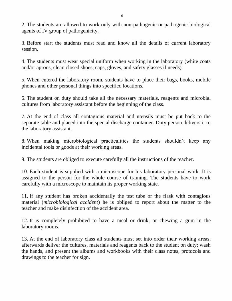
2. The students are allowed to work only with non-pathogenic or pathogenic biological
agents of IV group of pathogenicity.
3. Before start the students must read and know all the details of current laboratory
session.
4. The students must wear special uniform when working in the laboratory (white coats
and/or aprons, clean closed shoes, caps, gloves, and safety glasses if needs).
5. When entered the laboratory room, students have to place their bags, books, mobile
phones and other personal things into specified locations.
6. The student on duty should take all the necessary materials, reagents and microbial
cultures from laboratory assistant before the beginning of the class.
7. At the end of class all contagious material and utensils must be put back to the
separate table and placed into the special discharge container. Duty person delivers it to
the laboratory assistant.
8. When making microbiological practicalities the students shouldn’t keep any
incidental tools or goods at their working areas.
9. The students are obliged to execute carefully all the instructions of the teacher.
10. Each student is supplied with a microscope for his laboratory personal work. It is
assigned to the person for the whole course of training. The students have to work
carefully with a microscope to maintain its proper working state.
11. If any student has broken accidentally the test tube or the flask with contagious
material (microbiological accident) he is obliged to report about the matter to the
teacher and make disinfection of the accident area.
12. It is completely prohibited to have a meal or drink, or chewing a gum in the
laboratory rooms.
13. At the end of laboratory class all students must set into order their working areas;
afterwards deliver the cultures, materials and reagents back to the student on duty; wash
the hands, and present the albums and workbooks with their class notes, protocols and
drawings to the teacher for sign.

7
Personal work of students:
1. Preparing of slides of broth culture with staphylococci (methylene blue stain).
2. Preparing of slides of agar culture with Escherichia coli (fuchsin stain).
3. Demonstration: microscopy of slides with streptococci (methylene blue stain).
4. Drawing of smears.
The basic steps of slide preparing for microscopy
The preparation of the slide of agar culture
1. Put a drop of saline by sterile microbiological wire loop on the surface of defatted
glass slide. Sterilize the wire loop and loop holder.
2. Take the sample from the agar and disperse it by sterile loop in the drop of saline.
3. Thoroughly spread the culture upon the glass surface in the circle with the diameter
about 2.5-3 cm.
4. Sterilize the loop in the flame of burner.
5. Dry the slide at room temperature or with the help of ethanol burner.
6. Fix the slide passing it three times through the middle part of the flame.
7. Stain the slide with proper staining dye.
8. Wash it with tap water.
9. Dry the slide.
10. Drop the immersion oil on the slide.
11. Start bright field microscopy (immersion objective lens – 100x, eyepiece – 10x).
Laboratory class №2
The topic: Morphology and ultra-structure of prokaryotes. Differential methods of
stain
The main aim and the tasks of the work:
1. To learn the theoretical knowledge of the topic.
2. To know basic and advanced methods of microscopy.

8
3. To know the mechanisms and practical applications of differential staining methods:
Gram stain, Gins capsule stain, Neisser stain, Ziehl-Neelsen acid-fast bacilli stain,
Ozheshko spore stain.
The questions to the topic:
1. Structure of bacterial cell (obligate and non-obligate structural components).
2. Study of microbial morphology: basic and advanced methods of microscopy.
3. Nucleoid, its structure and functions, methods of detection.
4. Cytoplasm and ribosomes of bacteria. Cytoplasmic inclusions. Methods for detection
of volutin granules.
5. Bacterial envelope, its composition and function of different layers. Cytoplasmic
membrane, its structure and function.
6. Bacterial cell wall, its biological role. Structure of cell wall of gram-positive bacteria.
7. The cell wall of gram-negative bacteria. LPS, its functions.
8. Bacterial capsule, its structure and function.
9. Spores, stages of sporulation, methods of detection.
10. Flagella, methods for detection of bacteria motility.
11. Pili and fimbria. Injectisome, its structure and function.
12. Differential methods of stain (Gram stain, Gins capsule stain, Neisser stain, Ziehl-
Neelsen acid-fast bacilli stain, Ozheshko spore stain).
THE LITERATURE:
1. Lecture contents.
2. “Medical Microbiology, Virology & Immunology: Lecture Course”. Part 1.
I. I. Generalov, Vitebsk, 2016. Р. 26-42.
Personal work of students:
1. Preparing of smears with mixture of Sarcina flava and Escherichia coli (Gram stain).
2. Demonstration:
a) microscopy of smears with capsular bacteria (Gins capsule stain).
b) microscopy of smears with spore-forming bacteria (Ozheshko stain).
4. Drawing of smears.
9
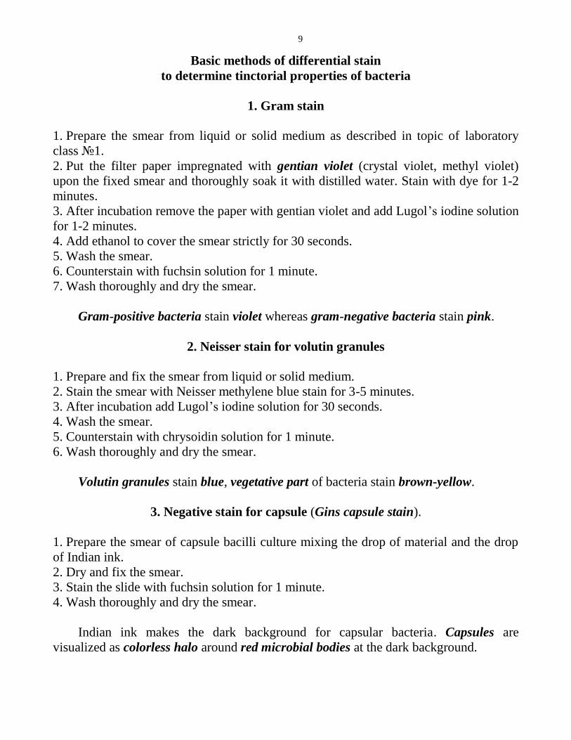
Basic methods of differential stain
to determine tinctorial properties of bacteria
1. Gram stain
1. Prepare the smear from liquid or solid medium as described in topic of laboratory
class №1.
2. Put the filter paper impregnated with gentian violet (crystal violet, methyl violet)
upon the fixed smear and thoroughly soak it with distilled water. Stain with dye for 1-2
minutes.
3. After incubation remove the paper with gentian violet and add Lugol’s iodine solution
for 1-2 minutes.
4. Add ethanol to cover the smear strictly for 30 seconds.
5. Wash the smear.
6. Counterstain with fuchsin solution for 1 minute.
7. Wash thoroughly and dry the smear.
Gram-positive bacteria stain violet whereas gram-negative bacteria stain pink.
2. Neisser stain for volutin granules
1. Prepare and fix the smear from liquid or solid medium.
2. Stain the smear with Neisser methylene blue stain for 3-5 minutes.
3. After incubation add Lugol’s iodine solution for 30 seconds.
4. Wash the smear.
5. Counterstain with chrysoidin solution for 1 minute.
6. Wash thoroughly and dry the smear.
Volutin granules stain blue, vegetative part of bacteria stain brown-yellow.
3. Negative stain for capsule (Gins capsule stain).
1. Prepare the smear of capsule bacilli culture mixing the drop of material and the drop
of Indian ink.
2. Dry and fix the smear.
3. Stain the slide with fuchsin solution for 1 minute.
4. Wash thoroughly and dry the smear.
Indian ink makes the dark background for capsular bacteria. Capsules are
visualized as colorless halo around red microbial bodies at the dark background.

10
4. Ziehl-Neelsen stain to detect acid-fast bacteria
1. Prepare and fix the smear from sputum specimen.
2. Stain it with Ziehl carbolic fuchsin solution for 5 minutes, or put the filter paper
impregnated with Ziehl fuchsin upon the fixed smear, thoroughly soak it with distilled
water and heat the slide upon burner until vapor appearance.
3. After incubation remove the paper and wash the smear with tap water.
4. Decolorize the smear with 5% sulfuric acid for 3-5 seconds.
5. Thoroughly wash the smear.
6. Counterstain the smear with methylene blue solution for 5 minutes.
7. Wash thoroughly and dry the smear.
Acid-fast bacteria retain the red stain while all other bacteria and the background
are stained blue.
5. Ozheshko method for spore stain
1. Prepare the smear of spore-containing bacilli culture.
2. Before fixing put 0.5% solution of hydrochloric acid upon the smear and heat the
slide on burner for 3-5 minutes.
3. After incubation wash the smear thoroughly with tap water.
4. Fix the smear.
5. Stain the slide with Ziehl-Neelsen method.
6. Wash thoroughly and dry the smear.
Spores stain red, the vegetative parts of microbial cell are blue.
6. Romanowsky-Giemsa stain
1. Prepare the smear of leukocyte culture incubated with bacterial or protozoan agents.
2. Fix the slide.
3. Stain it with Romanowsky-Giemsa’s solution for 15-30 min. Romanowsky-Giemsa’s
stain (mixture of azure, eosin, and methylene blue dyes) is prepared from primary stock
solution adding 1-2 drops of primary stock mixture into 1 ml of distilled water.
4. After incubation wash the slide with tap water and dry it.
The bacteria stain violet-purple, cell nuclei – red, cytoplasm – blue; protozoan
nuclei – red-violet, their cytoplasm – blue.

11
Laboratory class №3
The topic: Morphology and ultra-structure of prokaryotes and eukaryotes
(continuation). Differential methods of stain. Bacteriological method of examination
(isolation of microbial culture, 1st day)
The main aim and the tasks of the work:
1. To learn the theoretical knowledge of the topic.
2. To continue examination of the basic morphological forms of bacteria (spirochetes,
mycoplasmas, rickettsiae, chlamydiae, actinomycetes, fungi).
3. To get skills of microbial agar plating for isolation of pure bacterial culture.
The questions to the topic:
1. The morphology and structure of spirochetes. Romanowsky-Giemsa stain.
2. The morphology and structure of rickettsiae.
3. The morphology and structure of chlamydiae.
4. The morphology and structure of mycoplasmas.
5. The morphology and structure of actinomycetes.
6. Classification and structure of fungi.
7. The morphology of mould and yeast fungi.
THE LITERATURE:
1. Lecture contents.
2. “Medical Microbiology, Virology & Immunology: Lecture Course”. Part 1.
I. I. Generalov, Vitebsk, 2016. Р. 38-39, 43-53.
Personal work of students:
1. Demonstration:
a) microscopy of slides with C. trachomatis infected cells (methylene blue stain);
b) microscopy of slides with various mould fungi (Mucor mucedo, Aspergillus
fumigatus);
c) microscopy of slides with of Candida fungi (methylene blue stain).
d) laser scanning confocal fluorescent microscopy of Penicillium chrysogenum
culture (auramine stain, 3D reconstruction).

12
2. Drawing of slides.
3. Planting of microbial mixture on Petri dish with MPA for isolation of pure culture of
microorganisms. Protocol recording.
Protocol №1. Isolation of pure culture of microorganisms
Day of
examination
Material for
examination
Steps of examination Results
1. Mixture of
bacteria
Plating of specimen on Petri dish with
MPA. Incubation at 37°C for 24 h __
Laboratory class №4
The topic: Physiology of microorganisms. Nutrition of bacteria. Principles of
bacterial culturing. Classification of nutrient media. Anti-microbial measures:
sterilization, antisepsis, disinfection, asepsis. Isolation of pure culture of bacteria (2nd
day of examination)
The main aim and the tasks of the work:
1. To learn the theoretical knowledge of the topic.
2. To know the classification of nutrient media and their composition.
3. To make acquaintance with special equipment for sterilization and disinfection.
4. To be able to describe bacterial morphological, tinctorial, and cultural properties.
5. To get skills of inoculation of microbial specimen into slant agar.
The questions to the topic:
1. Metabolism of bacteria. Classification of bacteria according to their nutrition type and
energy gain.
2. The mechanisms of bacterial nutrition and transport of nutrients into bacterial cells.
3. Secretion systems for transport of proteins and other substances outside the bacterial
cells.
4. Main principles of microbial culture. Various groups of nutrient media.
5. Cultural characteristics of bacteria. Microbial colonies. Bacterial pigments, their
significance. Classification of pigments.
6. Sterilization, its purposes. Sterilizing factors.
7. Different methods of sterilization.
13
8. Antisepsis, definition. The basic requirements to antiseptic drugs.
9. Groups of antiseptics, mechanisms of action.
10. Disinfection, its main goal. Variants of disinfection.
11. Asepsis, definition and common principles.
THE LITERATURE:
1. Lecture contents.
2. “Medical Microbiology, Virology & Immunology: Lecture Course” Part 1.
I. I. Generalov, Vitebsk, 2016, р. 54-65, 141-146.
Personal work of students:
1. The acquaintance of students with laboratory equipment for disinfection and
sterilization.
2. Isolation of pure bacterial culture (2nd
day of examination) – continuation of test
protocol started at previous laboratory class:
a) description of bacterial growth, characteristics of colonies according to their
cultural properties.
b) inoculation of bacteria into slant agar for isolation of pure culture.
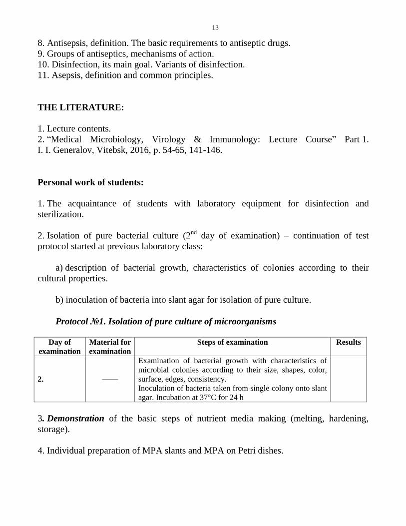
Protocol №1. Isolation of pure culture of microorganisms
Day of
examination
Material for
examination
Steps of examination Results
2.
––––
Examination of bacterial growth with characteristics of
microbial colonies according to their size, shapes, color,
surface, edges, consistency.
Inoculation of bacteria taken from single colony onto slant
agar. Incubation at 37°C for 24 h
3. Demonstration of the basic steps of nutrient media making (melting, hardening,
storage).
4. Individual preparation of MPA slants and MPA on Petri dishes.

14
Laboratory class №5
The topic: Physiology of microorganisms. Growth and reproduction of bacteria.
Bacterial pigments. Bacterial enzymes. Energy metabolism. Biological oxidation in
bacteria: fermentation and respiration. Isolation of pure culture of bacteria (3rd
and
4th
days of examination)
The main aim and the tasks of the work:
1. To learn the theoretical knowledge of the topic.
2. To know biochemical properties of bacteria.
3. To get acquaintance with the methods of anaerobic bacteria isolation.
4. To be able to identify isolated bacterial culture.
The questions to the topic:
1.Optimal conditions for bacterial growth and culture. Growth and reproduction of
microorganisms.
2. Microbial growth in biofilms.
3. Enzymes of bacteria, their properties and classification. The role of enzymes in
bacterial metabolism and pathogenicity.
4. Biological oxidation in bacteria. Fermentation.
5. Respiration in bacteria. Classification of bacteria according to their types of
respiration.
6. Methods of anaerobic bacteria culturing. Enzymatic differences between anaerobic
and aerobic bacteria.
7. Practical use of enzymatic properties of bacterial cultures. Assessment of microbial
carbohydrate hydrolysis. Hiss media.
8. Tests for detection of bacterial proteolytic activity. Determination of bacterial
catalase, oxidase and urease activities.
THE LITERATURE:
1. Lecture contents.
2. “Medical Microbiology, Virology & Immunology: Lecture Course”. Part 1.
I. I. Generalov, Vitebsk, 2016. Р. 59-61, 65-79.
15
Personal work of students:
1. Demonstration: bacterial pigments, methods for anaerobic bacteria culturing.
2. Isolation of pure bacterial culture (3nd
day of examination) – continuation of test
protocol started at previous classes:
a) examination of microbial growth on slant agar, assessment of culture purity
(preliminary visual examination, preparing of slides with Gram stain and bright field
immersion microscopy for determination of morphological and tinctorial properties of
isolated culture).
b) determination of catalase activity in reaction with hydrogen peroxide.
c) testing of microbial biochemical activity: inoculation of bacteria into Hiss media
and meat-peptone broth (MPB) for detection of carbohydrate hydrolysis and proteolytic
activity.
3. Isolation of pure bacterial culture (4th day of examination):
a) registering of bacterial biochemical activity according to the demonstration of
plate biochemical tests.
b) completion of bacteriological testing with final conclusion about the species of
isolated microbial culture.
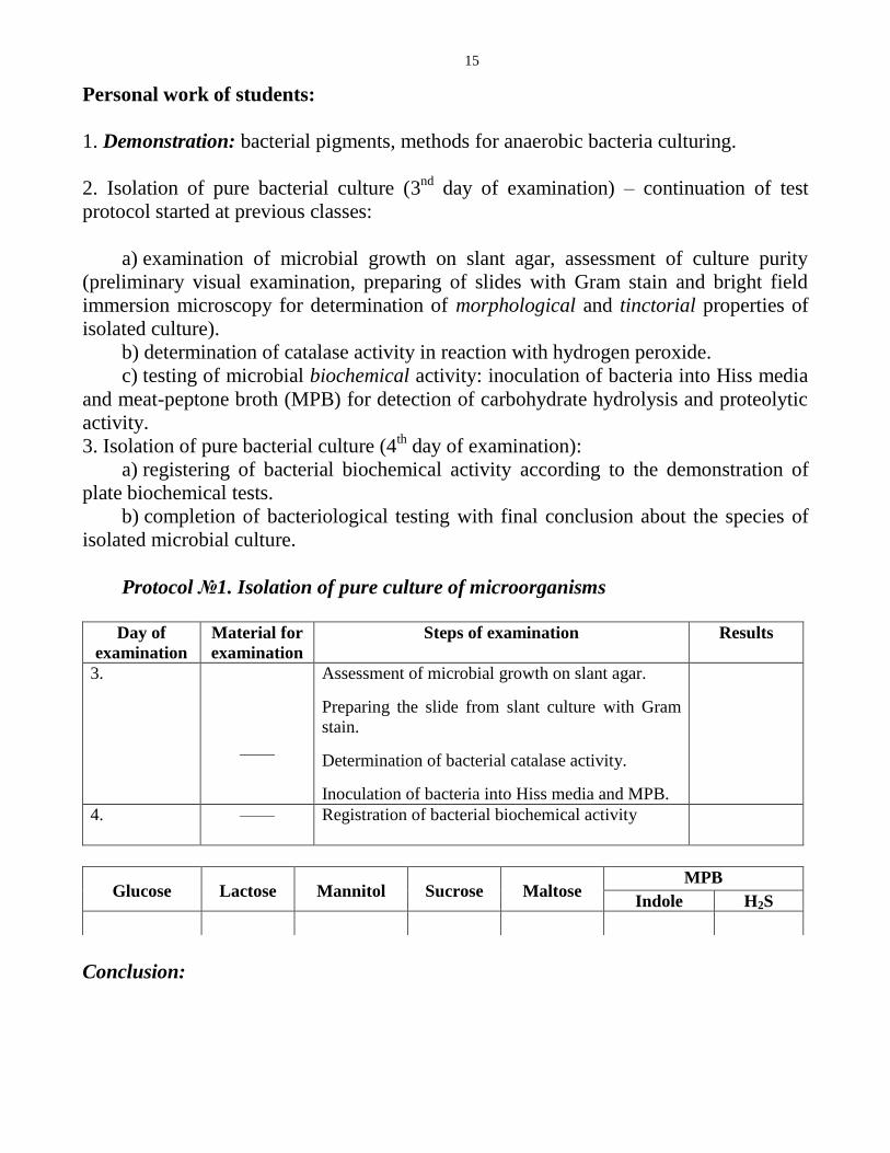
Protocol №1. Isolation of pure culture of microorganisms
Day of
examination
Material for
examination
Steps of examination Results
3.
––––
Assessment of microbial growth on slant agar.
Preparing the slide from slant culture with Gram
stain.
Determination of bacterial catalase activity.
Inoculation of bacteria into Hiss media and MPB.
4.
–––– Registration of bacterial biochemical activity
Conclusion:
Glucose
Lactose
Mannitol
Sucrose
Maltose MPB
Indole H2S

16
Laboratory class №6
The topic: Microbial genetics
The main aim and the tasks of the work:
1. To learn the theoretical knowledge of the topic.
2. To get skills of genetic transformation and transduction tests.
3. To be able to assess phenotypic variation of Proteus culture.
4. To be able to determine R- and S-forms of microbial colonies.
The questions to the topic:
1. Bacterial genotype and phenotype, their characteristics. Organization of bacterial
genome. Regulation of gene expression. Operon, its structure.
2. Plasmids and episomes, their structure and function.
3. Mobile genetic elements of bacterial genome. Transposons, IS-elements.
4. Genetic organization of adaptive immunity in bacteria – CRISPR/Cas system. 5. Phenotypic bacterial variations. Modifications, their characteristics.
6. Genotypic variations in bacteria, their classification. Bacterial dissociation.
7. Mutations: classification, mechanisms and biological significance.
8. Recombinations in bacteria, classification. Molecular mechanisms of recombinations.
Bacterial transformation.
9. Transduction in bacteria.
10. Bacterial conjugation.
11. Methods of molecular genetic analysis. Molecular hybridization of nucleic acids.
12. Polymerase chain reaction. Nucleic acid sequencing.
13. Principles of genetic engineering. Practical applications of recombinant technologies
in biology and medicine.
THE LITERATURE:
1. Lecture contents.
2. “Medical Microbiology, Virology & Immunology: Lecture Course”. Part 1.
I. I. Generalov, Vitebsk, 2016. Р. 80-108.

17
Personal work of students:
Protocol №1. Transformation test of B. subtilis culture
Day of
examination
Material for
examination
Steps of examination Results
1. 1. DNA of auxo-
autotrophic strain,
capable of
tryptophan
synthesis.
2. Recipient culture
of auxo-
heterotrophic
B. subtilis strain.
Bacterial suspension of slant culture of
B. subtilis is prepared by saline wash.
0.5 ml of bacterial suspension is added
into 2 test tubes. First one is
supplemented with 0.5 ml of DNA
solution, second – with 0.5 ml of saline
(control test).
Test tubes are placed for incubation at
37oC for 30 min.
After the end of incubation the samples
from both test tubes are plated onto
MPA without tryptophan.
__
Incubation at 37oC for 24 h
2. Assessment of transformation results.
Conclusion:
Protocol №2. Transduction test of E. coli strain (biovar Paracoli)
Day of
examination
Material for
examination
Steps of examination Results
1. 1. Bacteriophage,
able to transfer
genes for lactose
fermentation.
2. Recipient
non-fermenting
culture of E. coli
biovar Paracoli
Bacterial suspension of E. paracoli strain
is prepared by slant saline wash.
0.5 ml of bacterial suspension is added
into 2 test tubes. First is supplemented
with 0.5 ml of phage culture, second –
with 0.5 ml of saline (control test).
Test tubes are placed for incubation at
37oC for 30 min.
After incubation inoculation of samples
from both test tubes is made onto Endo
medium
__
Incubation at 37oC for 24 h
2. Evaluation of transduction results.
Conclusion:

18
Protocol №3. Examination of modification test of P. vulgaris culture
Day of
examina-
tion
Material for
examination
Steps of examination Results
1. 1. Broth culture of
P. vulgaris.
2. Petri dish with
MPA.
3. Petri dish with
MPA supplemented
with phenol.
One-streak plating of P. vulgaris culture
is made upon Petri dish with phenol-
supplemented MPA and control medium
(non-modified MPA).
__
Incubation at 37oC for 24 h
2. Assessment of “swarming” microbial
growth in experimental and control Petri
dishes.
Conclusion:
Laboratory class №7
The topic: Microorganisms and the environment. Normal microflora of human body
and its role. Sanitary microbiology. Microflora of air, soil and water. Sanitary control
of microbial pollution of air and water
The main aim and the tasks of the work:
1. To learn the theoretical knowledge of the topic.
2. To know the composition of normal human microbiota.
3. To know the standards of sanitary state of water and air.
4. To get skills of sanitary testing of water and air quality.
5. To know the composition of Kessler's and Endo media.
The questions to the topic:
1. Microbial Distribution in Nature. Symbiosis, its forms.
2. Antagonistic microbial relationships, their mechanisms. Types of microbial
antagonism.
3. Normal microflora of human body, its role in human physiology and pathology.
Microflora of skin and respiratory tract.

19
4. Microflora of oral cavity, general characteristics. Biological role of normal oral
microbiota. Microflora of human gut and urogenital tract, its role.
5. Dysbiosis (dysbacteriosis), predisposing factors and progression. Prophylaxis and
treatment of dysbiosis.
6. Sanitary indicator microorganisms, their characteristics.
7. Microflora of water, sources of water pollution. Water quality characteristics. Total
microbial count of water.
8. Sanitary indicator microorganisms for water. Laboratory testing of water sanitary
state. Identification of total coliform and thermotolerant bacteria.
9. Microflora of air. Microbial standards for air quality. Methods for determination of
air sanitary state.
10. Microflora of soil.
THE LITERATURE:
1. Lecture contents.
2. “Medical Microbiology, Virology & Immunology: Lecture Course”. Part 1.
I. I. Generalov, Vitebsk, 2016. Р. 109-117, 129-140.
Personal work of students:
1. Microscopy of specimen from dental plaque (Gram stain).
2. Bacteriological sanitary testing of hand wash.
Protocol №1. Bacteriological sanitary testing of hand wash
Day of
examination
Material for
examination
Steps of examination Results
1. Hand wash Sampling of hand wash by sterile swab soaked
with saline.
Inoculation of specimen into Kessler’s medium
for E. coli determination.
__
Incubation at 44oC for 24 h
2. Assessment of microbial growth in Kessler’s
medium.
Note. Composition of Kessler’s medium: MPB, lactose, bile salts, gentian violet and
tube for gas accumulation (float).
E. coli growth is indicated by gas accumulation within the float.
20
Laboratory class №8
The topic: Final control study of the section “Morphology of bacteria. Physiology of
bacteria. Microbial genetics. Sanitary microbiology”
The main aim and the tasks of the work:
To consolidate the basic knowledge of bacterial structure and metabolism (nutrition,
respiration, growth and reproduction, genetic alterations); methods of microbial
isolation, identification and molecular genetic analysis; principles and methods of
sanitary microbiology, asepsis, antisepsis, disinfection, and sterilization.
The questions:
1. Systematics and nomenclature of microorganisms. Principles of microbial
classification. Basic definitions in microbiology.
2. Study of bacterial morphology: modern methods of microscopy.
3. Characteristics of round (spherical) forms of bacteria.
4. Characteristics of rod-shaped forms of bacteria.
5. Characteristics of twisted (spiral) forms of bacteria.
6. Structure of bacterial cell (obligate and non-obligate components). Bacterial
envelope, its composition. Cytoplasmic membrane – structure and function.
7. Bacterial cell wall, its biological role. Structure of the cell wall of gram-positive
bacteria. Differential methods of staining – Gram stain.
8. The cell wall of gram-negative bacteria. LPS, its functions. Differential methods of
staining – Gram stain.
9. Bacterial capsule, its structure and function. Differential methods of staining – Gins
capsule stain.
10. Spores, stages of sporulation, methods of detection.
11. Flagella, pili, methods of bacteria motility detection. Injectisome, its structure and
functions.
12. Cytoplasm, ribosomes, inclusions, methods of volutin granules detection.
13. Nucleoid, its structure and function. Methods of detection.
14. Morphology and characteristics of actinomycetes. Differential methods of staining –
Ziehl-Neelsen acid-fast bacilli stain.
15. Morphology and characteristics of spirochetes. Differential methods of staining –
Romanowsky-Giemsa stain.
16. Morphology and characteristics of chlamydiae.
17. Morphology and characteristics of rickettsiae.
18. Morphology and characteristics of mycoplasmas.
19. Classification and structure of fungi.

21
20. Morphology and characteristics of mould and yeast fungi.
21. L. Pasteur, his outstanding contribution into microbiological science. R. Koch, his
work in microbiology.
22. Metabolism of bacteria. Classification of bacteria according to their nutrition type
and energy gain.
23. Mechanisms of bacterial trans-membrane transportation.
24. Secretion systems for transport of proteins and other substances outside the bacterial
cells.
25. Methods of bacterial culturing. Requirements to nutrient media. Classification of
nutrient media and their characteristics.
26. Biological oxidation in bacteria. Pathways of fermentation and respiration.
27. Classification of bacteria according to their types of respiration and their
characteristics.
28. Methods of anaerobic bacteria cultivation. Isolation of pure culture of anaerobes.
29. Bacterial enzymes, their properties and classification. The role of enzymes in
bacterial metabolism and pathogenicity.
30. Examination of bacterial carbohydrate hydrolysis. Determination of proteolytic
properties. Detection of catalase, oxidase, and urease activity of bacteria.
31. Growth and reproduction of bacteria.
32. Bacterial pigments, their significance. Classification of pigments.
33. Microorganisms, inhabiting the environment. Microbial ecology, microbial
communities, ecosystem, ecological variants. Symbiosis, its forms. Antagonistic
microbial relationships, their mechanisms. Types of microbial antagonism.
34. Normal microflora of human body, its role in human physiology and pathology.
Ontogenesis of normal microflora. Microflora of skin, its role.
35. Microflora of oral cavity, gut, respiratory, and urogenital tract; role in physiology
and pathology.
36. Dysbiosis (dysbacteriosis), predisposing factors and progression. Prophylaxis and
treatment of dysbiosis.
37. Sanitary indicator microorganisms, their common properties.
38. Microflora of water, sources of water pollution. Water quality characteristics. Total
microbial count of water.
39. Sanitary indicator microorganisms for water. Laboratory testing of water sanitary
state. Identification of total coliform and thermotolerant bacteria.
40. Microflora of air. Microbial standards for air quality. Methods for determination of
air sanitary state.
41. Asepsis and antisepsis – definitions, basic methods, and significance.
42. Classification of antiseptics. Requirements to antiseptic drugs.
43. Disinfection, its main goal. Variants of disinfection.
44. Sterilization, its purposes. Sterilizing factors. Physical methods of sterilization.
45. Methods of sterilization – mechanical and chemical sterilization. Sterilization by
irradiation.

22
46. Bacterial genotype and phenotype, their characteristics. Organization of bacterial
genome. Operon, its structure and function.
47. Plasmids and episomes, their structure and functions.
48. Mobile genetic elements of bacterial genome. Transposons, IS-elements.
49. Phenotypic bacterial variations. Modifications, their characteristics. Genotypic
variations in bacteria, their classification. Bacterial dissociation.
50. Mutations: classification, mechanisms and biological significance.
51. Recombinations in bacteria, classification and general characteristics. Molecular
mechanisms of recombinations. Bacterial transformation.
52. Tranduction in bacteria.
53. Bacterial conjugation.
54. Genetic organization of adaptive immunity in bacteria – CRISPR/Cas system.
55. Methods of molecular genetic analysis – molecular hybridization of nucleic acids.
Nucleic acid sequencing.
56. Polymerase chain reaction.
57. Principles of genetic engineering. Practical applications of recombinant technologies
in biology and medicine.
THE LITERATURE:
1. Lecture contents.
2. “Medical Microbiology, Virology & Immunology: Lecture Course”. Part 1.
I. I. Generalov, Vitebsk, 2016. Р. 9-117, 129-146.
Laboratory class №9
The topic: Immunology and immunity. Types of immunity. Structure of immune
system. Immune cell receptors (CD molecules). Cytokines. Differentiation of T- and
B cells
The main aim and the tasks of the work:
1. To learn the theoretical knowledge of the topic.
2. To know basic principles of immune system structure and function, T- and B cells
development and differentiation.
3. To get primary skills for laboratory testing of immune cells (assessment of peripheral
T cell count by rosette formation with sheep red blood cells, immunofluorescence).

23
4. To get acquaintance with high-throughput methods for immunocytes quantitation
(flow cytometry analysis).
The questions to the topic:
1. Immunology and immunity. Innate, acquired, artificial, natural immunity.
2. Anti-infectious immunity, its forms. Types of non-infectious immunity.
3. Structure and organization of immune system.
4. Cytokines, the basic features and classification.
5. Interleukins, their biological role and functions.
6. Interferons, TNF, growth factors.
7. CD molecules of immune cells, their significance.
8. T cells, their development and differentiation. Structure of TCR, its function.
9. Subpopulations of T lymphocytes, their role.
10. B lymphocytes, their development and functions.
11. Laboratory tests for quantitative analysis of immune cells. Flow cytometry.
THE LITERATURE:
1. Lecture contents.
2. “Medical Microbiology, Virology & Immunology: Lecture Course”. Part 1.
I. I. Generalov, Vitebsk, 2016. Р. 175-193, 243-244.
Personal work of students:
1. Demonstration:
a) microscopy of slides with T cell rosettes with sheep red blood cells
(Romanowsky-Giemsa stain);
b) microscopy of slides with immunofluorescence assay for B cell identification
(laser scanning confocal microscopy)
2. Drawing of slides.
3. Analysis of flow cytometry histogram for quantitation of immune cells.
24
Laboratory class №10
The topic: Antigens. Infectious and non-infectious antigens. HLA system.
Immunoglobulins and antibodies. Serological testing – the mechanisms and goals of
serological reactions. Precipitation tests
The main aim and the tasks of the work:
1. To learn the theoretical knowledge of the topic.
2. To be able to perform ring precipitation test for protein species identification.
3. To know the main goal and technique of Mancini’s single radial immunodiffusion
test, double immunodiffusion test and immune electrophoresis.
4. To know how to produce precipitin-containing antisera.
The questions to the topic:
1. Antigens: structure and main properties. Haptens.
2. Antigenic structure of microbial cell. Protective antigens, superantigens, antigenic
mimicry.
3. Non-infectious antigens, their classification. Auto-antigens and alloantigens, general
characteristics. Human blood group antigens.
4. HLA system, general characteristics. HLA molecules of I class, structure and
functions.
5. HLA molecules of II class, structure and functions. Biological role of HLA system.
6. Immunoglobulins, molecular structure and functions.
7. Classes of immunoglobulins, their characteristics.
8. Antibodies. Mechanisms of antibody action.
9. Genetic control of specificity and diversity of antibodies and T cell receptors (TCRs).
10. Serological reactions and their practical applications: general characteristics of
serological reactions. Immune reagents for serological reactions. Classification of
serological reactions.
11. Precipitation tests, reagents and main goal.
12. Variants of precipitation tests (ring precipitation, immune diffusion, immune
electrophoresis). Single radial immunodiffusion (Mancini’s test) for determination of
immunoglobulin concentrations.
THE LITERATURE:
1. Lecture contents.

25
2. “Medical Microbiology, Virology & Immunology: Lecture Course”. Part 1.
I. I. Generalov, Vitebsk, 2016. Р. 205-222, 246-248, 253-256.
Personal work of students:
1. Ring precipitation test for identification of species-specific proteins.
Reagents: 1) blood spot extraction
2) serum for precipitation of human proteins
3) serum for precipitation of chicken proteins
Reaction steps:
1. Put 1-2 ml of antiserum against human proteins on the bottom of test tube N1.
2. 1 ml of blood extraction is laid very carefully upon the serum.
3. The same manipulation should be made for the test tube N2, where antiserum against
chicken proteins is used.
4. Incubation for about 5 min at room temperature. Ring of precipitation is to be
formed.
5. Drawing of the results and making the conclusion.
2. Demonstration of double immunodiffusion test and single radial immunodiffusion
test; assessment of the results of immune electrophoresis
Laboratory class №11
The topic: Innate immunity: humoral and cellular response. Complement system.
Classical, alternative and lectin pathways of activation. Serological testing –
complement fixation test. Mononuclear phagocyte system. Granulocytes.
Phagocytosis, its stages. Laboratory testing of phagocytosis. Natural killer cells
The main aim and the tasks of the work:
1. To learn the theoretical knowledge of the topic.
2. To know the basic mechanisms of innate immune response.
3. To get skills of elaboration and results reading of complement fixation test.

26
4. To acquire skills of laboratory testing of phagocytosis.
The questions to the topic:
1. Humoral factors of non-specific (innate) immune response.
2. Complement system. Classical pathway of activation.
3. Alternative and lectin pathways of complement activation.
4. Serological tests – immune lysis reactions. Complement fixation test, principle of
analysis, reagents and medical applications.
5. Mononuclear phagocyte system, general characteristics and functions.
6. Phagocytosis, its stages. Mechanisms of microbial killing by phagocytes.
7. Granulocytes, their role in immune response.
8. Macrophage and granulocyte system assessment. Phagocytic number, phagocytic
index. NBT test, its main goal.
9. Natural killer cells (NK cells). NKT Cells.
THE LITERATURE:
1. Lecture contents.
2. “Medical Microbiology, Virology & Immunology: Lecture Course”. Part 1.
I. I. Generalov, Vitebsk, 2016. Р. 192-193, 196-204, 222-226, 245-246, 257-259.
Personal work of students:
1. Complement fixation test for determination of specific antibodies in patient’s serum
Reagents
Serum dilutions Controls
1:10 1:20 1:40 1:80 1:160 1:320 1:640 Hemol.
system Ag Compl.
1 2 3 4 5 6 7 8 9 10
Saline 0,05 0,05 0,05 0,05 0,05 0,05 0,05 0,15 0,05 0,1
Patient serum 0,05 0,05 0,05 0,05 0,05 0,05 0,05
Antigen in
working dose 0,05 0,05 0,05 0,05 0,05 0,05 0,05 0,05
Complement in
working dose 0,05 0,05 0,05 0,05 0,05 0,05 0,05 0,05 0,05
Incubation Incubation at 37оС 1 h or at 4
оС 18-20 h
Hemolytic system 0,1 0,1 0,1 0,1 0,1 0,1 0,1 0,1 0,1 0,1
Incubation Incubation at 37оС 1 h
Results**
Conclusion:

27
**Results are tested after control probes evaluation (test tubes №№ 8, 9, 10).
Hemolysis absence means positive complement fixation test result.
2. Demonstration:
a) microscopy of slides with complete phagocytosis of Escherichia coli
(Romanowsky-Giemsa stain);
b) microscopy of slides with incomplete phagocytosis of Neisseria gonorrhoeae
(methylene blue stain).
3. Drawing of demonstration slides.
Laboratory class №12
The topic: Acquired immunity. Toll-like receptors and pattern-based microbial
recognition. Dendritic and other antigen-presenting cells (APCs), their functions.T-
dependent and T-independent immune responses. Primary and secondary immune
response. Serological reactions – agglutination tests. Indirect hemagglutination assay.
Coombs` test
The main aim and the tasks of the work:
1. To learn the theoretical knowledge of the topic.
2. To know the mechanisms of acquired immune response.
3. To get acquaintance with basic agglutination tests and their medical applications.
4. To know the reagents for agglutination tests – agglutinating sera, microbial
diagnosticums, erythrocyte diagnosticums.
5. Laboratory training of slide agglutination and tube agglutination tests; indirect
hemagglutination assay.
The questions to the topic:
1. Pathogen-binding receptors and pattern-based recognition. Toll-like receptors in
humans, their functions. 2. Acquired immunity, general characteristics. T-independent immune response.
3. T-dependent immune response, stages. Antigen processing and presentation.
Dendritic and other antigen-presenting cells.
4. Inductive phase: activation and differentiation of T helper cells.
5. Activation of effector cells. Mechanisms of pathogen elimination.
6. Natural inhibition of immune response.
7. Primary and secondary immune response, their characteristics.
28
8. Serological reactions – agglutination tests, their main goals. Mechanisms of
agglutination. Slide agglutination and extended tube agglutination tests.
9. Indirect hemagglutination test. Reagents for indirect hemagglutination.
10. Reactions with incomplete antibodies. Coombs` test, its variants. Reagents for
Coombs` reaction.
THE LITERATURE:
1. Lecture contents.
2. “Medical Microbiology, Virology & Immunology: Lecture Course”. Part 1.
I. I. Generalov, Vitebsk, 2016. Р. 194-196, 227-234, 249-253.
Personal work of students:
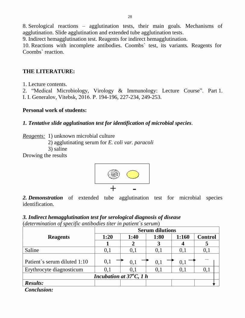
1. Tentative slide agglutination test for identification of microbial species.
Reagents: 1) unknown microbial culture
2) agglutinating serum for E. coli var. paracoli
3) saline
Drowing the results
2. Demonstration of extended tube agglutination test for microbial species
identification.
3. Indirect hemagglutination test for serological diagnosis of disease (determination of specific antibodies titer in patient`s serum)
Reagents
Serum dilutions
1:20 1:40 1:80 1:160 Control
1 2 3 4 5
Saline 0,1 0,1 0,1 0,1 0,1
Patient`s serum diluted 1:10
0,1
0,1
0,1
0,1 __
Erythrocyte diagnosticum 0,1 0,1 0,1 0,1 0,1
Incubation at 37oC, 1 h
Results:
Conclusion:

29
Laboratory class №13
The topic: Active and passive immunoprophylaxis and immunotherapy. Vaccines and
toxoids. Immune antisera and immunoglobulins. Monoclonal antibodies, their
practical use. Serological testing – reactions of toxin neutralization. Reactions with
labeled antibodies and antigens. Immunofluorescence assay. Enzyme-linked
immunosorbent assay. Radioimmunoassay. Immunoblotting (western blotting).
Laboratory testing of immune status. Assessment of cellular and humoral immunity
The main aim and the tasks of the work:
1. To learn the theoretical knowledge of the topic.
2. To know biological products for immunoprophylaxis and immunotherapy.
3. To get acquaintance with hybridoma technology.
4. To know the basic methods for immune status assessment.
5. To get skills of interpretation of blast transformation test.
6. To know the reagents for ELISA test and immunofluorescence assay.
7. To perform serological testing of specific antibodies by enzyme-linked
immunosorbent assay and data analysis by microplate reader.
The questions to the topic:
1. Active immunoprophylaxis. Vaccines and toxoids, their classification and
characteristics. Modern vaccines and toxoids for prophylaxis of infectious diseases.
2. Passive immunotherapy. Immune sera and immunoglobulins, their clinical use.
3. Monoclonal antibodies (mAbs). Stages of hybridoma technology. Medical
applications of mAbs for laboratory diagnosis and treatment of diseases. Humanized
monoclonal antibodies.
4. Serological testing – reactions of toxin neutralization. Reagents and main goals.
5. Serological testing – reactions with labeled antibodies and antigens.
Immunofluorescence assay, its variants and main applications.
6. Enzyme-linked immunosorbent assay (ELISA), its reagents, stages and applications in
immunological diagnosis.
7. Radioimmunoassay.
8. Western blotting analysis.
9. Immune status assessment – general characteristics. Humoral immunity evaluation.
10. Methods for assessment of quantity and functional activity of T- and B cells. Blast
transformation test.
30
THE LITERATURE:
1. Lecture contents.
2. “Medical Microbiology, Virology & Immunology: Lecture Course”. Part 1.
I. I. Generalov, Vitebsk, 2016. Р. 276-281, 218-219, 256-257, 259-260, 261-263, 243-
245.
Personal work of students:
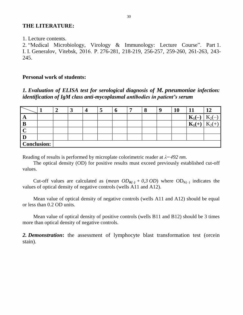
1. Evaluation of ELISA test for serological diagnosis of M. pneumoniae infection:
identification of IgM class anti-mycoplasmal antibodies in patient’s serum
1 2 3 4 5 6 7 8 9 10 11 12
A K1(–) K2(–)
B K1(+) K2(+)
C
D
Conclusion:
Reading of results is performed by microplate colorimetric reader at λ=492 nm.
The optical density (OD) for positive results must exceed previously established cut-off
values.
Cut-off values are calculated as (mean ODK(–) + 0,3 OD) where ODK(–) indicates the
values of optical density of negative controls (wells A11 and A12).
Mean value of optical density of negative controls (wells A11 and A12) should be equal
or less than 0.2 OD units.
Mean value of optical density of positive controls (wells B11 and B12) should be 3 times
more than optical density of negative controls.
2. Demonstration: the assessment of lymphocyte blast transformation test (orcein
stain).

31
Laboratory class №14
The topic: Immunopathology. Immediate and delayed types of hypersensitivity.
Coombs & Gell classification of hypersensitivity reactions. Anaphylactic
hypersensitivity. Allergy and allergic diseases. Cytotoxic hypersensitivity. Immune-
complex-mediated hypersensitivity. Cell-mediated (delayed) hypersensitivity. Skin tests
for laboratory diagnosis of infectious allergy. Stimulatory and blocking
hypersensitivity. Autoimmune diseases. Primary and secondary immunodeficiencies
The main aim and the tasks of the work:
1. To learn the theoretical knowledge of the topic.
2. To know basic mechanisms of immunopathology.
3. To be able to read and interpret the results of enzyme-linked immunosorbent assay
(ELISA) for determination of IgE allergen-specific antibodies.
4. To be able to evaluate the results of allergic skin tests.
The questions to the topic:
1. Immunopathology, classification. Immediate and delayed types of hypersensitivity,
general characteristics and mechanisms.
2. Coombs & Gell classification of hypersensitivity reactions.
3. Anaphylactic hypersensitivity. Allergy and allergic diseases, basic mechanisms and
stages of development.
4. Cytotoxic hypersensitivity, mechanisms. Autoimmune diseases, triggered by this type
of reactions.
5. Immune-complex-mediated hypersensitivity. Autoimmune diseases, stimulated by
this type of reactions.
6. Cell-mediated (delayed) hypersensitivity. Skin tests for laboratory diagnosis of
infectious allergy.
7. Stimulatory and blocking hypersensitivity. Autoimmune diseases, developed by these
reactions.
8. Primary immunodeficiencies. Combined immunodeficiencies. T- and B cell
immunodeficiencies.
9. Phagocyte and complement system immunodeficiencies. Secondary
immunodeficiencies, their mechanisms.
THE LITERATURE:
1. Lecture contents.

32
2. “Medical Microbiology, Virology & Immunology: Lecture Course”. Part 1.
I. I. Generalov, Vitebsk, 2016. Р. 264-275.
Personal work of students:
1. Evaluation of ELISA test for determination of IgE allergen-specific antibodies
1 2 3 4 5 6 7 8 9 10 11 12
A K1(–) K2(–)
B K1(+) K2(+)
C
D
Conclusion:
Reading of results is performed by microplate colorimetric reader at λ=492 nm.
The optical density (OD) for positive results must exceed previously established cut-off
values.
Cut-off values are calculated as (mean ODK(–) + 0,3 OD) where ODK(–) indicates the
values of optical density of negative controls (wells A11 and A12).
Mean value of optical density of negative controls (wells A11 and A12) should be equal
or less than 0.2 OD units.
Mean value of optical density of positive controls (wells B11 and B12) should be 3 times
more than optical density of negative controls.
Laboratory class №15
The topic: Final control study of the section “Immunology and immunity.
Immunological laboratory testing. Immunopathology. Immunoprophylaxis.
Immunotherapy”
The main aim and the tasks of the work:
To learn the principles of immune system function in normalcy and pathology; to
consolidate the knowledge of basic methods of immunological laboratory diagnosis,
immunoprophylaxis and immunotherapy.
33
The questions to the topic:
1. Immunology and immunity. Innate, acquired, artificial, natural immunity. Anti-
infectious immunity, its forms. Types of non-infectious immunity.
2. Immune system, its sub-systems and levels of organization. Central and peripheral
immune organs.
3. CD molecules of immune cells, their significance.
4. Cytokines, the basic features and classification. Groups of cytokines (interferons,
TNF, growth factors, chemokines).
5. Interleukins, their biological role and functions.
6. T cells, their development and differentiation. Structure of TCR, its function.
7. T cell subpopulations, their role.
8. B cells, their development and differentiation.
9. Antigens, their properties and general characteristics. Haptens.
10. Main bacterial and viral antigens. Protective antigens, superantigens, antigenic
mimicry.
11. Non-infectious antigens, their classification. Auto-antigens and alloantigens, general
characteristics. Human blood group antigens.
12. HLA system, general characteristics. HLA molecules of I class, structure and
functions. Biological role of HLA system.
13. HLA molecules of II class, structure and functions. Biological role of HLA system.
14. Immunoglobulins, molecular structure and functions.
15. Classes of immunoglobulins, their characteristics.
16. Structure and biological activity of secretory IgA.
17. Antibodies. Mechanisms of antibody action.
18. Monoclonal antibodies (mAbs). Stages of hybridoma technology. Medical
applications of mAbs for laboratory diagnosis and treatment of diseases.
19. Genetic control of specificity and diversity of antibodies and T cell receptors
(TCRs)
20. Complement system. Classical pathway of activation.
21. Alternative and lectin pathways of complement activation.
22. Mononuclear phagocyte system, general characteristics and functions. Granulocytes,
their role in immune response.
23. Phagocytosis, its stages. Mechanisms of microbial killing by phagocytes.
24. Toll-like receptors and pattern-based microbial recognition. Dendritic and other
antigen-presenting cells (APCs), their functions.
25. NK cells, mechanisms of activation and microbial killing.
26. Innate and acquired immunity, general characteristics. Thymus-independent immune
response.
27. Acquired immunity: T-dependent immune response, stages. Antigen processing and
presentation.
34
28. Acquired immunity: activation and differentiation of T helper cells. Th1 and Th2
control of various types of immune reactions.
29. Activation of effector cells in acquired immune response. Mechanisms of pathogen
elimination.
30. Primary and secondary immune response, their characteristics. Natural inhibition of
immune response.
31. Active immunoprophylaxis. Vaccines and toxoids, their classification and
characteristics. Modern vaccines and toxoids for prophylaxis of infectious diseases.
32. Passive immunotherapy. Immune antisera and immunoglobulins, their clinical use.
Medical applications of monoclonal antibodies for laboratory diagnosis and treatment of
diseases.
33. Immunopathology, classification. Immediate and delayed types of hypersensitivity,
general characteristics and mechanisms.
34. Coombs & Gell classification of hypersensitivity reactions, their general
characteristics.
35. Anaphylactic hypersensitivity. Allergy and allergic diseases, basic mechanisms and
stages of development.
36. Cytotoxic hypersensitivity, mechanisms. Autoimmune diseases, triggered by these
reactions.
37. Immune-complex-mediated hypersensitivity. Autoimmune diseases, stimulated by
this type of reactions.
38. Cell-mediated (delayed) hypersensitivity. Skin tests for laboratory diagnosis of
infectious allergy.
39. Stimulatory and blocking hypersensitivity. Autoimmune diseases, developed by
these reactions.
40. Primary immunodeficiencies. Combined immunodeficiencies. T- and B cell
immunodeficiencies.
41. Phagocyte and complement system immunodeficiencies. Secondary
immunodeficiencies, their mechanisms.
42. Serological tests, their goals, advantages and clinical value. Classification of
serological tests. Molecular mechanisms and conditions for serological reactions.
43. Precipitation tests, reagents and main goals. Variants of precipitation tests (ring
precipitation, immune diffusion, immune electrophoresis). Single radial
immunodiffusion (Mancini’s test) for quantitation of immunoglobulin classes.
44. Serological reactions – agglutination tests, their main goals. Mechanisms of
agglutination. Slide agglutination and extended tube agglutination tests.
45. Indirect hemagglutination test. Reagents for indirect hemagglutination. Coombs`
test, its variants. Reagents for Coombs` reaction.
46. Serological tests – immune lysis reactions. Complement fixation test, principle of
analysis, reagents and medical applications.
47. Serological testing – reactions of toxin neutralization. Reagents and main goals.

35
48. Serological testing – reactions with labeled antibodies and antigens.
Immunofluorescence assay, its variants and main applications.
49. Enzyme-linked immunosorbent assay (ELISA), its reagents, stages and applications
in immunological diagnosis.
50. Radioimmunoassay. Western blotting analysis.
51. Immune status assessment – general characteristics. Humoral immunity evaluation.
52. Laboratory tests for quantitative analysis of immune cells. Immunofluorescence
assay. Flow cytometry and automatic cell sorting.
53. Methods for assessment of functional activity of T- and B cells. Blast transformation
test.
54. Laboratory testing of phagocytosis. Phagocyte number, phagocytic index. NBT test,
its main goal.
THE LITERATURE:
1. Lecture contents.
2. “Medical Microbiology, Virology & Immunology: Lecture Course”. Part 1.
I. I. Generalov, Vitebsk, 2016. Р. 175-275.
Laboratory class №16
The topic: Infection and infectious process. Epidemic process. Microbial
pathogenicity and virulence. Virulence factors. Systemic bacterial infection (sepsis)
The main aim and the tasks of the work:
1. To learn the theoretical knowledge of the topic.
2. To know the principles of experimental infection (animal inoculation, or biological
method).
3. To get skills of experimental infection on mouse model.
4. To get skills of post-mortal examination of infected experimental animals (mice).
The questions to the topic:
1. Infection (or infectious process), its types. Basic conditions for emergence of
infectious process.
2. Characteristics of infectious diseases, their periods.

36
3. Various forms of infection, their characteristics. Classification of infections according
to their origin, localization and spread, clinical manifestations. Reinfection, relapse,
superinfection.
4. Microbial carrier state (microbial carriage).
5. Epidemic process. Conditions for epidemic process emergence and spread.
6. Mechanisms and routes of disease transmission, their characteristics.
7. Anthroponoses, zoonoses and sapronoses, their characteristics. Sporadic, epidemic,
pandemic, endemic, outbreak of infectious diseases.
8. Pathogenicity and virulence, their characteristics. Measurement of virulence.
9. Adhesion and invasion of bacteria. Bacterial adhesins. Invasive enzymes. Injectisome,
its role in bacterial invasiveness and pathogenesis of infections.
10. Bacterial endotoxins, their characteristics and molecular mechanism of action.
11. Bacterial exotoxins, their common properties. Classification of exotoxins.
12. Most active bacterial exotoxins, structure and mechanisms of action.
13. Systemic infectious process. Pathogenesis of sepsis. Laboratory diagnosis of sepsis.
THE LITERATURE:
1. Lecture contents.
2. “Medical Microbiology, Virology & Immunology: Lecture Course”. Part 1.
I. I. Generalov, Vitebsk, 2016. Р. 160-173.
Personal work of students:
1. Experimental infection of mice with K. pneumonia culture.
2. Post-mortem examination of infected dead mice with bacteriological testing: plating
of specimens from mouse inner organs on Petri dish with MPA.
3. Preparing of slides from animal organ specimens, fuchsin stain.
4. Assessment of microbial growth on Petri dishes.
6. Conclusion about the results of experimental infection.

37
Protocol № 1. Septicemia in mice resulted from experimental infection after intra-
peritoneal bacterial inoculation
Day of
examination
Material for
examination
Steps of examination Results
1. Klebsiella
pneumoniae
culture
Intraperitoneal inoculation of mouse with bacterial
culture in dose 1*109 cells per 0.5 ml of saline
–
2.
- Analysis of the results of experimental infection.
Post-mortem examination of infected dead mice with
isolation of bacterial culture: plating of mouse inner
organ samples on Petri dish with MPA.
Preparing of slides from organ specimens, fuchsin
stain, microscopy.
3. Registering of microbial growth on Petri dish.
Conclusion:
Laboratory class №17
The topic: Chemotherapy. Antibiotics. Antibiotic susceptibility testing
The main aim and the tasks of the work:
1. To learn the theoretical knowledge of the topic.
2. To get skills of disc diffusion test for assessment of bacterial susceptibility to
antibiotics.
3. To be able to determine end-point (minimum inhibitory concentration or MIC of
antibiotic) in broth dilution susceptibility testing.
4. To make acquaintance with agar dilution susceptibility test.
Blood
Lung Spleen
Liver Kidney

38
The questions to the topic:
1. Antimicrobial chemotherapy and chemoprophylaxis of infectious diseases.
Therapeutic ratio.
2. Antibiotics. Requirements to antibiotic drugs.
3. Classification of antibiotics according to their origin, their antibacterial effects,
spectrum of action and molecular mechanisms of their antibacterial activity.
4. Antimicrobial action by inhibition of cell wall synthesis: beta-lactam antibiotics
(cephalosporins, penicillins, carbapenems), vancomycin, linezolid, bacitracin.
5. Antimicrobial action by inhibition of cell membrane function: amphotericin B,
polyenes, polymyxins.
6. Antimicrobial action by inhibition of protein synthesis: chloramphenicol, macrolides
and azalides, lincomycins, tetracyclines, aminoglycosides.
7. Antimicrobial action by inhibition of nucleic acid synthesis: quinolones, rifampicin,
sulfonamides, trimethoprim.
8. Side effects of antibiotics.
9. Microbial drug resistance, mechanisms. Non-genetic and genetic (chromosomal and
extrachromosomal) resistance. Prevention of drug resistance.
10. Measurements of antimicrobial activity: disc diffusion test for determination of
bacterial susceptibility to antibiotics.
11. Broth and agar dilution susceptibility tests, their evaluation. E-test.
THE LITERATURE:
1. Lecture contents.
2. “Medical Microbiology, Virology & Immunology: Lecture Course”. Part 1.
I. I. Generalov, Vitebsk, 2016. Р. 147-159.
Personal work of students:
1. Demonstration of agar dilution test for assessment of microbial resistance to
antibiotics.
2. Disc diffusion test for determination of S. aureus resistance to antibiotics
Day of
examination
Material for
examination
Steps of examination Results
1.
Staphylococcus aureus
(1*109 cells/ml)
Plating of material on Petri dish with MPA. Placement
of disks with antibiotics on Petri dish __
Incubation at 37oC for 24 h
2. Assessment of microbial susceptibility testing:
measurement of diameters of growth inhibition zones.

39
Conclusion: Isolate of Staphylococcus aureus is susceptible to…
3. Measurement of microbial resistance to antibiotic with broth dilution test
1 2 3 4 5 6 7
Reagents Final antibiotic concentration, mkg/ml
32 16 8 4 2 1 К
Meat-peptone broth 0.1 0.1 0.1 0.1 0.1 0.1 0.1
Antibiotic (initial
concentration – 64 mkg/ml) 0.1 0.1 0.1 0.1 0.1 0.1
Microbial culture One loop of material is inoculated in all test tubes
Incubation at 37oC for 24 h
Results:
Conclusion:
Laboratory class №18
The topic: Causative agents of suppurative infections. Staphylococci, pseudomonads,
bacteroids and related agents
The main aim and the tasks of the work:
1. To learn the properties, and the role of various staphylococcal species in human
pathology.
2. To get skills of laboratory diagnosis of staphylococcal infections.
3. To know the properties of pseudomonads and bacteroids, their role in human
suppurative infections.
4. To know the principles of laboratory diagnosis of infections, caused by
pseudomonads and bacteroids.
The questions to the topic:
1. Classification, structure and properties of staphylococci.
2. Virulence factors of staphylococci.
3. Pathogenesis and clinical findings in staphylococcal infections.
4. Laboratory diagnosis of staphylococcal infections, specific prophylaxis and treatment.
5. Pseudomonas aeruginosa: classification, structure and properties.

40
6. Pathogenesis and clinical findings in infections caused by Pseudomonas aeruginosa.
Laboratory diagnosis, prophylaxis and treatment.
7. Classification of pathogenic gram-negative non-sporeforming anaerobes. Structure
and properties of bacteroids, prevotellae, porphyromonads.
8. Pathogenesis and clinical findings in bacteroidal infections. Laboratory diagnosis,
prophylaxis and treatment.
9. Other representatives of nonfermenting gram-negative bacteria –
Acinetobacter baumannii and Stenotrophomonas maltophilia.
THE LITERATURE:
1. Lecture contents.
2. “Medical Microbiology, Virology & Immunology: Lecture Course”. Part 2.
I. I. Generalov, Vitebsk, 2016. Р. 8-15, 24-31, 44-50.
Personal work of students:
1. Laboratory investigation of pus.
Day of
examination
Material for
examination
Steps of examination Results
1.
The pus taken
from patient’s
abscess.
Microscopy of Gram-stained slide from pus sample.
Plating of pus on blood agar and yolk-salt agar. __
2.
Assessment of microbial growth on blood agar and
yolk-salt agar.
Inoculation of material from hemolytic lecithinase-
positive colony upon slant agar.
3.
Determination of catalase and coagulase activity of
isolated culture.
Mannitol fermentation testing.
Antibiotic susceptibility testing of isolated culture.
4.
Assessment of mannitol fermentation test.
Evaluation of the results of antibiotic susceptibility
test.
Conclusion:
3. Demonstration: biopreparations for diagnosis, specific prophylaxis and treatment
of staphylococcal infections – staphylococcal toxoid, anti-staphylococcal donor’s
immunoglobulin, type-specific staphylococcal bacteriophages.
41
Учебное издание
Генералов Игорь Иванович
Железняк Наталья Васильевна
Фролова Аэлита Валерьевна
INSTRUCTIONS FOR LABORATORY TRAINING
in General Microbiology & Immunology
for Students of Medical Faculty
(English Medium)
МЕТОДИЧЕСКИЕ РЕКОМЕНДАЦИИ К ЛАБОРАТОРНЫМ ЗАНЯТИЯМ
ПО ОБЩЕЙ МИКРОБИОЛОГИИ И ИММУНОЛОГИИ Методические рекомендации
для студентов лечебного факультета
с английским языком обучения
высших медицинских учебных заведений
Редактор И.И. Генералов
Технический редактор И.А. Борисов
Подписано в печать
Формат бумаги 64х84 1/16 Бумага типографская №2.
Гарнитура ТАЙМС. Усл. печ. листов . Уч.-изд. л
Тираж экз. Заказ № .
Издатель и полиграфическое исполнение:
УО «Витебский государственный медицинский университет» ЛП № 02330/453 от 30.12.2013 г.
Пр. Фрунзе, 27, 210602, г. Витебск