instructions for use · title localization of minodronate in mouse femora through ... in mouse...
TRANSCRIPT
Instructions for use
Title Localization of Minodronate in Mouse Femora Through Isotope Microscopy
Author(s)Hongo, Hiromi; Sasaki, Muneteru; Kobayashi, Sachio; Hasegawa, Tomoka; Yamamoto, Tomomaya; Tsuboi, Kanako;Tsuchiya, Erika; Nagai, Tomoya; Khadiza, Naznin; Abe, Miki; Kudo, Ai; Oda, Kimimitsu; Henrique Luiz de Freitas,Paulo; Li, Minqi; Yurimoto, Hisayoshi; Amizuka, Norio
Citation Journal of Histochemistry & Cytochemistry, 64(10): 601-622
Issue Date 2016-10
Doc URL http://hdl.handle.net/2115/63234
Type article (author version)
File Information JHC64(10).pdf
Hokkaido University Collection of Scholarly and Academic Papers : HUSCAP

Localization of minodronate in bone
1
Localization of minodronate in mouse femora through isotope microscopy
Hiromi Hongo1#, Muneteru Sasaki 1,2#, Sachio Kobayashi3, Tomoka Hasegawa1,
Tomomaya Yamamoto1, Kanako Tsuboi1, Erika Tsuchiya1, Tomoya Nagai1, Naznin
Khadiza1, Miki Abe1, Ai Kudo1, Kimimitsu Oda4, Paulo Henrique Luiz de Freitas5,
Minqi Li6, Hisayoshi Yurimoto3 and Norio Amizuka1
1Department of Developmental Biology of Hard Tissue, Graduate School of Dental Medicine,
Hokkaido University, Sapporo, Japan, 2Department of Applied Prosthodontics, Unit of
Translational Medicine, Graduate School of Biomedical Sciences, Nagasaki University,
Nagasaki, Japan, 3Natural History Sciences, Isotope Imaging Laboratory, Creative Research
Institution, Hokkaido University, Sapporo Japan, 4Division of Biochemistry, Graduate School
of Medical and Dental Sciences, Niigata University, Niigata, Japan, 5Dental School, Federal
University of Sergipe at Lagarto, Aracaju, Brazil, 6Shandong Provincial Key Laboratory of
Oral Biomedicine, The School of Stomatology, Shandong University, Jinan, China
Running title: Localization of minodronate in bone
#These authors contributed equally to this study
Address for correspondence:
Norio Amizuka, DDS, PhD
Department of Developmental Biology of Hard Tissue
Graduate School of Dental Medicine, Hokkaido University
Kita 13 Nishi 7 Kita-ku
Sapporo, 060-8586, Japan
Tel: +81-11-706-4223
Fax: +81-11-706-4226
E-mail: [email protected]
Localization of minodronate in bone
2
Abstract
Minodronate is highlighted for its marked and sustained effects on osteoporotic bones. To
determine the duration of minodronate’s effects, we have assessed the localization of the drug
in mouse bones through isotope microscopy, after labeling it with a stable nitrogen isotope
(15N-minodronate). In addition, minodronate-treated bones were assessed by histochemistry
and TEM. Eight-weeks-old male ICR mice received 15N-minodronate (1mg/kg) intravenously
and were sacrificed after three hours, 24 hrs, one week and one month. Isotope microscopy
showed that 15N-minodronate was present mainly beneath osteoblasts rather than nearby
osteoclasts. At 3 hrs after minodronate administration, histochemistry and TEM showed
osteoclasts with well-developed ruffled borders. However, osteoclasts were roughly attached
to the bone surfaces and did not feature ruffled borders at 24 hrs after minodronate
administration. The numbers of TRAP-positive osteoclasts and ALP-reactive osteoblastic area
were not reduced suddenly, and apoptotic osteoclasts appeared in 1 week and 1 month after
the injections. Von Kossa staining demonstrated that osteoclasts treated with minodronate did
not incorporate mineralized bone matrix. Taken together, minodronate accumulates in bone
underneath osteoblasts rather than under bone-resorbing osteoclasts; therefore, it is likely that
the minodronate-coated bone matrix is resistant to osteoclastic resorption, which results in a
long-lasting and bone-preserving effect.
199 words
Key words: minodronate, osteoclast, osteoblast, isotope microscopy, bisphosphonate
Localization of minodronate in bone
3
Introduction
Bisphosphonates are mainstream drugs for the treatment of osteoporosis. Drugs of this class
have high affinity for crystalline calcium phosphates [1-3], thus binding to calcium
phosphates in the bone matrix upon administration. When osteoclasts degrade bone tissue
bound to bisphosphonates, bone resorption is inhibited as the drug induces osteoclast
apoptosis [4, 5].
Among the several commercially available bisphosphonates, alendronate is the most
commonly used one. In general, nitrogen-containing bisphosphonates (such as pamidronate,
alendronate, ibandronate, zoledronate and risedronate) act by inhibiting farnesyl
pyrophosphate synthase, an enzyme involved in the mevalonate pathway [6, 7], which then
impairs protein prenylation, especially that of the small GTPase of the Ras family [8]. These
small GTPase proteins are crucial for vesicular trafficking and cell survival and seem to be
involved in the cytoskeletal organization of bone-resorbing osteoclasts [9-11]. In short, the
cellular events that lead to osteoclastic inhibition seem to be shared by all nitrogen-containing
bisphosphonates. On the other hand, bisphosphonate-driven osteoclastic inhibition appears
to reduce osteoblastic population and bone formation, which can negatively affect bone
turnover as demonstrated by decreased bone formation and mineral apposition rates in rats
[12, 13], mice [14] and dogs [15].
Minodronate, a third-generation, nitrogen-containing bisphosphonate, is an approved
drug for treatment of osteoporosis in the Japanese market [16-18]. Studies showed that
minodronate suppressed bone resorption in ovariectomized rats and cynomolgus monkeys
[19-21] and affected cortical bone response to mechanical loading in rats [22]. In addition,
other in vivo and in vitro studies reported on the high potency of minodronate compared to
alendronate regarding the inhibition of bone resorption [23], with an intermediate
mineral-binding affinity [24].
Since patients seem to prefer weekly and monthly dosing regimens [25], long-acting
bisphosphonates such as minodronate are gradually becoming the first line of treatment for
many prescribing physicians. Still, a pervasive question among medical professionals and
researchers is how can a single dose of bisphosphonate sustain its effects for over a month?
Localization of minodronate in bone
4
Therefore, we postulated that the localization of minodronate may offer new insights that
might help address this issue. For instance, the use of radioisotope-labeled minodronate may
assist on determining the localization and distribution of minodronate in vivo; however,
animal studies involving radioisotopes are ethically questionable. We have explored the idea
of using minodronate’s own high affinity for binding crystalline calcium phosphates in the
bone matrix to circumvent that limitation by substituting 15N (very rare in nature) for 14N
(naturally abundant) in the minodronate molecule. 15N-minodronate was then injected into
mice to determine the localization of 15N-minodronate through using isotope microscopy.
Isotope microscopy is a mode of secondary ion mass spectrometry and two-dimensional
isotope detection technique [26-28]. Recently, isotope microscope systems have been used to
analyze living matter specimens [29].
To contribute for a better understanding on the mechanism of the sustained minodronate
effects, we have examined the localization of 15N-minodronate in murine bone through
isotope microscopy complemented by histochemistry and transmission electron microscopy
(TEM) observations.
Localization of minodronate in bone
5
Materials and methods
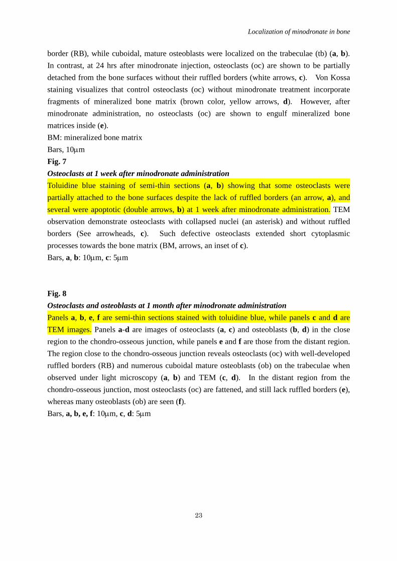
Preparation of 15N- and 13C-labelled minodronate
The 14N and 12C in the minodronate molecule were substituted with the stables isotopes 15N
and 13C to generate
{1-hydroxy-2-[(1-15N)imidazo[1,2-a]pyridin-3-yl](13C2)ethane-1,1-diyl}bis(phosphonic acid)
hydrate (Fig.1), which was kindly provided by Astellas Pharma Inc., Tokyo, Japan. The
isotope microscope of the Creative Research Institution, Hokkaido University [26] was used
for identification and localization of 15N of 15N- and 13C-labelled minodronate. For the sake of
convenience, 15N- and 13C-labelled minodronate will be referred to as 15N-minodronate from
here on.
Animals and tissue preparation
All animal experiments were approved by Hokkaido University and were conducted in
accordance with the standards of humane animal care (No. 15-0030). Mice were anesthetized
with an intraperitoneal injection of chloral hydrate, and then, were injected with 15N-minodronate (1mg/kg) through the external jugular vein. Three hours, 24 hrs, 1 week and
1month after the injection, mice (n=6 for each) were perfused with 4% paraformaldehyde
diluted in 0.1M phosphate buffer (pH 7.4, for histochemistry) or a mixture of 2%
paraformaldehyde + 2.5% glutaraldehyde diluted in 0.067M cacodylate buffer (pH7.4, for
isotope microscopy and transmission electron microscopy or TEM) through the heart’s left
ventricle. Femora were removed and immersed in the respective fixatives for 18 hurs (for
histochemistry) or 3 days (for isotope microscopy and TEM) at 4 oC. Specimens for
histochemistry were decalcified with 10% ethlenedimine tetraacedic disodium salt
(EDTA-2Na) and dehydrated in ascending ethanol solutions prior to paraffin embedding.
Samples for isotope microscopy were not decalcified, but instead immediately dehydrated,
and then embedded in epoxy resin (Epon 812, Taab, Berkshine, UK). Some TEM specimens
Localization of minodronate in bone
6
were decalcified with 5% EDTA-2Na, but others were not decalcified. All TEM specimens
were post-fixed with 1% osmium tetraoxide in 0.1M cacodylate buffer for 4 hrs at 4 oC, and
dehydrated in ascending acetone solutions prior to embedding in epoxy resin. Semi-thin
sections were placed on Si wafers for isotope microscope observation. Ultra-thin sections
were stained with uranyl acetate and lead citrate for TEM observations (Hitachi H-7100
Hitachi Co. Ltd, Tokyo, Japan) at 80 kV.
Isotope microscopy
The sections placed on Si wafers were coated with a 30-nm layer of gold to prevent the
accumulation of positive charges generated by the primary beam of the isotope microscopy.
Hokkaido University’s isotope microscope system (Cameca IMS 1270+SCAPS) was used to
visualize isotope distribution in the bone tissue, a technique known as isotopography [26, 27,
28, 29, 30].
Frequency histogram for osteoblast and osteoclast numbers and 15N/14N intensity ratio
To understand how much 15N-minodronate was deposited beneath osteoblasts (bone forming
surfaces) or osteoclasts (bone resorbing surfaces), we counted the osteoblasts and osteoclasts
seen on bone surfaces labeled with 15N-minodronate. Semi-thin sections of metaphyseal areas
were used to generate data for the histogram. The intensity of 15N (15N-minodronate) lines
seen underneath osteoblasts or osteoclasts was divided by the intensity of 14N labeling in the
same region. A series of 15N/14N ratios is shown on the horizontal axis of the histogram, while
the vertical axis shows the numbers of osteoblasts or osteoclasts located on the bone surfaces
with the corresponding 15N/14N ratio, thus indicating the volume of deposited 15N-minodronate per surface type (bone forming vs. bone resorbing).
Double staining for tissue nonspecific alkaline phosphatase (ALP) and tartrate-resistant
acid phosphatase (TRAP)
Dewaxed paraffin sections were treated for endogenous peroxidase inhibition with 0.3%
hydrogen peroxide in phosphate buffered saline (PBS) for 20 min and for nonspecific staining
blocking with 1% bovine serum albumin in PBS (1% BSA-PBS) for 30 min at room
temperature (RT). Section were incubated with rabbit polyclonal antisera against tissue
Localization of minodronate in bone
7
nonspecific alkaline phosphatase (ALP) [31] for 2 hrs at RT, and then incubated with
horseradish peroxidase-conjugated anti-rabbit IgG for 1 hr at RT. Immunoreactions were
detected with 3.3’-diaminobenzidine tetrahydrochloride (Dojindo Laboratorise, Kumamoto,
Japan). Following immunostaining, tartrate-resistant acid phosphatase (TRAP) was detected
as previously described [32]. In short, slides were rinsed with PBS and incubated in a
mixture of 2.5mg of naphthol AS-BI phosphate (Sigma-Aldrich, St. Louis, MO), 18mg of red
violet LB (Sigma-Aldrich) salt, and 100mML (+) tartaric acid (0.76g) diluted in 30 ml of a
0.1M sodium acetate buffer (pH 5.0) for 15min at 37 oC.
Von Kossa staining
Undecalcified semi-thin section were incubated with an aqueous solution of 5% silver nitrate
(Wako Pure Chemical Industries, Tokyo, Japan) for 60 min at RT under sunlight until taking
on a dark brown color, as previously described [33] .
Statistical analyses for osteoclast number, osteoblastic area and percentage of apoptotic
osteoclasts
Sagittal femora sections from samples of all time points (3 hrs, 24 hrs, 1 week and 1 month,
n=6 for each) were cut as shown in Fig. 2. The region of interest (ROI) for counting
TRAP-positive osteoclasts and measuring the area of ALP-reactive osteoblastic cells, as well
as the determination of percentage of apoptotic osteoclasts was set as a boxed area
neighboring the chondro-osseous junction and the endosteal surfaces of the cortical bone up
to a horizontal line 2 mm distant from the chondro-osseous junction. As reported previously
[33], ALP-positive cells located on bone surface were considered osteoblasts, while
multinucleated (more than two nuclei) TRAP-reactive cells were regarded as osteoclasts.
TRAP-positive osteoclasts with nuclear pyknosis were categorized as apoptotic osteoclasts.
We counted the numbers of TRAP-positive osteoclasts and also measured the ALP-reactive
area in the ROI by using the ImagePro Plus 6.2 software (Media Cybernetics, Silver Spring,
MD). The percentage of apoptotic osteoclasts was obtained after dividing the number of
pycknotic, TRAP-positive osteoclasts by the total osteoclast number.
Bone histomorphometry of BV/TV, Tb.N, Tb.Th and Tb.Sp
Localization of minodronate in bone
8
Bone histomorphometric static parameters were assessed as presented in a recent report from
our group [34]. In brief, a 400μm x 600μm region of interest (ROI) located 150μm below the
growth plate of the femoral metaphysis (n=6 for each) was used for assessing the following
static parameters: bone volume/tissue volume (BV/TV), trabecular number (Tb.N), trabecular
thickness (Tb.Th), trabecular separation (Tb.Sp). Whenever possible, abbreviations and
calculations followed the guidelines of the ASBMR Histomorphometry Nomenclature
Committee [35].
Statistical analysis
One-way ANOVA and the Tukey-Kramer multiple comparisons test were used to assess the
presence of statistical differences among groups. All values are presented as mean ± standard
deviation. The level of significance was set to p < 0.01.
Localization of minodronate in bone
9
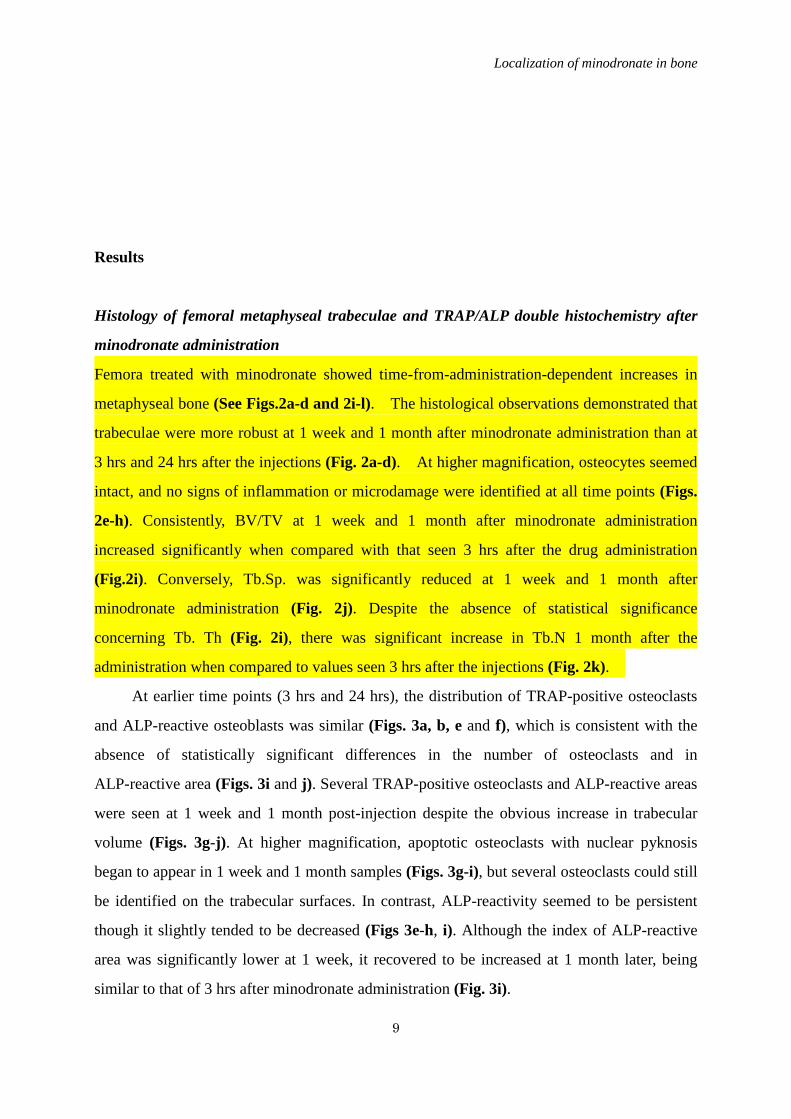
Results
Histology of femoral metaphyseal trabeculae and TRAP/ALP double histochemistry after
minodronate administration
Femora treated with minodronate showed time-from-administration-dependent increases in
metaphyseal bone (See Figs.2a-d and 2i-l). The histological observations demonstrated that
trabeculae were more robust at 1 week and 1 month after minodronate administration than at
3 hrs and 24 hrs after the injections (Fig. 2a-d). At higher magnification, osteocytes seemed
intact, and no signs of inflammation or microdamage were identified at all time points (Figs.
2e-h). Consistently, BV/TV at 1 week and 1 month after minodronate administration
increased significantly when compared with that seen 3 hrs after the drug administration
(Fig.2i). Conversely, Tb.Sp. was significantly reduced at 1 week and 1 month after
minodronate administration (Fig. 2j). Despite the absence of statistical significance
concerning Tb. Th (Fig. 2i), there was significant increase in Tb.N 1 month after the
administration when compared to values seen 3 hrs after the injections (Fig. 2k).
At earlier time points (3 hrs and 24 hrs), the distribution of TRAP-positive osteoclasts
and ALP-reactive osteoblasts was similar (Figs. 3a, b, e and f), which is consistent with the
absence of statistically significant differences in the number of osteoclasts and in
ALP-reactive area (Figs. 3i and j). Several TRAP-positive osteoclasts and ALP-reactive areas
were seen at 1 week and 1 month post-injection despite the obvious increase in trabecular
volume (Figs. 3g-j). At higher magnification, apoptotic osteoclasts with nuclear pyknosis
began to appear in 1 week and 1 month samples (Figs. 3g-i), but several osteoclasts could still
be identified on the trabecular surfaces. In contrast, ALP-reactivity seemed to be persistent
though it slightly tended to be decreased (Figs 3e-h, i). Although the index of ALP-reactive
area was significantly lower at 1 week, it recovered to be increased at 1 month later, being
similar to that of 3 hrs after minodronate administration (Fig. 3i).
Localization of minodronate in bone
10
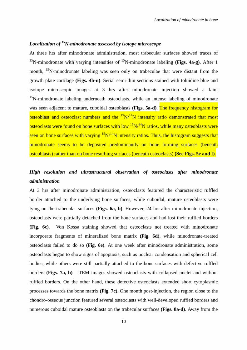
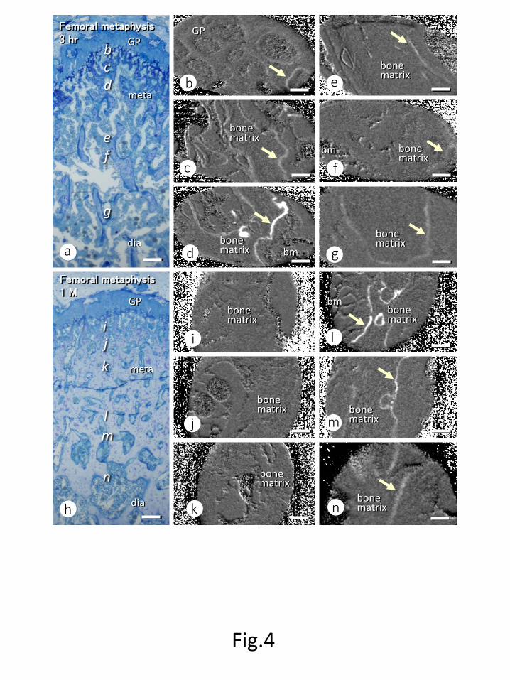
Localization of 15N-minodronate assessed by isotope microscope
At three hrs after minodronate administration, most trabecular surfaces showed traces of 15N-minodronate with varying intensities of 15N-minodronate labeling (Figs. 4a-g). After 1
month, 15N-minodronate labeling was seen only on trabeculae that were distant from the
growth plate cartilage (Figs. 4h-n). Serial semi-thin sections stained with toluidine blue and
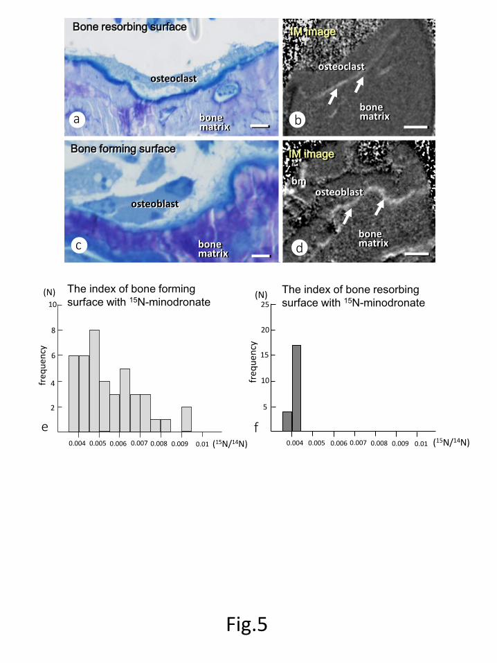
isotope microscopic images at 3 hrs after minodronate injection showed a faint 15N-minodronate labeling underneath osteoclasts, while an intense labeling of minodronate
was seen adjacent to mature, cuboidal osteoblasts (Figs. 5a-d). The frequency histogram for
osteoblast and osteoclast numbers and the 15N/14N intensity ratio demonstrated that most
osteoclasts were found on bone surfaces with low 15N/14N ratios, while many osteoblasts were
seen on bone surfaces with varying 15N/14N intensity ratios. Thus, the histogram suggests that
minodronate seems to be deposited predominantly on bone forming surfaces (beneath
osteoblasts) rather than on bone resorbing surfaces (beneath osteoclasts) (See Figs. 5e and f).
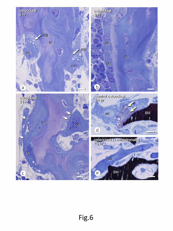
High resolution and ultrastructural observation of osteoclasts after minodronate
administration
At 3 hrs after minodronate administration, osteoclasts featured the characteristic ruffled
border attached to the underlying bone surfaces, while cuboidal, mature osteoblasts were
lying on the trabecular surfaces (Figs. 6a, b). However, 24 hrs after minodronate injection,
osteoclasts were partially detached from the bone surfaces and had lost their ruffled borders
(Fig. 6c). Von Kossa staining showed that osteoclasts not treated with minodronate
incorporate fragments of mineralized bone matrix (Fig. 6d), while minodronate-treated
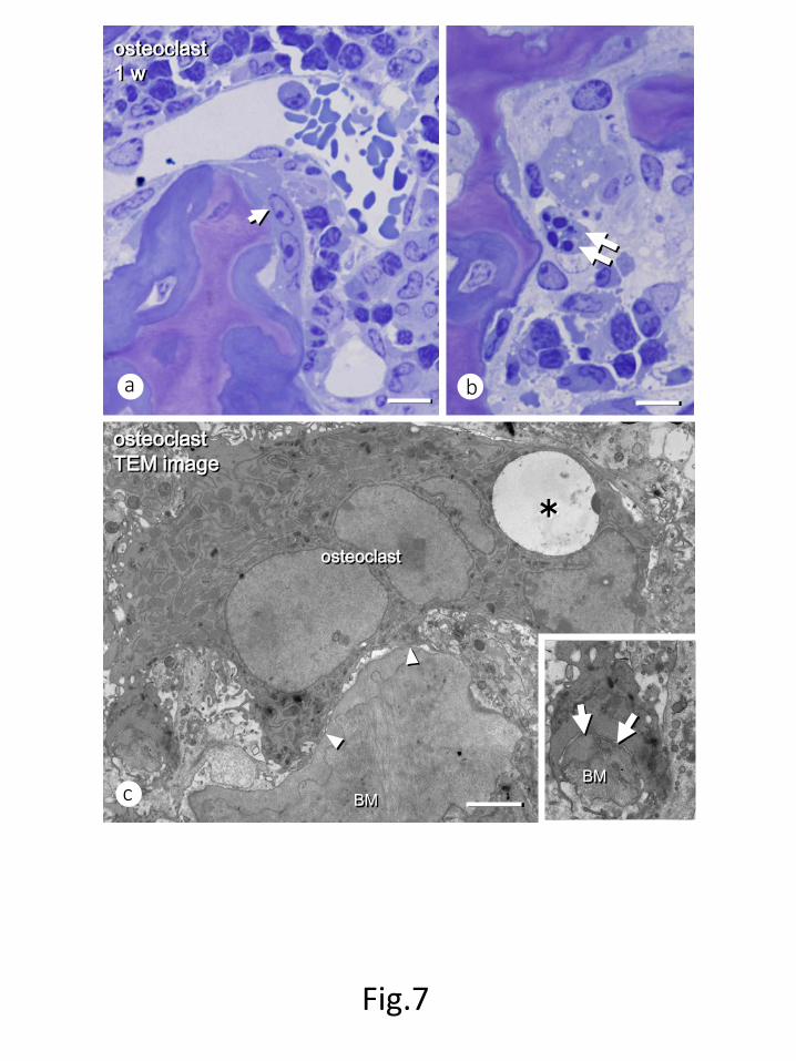
osteoclasts failed to do so (Fig. 6e). At one week after minodronate administration, some
osteoclasts began to show signs of apoptosis, such as nuclear condensation and spherical cell
bodies, while others were still partially attached to the bone surfaces with defective ruffled
borders (Figs. 7a, b). TEM images showed osteoclasts with collapsed nuclei and without
ruffled borders. On the other hand, these defective osteoclasts extended short cytoplasmic
processes towards the bone matrix (Fig. 7c). One month post-injection, the region close to the
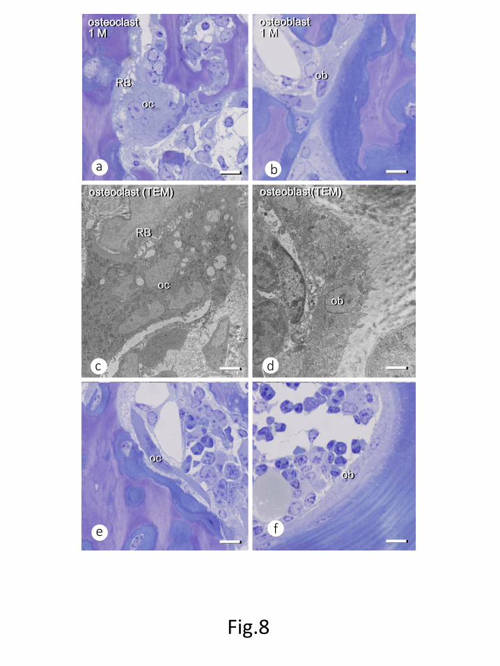
chondro-osseous junction featured several osteoclasts with well-developed ruffled borders and
numerous cuboidal mature osteoblasts on the trabecular surfaces (Figs. 8a-d). Away from the

Localization of minodronate in bone
11
chondro-osseous junction, most osteoclasts lacked ruffled borders, but there were many
mature osteoblasts on the nearby trabeculae (Fig. 8e, f).
Localization of minodronate in bone
12
Discussion
To our knowledge, this is the first report to show the localization of minodronate in vivo in
bone tissue. Isotope microscopy demonstrated that 15N-minodronate could be found
underneath osteoblasts, i.e., on bone formation surfaces, rather than near bone-resorbing
osteoclasts. This finding suggests that the bone-forming trabecular surfaces are coated by
minodronate, and we postulated that osteoclasts might somehow fail to resorb the
minodronate-coated bone matrix. If osteoclasts do not degrade the minodronate-coated bone
matrix, they would not be exposed to minodronate and would not, therefore, enter apoptosis -
at least immediately upon administration of the drug. Our histochemical findings
demonstrated that many TRAP-positive osteoclasts are seen at all time points studied here,
while apoptotic osteoclasts were not clearly identified until the later time points of 1 week and
1 month post-administration. Taken together, a single injection of minodronate seems to
produce a long-lasting effect that avoids osteoclastic bone resorption by coating the bone
surface and rendering it somewhat “resorption-proof”.
However, this particular tissue distribution seems to produce a “useful” side effect,
since maintaining osteoclast presence instead of forcing osteoclastic cells into apoptosis may
guarantee osteoblast activation through cell coupling [36]. As shown with the TRAP/ALP
double staining, not only TRAP-positive cell numbers were stable, but also many
ALP-reactive osteoblasts were persistent even at a later time point (1 month) following
minodronate administration, though it temporally decreased at 1 week. Then again,
bisphosphonates have been reported to reduce bone formation and bone turnover [12-15]. An
autoradiography study showed that alendronate accumulated on resorption surfaces [37],
which suggests that alendronate would be readily incorporated by bone-resorbing osteoclasts
and immediately halt bone resorption. While alendronate seems to target bone-resorbing
osteoclasts directly, minodronate’s distribution and localization on bone formation surfaces
indicates that the latter does not inhibit bone-resorbing osteoclasts immediately, but instead
protect the bone matrix from osteoclastic bone resorption. In addition, the dysfunctional,
non-resorbing osteoclasts may allow nearby osteoblasts somehow to be activated through cell
coupling. Retaining osteoblastic activity may favor combined therapy with an anabolic drug
such as teriparatide [hPTH(1-34)]. We have demonstrated that cell coupling with osteoclasts
Localization of minodronate in bone
13
during preosteoblastic differentiation into mature osteoblasts is necessary for parathyroid
hormone-driven anabolic effect to occur [38], as others have reported elsewhere [39, 40].
Indeed, daily alendronate administration appears to reduce the anabolic effect of PTH (1-84)
on hip bone mineral density (BMD) and cortical volume [41]; on the other hand, combining
teriparatide and long-term zoledronate or denosumab treatment produced positive effects on
lumbar spine and hip BMD [42, 43]. Likewise, the association of risedronate with
teriparatide increased cancellous bone mass in orchidectomized rats [44], as well as lumbar
spine, total hip and femoral neck BMD in men [45]. If minodronate sustains osteoblastic
activities, as suggested by our findings, it could also produce better results when combined
with teriparatide. In fact, one recent study described that a combination of minodronate and
teriparatide resulted in increased bone volume and trabecular number, while reducing
trabecular separation compared with teriparatide alone [20]. To date, unfortunately, reports
focusing on the cumulative effects of once-monthly minodronate and other anabolic drugs are
scarce.
In conclusion, minodronate accumulates in bone underneath osteoblasts rather than
under bone-resorbing osteoclasts; therefore, it is likely that the minodronate-coated bone
matrix is resistant to osteoclastic resorption, which results in a long-lasting and
bone-preserving effect.

Localization of minodronate in bone
14
Conflict of interest
Astellas Pharma Inc., Tokyo, Japan, provided us a 15N- and 13C-labeled minodronate for this
study. This study is partially supported by grant from Astellas Pharma Inc., Tokyo, Japan, and
grants-in-aid for scientific research and bilateral collaboration (Joint Research Projects and
Seminars) of Japan society for the promotion of science, Japan.
Localization of minodronate in bone
15
AUTHOR CONTRIBUTION STATEMENT HH and MS: Main researchers contributed equally to this work. Animal experiments, histochemical analysis, observation under isotope microscopy SK : operating the isotope microscopy TH and ET: analysis using transmission electron microscopy. TY and AK: bone histomorphometry and statistical analysis on histological analyses KT, TN, NK and MA : fixation of animals, extracts femora, decalcification of the specimens, and preparation paraffin sections KO : providing anti-tissue nonspecific alkaline phosphatase, and working on histogram of the minodronate localization PF and ML : discussion and preparation of this manuscript, and have been involved in preliminary experiments on the localization of minodronate HY : Chief of the research center of isotope microscopy, providing experimental protocol on the use of isotope microscopy NA : Chief of this research project, organizing collaborators and providing a whole idea of this experiment. All the above authors have read and approved the manuscript prior to submission
Localization of minodronate in bone
16
References
[1] Russell RGG, Mühlbauer RD, Bisaz S, Williams DA, Fleisch H (1970). The influence of pyrophosphate, condensed phosphates, phosphonates and other phosphate compounds on the dissolution of hydroxyapatitein vitro and on bone resorption induced by parathyroid hormone in tissue culture and in thyroparathyroidectomised rats. Calcif Tissue Res. 6(3): 183-196.
[2] Hansen NM, Felix R, Bisaz S, Fleisch H (1976). Aggregation of hydroxyapatite crystals.
Biochim Biophys Acta. 451(2): 549-559.
[3] Francis MD (1969). The inhibition of calcium hydroxyapatite crystal growth by polyphosphonates and polyphosphates. Calcif Tissue Res. 3(2): 151-162.
[4] Hughes DE, Wright KR, Uy HL, Sasaki A, Yoneda T, Roodman GD, Mundy GR, Boyce BF (1995). Bisphosphonates promote apoptosis in murine osteoclasts in vitro and in vivo. J Bone Miner Res. 10(10):1478-1487.
[5] Selander KS, Mönkkönen J, Karhukorpi EK, Härkönen P, Hannuniemi R, Väänänen HK (1996). Characteristics of clodronate-induced apoptosis in osteoclasts and macrophages. Mol Pharmacol. 50(5): 1127-1138.
[6] Amin D, Cornell SA, Gustafson SK, Needle SJ, Ullrich JW, Bilder GE, Perrone MH (1992). Bisphosphonates used for the treatment of bone disorders inhibit squalene synthase and cholesterol biosynthesis. J Lipid Res. 33(11):1657-1663.
[7] Rogers MJ (2003). New insights into the molecular mechanisms of action of bisphosphonates. Curr Pharm Des. 9(32):2643-2658.
[8] Luckman SP, Hughes DE, Coxon FP, Graham R, Russell G Rogers MJ (1998). Nitrogen-containing bisphosphonates inhibit the mevalonate pathway and prevent post-translational prenylation of GTP-binding proteins, including Ras. J Bone Miner Res. 13(11):581-589.
[9] Palokangs H, Mulari M, Vaananen HK (1997). Endocytic pathway from the basal plasma membrane to the ruffled border membrane in bone resorbing osteoclasts. J Cell Sci.
Localization of minodronate in bone
17
110:1767-1860.
[10] Abu-Amer Y, Teitelbaum SL, Chappel JC, Schlesinger P, Ross FP (1999). Expression and regulation of RAB3 proteins in osteoclasts and their precursors. J Bone Miner Res. 14(11):1855-1860.
[11] Alakangas A, Selander K, Mulari M, Halleen J, Lehenkari P, Mönkkönen J, Salo J, Väänänen K (2002). Alendronate disturbs vesicular trafficking in osteoclasts. Calcif Tissue Int. 70(1):40-47.
[12] Bikle DD, Morey-Holton ER, Doty SB, Currier PA, Tanner SJ, Halloran BP (1994). Alendronate increases skeletal mass of growing rats during unloading by inhibiting resorption of calcified cartilage. J Bone Miner Res. 9(11):1777-1787.
[13] Iwata K, Li J, Follet H, Phipps RJ, Burr DB (2006). Bisphosphonates suppress periosteal osteoblast activity independently of resorption in rat femur and tibia. Bone. 39(5): 1053-1058.
[14] Nakamura M, Udagawa N, Matsuura S, Mogi M, Nakamura H, Horiuchi H, Saito N, Hiraoka BY, Kobayashi Y, Takaoka K, Ozawa H, Miyazawa H, Takahashi N (2003). Osteoprotegerin regulates bone formation through a coupling mechanism with bone resorption. Endocrinology. 144(12): 5441-5449.
[15] Mashiba T, Turner CH, Hirano T, Forwood MR, Johnston CC, Burr DB (2001). Effects of suppressed bone turnover by bisphosphonates on microdamage accumulation and biomechanical properties in clinically relevant skeletal sites in beagles. Bone. 28(5):524-531.
[16] Hagino H, Nishizawa Y, Sone T, Morii H, Taketani Y, Nakamura T, Itabashi A, Mizunuma H, Ohashi Y, Shiraki M, Minamide T, Matsumoto T (2009). A double-blinded head-to-head trial of minodronate and alendronate in women with postmenopausal osteoporosis. Bone. 44(6):1078-1084.
[17] Matsumoto T, Hagino H, Shiraki M, Fukunaga M, Nakano T, Takaoka K, Morii H, Ohashi Y, Nakamura T (2009). Effect of daily oral minodronate on vertebral fractures in Japanese postmenopausal women with established osteoporosis: a randomized placebo-controlled double-blind study. Osteoporos Int. 20(8):1429-1437.
[18] Hagino H, Shiraki M, Fukunaga M, Nakano T, Takaoka K, Ohashi Y, Nakamura T, Matsumoto T (2012). Three years of treatment with minodronate in patients with
Localization of minodronate in bone
18
postmenopausal osteoporosis. J Bone Miner Metab. 30(4): 439-446.
[19] Yamagami Y, Mashiba T, Iwata K, Tanaka M, Nozaki K, Yamamoto T (2013). Effects of minodronic acid an alendronate on bone remodeling, microdamage accumulation, degree of mineralization and bone mechanical properties in overiectomized cynomolgus monkeys. Bone. 54(1):1-7.
[20] Iwamoto J, Seki A, Sato Y (2014). Effect of combined teriparatide and monthly minodronic acid therapy on cancellous bone mass in ovariectomized rats: A bone histomorphometry study. Bone. 64: 88-94.
[21] Tanaka M, Mori H, Kayasuga R, Ochi Y, Yamada H, Kawada N, Kawabata K (2014). Effect of intermittent and daily regimens of minodronic acid on bone metabolism in an ovariectomized rat model of osteoporosis. Calcif Tissue Int. 95(2):166-173.
[22] Nagira K, Hagino H, Kameyama Y, Teshima R (2013). Effects of minodronate on cortical bone response to mechanical loading rats. Bone. 53(1); 277-283.
[23] Dunford JE, Thompson K, Coxon FP, Luckman SP, Hahn FM, Poulter CD, Ebetino FH, Rogers MJ (2001). Structure-activity relationships for inhibition of farnesyl diphosphate synthase in vitro and inhibition of bone resorption in vivo by nitrogen-containing bisphosphonates. J Pharmacol Exp Ther. 296(2):235-242.
[24] Ebetino FH, Hogan AM, Sun S, Tsoumpra MK, Duan X, Triffitt JT, Kwaasi AA, Dunford JE, Barnett BL, Oppermann U, Lundy MW, Boyde A, Kashemirov BA, McKenna CE, Russell RG (2011). The relationship between the chemistry and biological activity of the bisphosphonates. Bone. 49(1); 20–33.
[25] Confavreux CB, Canoui-Poitrine F, Schott AM, Ambrosi V, Tainturier V, Chapurlat RD (2012). Persistence at 1 year of oral antiosteoporotic drugs: a prospective study in a comprehensive health insurance database. Eur J Endocrinol. 166(4):735–741.
[26] Yurimoto, H., Nagashima, K. and Kunihiro, T (2003). High precision isotope micro-imaging of materials. Appl. Surf. Sci. 203-204, 793-797.
[27] Nagashima K, Krot AN, Yurimoto H (2004). Stardust silicates from primitive meteorites. Nature. 428(6986): 921-924.
[28] Kunihiro T, Nagashima K, Yurimoto H (2005). Microscopic oxygen isotopic
homogeneity/heterogeneity in the matrix of the Vigarano CV3 chondrite. Geochim.
Localization of minodronate in bone
19
Cosmochim. Acta 69: 763-773.
[29] Hamasaki T., Matsumoto T, Sakamoto N, Shimahara A, Kato S, Yoshitake A, Utsunomiya A, Yurimoto H, Gabazza E.C, Ohgi T (2013). Synthesis of 18O-labeled RNA for application to kinetic studies and imaging. Nucleic Acids Research, doi:10.1093/nar/gkt1344.
[30] Kuga Y, Sakamoto N, Yurimoto H (2014). Stable isotope cellular imaging reveals that both live and degenerating fungal pelotons transfer carbon and nitrogen to orchid protocorms. New Phytologist 202: 594-605.
[31] Oda K, Amaya Y, Fukushi-Irie M, Kinameri Y, Ohsuye K, Kubota I, Fujimura S, Kobayashi J (1999). A general method for rapid purification of soluble versions of glycosylphosphatidylinositol-anchored proteins expressed in insect cells: An application for human tissue-nonspecific alkaline phosphatase. J. Biochem. 126(4):694-699.
[32] Amizuka N, Li M, Hara K, Kobayashi M, de Freitas PH, Ubaidus S, Oda K, Akiyama Y (2009). Warfarin administration disrupts the assembly of mineralized nodules in the osteoid.J Electron Microsc. 58(2):55-65.
[33] Sasaki M, Hasegawa T, Yamada T, Hongo H, de Freitas PH, Suzuki R, Yamamoto T, Tabata C, Toyosawa S, Yamamoto T, Oda K, Li M, Inoue N, Amizuka N (2013). Altered distribution of bone matrix proteins and defective bone mineralization in klotho-deficient mice. Bone. 57(1):206-219.
[34] Yamamoto T, Hasegawa T, Sasaki M, Hongo H, Tsuboi K, Shimizu T, Ota M, Haraguchi
M, Takahata M, Oda K, Luiz de Freitas PH, Takakura A, Takao-Kawabata R, Isogai Y, Amizuka N (2016). Frequency of teriparatide administration affects the histological pattern of bone formation in young adult male mice. Endocrinology. 157(7):2604-2620.
[35] Parfitt AM, Drezner MK, Glorieux FH, Kanis JA, Malluche H, Meunier PJ, Ott SM, Recker RR (1987). Bone histomorphometry: standardization of nomenclature, symbols, and units. Report of the ASBMR Histomorphometry Nomenclature Committee. J Bone Miner Res 2:595-610.
[36] Frost HM (1969). Tetracycline-based histological analysis of bone remodeling. Calcif Tissue Res. 3:211-237.
[37] Sato M, Grasser W, Endo N, Akins R, Simmons H, Thompson DD, Golub E, Rodan GA (1991). Bisphosphonate action. Alendronate localization in rat bone and effects on
Localization of minodronate in bone
20
osteoclast ultrastructure. J Clin Invest. 88(6):2095-2105.
[38] Luiz de Freitas PH, Li M, Ninomiya T, Nakamura M, Ubaidus S, Oda K, Udagawa N, Maeda T, Takagi R, Amizuka N (2009). Intermittent PTH administration stimulates pre-osteoblastic proliferation without leading to enhanced bone formation in osteoclast-less c-fos(-/-) mice. J Bone Miner Res. 24(9):1586-1597.
[39] Nishino I, Amizuka N, Ozawa H (2001). Histochemical examination of osteoblastic activity in op/op mice with or without injection of recombinant M-CSF. J Bone Miner Metab. 19(5):267-276.
[40] Sakagami N, Amizuka N, Li M, Takeuchi K, Hoshino M, Nakamura M, Nozawa-Inoue K, Udagawa N, Maeda T (2005). Reduced osteoblastic population and defective mineralization in osteopetrotic (op/op) mice. Micron. 36(7-8):688-695.
[41] Black DM, Greenspan SL, Ensrud KE, Palermo L, McGowan JA, Lang TF, Garnero P, Bouxsein ML, Bilezikian JP, Rosen CJ (2003). The effects of parathyroid hormone and alendronate alone or in combination in postmenopausal osteoporosis.N Engl J Med. 349(13):1207-1215.
[42] Cosman F, Eriksen EF, Recknor C, Miller PD, Guañabens N, Kasperk C, Papanastasiou P, Readie A, Rao H, Gasser JA, Bucci-Rechtweg C, Boonen S (2011). Effects of intravenous zoledronic acid plus subcutaneous teriparatide [rhPTH(1-34)] in postmenopausal osteoporosis.J Bone Miner Res. 26(3):503-511.
[43] Tsai JN, Uihlein AV, Lee H, Kumbhani R, Siwila-Sackman E, McKay EA, Burnett-Bowie SA, Neer RM, Leder BZ (2013). Teriparatide and denosumab, alone or combined, in women with postmenopausal osteoporosis: the DATA study randomised trial. Lancet. 82(9886):50-56.
[44] Iwamoto J, Seki A (2015). Effect of Combined Teriparatide and Monthly Risedronate Therapy on Cancellous Bone Mass in Orchidectomized Rats: A Bone Histomorphometry Study. Calcif Tissue Int. 97(1):23-31.
[45] Walker MD, Cusano NE, Sliney J Jr, Romano M, Zhang C, McMahon DJ, Bilezikian JP (2013). Combination therapy with risedronate and teriparatide in male osteoporosis. Endocrine. 44(1):237-246.
Localization of minodronate in bone
21
Figure Legends Fig.1 15N- and 13C-labelled minodronate As shown here, 15N- and 13C-labelled minodronate [{1-hydroxy-2-[(1-15N)imidazo[1,2-a]pyridin-3-yl](13C2)ethane-1,1-diyl}bis(phosphonic acid) hydrate] was generated by substitution of 14N and 12C in with the stables isotopes 15N and 13C to generate. Fig. 2 Femoral metaphyseal trabeculae after minodronate administration Femora treated with single administration of minodronate increased metaphyseal trabeculae as time goes on. Note many trabeculae at 1 week (c) and 1 month (d) when compared with those at 3 hrs (a) and 24 hrs (b) after injection. Panels e-h show highly magnified images of metaphyseal trabeculae. Osteocytes (ocy) seemed intact in all time points (See insets). Panels i-l show the statistical analyses on bone histomorphometry. Note the significant increases in BV/TV at 1 week and 1 month (i) and Tb.N at 1 month (k) when compared with those at 3 hrs after the administration, as well as the absence of statistical significance in Tb. Th (l). Tb.Sp. was significantly reduced at 1 week and 1 month after minodronate administration (j). meta: metaphysis Bars, a-d: 500µm, e-h: 100µm Fig. 3 Double staining of TRAP and ALP on femoral metaphyses after minodronate administration At all time points, there seemed the distribution of TRAP-positive osteoclasts (red color) and ALP-reactive osteoblasts (brown color) (a-d). When observing at a higher magnification, several TRAP-positive osteoclasts were seen throughout the experimental period despite the obvious increase in trabecular volume at 1 week and 1 month post-injection (e-h). Please note that apoptotic osteoclasts can be seen at 1 week and 1 month (insets in g and h). There was no
Localization of minodronate in bone
22
significant differences in the numbers of TRAP-positive osteoclasts in all the experimental periods (i). However, notice apoptotic osteoclasts significantly increased not at the early stage but at the later stage such as 1 week and 1 month of the minodronate injection. Although ALP-reactive area is decreased at 1 week later, it recovered to be increase similar to that at 3 hrs (j). meta: metaphysis, tb: trabecular bone Bars, a-d: 200µm, e-h: 50µm Fig.4 Localization of 15N-minodronate assessed by isotope microscope Panels a-g show the localization of 15N-minodronate at 3 hrs after minodronate administration, while panels h-n demonstrate 15N-minodronate at 1 month. Panels a and h show semi-thin sections stained with toluidine blue, which represent the areas of isotope microscopy observation. At 3 hrs, trabecular surfaces showed white lines indicative of 15N-minodronate in all the areas (yellow colored arrows, b-g). However, after 1 month later, the regions close to the growth plate do not show the labeling of 15N-minodronate, though the distant regions revealed 15N-minodronate (See white lines indicated by yellow arrows in l-n). meta: metaphysis, GP: growth plate, dis: diaphysis, bm: bone marrow Bars, a, h: 100µm, b-h, i-n: 10µm Fig.5 Localization of 15N-minodronate on bone-forming surface and bone-resorbing surface Panels a-d are serial semi-thin sections stained with toluidine blue (a, c) and isotope microscopic images (b, d) at 3 hrs after minodronate injection. Note a faint 15N-minodronate labeling underneath osteoclasts (a, b), while an intense labeling of minodronate adjacent to mature osteoblasts (c, d). Panels e and f are the histogram demonstrating the number of osteoblasts (bone forming surfaces) and osteoclasts (bone resorbing surfaces) located on a regions of varying 15N/14N intensity ratios. bm: bone marrow Bars, a: 10µm, b, d: 20µm, c: 5µm Fig. 6 Osteoclasts and osteoblasts at 3hrs and 24 hrs after minodronate administration Panels a-e show semi-thin sections stained with toluidine blue. At 3 hrs after minodronate administration, both osteoclasts (oc) and osteoblasts (ob) seem intact: osteoclasts have ruffled
Localization of minodronate in bone
23
border (RB), while cuboidal, mature osteoblasts were localized on the trabeculae (tb) (a, b). In contrast, at 24 hrs after minodronate injection, osteoclasts (oc) are shown to be partially detached from the bone surfaces without their ruffled borders (white arrows, c). Von Kossa staining visualizes that control osteoclasts (oc) without minodronate treatment incorporate fragments of mineralized bone matrix (brown color, yellow arrows, d). However, after minodronate administration, no osteoclasts (oc) are shown to engulf mineralized bone matrices inside (e). BM: mineralized bone matrix Bars, 10µm Fig. 7 Osteoclasts at 1 week after minodronate administration Toluidine blue staining of semi-thin sections (a, b) showing that some osteoclasts were partially attached to the bone surfaces despite the lack of ruffled borders (an arrow, a), and several were apoptotic (double arrows, b) at 1 week after minodronate administration. TEM observation demonstrate osteoclasts with collapsed nuclei (an asterisk) and without ruffled borders (See arrowheads, c). Such defective osteoclasts extended short cytoplasmic processes towards the bone matrix (BM, arrows, an inset of c). Bars, a, b: 10µm, c: 5µm Fig. 8 Osteoclasts and osteoblasts at 1 month after minodronate administration Panels a, b, e, f are semi-thin sections stained with toluidine blue, while panels c and d are TEM images. Panels a-d are images of osteoclasts (a, c) and osteoblasts (b, d) in the close region to the chondro-osseous junction, while panels e and f are those from the distant region. The region close to the chondro-osseous junction reveals osteoclasts (oc) with well-developed ruffled borders (RB) and numerous cuboidal mature osteoblasts (ob) on the trabeculae when observed under light microscopy (a, b) and TEM (c, d). In the distant region from the chondro-osseous junction, most osteoclasts (oc) are fattened, and still lack ruffled borders (e), whereas many osteoblasts (ob) are seen (f). Bars, a, b, e, f: 10µm, c, d: 5µm
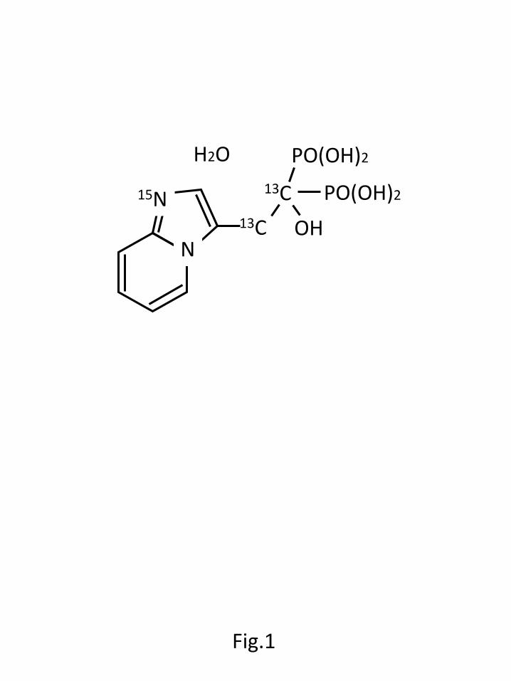
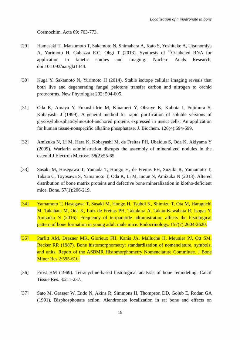
Fig.1
N
15N 13C
13C OH
PO(OH)2
PO(OH)2 H2O
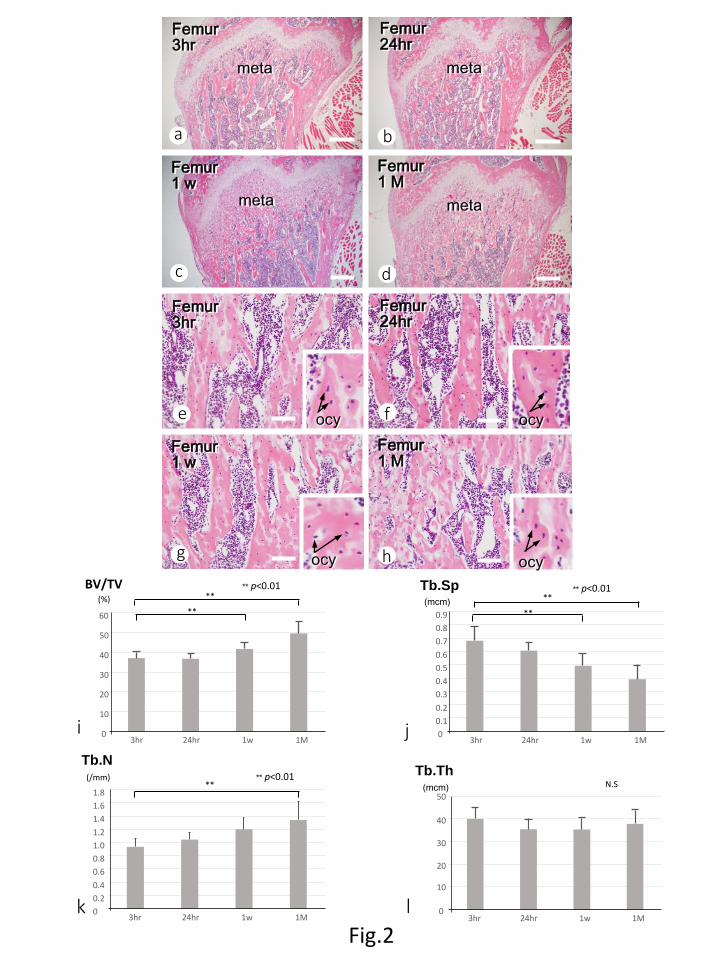
Fig.2
a
g
f e
d c
b
h
Femur 3hr
Femur 24hr
Femur 1 w
Femur 1 M
meta meta
meta meta
Femur 3hr
Femur 24hr
Femur 1 w
Femur 1 M
ocy ocy
ocy ocy
0 0.2 0.4 0.6 0.8 1.0 1.2 1.4 1.6 1.8
3hr 24hr 1w 1M
Tb.N (/mm)
** ** p<0.01
i j
k l
0
10
20
30
40
50
60
3hr 24hr 1w 1M
BV/TV (%) **
**
** p<0.01
0 0.1 0.2 0.3 0.4 0.5 0.6 0.7 0.8 0.9
3hr 24hr 1w 1M
Tb.Sp (mcm) **
**
** p<0.01
0
10
20
30
40
50
3hr 24hr 1w 1M
Tb.Th (mcm) N.S
30
25
20
15
10
5
0
35
(N) *
* *
* * *
* * P<0.05 P<0.01 *
3hr 24hr 1w 1M
Numbers of TRAP(+) osteoclasts Numbers of apoptotic osteoclasts
The numbers of TRAP-positive osteoclasts and apoptotic osteoclasts
a
g
f e
d c
b
h
Fig.3 i j
TRAP/ALP 3hr
TRAP/ALP 24hr
TRAP/ALP 1 w
TRAP/ALP 1 M
TRAP/ALP 3hr
TRAP/ALP 24hr
TRAP/ALP 1 w
TRAP/ALP 1 M
meta meta
meta meta
0
2000
4000
6000
8000
10000
12000
14000
16000
18000
20000
3hr 24hr 1w 1M
* *
ALP reactive area P<0.01 *
(µm2)
tb tb
tb
tb
Femoral metaphysis 3 hr GP
meta
dia
GP
meta
dia
a g
f
e
d
c
b
b
k
j
i
n
m
l
h
Fig.4
Femoral metaphysis 1 M
GP
bm
bone matrix
bone matrix
bone matrix
bone matrix
bone matrix
bone matrix
bone matrix
bone matrix
bone matrix
bm
bone matrix
bm
bone matrix
c d
e f
g
i j k
l m
n
0.004 0.005 0.006 0.007 0.008 0.009 0.01
2
4
6
8
10 (N)
freq
uenc
y
(15N/14N)
Bone resorbing surface
osteoclast
osteoblast
osteoclast
osteoblast
IM image
0.004 0.005 0.006 0.007 0.008 0.009 0.01
5
10
15
20
25 (N)
freq
uenc
y
(15N/14N)
a
e
d c
b
f
Fig.5
Bone forming surface IM image
The index of bone forming surface with 15N-minodronate
The index of bone resorbing surface with 15N-minodronate
bone matrix
bone matrix
bm
bone matrix
bone matrix
control osteoclast 24 hr
osteoclast (+ minodronate) 24 hr
BM
BM
oc
RB
osteoclast 3 hr
osteoclast 24 hr
a
e
d
c
b
Fig.6
osteoblast 3 hr
oc
RB
ob
ob
tb
tb
tb
oc
oc
oc
oc
osteoclast TEM image
osteoclast
BM BM
a
c
b
Fig.7
osteoclast 1 w
*
a
f e
d c
b
Fig.8
osteoclast 1 M
osteoblast 1 M
osteoclast (TEM) osteoblast(TEM)
RB
oc ob
oc ob
oc
ob RB