insulin: trigger and target of renal functions
TRANSCRIPT

fcell-08-00519 July 27, 2020 Time: 18:28 # 1
REVIEWpublished: 29 July 2020
doi: 10.3389/fcell.2020.00519
Edited by:Samar Abd ElHafeez,
Alexandria University, Egypt
Reviewed by:Sonia Michael Najjar,
Ohio University, United StatesKarem Salem,
Fayoum University, Egypt
*Correspondence:Maria Paula Macedo
Specialty section:This article was submitted to
Molecular Medicine,a section of the journal
Frontiers in Cell and DevelopmentalBiology
Received: 18 March 2020Accepted: 02 June 2020Published: 29 July 2020
Citation:Pina AF, Borges DO,
Meneses MJ, Branco P, Birne R,Vilasi A and Macedo MP (2020)
Insulin: Trigger and Target of RenalFunctions.
Front. Cell Dev. Biol. 8:519.doi: 10.3389/fcell.2020.00519
Insulin: Trigger and Target of RenalFunctionsAna F. Pina1,2,3, Diego O. Borges1,4, Maria João Meneses1,2, Patrícia Branco5,6,Rita Birne5,6, Antonio Vilasi7 and Maria Paula Macedo1,3,6*
1 Centro de Estudos de Doenças Crónicas, NOVA Medical School/Faculdade de Ciências Médicas, Universidade Novade Lisboa, Lisbon, Portugal, 2 ProRegeM Ph.D. Programme, NOVA Medical School/Faculdade de Ciências Médicas,Universidade Nova de Lisboa, Lisbon, Portugal, 3 Department of Medical Sciences, University of Aveiro, Aveiro, Portugal,4 Molecular Biosciences Ph.D. Programme, Instituto de Tecnologia Química e Biológica António Xavier, Universidade Novade Lisboa, Oeiras, Portugal, 5 Department of Nephrology, Centro Hospitalar Lisboa Ocidental, Lisbon, Portugal, 6 PortugueseDiabetes Association – Education and Research Center (APDP-ERC), Lisbon, Portugal, 7 Institute of Clinical Physiology– National Research Council, Reggio Calabria Unit, Reggio Calabria, Italy
Kidney function in metabolism is often underestimated. Although the word “clearance”is associated to “degradation”, at nephron level, proper balance between what is trulydegraded and what is redirected to de novo utilization is crucial for the maintenance ofelectrolytic and acid–basic balance and energy conservation. Insulin is probably one ofthe best examples of how diverse and heterogeneous kidney response can be. Kidneyhas a primary role in the degradation of insulin released in the bloodstream, but it is alsoincredibly susceptible to insulin action throughout the nephron. Fluctuations in insulinlevels during fast and fed state add another layer of complexity in the understanding ofkidney fine-tuning. This review aims at revisiting renal insulin actions and clearance andto address the association of kidney dysmetabolism with hyperinsulinemia and insulinresistance, both highly prevalent phenomena in modern society.
Keywords: diabetic nephropathy, insulin, insulin resistance, insulin clearance, albuminuria
INTRODUCTION
Insulin is a vital hormone with several functions among which its central role in energy andglucose homeostasis is fundamental (Plum, 2006). Beyond the traditional crosstalk between themost recognized insulin-responsive organs (liver, adipose tissue, and skeletal muscle), kidneyinsulin action and resistance have recently been suggested as critical components of metabolic and
Abbreviations: ADP, adenosine diphosphate; ATP, adenosine triphosphate; CEACAM-1, carcinoembryonic antigen-relatedcell adhesion molecule 1; cGMP, cyclic guanosine monophosphate; CKD, chronic kidney disease; CV, cardiovascular; DKD,diabetic kidney disease; DNL, de novo lipogenesis; eGFR, estimated glomerular filtration rate; ENaC, epithelial sodiumchannel; ER, endoplasmic reticulum; FFA, free fatty acid; FoxO1, forkhead box O1; GBM, glomerular basement membrane;GIT, glutathione-insulin transhydrogenase; GLUT1, glucose transporter 1; GLUT2, glucose transporter 2; GLUT4, glucosetransporter 4; GRB2, growth factor receptor-bound protein 2; GSK3, glycogen synthase kinase 3; GSV, GLUT storage vesicle;IDE, insulin-degrading enzyme; INSR, insulin receptor; IR, insulin resistance; IRS1, insulin receptor substrate 1; IRS2, insulinreceptor substrate 2; MAPK, mitogen activated protein kinase; mTOR, mammalian target of rapamycin; NAFLD, non-alcoholic fatty liver disease; NBCe1, sodium bicarbonate cotransporter 1; NHE-3, sodium/hydrogen exchanger 3; NKCC2,sodium-potassium chloride cotransporter-2; PGC1α, peroxisome proliferator-activated receptor-γ coactivator 1α; PI3K,phosphatidylinositol 3-kinase; PIP2, phosphatidylinositol 4,5 biphosphate; PIP3, phosphatidylinositol 3,4,5 triphosphate;PKB, protein kinase B; PKG, cGMP-dependent protein kinase G; PKGI, cGMP-dependent protein kinase G isoform I; PPARγ,peroxisome proliferator-activated receptor γ; PT, proximal tubule; PTEN, phosphatase and tensin homolog; PTP1B, proteintyrosine-phosphatase 1B; SGK1, serum/glucocorticoid regulated kinase 1; SGLT, sodium-glucose transport proteins; SGLT2,sodium-glucose co-transporter 2; SGLT2i, sodium-glucose co-transporter-2 inhibitors; SHIP2, SH2-domain-containinginositol phosphatase 2; SREBP-1c, sterol regulatory element binding protein-1c; T1DM, type 1 diabetes mellitus; T2DM,type 2 diabetes mellitus; TZDs, thiazolidinediones; Vamp2, vesicle-associated membrane protein 2.
Frontiers in Cell and Developmental Biology | www.frontiersin.org 1 July 2020 | Volume 8 | Article 519

fcell-08-00519 July 27, 2020 Time: 18:28 # 2
Pina et al. Insulin: Trigger and Target of Renal Functions
dysmetabolic states (Artunc et al., 2016). The important roleof insulin in the kidney is further corroborated by theincreased prevalence of chronic kidney disease in subjectswith type 2 diabetes mellitus (T2DM) and non-alcoholicfatty liver disease (NAFLD; Musso et al., 2014; Kim et al.,2018; Mok et al., 2019; Kiapidou et al., 2020). Furthermore,the kidney can adjust the levels of essential players ofnutrient homeostasis such as glucose and insulin (Krebs, 1963;Chamberlain and Stimmler, 1967).
First reports of insulin actions in the kidney were originatedin the 1950s (Farber et al., 1951). Since then, the knowledgeacquired has dramatically increased, nonetheless much remainsunveiled. It is now known that insulin impacts on tubularglucose reabsorption (Nakamura et al., 2015). In fact, awhole therapeutic class based on this mechanism was recentlydeveloped. Additionally, hyperinsulinemia driven by pancreatichypersecretion and/or impairments in hepatic insulin clearancecould explain changes in glomerular filtration rates and increasedrenal gluconeogenesis, which in turn causes fatty liver depositionand, as a direct consequence, may lead to dysglycemia (Brilet al., 2014; Naderpoor et al., 2017; Jung et al., 2018;Bergman et al., 2019).
Kidney is, in fact, the most critical organ responsible forinsulin clearance after the liver (Elgee and Williams, 1954;Narahara et al., 1958); still, its relevance in the maintenanceof proper insulinemia and insulin sensitivity is underestimated.The control of insulin clearance/action exerted by the kidney iscomplex. Furthermore, the diversity of insulin actions is onlypossible thanks to the differentiation of specific kidney segmentsin which insulin regulates different pathways. In this review, wewill briefly describe insulin production and its main actions inthe liver, adipose tissue, and skeletal muscle. Moreover, we willfocus on kidney and on renal insulin actions as well as insulinclearance/degradation. Overall, this work aims at highlightingthe critical role of kidney-insulin interplay in the developmentof dysmetabolism.
OVERVIEW OF INSULIN BIOSYNTHESISAND NON-RENAL ACTION ANDMETABOLISM
The insulin gene (INS) is represented by only one copy in thehuman genome, and its transcription is mainly regulated by thesame enhancers of other glucose-related genes (Andrali et al.,2008; Gao et al., 2014). In pancreas β-cell, INS is first transcribedas preproinsulin and, after cleavage and folding in the roughendoplasmic reticulum (ER), proinsulin is formed (Hutton,1994). The conversion into insulin occurs after the removal ofthe c-peptide in the Golgi apparatus, with a final maturationachieved by c-terminal amino acid elimination (Steiner, 2004).Mature insulin is composed of A and B chains linked by twodisulfide bonds, with a third bond within A chain (Mayer et al.,2007). For storage, insulin is complexed in hexamers with zinc,and an actin network is involved in the organization of matureinsulin granules for first and second insulin secretion phases(Kalwat and Thurmond, 2013).
Insulin secretion is triggered by cytoplasmic increments inATP levels, derived from glucose internalization and subsequentmetabolism (Prentki and Matschinsky, 1987). This increasein ATP shuts down membrane ATP-sensitive K+ channels,depolarizing plasma membrane, and activating voltage-gatedcalcium channels (Detimary et al., 1998). Indeed, the resultantCa2+ influx seems to be necessary for the fusion of insulingranules with the plasma membrane, ending up in its secretion(Hou et al., 2009). The first wave of insulin secretion is composedof primed granules that are in close proximity with plasmamembrane (Olofsson et al., 2002). However, most of thesegranules remain more distant from the plasma membrane andare further boosted with newly synthesized insulin that willculminate in 75–95% of total insulin secretion in the secondphase (Kou et al., 2014).
With the insulin pools ready to be released, insulin secretiondoes not occur in a constant manner, but in waves of 4–5 min interval (Song et al., 2000). The pulsatile secretion profileis considered primordial for proper insulin responsiveness, asconstant release would result in down-regulation of insulinreceptor (INSR) expression (Matveyenko et al., 2012). Afterbinding and activation of its receptor, insulin, as an anabolichormone, promotes increased biosynthesis of macromoleculesfurther needed to maintain energetic balance during nutrientprivation (Bedinger and Adams, 2015).
At cellular level, insulin mostly relies on the specificbind to its transmembrane receptor to perform its actions(Figure 1). Insulin receptor is a tyrosine kinase receptorwith two extracellular α subunits and two transmembrane β
subunits (Ye et al., 2017). Insulin binding to INSR producesan autophosphorylation that leads to the internalization of thecomplex and activation of downstream effectors. INSR substrate1 (IRS1) and 2 (IRS2) are the main targets that intracellularlycoordinate insulin action (Thirone et al., 2006; Kubota et al.,2016). In the liver, the activity of these proteins is differentlycontrolled by insulin. Although both IRS1 and IRS2 are degradedafter insulin activation, only IRS2 is transcriptionally repressed byinsulin activation (Hirashima et al., 2003). Further transductionof insulin signaling occurs via phosphatidylinositol 3-kinase(PI3K)/AKT phosphorylation. AKT or Protein kinase B (PKB)are considered the main effectors of metabolic insulin actions(Taniguchi et al., 2006; Molinaro et al., 2019). On the other hand,AKT1 and 3 are the leading players of mitogenic effects of insulinby RAS/mitogen activated protein kinase (MAPK) cascade andcrosstalk with growth hormones (Xu et al., 2006; Xu and Messina,2009; Qiu et al., 2017).
Given the scope of this review, we will summarize insulinactions on non-renal target organs, but detailed information isavailable in recent reviews (Gancheva et al., 2018; Tokarz et al.,2018). The first organ to be reached by insulin is the liver, where50–70% is removed at first pass (Duckworth et al., 1998). Inhepatocytes, insulin signaling recruits forkhead box transcriptionfactor 1 (FoxO1) to the cytoplasm, inhibiting the transcription ofgluconeogenic enzymes (Ling et al., 2018). In such an abundanceof glucose, insulin also stimulates the synthesis of glycogen byphosphorylation of glycogen synthase kinase 3 (GSK3), whichleads to dephosphorylation and activation of glycogen synthase
Frontiers in Cell and Developmental Biology | www.frontiersin.org 2 July 2020 | Volume 8 | Article 519

fcell-08-00519 July 27, 2020 Time: 18:28 # 3
Pina et al. Insulin: Trigger and Target of Renal Functions
FIGURE 1 | Canonical insulin signaling cascade. Insulin (gray circle) binds to its transmembrane kinase receptor at cell membrane and triggers insulin signalingcascade. Insulin receptor present two isoforms with distinct affinity for insulin already associated to differential insulin internalization (INSR A and INSR B; Calzi et al.,1997). Binding of insulin auto-phosphorylates INSR at Tyr-960. Further recruitment and phosphorylation of insulin receptor substrate (IRS) 1 and 2 will mostly result insubsequent activation of phosphoinositide-dependent kinase 1 (PDK1) through phosphoinositide-3 kinase (PI3K; Yamada et al., 2002). PDK1 is responsible forpropagation of insulin signal to one of the most important downstream effectors, Akt. Importantly, Akt has two distinct phosphorylation sites, Thr308 activated byPDK1 (Alessi et al., 1997) and Ser473 phosphorylated by mammalian target of rapamycin complex (mTORC) 2 protein (Bayascas and Alessi, 2005). Finally, fullyactivated Akt can interact with different proteins, eliciting different effects as stimulation of glucose uptake and glycogen synthesis by AS160 and glycogen synthasekinase 3 (GSK3), respectively (Ng et al., 2008). On the other hand, INRS activation also promote growth factor receptor-bound protein 2 (GRB2) interaction with Shcproteins and activation of mitogen activated protein kinases (MAPK; Skolnik et al., 1993; Xu et al., 2006). This as part of the insulin-mediated proliferative stimuli.PIP2, phosphatidylinositol 4,5 biphosphate; PIP3, phosphatidylinositol 3,4,5 triphosphate.
(McManus et al., 2005). After the replenishment of liver glycogenstores in the fed state, glucose starts to be degraded to pyruvate,which is the precursor of lipid biosynthesis via de novo lipogenesis(DNL; Sanders and Griffin, 2016). Herein, insulin signaling isalso crucial for upregulation of sterol regulatory element bindingprotein 1c (SREPB1c), a transcription factor associated to furthertranscription of DNL (Oh et al., 2003; Roder et al., 2007;Krycer et al., 2010).
Only a fraction of the insulin that reaches the liver willget to the periphery, to perform its action at skeletal muscle,adipose tissue, and other insulin-sensitive organs. Skeletal muscleis the greater insulin-dependent “kidnapper” of glucose, it isresponsible for ∼60 to 80% of whole-body glucose disposal(DeFronzo et al., 1976; Fernandes et al., 2011). Insulin signals tomyocytes through classical signaling, promoting glucose uptakeby the dynamic translocation of glucose transporter 4 (GLUT4;Zorzano et al., 1996). Glucose is here mainly used for glycolysisor stored as glycogen as already described for the liver.
During the fed state, a clear inhibitory effect in lipolysis andconsequent blunt of free fatty acid (FFA) release is mediatedby insulin acting on adipocytes (Dimitriadis et al., 2011). Theresultant decrease in FFA availability reduces its hepatic uptakeand fatty acid oxidation, changing the disposal of substrates thatare mostly used for gluconeogenesis (Galgani et al., 2008). Notexclusively for liver, the effect of insulin in reducing adipose tissueFFA release is important for other organs that, at fed state, changeenergy source to glucose and recover glycogen stores contributingto normoglycemia (Petersen and Shulman, 2018). In this context,glucose uptake is also stimulated by insulin in the fed state atadipocytes. Furthermore, translocation of glucose transporters tothe membrane of adipocytes was very well characterized in 3T3-L1 adipocytes by activation of the canonical PI3K/AKT2 pathway(Ng et al., 2008).
As mentioned above, all these organs expressing INSRare responsible for insulin clearance and local degradation.However, a significant part of insulin degradation is played in
Frontiers in Cell and Developmental Biology | www.frontiersin.org 3 July 2020 | Volume 8 | Article 519

fcell-08-00519 July 27, 2020 Time: 18:28 # 4
Pina et al. Insulin: Trigger and Target of Renal Functions
the liver (50–70%) by its specialized machinery of internalizationand degradation (Najjar and Perdomo, 2019). In the caseof internalization, carcinoembryonic antigen-related celladhesion molecule 1 (CEACAM-1) phosphorylation by insulinpromotes rapidly receptor-mediated uptake (Poy et al., 2002).Carcinoembryonic antigen-related cell adhesion molecule 1expression occurs preferentially in liver, and restoration of itsfunction only at liver of null CEACAM-1 mice reverse thehyperinsulinemia and fatty liver deposition observed in globalknockout CEACAM-1 model (Russo et al., 2017). Regardingdegradation, insulin-degrading enzyme (IDE) is considered themost critical protease to cleave insulin intracellularly and ishighly expressed in the liver (Kuo et al., 1993).
The amount of insulin that remains in circulation after liverfirst pass (30–50%) will be mostly degraded by the kidney(Elgee and Williams, 1954; Narahara et al., 1958). However,the kidney is not just an endpoint for insulin degradation, asinsulin also acts throughout the entire nephron. Whereas hepaticinsulin clearance is regaining attention as the very first alterationleading to hyperinsulinemia, compensatory mechanisms thatcould emerge from the kidney are not well known. Impairmentsin hepatic insulin clearance increase peripheral insulinemia, andvery little is described regarding the exact effects of the increasedamount of insulin that will further reach the kidney.
KIDNEY AND INSULIN ACTIONS
Kidney works together with the liver for maintenance of optimalinsulin levels. In humans, kidney removes 6–8 U of insulinper day by two major routes (Figure 2), that apparentlyhave distinct preferential physiological goals: post-glomerularsecretion (insulin signaling) and glomerular filtration (nutrientconservation and homeostasis) (Ritz et al., 2013). In the kidney,insulin acts at multiple sites along the nephron, from theglomerulus (Coward et al., 2005; Welsh et al., 2010; Mima et al.,2011; Lay and Coward, 2014) to the renal tubule (Tiwari et al.,2008, 2013; Li et al., 2013), to modulate different functionssuch as glomerular filtration, gluconeogenesis, renal sodiumhandling, among others.
Many alterations in insulin associated mechanisms in thekidney are driven by insulin resistance (IR), leading to diabeticnephropathy if not reestablished (Svensson and Eriksson, 2006).In the Asiatic population, it was described that the prevalenceof diabetic nephropathy with impaired estimated glomerularfiltration rate (eGFR) (with or without albuminuria) was 31.6%,and the prevalence of albuminuria (with or without impairedeGFR) was 16.9 and 22.0%, respectively (Mok et al., 2019).Patients may already show diabetic kidney disease (DKD) atthe time of T2DM diagnosis. Nevertheless, 10 years after thediagnosis of T2DM, low-level albuminuria is present in 24.9%of the patients and 5.3% already present macroalbuminuria(Adler et al., 2003).
Glomerular Insulin ActionsPodocytes are the primary constituent cell of the glomerulus, withtheir long finger-like projections to the glomerular capillaries
at the glomerular basement membrane (GBM). These cellshave intercellular junctions that form filtration barriers to helpmaintaining normal renal function. When damaged, podocyteslose their arrangement resulting in a reduction in barrierfunction. Indeed, one of the DKD features is the podocyte losswith consequent albuminuria (Pagtalunan et al., 1997; Wolfet al., 2005). Lately, it has been unveiled the role of insulin inpodocytes (Figure 3); in other words, podocyte IR gives rise tothe albuminuric feature.
Podocytes reveal to express proteins of the insulin signalingcanonical pathway, namely the INSR, and both IRS1 and IRS2(Coward et al., 2005; Santamaria et al., 2015), with IRS2 beingthe most prevalent one. Coward et al. (2005) depicted the effectof insulin in human podocytes, which results in glucose uptakenot only through GLUT4 but also through glucose transporter1 (GLUT1; Figure 3). As insulin promotes the translocationof GLUT4 to the membrane through the activation of PI3K-AKT2-PkB pathway, there is a remodeling of the cortical actinof the cytoskeleton with subsequent contraction (Welsh et al.,2010). In compliance, podocytes-specific deletion of INSR inmice revealed DKD features based on substantial albuminuriaand histological features as podocyte foot structure loss andglomerulosclerosis (Welsh et al., 2010). AKT seems to play acentral role as its phosphorylation appears to be severely affectedin several models of both type 1 diabetes mellitus (T1DM;streptozotocin-induced T1DM) and T2DM (db/db mice, Zuckerrats) (Tejada et al., 2008; Mima et al., 2011). Moreover, AKTisoform 2 deletion results in serious glomerular lesions in mice.This can lead to rapid disease progression, also associated withtubular dilatation and microalbuminuria (Canaud et al., 2013).Other relevant players that might contribute to podocyte IRis SH2-domain-containing inositol phosphatase 2 (SHIP2), adown regulator of the PI3K signaling pathway shown to beupregulated in the Zucker rats. Moreover, protein tyrosine-phosphatase 1B (PTP1B), a negative regulator of the INSRactivity, or phosphatase and tensin homolog when increased,appears to also compromise the insulin signaling pathway (Mimaet al., 2011; Garner et al., 2018).
Podocytes also present an insulin-dependent alternativepathway, the cyclic guanosine monophosphate(cGMP)-dependent protein kinase G (PKG), from which the PKG isoformI-alpha levels are increased in glomeruli of the hyperinsulinemicZucker rats (Piwkowska et al., 2013). These high insulin levelsincrease glomerular barrier albumin permeability througha PKGI-reliant mechanism via the NAD(P)H-dependentgeneration of superoxide anion.
An important player in podocytes physiology is the proteinnephrin, a podocyte-specific protein, which is responsible forthe maintenance of the integrity of the filtration barrier.In fact, nephrin mutations are involved in severe nephroticsyndrome (Lenkkeri et al., 1999). Nephrin appears to play a mostoutstanding role in the trafficking of GLUT4 and GLUT1 byinteracting with Vamp2 as well as by interacting with insulin-stimulated actin remodeling (Coward et al., 2007; Lay et al., 2017).Of interest, nephrin can also induce phosphorylation of p70S6Kin a PI3K-dependent manner independently of INSR/AKT2activation (Villarreal et al., 2016; Figure 3).
Frontiers in Cell and Developmental Biology | www.frontiersin.org 4 July 2020 | Volume 8 | Article 519

fcell-08-00519 July 27, 2020 Time: 18:28 # 5
Pina et al. Insulin: Trigger and Target of Renal Functions
FIGURE 2 | Continued
FIGURE 2 | Schematic representation of renal insulin handling. Insulin isdepicted in gray circles at different portions of the nephron. As insulin is asmall molecule, it will be fully filtered by the glomerular system until it reachesthe proximal tubule. At proximal tubule cells, almost all the filtered insulin willbe absorbed at the luminal membrane. In physiological conditions, only asmall percentage will be excreted in urine. Beyond glomerular filtration, insulinalso raise from the perivenous capillaries. In a closer look to the proximaltubule cells is represented both mechanisms of insulin clearance. Theincreased levels of insulin receptor (INSR) and consequent increased uptakeof insulin at the basolateral membrane is also depicted.
The development of glomerular IR is triggered by severalfactors: high glucose and/or insulin levels, increased FFAslevels, or an inflammatory milieu. Moreover, insulin signalingappears to be relevant for the adaptative ER stress responsein DKD; this is the case for mice with podocyte-specificheterozygous INSR deletion (Madhusudhan et al., 2015). Insupport of this view, stable overexpression of INSR or knock-down of PTP1B was protective against ER stress (Garner et al.,2018). Podocyte mitochondria play an essential role in cellularmetabolism. When dysfunctional as a result of reactive oxygenspecies production, mitochondria triggers apoptosis, which canalso be observed along with a compromised IR state (Susztaket al., 2006). Certainly, the preservation or reestablishment ofpodocyte integrity is essential in the prevention of the onset anddevelopment of DKD.
INSULIN TUBULAR ACTIONS
In the kidney tubule, insulin has several roles: metabolism,electrolyte and acid-base regulation and absorption of filteredsubstances. However, the exact mechanisms by which insulinperforms these distinct roles is not fully understood. Nonetheless,it seems that, at least some of them, are mediated by INSR, andcan be explained by the recruitment of specific IRS, as recentlyshown by Nakamura et al. (2020) specifically for gluconeogenesisand sodium reabsorption regulation. Still, there are overlappingmediators in downstream pathways. In the following paragraphswe will summarize the most relevant and well-known insulinactions in the tubular segment.
Insulin receptor is present throughout the entire nephron(Butlen et al., 1988; Stechi et al., 1994), however, insulin bindingcapacity of luminal and basolateral membrane tubule cells isdifferent. There is evidence showing same affinity of INSR inboth membrane sides of the cell, nonetheless its abundance isasymmetrical (Hammerman, 1985). In fact, the binding capacityof the contraluminal compared to luminal membrane seems tobe several times greater due to higher expression of INSR (Taloret al., 1982). Figure 4 summarizes insulin signaling in proximaltubule (PT), regarding its actions in both gluconeogenesis andsodium reabsorption. Additionally, insulin actions through INSRare thought to be different in the proximal and distal nephronregions. Tiwari et al. (2013) demonstrated, in a mouse modelwith deletion of INSR in the kidney tubule cells, that dependingon the segment targeted with INSR deletion, there were differentphenotypes further described. In case of decreased INSR at PT,
Frontiers in Cell and Developmental Biology | www.frontiersin.org 5 July 2020 | Volume 8 | Article 519

fcell-08-00519 July 27, 2020 Time: 18:28 # 6
Pina et al. Insulin: Trigger and Target of Renal Functions
FIGURE 3 | Podocyte insulin signaling. Podocytes are the first cells to interact with insulin at the nephron and express several proteins of the canonical insulinsignaling pathway. However, here the podocyte-specific protein nephrin is known to have a role in the trafficking of glucose transporters (GLUT1 or GLUT4) topodocyte membrane and consequently promote glucose uptake. The trafficking seems to involve Vamp2 and actin remodeling. On the other branch of insulinsignaling, an effect in the large-conductance Ca2+-activated K+ channels (BKCa) is also important for maintenance of podocyte integrity and proper glomerularfiltration. GRB2, growth factor receptor-bound protein 2; GSV, GLUT storage vesicle; VAMP2, Vesicle-associated membrane protein 2.
animals had a mild diabetic phenotype, without increased IRwhen compared to control. These animals shown to have anhigher activity of gluconeogenesis enzymes (Tiwari et al., 2013).On the other hand, in animals with the deletion of INSR targetedto distal parts of the tubule, elevated blood pressure and impairedsodium excretion was observed (Tiwari et al., 2008).
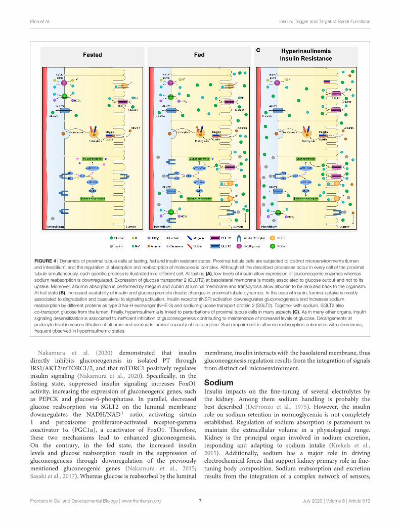
Glucose ReabsorptionGlucose is reabsorbed by the PT cells from the kidney tubulelumen to the bloodstream (Figure 4A). In the kidney, GLUT2is in the basolateral membrane and diffuses glucose out of thecell, contrary to the liver, where GLUT2 acts in glucose uptake.Sodium-glucose transporter proteins (SGLT) are responsible forglucose and sodium co-transport by the luminal membrane ofkidney cells. Sodium-glucose co-transporter 2 (SGLT2) is a high-capacity/low affinity sodium-glucose cotransporter present in theapical membrane of proximal convoluted tubule cells (Vallonet al., 2011). These transporters are responsible for approximately90% of filtered glucose reabsorption, and an important target forT2DM therapy (SGLT2 inhibitors; DeFronzo et al., 2012). Theremaining 10% of glucose in the tubule is absorbed by SGLT1,a low capacity and high affinity sodium-glucose cotransporter(Wright et al., 2011). Kidney has a threshold for glucose excretionwhich, in healthy conditions, relates to a glycemic value around180 mg/dl (Figure 4B). This threshold, however, can be alteredin diabetes (Rave et al., 2006). It is not clear if SGLT2 glucosetransport is or not directly dependent on insulin signaling(Ferrannini et al., 2020). Nonetheless, SGLT2 expression was
shown to be upregulated by insulin on human cultured PT cells,in a dose-dependent manner (Nakamura et al., 2015). Therefore,in hyperinsulinemic states, frequently observed in prediabetesand T2DM, an excessive glucose absorption can be observed. Ofnotice, glucose is still highly absorbed by SGLT2 in IR states,suggesting that this mechanism is not affected by IR, thoughit is upregulated by hyperinsulinemia. In this case, a viciouscycle can happen where increased insulin levels drive an increasein glucose SGLT2 overexpression increasing glycemic levelswhich in turn will increment the insulin secretion (Figure 4C).Therefore, SGLT2 overexpression inhibition is a potential newtarget for highly prevalent hyperinsulinemia related conditions,namely some dysglycemic phenotypes and obesity. In this case,lowering insulin levels could be paramount to prevent SGLT2overexpression. Thus, SGLT2 inhibitors might become a relevanttherapeutic approach for hyperinsulinemia related conditions,other than T2DM, namely obesity and prediabetes.
Gluconeogenesis RegulationKidney has a major role in gluconeogenesis along with the liverand the intestine. Gluconeogenesis occurs mainly in PT cellsessentially from lactate and glutamine (Figure 4A). Moreover,PT cells do not use glucose, as they get their energy mostlyfrom fatty acid oxidation (Gerich et al., 1963). Insulin regulatesgluconeogenesis in PT cells to meet the fluctuating needs ofthe body. While in the fasting state the kidney contributes in40% to overall gluconeogenesis, in the post-absorptive state itscontribution drops to 20% (Gerich et al., 1963; Figure 4B).
Frontiers in Cell and Developmental Biology | www.frontiersin.org 6 July 2020 | Volume 8 | Article 519
fcell-08-00519 July 27, 2020 Time: 18:28 # 7
Pina et al. Insulin: Trigger and Target of Renal Functions
FIGURE 4 | Dynamics of proximal tubule cells at fasting, fed and insulin resistant states. Proximal tubule cells are subjected to distinct microenvironments (lumenand interstitium) and the regulation of absorption and reabsorption of molecules is complex. Although all the described processes occur in every cell of the proximaltubule simultaneously, each specific process is illustrated in a different cell. At fasting (A), low levels of insulin allow expression of gluconeogenic enzymes whereassodium reabsorption is downregulated. Expression of glucose transporter 2 (GLUT2) at basolateral membrane is mostly associated to glucose output and not to itsuptake. Moreover, albumin absorption is performed by megalin and cubilin at luminal membrane and transcytosis allow albumin to be rerouted back to the organism.At fed state (B), increased availability of insulin and glucose promote drastic changes in proximal tubule dynamics. In the case of insulin, luminal uptake is mostlyassociated to degradation and basolateral to signaling activation. Insulin receptor (INSR) activation downregulates gluconeogenesis and increases sodiumreabsorption by different proteins as type 3 Na-H exchanger (NHE-3) and sodium-glucose transport protein 2 (SGLT2). Together with sodium, SGLT2 alsoco-transport glucose from the lumen. Finally, hyperinsulinemia is linked to perturbations of proximal tubule cells in many aspects (C). As in many other organs, insulinsignaling desensitization is associated to inefficient inhibition of gluconeogenesis contributing to maintenance of increased levels of glucose. Derangements atpodocyte level increases filtration of albumin and overloads luminal capacity of reabsorption. Such impairment in albumin reabsorption culminates with albuminuria,frequent observed in hyperinsulinemic states.
Nakamura et al. (2020) demonstrated that insulindirectly inhibits gluconeogenesis in isolated PT throughIRS1/AKT2/mTORC1/2, and that mTORC1 positively regulatesinsulin signaling (Nakamura et al., 2020). Specifically, in thefasting state, suppressed insulin signaling increases FoxO1activity, increasing the expression of gluconeogenic genes, suchas PEPCK and glucose-6-phosphatase. In parallel, decreasedglucose reabsorption via SGLT2 on the luminal membranedownregulates the NADH/NAD+ ratio, activating sirtuin1 and peroxisome proliferator-activated receptor-gammacoactivator 1α (PGC1α), a coactivator of FoxO1. Therefore,these two mechanisms lead to enhanced gluconeogenesis.On the contrary, in the fed state, the increased insulinlevels and glucose reabsorption result in the suppression ofgluconeogenesis through downregulation of the previouslymentioned gluconeogenic genes (Nakamura et al., 2015;Sasaki et al., 2017). Whereas glucose is reabsorbed by the luminal
membrane, insulin interacts with the basolateral membrane, thusgluconeogenesis regulation results from the integration of signalsfrom distinct cell microenvironment.
SodiumInsulin impacts on the fine-tuning of several electrolytes bythe kidney. Among them sodium handling is probably thebest described (DeFronzo et al., 1975). However, the insulinrole on sodium retention in normoglycemia is not completelyestablished. Regulation of sodium absorption is paramount tomaintain the extracellular volume in a physiological range.Kidney is the principal organ involved in sodium excretion,responding and adapting to sodium intake (Krekels et al.,2015). Additionally, sodium has a major role in drivingelectrochemical forces that support kidney primary role in fine-tuning body composition. Sodium reabsorption and excretionresults from the integration of a complex network of sensors,
Frontiers in Cell and Developmental Biology | www.frontiersin.org 7 July 2020 | Volume 8 | Article 519

fcell-08-00519 July 27, 2020 Time: 18:28 # 8
Pina et al. Insulin: Trigger and Target of Renal Functions
neural-hormonal stimuli and hemodynamic and metabolicmechanisms (Frame and Wainford, 2017).
Contrary to glucose, sodium is absorbed along the nephron bydistinct apical sodium transport proteins. Usually, approximately65% of filtered sodium is reabsorbed in the PT along withwater, mainly through type 3 Na-H exchanger (NHE-3) andSGLT proteins. In addition, the thick ascending limb isresponsible for approximately 25% of reabsorption throughsodium-potassium chloride cotransporter-2 (NKCC2). Finally,5–10% of sodium is reabsorbed in the collecting duct by epithelialsodium channel (ENaC) and less than 10% is excreted in urine(Esteva-Font et al., 2012).
The association of insulin with sodium absorption wassuggested almost a century ago (Atchley et al., 1933), nonethelessdiscrimination of insulin- and glucose-mediated effects hasnot been clarified (DeFronzo et al., 1976; Manhiani et al.,2011). It is still under debate if insulin has a causal effect onhypertension under normoglycemia. Nevertheless, it is knownthat insulin stimulates sodium absorption in all the tubulesegments where it takes place (Kirchner, 1988; Friedberg et al.,1991; Ghezzi and Wright, 2012). In the PT, insulin regulatesseveral sodium transporters, in both luminal (NHE-3, SGLT2)and basolateral membrane (Na/K-ATPase, NBCe1; Figure 4A;Gesek and Schoolwerth, 1991; Feraille et al., 1994; Ruiz et al.,1998). Recently, Nakamura et al. (2020) showed that, regardingsodium absorption in PT, insulin recruits an IRS (IRS2)different from the one orchestrating gluconeogenesis (IRS1).Insulin receptor substrate 2 acts through the AKT2/mTORC2pathway (Nakamura et al., 2020; Figure 4A). In fact, mTORC2is known to activate serum/glucocorticoid regulated kinase 1(SGK1), that will then stimulate ENaC and NHE-3, increasingsodium reabsorption (Satoh et al., 2015). It has been suggestedthat, in healthy conditions, with rising insulin levels in fedstate, IRS2 desensitize, suppressing sodium reabsorption at PTand increasing its delivery in the distal convoluted tubule(Figure 4B). Moreover, with IR, the desensitizing mechanism isabolished and therefore sodium will not reach the distal tubule(Ecelbarger, 2020; Figure 4C). Of notice, by recruiting distinctIRS, kidney cells can somehow dissociate pathways performingdistinct functions.
Finally, it must be kept in mind that insulin can interact withintrarenal and systemic renin-angiotensin-aldosterone system inseveral ways (Muscogiuri et al., 2008) and therefore, indirectlyinterfere with sodium reabsorption in different mechanism outof the scope of this review.
Albumin AbsorptionThe luminal membrane of PT cells is primarily responsible for thereabsorption of proteins that are freely filtered in the glomerulusby receptor-mediated endocytosis (Figure 4A; Christensen andGburek, 2004). This is the case of albumin reabsorptionthat can have an important role in energy conservation. Ithas been suggested that albumin endocytosis is a regulatedprocess, dependent on membrane receptors megalin and cubilin(Christensen and Birn, 2001). More recent evidence suggest thatinsulin might also have a role in the regulation of tubular albuminabsorption (Kumari et al., 2019).
Albuminuria is of major clinical relevance in diagnosis andfollow-up of kidney disease including subjects with diabetes.Insulin resistance was found to be associated with decreasedINSR expression in tubular cells in rat models (Wang et al.,2005). In these observations, Kumari et al. (2019) analyzedurine samples from mice with targeted deletion of INSR fromthe renal PT. These mice had an impaired uptake of albumin,without any glomerulopathy. They also demonstrated that inhealthy humans, albumin absorption capacity and excretion varyfrom the fast to the fed state. Moreover, IR was associated withmicroalbuminuria even in normoglycemia as described in theRISC study (Pilz et al., 2014) and thus can be present regardlessof diabetes diagnosis. Altogether, these evidences suggest thatalbuminuria might be an important marker of kidney tubulardysfunction and might reflect tubule cells IR (Figure 4C). Thesereinforces the kidney contribution to diabetes development andhighlights insulin and albumin dynamics prior and regardless ofthe development of diabetes.
KIDNEY INSULIN CLEARANCE
In the systemic circulation, besides insulin metabolization bythe liver, the kidney is the major site of insulin clearance(around 25%) (Elgee and Williams, 1954; Narahara et al., 1958);its action might be required to limit excessive insulin levels.Evidences supporting this theory started to rise in the middleof the 20th century (Zubrod et al., 1951; Elgee and Williams,1954; Narahara et al., 1958; Ricketts and Wildberger, 1962).In a study from 1966, Beck et al. (1966) found that wheninsulin was injected intravenously in mice, it concentrated inliver and kidney; however, with higher insulin doses, raisedinsulin levels were found in rat’s kidney, while these levels werefound to be reduced in liver. Nevertheless, this early studyhas some methodological limitations. Indeed, kidney insulinclearance remains constant in spite of insulinemia variations,but varies with creatinine clearance (Rubenstein et al., 1967).Globally, these evidences suggest that kidney insulin clearanceis a non-saturable process, although dependent on glomerularfiltration rate.
Insulin freely filtered in the glomerulus is absorbed by thelining cells of the PT (Figure 2). Upon entering the cell, insulin istransported through the luminal membrane into the PT cells andis degraded. Luminal insulin reabsorption limits urine insulinexcretion, thus less than 2% of insulin reaches the urine innormal fasting conditions (Rubenstein and Spitz, 1968). Insulinis transported through the luminal membrane by a receptor-mediated endocytic mechanism (Rabkin et al., 1984). Endocyticinternalization of insulin seems to be more related to insulindegradation than to insulin biological actions (Figure 2).
While glomerular filtration of insulin could not account forits total estimated renal extraction, a second mechanism waspostulated. In this context, it was observed that a significantamount of insulin was cleared by the post-glomerular peritubularcapillaries into the tubule cells (Chamberlain and Stimmler,1967). In humans, this route represents around one-third ofcleared insulin in the kidney, where it enters tubule cells not
Frontiers in Cell and Developmental Biology | www.frontiersin.org 8 July 2020 | Volume 8 | Article 519

fcell-08-00519 July 27, 2020 Time: 18:28 # 9
Pina et al. Insulin: Trigger and Target of Renal Functions
just by endocytosis, but also by INSR mediated uptake. In thiscase, through INSR binding, insulin signal to the kidneys’ tubularapparatus is crucial to maintain central physiologic functions,similarly to what happens in extra-renal tissues, namely regardingglucose homeostasis and blood pressure.
Insulin degrading activity has been observed at cytosol,lysosomes and mitochondria in addition to the membrane,indicating that it occurs in distinct cell sites. Degradation atthe membrane level, however, seems to represent less than 2%of total degrading activity (Rabkin et al., 1984). Insulin canbe initially hydrolyzed by an insulin protease followed by theaction of plasma-membrane-associated or lysosomal proteases.This pathway can degrade insulin entering through both luminaland contraluminal membrane. In another possible pathway,endocytic vesicles containing insulin fuse with lysosomes. Thispathway comprehends glutathione-insulin transhydrogenase(GIT) action, followed by hydrolysis of intact A and B chains bylysosomal proteases, and seems to need insulin internalization. Itmay act primarily on insulin delivered by luminal uptake and it ismost active when supraphysiological levels of insulin are present(Rabkin et al., 1984).
Azizi et al. (2015) demonstrated that, in a culture of humanadipose microvascular endothelial cells, insulin can go throughmicrovascular endothelial cells by transcytosis (Azizi et al.,2015). Regarding insulin handling in the kidney tubule, Dahlet al. (1989) hypothesized that insulin molecules could alsopass tubular cells by a retroendocytic pathway instead of beingdegraded. The authors demonstrated that cultured opossumkidney cells exhibited a retroendocytic pathway for insulin(Dahl et al., 1989). Using the same model, the authors laterdemonstrated that inhibition of insulin degradation divertedintact insulin from the degradative to the retroendocytic pathway(Dahl et al., 1990). Although captivating, especially regarding apotential contribution to hyperinsulinemic states, this hypothesiswas not further explored.
Whether insulin clearance mechanisms attributed to otherorgans affects renal function and insulin clearance it is not clear.For example, the lack of CEACAM-1, a key protein enrolled inhepatic insulin clearance driving hyperinsulinemia, in the kidneyleads to increased renin levels contributing to a potentiation ofthe RAS system and hypertension. These effects are exacerbatedupon high fat diet exposure. Hence, the described CEACAM-1renal effects can be due to the lack of its expression as well as theobserved hyperinsulinemia (Huang et al., 2013; Li et al., 2015).
Despite early conflicting results, further studies showed thatinsulin is excreted in urine. However, in physiological conditions,it represents a minimal proportion of insulin filtered in theglomerulus. In health, a minor amount of insulin appears inthe urine, as the majority is absorbed in PT. Tubule absorbingcapacity of insulin does not saturate and thus the insulinfraction excreted in urine is constantly small, regardless ofinsulin levels. However, the amount of insulin excreted in urinevaries physiologically (e.g., fasting and post-prandial) and inpathological conditions (obesity, diabetes) depending on theaffected nephron region (e.g., glomerulopathy vs. tubulopathy)(Rubenstein and Spitz, 1968). Considering that insulin isinternalized in the apical membrane by a receptor-mediated
endocytic mechanism, the increased urinary insulin excretionmight represent a tubular dysfunction. Subjects with tubulopathyshow large amounts of insulin in urine approximating theamount that is filtered (Rabkin et al., 1984). Conversely, subjectswith nephrotic syndrome show normal amounts of insulin inurine. When both glomerular and tubule lesion occur urineinsulin excretion increase (Rabkin et al., 1984).
CLINICAL INSIGHTS ON INSULINDYSREGULATION
Insulin resistance is a common feature in chronic kidney disease(CKD) patients, even in absence of diabetes (DeFronzo et al.,1981; Shinohara et al., 2002; Becker et al., 2005; Kobayashi et al.,2005; Landau et al., 2011), and it is a risk factor for CKDprogression (Fox et al., 2004). Its prevalence in CKD ranges from30 to 50%, and this mainly depends on the adopted methodof measurement (Spoto et al., 2016). Insulin resistance can bedetected at the very early stages, when eGFR is still within thenormal range, suggesting a potential role in triggering CKD(Fliser et al., 1998). A large study based on the AtherosclerosisRisk in Communities (ARICs) cohort confirmed that CKDdevelopment increases in strict parallelism with the number ofmetabolic syndrome criteria measured in non-diabetic adults,and this relationship remains significant even after controllingfor the development of diabetes and hypertension (Kurellaet al., 2005). Insulin resistance has also been associated withprevalent CKD and rapid decline in renal function in elderly,non-diabetic, Asian individuals (Cheng et al., 2012), and withmicroalbuminuria in the general population (Mykkänen et al.,1998), and in patients with T1DM (Yip et al., 1993; Ekstrandet al., 1998) and T2DM (Groop et al., 1993), indicating that thisrelationship is independent of diabetes (Mykkänen et al., 1998;Chen et al., 2003, 2004). The proposed mechanism by which IRcontributes to kidney damage involves the worsening of renalhemodynamics through activation of the sympathetic nervoussystem (Rowe et al., 1981), sodium retention, decreased Na+,K+-ATPase activity, and increased GFR (Gluba et al., 2013).
The etiology of IR in CKD is multifactorial, dependingon classical and CKD-peculiar risk factors, such as physicalinactivity, inflammation and oxidative stress, adipokinederangements, vitamin D deficiency, metabolic acidosis, anemiaand microbial toxins (Spoto et al., 2016).
Long-term hemodialysis has a positive effect on IR (DeFronzoet al., 1978), but there is little clinical data regarding the effect ofperitoneal dialysis.
In addition to being a risk factor for CKD onset andprogression, IR is also involved in the increased cardiovascular(CV) risk in this population. IR may be responsible for high bloodpressure via direct stimulation of renin–angiotensin–aldosteronesystem (Nickenig et al., 1998), activation of sympathetic system(Sowers et al., 2001) and downregulation of the natriureticpeptide system (Sarzani et al., 1999).
However, the association between IR and CV complicationsin CKD patients is still to be clarified, as well as the relationshipbetween IR and all-cause and CV mortality.
Frontiers in Cell and Developmental Biology | www.frontiersin.org 9 July 2020 | Volume 8 | Article 519

fcell-08-00519 July 27, 2020 Time: 18:28 # 10
Pina et al. Insulin: Trigger and Target of Renal Functions
A positive association between IR and all-cause mortalitywas found in smokers and physically inactive CKD patients(Xu et al., 2014) and in a small cohort of 170 Japanese,non-diabetic, dialysis patients (Shinohara et al., 2002), whereasassociation with CV mortality was found in a cohort of peritonealpatients. However, no association was found in the ULSAMcohort, including 3-4 stage CKD patients (Jia et al., 2014)and in a small study performed in peritoneal dialysis patients(Sánchez-Villanueva et al., 2013).
Even though the prognostic value of IR for death and CVevents need to be clarified, the association with CKD is wellestablished. Even in this case, however, if IR is responsible of theonset and progression of CKD, or if CKD is responsible for IR isstill to be clarified. A possible answer to this question could derivefrom clinical trials aiming at assessing the effect of drugs used forIR treatment on kidney function.
Thiazolidinediones (TZDs) are a class of oral diabeticmedications that increase insulin sensitivity by acting on PPARγ
(Yki-Järvinen, 2004). The effect of TZDs on kidney function hasbeen previously described in mice models (Fujii et al., 1997).Treatment with TZDs has been demonstrated to improve insulinsensitivity in patients with T2DM after a 3-month treatment,and to reduce albuminuria, the last effect likely mediated bythe concurrent increase in serum adiponectin concentration(Miyazaki et al., 2007). These results were confirmed in a meta-analysis of 15 double-blind, randomized, clinical trials involvingdiabetic patients (Sarafidis et al., 2010) and in a large studyinvolving 4351 diabetic patients (Lachin et al., 2011). Even thougha meta-analysis reported an increase in cardiovascular mortalitylinked to the use of TZDs in dialysis patients (Nissen and Wolski,2007), no definitive proof are available on the risk related to thismedication in this population.
Another interesting class of hypoglycemic drugs with positivekidney outcomes are the SGLT2 inhibitors (SGLT2i) whichinhibit glucose and sodium reabsorption in the PT (Ferrannini,2017). These drugs have a renoprotective effect in patients withT2DM (Perkovic et al., 2019) independently of glycemic control(Cherney et al., 2017). The renal protective effect can also beattributed to altered hemodynamics, reduced inflammation andfibrosis as well as controlled blood pressure and weight loss(Williams et al., 2020). In rats treated with SGLT2i the glycemicimprovement was accompanied by a decrease in insulin and lipidlevels (Huang et al., 2020). Moreover, the actions of SGLT2iare associated with increased insulin sensitivity and decreasedalbuminuria (Cherney et al., 2017). Interestingly, Jaikumkaoet al. observed that in an animal model of diet induced obesitycharacterized by IR and impaired renal function dapagliflozintreatment resulted in improved IR, renal function and renalinsulin signaling (Jaikumkao et al., 2018).
CONCLUSION
Insulin is a hormone which acts not only on the most recognizedinsulin-responsive organs (liver, adipose tissue, and skeletalmuscle), but also on the kidney. Moreover, the kidney has aprimordial role in insulin clearance and may impact on insulin
plasma levels. Whereas its main action is mainly related tohomeostasis of glucose, including modulation of gluconeogenesisand lipolysis, in kidney the effects of insulin and IR changeaccording to whether the target is in glomeruli or tubules. Morespecifically, if in glomerular podocytes insulin promotes glucoseuptake, with an involvement in barrier permeability, in tubulesit contributes to glucose reabsorption and gluconeogenesisregulation, and plays an important role in sodium homeostasis.
Importantly, insulin intervenes in albumin reabsorptionat tubular level. Moreover, IR has been associated withmicroalbuminuria even in normoglycemia, and thus can bepresent regardless of diabetes diagnosis. These findings reinforcethe kidney contribution to diabetes development and highlightsinsulin and albumin dynamics prior and regardless of thedevelopment of diabetes.
Insulin is cleared in the PT of kidney by two major routes,either by absorption of filtered insulin, or by post-glomerularcapillary secretion, and only a minor amount appears to beexcreted in urine. A decreased renal insulin clearance might leadto higher insulin levels, in the organ or systemically, favoringIR. Nonetheless, the impact of renal insulin clearance affectionin the kidney or in insulin plasma levels still needs to befurther unveiled.
It is clear now that kidney is not a mere target of insulin action,but insulin, more precisely IR, is also able to trigger CKD even inabsence of diabetes. IR has been associated with prevalent CKD,rapid decline in renal function and microalbuminuria in thegeneral population and in diabetic patients. In addition to beinga risk factor for CKD onset and progression, IR is also involvedin the increased cardiovascular risk in this population. However,if IR is responsible for the onset and progression of CKD, orif CKD is responsible for IR is still to be clarified. Preliminaryconfirmations come from clinical trials aiming at exploring theeffect of TZDs, a class of oral diabetic medications, on kidneyfunction. However, more focused studies, aiming also at testingthe safety of these medications in CKD patients, are needed tobetter understand if treatment of IR may improve renal functionin this population.
AUTHOR CONTRIBUTIONS
AP and MPM conceptualized the study. All authors originallydrafted the manuscript, reviewed, edited, and critically approvedthe final version of the manuscript.
FUNDING
This work was supported by “Fundação para a Ciência ea Tecnologia” – FCT to AP (PD/BD/136887/2018); MJM(PD/BD/114256/2016), MPM (PTDC/DTP-EPI/0207/2012),DOB e MPM (PTDC/BIM-MET/2115/2014); iNOVA4Health(UIDB/Multi/04462/2020), by the European Commission MarieSkłodowska-Curie Actions H2020 (grant agreements nos. 722619and 734719), and by the Sociedade Portuguesa de Diabetologia.
Frontiers in Cell and Developmental Biology | www.frontiersin.org 10 July 2020 | Volume 8 | Article 519

fcell-08-00519 July 27, 2020 Time: 18:28 # 11
Pina et al. Insulin: Trigger and Target of Renal Functions
REFERENCESAdler, A. I., Stevens, R. J., Manley, S. E., Bilous, R. W., Cull, C. A., Holman, R. R.,
et al. (2003). Development and progression of nephropathy in type 2 diabetes:The United Kingdom Prospective Diabetes Study (UKPDS 64). Kidney Int. 63,225–232. doi: 10.1046/j.1523-1755.2003.00712.x
Alessi, D. R., James, S. R., Downes, C. P., Holmes, A. B., Gaffney, P. R. J., Reese,C. B., et al. (1997). Characterization of a 3-phosphoinositide-dependent proteinkinase which phosphorylates and activates protein kinase Bα. Curr. Biol. 7,261–269. doi: 10.1016/S0960-9822(06)00122-9
Andrali, S. S., Sampley, M. L., Vanderford, N. L., and Özcan, S. (2008). Glucoseregulation of insulin gene expression in pancreatic β-cells. Biochem. J. 415, 1–10.doi: 10.1042/BJ20081029
Artunc, F., Schleicher, E., Weigert, C., Fritsche, A., Stefan, N., and Häring, H.(2016). The impact of insulin resistance on the kidney and vasculature. Nat.Publ. Gr. 12, 721–737. doi: 10.1038/nrneph.2016.145
Atchley, D. W., Loeb, R. F., Richards, D. W., Benedict, E. M., and Driscoll, M. E.(1933). ON DIABETIC ACIDOSIS: a detailed study of electrolyte balancesfollowing the withdrawal and reestablishment of insulin therapy. J. Clin. Invest.12, 297–326. doi: 10.1172/JCI100504
Azizi, P. M., Zyla, R. E., Guan, S., Wang, C., Liu, J., Bolz, S. S., et al. (2015). Clathrin-dependent entry and vesicle-mediated exocytosis define insulin transcytosisacross microvascular endothelial cells. Mol. Biol. Cell 26, 740–750. doi: 10.1091/mbc.E14-08-1307
Bayascas, J. R., and Alessi, D. R. (2005). Regulation of Akt/PKB Ser473Phosphorylation. Mol. Cell 18, 143–145. doi: 10.1016/j.molcel.2005.03.020
Beck, L. V., Zaharko, D. S., Roberts, N., and King, C. (1966). On insulin I-131metabolism in mice. Modifying effects of anti-insulin serum and of total insulindosage. Diabetes 15, 336–341. doi: 10.2337/diab.15.5.336
Becker, B., Kronenberg, F., Kielstein, J. T., Haller, H., Morath, C., Ritz, E., et al.(2005). Renal insulin resistance syndrome, adiponectin and cardiovascularevents in patients with kidney disease: the mild and moderate kidney diseasestudy. J. Am. Soc. Nephrol. 16, 1091–1098. doi: 10.1681/ASN.2004090742
Bedinger, D. H., and Adams, S. H. (2015). Metabolic, anabolic, and mitogenicinsulin responses: a tissue-specific perspective for insulin receptor activators.Mol. Cell. Endocrinol. 415, 143–156. doi: 10.1016/j.mce.2015.08.013
Bergman, R. N., Piccinini, F., Kabir, M., Kolka, C. M., and Ader, M. (2019).Hypothesis: role of reduced hepatic insulin clearance in the pathogenesis of type2 diabetes. Diabetes Metab. Res. Rev. 68, 1709–1716. doi: 10.2337/db19-0098
Bril, F., Lomonaco, R., Orsak, B., Ortiz-Lopez, C., Webb, A., Tio, F., et al.(2014). Relationship between disease severity, hyperinsulinemia, and impairedinsulin clearance in patients with nonalcoholic steatohepatitis. Hepatology 59,2178–2187. doi: 10.1002/hep.26988
Butlen, D., Vadrot, S., Roseau, S., and Morel, F. (1988). Insulin receptors along therat nephron: [125I] Insulin binding in microdissected glomeruli and tubules.Pflügers Arch. Eur. J. Physiol. 412, 604–612. doi: 10.1007/BF00583761
Calzi, S. L., Choice, C. V., and Najjar, S. M. (1997). Differential effect of pp120on insulin endocytosis by two variant insulin receptor isoforms. Am. J. Physiol.Metab. 273, E801–E808. doi: 10.1152/ajpendo.1997.273.4.E801
Canaud, G., Bienaimé, F., Viau, A., Treins, C., Baron, W., Nguyen, C., et al. (2013).AKT2 is essential to maintain podocyte viability and function during chronickidney disease. Nat. Med. 19, 1288–1296. doi: 10.1038/nm.3313
Chamberlain, M. J., and Stimmler, L. (1967). The renal handling of insulin. J. Clin.Invest. 46, 911–919. doi: 10.1172/JCI105597
Chen, J., Muntner, P., Hamm, L. L., Fonseca, V., Batuman, V., Whelton, P. K.,et al. (2003). Insulin resistance and risk of chronic kidney disease in nondiabeticUS adults. J. Am. Soc. Nephrol. 14, 469–477. doi: 10.1097/01.ASN.0000046029.53933.09
Chen, J., Muntner, P., Hamm, L. L., Jones, D. W., Batuman, V., Fonseca, V., et al.(2004). The metabolic syndrome and chronic kidney disease in U.S. adults. Ann.Intern. Med. 140, 167–174. doi: 10.7326/0003-4819-140-3-200402030-00007
Cheng, H. T., Huang, J. W., Chiang, C. K., Yen, C. J., Hung, K. Y., and Wu,K. D. (2012). Metabolic syndrome and insulin resistance as risk factors fordevelopment of chronic kidney disease and rapid decline in renal function inelderly. J. Clin. Endocrinol. Metab. 97, 1268–1276. doi: 10.1210/jc.2011-2658
Cherney, D. Z. I., Zinman, B., Inzucchi, S. E., Koitka-Weber, A., Mattheus, M., vonEynatten, M., et al. (2017). Effects of empagliflozin on the urinary albumin-to-creatinine ratio in patients with type 2 diabetes and established cardiovascular
disease: an exploratory analysis from the EMPA-REG OUTCOME randomised,placebo-controlled trial. Lancet Diabetes Endocrinol. 5, 610–621. doi: 10.1016/S2213-8587(17)30182-1
Christensen, E. I., and Birn, H. (2001). Megalin and cubilin: synergistic endocyticreceptors in renal proximal tubule. Am. J. Physiol. Physiol. 280, F562–F573.doi: 10.1152/ajprenal.2001.280.4.F562
Christensen, E. I., and Gburek, J. (2004). Protein reabsorption in renal proximaltubule - Function and dysfunction in kidney pathophysiology. Pediatr. Nephrol.19, 714–721. doi: 10.1007/s00467-004-1494-0
Coward, R. J. M., Welsh, G. I., Koziell, A., Hussain, S., Lennon, R., Ni, L., et al.(2007). Nephrin is critical for the action of insulin on human glomerularpodocytes. Diabetes Metab. Res. Rev. 56, 1127–1135. doi: 10.2337/db06-0693
Coward, R. J. M., Welsh, G. I., Yang, J., Tasman, C., Lennon, R., Koziell, A., et al.(2005). The human glomerular podocyte is a novel target for insulin action.Diabetes Metab. Res. Rev. 54, 3095–3102. doi: 10.2337/diabetes.54.11.3095
Dahl, D. C., Tsao, T., Duckworth, W. C., Frank, B. H., and Rabkin, R. (1990). Effectof bacitracin on retroendocytosis and degradation of insulin in cultured kidneyepithelial cell line. Diabetes Metab. Res. Rev 39, 1339–1346. doi: 10.2337/diab.39.11.1339
Dahl, D. C., Tsao, T., Duckworth, W. C., Mahoney, M. J., and Rabkin, R. (1989).Retroendocytosis of insulin in a cultured kidney epithelial cell line. Am. J.Physiol. Cell Physiol. 257, C190–C196. doi: 10.1152/ajpcell.1989.257.2.c190
DeFronzo, R. A., Alvestrand, A., Smith, D., Hendler, R., Hendler, E., and Wahren,J. (1981). Insulin resistance in uremia. J. Clin. Invest. 67, 563–568. doi: 10.1172/JCI110067
DeFronzo, R. A., Cooke, C. R., Andres, R., Faloona, G. R., and Davis, P. J. (1975).The effect of insulin on renal handling of sodium, potassium, calcium, andphosphate in man. J. Clin. Invest. 55, 845–855. doi: 10.1172/JCI107996
DeFronzo, R. A., Davidson, J. A., and del Prato, S. (2012). The role of the kidneys inglucose homeostasis: a new path towards normalizing glycaemia. Diabetes Obes.Metab. 14, 5–14. doi: 10.1111/j.1463-1326.2011.01511.x
DeFronzo, R. A., Goldberg, M., and Agus, Z. S. (1976). The effects of glucose andinsulin on renal electrolyte transport. J. Clin. Invest. 58, 83–90. doi: 10.1172/JCI108463
DeFronzo, R. A., Tobin, J. D., Rowe, J. W., and Andres, R. (1978). Glucoseintolerance in uremia. Quantification of pancreatic beta cell sensitivity toglucose and tissue sensitivity to insulin. J. Clin. Invest. 62, 425–435. doi: 10.1172/JCI109144
Detimary, P., Gilon, P., and Henquin, J.-C. (1998). Interplay between cytoplasmicCa2+ and the ATP/ADP ratio: a feedback control mechanism in mousepancreatic islets. Biochem. J. 333, 269–274. doi: 10.1042/bj3330269
Dimitriadis, G., Mitrou, P., Lambadiari, V., Maratou, E., and Raptis, S. A. (2011).Insulin effects in muscle and adipose tissue. Diabetes Res. Clin. Pract. 93,S52–S59. doi: 10.1016/S0168-8227(11)70014-6
Duckworth, W. C., Bennett, R. G., and Hamel, F. (1998). Insulin degradation:progress and potential. Endocr. Rev. 19, 608–624.
Ecelbarger, C. M. (2020). Refining insulin signaling in the proximal tubule at thelevel of the substrate. Kidney Int. 97, 256–258. doi: 10.1016/j.kint.2019.11.018
Ekstrand, A. V., Groop, P. H., and Grönhagen-Riska, C. (1998). Insulinresistance precedes microalbuminuria in patients with insulin-dependentdiabetes mellitus. Nephrol. Dial. Transplant. 13, 3079–3083. doi: 10.1093/ndt/13.12.3079
Elgee, N. J., and Williams, R. H. (1954). Degradation of Insulin-I131 by Liver andKidney in vivo. Exp. Biol. Med. 87, 352–355. doi: 10.3181/00379727-87-21380
Esteva-Font, C., Ballarin, J., and Fernández-Llama, P. (2012). Molecular biologyof water and salt regulation in the kidney. Cell. Mol. Life Sci. 69, 683–695.doi: 10.1007/s00018-011-0858-4
Farber, S. J., Berger, E. Y., and Earle, D. P. (1951). Effect of diabetes and insulin onthe maximum capacity of the renal tubules to reabsorb glucose. J. Clin. Invest.30, 125–129. doi: 10.1172/JCI102424
Feraille, E., Carranza, M. L., Rousselot, M., and Favre, H. (1994). Insulin enhancessodium sensitivity of Na-K-ATPase in isolated rat proximal convoluted tubule.Am. J. Physiol. Ren. Fluid Electrolyte Physiol. 267, F55–F62. doi: 10.1152/ajprenal.1994.267.1.f55
Fernandes, A. B., Patarrão, R. S., Videira, P. A., and Macedo, M. P. (2011).Understanding postprandial glucose clearance by peripheral organs: the roleof the hepatic parasympathetic system. J. Neuroendocrinol. 23, 1288–1295. doi:10.1111/j.1365-2826.2011.02226.x
Frontiers in Cell and Developmental Biology | www.frontiersin.org 11 July 2020 | Volume 8 | Article 519

fcell-08-00519 July 27, 2020 Time: 18:28 # 12
Pina et al. Insulin: Trigger and Target of Renal Functions
Ferrannini, E. (2017). Sodium-glucose co-transporters and their inhibition: clinicalphysiology. Cell Metab. 26, 27–38. doi: 10.1016/j.cmet.2017.04.011
Ferrannini, E., Pereira-Moreira, R., Seghieri, M., Rebelos, E., Souza, A. L., Chueire,V. B., et al. (2020). Insulin enhances renal glucose excretion: relation to insulinsensitivity and sodium-glucose cotransport. BMJ Open Diabetes Res. Care8:e001178. doi: 10.1136/bmjdrc-2020-001178
Fliser, D., Pacini, G., Engelleiter, R., Kautzky-Willer, A., Prager, R., Franek, E., et al.(1998). Insulin resistance and hyperinsulinemia are already present in patientswith incipient renal disease. Kidney Int. 53, 1343–1347. doi: 10.1046/j.1523-1755.1998.00898.x
Fox, C. S., Larson, M. G., Leip, E. P., Culleton, B., Wilson, P. W. F., and Levy,D. (2004). Predictors of new-onset kidney disease in a community-basedpopulation. J. Am. Med. Assoc. 291, 844–850. doi: 10.1001/jama.291.7.844
Frame, A. A., and Wainford, R. D. (2017). Renal sodium handling and sodiumsensitivity. Kidney Res. Clin. Pract. 36, 117–131. doi: 10.23876/j.krcp.2017.36.2.117
Friedberg, C. E., Van Buren, M., Bijlsma, J. A., and Koomans, H. A. (1991). Insulinincreases sodium reabsorption in diluting segment in humans: evidence forindirect mediation through hypokalemia. Kidney Int. 40, 251–256. doi: 10.1038/ki.1991.207
Fujii, M., Takemura, R., Yamaguchi, M., Hasegawa, G., Shigeta, H., Nakano, K.,et al. (1997). Troglitazone (CS-045) ameliorates albuminuria in streptozotocin-induced diabetic rats. Metabolism 46, 981–983. doi: 10.1016/S0026-0495(97)90264-X
Galgani, J. E., Moro, C., and Ravussin, E. (2008). Metabolic flexibility and insulinresistance. Am. J. Physiol. Metab. 295, E1009–E1017. doi: 10.1152/ajpendo.90558.2008
Gancheva, S., Jelenik, T., Álvarez-Hernández, E., and Roden, M. (2018). Interorganmetabolic crosstalk in human insulin resistance. Physiol. Rev. 98, 1371–1415.doi: 10.1152/physrev.00015.2017
Gao, T., McKenna, B., Li, C., Reichert, M., Nguyen, J., Singh, T., et al. (2014). Pdx1Maintains β cell identity and function by repressing an α cell program. CellMetab. 19, 259–271. doi: 10.1016/j.cmet.2013.12.002
Garner, K. L., Betin, V. M. S., Pinto, V., Graham, M., Abgueguen, E., Barnes,M., et al. (2018). Enhanced insulin receptor, but not PI3K, signalling protectspodocytes from ER stress. Sci. Rep. 8:3902. doi: 10.1038/s41598-018-22233-9
Gerich, J. E., Meyer, C., Hans, W. J., and Stumvoll, M. (1963). Renalgluconeogenesis. Adv. Enzyme Regul. 1, 385–400. doi: 10.1016/0065-2571(63)90034-7
Gesek, F. A., and Schoolwerth, A. C. (1991). Insulin increases Na(+)-H+ exchangeactivity in proximal tubules from normotensive and hypertensive rats. Am. J.Physiol. Physiol. 260, F695–F703. doi: 10.1152/ajprenal.1991.260.5.F695
Ghezzi, C., and Wright, E. M. (2012). Regulation of the human Na+-dependentglucose cotransporter hSGLT2. Am. J. Physiol. Cell Physiol. 303, 348–354. doi:10.1152/ajpcell.00115.2012
Gluba, A., Mikhailidis, D. P., Lip, G. Y. H., Hannam, S., Rysz, J., and Banach, M.(2013). Metabolic syndrome and renal disease. Int. J. Cardiol. 164, 141–150.doi: 10.1016/j.ijcard.2012.01.013
Groop, L., Ekstrand, A., Forsblom, C., Widén, E., Groop, P. H., Teppo, A. M., et al.(1993). Insulin resistance, hypertension and microalbuminuria in patients withType 2 (non-insulin-dependent) diabetes mellitus. Diabetologia 36, 642–647.doi: 10.1007/BF00404074
Hammerman, M. R. (1985). Interaction of insulin with the renal proximal tubularcell. Am. J. Physiol. Physiol. 249, F1–F11. doi: 10.1152/ajprenal.1985.249.1.F1
Hirashima, Y., Tsuruzoe, K., Kodama, S., Igata, M., Toyonaga, T., Ueki, K., et al.(2003). Insulin down-regulates insulin receptor substrate-2 expression throughthe phosphatidylinositol 3-kinase/Akt pathway. J. Endocrinol. 179, 253–266.doi: 10.1677/joe.0.1790253
Hou, J. C., Min, L., and Pessin, J. E. (2009). “Chapter 16 insulin granulebiogenesis, trafficking and exocytosis,” in Vitamins and Hormones, ed. G.Litwack (Amsterdam: Elsevier Inc), 473–506. doi: 10.1016/S0083-6729(08)00616-X
Huang, J., Ledford, K. J., Pitkin, W. B., Russo, L., Najjar, S. M., and Siragy,H. M. (2013). Targeted Deletion of Murine CEACAM 1 Activates PI3K-Aktsignaling and contributes to the expression of (pro)renin receptor via CREBFamily and NF-κB transcription factors. Hypertension 62, 317–323. doi: 10.1161/HYPERTENSIONAHA.113.01324
Huang, Z., Huang, L., Wang, C., Zhu, S., Qi, X., Chen, Y., et al. (2020). SUN-672 SGLT2 inhibitor reduces hyperinsulinemia and restores pulsatile growthhormone secretion in Obese MC4RKO mice. J. Endocr. Soc. 4:672. doi: 10.1210/jendso/bvaa046.1158
Hutton, J. C. (1994). Insulin secretory granule biogenesis and theproinsulin-processing endopeptidases. Diabetologia 37, S48–S56.doi: 10.1007/BF00400826
Jaikumkao, K., Pongchaidecha, A., Chueakula, N., Thongnak, L., Wanchai, K.,Chatsudthipong, V., et al. (2018). Renal outcomes with sodium glucosecotransporter 2 (SGLT2) inhibitor, dapagliflozin, in obese insulin-resistantmodel. Biochim. Biophys. Acta Mol. Basis Dis. 1864, 2021–2033. doi: 10.1016/j.bbadis.2018.03.017
Jia, T., Huang, X., Qureshi, A. R., Xu, H., Ärnlöv, J., Lindholm, B., et al. (2014).Validation of insulin sensitivity surrogate indices and prediction of clinicaloutcomes in individuals with and without impaired renal function. Kidney Int.86, 383–391. doi: 10.1038/ki.2014.1
Jung, S.-H., Jung, C.-H., Reaven, G. M., and Kim, S. H. (2018). Adapting to insulinresistance in obesity: role of insulin secretion and clearance. Diabetologia 61,681–687. doi: 10.1007/s00125-017-4511-0
Kalwat, M. A., and Thurmond, D. C. (2013). Signaling mechanisms of glucose-induced F-actin remodeling in pancreatic islet β cells. Exp. Mol. Med. 45:e37.doi: 10.1038/emm.2013.73
Kiapidou, S., Liava, C., Kalogirou, M., Akriviadis, E., and Sinakos, E. (2020).Chronic kidney disease in patients with non-alcoholic fatty liver disease: whatthe Hepatologist should know? Ann. Hepatol. 19, 134–144. doi: 10.1016/j.aohep.2019.07.013
Kim, K.-S., Park, S. W., Cho, Y.-W., and Kim, S.-K. (2018). Higher prevalenceand progression rate of chronic kidney disease in elderly patients with Type2 diabetes mellitus. Diabetes Metab. J. 42, 224–232. doi: 10.4093/dmj.2017.0065
Kirchner, K. A. (1988). Insulin increases loop segment chloride reabsorption in theeuglycemic rat. Am. J. Physiol. Ren. Fluid Electrolyte Physiol. 255, F1206–F1213.doi: 10.1152/ajprenal.1988.255.6.f1206
Kobayashi, S., Maesato, K., Moriya, H., Ohtake, T., and Ikeda, T. (2005).Insulin resistance in patients with chronic kidney disease. Am. J. Kidney Dis.2012:691369.doi: 10.1053/j.ajkd.2004.09.034
Kou, S.-W., Hu, Y.-H., and Wu, D.-A. (2014). The response of first and secondphase insulin secretion in newly diagnosed type 2 Diabetes Mellitus. Int. J.Diabetes Clin. Diagnosis 1:103. doi: 10.15344/2394-1499/2014/103
Krebs, H. A. (1963). Renal gluconeogenesis. Adv. Enzyme Regul. 1,385–400.
Krekels, M., Kroon, A., and de Leeuw, P. (2015). Sodium intake as amodulator of kidney function. Curr. Hypertens. Rev. 11, 57–60. doi: 10.2174/1573402111666150530204512
Krycer, J. R., Sharpe, L. J., Luu, W., and Brown, A. J. (2010). The Akt–SREBP nexus:cell signaling meets lipid metabolism. Trends Endocrinol. Metab. 21, 268–276.doi: 10.1016/j.tem.2010.01.001
Kubota, N., Kubota, T., Kajiwara, E., Iwamura, T., Kumagai, H., Watanabe, T., et al.(2016). Differential hepatic distribution of insulin receptor substrates causesselective insulin resistance in diabetes and obesity. Nat. Commun. 7:12977.doi: 10.1038/ncomms12977
Kumari, M., Sharma, R., Pandey, G., Ecelbarger, C. M., Mishra, P., and Tiwari, S.(2019). Deletion of insulin receptor in the proximal tubule and fasting augmentalbumin excretion. J. Cell. Biochem. 120, 10688–10696. doi: 10.1002/jcb.28359
Kuo, W. L., Montag, A. G., and Rosner, M. R. (1993). Insulin-degrading enzymeis differentially expressed and developmentally regulated in various rat tissues.Endocrinology 132, 604–611. doi: 10.1210/endo.132.2.7678795
Kurella, M., Lo, J. C., and Chertow, G. M. (2005). Metabolic syndrome and the riskfor chronic kidney disease among nondiabetic adults. J. Am. Soc. Nephrol. 16,2134–2140. doi: 10.1681/ASN.2005010106
Lachin, J. M., Viberti, G., Zinman, B., Haffner, S. M., Aftring, R. P., Paul, G., et al.(2011). Renal function in type 2 diabetes with rosiglitazone,metformin, andglyburide monotherapy. Clin. J. Am. Soc. Nephrol. 6, 1032–1040. doi: 10.2215/CJN.09291010
Landau, M., Kurella-Tamura, M., Shlipak, M. G., Kanaya, A., Strotmeyer, E., Koster,A., et al. (2011). Correlates of insulin resistance in older individuals with andwithout kidney disease. Nephrol. Dial. Transplant. 26, 2814–2819. doi: 10.1093/ndt/gfq817
Frontiers in Cell and Developmental Biology | www.frontiersin.org 12 July 2020 | Volume 8 | Article 519

fcell-08-00519 July 27, 2020 Time: 18:28 # 13
Pina et al. Insulin: Trigger and Target of Renal Functions
Lay, A., and Coward, R. J. (2014). Recent advances in our understanding ofinsulin signalling to the podocyte. Nephrol. Dial. Transplant. 29, 1127–1133.doi: 10.1093/ndt/gft471
Lay, A. C., Hurcombe, J. A., Betin, V. M. S., Barrington, F., Rollason, R., Ni, L.,et al. (2017). Prolonged exposure of mouse and human podocytes to insulininduces insulin resistance through lysosomal and proteasomal degradation ofthe insulin receptor. Diabetologia 60, 2299–2311. doi: 10.1007/s00125-017-4394-0
Lenkkeri, U., Männikkö, M., McCready, P., Lamerdin, J., Gribouval, O., Niaudet,P., et al. (1999). Structure of the Gene for Congenital Nephrotic Syndrome ofthe Finnish Type (NPHS1) and Characterization of Mutations. Am. J. Hum.Genet. 64, 51–61. doi: 10.1086/302182
Li, C., Culver, S. A., Quadri, S., Ledford, K. L., Al-Share, Q. Y., Ghadieh, H. E., et al.(2015). High-fat diet amplifies renal renin angiotensin system expression, bloodpressure elevation, and renal dysfunction caused by Ceacam1 null deletion. Am.J. Physiol. Metab. 309, E802–E810. doi: 10.1152/ajpendo.00158.2015
Li, L., Garikepati, R. M., Tsukerman, S., Kohan, D., Wade, J. B., Tiwari, S.,et al. (2013). Reduced ENaC activity and blood pressure in mice with geneticknockout of the insulin receptor in the renal collecting duct. Am. J. Physiol.Physiol. 304, F279–F288. doi: 10.1152/ajprenal.00161.2012
Ling, A. V., Gearing, M. E., Semova, I., Shin, D.-J., Clements, R., Lai, Z. W.,et al. (2018). FoxO1 is required for most of the metabolic and hormonalperturbations produced by hepatic insulin receptor deletion in male mice.Endocrinology 159, 1253–1263. doi: 10.1210/en.2017-00870
Madhusudhan, T., Wang, H., Dong, W., Ghosh, S., Bock, F., Thangapandi, V. R.,et al. (2015). Defective podocyte insulin signalling through p85-XBP1 promotesATF6-dependent maladaptive ER-stress response in diabetic nephropathy. Nat.Commun. 6:6496. doi: 10.1038/ncomms7496
Manhiani, M. M., Cormican, M. T., and Brands, M. W. (2011). Chronic sodium-retaining action of insulin in diabetic dogs. Am. J. Physiol. Ren. Physiol. 300,957–965. doi: 10.1152/ajprenal.00395.2010
Matveyenko, A. V., Liuwantara, D., Gurlo, T., Kirakossian, D., Dalla Man, C.,Cobelli, C., et al. (2012). Pulsatile portal vein insulin delivery enhances hepaticinsulin action and signaling. Diabetes Metab. Res. Rev 61, 2269–2279. doi:10.2337/db11-1462
Mayer, J. P., Zhang, F., and DiMarchi, R. D. (2007). Insulin structure and function.Biopolymers 88, 687–713. doi: 10.1002/bip.20734
McManus, E. J., Sakamoto, K., Armit, L. J., Ronaldson, L., Shpiro, N., Marquez,R., et al. (2005). Role that phosphorylation of GSK3 plays in insulin and Wntsignalling defined by knockin analysis. EMBO J. 24, 1571–1583. doi: 10.1038/sj.emboj.7600633
Mima, A., Ohshiro, Y., Kitada, M., Matsumoto, M., Geraldes, P., Li, C., et al. (2011).Glomerular-specific protein kinase C-β-induced insulin receptor substrate-1dysfunction and insulin resistance in rat models of diabetes and obesity. KidneyInt. 79, 883–896. doi: 10.1038/ki.2010.526
Miyazaki, Y., Cersosimo, E., Triplitt, C., and DeFronzo, R. A. (2007). Rosiglitazonedecreases albuminuria in type 2 diabetic patients. Kidney Int. 72, 1367–1373.doi: 10.1038/sj.ki.5002516
Mok, K. Y., Chan, P. F., Lai, L. K. P., Chow, K. L., and Chao, D. V. K.(2019). Prevalence of diabetic nephropathy among Chinese patients with type 2diabetes mellitus and different categories of their estimated glomerular filtrationrate based on the Chronic Kidney Disease Epidemiology Collaboration (CKD-EPI) equation in primary car. J. Diabetes Metab. Disord. 18, 281–288. doi:10.1007/s40200-018-00382-y
Molinaro, A., Becattini, B., Mazzoli, A., Bleve, A., Radici, L., Maxvall, I., et al.(2019). Insulin-Driven PI3K-AKT signaling in the hepatocyte is mediated byredundant PI3Kα and PI3Kβ activities and is promoted by RAS. Cell Metab.29:1400-1409.e5. doi: 10.1016/j.cmet.2019.03.010
Muscogiuri, G., Chavez, A., Gastaldelli, A., Perego, L., Tripathy, D., Saad, M.,et al. (2008). The crosstalk between insulin and renin-angiotensin-aldosteronesignaling systems and its effect on glucose metabolism and diabetes prevention.Curr. Vasc. Pharmacol. 6, 301–312. doi: 10.2174/157016108785909715
Musso, G., Gambino, R., Tabibian, J. H., Ekstedt, M., Kechagias, S., Hamaguchi,M., et al. (2014). Association of non-alcoholic fatty liver disease with chronickidney disease: a systematic review and meta-analysis. PLoS Med. 11:e1001680.doi: 10.1371/journal.pmed.1001680
Mykkänen, L., Zaccaro, D. J., Wagenknecht, L. E., Robbins, D. C., Gabriel, M., andHaffner, S. M. (1998). Microalbuminuria is associated with insulin resistance
in nondiabetic subjects: the insulin resistance atherosclerosis study. DiabetesMetab. Res. Rev. 47, 793–800. doi: 10.2337/diabetes.47.5.793
Naderpoor, N., Lyons, J. G., Mousa, A., Ranasinha, S., de Courten, M. P. J., Soldatos,G., et al. (2017). Higher glomerular filtration rate is related to insulin resistancebut not to obesity in a predominantly obese non-diabetic cohort. Sci. Rep.7:45522. doi: 10.1038/srep45522
Najjar, S. M., and Perdomo, G. (2019). Hepatic insulin clearance: mechanism andphysiology. Physiology 34, 198–215. doi: 10.1152/physiol.00048.2018
Nakamura, M., Tsukada, H., Seki, G., Satoh, N., Mizuno, T., Fujii, W., et al. (2020).Insulin promotes sodium transport but suppresses gluconeogenesis via distinctcellular pathways in human and rat renal proximal tubules. Kidney Int. 97,316–326. doi: 10.1016/j.kint.2019.08.021
Nakamura, N., Matsui, T., Ishibashi, Y., and Yamagishi, S. (2015). Insulin stimulatesSGLT2-mediated tubular glucose absorption via oxidative stress generation.Diabetol. Metab. Syndr. 7:48. doi: 10.1186/s13098-015-0044-1
Narahara, H. T., Everett, N. B., Simmons, B. S., and Williams, R. H. (1958).Metabolism of Insulin-I 131 and Glucagon-I 131 in the Kidney of the Rat. Am.J. Physiol. Content 192, 227–231. doi: 10.1152/ajplegacy.1958.192.2.227
Ng, Y., Ramm, G., Lopez, J. A., and James, D. E. (2008). Rapid Activation of Akt2 issufficient to stimulate GLUT4 translocation in 3T3-L1 adipocytes. Cell Metab.7, 348–356. doi: 10.1016/j.cmet.2008.02.008
Nickenig, G., Röling, J., Strehlow, K., Schnabel, P., and Böhm, M. (1998).Insulin induces upregulation of vascular receptor gene expression byposttranscriptional mechanisms. Circulation 98, 2453–2460. doi: 10.1161/01.CIR.98.22.2453
Nissen, S. E., and Wolski, K. (2007). Effect of rosiglitazone on the risk of myocardialinfarction and death from cardiovascular causes. N. Engl. J. Med. 356, 2457–2471. doi: 10.1056/NEJMoa072761
Oh, S.-Y., Park, S.-K., Kim, J.-W., Ahn, Y.-H., Park, S.-W., and Kim, K.-S. (2003).Acetyl-CoA Carboxylase β gene is regulated by sterol regulatory element-binding protein-1 in liver. J. Biol. Chem. 278, 28410–28417. doi: 10.1074/jbc.M300553200
Olofsson, C. S., Göpel, S. O., Barg, S., Galvanovskis, J., Ma, X., Salehi, A., et al.(2002). Fast insulin secretion reflects exocytosis of docked granules in mousepancreatic B-cells. Pflügers Arch. 444, 43–51. doi: 10.1007/s00424-002-0781-5
Pagtalunan, M. E., Miller, P. L., Jumping-Eagle, S., Nelson, R. G., Myers, B. D.,Rennke, H. G., et al. (1997). Podocyte loss and progressive glomerular injury intype II diabetes. J. Clin. Invest. 99, 342–348. doi: 10.1172/JCI119163
Perkovic, V., Jardine, M. J., Neal, B., Bompoint, S., Heerspink, H. J. L., Charytan,D. M., et al. (2019). Canagliflozin and Renal Outcomes in Type 2 Diabetes andNephropathy. N. Engl. J. Med. 380, 2295–2306. doi: 10.1056/NEJMoa1811744
Petersen, M. C., and Shulman, G. I. (2018). Mechanisms of Insulin Action andInsulin Resistance. Physiol. Rev. 98, 2133–2223. doi: 10.1152/physrev.00063.2017
Pilz, S., Rutters, F., Nijpels, G., Stehouwer, C. D. A., HØjlund, K., Nolan, J. J., et al.(2014). Insulin sensitivity and albuminuria: the risc study. Diabetes Care 37,1597–1603. doi: 10.2337/dc13-2573
Piwkowska, A., Rogacka, D., Kasztan, M., Angielski, S., and Jankowski, M.(2013). Insulin increases glomerular filtration barrier permeability throughdimerization of protein kinase G type Iα subunits. Biochim. Biophys. Acta Mol.Basis Dis. 1832, 791–804. doi: 10.1016/j.bbadis.2013.02.011
Plum, L. (2006). Central insulin action in energy and glucose homeostasis. J. Clin.Invest. 116, 1761–1766. doi: 10.1172/JCI29063
Poy, M. N., Yang, Y., Rezaei, K., Fernström, M. A., Lee, A. D., Kido, Y., et al.(2002). CEACAM1 regulates insulin clearance in liver. Nat. Genet. 30, 270–276.doi: 10.1038/ng840
Prentki, M., and Matschinsky, F. M. (1987). Ca2+, cAMP, and phospholipid-derived messengers in coupling mechanisms of insulin secretion. Physiol. Rev.67, 1185–1248. doi: 10.1152/physrev.1987.67.4.1185
Qiu, H., Yang, J.-K., and Chen, C. (2017). Influence of insulin on growth hormonesecretion, level and growth hormone signalling. Sheng Li Xue Bao 69, 541–556.doi: 10.13294/j.aps.2017.0062
Rabkin, R., Ryan, M. P., and Duckworth, W. C. (1984). The renal metabolism ofinsulin. Diabetologia 27, 351–357. doi: 10.1007/BF00304849
Rave, K., Nosek, L., Posner, J., Heise, T., Roggen, K., and van Hoogdalem, E. J.(2006). Renal glucose excretion as a function of blood glucose concentration insubjects with type 2 diabetes - Results of a hyperglycaemic glucose clamp study.Nephrol. Dial. Transplant. 21, 2166–2171. doi: 10.1093/ndt/gfl175
Frontiers in Cell and Developmental Biology | www.frontiersin.org 13 July 2020 | Volume 8 | Article 519

fcell-08-00519 July 27, 2020 Time: 18:28 # 14
Pina et al. Insulin: Trigger and Target of Renal Functions
Ricketts, H. T., and Wildberger, H. L. (1962). The effect of renal disease on thedegradation of insulin. Trans. Am. Clin. Climatol. Assoc. 74, 102–108.
Ritz, E., Adamczak, M., and Wiecek, A. (2013). Carbohydrate Metabolism in KidneyDisease and Kidney Failure. Amsterdam: Elsevier Inc, doi: 10.1016/B978-0-12-391934-2.00002-3
Roder, K., Zhang, L., and Schweizer, M. (2007). SREBP-1c mediates the retinoid-dependent increase in fatty acid synthase promoter activity in HepG2. FEBSLett. 581, 2715–2720. doi: 10.1016/j.febslet.2007.05.022
Rowe, J. W., Young, J. B., Minaker, K. L., Stevens, A. L., Pallotta, J., and Landsberg,L. (1981). Effect of insulin and glucose infusions on sympathetic nervous systemactivity in normal man. Diabetes Metab. Res. Rev. 30, 219–225. doi: 10.2337/diab.30.3.219
Rubenstein, A. H., Lowy, C., Welborn, T. A., and Fraser, T. R. (1967). Urineinsulin in normal subjects. Metabolism 16, 234–244. doi: 10.1016/0026-0495(67)90172-2
Rubenstein, A. H., and Spitz, I. (1968). Role of the kidney in insulin metabolism andexcretion. Diabetes Metab. Res. Rev. 17, 161–169. doi: 10.2337/diab.17.3.161
Ruiz, O. S., Qiu, Y.-Y., Cardoso, L. R., and Arruda, J. A. L. (1998). Regulation of therenal Na–HCO3 cotransporter: IX. Modulation by insulin, epidermal growthfactor and carbachol. Regul. Pept. 77, 155–161. doi: 10.1016/S0167-0115(98)00115-3
Russo, L., Muturi, H. T., Ghadieh, H. E., Ghanem, S. S., Bowman, T. A., Noh,H. L., et al. (2017). Liver-specific reconstitution of CEACAM1 reverses themetabolic abnormalities caused by its global deletion in male mice. Diabetologia60, 2463–2474. doi: 10.1007/s00125-017-4432-y
Sánchez-Villanueva, R., Estrada, P., Del Peso, G., Grande, C., Díez, J. J., Iglesias,P., et al. (2013). Repeated analysis of estimated insulin resistance usingthe HOMAIR index in nondiabetic patients on peritoneal dialysis and itsrelationship with cardiovascular disease and mortality. Nefrologia 33, 85–92.doi: 10.3265/Nefrologia.pre2012.Nov.11430
Sanders, F. W. B., and Griffin, J. L. (2016). De novo lipogenesis in the liver in healthand disease: more than just a shunting yard for glucose. Biol. Rev. 91, 452–468.doi: 10.1111/brv.12178
Santamaria, B., Marquez, E., Lay, A., Carew, R. M., González-Rodríguez, Á, Welsh,G. I, et al. (2015). IRS2 and PTEN are key molecules in controlling insulinsensitivity in podocytes. Biochim. Biophys. Acta Mol. Cell Res. 1853, 3224–3234.doi: 10.1016/j.bbamcr.2015.09.020
Sarafidis, P. A., Stafylas, P. C., Georgianos, P. I., Saratzis, A. N., and Lasaridis, A. N.(2010). Effect of thiazolidinediones on albuminuria and proteinuria in diabetes:a meta-analysis. Am. J. Kidney Dis. 55, 835–847. doi: 10.1053/j.ajkd.2009.11.013
Sarzani, R., Dessì-Fulgheri, P., Salvi, F., Serenelli, M., Spagnolo, D., Cola, G.,et al. (1999). A novel promoter variant of the natriuretic peptide clearancereceptor gene is associated with lower atrial natriuretic peptide and higherblood pressure in obese hypertensives. J. Hypertens. 17, 1301–1305. doi: 10.1097/00004872-199917090-00010
Sasaki, M., Sasako, T., Kubota, N., Sakurai, Y., Takamoto, I., Kubota, T., et al.(2017). Dual regulation of gluconeogenesis by insulin and glucose in theproximal tubules of the kidney. Diabetes Metab. Res. Rev 66, 2339–2350. doi:10.2337/db16-1602
Satoh, N., Nakamura, M., Suzuki, M., Suzuki, A., Seki, G., and Horita, S. (2015).Roles of Akt and SGK1 in the regulation of renal tubular transport. Biomed Res.Int. 2015, 1–8. doi: 10.1155/2015/971697
Shinohara, K., Shoji, T., Emoto, M., Tahara, H., Koyama, H., Ishimura, E.,et al. (2002). Insulin resistance as an independent predictor of cardiovascularmortality in patients with end-stage renal disease. J. Am. Soc. Nephrol. 13,1894–1900. doi: 10.1097/01.ASN.0000019900.87535.43
Skolnik, E. Y., Batzer, A., Li, N., Lee, C. H., Lowenstein, E., Mohammadi, M., et al.(1993). The function of GRB2 in linking the insulin receptor to Ras signalingpathways. Science 260, 1953–1955. doi: 10.1126/science.8316835
Song, S. H., McIntyre, S. S., Shah, H., Veldhuis, J. D., Hayes, P. C., and Butler, P. C.(2000). Direct measurement of pulsatile insulin secretion from the portal veinin human subjects 1. J. Clin. Endocrinol. Metab. 85, 4491–4499. doi: 10.1210/jcem.85.12.7043
Sowers, J. R., Epstein, M., and Frohlich, E. D. (2001). Diabetes, hypertension, andcardiovascular disease an update. Hypertension 37, 1053–1059. doi: 10.1161/01.HYP.37.4.1053
Spoto, B., Pisano, A., and Zoccali, C. (2016). Insulin resistance in chronic kidneydisease: a systematic review. Am. J. Physiol. Renal Physiol. 311, F1087–F1108.doi: 10.1152/ajprenal.00340.2016
Stechi, L. A., De Carll, S., and Bartoll, E. (1994). In situ characterization ofrenal insulin receptors in the rat. J. Recept. Res. 14, 347–356. doi: 10.3109/10799899409101509
Steiner, D. F. (2004). The proinsulin c-peptide—a multirole model. Exp. DiabesityRes. 5, 7–14. doi: 10.1080/15438600490424389
Susztak, K., Raff, A. C., Schiffer, M., and Bottinger, E. P. (2006). Glucose-inducedreactive oxygen species cause apoptosis of podocytes and podocyte depletionat the onset of diabetic nephropathy. Diabetes Metab. Res. Rev 55, 225–233.doi: 10.2337/diabetes.55.01.06.db05-0894
Svensson, M., and Eriksson, J. W. (2006). Insulin resistance in diabeticnephropathy — cause or consequence? Diabetes. Metab. Res. Rev. 22, 401–410.doi: 10.1002/dmrr.648
Talor, Z., Emmanouel, D. S., and Katz, A. I. (1982). Insulin binding and degradationby luminal and basolateral tubular membranes from rabbit kidney. J. Clin.Invest. 69, 1136–1146. doi: 10.1172/JCI110549
Taniguchi, C. M., Emanuelli, B., and Kahn, C. R. (2006). Critical nodes in signallingpathways: insights into insulin action. Nat. Rev. Mol. Cell Biol. 7, 85–96. doi:10.1038/nrm1837
Tejada, T., Catanuto, P., Ijaz, A., Santos, J. V., Xia, X., Sanchez, P., et al. (2008).Failure to phosphorylate AKT in podocytes from mice with early diabeticnephropathy promotes cell death. Kidney Int. 73, 1385–1393. doi: 10.1038/ki.2008.109
Thirone, A. C. P., Huang, C., and Klip, A. (2006). Tissue-specific roles of IRSproteins in insulin signaling and glucose transport. Trends Endocrinol. Metab.17, 70–76. doi: 10.1016/j.tem.2006.01.005
Tiwari, S., Sharma, N., Gill, P. S., Igarashi, P., Kahn, C. R., Wade, J. B., et al. (2008).Impaired sodium excretion and increased blood pressure in mice with targeteddeletion of renal epithelial insulin receptor. Proc. Natl. Acad. Sci. U.S.A. 105,6469–6474. doi: 10.1073/pnas.0711283105
Tiwari, S., Singh, R. S., Li, L., Tsukerman, S., Godbole, M., Pandey, G., et al.(2013). Deletion of the insulin receptor in the proximal tubule promoteshyperglycemia. J. Am. Soc. Nephrol. 24, 1209–1214. doi: 10.1681/ASN.2012060628
Tokarz, V. L., MacDonald, P. E., and Klip, A. (2018). The cell biology of systemicinsulin function. J. Cell Biol. 217, 2273–2289. doi: 10.1083/jcb.201802095
Vallon, V., Platt, K. A., Cunard, R., Schroth, J., Whaley, J., Thomson, S. C., et al.(2011). SGLT2 mediates glucose reabsorption in the early proximal tubule.J. Am. Soc. Nephrol. 22, 104–112. doi: 10.1681/ASN.2010030246
Villarreal, R., Mitrofanova, A., Maiguel, D., Morales, X., Jeon, J., Grahammer, F.,et al. (2016). Nephrin contributes to insulin secretion and affects mammaliantarget of rapamycin signaling independently of insulin receptor. J. Am. Soc.Nephrol. 27, 1029–1041. doi: 10.1681/ASN.2015020210
Wang, X. L., Lu, J. M., Pan, C. Y., Tian, H., and Li, C. L. (2005). A comparisonof urinary albumin excretion rate and microalbuminuria in various glucosetolerance subjects. Diabet. Med. 22, 332–335. doi: 10.1111/j.1464-5491.2004.01408.x
Welsh, G. I., Hale, L. J., Eremina, V., Jeansson, M., Maezawa, Y., Lennon, R.,et al. (2010). Insulin signaling to the glomerular podocyte is critical for normalkidney function. Cell Metab. 12, 329–340. doi: 10.1016/j.cmet.2010.08.015
Williams, D. M., Nawaz, A., and Evans, M. (2020). Renal outcomes in type 2diabetes: a review of cardiovascular and renal outcome trials. Diabetes Ther. 11,369–386. doi: 10.1007/s13300-019-00747-3
Wolf, G., Chen, S., and Ziyadeh, F. N. (2005). From the periphery of the glomerularcapillary wall toward the center of disease: podocyte injury comes of age indiabetic nephropathy. Diabetes Metab. Res. Rev. 54, 1626–1634. doi: 10.2337/diabetes.54.6.1626
Wright, E. M., Loo, D. D. F., and Hirayama, B. A. (2011). Biology of HumanSodium Glucose Transporters. Physiol. Rev. 91, 733–794. doi: 10.1152/physrev.00055.2009
Xu, H., Huang, X., Ärnlöv, J., Cederholm, T., Stenvinkel, P., Lindholm, B.,et al. (2014). Clinical correlates of insulin sensitivity and its association withmortality among men with CKD stages 3 and 4. Clin. J. Am. Soc. Nephrol. 9,690–697. doi: 10.2215/CJN.05230513
Frontiers in Cell and Developmental Biology | www.frontiersin.org 14 July 2020 | Volume 8 | Article 519
fcell-08-00519 July 27, 2020 Time: 18:28 # 15
Pina et al. Insulin: Trigger and Target of Renal Functions
Xu, J., Keeton, A. B., Franklin, J. L., Li, X., Venable, D. Y., Frank, S. J., et al.(2006). Insulin enhances growth hormone induction of the MEK/ERK signalingpathway. J. Biol. Chem. 281, 982–992. doi: 10.1074/jbc.M505484200
Xu, J., and Messina, J. L. (2009). “Crosstalk between growth hormone and insulinsignaling,” in Vitamins and Hormones, Ed. G. Litwack (Amsterdam: ElsevierInc), 125–153. doi: 10.1016/S0083-6729(08)00606-7
Yamada, T., Katagiri, H., Asano, T., Tsuru, M., Inukai, K., Ono, H., et al. (2002).Role of PDK1 in insulin-signaling pathway for glucose metabolism in 3T3-L1 adipocytes. Am. J. Physiol. Metab. 282, E1385–E1394. doi: 10.1152/ajpendo.00486.2001
Ye, L., Maji, S., Sanghera, N., Gopalasingam, P., Gorbunov, E., Tarasov, S.,et al. (2017). Structure and dynamics of the insulin receptor: implications forreceptor activation and drug discovery. Drug Discov. Today 22, 1092–1102.doi: 10.1016/j.drudis.2017.04.011
Yip, J., Mattock, M. B., Morocutti, A., Sethi, M., Trevisan, R., and Viberti,G. (1993). Insulin resistance in insulin-dependent diabetic patients withmicroalbuminuria. Lancet 342, 883–887. doi: 10.1016/0140-6736(93)91943-G
Yki-Järvinen, H. (2004). Thiazolidinediones. N. Engl. J. Med. 351, 1106–1118.doi: 10.1056/NEJMra041001
Zorzano, A., Munoz, P., Camps, M., Mora, C., Testar, X., and Palacin, M. (1996).Insulin-Induced Redistribution of GLUT4 Glucose Carriers in the Muscle Fiber:in Search of GLUT4 Trafficking Pathways. Diabetes 45, S70–S81. doi: 10.2337/diab.45.1.S70
Zubrod, C. G., Eversole, S. L., and Dana, G. W. (1951). Amelioration of diabetes andstriking rarity of acidosis in patients with kimmelstiel-wilson lesions. N. Engl. J.Med. 245, 518–525. doi: 10.1056/NEJM195110042451402
Conflict of Interest: The authors declare that the research was conducted in theabsence of any commercial or financial relationships that could be construed as apotential conflict of interest.
Copyright © 2020 Pina, Borges, Meneses, Branco, Birne, Vilasi and Macedo. Thisis an open-access article distributed under the terms of the Creative CommonsAttribution License (CC BY). The use, distribution or reproduction in other forumsis permitted, provided the original author(s) and the copyright owner(s) are creditedand that the original publication in this journal is cited, in accordance with acceptedacademic practice. No use, distribution or reproduction is permitted which does notcomply with these terms.
Frontiers in Cell and Developmental Biology | www.frontiersin.org 15 July 2020 | Volume 8 | Article 519