integumentary aka skin. introduction skin comprises the largest organ system in the body weighs...
Post on 21-Dec-2015
216 views
TRANSCRIPT

Integumentary AKA Skin
Introduction
Skin comprises the largest organ system in the body
Weighs approximately 17 Kg Takes up 2.0 square meters
Functions
Regulates body temp. Acts as an excretory organ Stores chlorides Manufactures vitamin D which is essential for
the mineralization of bones and teeth Serves as the most extensive and varied of
the sense organs – high concentration of sensory receptors, especially where this is critical – lips, finger tips
The Envelope
Prevents the loss of essential host constituents or the entry into the host of toxic physical, chemical and microbiotic agents
Properties go beyond a mere envelope• Is waterproof and prevents the evaporation and
escape of tissue fluids
• Becomes thick when subjected to rough treatment
• Fastened down where most likely to be pulled off
• Has friction ridges to lessen slippage – e.g., finger tips
Anatomy
Non-Homogeneous Large differences in form, color, and
consistency in various parts of the same body
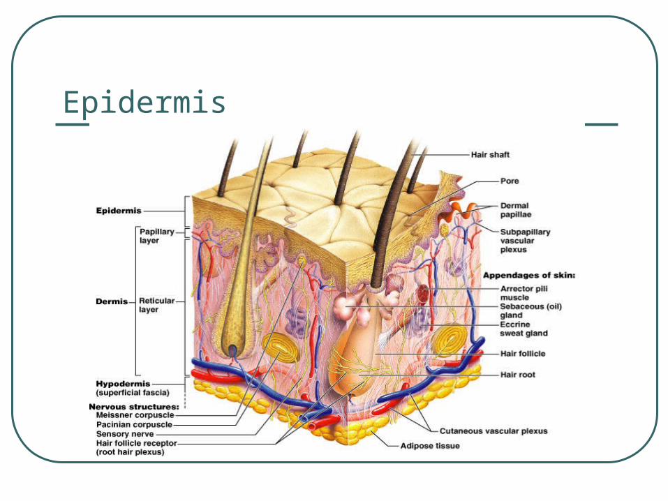
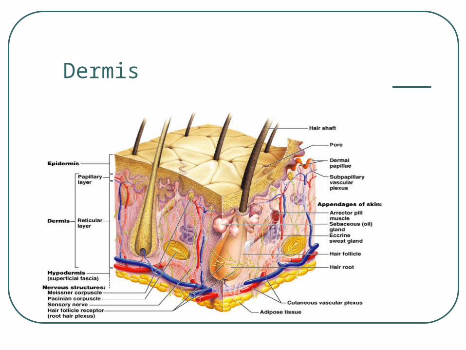
Three distinct types of tissue – epidermis, dermis, and subcutaneous
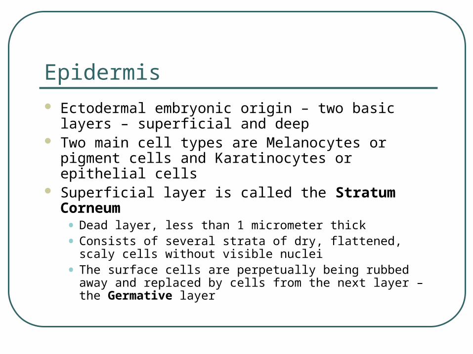
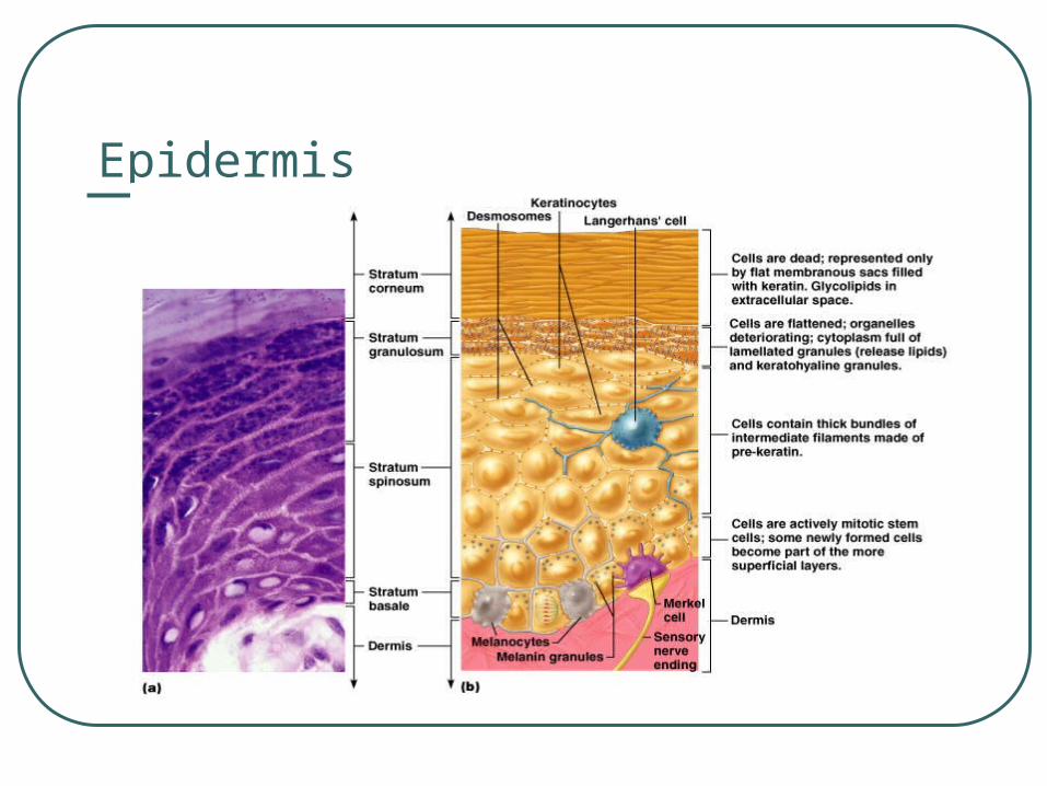
Epidermis Ectodermal embryonic origin – two basic layers –
superficial and deep Two main cell types are Melanocytes or pigment
cells and Karatinocytes or epithelial cells Superficial layer is called the Stratum Corneum
• Dead layer, less than 1 micrometer thick
• Consists of several strata of dry, flattened, scaly cells without visible nuclei
• The surface cells are perpetually being rubbed away and replaced by cells from the next layer – the Germative layer
Epidermis
Epidermis
Germative Layer
AKA Basal Layer This layer is living Cell division occurs here and the cells
biochemically and functionally mature as they ascend through the more superficial layers
Elapsed time from the cell division to shedding is at least 4 weeks – in abnormal states such as psoriasis = may turn over in 4 days
Dermis
Has a mesodermal embryonic origin, similar to CT
Primarily made up of collagen, elastin, and reticulin fiber
Dermis accounts for 5% of body mass The outer aspect of dermis nearest to the
epidermis is called the papillary dermis – rich in nerves, vessels, and various sensory receptors
Dermis
Dermis
The deeper dermis is called the Reticular Dermis that contains the cutaneous vascular network
These vessels subserve the thermoregulatory functions
Also, there are more neuroreceptors and lymph glands
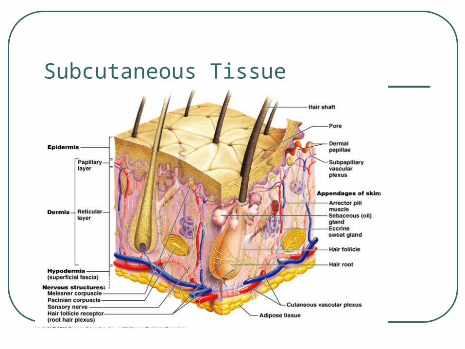
Subcutaneous Tissue
Deep to dermis Fatty structure of various thickness Functions as a thermal barrier and
protective cushion AKA superficial fascia or tela subcutanea
Subcutaneous Tissue
Skin Color
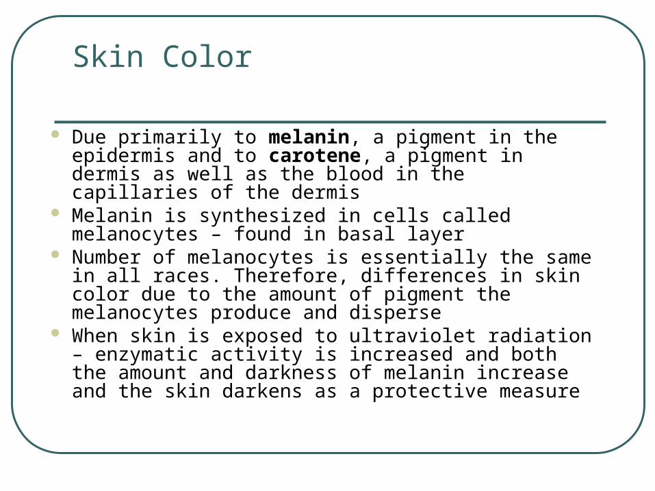
Due primarily to melanin, a pigment in the epidermis and to carotene, a pigment in dermis as well as the blood in the capillaries of the dermis
Melanin is synthesized in cells called melanocytes – found in basal layer
Number of melanocytes is essentially the same in all races. Therefore, differences in skin color due to the amount of pigment the melanocytes produce and disperse
When skin is exposed to ultraviolet radiation – enzymatic activity is increased and both the amount and darkness of melanin increase and the skin darkens as a protective measure
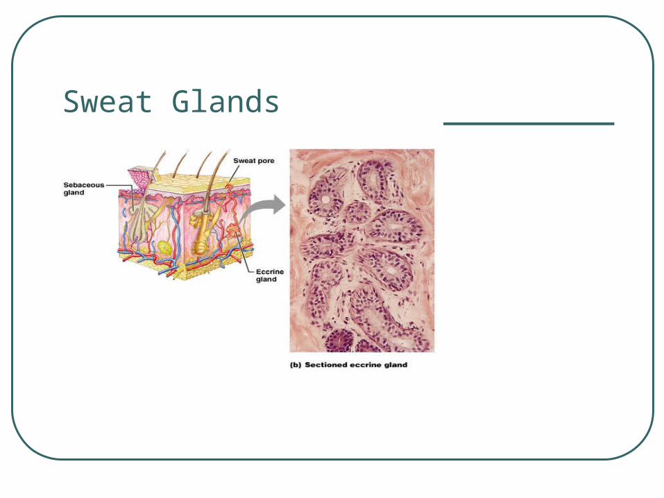
Sweat Glands
Two types – eccrine and sebaceous Eccrine – produce watery sweat that reaches
the surface of skin – found in very high concentration throughout the body and serve to regulate temperature
Sebaceous – are outgrowths of hair follicles into the dermis• Are filled with cells which secrete greasy substance
called sebum and give skin a greasy feels and make the skin waterproof
• Are found in high concentration in upper torso
Sweat Glands
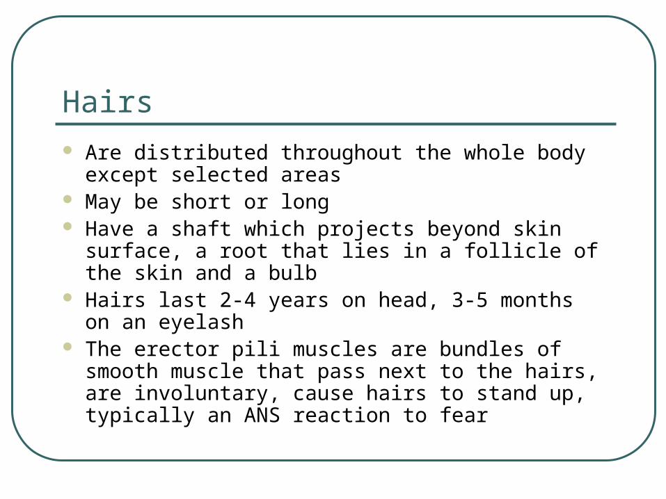
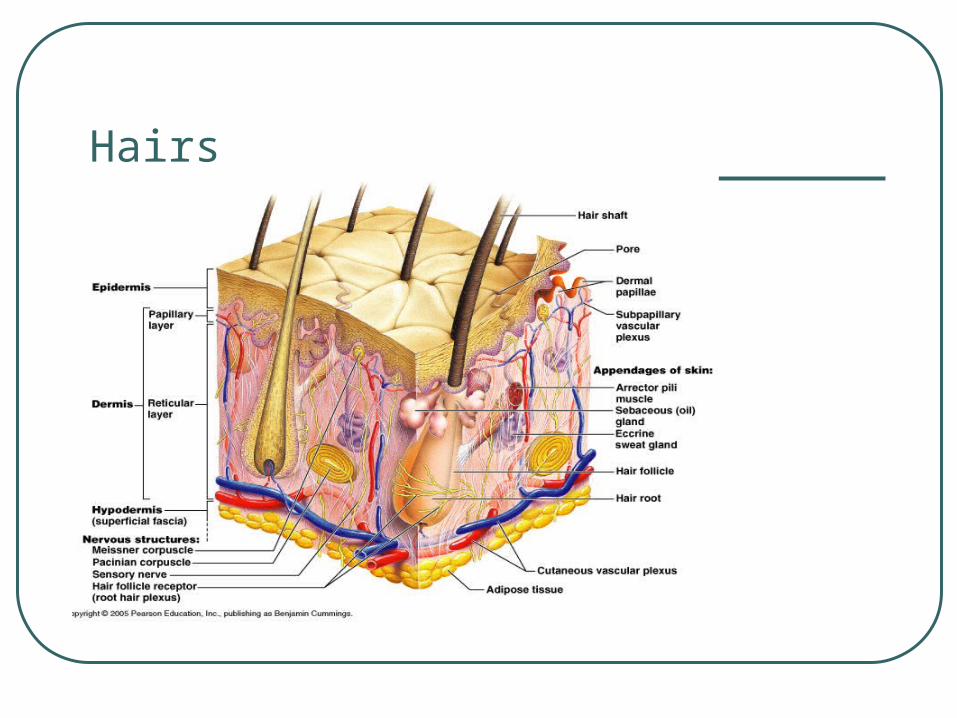
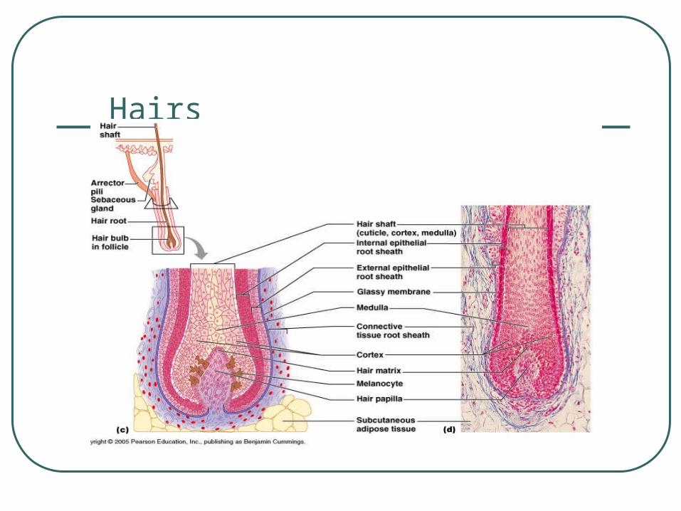
Hairs Are distributed throughout the whole body except
selected areas May be short or long Have a shaft which projects beyond skin surface, a
root that lies in a follicle of the skin and a bulb Hairs last 2-4 years on head, 3-5 months on an
eyelash The erector pili muscles are bundles of smooth
muscle that pass next to the hairs, are involuntary, cause hairs to stand up, typically an ANS reaction to fear
Hairs
Hairs
Brief Overview of Burns
1st degree – damages only epidermis, a sunburn with reddening of the skin
2nd degree – burn destroys much of the epidermis but leaves some epidermal remnants• Re-growth from remnants is possible
• Blisters are common and pain is often severe since the skin nerves are irritated by the products of cellular destruction
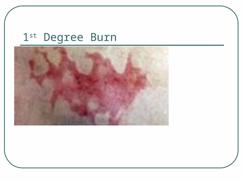
1st Degree Burn
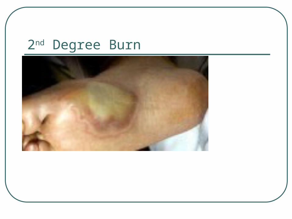
2nd Degree Burn
Burns
3rd Degree – reaches to and thru dermis – often exposing muscle and bone
No epidermal remnants are present Little or no feeling because of destruction
of nerves Treatment requires skin grafts to provide
epidermal cells Healing is slow at best

3rd Degree Burn
Major Problems of Burns
Infection Maintaining fluid Maintaining electrolyte balance which
requires food and fluid intake Contractures of skin and underlying CT
and muscle (all soft tissue) due to intense scarring
Other Common Problems with Skin
Decubitus Ulcers – AKA bed or pressure sores• Caused by constant deficiency of blood to
tissues overlying a bony prominence subjected to prolonged pressure – especially if CNS or PNS not functioning fully
• Tissue breakdown leads to infection, necrosis, etc.
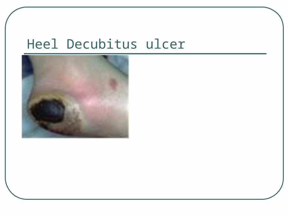
Heel Decubitus ulcer
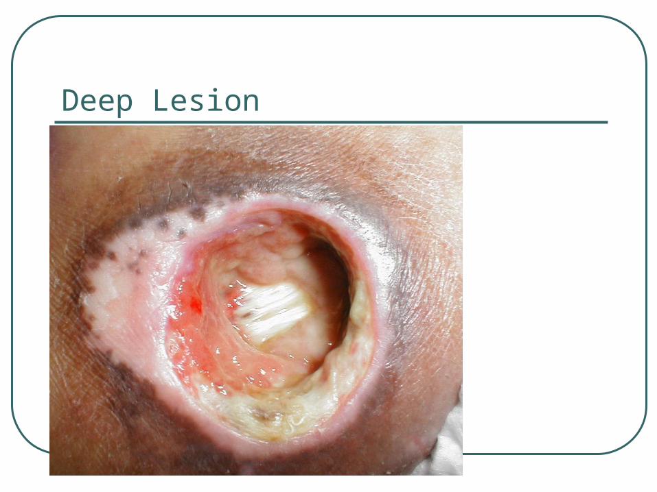
Deep Lesion
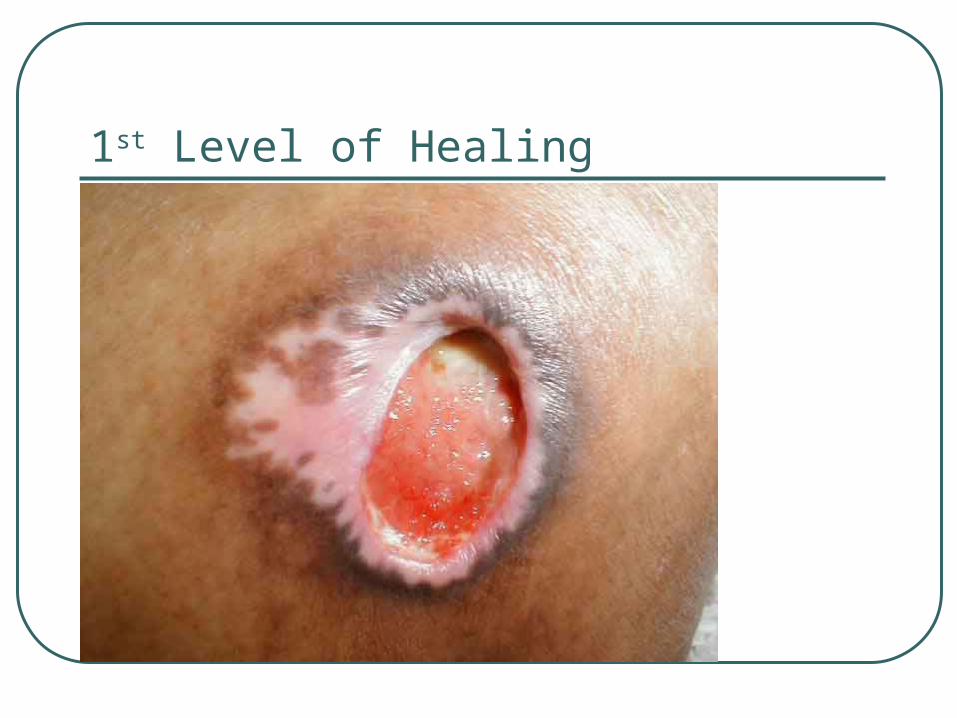
1st Level of Healing
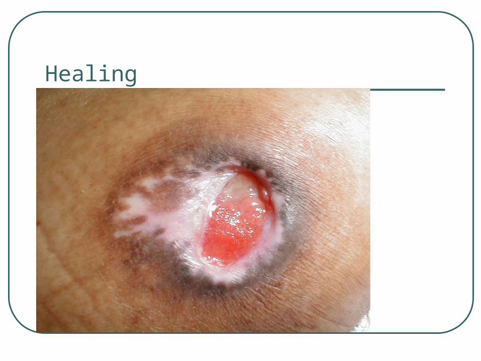
Healing
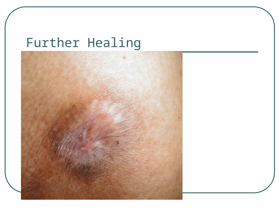
Further Healing
Skin Problems Skin Cancer Prolonged overexposure to sunlight is primary cause
with higher risk for fair skin South and southwest have higher incidence Main types Basal Cell – most common
• Appears as small, shiny, fleshy nodules on the head, neck and/or hands
• Untreated, the nodular lesions will begin to bleed, crust over, and repeat the cycle
• Does not metastasize – may cause localized destruction of tissue
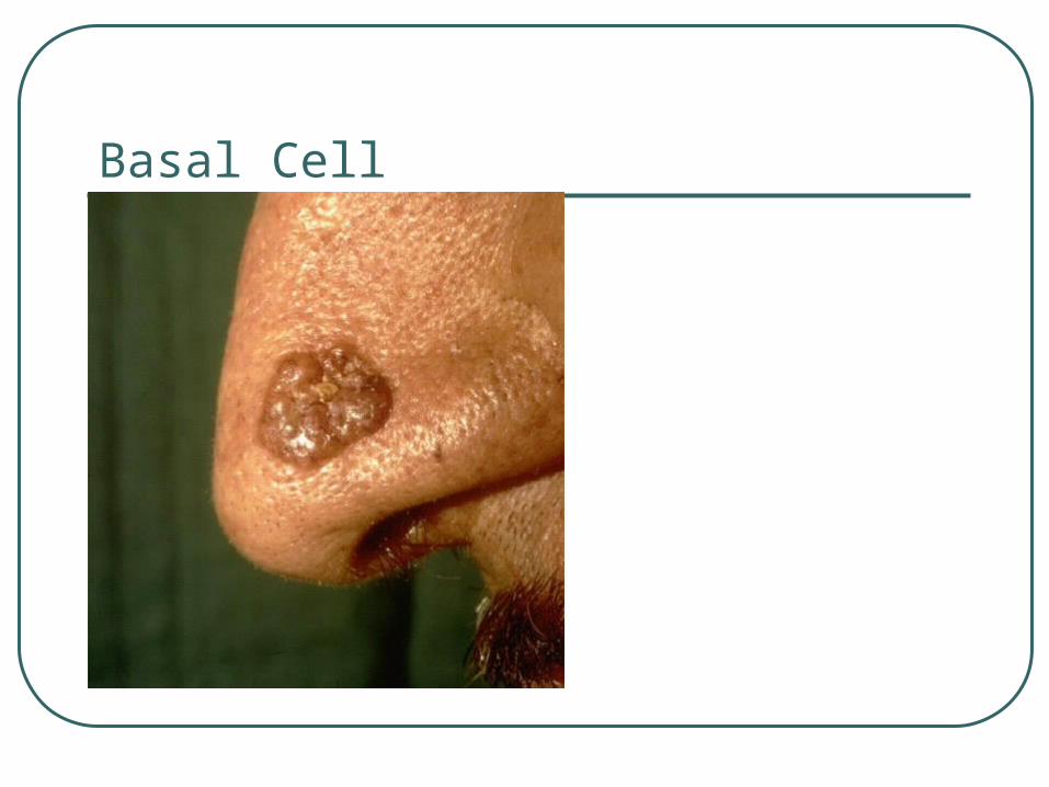
Basal Cell
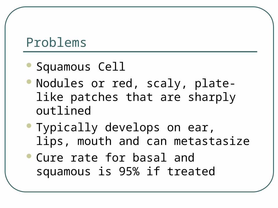
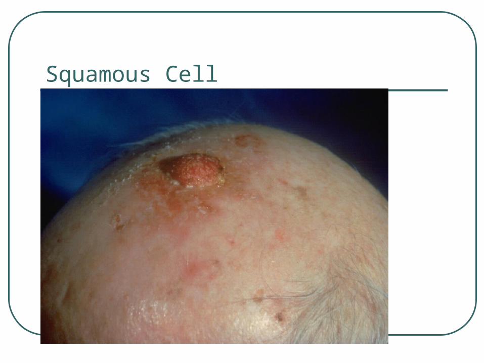
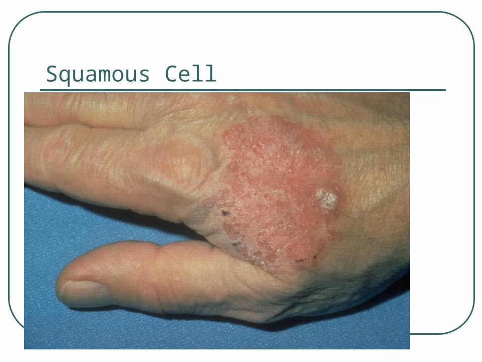
Problems
Squamous Cell Nodules or red, scaly, plate-like patches
that are sharply outlined Typically develops on ear, lips, mouth
and can metastasize Cure rate for basal and squamous is
95% if treated
Squamous Cell
Squamous Cell
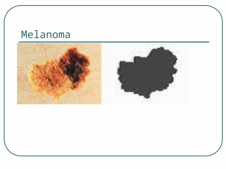
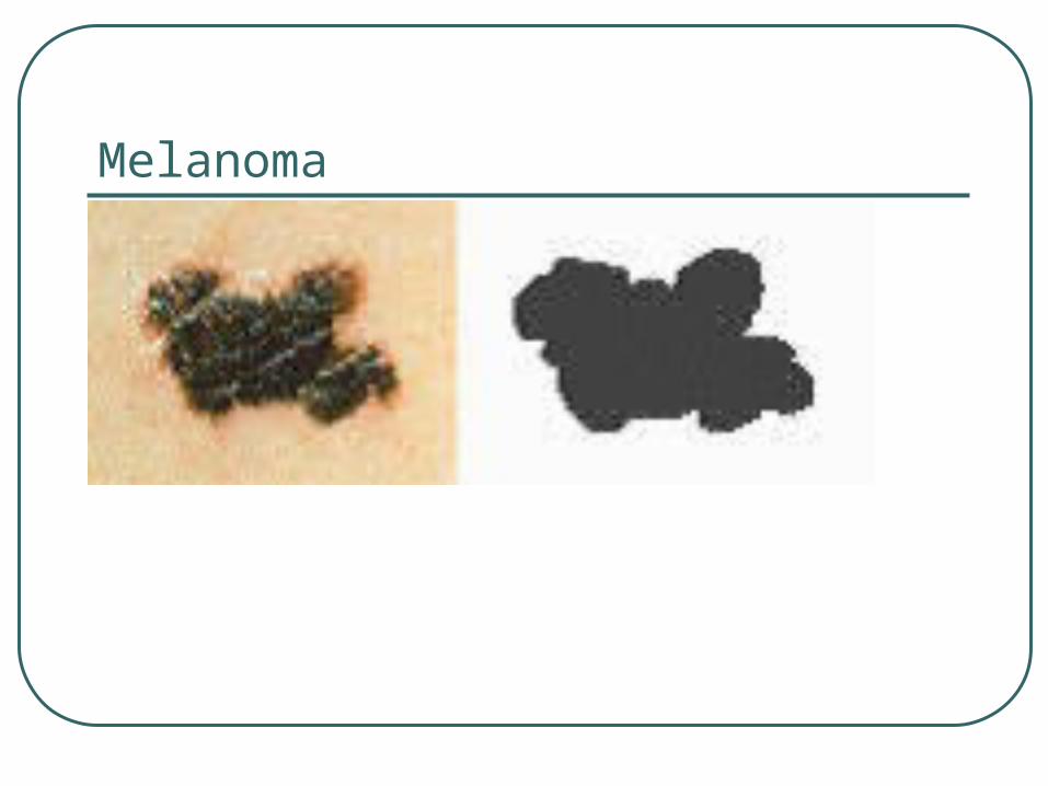
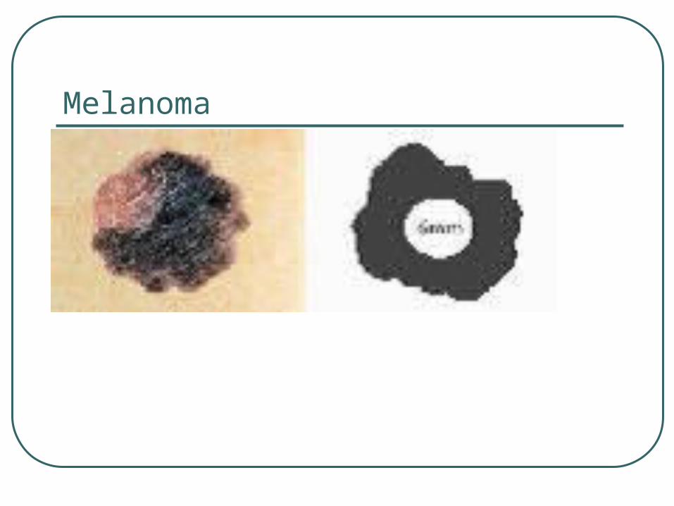
Problems Melanoma Uncommon Serious, can metastasize, can lead to death Appears as a dark brown or black mole-like growth
with irregular borders and irregular pigmentation Lesions may turn shades of gray, blue and white Most common sites are upper back in males and
female, anterior body in males, lower legs in females
Melanoma
Melanoma
Melanoma