intensive care unit to outpatient: a case and physical
TRANSCRIPT

Received 09/02/2017 Review began 09/24/2017 Review ended 12/10/2017 Published 12/14/2017
© Copyright 2017Wilson et al. This is an open accessarticle distributed under the terms ofthe Creative Commons AttributionLicense CC-BY 3.0., which permitsunrestricted use, distribution, andreproduction in any medium, providedthe original author and source arecredited.
Cerebellar Stroke Occupational Therapyand Physical Therapy Management fromIntensive Care Unit to Outpatient: A CaseReportChristopher M. Wilson , Christina L. Mitchell , Katherine M. Hebert
1. Physical Therapy, Oakland University, Rochester, USA 2. Rehabilitation Services, Beaumont Hospital,Troy, Mi 3. Physical Therapy Program, School of Health Sciences, Oakland University, Rochester, Mi
Corresponding author: Christopher M. Wilson, [email protected] Disclosures can be found in Additional Information at the end of the article
AbstractCerebellar stroke increases the risk of extensive physical disability and long-terminstitutionalization. The purpose of this case report is to describe the 14-month longitudinalrehabilitation management and outcomes from the intensive care unit, inpatient rehabilitationunit and outpatient care of a patient after cerebellar stroke. A goal of this case report is toprovide rehabilitation clinicians with a long-term perspective and understanding of the courseof recovery for a patient after cerebellar cerebrovascular accident or related injury. A 51-year-old healthy athletic female experienced acute bilateral cerebellar infarcts with subsequentcraniotomy to remove infarcted areas. The patient had postoperative hemorrhages andhydrocephalus and was deemed to have a poor prognosis. Multimodal sensory stimulation andearly mobility was performed until conventional neuromuscular reeducation interventionscould be tolerated. Primary deficits included decreased proximal strength, whole body ataxia,vertical diplopia, dysphagia, difficulty communicating, and emotional lability. Fourteenmonths after the initial infarcts, the patient was able to reside in her own home with herhusband, ambulate, and stand with assistance and perform most activities of daily living withstandby or set-up assistance. This patient made significant progress toward safety and mobilityand was able to return home despite the early discussion about a poor prognosis and a palliativecare consultation. The complex, intensive course of rehabilitation elicited slow, steady,consistent gains. The patient’s motivation and family involvement likely facilitated optimumoutcomes.
Categories: Neurology, Physical Medicine & Rehabilitation, NeurosurgeryKeywords: cerebellar stroke, multimodal sensory stimulation, early mobility, cerebellar diplopia,stroke, cerebellum, palliative, icu, physical therapy, occupational therapy
IntroductionCerebellar infarction represents approximately 2.3% of acute strokes overall [1]. Largercerebellar infarcts produce symptoms and signs localizing to the brainstem, such as diplopia,dysarthria, limb ataxia, dysphagia, and weakness or numbness. Approximately 10% of patientswith cerebellar infarction can present with isolated vertigo, that is, vertigo with no localized ordiagnostic findings on motor, sensory, reflex, cranial nerve, or limb coordination uponexamination. Most (96%) cerebellar strokes are infarcts of the medial branch of the posteriorinferior cerebellar artery (PICA) [2]. Mortality after cerebellar stroke is higher than that of othervascular territories. This is generally due to concomitant brainstem infarction or compressive
1 2 3
Open Access CaseReport DOI: 10.7759/cureus.1949
How to cite this articleWilson C M, Mitchell C L, Hebert K M (December 14, 2017) Cerebellar Stroke Occupational Therapy andPhysical Therapy Management from Intensive Care Unit to Outpatient: A Case Report. Cureus 9(12):e1949. DOI 10.7759/cureus.1949

hydrocephalus, rather than cerebellar infarction in itself [3].
Kelly et al. [4] examined functional recovery after cerebellar stroke and found that outcomeswere positively correlated with admission functional status into inpatient rehabilitation.Functional gains were negatively correlated with presence of pre-existing comorbidities, analtered level of conciousness upon intial presentation, and the infarct occuring at the superiorcerebellar artery. Mean admission functional independence measure (FIM) score wassignificanlty lower for hemmhorages as compared to infarctions (p=0.006) [4]. Kelly et al.reported a mean improvement in FIM score from admission to discharge of 70 to 93 forcerebellar infarcts [4]. Cerebellar hemmhorage FIM scores also improved from 43 to 74. There isa gap in the literature as it relates to long term progress and comprehensive rehabilitationmanagement for patients after cerebellar stroke.
The purpose of this case report was to describe the longitudinal management for occupationaltherapy (OT) and physical therapy (PT) for a patient with a cerebellar stroke over a 14-monthtreatment course. This case report will provide a long term perspective and outcomes after acomplex and lengthy rehabilitation course to guide rehabilitation clinicians in long-term andshort-term management after a cerebellar stroke. Although speech language pathology (SLP),nursing, social work, medical care, and recreation therapy were integral to the recovery of thispatient, the focus of this article was on OT and PT services. According to the Beaumont HealthInstitutional Review Board (Royal Oak, MI, USA), a case report does not constitute humansubjects research. The patient and her spouse provided written informed consent to publish thiscase report and the associated photographs.
Case PresentationA 51-year-old healthy female arrived at the emergency center presenting with acute dizziness,a severe headache, diaphoresis, diffuse photophobia, nausea, and emesis. The patient reportedthat she had been doing pushups during a televised total body workout and was suddenlyunable to lift herself up. A head computerized tomography (CT) demonstrated that there wasno acute intracranial event. Her symptoms were initially believed to be a vasovagal reaction.She was bradycardic at 25 to 39 beats per minute. The emergency room plan was to monitororthostatic vital signs and aggressively hydrate overnight. The next day a repeat head CTdemonstrated acute ischemia of the left cerebellum mainly in the PICA distribution. Upon thisfinding, the patient was admitted into the hospital diagnosed with an acute cerebrovascularaccident (CVA) in the cerebellum.
Prior to the cerebellar CVA, the patient was active and independent with both basic andadvanced activities of daily living (ADL). Her social history included being a former smoker foreight years, consuming two to three drinks per week, and completing high intensity exercise sixtimes a week (mostly aerobic). Her health history included a patent foramen ovale, twosuperficial venous thrombi, and a history of depression. Prior to admission, her medicationsincluded aspirin 81 mg daily, estradiol 0.5 mg, ibuprofen 200 mg, a daily multivitamin, nystatinsuspension, and 500 mg of vitamin C. The patient was living with her spouse and four childrenin a two story home with a two-step entry. Her bedroom and full bath were on the second floorwith a half bath on the first floor. Two of her children were attending college and two teenagedchildren were living at home. Her husband worked full time as an engineer but had a veryflexible schedule and she had a large social network for support after discharge.
Initial emergency room visit and symptom progressionBased on her diagnosis and symptoms, the patient received an evaluation by SLP as well as PTand OT. She was found to have double vision, dizziness when sitting up, and complained of aheadache that she subjectively rated as a 7/10 (0 = no pain and 10 = worst pain). She
2017 Wilson et al. Cureus 9(12): e1949. DOI 10.7759/cureus.1949 2 of 23

demonstrated an initial score of three on the National Institute of Health Stroke Scale and wasplaced on the hospital stroke protocol. She had normal range of motion (ROM), musclestrength, tone and sensation. It was found that her coordination was impaired, her gait wasataxic without an assistive device and supervision was required for bed mobility and transfers.The patient scored a 2/6 (0=no impairment, 6=severe disability/bedridden) on the modifiedRankin scale. A PT treatment plan was established for balance retraining during her initial visitsbut subsequent medical events did not allow for PT interventions to be performed.
Four days after her admission, she was found face down in bathroom after ambulating thereindependently. She was alert, oriented, and responding appropriately. A head CT revealedhydrocephalus with dilation of the lateral ventricles including temporal horns and thirdventricle, along with fourth ventricle compression and brain stem compression due to edema ofa left cerebellar infarct. She became progressively more lethargic and almost non-verbal, butstill was able to follow commands. The patient was then transferred to the intensive care unit(ICU) and had emergency surgery for a left suboccipital craniotomy with removal of theinfarcted areas in the cerebellum. After surgery, a head CT demonstrated a new hemorrhageand gas within the operative cavity along with an increased mass effect on the left side of thefourth ventricle which was completely effaced with moderate obstructive hydrocephalus. Laterin the day the patient demonstrated a reduced level of consciousness and was taken to theoperating room again where a right frontal ventriculostomy was performed to decrease thehydrocephalus. Postoperative cerebellar damage is depicted via magnetic resonance imaging(MRI) on Figure 1. She subsequently had a posterior fossa decompression and she was deemedto have an overall poor prognosis.
2017 Wilson et al. Cureus 9(12): e1949. DOI 10.7759/cureus.1949 3 of 23

FIGURE 1: Brain Magnetic Resonance Imaging afterCraniotomyBrain MRI (T2 Weighted) without gadolinium completed two months after left occipital craniotomyand evacuation of infarcted left cerebellar tissue. Images demonstrate left cerebellar subacute andchronic infarctions as well as an area of acute infarction within the pons.
Three days after her fall and subsequent surgeries, the patient remained orally intubated andunresponsive. The patient’s intracranial pressure (ICP) was found to be elevated and she had anexternal ventricular drain (EVD) placed for therapeutic drainage of her cerebrospinal fluid(CSF). She subsequently developed a hemorrhage surrounding her EVD in the right frontal lobeand a hemorrhage within the right lateral ventricle. The next day she underwent surgery toreopen her suboccipital craniotomy for evacuation of a clot. Seven days after her fall andsurgery, she remained intubated and on a ventilator. By this time, her medical status became
2017 Wilson et al. Cureus 9(12): e1949. DOI 10.7759/cureus.1949 4 of 23

more stable overall, however she remained verbally unresponsive and demonstrated a positiveBabinski sign on the left foot. In light of her poor prognosis, the patient received a palliativecare consultation to assist in coordinating care and decision making 11 days after her fall andsurgery. Twelve days after her initial fall, a tracheostomy was completed and she remained onventilator support. Two days after the tracheostomy, she was successfully transferred tosupplemental oxygen at 28% FiO2 via a tracheostomy collar.
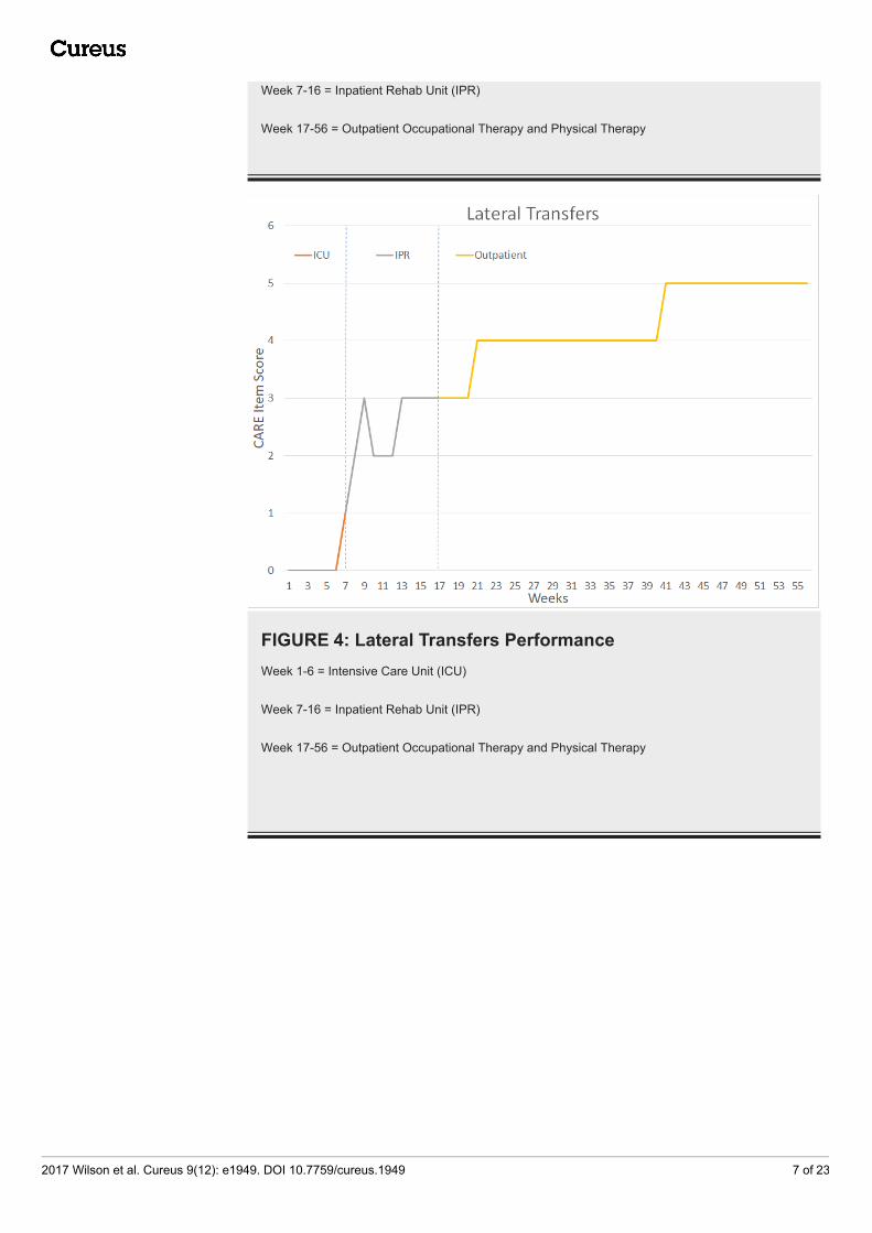
Results and interventions during occupational therapy andphysical therapyThe patient underwent a total of 14 months of PT and OT – six weeks in the intensive Care Unit(ICU), approximately 10 weeks in Inpatient Rehabilitation (IPR), and 41 weeks of outpatientrehabilitation. Figure 2 outlines key milestones within her ICU and IPR rehabilitation. Figures3-7 depict the patient’s overall progress using the Continuity Assessment Record andEvaluation (CARE) item scoring for major activities of daily living (ADL) and physical mobilitytasks across the continuum of care. See Table 1 for CARE item scoring and interpretation.
FIGURE 2: Intensive Care Unit and Inpatient Rehabilitation UnitTimelineOT = occupational therapy, PT = physical therapy, RX = treatment, PEG = percutaneousendoscopic gastrostomy, ICU = intensive care unit, IPR = inpatient rehabilitation unit, D/C =discharge
2017 Wilson et al. Cureus 9(12): e1949. DOI 10.7759/cureus.1949 5 of 23

Score Description
6 Independent – Patient completes the activity by him/herself with no assistance from a helper.
5Setup or clean-up assistance – Helper SETS UP or CLEANS UP; patient completes the activity. Helper assistsonly prior to or following the activity.
4Supervision or touching assistance – Helper provides VERBAL CUES or TOUCHING/STEADYING assistanceas patient completes activity. Assistance may be provided throughout the activity or intermittently.
3Partial/moderate assistance – Helper does LESS THAN HALF the effort. Helper lifts, holds or supports trunk orlimbs, but provides less than half the effort.
2Substantial/maximal assistance – Helper does MORE THAN HALF the effort. Helper lifts or holds trunk or limbsand provides more than half the effort.
1 Dependent – Helper does all of the effort. Patient does none of the effort to compete the task.
0Activity not attempted/completed due to medical condition, safety concerns, environmental constraints, or patientrefused.
TABLE 1: Continuity Assessment Record and Evaluation (CARE) Item Coding Keyhttps://www.cms.gov/Medicare/Quality-Initiatives-Patient-Assessment-Instruments/Post-Acute-Care-Quality-Initiatives/Downloads/CARE-Institutional-Admission-Assessment-Tool.pdf
FIGURE 3: Bed Mobility PerformanceWeek 1-6 = Intensive Care Unit (ICU)
2017 Wilson et al. Cureus 9(12): e1949. DOI 10.7759/cureus.1949 6 of 23
Week 7-16 = Inpatient Rehab Unit (IPR)
Week 17-56 = Outpatient Occupational Therapy and Physical Therapy
FIGURE 4: Lateral Transfers PerformanceWeek 1-6 = Intensive Care Unit (ICU)
Week 7-16 = Inpatient Rehab Unit (IPR)
Week 17-56 = Outpatient Occupational Therapy and Physical Therapy
2017 Wilson et al. Cureus 9(12): e1949. DOI 10.7759/cureus.1949 7 of 23

FIGURE 5: Sit-To Stand Transfers PerformanceWeek 1-6 = Intensive Care Unit (ICU)
Week 7-16 = Inpatient Rehab Unit (IPR)
Week 17-56 = Outpatient Occupational Therapy and Physical Therapy
2017 Wilson et al. Cureus 9(12): e1949. DOI 10.7759/cureus.1949 8 of 23

FIGURE 6: Ambulation PerformanceWeek 1-6 = Intensive Care Unit (ICU)
Week 7-16 = Inpatient Rehab Unit (IPR)
Week 17-56 = Outpatient Occupational Therapy and Physical Therapy
FIGURE 7: Grooming and Dressing PerformanceWeek 1-6 = Intensive Care Unit (ICU)
Week 7-16 = Inpatient Rehab Unit (IPR)
Week 17-56 = Outpatient Occupational Therapy and Physical Therapy
Intensive Care Unit Rehabilitation
The patient was in the ICU for a total of six weeks and received 19 visits of OT, 12 visits of PT,and 17 visits of SLP. The OT sessions lasted 30 to 45 minutes while PT sessions lasted 30minutes. Occupational therapy was focused on multimodal sensory stimulation (MSS) and oncethe patient was responsive enough to benefit from early mobility, PT wasinitiated. Occupational therapy was performed immediately prior to PT to optimizewakefulness and participation in early mobility activities. After PT/OT treatment plans wereestablished, they prescribed complimentary restorative aide services (trained technicianssupervised by OTs and PTs who assist patients with exercises, general mobility and ROM) for atotal of eight times while in the ICU.
2017 Wilson et al. Cureus 9(12): e1949. DOI 10.7759/cureus.1949 9 of 23

Occupational therapy evaluation, interventions and outcomes: Three days after the initial falland surgery, the patient was evaluated by OT. Initially she presented as minimally responsivewith her eyes closed throughout the session; she was non-verbal with no attempts tocommunicate and was flaccid in both arms and legs with mild edema, and dependent with allADL and functional mobility. Goals of OT were to increase environmental engagement andresponse to stimuli to begin early mobility with physical therapy.
The majority of the ICU OT treatment sessions consisted of MSS, which is an application of aseries of sensory techniques that are customized in occurrence and amount in order to increasearousal and awareness to elicit a purposeful behavior. MSS is also known as multi-sensorystimulation or “coma stim” [5]. The MSS therapy consisted of interventions such as wiping acold washcloth along the patient’s face, neck and wrist and clapping loudly near the patient’sears. The patient initially responded to the MSS with an elevated heart rate and blood pressure,and then began communicating by nodding and biting on her endotracheal tube, turning herhead away, and wiggling her fingers. Subsequent OT treatments in the ICU included bedmobility and ADL retraining for which the patient was initially dependent. As the patientbecame more responsive, neuromuscular re-education techniques were used such as thetherapist placing the patient’s right hand on her hair and asking her to lower her arm to touchher eyes, nose and mouth, use of a universal cuff on the right upper extremity to assist withbrushing her teeth and preparing to eat. Other various treatments that were utilized in the ICUincluded grasping and releasing cones and sponges, muscle tapping/brushing, a bilateralpectoral myofascial release, joint compression in order to increase proprioceptive output,visual tracking, and the application of L’nard boots (AKA preventative ankle foot orthoses) toprevent foot drop and a right eye patch to address double vision. In the last week of thepatient’s stay in the ICU, she demonstrated moderate to maximal assistance with grooming,she was dependent with dressing and toileting, required maximal assistance with side-to-sidebed mobility and supine-to-sit transfers, and continued to have poor static and dynamic seatedbalance. At discharge from the ICU, her alertness improved and she displayed consistentcommand following and was progressing to using one or two fingers and head motions forno/yes with good accuracy and she was consistently alert and oriented to one to two.
Physical therapy evaluation, interventions and outcomes: Physical therapy treatments wereinitiated 30 days after the patient’s initial fall and eight days after OT was initiated in the ICU.Key evaluation findings demonstrated full passive range of motion but lack of purposefulmovement, however the patient did occasionally move her arms and legs spontaneously againstgravity. Initial PT treatments consisted of passive range of motion (PROM) and progressing toactive assistive range of motion (AAROM) and stretching. The physical therapist also provideda home exercise program and fall prevention education to the family and nursing. In addition,early mobility procedures were initiated including progressive sitting up in bed, at the edge ofthe bed and tilt table [6]. At week five, the tilt table was introduced with intervals of two to tenminute holds to allow her vitals and symptoms to stabilize. At the end of her ICU stay (or after aweek-and-a-half of treatment) she was able to tolerate a 70 degree tilt-table angle for 45 to 55minutes three times per day.
Inpatient Rehabilitation Unit
The patient was directly admitted to the IPR unit after a six week ICU stay. During inpatientrehabilitation, the patient received a total of 54 visits of OT, 51 visits of PT, 47 visits of SLP and22 visits of recreation therapy. The patient was in the inpatient rehab setting for 10 weeks andconsistently maintained the admission criteria for three hours per day averaging five to sevendays per week. Throughout the IPR stay, the patient’s husband was active, engaged andparticipated in the entirety of the educational process of rehabilitation.
2017 Wilson et al. Cureus 9(12): e1949. DOI 10.7759/cureus.1949 10 of 23

Occupational therapy evaluation, interventions and outcomes: The OT evaluation founddecreased muscle strength grossly to both shoulders at 1/5, elbow and wrist strength was 2/5,while the forearm and hands were 3-/5. The patient demonstrated hypotonicity which wasaccentuated proximally, as well as dymetria and dysdiadochokinesia of both arms. Shedemonstrated truncal ataxia resulting in poor sitting balance and required total assist withupper body and lower body dressing, bathing, toileting, and all bed mobility. The patientrequired total assist of three clinicians during a partial-stand bed-to-chair transfer using asliding board. She was unable to tolerate static sitting without total assistance of two people tomaintain her balance. She had not advanced to wheelchair propulsion by this time and was stillutilizing a trach collar with humidified air. She required a soft cervical collar during transfersand upright activities as she did not have the neck extensor strength to lift her chin off of hersternum against gravity. The patient denied any pain during the IPR series of visits with theexception of pain reproduced during weight bearing on the upper extremities during sitting atthe edge of the mat.
Her initial series of OT visits emphasized control of upright sitting with cueing as well asutilizing kinesiology tape to facilitate cervical extension. After two weeks in IPR, the patient nolonger required the soft collar during transfers. The patient participated in upper extremityROM progressing from gravity-eliminated/AAROM and also received scapular mobilizations.Vision treatments included application of tactile and auditory stimulation for coordination ofeye active range of motion (AROM) and a trial of clear eyeglasses with a unilateral gauze patchover the right eye for diplopia. The patient also performed gaze stabilization exercises as well asprogressive transfer activities incorporating tracking and planning. By week four in IPR, thepatient demonstrated improvement in bed mobility from maximum to moderate assist withimproved motor control. She had also improved to requiring moderate assistance during staticsitting balance. At this time, the patient was able to perform a standing pivot transfer from abed to a chair with maximum assistance of one person.
The patient demonstrated static sitting balance of 40 seconds with steadying assist. By weeksix, the patient was able to propel the wheelchair 30 feet utilizing only her legs for propulsion asshe was unable to grasp or elevate her upper extremities to the propulsion level of thewheelchair at this point; this improved to 50 feet by week seven of IPR. During week seven andeight in IPR, specific treatments included emphasizing wheelchair seating and positioning withtasks such as leaning forward in her wheelchair to rest her elbows on tops of her thighs andreturning to upright as well as wheelchair push-ups and lower body dressing. Vision treatmentsincluded activities such as visual fixation on targets with lateral head movements, letter andnumber reading, and utilization of the Nintendo Wii emphasizing hand-eye coordination.
By week eight, the patient only required moderate assistance with transfers and brief staticstanding. Her wheelchair sitting tolerance improved to 20 minutes. By week nine, OTtreatments emphasized reaching forward and down to the ground while in a wheelchair withreturn and upper extremity stretching and strengthening focusing on optimizing functionalreaching activities. Intervention strategies also included reaching activities while in a standingposition utilizing steadying assist and incorporating hand-eye coordination. During her lastweek in inpatient rehabilitation, she displayed active left shoulder elevation of 110° and 140°on the right and gross left arm strength of 3/5 while the right arm was grossly 3+/5. The patientwas rolling side-to-side with standby assist and her static sitting balance was also standbyassist. Static standing required minimum assist while dynamic balance required maximalassistance. Her tub/shower and bed-to-chair transfers required moderate assistance.
Physical therapy evaluation, interventions and outcomes: The patient’s lower extremity musclestrength via manual muscle testing on initial admission into IPR was 2-/5 for hip flexors whileknee extensors, knee flexors and dorsiflexors were 3/5. As noted in the OT evaluation, thepatient required maximum assistance/total assistance of two to three people and utilized a
2017 Wilson et al. Cureus 9(12): e1949. DOI 10.7759/cureus.1949 11 of 23

cervical collar to prevent neck flexion during transfers. The patient demonstrated a standingtolerance of five to ten minutes with maximum assistance. During exercises the patientrequired continuous verbal cueing and emphasis on breathing techniques. The initial series oflower extremity exercises were AAROM and as the patient progressed to tolerating increasedweightbearing and upright activity, open chain activities were modified to be performed inclosed chain in varying planes of movement. Upon week two, the patient received assistancewith standing activities while using a standing frame and SARA Plus mechanical standing unit(ArjoHuntleigh, Addison, IL http://www.arjohuntleigh.us/) for assisted pre-gait activity withmoderate assistance.
By week two, the patient was tolerating light ankle weights of one pound for long arc quads andprogressing to active range of motion from active-assistive. Throughout the treatmentsequence, the patient was assisted in use of a recumbent elliptical machine; this initiallyfocused on the lower extremities but as the patient’s grasp improved, upper extremities werealso incorporated. The patient also transitioned to using a mechanical ceiling-mounted gaitharness and was able to tolerate ambulation over 18 m (60 ft) with maximum assistance of twopeople. The patient was propelling her wheelchair with moderate assistance for 30 m (100 ft)forward and backwards (Figure 8). During this propulsion, she demonstrated limitations withtrunk and hand control that affected her ability to maintain a midline position.
2017 Wilson et al. Cureus 9(12): e1949. DOI 10.7759/cureus.1949 12 of 23

FIGURE 8: Rest Break during Gait TrainingSeated rest break during gait training in inpatient rehabilitation unit. Gait training was performedusing a rolling walker and ceiling-mounted harness for partial unweighting and fall prevention
By week four of PT in IPR, the patient had progressed to gait training activities utilizing theLiteGait assisted gait system (LiteGait, Tempe AZ, http://www.litegait.com/) and the patientwas able to support 60-90% of her body weight with 4.5-8 kg (10-40 lbs) of assistance from theharness for 92 m (301 ft) on even surfaces. This progressed to 227 m (745 ft) later that weekutilizing a platform rolling walker. By week four, she was able to participate in sit-to-standtransfers with minimum to moderate assistance however pivot transfers and sustained standingcontinued to require maximum assistance. By week six, the patient was performing gait
2017 Wilson et al. Cureus 9(12): e1949. DOI 10.7759/cureus.1949 13 of 23

training with the SARA Plus walking sling for distances up to 366 m (1,200 ft) with threestanding rest breaks with moderate assist with 0-11 kg (0-25 lbs) of unweighting at 0.22 m/s (0.5mph). She began unsupported gait training with a platform walker and bilateral hand splintsbut required two seated rest breaks over 60 m (190 ft).
Upon week seven, the patient was able to sit at the edge of the mat with her hands on her lapfor 90 seconds with supervision while external perturbations and isometric resistance wereapplied. The patient was also able to begin car transfer training with moderate to maximumassistance. She was now able to propel her wheelchair 24 m (80 ft) but as noted above in the OTsection, this task mainly utilized her lower extremities due to poor handgrip and upperextremity strength. The patient performed sit-to-stand transfers with minimum assistance andrequired moderate to maximum assistance for overall gait training. Standing pivot transfershad progressed to minimum assistance and she was able to roll in bed with steadying assist,however, she still required moderate assistance for other bed mobility tasks. The LiteGait forgait training continued to be emphasized and the patient was able to ambulate 426 m (1400 ft)with 0-11 kg (0-25 lbs) of unweighting at 0.22-0.27 m/s (0.5-0.6 mph). During gait training withher platform walker, she no longer required a hand splint for the right upper extremity but stillrequired this for the left hand to facilitate grasp. She was able to begin stair training with a 15cm (6 in) step with maximum assistance of two people.
By week nine, the patient was safe enough to perform gait training with one person assistancewithout a wheelchair following her. She was able to manage her rolling walker independentlyand was displaying improvement in balance during dynamic standing activities and stairtraining. The patient’s therapeutic exercise had subsequently progressed to standing heel andtoe raises, marching, mini-squats, ankle pumps, seated alternating hip flexion, supine bridgingand balance and core strengthening activities while reaching outside of her base of support.The patient’s sit-to-stand transfers had improved to minimum/moderate assist (depending onfatigue) and her bed mobility also required only minimum assistance. Upon discharge from IPR,the patient was sidestepping in the parallel bars 9 m (30 ft) for two repetitions with moderateassistance. The patient was able to perform wheelchair propulsion using both legs and arms for46 m (150 ft) with standby assistance. She was able to ascend and descend a 15 cm (6 in) stepfour times using a handrail with maximum assistance of two individuals. She was able toperform a five-times-sit-to-stand test in 33 seconds with minimum assistance in the parallelbars. By week 10, the patient could perform daily bathing, grooming, showering, and functionalmobility tasks with moderate assistance from her husband without cues from the therapists.This allowed for safe discharge to her adapted home environment. Home adaptations included awheelchair ramp and was discharged to home with a wheelchair, platform walker, shower chair,tub transfer bench and subsequently received outpatient PT and OT after a one week gap intreatment due to the holidays.
Outpatient Rehabilitation
Upon initiation of outpatient PT and OT seventeen weeks after her fall and craniotomy surgery,the patient presented with decreased core strength requiring a specialized wheelchair forpositioning and stability. During outpatient rehabilitation, the patient received a total of 117visits of OT, 115 visits of PT, and 110 visits of SLP over 41 weeks of rehabilitation.
She displayed whole body ataxia as well as bilateral upper and lower extremity weakness withthe left side weaker than the right. She required moderate assistance from her family for all ADLand transfers. The patient relied on a percutaneous endoscopic gastrostomy (PEG) tube fornutrition due to risk of aspiration and received patching of the left eye to address her doublevision and left eye palsy. It should be noted that in IPR the patient's right eye was patched asher right eye dysfunction was limiting her performance on functional retraining but in
2017 Wilson et al. Cureus 9(12): e1949. DOI 10.7759/cureus.1949 14 of 23

outpatient occupational therapy, retraining the visual musculature could be emphasized andthe patch was switched to the left eye. Her cognitive status allowed her to follow multiple stepcommands to perform moderately complex tasks.
Occupational therapy evaluation, interventions and outcomes: Occupational therapy wasinitiated three times a week for 45 to 60 minutes per session. Her OT intervention initiallyfocused on family education for home exercise program (HEP), core stability activities whilelaying supine on a mat with moderate to maximal assist, and unsupported sitting withmoderate assistance. Improvement in ADL were monitored via the Patient-Specific FunctionalScale (PSFS) and are noted in Table 2. During sitting she displayed frequent losses of balanceand continued truncal ataxia. Scapular mobilization and kinesiology taping was utilized toencourage upright posture as well as to facilitate normalized motion of both arms. Monocularvisual perception activities and tracking activities were utilized in conjunction with taping tothe central portion of the patient’s left glasses lens to allow for continued peripheral vision andlight input.
Outpatientevaluation
Month1
Month2
Month3
Month4
Month5
Month6
Month7
Month8
Month9
Transfer from w/c tosurfaces
1 2 4 5 6 6 7 6-7
8(bed),5(toilet)
8(bed), 7(toilet)
Dressing UB/LB 1 3 4 5 6
Toileting 0 3 4 5 6 6 6 7 7 7
Dressing UB 8 8 8 9 9
Dressing LB 5 6 8 8 8
Feeding self 6 8 8 9
Handwriting 2 4 4
TABLE 2: Outpatient Occupational Therapy Patient Specific Functional ScalePSFS scoring: 0 = inability to perform the activity at all; 10 = ability to perform the activity at the same level as before the injury orcondition
Key: w/c = wheelchair, UB = upper body, LB = lower body
By the second month, fine motor coordination (FMC) activities were initiated utilizing largesized pegs and beads requiring hand-over-hand assistance for fine motor tasks to facilitatenormalized movement patterns secondary to ataxia (Table 3). Interventions also focused onmodified ADL tasks with an emphasis on weight bearing through alternating upper extremitiesto incorporate into HEP. Neuromuscular re-education with weight bearing and proprioceptiveneuromuscular facilitation (PNF) D1/D2 patterns were completed while sitting at the edge ofthe mat with minimal to moderate assistance to avoid loss of balance. During the third month,a resisted upper extremity exercise program was initiated with medium resistance elastic bandwith scapular retraction and shoulder horizontal abduction/adduction with trunk rotation to
2017 Wilson et al. Cureus 9(12): e1949. DOI 10.7759/cureus.1949 15 of 23

strengthen her core. She required minimal assistance to maintain her posture during theseactivities. She also participated in standing level neuromuscular re-education activities andgross motor coordination (GMC) tasks. The patient demonstrated an increased ability to self-correct her posterior loss of balance while in standing and this required minimal assistancewhile reaching minimally outside her base of support. A re-evaluation was completed after thethird month which provided evidence of an increase in control and strength for completion ofmodified ADLs and transfers with minimal to moderate assistance with perturbations 50% ofthe time.
Month of Testing (units inkg unless otherwise noted)
Hand griptesting(R/L)
Three JawChuck(R/L)
Twopoint(R/L)
LateralPinch(R/L)
Nine Hole Peg(seconds)(R/L)
Box and Block Test(number of blocks)(R/L)
Outpatient evaluation 24/5unable/unable
unable/unable
11/6 unable /195 N/A
1 month 24/8 8/5 7.5/4.75 11/7 280/141 N/A
3 months 36/25 11/8 9/6 14/11.5 150/48 12/19
6 months N/A N/A N/A N/A 111/56 14/22
9 months 44.3/35 15/11 12/8.5 18/12 60.6/39 17/26
10 months 53.3/ 36.6 15/11.5 12.5/8.5 18.5/13.5 85.7/38.3 17/24
Discharge - 10.5 months 54.3/33 17/13 13/10 20/14 87/39 16/24
TABLE 3: Outpatient Hand Function Results
During months four and five, she demonstrated an increase in core stability and improvedcoordination and control of her upper extremities. Core strengthening interventions includedquadruped positioning on the mat with alternating arm-and-leg-lifts with minimal assistanceas well as neuromuscular re-education techniques while seated on a foam pad reaching outsideher base of support with minimal assistance to maintain balance. During FMC training withmedium-sized pegs, she was overshooting 35% of the time. In addition, the patient performedlight resistance clothespins and therapeutic putty to improve hand strength and dexterity.
By six months after initiation of outpatient OT, her upper extremity strength and core controlbecame functional enough to focus on achievement of independence with basic self-care needsincluding dressing, toileting and feeding. Her interventions continued to include graded coreand upper extremity strengthening and FMC/GMC tasks. The patient was introduced tomodified techniques for dressing in supine as well as adaptive equipment for self-feeding asshe was now able to eat with a modified diet and no longer required a feeding tube. Due to hercontinued need for a wheelchair for mobility, during months seven and eight, she receivedinstruction in adaptation of kitchen and laundry tasks at the wheelchair level with emphasis onsafety and adapted performance. She also started a writing program with her non-dominant lefthand as coordination was improving faster than her right hand. Through months nine and ten,interventions continued to focus on modified techniques and safety with both basic andadvanced ADLs as well as functional transfers and independence at wheelchair level inpreparation for discharge.
2017 Wilson et al. Cureus 9(12): e1949. DOI 10.7759/cureus.1949 16 of 23

Physical therapy evaluation, interventions and outcomes: Physical therapy was initiated inconjunction with OT and SLP, three times a week for 45 minute sessions each. Upon initialevaluation, the patient’s Postural Assessment for Stroke Scale (PASS) score was 11/36 (0=maximal disability, 36= no disability). Interventions for physical therapy focused on dynamicand static sitting and standing balance activities, pre-gait and family education. Improvementin physical mobility were monitored via the Patient-Specific Functional Scale (PSFS) and arenoted in Table 4.
Activity Month 6 Month 7 Month 8 Month 9
Independent bed mobility: supine to sit 9 10 10 10
Gait with U-step with assist 7 7 8 9
Gait with U-step up and down 6-8 inch step in home 0 1 3 5
Standing balance unsupported with assist 3-4 4 5 7
Transfers: w/c to mat 7-8 8-10 9-10 9
Car transfer 9 10 8 10
Ascend stairs with bilateral handhold 7 (min) 5 (SBA) 6 (SBA) 6 (SBA)
Down stairs with bilateral handhold/sideways 6 (min) 3-4 (SBA) 5 (SBA) 6 (SBA)
Sit to stand from w/c using U-step 3 (SBA) 5 (SBA)
Standing dynamic balance for ADL 1 (SBA) 1
TABLE 4: Outpatient Physical Therapy Patient Specific Functional Scale (PSFS)PSFS scoring: 0 = inability to perform the activity at all; 10 = ability to perform the activity at the same level as before the injury orcondition
Key: w/c = wheelchair, ADL = activities of daily living, min = minimal assistance, SBA = standby assistance
Therapeutic activities included unsupported trunk rotation with weighted ball and seatedmarching with alternating upper extremity requiring moderate assistance for support andupright posture. The patient also participated in parallel bar side-stepping with moderateassistance at the hips to decrease overstepping. A rolling walker weighted with 2.27 kg (5 lbs)was utilized during gait training with moderate assistance for 46 m (150 ft). She alsodemonstrated standing pivot transfers with moderate assistance with external perturbations.After one month, her PASS score was 26/36. Months two and three focused on core stabilityexercises in supine and quadruped via crawling forward and backward, supine-to-kneeling andmini push-ups with moderate assistance. Interventions also focused on fall recovery as thepatient demonstrated a risk of anterior loss-of-balance from the toilet at home. Her timed-up-and-go score progression is noted in Table 5.
2017 Wilson et al. Cureus 9(12): e1949. DOI 10.7759/cureus.1949 17 of 23

Month Time and relevant details
2months
131 seconds with rolling walker and partial/moderate assist
3months
100 seconds with rolling walker and supervision/touching assist
5months
71 seconds with rolling walker and supervision/touching assist
6months
91 seconds with U-step walker and partial/moderate assist
6.5months
72 seconds with U-step walker and partial/moderate assist
7months
78 seconds with U-step walker and supervision/touching assist (increased time, however improved motorcontrol and less physical assistance needed with turns)
8months
63 seconds with U-step and supervision/touching assist
9months
59 seconds without assistive device and supervision/touching assist which increased to parttial/moderate assist during turning
10months
51 seconds with no assistive device and supervision/touching assist
TABLE 5: Timed Up and Go (TUG) Results
At four months, her PASS score was 28/36 and was not repeated after this date. Pre-gait in theparallel bars was advanced to incorporate single leg stance, step tap with alternating lowerextremities in both the ascending and descending direction; her anterior step-downs andposterior step-ups with external perturbations required moderate to maximal assistance for fallprevention. As her core stability increased in months five and six and she was no longer fallingforward during transitional movements, her gait training progressed to outdoor, unevensurfaces, and stair training. At the beginning of month five, the patient’s Berg balance scorewas 13/56 (56 = normal balance) and by the end of the sixth month, it had improved to 17/56and was no longer reported in the medical record. More advanced strengthening and balanceactivities were initiated including lunges, squats, standing ball bouncing and tandem weightshift with minimal assistance to maintain balance. She was also started on a program with theLiteGait and treadmill as well as an elliptical machine. By month seven, the patient was startedon a U-step walker (In-Step Mobility Products, Skokie, IL, 60076. www.ustep.com) to aid inambulatory control and balance. Transfer training shifted focus toward family education andsafety to prevent falls. Months eight through ten focused on core strength and stability, lowerextremity strengthening, gait training with the U-step with minimal assistance from family,and independence at wheelchair level with self-propulsion.
By month ten, the patient had started to plateau with her functional level and was beingprepared for discharge. Overall she progressed in her independence for both basic and advancedADL and transfers. She was able to self-propel her wheelchair independently. Initially requiring
2017 Wilson et al. Cureus 9(12): e1949. DOI 10.7759/cureus.1949 18 of 23
moderate to maximal assistance, her transfers to and from common household surfaces (bed,toilet and kitchen chair) now only required steadying assist/close supervision. Shedemonstrated increased core stability and upper extremity control and coordination toparticipate with upper and lower body dressing and toileting needs which only requiredsteadying assist (she initially required moderate to maximal assist). She was able to staticallystand while leaning her trunk against a counter to participate with light advanced ADL taskssuch as folding laundry and simple meal preparation with close supervision (Figure 9). She wasable to propel her wheelchair around the house and assist with setting and clearing the table formeals. She resumed modified leisure activities such as working in her garden, painting and bikeriding. She was able to utilize the U-step walker with minimal assistance from her family andcaregivers to move around the house and community with good tolerance for activity. Shecontinued to require support and supervision at home from family and hired assistance toensure safety although she was able to function in her environment at the wheelchair level withlittle to no assistance.
2017 Wilson et al. Cureus 9(12): e1949. DOI 10.7759/cureus.1949 19 of 23

FIGURE 9: Standing-Level Meal PreparationPatient demonstrating ability to complete independent meal preparation in her own home
DiscussionThe purpose of this case study was to describe the longitudinal management and outcomes forOT and PT for a patient with a cerebellar stroke with surgical excision over a 14-monthtreatment course. This patient had an exceptionally long and complex continuum of care thatrequired close coordination and facilitation between disciplines and care settings. A goal of thiscase report was to provide rehabilitation clinicians with a long-term perspective andunderstanding of the course of recovery for a patient after cerebellar cerebrovascular accident
2017 Wilson et al. Cureus 9(12): e1949. DOI 10.7759/cureus.1949 20 of 23

or related injury. Kelly et al. [4] examined outcomes after cerebellar stroke in 58 patients. Theresults of this case study are consistent with this finding as this patient demonstratedsignificant recovery, however, it did not appear that the patients in the Kelly study had such aninitially complex ICU and surgical course.
The patient’s early recovery and ability to begin early mobility in the ICU may have beenhastened by the implementation of MSS. In the acute care setting, MSS can promote brainreorganization specifically targeting the reticular activating system (RAS) [7]. The RAS iscritical due its’ role in consciousness, attention, sleep/wake cycle, motivation, andphysiological function. These interventions are aimed at facilitating the patient through thelevels of recovery with the intention of improving prognosis and earlier rehabilitation [8]. Thepopulations that benefit from multimodal sensory stimulation include patients in the ICU whoare receiving minimal to no sedative medications and are physiologically responsive.Appropriate diagnoses to employ MSS include patients with a neurological injury that result ina change of baseline of the patient such as a stroke, traumatic brain injury, anoxic brain injury,and encephalopathy. Prior to administering MSS, it is important to determine that the client ismedically stable enough to tolerate the services and that the environment (noise, lighting,temperature) is able to be controlled [5]. Treatment includes stimulating the auditory, visual,tactile, olfactory, gustatory and kinesthetic systems. Components of MSS may include speaking,ringing a bell, exposing the patient to the smell or taste of fruit, light moving touch on varyingparts of the body and purposeful movement in the early stages of medical stability after theneurological insult. It is important to have an occupational profile to ensure that treatmentincludes stimulation that is meaningful to the client.
Within the rehabilitation literature, it is well established that early mobility in the ICU iseffective, shortens length of stay and provides better and more cost effective outcomes for theICU [6]. Early mobility in the ICU is often predicated on the patient being able to be physically,psychologically and medically stable enough to participate [7]. It should be noted that there is adearth of literature on early mobility regarding brain injury rehabilitation, especially cerebellarstroke. The early initiation of MSS was felt to be a key component in assisting in the earlyrestoration of purposeful cognitive status to be able to follow instructions for early mobility inthe ICU. For these services, the physical therapist had daily communication and rounded withthe occupational therapist who participated in the MSS. Once appropriate medical andcognitive stability was achieved through MSS then physical therapy was initiated. This closecoordination of care may be an effective management strategy of the use of physical therapistand occupational therapist skill sets especially in the era of limited personnel resources withinthe hospital.
A key component of this patient’s positive outcomes after such a long and intensiverehabilitation process was likely the strong social and economic support network available toher. Throughout the entire rehabilitation process, the patient’s husband and childrenwere actively involved and frequently assisted in and participated with treatments in allrehabilitation settings. Even after discharge from outpatient PT/OT, the patient’s spouse set upan unweighting harness for her home treadmill by installing asupport system in the ceiling oftheir living room. The patient also had adequate and appropriate insurance to cover extensiverehabilitation services especially for such a long duration of time. Another factor in thepatient’s recovery was the absence of serious medical complications post-surgically, allowingfor consistent, progressive, aggressive rehabilitation. Finally, the patient was a high-levelathlete prior to her injury and the patient translated this work ethic and response to adversityin her recovery. This may have provided her with the intrinsic motivation to continue toparticipate in rehabilitation when other patients may have not applied that intrinsic motivationand achieved as significant of outcomes.
Unilateral eye patching during acute neurological rehabilitation remains controversial with
2017 Wilson et al. Cureus 9(12): e1949. DOI 10.7759/cureus.1949 21 of 23

limited evidence of clinical efficacy with some investigators noting that acute eye patching maylimit long term vision recovery [9]. Although there is no consensus on eye patching, patientcare in the IPR environment significantly emphasized an expedited return to safe function athome. During early attempts to rehabilitate the patient’s diplopia, her vision was notdemonstrating significant recovery and was delaying rehabilitation of functional mobility andADL retraining. After this finding, the physiatrist, care team, patient, and family determinedthat the patient’s diplopia was a primary barrier to restoration of safe ADL. This wasanticipated to increase the IPR length-of-stay and delay safe discharge home to outpatient OTand formal vision rehabilitation [10]. The patient was educated in the relative risks and benefitsof patching vs. not patching and she preferred to focus on functional recovery utilizing theeyepatch. After discharge from IPR, outpatient OT was initiated which included a visionrehabilitation component and employing a transparent central field patch to facilitate scanningand ambient light. This area of practice would benefit from further research into acuteeyepatching and its impact on functional recovery in the presence of severe disability.
Limitations and future researchAs this case report focused on one highly motivated and healthy patient along the entirecontinuum of care, outcomes may not be generalizable to other patients with similar diagnoses.In addition, although standardized, reliable functional outcome measures were employedacross the continuum of care, there was not consistency of use of a single functional outcometo track and trend the patient’s functional recovery over her entire rehabilitation journey aseach therapist applied what he or she anticipated to be the most clinically applicable andsensitive tool at that point in the patient’s recovery. Future research about this diagnosisshould examine outcomes after standard care without strong interdisciplinary communicationand collaboration as compared to highly coordinated, facilitated interdisciplinary care andbetween care settings. Finally, further examination of the applicability and validity ofcommonly utilized stroke outcome measures (PASS, National Institute of Health Stroke Scale,etc.) should be evaluated for patients with cerebellar involvement.
ConclusionsThis patient demonstrated substantial gains and recovery after a life threatening cerebellarstroke with an initially poor survival prognosis. In the early stages of rehabilitation, multimodalsensory stimulation closely coordinated with early mobility in the ICU were utilized to assistwith eliciting a state of arousal from a comatose state to transition to the inpatientrehabilitation unit. Although the primary mean of mobility was via wheelchair, the patient wasable to return home and be independent in modified ADL and instrumental ADL.
Additional InformationDisclosuresHuman subjects: Consent was obtained by all participants in this study. Beaumont HealthInstitutional Review Board issued approval. This case report is exempt from IRB review asBeaumont Health does not consider case reports to be human subjects research. . Conflicts ofinterest: In compliance with the ICMJE uniform disclosure form, all authors declare thefollowing: Payment/services info: All authors have declared that no financial support wasreceived from any organization for the submitted work. Financial relationships: All authorshave declared that they have no financial relationships at present or within the previous threeyears with any organizations that might have an interest in the submitted work. Otherrelationships: All authors have declared that there are no other relationships or activities thatcould appear to have influenced the submitted work.
2017 Wilson et al. Cureus 9(12): e1949. DOI 10.7759/cureus.1949 22 of 23

References1. Tohgi H, Takahashi S, Chiba K, Hirata Y: Cerebellar infarction. Clinical and neuroimaging
analysis in 293 patients. The Tohoku Cerebellar Infarction Study Group. Stroke. 1993,24:1697–701. 10.1161/01.STR.24.11.1697
2. Lee H, Sohn SI, Cho YW, Lee SR, Ahn BH, Park BR, Baloh RW: Cerebellar infarctionpresenting isolated vertigo frequency and vascular topographical patterns. Neurology. 2006,67:1178–83. 10.1212/01.wnl.0000238500.02302.b4
3. Shenkin H, Zavala M: Cerebellar strokes: mortality, surgical indications, and results ofventricular drainage. The Lancet. 1982, 320:429–32. 10.1016/S0140-6736(82)90453-6
4. Kelly PJ, Stein J, Shafqat S, Eskey C, Doherty D, Chang Y, Kurina A, Furie KL: Functionalrecovery after rehabilitation for cerebellar stroke. Stroke. 2001, 32:530–4.10.1161/01.STR.32.2.530
5. Gerber CS: Understanding and managing coma stimulation: are we doing everything we can? .Crit Care Nurs Quarterly. 2005, 1:94–108.
6. Needham DM: Mobilizing patients in the intensive care unit: improving neuromuscularweakness and physical function. JAMA. 2008, 300:1685–90. 10.1001/jama.300.14.1685
7. Li N, Yang Y, Glover DP, Zhang J, Saraswati M, Robertson C, Pelled G: Evidence for impairedplasticity after traumatic brain injury in the developing brain. J Neurotrauma. 2014, 31:395–403. 10.1089/neu.2013.3059
8. Lombardi F, Taricco M, De Tanti A, Telaro E, Liberati A: Sensory stimulation for brain injuredindividuals in coma or vegetative state. Cochrane Database Syst Rev. 2002,10.1002/14651858.CD001427
9. Khan S, Leung E, Jay WM: Stroke and visual rehabilitation. Top Stroke Rehabil. 2008, 15:27–36. 10.1310/tsr1501-27
10. Wolter M, Preda S: Visual deficits following stroke: maximizing participation inrehabilitation. Top Stroke Rehabil. 2006, 13:12–21. 10.1310/3JRY-B168-5N49-XQWA
2017 Wilson et al. Cureus 9(12): e1949. DOI 10.7759/cureus.1949 23 of 23