interaction between toothpaste abrasivity and …
TRANSCRIPT

INTERACTION BETWEEN TOOTHPASTE ABRASIVITY AND
TOOTHBRUSH FILAMENT STIFFNESS ON THE
DEVELOPMENT OF EROSIVE-ABRASIVE
LESIONS
by
Mona Arrageg
Submitted to the Graduate Faculty of the School of Dentistry in partial fulfillment of the requirements for the degree of Master of Science in Dentistry, Indiana University School of Dentistry, October 2015.

ii
Thesis accepted by the Department of Preventive and Community Dentistry, Indiana University School of Dentistry, in partial fulfillment of the requirements for the degree of Master of Science in Dentistry.
Tien-Mien Chu
Kelton Stewart
Norman Cook
Frank Lippert Chair of the Research Committee
Anderson Hara Program Director
Date

iii
DEDICATION

iv
To my Father and Mother for their prayers and encouragement. To my husband, Mahmud, for love, support, and encouragement. To my princess,Malak, for her
smiles and laughs. To my cousin Anecie for his support and who encouraged me to pursue this degree.
Thank you all.

v
TABLE OF CONTENTS

vi
Introduction…………………………………………………………………… 01
Review of Literature………………………………………………………….. 06
Methods and Materials………………………………………………………... 18
Results……………………………………………………………………….... 23
Tables and Figures……………………………………………………………. 26
Discussion…………………………………………………………………….. 48
Summary and Conclusion…………………………………………………….. 57
References……………………………………………………………………. 59
Abstract………………………………………………………………………. 67
Curriculum Vitae

vii
LIST OF ILLUSTRATIONS

viii
TABLE I Summary of the experiments of the project………………… 27
TABLE II Artificial saliva composition……………………………….. 27
TABLE III Abrasive slurry composition……………………………….. 27
TABLE IV Characteristics of experimental toothbrushes……………… 27
TABLE V Daily treatment schedule…………………………………. 28
TABLE VI Mean and standard deviation for study groups …………… 29
TABLE VII Mean and P-value for dentin groups (abrasive level)……... 29
TABLE VIII Mean and P-value for dentin groups (cycling time)………. 29
TABLE IX Means and P-value for the interaction between all variables. 30
TABLE X Mean and P-value of enamel groups (cycling time)……….. 31
FIGURE 1 Struers RotoPol 31/RotoForce (polishing machine, top)...... 32
FIGURE 2 Slabs of dentin, enamel and dentine embedded in acrylic resin.......................................................................................
33
FIGURE 3 A photograph of UPVC tapes placed on the surface of the specimens…………………………………………………..
34
FIGURE 4 A photograph of brushing machine……………………….. 35
FIGURE 5 A photograph of experimental toothbrush (Lactona Dental Care)......................................................................................
36
FIGURE 6 Photographs of bristles of different types of toothbrushes used in the project………………………………………….
37
FIGURE 7 Different types of toothbrush bristles with different diameter...............................................................................
38
FIGURE 8 Electromicroscopic image of low abrasive (Zeodent 113) used in the project................................................................
39
FIGURE 9 Electromicroscopic image of high abrasive (Zeodent 103) used in the project.................................................................
40

ix
FIGURE 10 Specimen after removing the tape ……….............................
42
FIGURE 11 Optical profilometer used in the project................................ 43
FIGURE 12 An output screen from the optical profilometer analysis software.................................................................................
44
FIGURE 13 Line graphs showing means of surface loss of eroded dentin for different experimental groups brushed with two different abrasive slurries (high and low)............................................
45
FIGURE 14 Line graphs showing means of surface loss of eroded enamel for different experimental groups brushed with two different abrasive slurries (high and low)..............................
46
FIGURE 15 Bar graphs showing the mean of surface loss of eroded dentin for different groups....................................................................................
47
FIGURE 16 Bar graphs showing the mean of surface loss of eroded enamel....................................................................................
48

1
INTRODUCTION

2
Tooth brushing is considered the most common method used to maintain good
oral hygiene.1 However, several studies have indicated that tooth brushing with
toothpaste is a major contributor to dental abrasion. Moreover, it should be clarified that
toothbrush abrasion is of minor importance to maintaining sound dental hard tissue,2-5
especially for enamel, because most abrasives in toothpaste are softer than enamel, except
for the rarely used hydrated alumina.6 It has been reported, based on several in-situ
studies, that the mean of enamel loss after 10 years is around 20 μm, which is considered
clinically irrelevant.1 On the other hand, various researchers have indicated that dentin is
less resistant to abrasion, and the mean of dentin loss after 10 years of tooth brushing was
estimated to be 1 mm.1 However, several in-vitro and in-situ studies have shown that
toothbrush abrasion is considered a significant risk factor for tooth wear and especially
when associated with acid softening of enamel and dentin.2,5,7 It has been suggested that
after a short exposure to an erosion challenge (1 min to 3 min) the tooth surface will be
softened by up to several hundred nanometers. This softened layer can be completely or
partially removed depending on several factors related to tooth brushing.1
A number of factors have a potential impact on the abrasion process of dental
hard tissue. These factors include the abrasivity and concentration of the toothpaste,
brushing frequency, brushing duration, force of brushing, and toothbrush bristle
stiffness.5 However, the abrasivity of the toothpaste is the most important parameter that
affects the abrasion process of dental hard tissue.

3
Toothpaste abrasivity is determined by the radioactive dentine abrasivity (RDA)
value and radioactive enamel abrasivity (REA) value. RDA and REA values are
numerical scales that determine the abrasivity of toothpaste, and they are used for
comparison between toothpastes.6,8 However, because sound dentin is more susceptible
than enamel to abrasion, the RDA value is the main factor determining the abrasivity of
toothpaste.1 The REA and RDA impact on dental hard tissue has been investigated in
several studies. These studies investigated whether toothpastes of high abrasivity might
cause more harmful effects to the tooth surface than low-abrasion toothpastes. For sound
and eroded dentin, the RDA of toothpaste was shown to be associated significantly with
the amount of dentin wear in many studies.9-11 However, for enamel, an in-situ study
suggested that the RDA of toothpaste was of minor relevance to enamel wear, 2 whereas
an in-vitro study reported a high association between toothpaste abrasivity and the
abrasion of acid-softened enamel.12
Overall, it has been indicated that interpreting toothpaste abrasivity studies is very
difficult, especially because the RDA and REA of brands obtained from different sources
may differ depended on the country of origin.13,14
Toothbrushes also play a role in the abrasive process; they are considered to be
the delivery vehicles that modulate the action of toothpaste abrasivity.4,12 Several
toothbrush variables may have an influence on dental hard tissue, such as filament
stiffness, the brand of tooth brush (as hardness among brands is not consistent), time and
frequency of brushing, and brushing force.11,12,15 Several studies indicated that toothbrush
filament stiffness is an important parameter that may affect tooth wear and its extent,
depending on toothpaste abrasivity.9,16
4
Patients suffering from erosion are typically instructed to brush their teeth with a
low-RDA toothpaste and a toothbrush with soft filaments to reduce abrasion of the
eroded tooth surface.11 This is done even though the impacts of toothbrush filament
stiffness and toothpastes of different abrasivities are not fully understood.
A need exists to accurately assess the interaction between toothbrushes and
different abrasive levels on dental tissue. Little research has taken place using a clinically
relevant erosion-abrasion cycling model. Previous studies were limited in their approach;
many studies used non-fluoridated toothpaste, several hundred to several thousand brush
strokes, and erosion-abrasion treatments without remineralization in between. Our
present in-vitro investigation aimed to resolve the interaction between toothbrushes and
the abrasives used in toothpastes using a clinically relevant erosion/abrasion cycling
model.
OVERALL AIM
The objective of this in-vitro study was to investigate the interaction between
different abrasive levels of toothpaste and toothbrush filament stiffness on the
development of erosion/abrasive lesions. Specific focus: The surface loss of eroded
enamel and dentin surfaces subjected to brushing abrasion with soft, medium and hard
toothbrush associated to high and low abrasive slurries.
Null Hypotheses
1. The interplay between the abrasive levels of toothpaste and different types
of toothbrushes cannot modulate tooth-brushing abrasive wear on eroded enamel and
dentin.

5
2. The amount of enamel and dentin loss is not dependent on the abrasivity
of the toothpaste.
3. The amount of enamel and dentin loss is not affected by the filament
stiffness of the toothbrush.
4. The amount of enamel and dentin loss is not affected by the cycling time.
Alternative Hypothesis
1. The interplay between the abrasive levels of toothpaste and different types
of toothbrushes can modulate tooth-brushing abrasive wear on eroded enamel and dentin.
2. The amount of enamel and dentin loss is dependent on the abrasivity of
toothpaste.
3. The amount of enamel and dentin loss is dependent on the filament
stiffness of the toothbrush.
4. The amount of enamel and dentin loss is affected by cycling time.

6
REVIEW OF LITERATURE

7
Tooth wear can be described as a loss of dental hard tissue by processes other
than dental caries. The term tooth wear is also used to describe a variety of mechanisms
that cause tooth surface loss, e.g. attrition, abrasion, erosion and abfraction. These terms
have been used when the specific etiological factor are known.17 It has been shown in a
number of clinical and experimental observations that the interaction between different
types of tooth wear is very common, especially between abrasion and erosion. In
addition, this interaction is considered the major factor in tooth wear,18,19 and leads to
significant increased surface loss of dental hard tissue.3,20
THE ROLE OF EROSION IN THE ETIOLOGY OF TOOTH WEAR
Dental erosion is a multifactorial condition that plays a major role in tooth wear.
It is defined as a chemical process characterized by chronic, localized loss of dental hard
tissue as a result of acid dissolution and/or chelation absent any bacterial
involvement.21,22 During the erosion process, acid attack results in bulk loss of dental
tissue and the remaining partially demineralized surface layer. This demineralized layer is
highly susceptible to mechanical wear.16 Currently, there is increased attention to dental
erosion by clinicians and researchers. This is probably due to an increased prevalence of
dental erosion and an increase in knowledge about its etiology and diagnosis.23,24
The erosive acids that cause dental erosion can be of intrinsic or extrinsic origin.
Intrinsic sources of acid are related to gastric abnormalities such as bulimia, anorexia
nervosa, and regurgitation.25 Extrinsic sources of acid may be from dietary components

8
including citrus fruits, carbonated soft drinks, fruit juices, and from medication and/or
environmental acid sources.26,27 Several studies found correlations between erosion and
dietary habits. For example, it has been shown that the risk of erosion increased three-
fold for children and young adults who drink Coca-Cola several times a week. This is of
great concern, especially because young adults and children are the primary consumers of
soft drinks.28 Dietary acids that have naturally low pH values, such as citric and malic
acids (organic hydroxyl acids found in the fruits), phosphoric acid (weak mineral acid),
and ascorbic acid (contained in sport drinks) are the most consumed erosive acids and
play an important role in the erosion process.29 Citric acid is considered to have more
erosive potential than phosphoric acid due to its chelating properties. It has been shown
that the citric acid in orange juice depletes up to 32 percent of calcium in saliva, leading
to decreased saturation of saliva and increased dissolution of tooth minerals.30
Factors Involved in Dental Erosion
Dental erosion is a result of the interaction between different chemical, biological,
and behavioral factors. In situ studies showed different susceptibilities in individuals
exposed to the same acidic challenge.29 This may be explained by the modifying role of
chemical, behavioral, and biological factors in the erosive process.25,31
Chemical Factors
There are a number of chemical characteristics that influence the erosive potential
of extrinsic acids. These include pH value, pKa, titratable acidity or buffering capacity,
concentration, temperature, and calcium-chelation ability.29 It has been shown that the pH
value of acids is probably one of the most important parameters affecting the erosive
9
potential of acidic products. However, other parameters, such as titratable acidity and
concentration, should also be considered. Hughes and collaborators demonstrated in their
in-vitro study that by decreasing pH value and increasing the concentration of acid, the
rate of enamel dissolution increased.29 Barbour et al. investigated enamel dissolution in
citric acid with a pH range over 2.30 <pH < 6.30. They found that enamel reached the
lowest hardness when the pH value was less than 2.90.28
Behavioral Factors
Time of and frequencies of acid exposure contribute to the development of dental
erosion, and are significantly important, especially for the protective measurement of
dental erosion. Exposure to acid before sleeping (from drinking acidic beverages) may
increase the risk of dental erosion due to decreased salivary flow during nighttime.
Furthermore, any behavioral factors that increase the contact time of acid with the tooth
surface probably lead to an increased risk of erosion.
Biological Factors
Saliva, dental pellicle, and tooth structure play important roles in the erosion
process. Saliva is considered the most relevant factor for the prevention of dental erosion.
It acts against dental erosion in different ways, including decreasing demineralization by
forming the acquired dental pellicle, and increasing remineralization by providing
phosphate and calcium to the demineralized tissue.32,33 It has been established in vitro, in
situ and in vivo that acid-softened dental tissue can be rehardened after exposure to
saliva.34 However, remineralization of early eroded dental tissue can be achieved not only

10
from natural saliva but from artificial saliva too, and the potential remineralization effect
of artificial saliva has been well established.34
THE ROLE OF ABRASION IN THE ETIOLOGY OF TOOTH WEAR
Abrasion is defined as the physical wear of dental hard tissue produced by the
interaction between teeth and other materials, such as toothpaste and toothbrushes.35,36
Tooth brushing with toothpaste is considered the major contributor to dental abrasion.2-4
In 1907 Miller was the first to state the effects of toothpaste abrasivity on dental hard
tissue.9,37 Since that time, it has become obvious that dentin is probably affected by the
different toothpastes available on the international market.38 However, toothpaste
abrasivity seems to have a negligible effect on sound enamel.
It is well known that enamel is the hardest tissue in the human body.39 The
hardness of enamel ranges between 272 KHN to 440 KHN (Knoop Hardness Number),
while that of dentin is between 50 KHN to 70 KHN.40 The hardness of the commonly
used toothpaste abrasives in developed countries ranges between 50 KHN to 150 KHN.41
Furthermore, some studies have indicated a strong correlation between the microhardness
of enamel and its resistance to the abrasives.38,42 Consequently, the abrasive effect of
toothpastes on sound enamel is generally very low.38,39
Several authors have studied the effects of different toothpastes on enamel. In
2002 Joiner et al. stated in a study comparing whitening toothpaste with a conventional
one that it is very difficult to wear away any significant amount of enamel by brushing
with toothpaste. In addition, several in-vitro and in-situ studies have indicated that less
than 1 μm of enamel wear by toothpaste may not be clinically significant.26,43,44

11
This may just apply to areas covered with enamel; but in patients with exposed
dentin, toothpaste abrasivity could be considered a problem because dentin has shown
more susceptibility to abrasion than has sound enamel.45,46
Factors Involved in Dental Abrasion
It is well documented that tooth brushing abrasion is significantly related to the
abrasiveness of toothpastes.45 However, toothpaste abrasivity is dependent on several
parameters including the type of abrasive (chemical composition), concentration, particle
size, size distribution, surface structure of the abrasive particle, diluents, and the dilution
rate of toothpaste.47,48 There is a linear relation between abrasive wear and size and the
concentration of abrasive particles. As particle size and concentration increase, abrasive
wear increases as well.46,49,50 Furthermore, an increased dilution rate of toothpaste may
lead to a decrease in wear, as the concentration of abrasive particles decreases as a result
of slurry dilution.51
Different studies have investigated the abrasivity of toothpaste, which can be
described by REA and RDA.52 RDA and REA values are determined by the radiotracer
method (ISO 11609),7,9,11 which is the most commonly used method to determine RDA
and REA as compared with the standard abrasive, calcium pyrophosphate.2,11 However,
there are other methods that have been developed to assess abrasivity of toothpastes to
enamel and dentin, and each method has its advantages and disadvantages.13 These
methods include surface profilometry, microscopic methods, and the weight-loss
technique. 13
Several studies have investigated whether toothpastes of high abrasivity (RDA)
cause more damaging effects to tooth surface than do low-abrasion toothpastes. For

12
eroded and sound dentin, many studies have shown that abrasive wear of dentine is
associated significantly with RDA.9,11 For instance, Hooper et al. in their in-situ study
found a positive correlation between RDA value and dentin wear.2 However, for the
eroded enamel, Hooper and colleagues reported in the same study that there is no
difference in enamel wear when brushed with toothpastes of different RDA values.2
Philpotts et al. also tested toothpastes with a range of REA and RDA values in vitro and
demonstrated similar results as Hooper et al. 13 In contrast, a separate in-vitro
investigation suggested that the abrasion of eroded enamel is mainly affected by
toothpaste abrasivity.12
There are other factors that could affect toothpaste abrasivity and consequently
affect abrasive wear of teeth, including tooth brush characteristics, brushing time, force
of brushing, temperature during brushing, and slurry viscosity. The toothbrush is
considered the delivery vehicle which modulates the action of toothpaste abrasivity.4,12
Several toothbrush variables could have an influence on dental hard tissue, such as
filament stiffness, brand of tooth brush (as hardness among brands is not consistent), time
and frequency of brushing, and brushing force.11,12,15 Toothbrush stiffness is also
dependent on several factors such as bristle diameter and modulus of elasticity, number
of tufts, number of bristles packed into tuft holes, and tuft diameter. These characteristics
of the toothbrush identify their efficiency as delivery aids for the various toothpastes.53
A number of in-situ and in-vitro studies have indicated that filament diameter or
stiffness may affect the abrasion process. In 2000 Dyer et al. tested different types of
toothbrushes on acrylic plates that have hardness similar to that of dentin. They stated
that a soft toothbrush may abrade dental tissue more than a hard one.15 In 2009 Wiegand

13
et al. investigated the impact of soft, medium, and hard toothbrushes on eroded dentin.
Their findings were similar to those of Dyer et al.11 Furthermore, in 2011 Teche et al.
also tested the abrasion capacity of four different brands of soft toothbrushes. They found
a positive association between softer bristles and abrasion (the softer the bristles the
greater the abrasion capacity).53 Dyer et al. have given an explanation for these results.15
They assumed that during brushing, the bristles of the toothbrush harbor abrasive
particles of toothpaste across the tooth surface. Since a soft toothbrush (approximate
bristle diameter of 0.15 mm) contains a high number of bristles, it probably retains a
greater amount of the abrasive.10 In addition, a soft toothbrush is more flexible and could
create greater contact area with the tooth surfaces, thereby increasing the amount of
abrasive moving across the tooth surface.4,15
In contrast, very few studies support the opposite theory, that a hard toothbrush
might cause more abrasion to the tooth surface than a soft toothbrush.4 This result might
be due to using extra-hard natural bristle toothbrushes in some studies.37 For instance,
Manly and Hart found that a hard toothbrush caused more abrasion on sound enamel than
a soft one.9 Furthermore, a recent clinical evaluation also strengthened the common idea
held by many dentists, that hard toothbrushes cause greater abrasion.54
On the other hand, one in-vitro study has indicated that filament stiffness is
considered a secondary factor affecting toothbrush abrasion,15,55 and enamel loss is more
correlated to toothpaste abrasivity than to filament stiffness.12 Voronets et al. found no
significant difference between hard and soft toothbrushes. In an in-vitro assessment, it
has been reported that soft, medium, and hard toothbrushes are not capable of
significantly abrading the sound enamel surface.56
14
Tellefsen et al. showed that toothbrush abrasion was negligible when the brushing
was performed with water, and that only small differences were found between different
toothbrush types. However, when brushing was performed with toothpaste, the wear
increased up to 10 times depending on the toothpaste and the toothbrush (shape cut of
bristle and roughness).4
Even though toothbrush abrasion is significantly related to the abrasivity of
toothpaste, it can be concluded from the previous information that it is difficult to
distinguish the effect of the toothpaste from the effect of the toothbrush on the abrasion
process. Furthermore, abrasivity probably depends on the interaction between both
toothpaste and toothbrush.
COMBINED ROLE OF EROSION AND ABRASION
It has been shown in a number of clinical and experimental observations that the
interaction between different types of tooth wear is very common, especially between
abrasion and erosion. In addition, this interaction is considered the major factor in tooth
wear.18,19,57 In 1980 Davis and Winter were the first ones who introduced the concept that
toothbrush abrasion is accelerated by acid softening of enamel and dentin. The acid-
softened surface becomes highly susceptible to mechanical abrasion due to mineral loss
that forms a softened layer with reduced surface hardness. This layer can be partially or
completely removed by toothbrushing.1 Since that time, considerable evidence drawn
largely from in-vitro and in-situ studies has supported their concept.3,58,59 For instance,
one study indicated that tooth wear will increase 50 percent with the combined effect of
erosion and abrasion over erosion or abrasion alone.57 Furthermore, Ganss et al. have
shown that abrasion can increase surface loss in association with 1-min and 15-min
15
erosion challenges with an erosive beverage four-fold and thirteen-fold, respectively.58 In
contrast, De Menezes and collaborators stated that there is no statistical difference in
surface loss between uneroded and eroded dentin subjected to brushing abrasion.
However, these results may be related to the study design, in which specimens were
exposed to remineralization solution for 1 minute after 5 minutes exposure to the erosive
challenge (Diet Sprite, Coca-Cola Co., USA).45
Several studies have assessed the interplay between erosion and abrasion in vitro,
in situ or under combined in-vitro and in-situ conditions. In addition, the impact of
different parameters on the development of erosion/abrasion lesions have been evaluated,
such as fluoride, different abrasive levels of toothpaste (low, medium, and high), and
different toothbrush characteristics.19 These studies have used erosion/abrasion cyclic
models to simulate everyday life situations, where teeth are subjected to erosion (from
acidic food or beverages) and abrasion (tooth brushing or hard food) several times a day.
However, there were some differences in these erosive/abrasion models, including a
number of erosion/abrasion cycles, acid type and exposure time, and number of strokes
during brushing.
The in-vitro model has been preferred in many studies due to its advantage over
in-situ and in-vivo models. For instance, the in-vitro model provides good control of the
specific parameters involved in a study. 60 The experiment also can be conducted within a
short-time period, with a small budget and the use of fewer staff members. However,
interpretation of in-vitro studies should be done very carefully to avoid over- or
underestimating the actual tooth wear. Furthermore, since the behavioral and biological
aspects of the erosion/abrasion process cannot be fully applied, the clinical relevance of

16
clinical aspects is limited. Nevertheless, valid and reasonable data can be obtained from
in-vitro erosion/abrasion studies.60
Bovine teeth have been widely used as a substitute for human dental hard tissue in
many erosion/abrasion experiments.61 For a variety of reasons, about 50 percent of in-
vitro studies have used bovine teeth.19 It is difficult to obtain a sufficient number of
human teeth, and to manipulate and standardize their use, especially in regard to the
dentin substrate.45 On the other hand, a sufficient number of bovine teeth can be collected
easily. Furthermore, bovine incisor teeth have a wide surface area, so more than one
specimen can be obtained from one tooth. This increases homogeneity among the
separate groups, as different specimens from one tooth can be distributed among the
different groups.62 However, the difference between human and bovine samples must be
taken into consideration, especially as bovine enamel shows more susceptibility to wear
than human enamel.61 However, it has been shown that there is no difference in
performance between human dentin substrate and bovine dentin substrate under the in-
vitro erosion/abrasion model.62
Different erosion challenges have been used in erosion/abrasion laboratory
studies, including commercial beverages, citric acid, and hydrochloric acid. The use of
specific erosive media depends on the objective of the study. For instance, to simulate the
extrinsic source of acid, soft drinks, sport beverages, and citric acid have been used.
However, citric acid (pH 2.3 to pH 3.8) is generally used because it is in wide use as an
additive to foodstuff and drink.7 To simulate intrinsic sources of acid, hydrochloric acid
has been used. The duration of erosion challenges varied from between 15 s to 40 min per

17
cycle. However, 1 min to 5 min was the most common erosion duration used in the
studies.19
Several in-vitro erosion/abrasion cycling models have included remineralization
phases. Both artificial and human saliva have been used. However, artificial saliva was
used more often than human saliva, because artificial saliva can be prepared in constant
composition, creating a high degree of standardization. In addition, high concentrations
of calcium and phosphate in artificial saliva exhibit supersaturation with respect to
different calcium and phosphate minerals, such as hydroxyapatite.19,63

18
MATERIALS AND METHODS

19
STUDY DESIGN
In this study, an established erosion/abrasion model was used.20,64 Three
experimental factors were tested in a 2 × 3× 2 factorial design: abrasive, at two levels
(high (Z103) and low (Z113)), toothbrushes at three levels determined by bristle stiffness
(soft, medium, and hard), and cycling time (third vs fifth days of erosion/abrasion
cycles). The response variables were enamel and dentin surface loss, in microns, as
measured by optical profilometry following the third and fifth erosion/abrasion cycles.
Specimen preparation
Enamel and dentin slabs (4 mm width × 4 mm length × 2 mm thickness) obtained
from bovine incisors stored in 0.1-percent thymol solution were prepared. The bottom
and top of the enamel and dentin sides of the slabs were sequentially ground flat using
silicon carbide grinding papers (Struers RotoPol 31/RotoForce 4 polishing unit, USA)
(Figure 1). Uniform thicknesses of approximately 2 mm have been created. Both slabs of
enamel and dentin have been embedded in acrylic resin (Varidur acrylic system, Buehler,
USA) utilizing a custom-made silicon mold, leaving the enamel and dentin surfaces
exposed (Figure 2). The embedded blocks have been serially ground and polished up to
4000-grit grinding paper followed by a 1-µm diamond polishing suspension. Specimens
have been selected based on the quality of enamel and dentin and randomized into the 6
experimental groups (n = 8) (Table I).

20
Surface area delimitation
UPVC tapes have been placed on the surface of the specimens, leaving an area of
1 mm x 4 mm exposed in the center of the each enamel/dentin slab (Figure 3).
Erosive Solution
A solution of 0.3-percent citric acid anhydrous (Sigma C1857) in DI water (pH
approx. 3.75) has been used as an erosive challenge in this study.
Remineralization Media
The artificial saliva (pH adjusted to 7.0 with HCl) formulation has been prepared
and used as the remineralization medium (Table II).64
Abrasive Slurries
Two abrasive slurries have been prepared, as described in Table III with two
levels of abrasives (low, REA = 4.01±0.79/RDA = 69.24 and high, REA =
7.14±1.96/RDA = 208.03±26.57). Two hundred and seventy-five ppm fluoride (as NaF)
has been added to simulate the fluoride concentration in toothpaste after dilution during
brushing. The slurries have been prepared by mixing the ingredients mentioned above
with an aqueous suspension containing 0.5% (w/w) Blanose 7MF
carboxymethylcellulose (CMC) and 10% (w/w) glycerol. Sixty grams of the slurry have
been used in each slot of the brushing machine.
Brushing abrasion
Specimens have been positioned in an automated brushing machine (Figure IV).
They have been brushed for 15 s (45 strokes, OHRI brushing machine) with one of three
test toothbrushes - soft, medium and hard (Lactona, Dental Care Clinic) (Figure 5, Figure

21
6), using 150 g of load with their respective abrasive slurries. Additional information
about the toothbrushes is given in Table IV, and Figure 7.
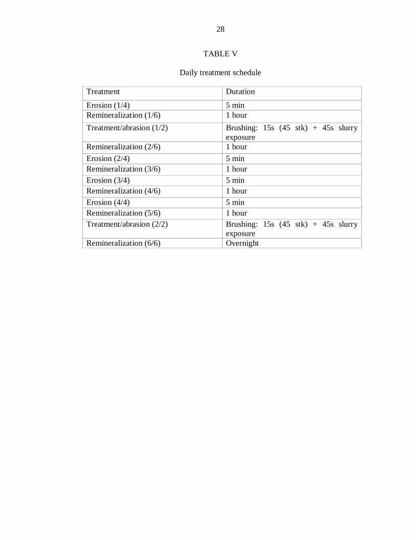
Daily Treatment Regimen
The daily treatment regimen consisted of four acid challenges and two tooth-
brushing treatments with the abrasive slurries. At the end of the cycle each day,
specimens were stored in a closed container with a humid environment at 5ºC, until the
next test day. The daily treatment schedule is highlighted in Table V.
Experimental setup
The samples have been subjected to five treatment days each consisting of 4 x 5
min of erosion, 5 x 1 h storage in artificial saliva, 2x tooth brushing abrasion followed by
slurry exposure without brushing, and storage in artificial saliva overnight. Enamel and
dentin surfaces have been eroded by unstirred storage in 0.3-percent citric acid for 5 min
and were then rinsed for 10 s with distilled water. Thereafter, the samples were brushed
in an automatic brushing machine with a load of 150 g using manual toothbrushes with
different filament stiffness and slurries with different abrasivity. After the 3rd and 5th day
of cycling, enamel and dentin surface loss were measured by profilometry.
Profilometry
Surface loss (SL) was measured using an optical profilometer (Proscan 2000,
Scantron, England; (Figure 8) after 3 days and 5 days of cycling. Tapes were removed
from the specimens and an area of 1 mm x 4 mm in the center of the specimen (including
both exposed and tape-covered areas) was scanned (Figure 8). Dedicated software
(Proscan 2000, Scantron) was used to analyze surface loss (Figure 9).

22
Statistical Analysis
The effects of cycling time, slurry abrasiveness, and toothbrush filament hardness
on surface loss have been examined using ANOVA. Separate analyses have been
performed for enamel and dentin surfaces. The ANOVA has included main effect terms
for each of the three factors, all interactions among the factors, and a random effect to
correlate the results from the two cycles within a sample. Fisher’s Protected Least
Significant Differences has been used to control the overall significance level of the tests.
A 5-percent significance level will be used.
Sample size
Based on a prior study the within-group standard deviation of the surface loss is
expected to be 1.5 µm. With a sample size of 8 specimens per abrasiveness-hardness
combination, the study will have 80-percent power to detect differences of 2.3 µm
between any two abrasiveness-hardness combinations for each cycling time, assuming
two-sided tests conducted at a 5-percent significance level.

23
RESULTS
24
DENTIN RESULTS
The Effect of Abrasivity of Toothpaste Slurries
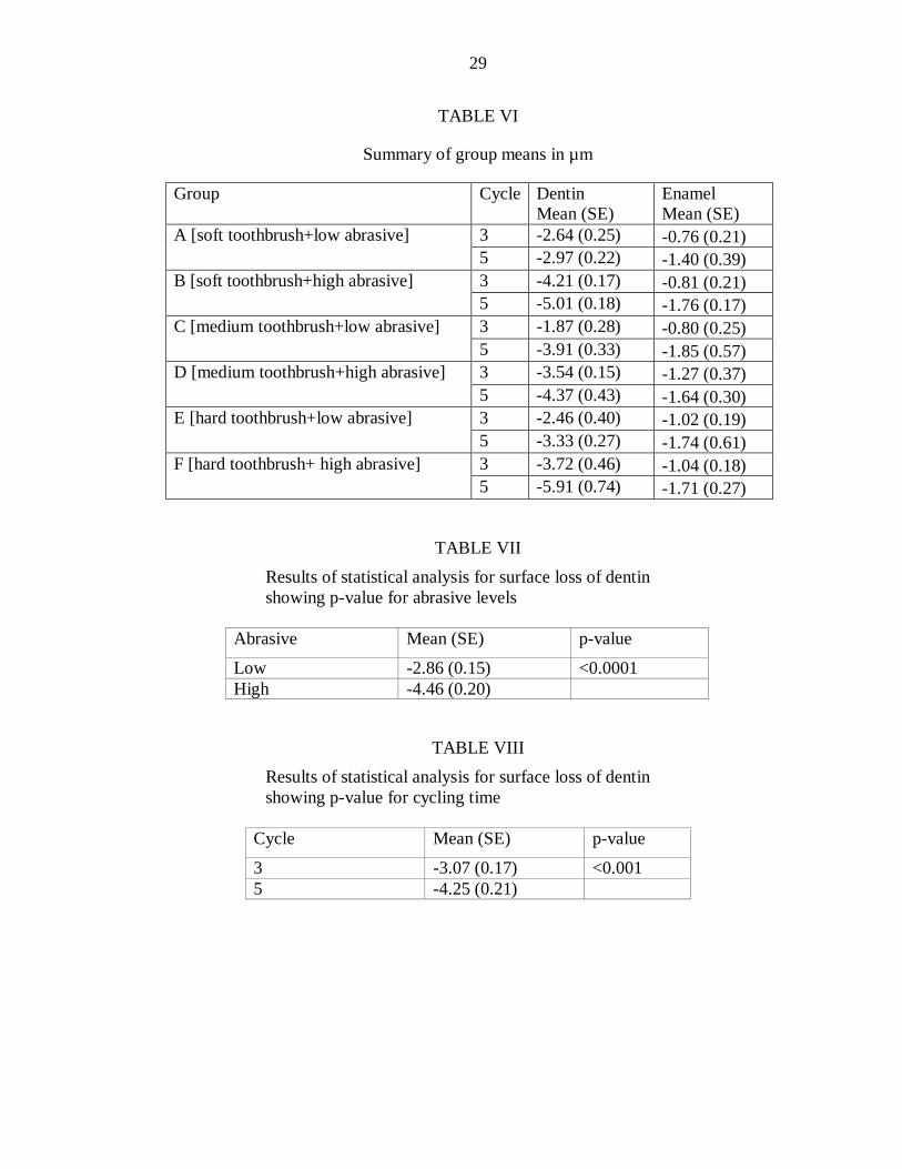
Mean (SD) dentin loss (µm) by brushing of eroded dentin specimens in different
group is presented in Table VI and Figures 12, 14. ANOVA showed that dentin loss
increases along with the RDA-value of toothpaste slurries; high abrasive (RDA) had
significantly higher dentin surface loss than low abrasive (RDA) in all the groups (A, B,
C, D, E, and F) (p < 0.0001) (Table VII).
The Effect of Toothbrush Filament Stiffness
Only the Hard toothbrush had significantly higher surface loss than the medium
toothbrush for the high abrasive at Cycle 5 (p = 0.0088) with no other significant
toothbrush differences (p > 0.18).
The Effect of Cycling Time
The fifth Cycle had significantly higher dentin surface loss than Cycle 3 overall (p
< 0.0001), with particularly large differences for Group F (hard toothbrush/high abrasive,
p < 0.0001) and Group C (medium toothbrush/low abrasive, p = 0.0001) (Table VIII).
Overall, the data did not show significant interaction between the two factors
(abrasivity of toothpaste slurries and filament stiffness of toothbrushes) (p = 0.1948).
However, the data showed that the impact effect of all factors (abrasivity, toothbrush

25
filament stiffness, and cycling time) had a strong effect on dentin loss, except for the
medium toothbrush on fifth cycle (Table IX).
ENAMEL RESULT
The Effect of Abrasivity of Toothpaste Slurries
Mean (SD) enamel loss (µm) by brushing of the eroded enamel specimens in the
different groups is presented in Table VI and Figures 13, 15. This data showed that the
abrasivity of toothpaste slurries did not affect enamel surface loss, there is no significant
difference between low and high abrasive on enamel wear (p = 0.2380).
The Effect of Cycling Time
The fifth cycle had significantly higher enamel surface loss than the third cycle (p
= 0.0003) (Table 9).
The Effect of the Toothbrush
Different toothbrushes (high, medium, and low) did not significantly affect
enamel surface loss (p = 0.6204).

26
TABLES AND FIGURES

27
TABLE I
Experimental groups
Toothbrush Low Abrasive High Abrasive Soft toothbrush A B Medium toothbrush C D Hard toothbrush E F
TABLE II
Artificial saliva composition
Chemicals Quantity (in g/l) CaCl2*2H2O 0.213g H2PO4 0.738g KCl 1.114g NaCl 0.381g Tris buffer 12g
TABLE III
Abrasive slurry compositions
Abrasive AbrasiveLoad (%)
Fluoride(ppm NaF)
Abrasive Amount (g)
Zeodent 113(Low)
5 275 3
Zeodent 103(High)
15 275 9
TABLE IV
Characteristics of the experimental toothbrushes, the measurements have been taken at OHRI
Parameter Lactona/ Soft Lactona/ Medium Lactona/ Hard
Filament Diameter 212.8 µm 228.6 µm 310.4 µm
Bristle Length 11 mm 11mm 11mm Tufts 43 43 43 Bristles/tuft (no) 50 36 16
28
TABLE V
Daily treatment schedule
Treatment Duration
Erosion (1/4) 5 min Remineralization (1/6) 1 hour Treatment/abrasion (1/2) Brushing: 15s (45 stk) + 45s slurry
exposure Remineralization (2/6) 1 hour Erosion (2/4) 5 min Remineralization (3/6) 1 hour Erosion (3/4) 5 min Remineralization (4/6) 1 hour Erosion (4/4) 5 min Remineralization (5/6) 1 hour Treatment/abrasion (2/2) Brushing: 15s (45 stk) + 45s slurry
exposure Remineralization (6/6) Overnight
29
TABLE VI
Summary of group means in µm
Group Cycle Dentin Mean (SE)
Enamel Mean (SE)
A [soft toothbrush+low abrasive] 3 -2.64 (0.25) -0.76 (0.21) 5 -2.97 (0.22) -1.40 (0.39)
B [soft toothbrush+high abrasive] 3 -4.21 (0.17) -0.81 (0.21) 5 -5.01 (0.18) -1.76 (0.17)
C [medium toothbrush+low abrasive] 3 -1.87 (0.28) -0.80 (0.25) 5 -3.91 (0.33) -1.85 (0.57)
D [medium toothbrush+high abrasive] 3 -3.54 (0.15) -1.27 (0.37) 5 -4.37 (0.43) -1.64 (0.30)
E [hard toothbrush+low abrasive] 3 -2.46 (0.40) -1.02 (0.19) 5 -3.33 (0.27) -1.74 (0.61)
F [hard toothbrush+ high abrasive] 3 -3.72 (0.46) -1.04 (0.18) 5 -5.91 (0.74) -1.71 (0.27)
TABLE VII Results of statistical analysis for surface loss of dentin showing p-value for abrasive levels
Abrasive Mean (SE) p-value
Low -2.86 (0.15) <0.0001 High -4.46 (0.20)
TABLE VIII Results of statistical analysis for surface loss of dentin showing p-value for cycling time
Cycle Mean (SE) p-value
3 -3.07 (0.17) <0.001 5 -4.25 (0.21)

30
TABLE IX
Statistical analysis for the surface loss of eroded dentin resulted from the interaction between study variables
Toothbrush Abrasive Cycle Mean (SE)
p-Value
Hard High 3 -3.72 (0.46)
<0.0001
5 -5.91 (0.74)
Hard High 3 -3.72 (0.46)
0.0048
Low -2.46
(0.40) Hard High 5 -5.91
(0.74) 0.0001
Low -3.33 (0.27)
Medium Low 3 -1.87 (0.28)
0.0001
5 -3.91 (0.33)
Medium High 3 -3.54 (0.15)
0.0002
Low -1.87 (0.28)
Soft High 3 -4.21 (0.17)
0.0005
Low -2.64 (0.25)
Soft High 5 -5.01 (0.18)
0.0006
Low -2.97 (0.22)
Hard High 5 -5.91 (0.74)
0.008
Medium -4.37 (0.43)

31
TABLE X
Results of statistical analysis for the surface loss of enamel showing p-value for cycling time
Cycle Mean (SE) p-Value 3 -0.95 (0.10) 0.0003 5 -1.68 (0.16)

32
FIGURE 1. Photographs of the Struers RotoPol 31/RotoForce (polishing machine).

33
FIGURE 2. (Top) Photograph of dentin slabs mounted in acrylic resin. (Bottom) Photograph of slabs of enamel and dentine embedded in acrylic resin.
Enamel
Dentin Acrylic resin

34
UPVC tapes
FIGURE 3. A photograph of UPVC tapes placed on the surface of the specimens, leaving an area of 1 x 4 mm exposed in the center of the each enamel and dentin slab.

35
FIGURE 4. A photograph of brushing machine.

36
FIGURE 5. A photograph of experimental toothbrush (Lactona Dental Care).

37
Soft toothbrush Medium toothbrush Hard toothbrush
FIGURE 6. Photographs of bristles of different types of toothbrushes used in the project.

38
Soft toothbrush Medium toothbrush Hard toothbrush
FIGURE 7. Photographs of different types of toothbrush bristles with different diameter

39
FIGURE 8. Electromicroscopic image of low abrasive (Zeodent 113) used in the project.

40
FIGURE 9. Electromicroscopic image of high abrasive (Zeodent 103) used in the project.

41
FIGURE 10. A photograph shows the specimen after removing tape.
Reference Area
Demineralized area

42
FIGURE 11. A photograph shows the optical profilometer used in the project.
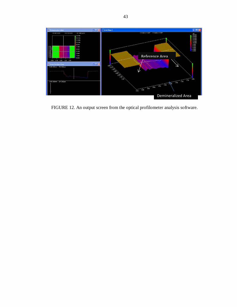
43
FIGURE 12. An output screen from the optical profilometer analysis software.
Reference Area
Demineralized Area

44
FIGURE 13. Line graphs showing means of surface loss of eroded dentin for different experimental groups brushed with two different abrasive slurries (high and low).

45
FIGURE 14. Line graphs showing means of surface loss of eroded enamel for different experimental groups brushed with two different abrasive slurries (high and low).

46
A: Low abrasive/Soft toothbrush. B: High abrasive/soft toothbrush. C: Low abrasive/soft toothbrush D: High abrasive/soft toothbrush. E: Low abrasive/soft toothbrush. F: High abrasive/ hard toothbrush
FIGURE 15. Bar graphs showing the mean of surface loss of eroded dentin for different groups.
A B C D E F

47
A: Low abrasive/Soft toothbrush. B: High abrasive/soft toothbrush. C: Low abrasive/soft toothbrush. D: High abrasive/soft toothbrush. E: Low abrasive/soft toothbrush. F: High abrasive/ hard toothbrush
FIGURE 16. Bar graphs showing the mean of surface loss of eroded enamel for different groups.
A B C D E F

48
DISCUSSION

49
Justifications for Experimental Parameters
The present study was aimed at investigating the surface loss of eroded enamel
and dentin resulting from the interaction between toothpaste abrasivity and toothbrush
filament stiffness. To investigate the study questions, a five-day established
erosion/abrasion cycling protocol was used, involving episodes of erosion challenges
(five times a day, for five min each), remineralization in artificial saliva (six times a day,
for 1 h each), and brushing abrasion (two times a day, for 15 s each).64,65 The brushing of
eroded enamel and dentin occurred after storage of the samples in artificial saliva. In
addition, brushing was conducted two times in each cycle instead of after each erosion
treatment. This experimental approach is more representative of the everyday clinical
situation than previous studies, because most people brush their teeth twice daily rather
than after each contact with erosive food stuff.
In this study, we attempted to simulate the recommended brushing time of nearly
two min. Each specimen was brushed for 30 s, the equivalent of 15 s, or 45 brushing
strokes for the facial/buccal surface, and the palatal/lingual surface.66 The 45 brushing
strokes equated to 450 brushing strokes at the end of each cycle and represented 5 d of
brushing. The majority of in-vitro and in-situ studies exaggerate the clinical situation by
applying a high number of brushing strokes ranging from 300 strokes to 400 strokes in
their brushing treatment.67 This can easily lead to the removal of the fragile outer layer of
softened dental hard tissue, regardless of the effects of different abrasivity values of
toothpaste and toothbrush filament stiffness.

50
In the majority of studies, the brushing was performed with manual
toothbrushes.19 The toothbrushes were attached to a brushing machine in order to
standardize the movement of the toothbrush and to ensure that the surfaces of all the
specimens were brushed under constant load. The use of 150-g brushing load for testing
the abrasivity of toothpaste is in agreement with previous recommendations of Wiegand
et al.19as well as the International Standards Organization (ISO11609). The toothpaste
slurries in this experiment were prepared with fluoride because most toothpaste available
internationally contain fluoride.
In order to control variability among toothpaste slurries, we have prepared our
own slurries in this experiment. Under clinical conditions, toothpaste will be diluted by
saliva during brushing. Therefore, this investigation used one part of toothpaste to three
parts of artificial saliva (1:3).38 For this reason, 275 ppm of fluoride was used,
representing 1100 ppm fluoride of regular toothpaste after saliva dilution.68
Non-contact surface profilometry was used in this study for two reasons. First, the
profilometer offers good flexibility for analyzing combined erosion-abrasion tissue loss.
In this study, the specimens were polished and flattened to obtain a profilometer
measurement with maximum sensitivity and accuracy.69 Secondly, it has been suggested
that the contact profilometers may cause damage to the eroded dental surfaces.70
Therefore, by using a non-contact profilometer we eliminated any possible interference
that may cause damage to the eroded surfaces of enamel and dentin.69,71
The Effect of Abrasivity Levels on the Abrasion of Eroded Dentin
In this study, the high abrasive slurries caused more surface loss of eroded dentin
than the low abrasive one. These results are in agreement with previous findings of

51
Wiegand et al., Hooper et al., and Dyer et al., who found that abrasion of eroded dentin
increased along with the RDA-value of the toothpaste slurry.2,11,15
From the present data, it can be assumed that low abrasive slurries may only
abrade the superficial layer of softening dentin, whereas their high abrasive counterpart
may probably wear the deeper part of the eroded dentine. Under this assumption, and
taking into consideration that Addy et al. considered that abrasivity degree of toothpastes
found in-vitro studies could be applied to clinical settings,72 patients with dentine erosive
lesions should be advised to use toothpaste with low RDA.
Although there is a remineralization period before and after brushing abrasion,
and considering the abrasive slurries were prepared with fluoride (which has been shown
to decrease brushing abrasion of eroded dentine),73 eroded dentine is still susceptible to
wear. This may be related to the fact that dentine is vulnerable and difficult to protect.45,74
Vanuspong et al. indicated that re-hardening of dentine after acid attacks may not occur.74
Furthermore, Hara et al. have concluded that 60 min of remineralization between the
erosion and the abrasion treatments has no effect on the prevention of surface loss of
eroded dentine.75 However, in this study we did not aim to study the effect of
remineralization on eroded dentine.
The Effect of Toothbrush Stiffness on the Abrasion of Eroded Dentin
It has been established previously that nylon toothbrushes alone have negligible
effects on dental hard tissue,76 but might indirectly influence the abrasion process by
modulating the action of toothpaste. This is related to the previous indication that
different types of toothbrushes probably differ in their capacity to hold toothpaste
abrasives, which may result in differences in abrasion of the dental substrate.77 Dyer et al.

52
suggested two reasons related to this indication: filament stiffness and density of the
brush, and second, the filament area of the brush head.15
The present study involved one brand of three different types of toothbrush (soft,
medium, and hard) with nylon bristles and end rounded tips. Only one brand of
toothbrush was used in this experiment, because stiffness (soft, medium, hard) among
the brands of toothbrushes is not constant.37
In a study by Wiegand et al., toothbrushes with a filament diameter of 0.15 mm,
0.20 mm, and 0.25 mm were applied on eroded dentine using non-fluoridated toothpaste
slurries. Wear of the eroded dentin increased along with the decreased diameter of
toothbrushes. However, from our data, only with the high-abrasive toothpaste slurries did
the hard toothbrush cause more dentin wear than the medium toothbrush at the fifth
cycle; there were no other significant toothbrush differences. This finding is somewhat
similar to the previous study of Manly and Harte, who showed that hard toothbrushes
cause more surface loss than medium ones on sound dentine.9
Surprisingly, our data showed that toothbrush by itself was not significant nor by
interacting with abrasives and the highest surface loss resulted only when all variables
interacted together (abrasives, toothbrushes, and time). This is related to the effect of
time, since Addy and Hunter have indicated that abrasion process is time-dependent.3
Our data confirmed a previous finding that the correlation between dentin wear
and toothbrush stiffness is low, and dentine wear mainly depends on the abrasivity of the
toothpaste.11

53
The Effect of Abrasivity Levels on the Abrasion of Eroded Enamel
In the comparison between high and low toothpaste slurries, the wear of eroded
enamel specimens was not statistically different. These results are in agreement with
those reported by Hooper et al.,2 who investigated the interplay between erosion and
abrasion of dental hard tissue using toothpaste with different abrasivities. They have
stated that fluoridated toothpastes with differing RDA values display similar abrasiveness
on previously eroded enamel. The authors speculated that any mechanical force may
remove the softened layer of enamel, regardless of different abrasivity levels (RDA). In
addition, they have calculated the lifetime brushing (100 years) based on the highest
mean of enamel abrasion in their studies, which would be equivalent to 38 µm. Since the
thickness of enamel at the cervical area is equal to 130 µm, a result of less than 100 µm
enamel wear is considered to be clinically irrelevant. This might be an explanation of our
results.2 However, our data is in contrast to the previous findings of Wiegand et al.,12 who
showed that the abrasivity of toothpaste slurries is considered to be the major factor for
eroded enamel wear. Wiegand et al. attempted to simulate the worst case scenario in
evaluating the effects of slurries exhibiting different abrasivity levels on softened enamel.
Their samples were subjected to 60 cycles each consisting of 15 s of erosion in 1 ml of
hydrochloric acid , and toothbrushing abrasion performed at 250-g load. This is in
contrast to the less aggressive nature of our model. Furthermore, in their study, the
brushing was performed with non-fluoridated toothpaste to focus on the abrasivity of the
toothpaste itself. On the other hand, in our model, we brushed our specimens with
fluoridated toothpaste slurries, given that most available toothpaste contains fluoride.
Hara et al. reported that fluoridated toothpaste reduces the wear of erosive-abrasive

54
lesions.64 Magalhaes et al. had suggested in their in situ/ex vivo study, which assessed the
effect of low-dosage fluoridated dentifrices on the abrasion of eroded enamel, that
brushing with fluoridated toothpaste had a protective effect on eroded enamel surface.78
Moreover, it has been demonstrated that there is a tendency toward differences between
toothpastes of different abrasivities in the absence of fluoride.79 Although Hara et al. in
2009 indicated that the presence of fluoride in toothpaste increased the difference
between high and low abrasives,64 we assumed in our experiment the presence of fluoride
could mask the abrasiveness of toothpaste slurries. This could explain our results
compared with those of Wiegand.
However, it is important to point out that we did not intend to investigate whether
fluoridated toothpaste is able to promote the complete rehardening of eroded enamel.
Further studies should be carried out to compare fluoridated toothpaste of
different abrasivities with non-fluoridated toothpaste to confirm the present data.
It has been shown that saliva can remineralize the acid-softened tooth surface.79
Furthermore, Attin et al. and Jaeggi found significant differences between brushing
immediately compared with brushing after 60 min of exposure to saliva.22,80 For this
reason, the majority of in-vitro studies did not include a remineralization period in-
between erosion and abrasion treatments in order to avoid rehardening of eroded enamel.
However, a remineralization treatment was used in this investigation to simulate clinical
conditions, which resulted in decreased eroded enamel wear.
The brushing load of 150 g may have affected negatively the extent of enamel
wear in our investigations. Parry et al. have suggested that using 150-g brushing load had
an influence on the abrasion level of the enamel in his investigation. They also indicated

55
that, using a greater load is more appropriate in assessing the toothpaste abrasivity on
enamel.81
Finally, by using a clinically relevant brushing time, the softened layer is unlikely
to be completely removed. This probably confirms Wiegand et al., who assumed the
outermost layer of softened enamel may behave differently compared with deeper layers.
The Effect of Toothbrush Stiffness on Abrasive Wear of Eroded Enamel
Regarding the abrasion of eroded enamel, Wiegand et al. have indicated that
toothbrush filament stiffness is of secondary importance, because only a medium
toothbrush and toothpaste with REA 6 caused more abrasion of the eroded enamel than
did either soft or hard toothbrush.12 In our study, toothbrush stiffness did not significantly
affect enamel surface loss. This is not surprising because many investigators reported that
filament stiffness was not a factor affecting toothpaste abrasivity. Voronets et al. have
compared abrasion of softened human enamel brushed with two different types of
toothbrushes (hard and soft). They stated that there was not any significant difference
between hard and soft toothbrushes on the abrasion of eroded enamel.82
From these data we suggest that the choice of soft, medium, or hard toothbrush is
of lesser relevance to enamel than to dentin abrasion.
The Effect of Cycling Time on the Abrasion of Eroded Enamel and Dentin
Regarding the variable of time, the measurement of surface loss was taken at two
different times (third and fifth cycles). This allowed us to see the significant increase of
surface loss of eroded enamel and dentin in the fifth cycle versus the third cycle. This

56
was not surprising because the total exposed time to erosion in the third cycle was 1 hour,
and in the fifth cycle, 1 hour and 40 min. Based on this, we may consider that the depth
of softening of the surface of enamel and dentin specimens in the fifth cycle was
probably more than that of the third cycle. As result, significantly greater surface loss was
observed in the fifth cycle compared with the third.

57
SUMMARY AND CONCLUSIONS

58
The surface loss of eroded enamel and dentin has been assessed using a clinically
relevant in-vitro cycling model for two surrogate toothpastes of varying abrasivity (low,
high) and three different types of toothbrushes (soft, medium, hard). As for dentin, the
results of our data showed the surface loss of softened dentin is mainly affected by
toothpaste abrasivity, because more abrasive toothpaste presents greater surface loss. For
enamel, neither abrasive levels nor toothbrushes affected the surface loss of softened
enamel under the conditions of the present study.
Within the limitations of the present study, the following conclusion can be
drawn:
1. The surface loss of eroded dentine is strongly correlated to relative dentine
abrasivity (RDA).
2. The correlation between toothbrush filament stiffness and eroded dentine
surface loss is very low.
3. The surface loss of eroded enamel is not correlated to the RDA. In other words,
the wear of superficial, acid-softened enamel is not dependent on the abrasivity of
toothpaste.
4. The toothbrush stiffness was not a factor affecting eroded enamel surface loss.
5. The extent of enamel and dentin loss is affected by the cycling time.

59
REFERENCES
60
1. Wiegand A, Schlueter N. The role of oral hygiene: does toothbrushing harm? Monogr Oral Sci 2014;25:215-9.
2. Hooper S, West NX, Pickles MJ, et al. Investigation of erosion and abrasion on
enamel and dentine: a model in situ using toothpastes of different abrasivity. J Clin Periodontol 2003;30(9):802-8.
3. Addy M, Hunter ML. Can tooth brushing damage your health? Effects on oral and
dental tissues. Int Dent J 2003;53 Suppl 3:177-86. 4. Tellefsen G, Liljeborg A, Johannsen A, Johannsen G. The role of the toothbrush
in the abrasion process. Int J Dent Hyg 2011;9(4):284-90. 5. Wiegand A, Burkhard JP, Eggmann F, Attin T. Brushing force of manual and
sonic toothbrushes affects dental hard tissue abrasion. Clin Oral Investig 2013;17(3):815-22.
6. Addy M. Tooth brushing, tooth wear and dentine hypersensitivity--are they
associated? Int Dent J 2005;55(4 Suppl 1):261-7. 7. Voronets J, Lussi A. Thickness of softened human enamel removed by toothbrush
abrasion: an in vitro study. Clin Oral Investig 2010;14(3):251-6. 8. Barbakow F, Imfeld T, Lutz F, Stookey G, Schemehorn B. Dentin abrasion
(RDA), enamel abrasion (REA) and polishing scores of dentifrices sold in Switzerland. Schweiz Monatsschr Zahnmed 1989;99(4):408-13.
9. Harte DB, Manly RS. Effect of toothbrush variables on wear of dentin produced
by four abrasives. J Dent Res 1975;54(5):993-8. 10. Dyer D, MacDonald E, Newcombe RG, et al. Abrasion and stain removal by
different manual toothbrushes and brush actions: studies in vitro. J Clin Periodontol 2001;28(2):121-7.
11. Wiegand A, Kuhn M, Sener B, Roos M, Attin T. Abrasion of eroded dentin
caused by toothpaste slurries of different abrasivity and toothbrushes of different filament diameter. J Dent 2009;37(6):480-4.
12. Wiegand A, Schwerzmann M, Sener B, et al. Impact of toothpaste slurry
abrasivity and toothbrush filament stiffness on abrasion of eroded enamel - an in vitro study. Acta Odontol Scand 2008;66(4):231-5.

61
13. Philpotts CJ, Weader E, Joiner A. The measurement in vitro of enamel and dentine wear by toothpastes of different abrasivity. Int Dent J 2005;55(3 Suppl 1):183-7.
14. Magalhaes AC, Wiegand A, Buzalaf MA. Use of dentifrices to prevent erosive
tooth wear: harmful or helpful? Braz Oral Res 2014;28 Spec:1-6. 15. Dyer D, Addy M, Newcombe RG. Studies in vitro of abrasion by different manual
toothbrush heads and a standard toothpaste. J Clin Periodontol 2000;27(2):99-103.
16. Wiegand A, Kowing L, Attin T. Impact of brushing force on abrasion of acid-
softened and sound enamel. Arch Oral Biol 2007;52(11):1043-7. 17. Kelleher M, Bishop K. Tooth surface loss: an overview. Br Dent J
1999;186(2):61-6. 18. Vieira A, Overweg E, Ruben JL, Huysmans MC. Toothbrush abrasion, simulated
tongue friction and attrition of eroded bovine enamel in vitro. J Dent 2006;34(5):336-42.
19. Wiegand A, Attin T. Design of erosion/abrasion studies--insights and rational
concepts. Caries Res 2011;45 Suppl 1:53-9. 20. Hara AT, Lippert F, Zero DT. Interplay between experimental dental pellicles and
stannous-containing toothpaste on dental erosion-abrasion. Caries Res 2013;47(4):325-9.
21. Wiegand A, Wegehaupt F, Werner C, Attin T. Susceptibility of acid-softened
enamel to mechanical wear--ultrasonication versus toothbrushing abrasion. Caries Res 2007;41(1):56-60.
22. Jaeggi T, Lussi A. Toothbrush abrasion of erosively altered enamel after intraoral
exposure to saliva: an in situ study. Caries Res 1999;33(6):455-61. 23. Comar LP, Gomes MF, Ito N, et al. Effect of NaF, SnF(2), and TiF(4)
Toothpastes on bovine enamel and dentin erosion-abrasion in vitro. Int J Dent 2012;2012:134350.
24. Kreulen CM, Van 't Spijker A, Rodriguez JM, et al. Systematic review of the
prevalence of tooth wear in children and adolescents. Caries Res 2010;44(2):151-9.
25. Lussi A, Schlueter N, Rakhmatullina E, Ganss C. Dental erosion--an overview
with emphasis on chemical and histopathological aspects. Caries Res 2011;45 Suppl 1:2-12.

62
26. Bartlett DW, Shah P. A critical review of non-carious cervical (wear) lesions and the role of abfraction, erosion, and abrasion. J Dent Res 2006;85(4):306-12.
27. Harpenau LA, Noble WH, Kao RT. Diagnosis and management of dental wear.
Todays FDA 2012;24(5):50-7. 28. Barbour ME, Parker DM, Allen GC, Jandt KD. Human enamel dissolution in
citric acid as a function of pH in the range 2.30< or =pH< or =6.30--a nanoindentation study. Eur J Oral Sci 2003;111(3):258-62.
29. Hughes JA, West NX, Parker DM, van den Braak MH, Addy M. Effects of pH
and concentration of citric, malic and lactic acids on enamel, in vitro. J Dent 2000;28(2):147-52.
30. Meurman JH, ten Cate JM. Pathogenesis and modifying factors of dental erosion.
Eur J Oral Sci 1996;104(2 ( Pt 2)):199-206. 31. Lussi A, von Salis-Marincek M, Ganss C, et al. Clinical study monitoring the pH
on tooth surfaces in patients with and without erosion. Caries Res 2012;46(6):507-12.
32. Hara AT, Lussi A, Zero DT. Biological factors. Monogr Oral Sci 2006;20:88-99. 33. Buzalaf MA, Hannas AR, Kato MT. Saliva and dental erosion. J Appl Oral Sci
2012;20(5):493-502. 34. Amaechi BT, Higham SM. In vitro remineralisation of eroded enamel lesions by
saliva. J Dent 2001;29(5):371-6. 35. Addy M, Shellis RP. Interaction between attrition,abrasion and erosion in tooth
wear. Monogr Oral Sci 2006;20:17-31. 36. Kaifu Y, Kasai K, Townsend GC, Richards LC. Tooth wear and the "design" of
the human dentition: a perspective from evolutionary medicine. Am J Phys Anthropol 2003;Suppl 37:47-61.
37. Harte DB, Manly RS. Four variables affecting magnitude of dentrifice
abrasiveness. J Dent Res 1976;55(3):322-7. 38. Kielbassa AM, Gillmann L, Zantner C, et al. Profilometric and microradiographic
studies on the effects of toothpaste and acidic gel abrasivity on sound and demineralized bovine dental enamel. Caries Res 2005;39(5):380-6.
39. Pickles MJ. Tooth wear. Monogr Oral Sci 2006;19:86-104.

63
40. Meredith N, Sherriff M, Setchell DJ, Swanson SA. Measurement of the microhardness and Young's modulus of human enamel and dentine using an indentation technique. Arch Oral Biol 1996;41(6):539-45.
41. Tellefsen G. surface roughness: causual factors: and its relation to bacterial
adhesion [karoliska institute: karoliska institute; 2013.] 42. Attin T, Koidl U, Buchalla W, et al. Correlation of microhardness and wear in
differently eroded bovine dental enamel. Arch Oral Biol 1997;42(3):243-50. 43. Svinnseth PN, Gjerdet NR, Lie T. Abrasivity of toothpastes. An in vitro study of
toothpastes marketed in Norway. Acta Odontol Scand 1987;45(3):195-202. 44. Joiner A, Pickles MJ, Tanner C, Weader E, Doyle P. An in situ model to study the
toothpaste abrasion of enamel. J Clin Periodontol 2004;31(6):434-8. 45. De Menezes M, Turssi CP, Hara AT, Messias DC, Serra MC. Abrasion of eroded
root dentine brushed with different toothpastes. Clin Oral Investig 2004;8(3):151-5.
46. De Boer P, Duinkerke AS, Arends J. Influence of tooth paste particle size and
tooth brush stiffness on dentine abrasion in vitro. Caries Res 1985;19(3):232-9. 47. Schemehorn BR, Moore MH, Putt MS. Abrasion, polishing, and stain removal
characteristics of various commercial dentifrices in vitro. J Clin Dent 2011;22(1):11-8.
48. Franzo D, Philpotts CJ, Cox TF, Joiner A. The effect of toothpaste concentration
on enamel and dentine wear in vitro. J Dent 2010;38(12):974-9. 49. Joiner A. Whitening toothpastes: a review of the literature. J Dent 2010;38 Suppl
2:e17-24. 50. Davis WB. Cleaning and polishing of teeth by brushing. Community Dent Oral
Epidemiol 1980;8(5):237-43. 51. Turssi CP, Messias DC, Hara AT, Hughes N, Garcia-Godoy F. Brushing abrasion
of dentin: effect of diluent and dilution rate of toothpaste. Am J Dent 2010;23(5):247-50.
52. Hefferren JJ. A laboratory method for assessment of dentrifrice abrasivity. J Dent
Res 1976;55(4):563-73. 53. Teche FV, Paranhos HF, Motta MF, Zaniquelli O, Tirapelli C. Differences in
abrasion capacity of four soft toothbrushes. Int J Dent Hyg 2011;9(4):274-8.

64
54. Brandini DA, de Sousa AL, Trevisan CI, et al. Noncarious cervical lesions and their association with toothbrushing practices: in vivo evaluation. Oper Dent 2011;36(6):581-9.
55. Litonjua LA, Andreana S, Cohen RE. Toothbrush abrasions and noncarious
cervical lesions: evolving concepts. Compend Contin Educ Dent 2005;26(11):767-8, 70-4, 76 passim.
56. Azevedo AM, Panzeri H, Prado CJ, et al. Assessment in vitro of brushing on
dental surface roughness alteration by laser interferometry. Braz Oral Res 2008;22(1):11-7.
57. Eisenburger M, Shellis RP, Addy M. Comparative study of wear of enamel
induced by alternating and simultaneous combinations of abrasion and erosion in vitro. Caries Res 2003;37(6):450-5.
58. Ganss C, Schulze K, Schlueter N. Toothpaste and erosion. Monogr Oral Sci
2013;23:88-99. 59. Lussi A, Lussi J, Carvalho TS, Cvikl B. Toothbrushing after an erosive attack:
will waiting avoid tooth wear? Eur J Oral Sci 2014;122(5):353-9. 60. West NX, Davies M, Amaechi BT. In vitro and in situ erosion models for
evaluating tooth substance loss. Caries Res 2011;45 Suppl 1:43-52. 61. White AJ, Yorath C, ten Hengel V, et al. Human and bovine enamel erosion under
'single-drink' conditions. Eur J Oral Sci 2010;118(6):604-9. 62. Wegehaupt F, Gries D, Wiegand A, Attin T. Is bovine dentine an appropriate
substitute for human dentine in erosion/abrasion tests? J Oral Rehabil 2008;35(5):390-4.
63. Attin T, Buchalla W, Gollner M, Hellwig E. Use of variable remineralization
periods to improve the abrasion resistance of previously eroded enamel. Caries Res 2000;34(1):48-52.
64. Hara AT, Gonzalez-Cabezas C, Creeth J, et al. Interplay between fluoride and
abrasivity of dentifrices on dental erosion-abrasion. J Dent 2009;37(10):781-5. 65. Hara AT, Gonzalez-Cabezas C, Creeth J, Zero DT. The effect of human saliva
substitutes in an erosion-abrasion cycling model. Eur J Oral Sci 2008;116(6):552-6.
66. van der Weijden GA, Timmerman MF, Reijerse E, Snoek CM, van der Velden U.
Toothbrushing force in relation to plaque removal. J Clin Periodontol 1996;23(8):724-9.
65
67. Ganss C, Schlueter N, Preiss S, Klimek J. Tooth brushing habits in uninstructed adults--frequency, technique, duration and force. Clin Oral Investig 2009;13(2):203-8.
68. Duke SA, Forward GC. The conditions occurring in vivo when brushing with
toothpastes. Br Dent J 1982;152(2):52-4. 69. Schlueter N, Hara A, Shellis RP, Ganss C. Methods for the measurement and
characterization of erosion in enamel and dentine. Caries Res 2011;45 Suppl 1:13-23.
70. Heurich E, Beyer M, Jandt KD, et al. Quantification of dental erosion--a
comparison of stylus profilometry and confocal laser scanning microscopy (CLSM). Dent Mater 2010;26(4):326-36.
71. Ganss C, Schlueter N, Hardt M, von Hinckeldey J, Klimek J. Effects of
toothbrushing on eroded dentine. Eur J Oral Sci 2007;115(5):390-6. 72. Addy M, Hughes J, Pickles MJ, Joiner A, Huntington E. Development of a
method in situ to study toothpaste abrasion of dentine. Comparison of 2 products. J Clin Periodontol 2002;29(10):896-900.
73. Munoz CA, Feller R, Haglund A, Triol CW, Winston AE. Strengthening of tooth
enamel by a remineralizing toothpaste after exposure to an acidic soft drink. J Clin Dent 1999;10(1 Spec No):17-21.
74. Vanuspong W, Eisenburger M, Addy M. Cervical tooth wear and sensitivity:
erosion, softening and rehardening of dentine; effects of pH, time and ultrasonication. J Clin Periodontol 2002;29(4):351-7.
75. Hara AT, Turssi CP, Teixeira EC, Serra MC, Cury JA. Abrasive wear on eroded
root dentine after different periods of exposure to saliva in situ. Eur J Oral Sci 2003;111(5):423-7.
76. Manly RS, Brudevold F. Relative abrasiveness of natural and synthetic toothbrush
bristles on cementum and dentin. J Am Dent Assoc 1957;55(6):779-80. 77. Phaneuf EA, Harrington JH, Dale PP, Shklar G. Automatic toothbrush: a new
reciprocating action. J Am Dent Assoc 1962;65:12-25. 78. Magalhaes AC, Rios D, Delbem AC, Buzalaf MA, Machado MA. Influence of
fluoride dentifrice on brushing abrasion of eroded human enamel: an in situ/ex vivo study. Caries Res 2007;41(1):77-9.
66
79. Amaechi BT, Higham SM. Eroded enamel lesion remineralization by saliva as a possible factor in the site-specificity of human dental erosion. Arch Oral Biol 2001;46(8):697-703.
80. Attin T, Knofel S, Buchalla W, Tutuncu R. In situ evaluation of different
remineralization periods to decrease brushing abrasion of demineralized enamel. Caries Res 2001;35(3):216-22.
81. Parry J, Harrington E, Rees GD, McNab R, Smith AJ. Control of brushing
variables for the in vitro assessment of toothpaste abrasivity using a novel laboratory model. J Dent 2008;36(2):117-24.
82. Voronets J, Jaeggi T, Buergin W, Lussi A. Controlled toothbrush abrasion of
softened human enamel. Caries Res 2008;42(4):286-90.

67
ABSTRACT

68
INTERACTION BETWEEN TOOTHPASTE ABRASIVITY AND
TOOTHBRUSH FILAMENT STIFFNESS ON THE
DEVELOPMENT OF EROSIVE-ABRASIVE
LESIONS
by
Mona Arrageg
Indiana University School of Dentistry
Indianapolis, Indiana
Background: Toothpaste abrasivity is considered the major contributor in
toothbrushing abrasive wear, while toothbrush stiffness can be considered a secondary
factor that may modify the abrasivity of toothpaste.
Objectives: To investigate the longitudinal enamel and dentin surface loss caused
by the interaction between the abrasives in toothpaste and toothbrush filament stiffness.
Study Hypothesis: The amount of enamel and dentin loss depends on the
abrasivity of the toothpaste and the filament stiffness of toothbrush.

69
Materials and Methods: The following experimental factors were considered:
abrasive suspension, at two levels (L-low: Z113 and H-high: Z103); and toothbrushes at
three levels determined by bristle stiffness (soft, medium, and hard) generating 6 testing
groups (n = 8). Slabs of bovine enamel and dentin were cut, embedded in acrylic resin,
and polished. UPVC tapes were placed on the surface of the specimens, leaving an area
of 1 × 4 mm exposed in the center of the each enamel slab. Specimens (n = 8) were
subjected to 5 d of erosion/abrasion cycling: erosion (5min, 4×/d, 0.3% citric acid, pH
3.75), abrasion (15 s, 2×/d, 45 strokes each, 150-g load, automated brushing machine),
fluoride treatment (15 s with abrasion and 45 s without abrasion; 275 ppm F as NaF in
abrasive slurry) with exposure to artificial saliva between erosion and abrasion (1h) and
all other times (overnight). Surface loss (SL, in micrometers) was determined by optical
profilometry, after the third and fifth days of cycling. Data were analyzed using three-
way ANOVA (alpha = 0.05). For enamel, only cycling time was found to affect surface
loss with 5 d > 3 d. Overall, there was little SL (mean range: 0.76 µm to 1.85 µm). For
dentin (mean SL range: 1.87 µm to 5.91 µm), significantly higher SL was found for 5 d
vs. 3 d, with particularly large differences for hard toothbrush high abrasive, and medium
toothbrush/low abrasive. Hard toothbrush resulted in significantly higher SL than
medium toothbrush for high abrasive after 5 d, with no other significant stiffness
differences. High abrasive had significantly higher SL than low abrasive overall with
strong effects for all combinations, except medium stiffness after 5 d. In conclusion, the
interplay between abrasivity and filament stiffness appears to be more relevant for dentin
than enamel.

CURRICULUM VITAE

Mona Arrageg
1982 Born in Tripoli, Libya
2000 to 2006 BDS AL-Fath University Faculty of Dentistry
Tripoli, Libya
2008 to 2010 Clinical demonstrator AL-Fath University Faculty of Dentistry Tripoli, Libya