interactions between angiotensin ii and prostaglandin e2
TRANSCRIPT

Interactions between Angiotensin II and Prostaglandin E2: Mechanisms for Regulation of
Vascular Reactivity
By
Maria Palazzo Kraemer
Dissertation
Submitted to the Faculty of the
Graduate School of Vanderbilt University
in partial fulfillment of the requirements
for the degree of
DOCTOR OF PHILOSOPHY
in
Biochemistry
August, 2016
Nashville, Tennessee
Approved:
Richard M. Breyer, Ph.D.
John D. York, Ph.D.
Bruce D. Carter, Ph.D.
Fred D. Lamb, M.D., Ph.D.
Jeff Reese, M.D.

ii
To my very first and most cherished mentors, Cindy and Joe Palazzo
&
To my best friend and enthusiast, Bradley Kraemer

iii
ACKNOWLEDGEMENTS
I would like to express my sincere appreciation for the many people who have
assisted and supported me during my Ph.D. work. I would first like to express my gratitude
for my advisor Dr. Rich Breyer for allowing me to join the lab and for training me to be
resilient and independent. I am also very appreciative to the Breyer lab: Sarah Davis,
Ryan Ceddia, Kelli Simpson, Jason Downey, and Christie Bartlett. Sarah has been my
sidekick for the last couple years. Ryan provided comradery and a willingness to brain-
storm with me. Jason and Christie helped train me to perform several in vivo techniques
including osmotic pump implantation and intracarotid blood pressure readings, and have
provided much needed advice and encouragement from time to time.
My dissertation committee deserves a great deal of recognition for their
overwhelming amount of care and advice. I am grateful to Dr. Fred Lamb who has been
an invaluable collaborator. I appreciate his availability even with his busy schedule,
providing endless amounts of guidance and advice, and always remaining incredibly kind.
I am grateful to Dr. John York as the chair of my committee and for providing guidance in
my scientific endeavors. Dr. York has been a great addition to the Biochemistry
department. I, along with many students, appreciate his recognition of student’s needs
and his efforts to improve upon the department. Thank you to Dr. Bruce Carter who not
only from my very first days at Vanderbilt has provided much professional support to me
but has also helped out my family with his mentorship of Brad. Last but not least, I am
appreciative of Dr. Jeff Reese for his constant encouragement, his scientific support and
collaboration, as well as for boosting my confidence once in a while by calling me Dr.
Palazzo before I deserved such a title.

iv
My research and training has developed as a collective effort of numerous people.
Thank you to Dr. David Harrison and his staff, in particular Hana Itani, Jing Wu, and Liang
Xiao for collaborations and technical advice. Thank you to Stan and Naoko from Dr.
Reese’s lab who helped train me in specific techniques and are always willing to help. I
would also like to mention Yahua Zhang, who took time from her own research to trouble-
shoot and teach me some tricks of the trade to performing intracarotid blood pressure
recordings. Thank you especially to Hong Nguygen and Hye Choi from Dr. Lamb’s lab,
who have not only provided their time and efforts to help with technical advice and training
for the wire myography studies, but have also been great friends. I would like to
acknowledge everyone in the Carter lab, past and current lab members, Chelsea, Alison,
Jami, Anna, Uzma, Amrita, Malathi, Emily, Rose, Alex, Eddie, and BRAD for being my
support, entertainment, captive audience when necessary, and scientific resource. I
would also like to acknowledge my funding support provided in part by an American Heart
Association pre-doctoral training grant as well as support from the Nephrology
department and research funds provided from a Veteran’s Affairs Merit Grant.
I would like to recognize my vast friends and family network who without their
words of motivation, tolerance, and confidence in me I would never have completed this
work. I am so proud to say I have some the most loyal, intelligent, supportive friends that
anyone would hope to have in their lifetime. Asuka has been my non-genetic sister and a
great model of a young scientist that I hope to develop the relentless motivation and
passion for science that she embodies. I am grateful to Stephanie for always being there,
always listening, and knowing what is best. Chelsea, thank you for all the coffee breaks
and decompression, and more recently for all the phone calls and texts to keep me going.

v
I would like to especially thank the Kraemer family, everyone has welcomed me with open
arms, and provided me with love, support, and advice that is unconditional and tenacious.
Words cannot describe how remarkable my parents have been through this
experience. I am forever grateful for all the love, support, advice, hugs, etc. To my
parents, thank you for raising me to be determined, inquisitive, and independent. I am
also very appreciative of my sisters, Amanda and Rachel. Both of them have been my
greatest companions, and frequently visited and offered much needed release. Lastly, I
would like to thank my husband, Bradley. Thank you for enduring the storm with me for
the last 6 years, and committing to continue onward along my side. You have kept me
rational, and never let me lose sight of my full potential and worth. My confidence in myself
has never been greater.

vi
LIST OF TABLES
Table Page
1. High Blood Pressure Categories .......................................................................... 6
2. Summary of EP receptor Functions .................................................................... 31

vii
LIST OF FIGURES
Figure Page
1.1 Brief summary of end-organ damage resulting from chronic hypertension ........... 9
1.2 Diagram of the RAAS system indicating angiotensin processing and key
organ systems .................................................................................................... 11
1.3 AT1 receptor signaling cascades ........................................................................ 18
2.1 Determination of optimal wire myograph basal tension setting for mouse
femoral arteries ................................................................................................... 44
2.2 Ang II priming enhances PGE2-mediated vasoconstriction................................. 46
2.3 PGE2-facilitated contraction at varying times after Ang II pretreatment .............. 48
2.4 PGE2 pre-treatment enhances Ang II-induced vasoconstriction ......................... 50
2.5 Mesenteric vascular beds but not aorta exhibit PGE2-mediated contraction
after Ang II pre-treatment ................................................................................... 52
2.6 Vascular reactivity of PGE2 on mouse femoral arteries ...................................... 54
2.7 Effects of constrictor EP-receptors on PGE2-induced contraction of Ang II-
primed arteries .................................................................................................... 56
2.8 Effects of EP-receptors on PGE2-induced contraction of Ang II-primed
arteries ............................................................................................................... 58
2.9 Effects of EP-receptors on Ang II-induced vasoconstriction ............................... 60
2.10 AT1 antagonist inhibits Ang II priming of PGE2 .................................................. 62
2.11 AT1 and AT2 antagonists do not compete for EP receptor ligand binding ......... 63
2.12 SII does not potentiate PGE2-mediated contraction ........................................... 65
2.13 Tempol reduces Ang II potentiation of PGE2-mediated vasoconstriction ............ 67
2.14 Role of reactive oxygen species and ER stress in Ang II priming of PGE2 ......... 69
2.15 EGFR does not contribute to Ang II potentiation of PGE2-mediated
vasoconstriction .................................................................................................. 71
2.16 Endothelial dysfunction does not affect Ang II potentiation of PGE2-mediated
vasoconstriction .................................................................................................. 73
2.17 Contribution of extracellular calcium and Rho-kinase to PGE2 primed
vasoconstriction .................................................................................................. 75

viii
2.18 Chloride channel contributions to vasoconstriction induced by PGE2
and Ang II ........................................................................................................... 77
2.19 Contribution of endogenous prostaglandin production ....................................... 79
2.20 Pyk2 is necessary for PGE2-mediated contraction of Ang II-primed vessels ...... 81
2.21 Schematic of key proteins involved in PGE2-induced contractile responses ...... 82
3.1 Experimental design of ICBP studies investigating the effects of acute
intravenous infusion of PGE2 in combination with other vasoconstrictors .......... 92
3.2 PGE2 acts a transient vasodepressor when infused during continuous infusion
of other vasoconstrictors in wildtype mice .......................................................... 93
3.3 Mice given subcutaneous slow-pressor doses of Ang II develop high blood
pressure ............................................................................................................. 95
3.4 Chronic Ang II effects on PGE2-mediated blood pressure responses ................ 96
3.5 ICBP experiments using the euvolemic protocol used to investigate chronic
Ang II effects on PGE2-mediated blood pressure responses .............................. 98
3.6 PGE2 is a vasodepressor in EP3-/- mice ............................................................ 100
3.7 Comparison of heart rate and MAP during PGE2 bolus in mice undergoing continuous Ang II intravenous infusion between two different
experimental ICBP methods .............................................................................. 102 3.8 Catecholamine signaling did not contribute to Ang II potentiation of PGE2-
mediated vasoconstriction ................................................................................. 104

ix
LIST OF ABBREVIATIONS
15-PGDH 15-hydroxyprostaglandin dehydrogenase
AA Arachidonic acid
ACE Angiotensin-converting enzyme
ADH Anti-diuretic hormone
AGT Angiotensinogen
Ang I Angiotensin I
Ang II Angiotensin II
ARB AT1 receptor blocker
AT1 Angiotensin II type-1 receptor
AT2 Angiotensin II type-2 receptor
AUC Area under the curve
BP Blood pressure
cAMP Cyclic adenosine monophosphate
CNS Central nervous system
cPLA2 Cytosolic phospholipase A2
CO Cardiac output
COX Cyclooxygenase enzyme
CRC Concentration response curve
DMSO Dimethyl sulfoxide
DP Prostaglandin D receptor
EC Effective concentration
EGFR Epidermal growth factor receptor
EGTA Ethylene glycol-bis tetraacetic acid
eNOS Endothelium nitric oxide synthase
EP E prostanoid
ER stress Endoplasmic reticulum stress
EtOH Ethanol

x
FAK Focal adhesion kinase
FP Prostaglandin F receptor
GFR Glomerular filtration rate
GRK G protein-coupled receptor kinase
ICBP Intracarotid blood pressure
Kd Dissociation constant
L-NAME Nω-Nitro-L-arginine methyl ester hydrochloride
LOX Lipoxygenase
MAP Mean arterial pressure
MAPK Mitogen-activated protein kinase
MLCK Myosin light chain kinase
MLCP Myosin light chain phosphatase
NSAIDs Non-steroidal anti-inflammatory drugs
NFA Niflumic acid
PE Phenylephrine
PGD2 Prostaglandin D2
PGE2 Prostaglandin E2
PGES Prostaglandin E synthase
PGF2α Prostaglandin F2α
PGG2 Prostaglandin G2
PGH2 Prostaglandin H2
PGI2 Prostacyclin
PLA2 Phospholipase A2
PRR (Pro)renin receptor
Pyk2 Proline-rich tyrosine kinase 2
RAG-1 Recombination activating gene 1
RAAS Renin-Angiotensin-Aldosterone System
ROCK Rho kinase

xi
ROS Reactive oxygen species
SBP systolic blood pressure
SEM Standard error of the mean
SFO Subfornical organ
SHR Spontaneously hypertensive rat
SII Sar1,Ile4,8-Angiotensin II
SMC Smooth muscle cells
SNP Single nucleotide polymorphism
SVR Systemic vascular resistance
Tempol 4-hydroxy-2,2,6,6-tetramethylpiperidin-1-oxyl
TP Thromboxane receptor
TUDCA Tauroursodeoxycholic acid
TXA2 Thromboxane

xii
TABLE OF CONTENTS
Page
DEDICATION ................................................................................................................... ii
ACKNOWLEDGEMENTS ............................................................................................... iii
LIST OF TABLES ............................................................................................................ vi
LIST OF FIGURES ......................................................................................................... vii
LIST OF ABBREVIATIONS ............................................................................................. ix
Chapter
I. INTRODUCTION .................................................................................................. 1
Historical Investigations of Blood Pressure Regulation ..................................... 1
Treatments for Hypertension .................................................................... 2
Components of Blood Pressure Regulation .............................................. 5
Renin-Angiotensin System ........................................................................ 9
Angiotensinogen ........................................................................... 12
Renin ............................................................................................. 13
Angiotensin I & angiotensin converting enzyme .................................. 15
Angiotensin II ................................................................................ 16
Prostaglandins ......................................................................................... 20
Initial Indications of Prostaglandin Involvement in Blood Pressure ......... 22
Prostaglandin E2 .................................................................................... 23
The EP Receptors and Their Role in Blood Pressure Regulation ............. 24
The EP1 Vasopressor Receptor ............................................................... 25
The EP3 Vasopressor Receptor ............................................................... 26
The EP2 Vasodepressor Receptor .......................................................... 28
The EP4 Vasodepressor Receptor .......................................................... 29
The Interplay of Prostaglandin E2 and Angiotensin II ............................. 31
Specific Aims .......................................................................................... 34
II. REGULATON OF ARTERIAL REACTIVITY BY CONCURRENT SIGNALING OF
PGE2 WITH ANGIOTENSIN II ..................................................................................... 37
Introduction ............................................................................................. 37
Experimental Procedures ........................................................................ 39
Results .................................................................................................... 42

xiii
Discussion ............................................................................................... 83
III. ANG II EFFECTS ON PGE2-MEDIATED BLOOD PRESSURE RESPONSES . 87
Introduction ............................................................................................. 87
Experimental Procedures ........................................................................ 88
Results .................................................................................................... 90
Discussion ............................................................................................. 105
IV. FUTURE DIRECTIONS AND CONCLUSIONS ........................................................... 108
Future Directions ................................................................................... 108
Conclusions .......................................................................................... 113
REFERENCES .................................................................................................................................... 115

1
CHAPTER I
INTRODUCTION
Historical Investigations of Blood Pressure Regulation
Currently, the prevalence of hypertension in the United States for individuals
eighteen or older is 30.9%.1 High blood pressure is a major risk factor for cardiovascular
diseases including myocardial infarction, stroke, and renal failure.2, 3 Since heart disease
and stroke are among the leading causes of death not only in the United States but also
around the world, it is critical that more research is conducted to reveal the multifaceted
mechanisms resulting in onset of hypertension in order to prevent its deleterious effects
on health. However, the importance of blood pressure-related research has not always
been apparent and at one point was even discouraged.
The first mean arterial pressure reading is credited to Reverend Stephan Hales in
1733, who inserted a glass tube into an artery of a horse and measured the height to
which the blood in the column rose.4 By the late 1800s to- early 1900s more studies on
blood pressure ensued leading to the invention of the instrument we use today to measure
blood pressure, the sphygmomanometer.5 Although the invention of the
sphygmomanometer allow accurate measurements of blood pressure, there was still
much debate regarding the clinical significance of hypertension.
Drs. Fred Mahomed and Otto Frank are credited for coining the term “essential
hypertension” during the early 1900s which inferred that elevation of blood pressure was
an essential compensatory response to overcome ischemia of tissues caused by
constricted arterioles.6, 7 Then in 1912, Dr. Sir William Osler addressed the Glasgow

2
Southern Medical Society in Great Britain, discouraging treatment of high blood pressure
that accompanied atherosclerosis. Dr. Osler indicated that, “…the extra pressure is a
necessity - as purely a mechanical affair as in any great irrigation system with old
encrusted mains and weedy channels…”.8 This notion of high blood pressure being
protective continued, and in 1931 in the British Medical Journal, Dr. John Hay notably
wrote:
“The greatest danger to a man with high blood pressure lies in its discovery,
because then some fool is certain to try and reduce it.”9
It wasn’t until the late 1960s to -early 1970s that hypertension began to be considered a
disorder that required treatment. The Veteran’s Administration Cooperative Clinical
Studies on hypertension were a major breakthrough yielding published results of studies
that revealed reduced occurrence of cardiovascular events following drug therapy that
lowered high blood pressure in patients with elevated diastolic blood pressure.10, 11 These
initial studies along with subsequent investigations have provided a basis of
understanding that hypertension is a major modifiable risk factor for cardiovascular
disease, and that treatment is associated with reduced risk of associated complications
such as stroke, myocardial infarction and heart failure.12, 13 Today the term essential
hypertension refers to high blood pressure that has no known secondary cause and is
often renamed primary hypertension.
Treatments for Hypertension
Although the clinical benefits of lowering blood pressure have only been widely
appreciated within the last half-century, treatments for hypertension actually began in

3
ancient times. There are records, some dating as early as 2600 B.C., indicating the use
of either phlebotomy, acupuncture, or bleeding by leeches as treatment for ‘hard pulse
disease’.14 Treatment plans for arterial ‘pulse’ related diseases were suggested and used
by a range of civilizations from the Yellow Emperor of China to the ancient Romans and
Greeks.15-17 One monumental Chinese text on early medicine, the Yellow Emperor’s
Classic of Internal Medicine (~2600 B.C.), indicated that people noticed how the heart
influences pulse and even went so far as to note that eating too much salt resulted in a
hardening of the pulse.15 It wouldn’t be until the mid-1940s until dietary restriction of salt
was shown to reverse some pathophysiology of hypertension; however, a low salt diet is
difficult for patients to maintain due to its monotonous flavoring.18 The Greek scholar
Herophilus seems to have been the first person to truly study pulse and link arterial pulse
to functions of the heart.19 Although early treatments seem barbaric, scholars from that
time believed that if the patient survived treatment then their outcome would be promising.
Fortunately, in the 1900s, pharmacological treatments for essential hypertension
were investigated and implemented, eventually progressing into the therapeutics that we
know and use today. Prior to pharmacological treatments, patients were advised to get
plenty of rest, eat simple diets, and sometimes physicians would administer sedatives.20
One of the earliest drugs used to lower blood pressure was thiocyanate.20 Although the
mechanism for its beneficial effects is unclear, evidence suggests that thiocyanate may
elicit depressant actions on nerves and smooth muscle.21 However, the adverse side-
effects such as fatigue, aching muscles and in some cases chest pain, along with the
discovery of better anti-hypertensive drugs, lead to the discontinuation of its use.20

4
Surgical sympathectomy interventions suggested there were benefits from
sympathetic nerve blockade in the most severe cases of hypertension; thus, ganglion-
blockers were developed and prescribed in the 1950s.22, 23 However, these drugs also
had severe side effects, and their use was restricted to extreme cases of high blood
pressure.24 Eventually, as the benefits from treatment of hypertension became more
widely accepted, so did the discovery of better treatment options. Thiazides (diuretics)
and adrenergic blocking agents were discovered and used through the 1960s-1970s
which allowed physicians to control hypertension better in their patients with only minor
side-effects.25, 26
A major advance in treatment options came with the discovery of renin and
angiotensin as potent systemic regulators of blood pressure, as discussed in greater
detail below. Drugs targeting the renin-angiotensin system, including renin inhibitiors,
angiotensin converting enzyme (ACE) inhibitors, and angiotensin II type 1 (AT1) receptor
antagonists (ARB), have been effective for lowering high blood pressure. Around the
same time frame came the discovery of calcium channel blockers, which further added to
the possible treatment options. Although anti-hypertensive drugs are currently available
to reduce morbidity and mortality associated with hypertension, no specific therapy will
work well for every patient. Thus, the prevalence of end-organ damage and death
resulting from chronic hypertension persists. Of the 70 million American adults with high
blood pressure, only about half of these cases have their condition under control.27
Research exploring novel pharmacological targets would therefore be beneficial for the
development of better treatments for hypertension.

5
Components of Blood Pressure Regulation
There are many different physiological systems involved in blood pressure
homeostasis since changes in blood flow results from specific tissue needs. In general,
arterial pressure is a combination of blood volume, heart rate, and vascular resistance
that can be defined in the simplest form as a derivation of Ohm’s Law:
Blood pressure (BP) = cardiac output (CO) x systemic vascular resistance (SVR)28
Blood pressure is affected by changes in either the volume of blood pumped into the aorta
with each minute, also known as cardiac output, and systemic vascular resistance. The
ventricles fill with blood during a resting period known as diastole, followed by a ventricular
contraction and ejection of blood into arteries known as systole. In the clinical setting
blood pressure is reported as systolic versus diastolic pressure or the pressure in the
arteries when the heart ventricles contract versus the pressure in the arteries between
ventricular contractions. Normal blood pressure is considered as a systolic pressure
reading of < 120 mm Hg and a diastolic pressure reading of < 80 mm Hg. Hypertension
can be classified into various stages that are defined in Table 1 below.

6
Table 1 - High Blood Pressure Categories
Blood Pressure
Category
Systolic
mm Hg
Diastolic
Mm Hg
Pre-hypertensive 120-139 or 80-89
Stage 1 140-159 or 90-99
Stage 2 ≥160 or ≥100
Hypertensive Crisis ≥180 or ≥110
Based on American Heart Association recommendations (last updated 2014).
One key component in the regulation of blood pressure is peripheral vascular
resistance. Many stimuli and hormones modulate blood vessel constriction and thereby
regulate vascular resistance. Active contraction of vascular smooth muscle cells is
facilitated by increases in intracellular calcium, leading to activation of the calcium-binding
protein calmodulin. In turn calmodulin binds and activates myosin light chain kinase
(MLCK).29 MLCK is a serine/threonine kinase that phosphorylates myosin light chain (20
kD). Subsequently, phosphorylated myosin interacts with actin filaments and utilizes ATP
hydrolysis to promote contraction. Additionally, Rho kinase has been shown to
phosphorylate myosin light chain at the same phosphorylation site as MLCK, thus
providing a mechanism of calcium-independent driven contraction.30 There are also
calcium sensitization pathways that induce vascular contraction through inhibition of
myosin light chain phosphatase (MLCP). For example, activation of Rho kinase and/or
PKC inhibits MLCP-mediated removal of phosphate from myosin.31, 32 In contrast, smooth
muscle relaxation reduces vascular resistance. Two common mechanisms of vasodilation

7
include dephosphorylation of myosin by MLCP, as previously mentioned, or release of
nitric oxide from endothelial cells, which activates guanylyl cyclase in smooth muscle
cells, thereby increasing intracellular levels of the secondary messenger cGMP.
Consequently, cGMP mediates relaxation by inhibiting calcium entry into the cell,
enhancing potassium channels conductance, and inducing kinases that activate MLCP.29,
33, 34
Although the vasculature has an important influence, there is still much debate
about whether vascular dysfunction alone could lead to hypertension. Mean arterial
pressure is a result of many factors such as renal and heart function, neuronal
sympathetic drive, as well as central nervous system (CNS) activity. Historically, the
primary regulator of long-term blood pressure has been credited to the functions of the
kidneys.35 However, a study by Dr. Thomas Coffman’s lab demonstrated that mice
deficient for the AT1 receptor, a receptor that mediates the blood pressure raising actions
of the hormone angiotensin II, display reduced blood pressure even after undergoing a
wildtype kidney transplantation.36 This study implies that alterations in tissues other than
the kidneys can in fact have blood pressure altering effects.
Blood supply is essential to virtually every organ, and therefore chronic
hypertension can result in end-organ damage. Patients with untreated hypertension
develop hypertrophy of the heart marked by enlargement of the left ventricle in response
to mechanical stress from elevated blood pressure. This stiffness and enlargement of the
heart results in elevated ventricle filling pressure and transmission of elevated pressure
into the pulmonary vascular beds that can lead to angina, dyspnea, arrhythmia, and
ultimately heart failure.37 Patients can have increased blood volume. In turn, repeated

8
distension of the aorta and arteries from the larger blood volume promotes build-up of
elastin and collagen, resulting in arterial stiffness. This stiffness provides more support to
the vasculature; however, vascular resistance increases and further contributes to the
higher pressure. Eventually, stiffness of the vasculature in combination with the high
pressure results in endothelial damage and remodeling, leading to loss of compensatory
endothelial-mediated vasodilation, arteriosclerotic stenosis, and potential aneurysms.37
Damage to the vascular beds of many organs can result in critical functional damage to
tissue. Hypertension is a significant risk factor for strokes and can cause retinopathy. The
kidneys, which regulate blood volume, can undergo significant damage to the filtration
components, or glomeruli, resulting in reduced glomerular filtration rates (GFR) and
chronic renal failure.

9
Figure 1.1 – Brief summary of end-organ damage resulting from chronic hypertension.
Renin-Angiotensin System
One key hormonal signaling pathway that is a critical regulator of blood pressure
is the renin-angiotensin-aldosterone system (RAAS). Dysregulation of this pathway is
often credited as a contributor to the pathophysiology of high blood pressure and thus is
a common anti-hypertensive pharmacological target. This hormonal system produces a
wide range of physiological effects. Historically, the RAAS was thought of as a systemic
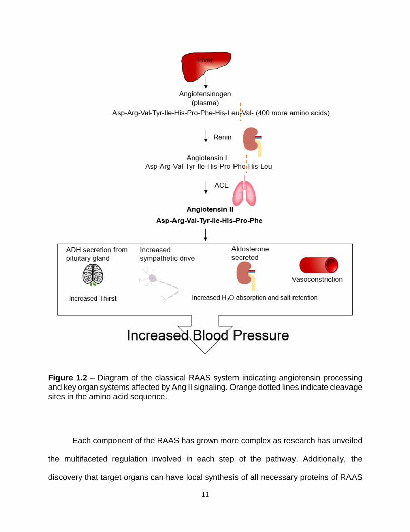
circulating pathway that is activated upon a drop in blood pressure. Briefly, this system
involves the liver constantly producing an α-2-globulin protein angiotensinogen (AGT) into
the plasma. Upon a reduction in blood pressure, the kidney secretes the active form of

10
the enzyme renin, which cleaves angiotensinogen into a smaller 10-amino’acid peptide
angiotensin I (Ang I). Renin’s conversion of angiotensinogen to Ang I is the rate-limiting
step of the RAAS. Ang I is further cleaved in the plasma by ACE enzymes contained
within the pulmonary blood vessel endothelium to create the 8-amino acid peptide
angiotensin II (Ang II). Ang II is the principal effector of the RAAS that can cause a wide
range of physiological effects through regulation of the vascular, renal, endocrine, and
neural systems. Ang II increases water and salt reabsorption of the kidneys by directly
activating specific G-protein coupled receptors and indirectly by causing secretion of anti-
diuretic hormone (ADH) from pituitary glands and release of aldosterone from the adrenal
cortex. Ang II increases activation of thirst centers in the CNS as well as sympathetic
drive. Ang II also stimulates vasoconstriction by directly acting on smooth muscle cells
(SMCs) of the vasculature (Figure 1.2).
11
Figure 1.2 – Diagram of the classical RAAS system indicating angiotensin processing and key organ systems affected by Ang II signaling. Orange dotted lines indicate cleavage sites in the amino acid sequence.
Each component of the RAAS has grown more complex as research has unveiled
the multifaceted regulation involved in each step of the pathway. Additionally, the
discovery that target organs can have local synthesis of all necessary proteins of RAAS

12
has extended the understanding of RAAS from a systemic system to also include a local
version of the RAAS. Several organs have been determined to express the necessary
components of the RAAS including the heart, vasculature, brain, reproductive organs,
skin, digestive organs, adipose, and eyes.38
Angiotensinogen. AGT has historically been described as a biologically inactive pre-
cursor for the active angiotensin cleavage products of the RAAS; however, more recent
studies are beginning to identify potential activity of AGT itself. There are several single
nucleotide polymorphisms (SNPs) in AGT that have been linked to cardiovascular
disease-related risks including M235T (rs699).39-42 Particular SNPs, such as rs7079
occurring in the 3’-untranslated region, have been shown to correlate with increased
effectiveness of ACE inhibitors in lowering high blood pressure in a subset of hypertensive
patients.43 Plasma levels of AGT are credited to hepatic synthesis, and various hormones
and cytokines such as Ang II, glucocorticoids, estrogens, insulin, and IL-6 can affect
plasma concentrations.44-49 Recent discoveries have indicated synthesis of AGT is not
limited to the liver, and AGT mRNA has been detected in many different tissues.38 The
structure of AGT indicates that it is a member of the non-inhibitory serpin (serine protease
inhibitors) superfamily (SERPINA8) and of the ~453 amino acids the most conserved
portion of the protein among species is the angiotensin cleavage product.50 The
angiotensin cleavage site of AGT seems to be buried inside the structure of the protein
until bound to renin, which then allows conformational changes in AGT to move this
cleavage site into the enzymatic active cleft of renin.51

13
Renin. Renin is an aspartic protease with high substrate specificity that hydrolyzes the N-
terminus of AGT to form the decapeptide Ang I. Renin is one of the earliest studied
proteins of the RAAS. Its discovery is credited to Robert Tigerstedt and his student at the
time, Per Bergman, after they reportedly observed increased blood pressure in recipient
rabbits after injecting them with rabbit kidney homogenate. This led to their further
characterization of “...a blood-pressure raising substance…” that they then named
renin.52, 53 It was not until the 1970s that renin was purified and its structure determined
by Tadashi Inagami.54-57
Renin expression and secretion are regulated by a complex network of cellular
signaling cascades occurring in response to physiological changes. Plasma renin
production is mostly localized to juxtaglomerular cells of the renal afferent arterioles.
Renin begins as a precursor form of the protein called (pro)renin that becomes packaged
into specific secretory pathway granules. Some of these granules secrete (pro)renin
constitutively into the plasma, while a more regulated granule pathway will result in
cleavage of (pro)renin by proteases, such as cathepsin B, that are localized in secretory
granules to form active renin followed by controlled exocytosis.58, 59 Secretion of active
renin can be triggered by tubular chloride concentrations, sympathetic nerve activation of
β-adrenergic receptors on juxtaglomerular cells, pressure-sensing mechanisms
(baroreceptors), and cell membrane potentials.60 A number of signaling events that
regulate renin secretion also affect cAMP levels, such as activation of β-adrenergic
receptors, suggesting cAMP regulates the rate of renin secretion. 61, 62
More recently, the discovery of a receptor for renin, (pro)renin receptor (PRR), has
further enhanced our understanding of renin’s functions and regulation.63 PRR is a single-
14
pass transmembrane protein that binds active renin or (pro)renin.64 Binding of active renin
to PRR enhances renin’s catalytic conversion of AGT to Ang I. Furthermore, independent
of the RAAS, PRR can activate mitogen-activated protein kinase (MAPK) signaling
pathways when either prorenin or renin is bound. PRR can also serve as a subunit of the
proton pump V-ATPase and contribute to Wnt/β-catenin signaling.65, 66 PRR may also
affect the pathophysiology of hypertension, as mice with a specific knockout of PRR in
renal tubular cells are protected from Ang II-induced high blood pressure and exhibit
reduced renal sodium retention.67 Other studies have shown that when the aldosterone
mimetic, DOCA-salt, is used as a model of hypertension, it leads to increased expression
of PRR in the brain.68 In this model, PRR expression is up-regulated by Ang II and
required induction of cAMP response element-binding proteins.68 The gene for renin,
REN, has been reported to contain a cAMP response element in the promotor region,
indicating that not only secretion of renin but also transcription of REN is modulated by
cAMP.69
Renin is an attractive target for novel anti-hypertensive medications because of
the potential to achieve complete blockade of the RAAS as compared to partial inhibition
observed with ACE inhibitors and ARBs. Current renin inhibitors target the interaction of
renin with AGT. In the 1970s, the peptide pepstatin was identified as a natural competitive
renin inhibitor for the active site of renin.70 However, pepstatin also inhibits other aspartic
proteases; thus, a variety of pepstatin derivatives, commonly referred to as AGT
analogues, have been developed.71 Unfortunately, the benefits of AGT analogs toward
hypertension and cardiovascular disease have mostly been observed only
experimentally, and these types of inhibitors often have low bioavailability.72 The first

15
direct renin inhibitor for use in humans was approved in 2007; the United States Food
and Drug Administration approved the use of Aliskiren, a non-peptide inhibitor for the
treatment of high blood pressure.73-75 It is reported to be an effective oral monotherapy
for lowering blood pressure; however, recent concerns have developed when considering
Aliskiren for combinational drug therapies.76 One clinical study testing the effectiveness
of Aliskiren along with ACE inhibitors and/or ARBs in diabetic patients had to be
terminated prematurely due to safety concerns.77 Research continues to assess the
extent of Aliskiren efficacy toward different disease states and explore the development
of next generation renin inhibitors.
Angiotensin I & Angiotensin-converting enzyme. The cleavage product of AGT by renin
is an inactive decapeptide, Ang I, which was formerly called hypertensin I/angiotonin I
until a uniform nomenclature was later adopted. Ang I was found to have very minimal
contractile or pressor effects on the isolated kidney; however, intravenous infusion of Ang
I had similar blood pressure effects as Ang II.78 Ang I was thus identified as simply a
precursor that is quickly hydrolyzed by angiotensin-converting enzyme (ACE), a zinc
metalloprotease, to form Ang II.
ACE was discovered by Leonard T. Skeggs Jr. who is also credited with
determining the organization of substrates/enzymes of the RAAS.78-81 Unlike renin, ACE
does not have a strong substrate specificity. Although the most well-known substrate for
ACE is Ang I, it can also hydrolyze bradykinin, substance P, neurotensin, angiotensin
metabolites such as angiotensin(1-7), and other small peptides.82, 83 ACE expression has
been detected in the lung, vascular endothelium, renal proximal tubular epithelium,

16
intestinal epithelium, ovary, male germ cells, brain, and immune cells.38 Additionally, a
novel homolog of ACE termed ACE2 has been discovered in the last decade that seems
to counter the functions of ACE by generating the metabolites Ang(1-7) and Ang(1-9) from
Ang I and Ang II.84, 85
One of the first classes of drugs to clinically target the RAAS were ACE inhibitors.
The first ACE inhibitor was identified by Sergio Ferreira, who noticed that snake venom
peptides inhibited the conversion of Ang I to Ang II and the degradation of bradykinin.86
The discovery of this inhibitory peptide eventually facilitated the development of orally
active drugs with Captopril being the first ACE inhibitor used for the treatment of
hypertension.87 ACE inhibitors are recommended by the American Diabetes Association
for patients with diabetes that also have hypertension as ACE inhibitors offer fewer side-
effects in this population as compared to other anti-hypertensive drugs.88-90 Yet their
average effectiveness in lowering blood pressure in the general population is reported to
be modest with a -8 mm Hg for systolic blood pressure and -5 mm Hg for diastolic blood
pressure.91
Angiotensin II. The principal effector of the RAAS is Ang II, an octapeptide hormone
product derived from ACE-mediated hydrolysis of Ang I. As depicted in Figure 1.2, Ang II
can facilitate vasoconstriction, thirst, renal salt and water reabsorption, and increase
sympathetic activity.92
In mammalian cells, Ang II activates cellular responses by signaling through two
distinct G protein-coupled receptors designated angiotensin type 1 and type 2 receptors
(AT1 and AT2). The widely expressed AT1 receptor mediates the classical actions of Ang

17
II and is inhibited by the commonly prescribed anti-hypertensive drugs, ARBs. There is at
least one known human variant (A1166C) of the gene encoding the AT1 receptor
(ATGER1) that has been implicated in essential hypertension.93, 94 Uniquely, rodents
express two isoforms of the AT1 receptor, AT1a and AT1b, that share 95% of their amino
acid sequence.95 The AT1a receptor is significantly expressed in vascular smooth muscle
cells, brain, kidney, and heart, whereas the AT1b receptor is primarily expressed in
adrenal and pituitary glands.96 Despite their similarity, in vivo studies have indicated that
the AT1a receptor contributes more to Ang II-induced blood pressure effects as AT1b-/-
mice lack any compensatory phenotypes and display blood pressures similar to wildtype
mice.97, 98
Ang II signaling through the AT1 receptor is tightly regulated which is evident by
its ability to induce potent yet transient vasoconstriction. Upon activation, the AT1
receptor is quickly desensitized by β-arrestin-dependent receptor internalization.99 The
C-terminal tail is rich in serine/threonine phosphorylation sites that are targeted by G
protein-coupled receptor kinases (GRKs) to initiate recruitment of β-arrestin.100, 101
Interestingly, in addition to promoting receptor internalization, β-arrestin recruitment to
AT1 receptors can activate MAPKs via β-arrestin scaffolding, which has been shown to
have beneficial cardiovascular effects.102-104 These findings have led to the development
of biased agonists for the AT1 receptor that would provide anti-hypertensive effects from
antagonizing the G-protein signaling cascades while maintaining the advantageous β-
arrestin signaling. One of the leading biased agonists, TRV027, is currently in phase II
clinical trials as a novel therapeutic for hypertensive acute heart failure.105
18
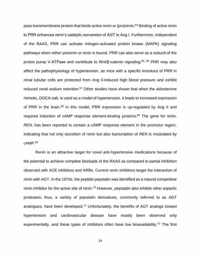
Figure 1.3 – The angiotensin II receptor 1 (AT1) signaling cascades and key proteins activated by the AT1 receptor.
The AT1 receptor couples to many different signaling cascades to mediate cellular
responses. AT1 receptors can couple to Gq and activate phospholipase C, leading to
production of inositol trisphosphate and increased intracellular calcium. Through coupling
with G12/13, AT1 can activate extracellular voltage-gated calcium channels and the
Rho/ROCK cascade.106, 107 AT1 has also been shown to activate Ras/Raf through
coupling with Gi.108 In addition to coupling to different G proteins, various downstream
signaling cascades are activated by the AT1 receptor. Ang II via AT1 activates
phospholipases A2, which stimulates endogenous prostaglandin production and
enhances inflammation.109, 110 The AT1 receptor has been shown to transactivate the
epidermal growth factor receptor (EGFR), which specifically has been linked to
hypertension-associated hypertrophy of tissues.111, 112 AT1 receptor activation can also

19
lead to reactive oxygen species production by activation of NADPH oxidase, which has
been linked to endothelial cell dysfunction associated with hypertension.113, 114 Numerous
other kinases, such a Pyk2, Src, JAK, and MAPKs have been demonstrated to be
regulated by AT1 signaling (Fig. 1.3).115-118
The characterization of and physiological implications for the presence of a second
Ang II G-protein coupled receptor, the AT2 receptor, is currently an area of intense
investigation. Some of the AT2 receptor’s functions have been inferred from expression
data. The AT2 receptor is ubiquitously expressed in fetal tissues; however, after birth,
expression of AT2 is dramatically reduced in many regions to almost undetectable levels
consistent with a role for AT2 in development.119-121 In adults, the existence of the AT2
receptor has been detected in uterus, heart, lung, kidney, brain, and adrenals.119-124
Despite low levels of expression in adult tissues, the AT2 receptor has been determined
to counter-balance many of the actions resulting from Ang II signaling through the AT1
receptor. Some of the identified functions of the AT2 receptor include inhibiting cell
growth, inducing apoptosis, and mediating vasodilation.125-127 In the kidneys, AT2
stimulates the release of vasodilators such as bradykinin and nitric oxide.128, 129 In
neurons, the AT2 receptor has been found to inhibit norepinephrine release from
catecholaminergic neurons and increase neurite outgrowth.130 From studies investigating
deletion of the AT2 receptor, the opposing function of AT2 receptors against Ang II-AT1
actions are apparent as AT2-/- mice exhibit elevated baseline blood pressures as well as
exacerbated Ang II-induced pressor and antidiuretic effects.131, 132 The AT2 receptor was
found to signal through coupling to Gi.133 Further studies have suggested that the AT2
receptor may dimerize and provide direct antagonism of the AT1 receptor.134

20
In healthy humans Ang II is cleared from circulation within 48 hours, this clearance
can be altered in hypertensive patients.135 Ang II was initially thought to be degraded into
several different smaller inactive peptides; however, additional research has revealed that
these metabolites are yet another way by which the RAAS affects physiology. One branch
of angiotensin metabolism involves the above mentioned novel variant of ACE, ACE2,
that can breakdown Ang I and Ang II to form Ang(1-7) and Ang(1-9). Ang(1-7) functionally
opposes many AT1-mediated actions and has cardioprotective effects that are thought to
be mediated through activation of the G protein–coupled Mas receptor.136, 137 Evidence
suggests Ang(1-9) either can be converted to Ang II or has Ang II-like functions.138
Another branch of angiotensin metabolism involves the sequential cleavage of Ang II by
aminopeptidases A and N to form Ang III and Ang IV, both of which have been found to
have physiological functions.139 Furthermore, a selective receptor for Ang IV, which was
termed AT4 and later identified to be the insulin-regulated membrane aminopeptidase
(IRAP), has been discovered that has been shown to have a significant role in neuronal
plasticity.140, 141 These metabolites of Ang II are processed further to smaller non-active
peptides.142 Despite the many investigations and accumulation of knowledge obtained
regarding the RAAS pathway and the contributions of Ang II to hypertension, there
remains an unmet need for novel pharmacological targets which may provide therapeutic
options for the many unmanageable cases of essential hypertension.
Prostaglandins
Prostaglandins are potent lipid mediators that comprise a subclass of the
eicosanoids, a family of signaling molecules derived from oxidation of 20-carbon

21
polyunsaturated fatty acids. Eicosanoids are synthesized from arachidonic acid (AA),
eicosapentaenoic acid, or dihomo-gamma-linolenic acid, and can be further classified into
two main groups, the prostanoids (including prostaglandins) and leukotrienes/lipoxins.
Each group requires specific catalytic enzymes for their elaboration; cyclooxygenase
(COX) enzymes generate the prostanoids, which include prostaglandins, thromboxanes,
and prostacyclins, while lipoxygenase (LOX) enzymes generate the leukotrienes.
Prostaglandins are produced upon the release of AA from phospholipids of the plasma
membrane by phospholipase A2 (PLA2). AA is further processed by COX enzymes to form
the prostaglandin precursors, prostaglandin G2 (PGG2) and prostaglandin H2 (PGH2), that
are subsequently converted by specific prostaglandin synthases into the five bioactive
prostanoids: prostaglandin E2 (PGE2), prostacyclin (PGI2), prostaglandin D2 (PGD2),
prostaglandin F2α (PGF2α) and thromboxane (TXA2).
The term “prostaglandin” was first used by Ulf von Euler after he isolated a
substance from sheep seminal vesicles that exerted both depressor and vasocontractile
effects.143 Believing its constituents to be secreted from the prostate gland, he named the
substance prostaglandin. Following further purification, specific prostaglandins were
eventually named based on the fraction from which they originated; prostaglandin E2
was described as remaining in the ether fraction and prostaglandin F2 in the phosphate
fraction (the Swedish word for phosphate is “fosfat”, hence the “F”).144-146 Later identified
prostaglandins were termed to align within the initial alphabetical nomenclature, with the
exception of thromboxane, which was termed after its role in clot formation. Subscripts
are included in the name to indicate the number of double bonds in the molecule.

22
Structurally, prostaglandins are hydrophobic molecules that differ by slight variations in
their cyclcopentanone ring and/or the two carbon side chains.
Prostaglandins are synthesized ubiquitously in response to various stimuli and
released to induce autocrine and/or paracrine signaling events by binding to distinct cell
surface G-protein coupled receptors. Prostaglandins have diverse physiological
functions, from regulating fertility to modulating inflammation and blood pressure;
however, the extent of their signaling is efficiently controlled by first-pass clearance from
tissue and circulation initiated by 15-hydroxyprostaglandin dehydrogenase (15-
PGDH).147-149
Initial Indications of Prostaglandin Involvement in Blood Pressure
Non-steroidal anti-inflammatory drugs (NSAIDs) are over-the-counter analgesia
medications that prevent the endogenous synthesis of prostaglandins by inhibiting
cyclooxygenase (COX) enzymes. There are two isoforms of COX, and each is classically
described as homodimers that catalyze the oxygenation and subsequent reduction of AA
to form the prostaglandin precursors, although recent studies have suggested the
existence of conformational heterodimers.150 Preliminary findings suggested that COX-1
is constitutively expressed, whereas COX-2 expression is inducible and stimulated by
pathophysiological conditions.151 This initial evidence drove the formulation of selective
COX-2 inhibitors to prevent the adverse gastrointestinal effects that were initially noted
with non-selective NSAIDs.152 However, this early understanding of COX isoforms may
be outdated, since more recent studies have reported a role for COX-1 in inflammatory

23
states and determined that inhibition of COX-1 does not increase the risk of gastric
complications.153, 154
Clinical studies investigating the effects of selective and non-selective COX
inhibitors have revealed that prostaglandins affect cardiovascular homeostasis. Although
the use of NSAIDs are well tolerated by many individuals, chronic use of primarily COX-
2 selective inhibitors and of some non-selective NSAIDs have been shown to increase
the risk of myocardial infarction, stroke, heart failure, arrhythmias, and hypertension.155-
157 This risk is most pertinent to patients with a pre-existing condition, potential drug-
interaction, or a family history of cardiovascular complications.155-158 Cardiovascular risks
are also higher amongst the elderly population who are often prescribed NSAIDs more
frequently. Rofecoxib (Vioxx, Merck), a COX-2 inhibitor that was prescribed for the
alleviation of symptoms associated with osteoarthritis, was withdrawn from the market
after it was reported that cardiovascular events occurred in 30,000 people taking the
medication.159 These unfortunate complications reveal that prostaglandins play an
essential role in mediating cardiovascular health.
Prostaglandin E2
Prostaglandin E2 (PGE2) is an abundant and extensively investigated prostanoid.
PGE2 is produced from the isomerization of PGH2 by prostaglandin E synthases (PGES)
and was initially described to facilitate systemic vasodepression as well as smooth muscle
contraction.160, 161 Many other biological functions have since been identified to be
affected by PGE2. Most notable are its roles in blood pressure regulation,
neurogeneration, bone formation, inflammation, maintenance of gastrointestinal integrity,

24
patency of fetal ductus arteriosus, cervical ripening, and uterine contractions.160-168
However, fully understanding the physiological roles of PGE2 has often been challenging,
since many opposing functions have been attributed to PGE2. For example, PGE2 has
been shown to exhibit both vasodepressor and vasopressor effects.169-175 In the immune
system, PGE2 is well-established to mediate pro-inflammatory effects, but some reports
identify an immunosuppressor function.165 These complex and counterregulatory actions
of PGE2 can be attributed to its ability to signal through four distinct G protein-coupled
receptors, the E prostanoid (EP) receptors subtypes 1 – 4 (EP1 – EP4). While PGE2 has
the greatest affinity for the EP receptors (Kd values ranging from 1 – 40 nM), at higher
concentrations PGE2 can also activate the prostaglandin F2α (FP), prostaglandin D2 (DP),
and thromboxane (TP) receptor (Kd = 100 nM, 300 nM, 30 µM respectively).176-178
Different subtypes of EP receptors activate specific cellular transduction pathways and
have unique tissue and cellular distribution, which collectively allows PGE2 to facilitate a
wide variety of cellular functions.
The EP Receptors and Their Role in Blood Pressure Regulation
PGE2 has been shown to exhibit both vasodepressor and vasopressor effects
indicating that PGE2 has a counter-balancing role in maintaining blood pressure
homeostasis. Much of the current understanding of PGE2-mediated blood pressure
effects originated from investigations of the specific EP subtypes. EP1 and EP3 receptors
were recognized as mediating the vasopressor effects and the vasodepressor responses
were attributed to activation of the EP2 and EP4 receptors.
25
The EP1 Vasopressor Receptor. The EP1 receptor is a smooth muscle contractile
receptor that is thought to primarily couple with Gq leading to activation of PLC, production
of inositol triphosphate, and increased intracellular calcium.179, 180 However, in one study,
EP1 was found to couple with Gi leading to increased expression of hypoxia-inducible
factor-1α.181 With the lack of specific antibodies for any of the EP receptors, expression
patterns are limited to radioligand binding assays and quantification of mRNA. EP1
receptor mRNA is ubiquitously expressed with high transcript levels observed in blood
vessels.182 However, expression does not necessarily correlate with PGE2 function. For
example, the mRNA levels for the contractile EP3 receptor was found to be greater than
the other EP receptors in the mouse aorta; however, PGE2 on ex vivo aortic vessel rings
exhibits vasodilation.183, 184 In the rat, a variant form of the EP1 receptor has been cloned
that is highly expressed in the kidney and has similar ligand specificity, yet exhibits
reportedly lower calcium mobilization.185 The EP receptors can also be differentiated by
drug selectivity. Classically, 17-phenyl-PGE2 has been used as an agonist for the EP1
receptor, but it, along with many of the EP1 agonists, has been shown to have activity at
the EP3 receptor as well.178 The most recent and selective EP1 agonist is ONO-D1-004
while the most used EP1 antagonists are SC-51322, ONO 8711 and ONO 8713.177, 186-
188
Discovery of the EP1 receptor’s role in blood pressure originated from
investigations of EP1 receptor knockout mice. EP1-/- mice are essentially healthy, fertile,
exhibit PGE2-mediated vasodepressor responses, and have lower baseline MAP.174, 189,
190 Additionally, EP1-/- mice from a DBA/1lacJ strain exhibited a reduction in pain
sensitivity.190 The EP1 receptor has been found in part to mediate Ang II-induced blood

26
pressure effects, as EP1-/- mice have a blunted response to acute intravenous infusion or
chronic osmotic pump infusion of Ang II.174 In a mouse model of end-organ damage
(uninephrectomy/DOCA-NaCl/Ang II osmotic pump), EP1-/- mice displayed reduced
mortality and organ damage as well as lower MAP.191 The EP1 receptor seems to mediate
Ang II-induced hypertension in the CNS.192 Cerebrovascular dysfunction has been shown
to involve ROS production and increased calcium influx; both of which have also been
shown to require activation of central EP1 receptors.192-194
The EP3 Vasopressor Receptor. The other contractile EP receptor is the EP3 sub-type
that is known to primarily couple to Gi leading to inhibition of adenylyl cyclase and reduced
cAMP production.180, 195-197 The EP3 receptor is widely expressed and contributes to
diverse functions including regulation of the febrile response, kidney functions,
maintenance of gastrointestinal health, and hepatic glucose metabolism.173, 198-201
Abundant levels of (PTGER3) mRNA are found in metabolic and gastroenteric tissues
including the kidneys, adrenal glands, pancreas, adipose tissue, and stomach.182, 202, 203
Uniquely, multiple splice variants of the EP3 receptor have been identified that have
similar ligand binding properties but differ by their C-terminal tail, enabling coupling to
several different G proteins.204 The number of EP3 variants differs with various species,
with 9 identified in humans, 3 in mice, 4 in rats, and 5 in rabbits.203, 205-209 The EP3 variants
found in mice have been well investigated, and of the three variants, EP3γ and EP3α
have been shown to have partial constitutive Gi activity, whereas EP3β can couple to Gi
but does not exhibit constitutive activity.210, 211 The EP3γ isoform has been demonstrated
to couple to Gs as well.209 Evidence also supports the EP3 receptor coupling to G13,

27
leading to activation of Rho kinase (ROCK).212, 213 Additionally, the EP3 receptor has been
shown to mediate calcium mobilization via the Gβγ subunit of G proteins activating PLC.214
More recent studies have suggested that the EP3 receptor may also couple to Gq since
its calcium mobilization effects have been shown to be independent of Gi and is a result
of PLC activation.215 Despite what is known about the EP3 variants and their signal
transduction properties, the physiological significance for each variant remains to be
determined. There are selective agonists for the EP3 receptor with sulprostone (Kd = 0.6
nM) being the most used, although it has affinity for the EP1 receptor (Kd = 20 nM) as
well.178 ONO-AE-248 is a more recent and selective EP3 agonist.216 There are selective
antagonists for the EP3 receptor such as L-796,106 and DG-041; moreover, DG-041 has
been tested in clinical trials for effectiveness as an antiplatelet drug.217, 218
Several studies have revealed that EP3 receptors regulate hemodynamic effects.
In EP3-/- mice PGE2 exhibits a slightly enhanced vasodepressor response when
compared to wildtype mice indicating that the EP3 receptor counter-regulates the PGE2-
mediated vasodepressor response.173 Furthermore, in knockout mice in which the primary
EP vasodepressor receptor (EP2) is absent, PGE2 exhibits vasopressor responses that
are attributed to activation of the EP3 receptor as these responses can be reproduced
with sulprostone in wildtype and EP2-/- mice.169 The EP3 receptor also plays a role in
regulating renal blood flow as EP3-/- mice have enhanced blood flow in response to
PGE2.173 Similar to the EP1-/- mice, EP3-/- mice have been found to exhibit attenuated
rises in blood pressure and vascular contractility in response to Ang II.175 Furthermore,
intracerebroventricular injection of PGE2 in rats elicits an EP3-driven pressor response
involving sympathetic nerve activation.219, 220 The EP3 receptor has been shown to

28
mediate PGE2-induced vasoconstriction in many different vascular beds including
mesenteric, cerebral, intercostal, pulmonary, and femoral arteries.180, 221-224 EP3-
mediated vasoconstriction seems to in part require activation of protein kinase C δ and
ROCK.221, 222, 225
Vasodepressor EP Receptors
EP2 receptor. The EP2 receptor is the primary vasodilator/vasodepressor EP receptor
that couples to Gs leading to activation of adenylyl cyclase. This causes increases in
cAMP that, in turn, induces vasodilation by inhibiting myosin light chain kinase.226 Two
different “EP2 receptors” were originally cloned by independent groups that claimed to
have identified a Gs-coupled EP receptor; one clone was sensitive to butaprost and the
other was not.227, 228 Eventually, it was decided that the butaprost sensitive clone was the
EP2 receptor and the insensitive clone was termed the EP4 receptor. The EP2 receptor
has been found to be most abundantly expressed in bone marrow, spleen, ovary,
adipose, lung, and olfactory epithelium.182 Physiological functions regulated by the EP2
receptor include smooth muscle relaxation and stimulation of cell differentiation.229-231 The
EP2 receptor has been identified to be involved in cancer progression and tumorigenesis
as a result of its identified role in stimulating cell growth.232 The EP2 receptor may also
contribute to fertility as female EP2-/- mice are infertile; however, human studies have yet
to confirm the relevance of these findings.169, 172
As previously mentioned, the EP2 receptor is the primary receptor mediating
PGE2-induced vasodilator responses; thus, EP2-/- mice exhibit PGE2-induced
vasopressor responses upon intravenous infusion.169 Mice lacking EP2 receptors are

29
more susceptible to salt-induced hypertension. Moreover, in a cohort of Japanese men,
a SNP (rs17197) in the untranslated region of the PTGER2 gene has been correlated
with essential hypertension.169, 233 Although the EP2 receptor is not highly expressed in
the kidney, intramedullary infusion of PGE2 results in natriuresis and diuresis that is
absent in EP2-/- mice.234 Additionally, PGE2 induces vasodilation of the afferent arterioles
of wildtype mice that is switched to vasoconstriction in EP2-/- mice.235 Interestingly, the
renal afferent arteriolar vasoconstriction observed in EP2-/- mice can be inhibited with an
ACE inhibitor indicating that this vasoconstriction is dependent on Ang II elaboration.235
Collectively, these studies underscore a role for the EP2 receptor in regulating kidney
function.
EP4 receptor. As previously specified, the EP4 relaxant receptor was originally
designated as the EP2 receptor by initial cloning studies.227 The EP4 receptor was also
determined to couple to Gs and increase cAMP levels similar to the EP2 receptor.195 The
EP4 receptor is more abundantly expressed across tissue types when compared to the
EP2 receptor; however, EP4 mRNA is enriched in the pancreas, large intestine, uterus,
skin, ductus arteriosus, and thymus.182, 236, 237 The EP4 receptor differs from the EP2
receptor by the presence of a longer C-terminal tail that includes six serine residues that
are thought to allow increased phosphorylation leading to quick desensitization which is
not observed with EP2 receptor activation.238, 239 Additionally, agonists and antagonists
of the EP4 receptor have been identified. The agonists include: ONO-AE1-329, TCS-
2510, and L-902688.240 The standard EP4 antagonist was previously AH-23848, which
has since been superseded by more selective ligands.240

30
Investigating a role for the EP4 receptor in blood pressure regulation has been a
challenge since global knockout of the EP4 receptor in mice from an inbred strain are
perinatal lethal as a result of patent ductus arteriosus.236, 237, 241 However, studies using
mixed background EP4-/- mice and/or EP4 receptor agonists and antagonists have
revealed the vasodilator actions of the EP4 receptor. The EP4 receptor has been shown
to mediate endothelium-dependent vasodilation of isolated mouse aorta in response to
PGE2.184 EP4-induced vasodilation is achieved by increased activation of endothelium
nitric oxide synthase (eNOS) resulting in production of endothelium-derived nitric oxide
that induces activation of guanylyl cyclase in smooth muscle cells and subsequent
relaxation.184 EP4-/- mice on a mixed background exhibit blunted depressor responses to
PGE2, and infusion of an EP4 agonist, ONO-AE1-329, into rats reveals a lowering of
MAP.189, 242 More recently, PGE2 acting through the EP4 receptor has been shown to
inhibit pig ciliary arterial contractions.243 Additionally, intravenous infusion of ONO-AE1-
329 into anesthetized dogs has been shown to reduce peripheral resistance, diastolic
pressure and MAP.244 Studies have indicated a role of the EP4 receptor in regulating
kidney function which in turn could alter long-term blood pressure. In separate studies,
the EP4 receptor has been shown to mediate renal activation of the RAAS under
conditions of low salt diet and promote exocytosis of renin from juxtaglomerular cells in
response to PGE2.245, 246

31
Table 2 – Summary of cited EP receptor functions in regulating blood pressure and relevant phenotypes of EP receptor knockout mice. Commonly used agonists and antagonists are listed.
EP Receptor
Agonists Antagonists Vascular Functions Knockout Mice Phenotypes
EP1 Sulprostone 17-phenyl-PGE2
ONO-D1-004
SC-51322 ONO 8711 ONO 8713
Smooth muscle contraction Plays a major role in CNS in mediating Ang II-induced hypertension and cerebrovascular dysfunction
Lower baseline MAP Reduced response to Ang II-induced hypertension Reduced hypertensive end-organ damage Reduced pain sensitivity
EP2 Butaprost PF-04418948
AH-6809
Smooth muscle relaxation Mediates PGE2 -induced vasodepression
Female mice are infertile Exhibits vasopressor responses to intravenous infused PGE2 Lack renal natriuresis and diuresis Afferent arterioles exhibit vasoconsitrction in response to PGE2
EP3 17-phenyl-PGE2
Sulprostone ONO-AE-248
L-796,106 DG-041
Smooth muscle contraction Mediates PGE2 -induced vasopression Drives PGE2-mediated intracerebroventricular-induced rise in blood pressure
Enhanced vasodepressor response to PGE2 Enhanced renal blood flow Reduced response to Ang II-induced hypertension
EP4 ONO-AE1-329 TCS-2510 L-902688
AH-23848 CJ-023423 GW-627368 L-161982 ONO-AE3-20
Endothelium-dependent vasodilation Vasodepression Reduce peripheral resistence Promotes renin release and activation of RAAS
Global knockout is perinatal lethal
The Interplay of Prostaglandin E2 and Angiotensin II
An early indication that Ang II and PGE2 signaling may interact came from the
discovery that Ang II can activate PLA2, leading to endogenous PGE2 production via COX

32
and PGES enzymes.247 Initial studies demonstrated that inhibition of PGE2 with NSAIDs
augmented the actions of Ang II in the kidney; thus PGE2 was thought to negatively
regulate the actions of Ang II.248 Other studies demonstrated that that exogenous
application of PGE2 to the medullary vasa recta or renal artery generally provides negative
feedback to the vasoconstrictor effects of Ang II.249, 250 Although these early investigations
led to the hypothesis that PGE2 functions to buffer the actions of Ang II, more recent
studies have revealed that the interaction between Ang II and PGE2 is much more
complex.251 The notion that PGE2 only counteracts Ang II’s pro-hypertensive effects
changed after PGE2 was shown to increase the secretion of renin and subsequently
increase levels of Ang II.109, 170, 252, 253
Since COX enzymes lead to the synthesis of PGE2, many studies have been
conducted investigating the effects of Ang II on these enzymes. Studies carried out by
Dr. Matt Breyer’s lab have indicated unique roles for each COX isoform in regulating the
effects of Ang II on blood pressure and renal blood flow. Pharmacological or genetic
inhibition of COX-2 was shown to potentiate Ang II-induced increase in blood pressure,
as well as exacerbate Ang II-induced reduction in medullary blood flow.254 Conversely,
inhibition of COX-1 attenuated Ang II responses.254 These data suggest that COX-2-
derived products counteract Ang II while COX-1-derived products sustain the effects of
Ang II. Additionally, Ang II has been demonstrated to differentially regulate COX-2
expression in the kidney with AT1 receptor activation leading to reduction in COX-2
expression and AT2 receptor activation leading to increases in COX-2.255
One caveat of investigations focused on COX enzymes is that COX enzymes lead
to the synthesis of all prostanoids; thus, studies investigating COX enzyme expression

33
do not conclusively describe the contributions of PGE2 specifically. Additional
investigations of mice deficient for mPGES, the specific synthase that produces PGE2,
have demonstrated that these mice exhibit hypersensitivity to chronic low-doses of
Ang.256 However, even these studies over-generalize the role of PGE2, as there are four
EP receptors that can mediate diverse physiological effects upon activation by PGE2.
Depending on the specific tissue type and the EP receptors stimulated, concurrent Ang II
and PGE2 signaling can facilitate different outcomes.
Accumulating evidence indicates that Ang II is at least in part dependent on PGE2
to facilitate its systemic blood pressure and vasculature responses. The EP contractile
receptors, EP1 and EP3, have been identified to play a role in mediating Ang II-dependent
hypertension. EP1-/- mice have a reduced rise in MAP in response to acute or chronic
Ang II.174 In isolated mesenteric arteries and preglomerular arterioles, treatment with the
EP1 antagonist SC51322 significantly blunts Ang II-induced constriction.174 Additional
studies have indicated a significant role for the EP1 receptor in mediating the CNS effects
of Ang II, including its pathological mechanisms. Ang II-induced cerebrovascular
dysfunction requires PGE2-induced activation of EP1.194 More recently analyses
conducted by Dr. Robin Davisson’s lab have identified the requirement of PGE2 signaling
through EP1 receptors in the subfornical organ (SFO) of the brain for Ang II-induced
hypertension.192, 193 In these studies, mice that were given intracerebroventricular infusion
of the EP1 antagonist, SC-51089, were protected from subcutaneous micro-osmotic
pump Ang II-induced hypertension.192 In follow-up studies using cultured SFO neurons,
they showed that Ang II-induced ROS formation reportedly required AT1 activation of
PLA2 and COX-1 derived PGE2 signaling through the EP1 receptor.193 The EP3 receptor

34
has been indicated to play a role in Ang II-induced hypertension as well, as EP3-/- mice
display a reduced rise in MAP in response to acute or chronic Ang II.175 Additionally, EP3
receptor antagonists and EP3-/- mice have been shown to exhibit reduced Ang II-induced
vasoconstriction of mouse mesenteric arteries.175 Even though studies have indicated
that PGE2 signaling through the EP1 and EP3 receptors is to some extent necessary for
the hypertensive and contractile effects of Ang II, it is still unclear how PGE2 contributes
to Ang II responses since systemic infusion of PGE2 exhibits an EP2-induced
vasodepressor effect. Further characterization of the complex balance of PGE2 signaling
through its four EP receptors and the mechanisms by which PGE2 contributes to Ang II-
induced hypertension is an active area of investigation that the work presented in this
thesis elaborates upon.
Specific Aims
Hypertension is a major risk factor for renal damage and cardiovascular disease.
Although anti-hypertensive drugs are available to reduce morbidity and mortality
associated with hypertension, the prevalence of end organ damage and death resulting
from chronic hypertension persists. Prostaglandins are potent lipid modulators of blood
pressure. Prostaglandin E2 (PGE2), a prostanoid that normally acts as a systemic
vasodepressor, has been shown to have vasopressor effects under certain
circumstances. The objective of my doctoral research was to gain insights into the
complex interaction of PGE2 and Ang II in regulating vascular reactivity and blood
pressure homeostasis. I hypothesized that Ang II, a principal effector of the renin-
angiotensin-aldosterone system, potentiates PGE2-mediated vasoconstriction.

35
Additionally, I hypothesized that angiotensin II priming could switch PGE2 from its
functional role as a vasodepressor to instead act as a vasopressor, ultimately contributing
to high blood pressure. To investigate this interaction, I formulated the following aims:
Aim 1: Evaluate synergism between angiotensin II and PGE2 pressor receptors in
modulating vessel reactivity. To evaluate the ability of submaximal doses of angiotensin
II to potentiate PGE2-mediated vasoconstriction, vessel reactivity will be determined ex
vivo by wire myography. Using receptor antagonists and knockout mice, the specific
angiotensin receptor and EP receptors required for PGE2-mediated vasoconstriction will
be determined.
Aim 2: Determine the signaling pathways by which angiotensin II and PGE2 synergize.
Understanding the mechanism of Ang II and PGE2 synergism will advance our knowledge
of the complexity of vascular reactivity. In terms of therapy for hypertension, examining
other key proteins besides the receptors themselves will allow the discovery of other,
possibly novel, drug targets. Furthermore, the generation of biased agonists, drugs that
exclusively stimulate a subset of beneficial pathways, may be possible.257
Aim 3: Determine PGE2 vasopressor effects in a mouse model of hypertension. To
investigate the relevance of this synergism to the physiology of the whole animal, in vivo
experiments using a mouse model of chronic Ang II hypertension will be used to test
PGE2 blood pressure effects. These studies will investigate whether PGE2 functionally
behaves as a pressor under conditions of Ang II in wildtype mice and determine whether

36
this effect would be mediated by the EP3 receptor by incorporating EP3-/- mice into these
experiments.

37
CHAPTER II
REGULATION OF ARTERIAL REACTIVITY BY CONCURRENT SIGNALING OF PGE2
WITH ANGIOTENSIN II
Introduction
High blood pressure is a major risk factor for cardiovascular diseases, including
myocardial infarction, stroke and renal failure.2, 3 The renin-angiotensin-aldosterone-
system (RAAS) has long been regarded as a major drug target for treatment of
hypertension due to its critical role in maintaining blood pressure through regulation of the
vascular, renal, endocrine, and neural systems.258, 259 Angiotensin II (Ang II), a principal
effector of the RAAS system, signals via angiotensin II type 1 and type 2 (AT1 and AT2)
G-protein coupled receptors. AT1 receptors mediate classical Ang II pressor effects
including smooth muscle contraction, increased sympathetic nerve activity, and renal
tubular reabsorption of Na+.260 The AT2 receptor, although not fully characterized, has
been shown to mediate opposing responses compared to AT1 function.261, 262 The AT1
receptor induces vasoconstriction by coupling to Gq, thereby leading to activation of
phospholipase C and increasing intracellular calcium.262, 263 These resulting calcium
increases then activate kinases, such as myosin light chain kinase and proline-rich
tyrosine kinase 2, that promote an increase in myosin light chain phosphorylation and
ultimately induce smooth muscle contraction. The AT1 receptor is also known to increase
phospholipase A2 activation, resulting in liberation of arachidonic acid and synthesis of
prostaglandins.264

38
Prostaglandin E2 (PGE2) is an oxygenated metabolite of arachidonic acid, which
modulates blood pressure among many functions.265, 266 PGE2 acts primarily as a
vasodilator and vasodepressor; however, under certain circumstances PGE2 can induce
vasopressor effects.169, 170, 183, 267-269 These divergent functions are due in part to the
ability of PGE2 to activate four distinct E-prostanoid receptors, EP1 - EP4.265, 270 EP1 and
EP3 receptors facilitate vasoconstriction while the EP2 and EP4 receptors mediate
vasodilation.183, 267 As with the AT1 receptor, activation of the EP1 receptor leads to a rise
in intracellular calcium via Gq signaling.271 The EP3 receptor couples to Gi and G12/13,
which inhibits cAMP production, activates Rho GTPase, and elevates intracellular calcium
levels through its Gβγ subunit.270 EP2 and EP4 receptors couple to Gs and upon activation
increase cAMP.
The Breyer lab and Guan lab have previously demonstrated that blockade of the
EP1 or EP3 receptor has salutary effects in mouse models of Ang II-mediated
hypertension. EP1 receptor knockout mice display a reduced rise in mean arterial blood
pressure in response to Ang II infusion and are protected against end-organ damage in
hypertensive mouse models.174, 191 Similarly, EP3 receptor knockout mice have a blunted
blood pressure response to acute administration of Ang II.175 Although these results
indicate an interaction between Ang II and PGE2 signaling, the molecular mechanisms
through which these systems synergize to regulate vascular tone is incompletely
characterized.
Previous studies have indicated a unique characteristic of PGE2-mediated vessel
reactivity in which, pre-stimulation of rat arterial tissue with a contractile agent followed
by PGE2 unmasks a significant vasoconstrictive response to PGE2.221 Thus, we

39
hypothesized that Ang II, a potent vasoconstrictor, could potentiate PGE2-mediated
vasoconstriction. Additionally, we postulated that this enhanced vasoconstriction would
be mediated by potentiation of EP3 or EP1 receptor signaling rather than through
desensitization of the vasodilator EP receptors. In the present study, we investigated the
effects of low dose Ang II “priming” on PGE2-induced contraction in isolated blood vessels
to determine whether Ang II can alter PGE2 vessel responses. We demonstrated that
Ang II primes vessels to exhibit potentiated vasoconstriction in response to PGE2. This
effect is mediated by the AT1 and EP3 receptors and requires calcium-dependent
mechanisms involving Rho-kinase (ROCK) and the proline-rich tyrosine kinase 2 (Pyk2).
Experimental Procedure
Animals
All studies were performed in accordance with the National Institutes of Health
animal care standards and approved by the Vanderbilt University Institutional Animal
Care and Use Committee. Male mice 9 to 15 weeks old on a C57BL/6 background were
used for all studies unless noted otherwise. Generation of EP1 (ptger1), EP2 (ptger2),
and EP3 (ptger3) receptor knockout mice has been previously described.169, 174, 198 Mice
deficient for the EP4 receptor (ptger4) have a lethal newborn phenotype when maintained
on a C57BL/6 genetic background.241 We bred C57BL/6 EP4+/- male mice to an outbred
strain (CD-1, Charles River laboratories; 10-generation outcross) and observed EP4-/-
mice viability into adulthood; thus, these mice were used for the EP4 studies.236, 272

40
Wire myography with isolated femoral arteries
Following euthanasia, femoral arteries were excised and placed into ice-cold
Modified Krebs-Henseleit solution (NaHCO2 25, KH2PO4 1.2, MgSO4 1.2, KCl 4.7, NaCl
118, CaCl2 2.5, Dextrose 10 mM). Vessel rings (~2 mm long) were cut and suspended
between 40 µm stainless steel wire in a Danish Myo Technology Model 620M myograph
system to record tension. Tissue baths contained 5.0 mL of Krebs-Henseleit at 37 ᵒC
aerated with 95% O2, 5% CO2 gas. Vascular preparations were allowed to stabilize for 45
minutes at a resting tension of 2.5 mN, which had been determined to induce about 90%
of maximal response to 50 mM K+ in mouse femoral arteries. Each preparation was
initially challenged with 50 mM K+, followed by 3 washes of Krebs-Henseleit solution
before testing experimental reagents. Separate rings were used for each data point to
avoid the rapid tachyphylaxis of the AT1 receptors. For Ang II priming, Ang II was added
to vessel ring baths for 10 minutes followed by addition of PGE2. At the end of each
experiment, endothelium integrity was confirmed by reversal of 1 µM phenylephrine-
evoked constriction with 10 µM acetylcholine. Rings were excluded that did not dilate at
least 70% of the phenylephrine contraction (tension reduced to < 30% of the
phenylephrine response). Finally, rings were washed and retested with 50 mM K+ to
assure maintained responsiveness throughout the entire experiment. All drugs were
dissolved in water, ethanol (PGE2, nifedipine, ibuprofen, SC-236), or DMSO (DG-041,
PF-431396) and then further diluted to ≥ 1:1000 or more in Krebs buffer before being
added to the vessel bath. Data were collected and stored using ADInstuments LabChart
7 software.

41
Chemicals
Angiotensin II (human), acetylcholine, losartan, ibuprofen, nifedipine, PF-431396
hydrate, salicylic acid, and phenylephrine were purchased from Sigma (St. Louis, MO).
Prostaglandin E2 and SC-236 were purchased from Cayman Chemical (Ann Arbor, MI).
PD123319 was purchased from Tocris (Bristol, UK). Y-27632 was purchased from Abcam
Biochemicals (Cambridge, MA). EGTA was purchased from Thermo Fisher Scientific
(Waltham, MA). FAK inhibitor 14 was purchased from Santa Cruz Biotechnology (Dallas,
TX). (Sar1,Ile4,8)-Angiotensin II Trifluoroacetate salt was purchased from Bachem
(Bubendorf, Switzerland). The Vanderbilt Chemical Synthesis Core synthesized DG-041.
Salicylamine was a gift from L. Jackson Roberts (Vanderbilt University).
Competitive binding of [3H]PGE2
Total kidney membranes (100 µg) from wildtype C57BL/6 mice were incubated
with [3H]PGE2 (2 nM) in binding buffer (25 mM potassium phosphate, 1mM EDTA, and
10mM MgCl2, pH 6.2) at a total volume of 200 µl for 1 hr at 30 ᵒC. Nonspecific binding
was determined in the presence of excess unlabeled PGE2 (1 µM). EP1/3 selective
agonist sulprostone (1 µM) was used as a positive control. Competitive binding
experiments were performed in the presence of 2 nM [3H]PGE2 and 1 µM of competing
ligands, either Losartan or PD123319. Reactions were terminated by vacuum filtration
and radioactivity bound was quantified.273, 274

42
Data analysis
Vascular responses were quantified as a change in force (maximal contraction-
baseline tension) normalized to the first 50 mM K+ response (K-standard) of each ring. All
data were graphed as means ± SEM using GraphPad Prism 5 software. Data were fitted
with variable slope sigmoidal curves using GraphPad Prism. Area under the curve (AUC)
(% contraction/Log[treatment]) values were used to statistically compare concentration
response curves (CRC) of PGE2 with or without priming agents. Student’s t-test was used
to determine significance between AUCs as well as for the [3H]PGE2 binding data. Ang II
CRCs were fitted to a variable-slope sigmoidal curve. Comparison of single dose PGE2
to various conditions was performed using one-way ANOVAs with a Bonferroni post-hoc
test. For all studies, P ≤ 0.05 was considered statistically significant. Significance was
represented throughout each figure as the following: *= P ≤ 0.05, **= P ≤ 0.01, and ***=
P ≤ 0.001.
Results
Ang II potentiation of PGE2-mediated vasoconstriction
To investigate the effect of Ang II on PGE2-mediated vasoconstriction, mouse
femoral arteries (I.D. ~0.25-0.3 mm) were studied ex vivo using a wire myograph. Femoral
arteries were selected for our studies as a model of peripheral vasculature in order to
compare to previous studies conducted using rat femoral arteries.221 To begin
investigating the vascular responsiveness of arteries to PGE2, the optimal resting tension
for mouse femoral arteries was determined. This was conducted by recording the arterial
force exhibited in response to 50 mM K+ at different manually set basal tensions. From
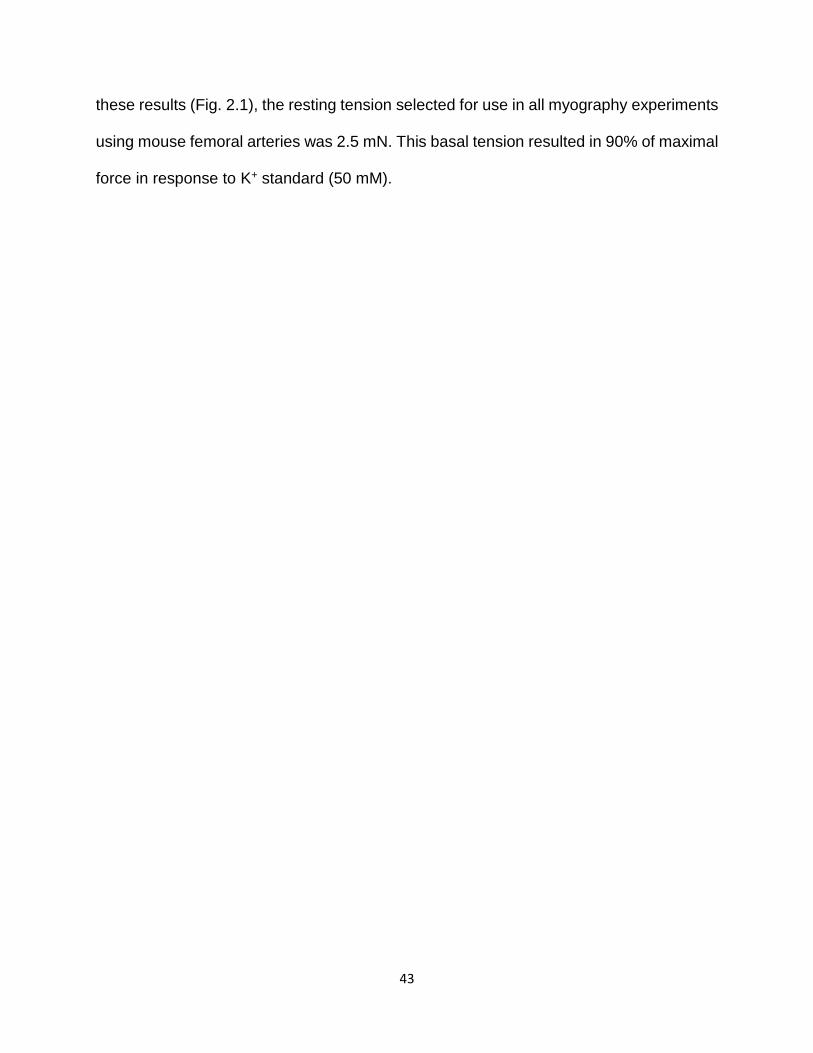
43
these results (Fig. 2.1), the resting tension selected for use in all myography experiments
using mouse femoral arteries was 2.5 mN. This basal tension resulted in 90% of maximal
force in response to K+ standard (50 mM).

44
2.0 2.5 3.00
2
4
6
Basal Tension (mN)
K
+ F
orc
e (
mN
)
Figure 2.1 – Determination of optimal wire myograph basal tension setting for mouse
femoral arteries. Graph represents the change in force resulting from vessel ring
contraction in response to 50 mM K+ at various tensions (2.0 - 3.0 mN) in wildtype mice.
Vertical bars indicate SEM. (N = 3)

45
Using the determined optimal baseline tension, arterial rings were assayed for
responsiveness to a range of single Ang II concentrations to formulate a concentration
response curve (CRC) (Fig. 2.2A). Arterial rings were then primed with a sub-threshold
constrictor concentration of Ang II (1 nM, ~ EC15) followed by a single concentration of
PGE2 at varying doses to construct a PGE2 CRC. Exposure of arterial rings to PGE2
alone, even at a high concentration of 1 µM (3.326 ± 0.647 % contraction of K-standard,
N = 5) did not cause significant vasoconstriction. However, priming of arterial rings with a
submaximal concentration of Ang II (1 nM) resulted in robust PGE2-mediated vessel
contraction (Fig. 2.2B, C). Ang II priming facilitated arterial contraction following exposure
to doses of 2 nM PGE2 or greater (AUCuntreated 2.778 ± 2.091, AUCAng II primed 22.830 ±
8.560, t test ***P<0.001) (Fig. 2.2, C).

46
Figure 2.2 - Ang II priming enhances PGE2-mediated vasoconstriction. (A) Ang II CRC in
wildtype mice. (N = 5-7) Dotted line indicates concentration of Ang II used to prime
vessels in B and C. (B) Representative trace of 30 nM PGE2-induced contraction of
vessels primed with 1 nM Ang II. (C) Comparison of PGE2 concentration-response curve
with (filled circles, N = 5-6) or without (open circles, N = 5) 1 nM Ang II priming. Data for
each concentration was collected on separate rings. Results were normalized to each
ring’s response to 50 mM KCl. Vertical bars indicate SEM.275

47
Additional studies were performed to test the time duration that PGE2-mediated
vasoconstriction would occur after Ang II pre-treatment. Vessels rings were treated with
1 nM Ang II followed by addition of 100 nM PGE2 at varying time points (10 – 60 minutes).
PGE2-induced contraction was greatest when PGE2 was added at approximately 10
minutes after Ang II and was significantly reduced at 30 minutes after Ang II (Fig. 2.3). It
was also of interest to determine whether the presence of or continuous stimulation by
Ang II was necessary for the ability of PGE2 to result in contractile responses or whether
Ang II would predispose the vessel to PGE2-mediated vasoconstriction even after Ang II
was removed from the bath. In experiments in which 1 nM Ang II was incubatd (5 minutes)
and then quickly washed from the vessel bath followed by immediate addition of 100 nM
PGE2, contractile responses induced by PGE2 were significantly attenuated (Fig. 2.3, right
bar) when compared to contractile responses observed at the 10-minute time point.

48
0 pr
iming
10m
in
20m
in
30m
in
60m
in
was
h ou
t Ang
II
0
10
20
30
** ***
Co
ntr
actio
n
(% K
-sta
nd
ard
)
Figure 2.3 – PGE2-facilitated contraction at varying times after Ang II pretreatment. 100 nM PGE2 responses with (black bars) or without (white bar) 1 nM Ang II priming. Minutes indicate time after Ang II that PGE2 was added to vessel bath. The far right bar indicates treatment of vessel rings with Ang II for 5 minutes then immediately removing Ang II from the bath prior to PGE2 addition. Vertical bars indicate SEM. (N = 3-5) *= P<0.05, **= P<0.01

49
Reversal effects: PGE2 potentiation of Ang II-mediated vasoconstriction
To further characterize vascular interactions between Ang II and PGE2, studies
were conducted to determine whether priming femoral arteries with PGE2 for 10 minutes
could potentiate Ang II-mediated vasoconstriction. Priming of femoral arteries with 30 nM
PGE2 resulted in a slight leftward shift in the Ang II CRC (Fig. 2.4, Ang II EC50 = 1.8 nM,
Ang II+PGE2 EC50 = 1 nM). These results indicated that PGE2 is able to modestly potentiate
Ang II-mediated vasoconstriction.

50
-9.5 -9.0 -8.5 -8.0 -7.50
20
40
60
80
100 30 nM PGE2 1
no 1
log [Ang II]
Co
ntr
actio
n
(%
K-s
tan
da
rd)
Figure 2.4 – PGE2 pre-treatment enhances Ang II-induced vasoconstriction. Ang II CRC with (grey triangles) or without (black circles) 30 nM PGE2 priming (Ang II-PGE2 data from Fig. 2.2). Ang II-PGE2 LogEC50 = -8.74 ± 0.05 and Ang II+PGE2 LogEC50 = -9.00 ± 0.06 (N = 5) The EC50 values are significant with a P < 0.01.

51
Mesenteric vessels exhibited PGE2-mediated vasoconstriction after Ang II priming
Femoral arteries were selected in order to compare to previous studies conducted
using rat femoral arteries.221 Femoral arteries are a model for peripheral vasculature, but
in other studies we also investigated whether different vascular beds would exhibit Ang
II-induced potentiation of PGE2-mediated vasoconstriction. PGE2 facilitated
vasoconstriction in response to Ang II priming of rings from 1st order mesenteric (Fig.
2.5A), but induced apparent vasodilation in aortic vessel rings (Fig. 2.5B). PGE2-mediated
vasodilation of the aorta was also observed when priming arterial rings with K+ or
phenylephrine (Fig. 2.5C, D). All other wire myography studies were conducted using
mouse femoral arteries.

52
Aorta
0 250 5004.5
5.0
5.5
6.0
6.5
100 nM PGE2
wash
30 nM Ang II
Time (sec)
Fo
rce
(m
N)
Mesenteric
0 250 500 750 1000 1250
2
3
4
5
6
10 nMAng II 100 nM
PGE2
wash
Time (sec)
Fo
rce
(m
N)
B.A.
Aorta
0 250 500 750 1000
4
6
8
10
12
100 nM PGE 2
wash
20 mM KCl
Time (sec)
Fo
rce
(m
N)
Aorta
0 250 500 750 1000 12500
5
10
15
20
100 nM PGE2
wash
2 M PE
Time (sec)
Fo
rce
(m
N)
D.C.
Figure 2.5 – Mesenteric vascular beds but not aorta exhibit PGE2-mediated contraction
after Ang II pre-treatment. (A) Wire myography trace from a 1st order mesenteric vessel
ring showing PGE2-mediated contraction after 10 nM Ang II pre-treatment. (B) Wire
myography trace from an aortic vessel ring showing PGE2-mediated vascular response
after 30 nM Ang II pre-treatment. (C) Wire myography trace from an aortic vessel ring
showing PGE2-mediated vascular response after 20 mM K+ pre-treatment. (D) Wire
myography trace from an aortic vessel ring showing PGE2-mediated vascular response
after 1 µM PE pre-treatment. Drug additions are indicated with arrows. (N = 1)

53
Other contractile agents potentiate PGE2-mediated vasoconstriction
Additional studies were conducted to test whether other known contractile agents
could enhance PGE2-mediated vasoconstriction of mouse femoral arteries. Indeed, sub-
maximal concentrations of K+ (20 mM), a smooth muscle cell depolarizing agent;
phenylephrine (100 nM), an adrenergic receptor agonist; or vasopressin (10 nM), a
hormone also referred to as anti-diuretic hormone (ADH), were also able to reveal PGE2-
mediated contraction (Fig. 2.6). These studies indicate that other contractile agents can
potentiate PGE2-induced vasoconstriction of the mouse femoral artery.

54
10
KCl 30
100
300
1000
5 mN
5 min
10 100 300 100030
PGE2 only
-9 -8 -7 -6
0
20
40
60
80
PGE2 CRC
20 mM KCl priming + PGE 2
100 nM PE priming + PGE 2
10 nM Vasopressin + PGE2
Log [PGE2] M
Co
ntr
actio
n
(% K
-sta
nd
ard
)
A. B.
456789
-2
0
2
4
6
8
-log [Phenylephrine M]
m
N F
orc
e
0123
-2
0
2
4
6
8
-log [KCl M]
m
N F
orc
e
C. D.
Figure 2.6 - Vascular reactivity of PGE2 on mouse femoral arteries. (A) K+ CRC and (B) PE CRC with dotted lines indicating doses used for priming experiments in C & D (N = 1). (C) Example traces from wire myography studies showing effects of PGE2 alone and PGE2 with 20 mM K+ priming. Arrows indicate when drugs were added, and values are in units of nM. (D) PGE2 CRC with or without K+, PE, or vasopressin priming. (N = 2-5). Vertical bars indicate SEM. Data are represented as the change in force from the maximum response minus the baseline tension. Baseline for the primed vessels was set at the response of the priming agent.
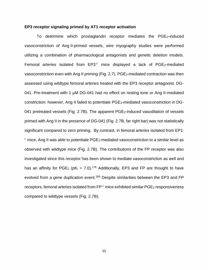
55
EP3 receptor signaling primed by AT1 receptor activation
To determine which prostaglandin receptor mediates the PGE2-induced
vasoconstriction of Ang II-primed vessels, wire myography studies were performed
utilizing a combination of pharmacological antagonists and genetic deletion models.
Femoral arteries isolated from EP3-/- mice displayed a lack of PGE2-mediated
vasoconstriction even with Ang II priming (Fig. 2.7). PGE2-mediated contraction was then
assessed using wildtype femoral arteries treated with the EP3 receptor antagonist, DG-
041. Pre-treatment with 1 µM DG-041 had no effect on resting tone or Ang II-mediated
constriction: however, Ang II failed to potentiate PGE2-mediated vasoconstriction in DG-
041 pretreated vessels (Fig. 2.7B). The apparent PGE2-induced vasodilation of vessels
primed with Ang II in the presence of DG-041 (Fig. 2.7B, far right bar) was not statistically
significant compared to zero priming. By contrast, in femoral arteries isolated from EP1-
/- mice, Ang II was able to potentiate PGE2-mediated vasoconstriction to a similar level as
observed with wildtype mice (Fig. 2.7B). The contributions of the FP receptor was also
investigated since this receptor has been shown to mediate vasoconstriction as well and
has an affinity for PGE2 (pKi = 7.0).178 Additionally, EP3 and FP are thought to have
evolved from a gene duplication event.265 Despite similarities between the EP3 and FP
receptors, femoral arteries isolated from FP-/- mice exhibited similar PGE2 responsiveness
compared to wildtype vessels (Fig. 2.7B).

56
0 5 10 152
4
6
8
1 nM Ang II30 nM PGE2
EP3-/-
1 nM Ang II
Time (min)
Fo
rce
(m
N)
-10
0
10
20
30
40
WT EP1-/-
EP3-/-
30 nM PGE2
30 nM PGE2 + 1nM Ang II 1
DG
*** ***
Veh1
Veh2
FP-/-
PG
E2 c
on
tra
ctile
re
sp
on
se
(% K
-sta
nd
ard
)
A.
B.
Figure 2.7 - Effects of constrictor EP-receptors on PGE2-induced contraction of Ang II-primed arteries. (A) Representative wire myography trace of an EP3-/- femoral artery that shows the lack of PGE2 response when primed with Ang II. (B) Comparison of % contraction during 30 nM PGE2-induced vasoconstriction with or without 1 nM Ang II priming in EP1-/-, EP3-/-, or FP-/- vessels or in wildtype vessels treated with or without the EP3 antagonist DG-041 (DG) (N = 4-5 for EP1-/-, N = 3 for FP-/-, all other data sets N = 5). PGE2 alone did not cause significant vasoconstriction in isolated femoral arteries from any of the mouse strains (wildtype, EP1-/-, EP3-/-, FP-/-). Veh1 indicates Ang II priming of PGE2 solvent (ethanol). Veh2 indicates pre-treatment with DG-041 solvent (DMSO). Wildtype data with or without Ang II are taken from Fig 2.2. Vertical bars indicate SEM.275 ***= P < 0.001

57
Although these data indicated that upon Ang II priming the subsequent PGE2-
induced contraction was EP3 receptor-mediated, Ang II promoting desensitization of
either vasodilator receptor, EP2 or EP4, could also explain the PGE2-induced contraction.
Thus, to test the contribution of the EP2 or EP4 receptor, we assessed the effect of PGE2
on EP2-/- and EP4-/- femoral arteries. In the absence of Ang II priming, deletion of either
EP2 or EP4 was insufficient to facilitate comparable PGE2-induced vasoconstriction to
that observed in wildtype vessels primed with Ang II (Fig. 2.8, EP2-/- & EP4-/- open bars).
PGE2-mediated contraction of Ang II-primed vessels was notably greater in
EP2-/- vessels, presumably from loss of the EP2 receptor vasodilator function (Fig. 2.8,
EP2-/- black bar, P ≤ 0.01). Arterial rings isolated from EP4-/- mice exhibited responses
that were indistinguishable from the responses observed in wildtype vessels.

58
-10
0
10
20
30
40
WT
30 nM PGE2 + 1nM Ang II 1
**
EP2-/-
Veh1
EP4-/-
30 nM PGE2
PG
E2 c
on
tra
ctile
re
sp
on
se
(% K
-sta
nd
ard
)
Figure 2.8 - Effects of EP-receptors on PGE2-induced contraction of Ang II-primed arteries. Comparison of % contraction during 30 nM PGE2-induced vasoconstriction with or without 1 nM Ang II priming in EP2-/- or EP4-/- vessels or in wildtype vessels (N = 5; two EP2-/- mice were one year old). PGE2 alone did not cause significant vasoconstriction in any of the mouse strains (wildtype, EP2-/-, EP4-/-). Veh1 indicates Ang II priming of PGE2 solvent (ethanol). Wildtype data with or without Ang II and Veh1 are taken from Fig 2.7. Vertical bars indicate SEM.275 **= P<0.01

59
Ang II vascular responsiveness in EP1-/- and EP3-/- mice is similar to wildtype
To investigate whether EP3-/- mice have altered contractile responses to Ang II,
concentration-response relationships for wildtype or EP3-/- vessels were compared. The
EC50 and Emax for Ang II-induced vasoconstriction were not different between wildtype
and EP3-/- mice (Fig. 2.9A). Interestingly, the CRC to Ang II exhibited a steeper slope in
wildtype vessels (Hill coefficient of 2.581) as compared to that observed in vessels
isolated from EP3-/- mice (Hill coefficient of 1.198). In conducting these studies, I also
wanted to determine whether vessels from EP1-/- mice have altered Ang II-mediated
vasoconstriction since previous studies had indicated that EP1-/- mice were protected
from Ang II-mediated hypertension.174, 191 Ang II vascular reactivity was essentially
unchanged in EP1-/- mice compared to wildtype Ang II CRC (Fig. 2.9B). PGE2 alone did
not cause significant vasoconstriction in any of the mice (wildtype, EP1-/-, EP2-/-, EP3-/-,
EP4-/-). Taken together, these results indicate the EP3 receptor is necessary for PGE2-
induced vasoconstriction potentiated by Ang II.

60
-9.5 -9.0 -8.5 -8.0 -7.50
20
40
60
80
100
EP3 -/-
WT
Log [Ang II] M
Co
ntr
actio
n
(%
K-s
tan
da
rd)
-9.5 -9.0 -8.5 -8.0 -7.50
20
40
60
80
100 WT
EP1-/-
Log [Ang II] M
Co
ntr
actio
n
(%
K-s
tan
da
rd)
A.
B.
Figure 2.9 - Effects of EP-receptors on Ang II-induced vasoconstriction. (A) Ang II concentration-response curves comparing contraction of wildtype (taken from Fig 2.2,
N = 5-7) and EP3-/- vessels (N = 5-6). LogEC50 for each was -8.74 and -8.70, respectively.
(B) Ang II concentration-response curves comparing contraction of wildtype (taken from
Fig 2.2, N = 5-7) and EP1-/- vessels (N = 3-8). LogEC50 for each was -8.74 and -8.86.
Vertical bars indicate SEM.275

61
AT1 receptors mediate the Ang II priming effect
To identify the Ang II receptor responsible for potentiation of PGE2-mediated
vasoconstriction by Ang II, vessel contraction was measured in arterial rings pre-treated
for 30 minutes with the AT1 antagonist losartan or the AT2 antagonist PD123319 prior to
Ang II priming and PGE2 addition. Losartan blocked the ability of Ang II to potentiate
PGE2-induced vasoconstriction, while PD123319 had no significant effect. (Fig. 2.10A, B,
C) To verify that the effect of losartan was not due to off target competitive inhibition of
the prostanoid receptors, the ability of either losartan or PD123319 to compete with
[3H]PGE2 binding in mouse kidney membrane preparations was assessed. In contrast to
the EP1/3 selective agonist, sulprostone, neither losartan nor PD123319 competed with
[3H]PGE2 binding (Fig. 2.11). Together, these results indicate that the AT1 receptor
facilitates the Ang II priming effect on EP3 receptor-mediated contraction.

62
0 500 1000 15002
4
6
8100 nMPGE2
wash
1 nM Ang II
Time (sec)
Fo
rce
(m
N)
0 500 1000 15002
4
6
8
100 nMPGE2
wash
1 nM Ang II
1M Losartan Pretreatment
Time (sec)
Fo
rce
(m
N)
0
10
20
30
40
***
X X X XX X X
100 nM PGE 2
1 nM Ang II
1 M LosartanX1 M PD123319
*** ***
X
Co
ntr
actio
n
(%
K-s
tan
da
rd)
A. B.
C.
Figure 2.10 - AT1 antagonist inhibits Ang II priming of PGE2. (A) Representative wire myography trace of femoral arterial rings primed with 1 nM Ang II, followed by addition of 100 nM PGE2. (B) Representative trace of femoral artery with 30-minutes pretreatment with 1 µM losartan followed by addition of 1 nM Ang II priming of 100 nM PGE2. (C) Comparison between femoral arterial ring responses to 100 nM PGE2 with and without 1 nM Ang II priming, as well as with priming in the presence of an AT1 (losartan) or AT2 antagonist (PD123319) (N = 5). 275 ***= P<0.001

63
Non
e
Los
arta
n
PD12
3319
Sulpr
osto
ne
-200
0
200
400
600
800
1000
Competitor
*
[3H
]PG
E2 B
ou
nd
- c
old
PG
E2
(cp
m)
Figure 2.11 – AT1 and AT2 antagonists do not compete for EP receptor ligand binding. Competition binding in wildtype mouse kidney membranes; displacement of [3H]PGE2 by the EP3 agonist sulprostone, losartan, and PD123319. Nonspecific binding was determined in the presence of excess unlabeled PGE2. (N = 3). * = P < 0.05. 275
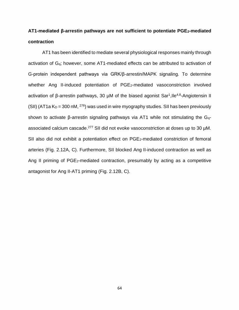
64
AT1-mediated β-arrestin pathways are not sufficient to potentiate PGE2-mediated
contraction
AT1 has been identified to mediate several physiological responses mainly through
activation of Gq; however, some AT1-mediated effects can be attributed to activation of
G-protein independent pathways via GRK/β-arrestin/MAPK signaling. To determine
whether Ang II-induced potentiation of PGE2-mediated vasoconstriction involved
activation of β-arrestin pathways, 30 µM of the biased agonist Sar1,Ile4,8-Angiotensin II
(SII) (AT1a KD = 300 nM, 276) was used in wire myography studies. SII has been previously
shown to activate β-arrestin signaling pathways via AT1 while not stimulating the Gq-
associated calcium cascade.277 SII did not evoke vasoconstriction at doses up to 30 µM.
SII also did not exhibit a potentiation effect on PGE2-mediated constriction of femoral
arteries (Fig. 2.12A, C). Furthermore, SII blocked Ang II-induced contraction as well as
Ang II priming of PGE2-mediated contraction, presumably by acting as a competitive
antagonist for Ang II-AT1 priming (Fig. 2.12B, C).

65
Figure 2.12 - SII does not potentiate PGE2-mediated contraction. (A) Representative wire myography trace showing that 10-minute pre-treatment with 30 µM SII did not potentiate 100 nM PGE2-mediated contraction. (B) Representative trace of arterial rings preincubated with 30 µM SII for 10 minutes, followed by priming with 1 nM Ang II and addition of 100 nM PGE2. (C) Quantification of SII results, showing a lack of potentiation by SII on PGE2-induced contraction (3rd bar from left), and SII blockade of PGE2-mediated contraction of Ang II-primed vessels (4th bar from left) (N = 5).275 ***= P<0.001

66
A role for reactive oxygen species in Ang II potentiated PGE2-induced contraction
Ang II has been shown to induce production of reactive oxygen species (ROS) via
NADPH oxidases as well as increase expression of several NADPH oxidase
subunits.278, 113 To determine whether Ang II-induced potentiation of PGE2-mediated
vasoconstriction involved the elaboration of different sources of ROS, several different
studies were conducted. First, 1 mM Tempol, a superoxide dismutase mimetic, was
added to the bath 20 minutes before Ang II priming of PGE2.279 Pre-treatment with Tempol
resulted in significant reduction in 100 nM PGE2-mediated vasoconstriction (Fig. 2.13A,
B). 1 mM Tempol had no effect on 30 nM Ang II-mediated vasoconstriction (Fig. 2.13A,
C). These data suggest that Ang II mediates PGE2-induced vasoconstriction through a
mechanism involving ROS.

67
Figure 2.13 - Tempol reduces Ang II potentiation of PGE2-mediated vasoconstriction.
(A) Example traces of Ang II priming in the presence of 1 mM 4-hydroxy-2,2,6,6-
tetramethylpiperidin-1-oxyl (Tempol), a superoxide dismutase mimetic. (B) PGE2
responses with or without Ang II priming as compared to priming in the presence of 1 mM
Tempol. (N = 3) (C) Ang II-mediated vasoconstriction with or without 1 mM Tempol. No
significant differences. (N = 3) Vertical bars indicate SEM. *= P<0.05

68
To further investigate the contribution of ROS in PGE2-mediated vasoconstriction
vessels rings were treated with pharmacological inhibitors or stimulators of ROS. Since
Tempol is known to metabolize superoxide anions, I was interested in determining
whether the source of superoxide contributing to PGE2-mediated contraction was from
Ang II activation of NADPH oxidase.279 In vessels pre-treated with apocynin, an NADPH
oxidase inhibitor, PGE2-mediated contraction following Ang II priming was unaffected
(Fig. 2.14A). To investigate whether ROS are sufficient to induce PGE2-mediated
vasoconstriction, vessels were primed with menadione, which generates ROS through
redox cycling.280 Menadione pretreatment (10 µM) did not result in enhanced PGE2-
mediated vasoconstriction comparable to that observed with Ang II priming. Moreover, a
higher dose (50 µM) resulted in no PGE2-induced contraction (Fig. 2.14A), likely from cell
death induced by high concentrations of menadione since these rings did not respond to
either KCl or phenylephrine (data not shown). In other studies, we tested whether
inhibition of endoplasmic reticulum stress (ER stress) via pre-treatment with
tauroursodeoxycholic acid (TUDCA) would affect PGE2-induced contraction since Ang II
is reported to cause ER stress.281, 282 TUDCA blunted Ang II potentiation of PGE2-
mediated contraction without affecting Ang II-induced contraction (Fig. 2.14A, B).

69
0
10
20
30
40
100 nM PGE2
30 nM Ang II
100 M Apocynin
+ + +
+
+
+-
- -100 M TUDCA
+
+
-
+
- -
- - -10 M Menadione
50 M Menadione
-- - --- - -
+
-
--
-+-
+
-
--
--+
Co
ntr
actio
n
(% K
-sta
nd
ard
)
No
pre-
treat
men
t
M A
pocy
nin
100
M T
UDCA
100
0
10
20
30
40
50
60
70
80
90Ang II
Co
ntr
actio
n
(% K
-sta
nd
ard
)
A. B.
Figure 2.14 - Role of reactive oxygen species and ER stress in Ang II priming of PGE2.
(A) PGE2 responses with or without 30 nM Ang II priming as compared to priming in the
presence of apocynin or TUDCA. The last two bars on the right depict 100 nM PGE2
response on vessels primed with menadione instead of Ang II. (N = 1-2) (B) Ang II-
mediated vasoconstriction with or without apocynin or TUDCA. (N = 1-4)

70
EGFR activation does not contribute to PGE2-mediated vasoconstriction
Ang II transactivation of epidermal growth factor receptors (EGFR) is a well
identified signaling mechanism involved in Ang II-mediated physiological responses such
as kidney fibrosis and cardiac hypertrophy.283-285 There is evidence to support a role for
EGFR in mediating vasoconstrictor responses such as contributing to Ang II-mediated
contraction of rat renal artery or aorta and contributing to endothelin-1-induced
contractions of rabbit basilar artery.286-288 Furthermore, studies have indicated a link
between ROS and EGFR in mediating vascular contraction of ductus arteriosus.289 Thus,
we performed an experiment to determine whether inhibition of EGFR would affect PGE2-
mediated vasoconstriction. Vessels were pretreated with 1 µM erlotinib to inhibit EGFR
activation; however, erlotinib had no significant effects on PGE2-induced contraction (Fig.
2.15A).

71
0
10
20
30
40
100 nM PGE2
1 nM Ang II
1 M Erlotinib
x x x
xx
x
Co
ntr
actio
n
(% K
-sta
nd
ard
)
Ang II
No
Erlo
tinib
M E
rlotin
ib
1
0
10
20
30
40
50
Co
ntr
actio
n
(% K
-sta
nd
ard
)
A. B.
Figure 2.15 - EGFR does not contribute to Ang II potentiation of PGE2-mediated
vasoconstriction. (A) PGE2 responses with or without 1 nM Ang II priming as compared
to priming in the presence of erlotinib. (N = 3) (B) Ang II-mediated vasoconstriction with
or without erlotinib. No significant differences. (N = 3)
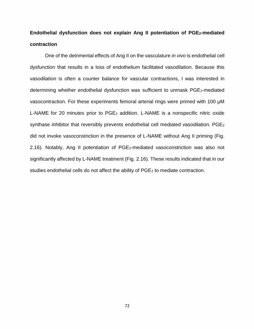
72
Endothelial dysfunction does not explain Ang II potentiation of PGE2-mediated
contraction
One of the detrimental effects of Ang II on the vasculature in vivo is endothelial cell
dysfunction that results in a loss of endothelium facilitated vasodilation. Because this
vasodilation is often a counter balance for vascular contractions, I was interested in
determining whether endothelial dysfunction was sufficient to unmask PGE2-mediated
vasocontraction. For these experiments femoral arterial rings were primed with 100 µM
L-NAME for 20 minutes prior to PGE2 addition. L-NAME is a nonspecific nitric oxide
synthase inhibitor that reversibly prevents endothelial cell mediated vasodilation. PGE2
did not invoke vasoconstriction in the presence of L-NAME without Ang II priming (Fig.
2.16). Notably, Ang II potentiation of PGE2-mediated vasoconstriction was also not
significantly affected by L-NAME treatment (Fig. 2.16). These results indicated that in our
studies endothelial cells do not affect the ability of PGE2 to mediate contraction.

73
0
10
20
30
40
50
100 nM PGE2
1 nM Ang II
100 M L-NAME
X X X X
X X
X X
**
ns
**
Co
ntr
actio
n
(% K
-sta
nd
ard
)
Figure 2.16 – Endothelial dysfunction does not affect Ang II potentiation of PGE2-
mediated vasoconstriction. 100 nM PGE2 responses with or without 1 nM Ang II priming
as compared to Ang II priming in the presence of 100 µM L-NAME or to PGE2 responses
from arterial rings primed with L-NAME instead of Ang II. (N = 3) ns = not significant,
**= P<0.01

74
Extracellular calcium stores and Rho-kinase are required for contraction
The contribution of extracellular stores of calcium to Ang II-induced potentiation of
PGE2-mediated vasoconstriction was assessed by analyzing vessels in Ca2+ free buffer.
Arterial rings were incubated in calcium free Krebs buffer containing 1 mM ethylene
glycol-bis tetraacetic acid (EGTA). EGTA was subsequently removed from the bath, and
the contractile response of Ang II-primed vessels to PGE2 was determined. In control
vessels without Ang II pre-treatment, 100 nM PGE2 did not cause significant contraction
in normal Krebs or upon addition of higher calcium concentrations to the bath (3.5 or 4.5
mM), demonstrating that increasing extracellular calcium concentration is not sufficient to
evoke potentiated PGE2-mediated contraction (Fig. 2.17A). In the absence of extracellular
Ca2+, femoral arteries exhibited minimal responsiveness to Ang II. Furthermore, Ang II
did not potentiate PGE2-induced contraction in Ca2+ free media. However, re-addition of
Ca2+ into the bath restored PGE2-mediated contraction (Fig. 2.17B, C). Pre-treatment (30
minutes) of arteries with the L-type calcium channel blocker, nifedipine, inhibited the
PGE2-mediated contraction (Fig. 2.17C, far right bar). Collectively, these studies indicate
a significant contribution of extracellular sources of calcium, particularly from calcium
influx by L-type channels, in mediating PGE2-induced contraction after priming with Ang
II. Due to its known role in sensitizing smooth muscle cells to calcium-induced contraction,
ROCK was evaluated for its effect on PGE2-induced vasoconstriction of Ang II-primed
vessels. Addition of the ROCK inhibitor Y-27638 to PGE2-induced vasoconstriction
returned vascular tone to baseline (Fig. 2.17D & E). These data indicate that ROCK is
required for EP3-induced arterial contraction.

75
Figure 2.17 - Contribution of extracellular calcium and Rho-kinase to PGE2 primed vasoconstriction. (A) Representative wire myography trace (N = 5) of PGE2-mediated contraction with additional calcium added (normal Krebs 2.5 mM, additional calcium 3.5 or 4.5 mM). (B) Example trace of femoral arterial ring maintained in calcium-free Krebs. Vessels were briefly treated with EGTA, washed, and subsequently administered Ang II, followed by PGE2. Calcium was then added back to the bath to restore contraction. (C) Quantification of PGE2 response with and without Ang II priming, in the presence or absence of calcium, compared to vessels pre-incubated with 300 nM nifedipine or vehicle control (ethanol) (N = 5-8). (D) Quantification of the effects of the ROCK inhibitor Y-27632 on PGE2-mediated contraction (N = 5). (E) Example trace of Ang II priming of PGE2-induced contraction with ROCK inhibitor, Y-27632, addition during PGE2 response.275 * = P < 0.05, **= P < 0.01, ***= P < 0.001

76
Chloride channels are involved in contraction induced by Ang II and PGE2
Along with determining the contributions of calcium to vasoconstriction, further
studies were completed to assess the contributions of intracellular chloride. Chloride
levels within smooth muscle cells (SMC) are typically higher than extracellular levels;
consequently, SMCs depolarize upon efflux of chloride through chloride channels
resulting in activation of voltage-dependent calcium channels resulting in contraction.290
To assess the contribution of different chloride channels in Ang II potentiation of PGE2-
mediated vasoconstriction, vessel rings were pre-treated with either DCPIB, an inhibitor
of volume-sensitive anion channels, or niflumic acid (NFA), an inhibitor of calcium-
activated chloride channels.291 NFA inhibited PGE2-mediated contraction potentiated by
Ang II (P < 0.05, Fig. 2.18A). Ang II-mediated contraction was not observed with vessel
rings treated with NFA (P < 0.1, Fig. 2.18B). DCPIB treatment also blunted Ang II
potentiation of PGE2-mediated vasoconstriction (P < 0.1, Fig.2.18A). Although reporting
these results with a 95% confidence interval would require increasing the sample size,
these preliminary studies (N = 3-4) indicated that chloride channels may contribute to the
enhanced PGE2-mediated vasoconstriction along with the calcium response.

77
PGE2 response
0
10
20
30
40
50
**
100 nM PGE2
1 nM Ang II
30 M DCPIB
+ + +
+
+
+-
- -100 M NFA
+
+
-
+
- -
- - -Veh. (DMSO) -- - -
+
+
--
-+
***
*
Co
ntr
actio
n
(%
K-s
tan
da
rd)
1 nM Ang II response
No
pre-
treat
men
t
M D
CPIB
30
M N
FA
100
Veh (D
MSO
)
-10
0
10
20
30
40
50
Co
ntr
actio
n
(%
K-s
tan
da
rd)
A. B.
Figure 2.18 - Chloride channel contributions to vasoconstriction induced by PGE2 and
Ang II. (A) PGE2 responses with or without 1 nM Ang II priming as compared to priming
in the presence of DCPIB or NFA. (N = 3-4) (B) Ang II-mediated vasoconstriction with or
without DCPIB or NFA. Asterisks indicate significance of at least P < 0.05 (N = 3-4, ns)
*= P < 0.05, **= P < 0.01

78
Salicylate diminishes Ang II potentiation of PGE2-mediated contraction
To determine whether Ang II potentiates PGE2-mediated vasoconstriction through
its action on cPLA2, resulting in increased prostaglandin biosynthesis, femoral arterial
rings were pre-incubated with nonsteroidal anti-inflammatory drugs (NSAID). 100 µM
ibuprofen, a potent COX inhibitor (COX-1 IC50 = 7.6 µM, COX-2 IC50 = 7.2 µM) 292, did
not affect PGE2-induced constriction of Ang II-primed vessels (Fig. 2.19 A, P>0.05), nor
did 100 nM SC-236 (COX-1 IC50 = 17.8 µM, COX-2 IC50 = 10 nM) 293, a COX-2 specific
inhibitor. However, 10 mM salicylate, an active metabolite of aspirin and poor COX
enzyme inhibitor (COX-1 IC50 = 5 mM, COX-2 IC50 = 0.5-34 mM) 292, significantly
attenuated PGE2-mediated vasoconstriction of Ang II-primed vessels (Fig. 2.19 A,
P<0.01). Pretreatment with 100 µM – 10 mM salicylamine, a structurally similar compound
to salicylate and identified isoketal scavenger, did not inhibit PGE2-mediated
vasoconstriction (Fig. 2.19 A & B, P>0.05).294 Inhibition of contraction by salicylate but
not ibuprofen or SC-236, suggests that the salicylate effect was a result of a non-COX
mechanism.

79
0
10
20
30
40
100 nM PGE2
1 nM Ang II
100 M Ibuprofen100 nM SC-23610 mM Salicylate
100 M SalicylamineVehicle (EtOH)
X X X X X
X X X X
X
X
X
****
X
X
X
X
X
X
Co
ntr
actio
n
(% K
-sta
nd
ard
)
0
10
20
30
40
100 nM PGE2
1 nM Ang II
Salicylate Salicylamine
100uM 1mM 5mM 10mM 10mM
X X X X X X XX X X X X X
% o
f 50 m
M K
Cl S
tandard
A.
B.
Figure 2.19 – Contribution of endogenous prostaglandin production. (A) PGE2-induced contraction of femoral arterial rings pretreated for 30 minutes with the indicated concentrations of ibuprofen, SC-236, salicylate, salicylamine, or control solvent for ibuprofen/SC-236 (EtOH) followed by priming with Ang II and subsequent addition of PGE2 (N = 7-10). Salicylate attenuated PGE2-induced constriction of vessels primed with Ang II (P<0.05 1way ANOVA).275 (B) Varying doses of salicylate and salicylamine to determine whether concentration differences had an effect on vascular function. Salicylamine did not inhibit PGE2-induced contraction even at 10 mM (far right bar). * = P < 0.05, ***= P < 0.001

80
Effects of proline-rich tyrosine kinase 2 inhibitors
Salicylate has been shown to inhibit the non-receptor tyrosine kinase Pyk2. 115, 295
To test whether salicylate exerted its effect by inhibition of Pyk2, the Pyk2 inhibitor PF-
431396 was tested. PF-431396 abolished PGE2-induced contraction of Ang II-primed
vessels (Fig 2.20). Since Pyk2 is structurally similar to focal adhesion kinase (FAK),
arteries were pre-treated with a FAK specific inhibitor, 10 µM FAK inhibitor 14, to
determine the contribution of FAK (Fig 2.20). The FAK inhibitor did not affect Ang II
priming of PGE2, indicating that Pyk2 underlies PGE2-mediated contraction. Collectively,
these findings demonstrate that Ang II potentiates vasoconstriction induced by PGE2 via
convergent signaling between the AT1 and EP3 receptors. These convergent events
occur through a Ca2+-Pyk2-ROCK mechanism and are independent of β-arrestin
signaling and AT1-mediated activation of PLA2 (Fig. 2.21).

81
0
10
20
30
40
X X X
X X X
X
100 nM PGE2
1 nM Ang II
10 M PF-431396
10 M FAK inhibitor
X
******
Vehicle (DMSO)
X
X
X
XC
on
tra
ctio
n
(% K
-sta
nd
ard
)
Figure 2.20 - Pyk2 is necessary for PGE2-mediated contraction of Ang II-primed vessels. Preincubation with the Pyk2 inhibitor PF-431396 blocked PGE2-induced contraction of Ang II-primed vessels, while inhibiting FAK with FAK inhibitor 14 did not affect contraction (N = 5). Vehicle represents control solvent for PF-431396 (N = 5). Vertical bars indicate SEM.275 ***= P < 0.001

82
Figure 2.21 - Schematic of key proteins involved in PGE2-induced contractile responses. Prior activation of the AT1a receptor by Ang II facilitates vasoconstriction via a PGE2 - EP3 receptor pathway. PGE2-induced vasoconstriction is dependent upon signaling through a Ca2+-Pyk2-ROCK cascade, as opposed to other defined pathways such as activation of phospholipase A2, EGFR, NADPH oxidase, or signaling via β-arrestin. EP3 and AT1 signaling converges through Pyk2-ROCK to sensitize smooth muscle cells to calcium and thus trigger a significant contractile response.

83
Discussion
In the present study, we demonstrated that PGE2 functions as a vasoconstrictor in
femoral arteries pretreated with low-dose Ang II. In contrast, PGE2 was shown to have no
detectable effect on vascular tone in the absence of priming by Ang II. These findings
reveal a novel interaction between Ang II and PGE2 in regulating peripheral vascular
reactivity. Other constrictors may also potentiate EP3 receptor-induced contraction,
indicated by published results with rat femoral arteries primed with thromboxane or
phenylephrine 221, Ang II is of particular interest due to its central role in the development
of cardiovascular disease. One contributing factor of high blood pressure is increased
peripheral vascular resistance.296 Increased formation of vasoconstrictors and altered
effectiveness of vasodilators may underlie such impairments. PGE2 has been
demonstrated to function primarily as a vasodilator and vasodepressor when
intravenously infused into many species, including humans, dogs, rabbits, mice and rats;
however, in certain physiologic settings PGE2 acts as a vasoconstrictor.161, 169, 170, 183, 225,
297-300 Our studies indicate that in the presence of Ang II, PGE2 exhibits vasoconstrictor
effects in mouse femoral arteries. Because Ang II and PGE2 are well known to regulate
peripheral vasculature, contractile responses mediated by concurrent signaling by Ang II
and PGE2 may contribute to vascular dysfunction associated with hypertension.
The Ang II concentration of 1 nM used for priming femoral arteries in our studies
is greater than typical reported plasma concentrations of Ang II, which have been
measured to be in the pM range 301, 302. However, evidence indicates that tissue sources
of Ang II may contribute more to vascular contractions than circulating Ang II, and
interstitial levels of Ang II have been detected in the nM range in femoral arteries and
84
kidneys.303, 304 Therefore, our priming concentration of Ang II, which produced only a
modest contractile response prior to PGE2 administration (Fig. 2.2), may approximate
levels observed by AT1 receptors in vivo and could thus be relevant to vascular
responsiveness.
Studies presented here demonstrate that the EP3 receptor is required for PGE2-
mediated vasoconstriction. We found that PGE2-induced vasoconstriction following Ang
II priming was lost upon loss or blockade of the EP3 receptor. Previous studies indicate
that EP3-/- mice have lower mean arterial pressure (MAP) in response to acute or chronic
Ang II infusions.175 The observed changes in vascular reactivity described in the present
studies may contribute to the reduction in MAP in response to Ang II observed in EP3-/-
mice.175 Of note, femoral arteries from EP3-/- mice exhibited Ang II concentration
response curves similar to wildtype femoral arteries, suggesting the alterations in vascular
reactivity to PGE2 observed in EP3-/- femoral vessels are not due to a loss of Ang II
responsiveness. In contrast, a previously reported study investigating the effect of EP3
receptor signaling on mouse mesenteric vasculature demonstrates that Ang II-mediated
contraction was diminished by knockout of the EP3 receptor.175 Considering these
findings, our data suggest differences in the interactions between Ang II and PGE2
signaling that are specific to particular vascular beds. Additionally, differences in the
steepness of the Ang II dose response curve was noted in the EP3-/- vessels indicating
that the EP3 receptor may contribute to Ang II binding to the AT1 receptor. More studies
would need to be done to determine the effects of the EP3 receptor in regulating vascular
sensitivity to Ang II.

85
Our findings support a model, diagrammed in Figure 2.21, in which activation of
the AT1 receptor by Ang II sensitizes smooth muscles cells to calcium, thereby facilitating
a contractile response to PGE2. This vasoconstriction does not require AT1-mediated
production of endogenous prostaglandins or β-arrestin scaffolding. Importantly, our
studies identified Pyk2 as a tyrosine kinase crucial for PGE2-induced contraction. This
finding aligns well with previous investigations of Pyk2 function. Pyk2 is a calcium-
dependent tyrosine kinase that acts upstream of the RhoA/ROCK pathway.115, 295 Pyk2
can be activated by Ang II and plays a role in the ability of Ang II to mediate many cellular
functions, including vasoconstriction.115, 305 The EP3 receptor has been identified to
mediate contractile responses through activation of ROCK.175, 221, 222 Therefore, Ang II
signaling may activate Pyk2, thereby sensitizing the ROCK pathway to EP3 signaling
(depicted in Figure 2.21); alternatively, EP3 and AT1 signaling may concurrently converge
through a Pyk2-RhoA-ROCK pathway. Activation of ROCK would ultimately lead to
inhibition of myosin phosphatase and enhance Ca2+ sensitivity of myosin filaments. The
inhibition of Ang II priming of PGE2-mediated contraction observed with salicylate and the
specific Pyk2 inhibitor (PF-431396) may also explain some of the beneficial effects of
salicylate on the cardiovascular system.
Although the present studies focused on peripheral vascular reactivity, the
observed interaction between the EP3 and AT1 receptors may be important in other
vascular beds (Fig. 2.5), tissue types, and physiologic events associated with
hypertension. For example, studies using rats have indicated that intracerebroventricular
infusion of PGE2 results in a rise in blood pressure and heart rate.306 Interestingly, these
responses were exacerbated in spontaneously hypertensive rats (SHRs), a hypertension
86
model known to have elevated brain levels of Ang II.307 These findings suggest a potential
interaction of Ang II and PGE2 in the central nervous system that might affect blood
pressure. Future studies investigating additional mechanisms through which the EP3 and
AT1 receptors produce synergistic responses and how these interactions relate to the
pathology of hypertension may reveal novel therapeutic strategies for treatment.

87
CHAPTER III
ANG II EFFECTS ON PGE2-MEDIATED BLOOD PRESSURE RESPONSES
Introduction
Prostaglandin E2 (PGE2) is a prostanoid that normally acts as a systemic
vasodepressor when infused into mice. In wildtype mice a 100 µg/kg intravenous bolus
of PGE2 exhibits a transient depressor effect that last several minutes before blood
pressure is fully restored to baseline mean arterial pressure (MAP). This vasodepressor
effect can be attributed primarily to PGE2-mediated activation of EP2 receptors. PGE2
intravenous infusion into mice lacking the EP2 receptor induces a pressor response.169,
298 This pressor response is attributed to activation of EP3 receptors.183 From studies
conducted using EP receptor knockout mice, complex blood pressure regulating effects
of PGE2 have been revealed. For example, EP2-/- mice are more susceptible to dietary
salt induced hypertension169, and knockout of either vasopressor receptor, EP1 or EP3,
results in salutary effects against angiotensin II (Ang II)-induced increases in blood
pressure.174, 175 Although studies of EP receptor knockout mice have revealed some of
the physiological and pathophysiological roles of these receptors in blood pressure
regulation, there are no studies that have examined altered PGE2 responses in wildtype
mice under different physiological conditions.
The vessel reactivity studies presented in Chapter II indicated that PGE2 exhibits
contractile responses in wildtype mice when the arterial tissue is primed with Ang II.
However, the relevance of Ang II synergism with PGE2 regulating vascular reactivity to

88
the physiology of the whole animal remains incompletely characterized. Thus, it was
hypothesized that Ang II in wildtype mice would alter the vascular responsiveness to
PGE2 resulting in a vasopressor effect upon PGE2 intravenous infusion. Furthermore,
since ex vivo vascular results presented in Chapter II indicated the involvement of the
EP3 receptor in PGE2-induced contraction, it was hypothesized that PGE2 vasopressor
effects would be dependent on the EP3 receptor.
Experimental Procedure
Animals
All studies were performed in accordance with the National Institutes of Health
animal care standards and approved by the Vanderbilt University Institutional Animal
Care and Use Committee. Male C57BL/6 strain wildtype or EP3-/- mice, 12 to 15 weeks
old, were used for all studies.
Intracarotid Blood Pressure Measurements
Mice were anesthetized using ketamine (100 mg/kg) and inactin (100 mg/kg) via
intraperitoneal injections. Mice were positioned onto a heating pad and a PE10 indwelling
catheter was secured into the left carotid artery. The catheter was connected to a
pressure transducer (Digi-Med TXD-310, Micro-Med) and blood pressure was measured
using a blood pressure analyzer (Digi-Med BPA 400, Micro-Med). For drug infusions, a
PE50 indwelling catheter was secured into the right jugular vein and attached to a blunt
needle of a 1 mL saline syringe. Drug infusion rates were controlled by a Genie Plus
Syringe Pump (Kent Scientific). For experiments in which two drugs were simultaneously

89
infused, a bifurcated indwelling catheter attached to separate syringes regulated by
individual syringe pumps was used. To improve respiration and survival during blood
pressure recordings, tracheotomy was performed by inserting a 1 inch in length PE60
tube into an incision between two superior tracheal cartilages down into the trachea.
Immediately after surgeries, mice were equilibrated for 45 minutes until stable MAP
values were attained. For continuous intravenous infusions, drugs were injected at a rate
of 5 µl/min. For bolus injections, the jugular catheter was able to hold 50 µl without going
into the vein. Drugs were slowly injected into the catheter by hand and then attached to
a continuous pump of saline at a controlled rate of 50 µl/min to infuse the bolus into the
mouse. All drugs were diluted into sterile saline.
Mouse model of chronic Ang II hypertension
Micro-osmotic pumps (alzet, model 1002) filled with either saline or Ang II (300ng/kg/min)
were inserted subcutanously into wildtype mice 14 days prior to intracarotid blood
pressure (ICBP) recordings. Tail cuff blood pressure measurements were collected
throughout the 14 days leading up to ICBP experiments. Two different protocols were
used for ICBP on the osmotic pump treated mice. Protocol 1 was conducted as described
above (Intracarotid Blood Pressure Measurements). Protocol 2 included additional steps
to ensure mice did not experience fluid loss during ICBP surgery. To prevent fluid loss,
mice were administered saline (5 µl/min) as a continuous intravenous infusion through
the jugular catheter during the 45-minute post-surgical MAP equilibration period, and
saline was added to the exposed tissue at the surgical site of the throat and covered with
Parafilm in which an opening was cut to allow for projection of the tracheostomy tube.

90
Wire myography with isolated femoral arteries
Methods were performed as described in Chapter II.
Results
PGE2 blood pressure effects are unaltered by acute continuous infusion of Ang II
In order to determine whether Ang II would alter PGE2-mediated vasodepressor
effects in vivo, we first determined whether intravenous infusion of PGE2 along with Ang
II would alter PGE2-induced blood pressure changes. Figure 3.1 depicts the experimental
protocol used for these studies. Briefly, continuous infusion of Ang II or saline was
administered for 30 minutes prior to infusing a bolus of vehicle control (saline) followed
by a bolus of 100 µg/kg PGE2. Infusion of the vehicle control did not alter blood pressure
responses in any of the mice; however, infusion of Ang II did raise the baseline blood
pressure (Fig. 3.2 A, B). PGE2 typically induces a transient vasodepressor response
lasting about 180 seconds that is similar to responses observed with the vehicle control
mice (Fig. 3.2A).169 In mice that received continuous infusion of Ang II (75 pmol/kg/min),
PGE2 also induced a vasodepressor response (Fig. 3.2B). Of note, the PGE2 response
in combination with Ang II exhibited a quicker recovery to baseline that was followed by
a gradual decline in blood pressure over time (Fig. 3.2B). For comparison, in an additional
cohort of wildtype mice, the α-adrengeric receptor agonist phenylephrine (100 µg/kg/min)
was continuously infused to determine whether another contractile agent, which was
previously shown in ex vivo studies to prime the vasculature to PGE2-induced
vasoconstriction, would alter PGE2 blood pressure effects. Although phenylephrine did

91
raise baseline blood pressure (MAP = 120 mmHg, Fig. 3.2, C), it did not cause PGE2 to
exhibit vasopressor responses either (Fig. 3.2C).

92
-50
0
50
100
150
200Ang II, PE, or Saline
Veh PGE2
30 min 30 min 30 min
Time (s)
Me
an
Art
eri
al P
ressu
re
(mm
Hg
)
Figure 3.1 – Experimental design of ICBP studies investigating the effects of acute intravenous infusion of PGE2 in combination with other vasoconstrictors. Wildtype mice were given intravenous continuous infusion of either saline (5 µl/min), Ang II (75 pmol/kg/min), or phenylephrine (PE) (100 µg/kg/min). After 30 minutes of continuous infusion, a bolus of vehicle (saline) followed by a bolus of PGE2 (100 µg/kg) was infused without interfering with the continuously infused drug. Arrow indicates continuous infusion time and dotted lines indicate bolus infusion time-points. (N = 3)
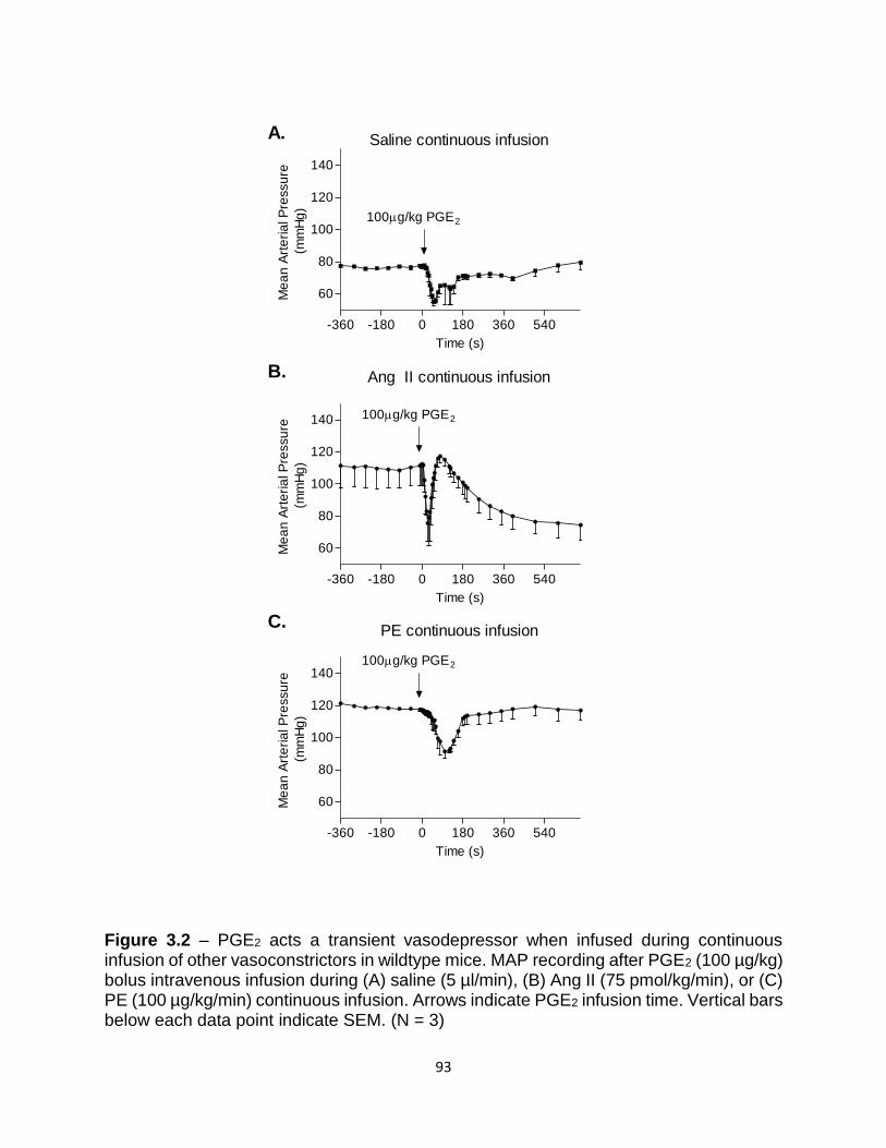
93
Ang II continuous infusion
-360 -180 0 180 360 540
60
80
100
120
140 100g/kg PGE2
Time (s)
Me
an
Art
eri
al P
ressu
re
(mm
Hg
)
PE continuous infusion
-360 -180 0 180 360 540
60
80
100
120
140100g/kg PGE2
Time (s)
Me
an
Art
eri
al P
ressu
re
(mm
Hg
)
Saline continuous infusion
-360 -180 0 180 360 540
60
80
100
120
140
100g/kg PGE2
Time (s)
Me
an
Art
eri
al P
ressu
re
(mm
Hg
)
A.
B.
C.
Figure 3.2 – PGE2 acts a transient vasodepressor when infused during continuous infusion of other vasoconstrictors in wildtype mice. MAP recording after PGE2 (100 µg/kg) bolus intravenous infusion during (A) saline (5 µl/min), (B) Ang II (75 pmol/kg/min), or (C) PE (100 µg/kg/min) continuous infusion. Arrows indicate PGE2 infusion time. Vertical bars below each data point indicate SEM. (N = 3)

94
PGE2 is a vasodepressor in a chronic Ang II mouse model of hypertension
Since acute Ang II treatment did not appear to alter PGE2 blood pressure
responses, I sought to determine whether a longer exposure to Ang II would produce an
effect. For these studies, a chronic Ang II pump model of hypertension was used in which
wildtype mice were implanted with a micro-osmotic pump that slowly diffused Ang II
subcutaneously over two weeks to induce hypertension. This Ang II model of
hypertension has been reported to closely simulate the onset of human hypertension.308,
309 Mice that were implanted with an Ang II pump develop a slow rise in baseline blood
pressure that was observed to reach a maximum (30 % rise in SBP) by 9 days after pump
implantation whereas baseline blood pressure in vehicle control mice displayed modest
changes (< 10 % change in SBP, Fig. 3.3). After 14 days of Ang II or saline infusion,
ICBP was recorded while infusing an intravenous bolus of PGE2. PGE2-evoked an
immediate and sustained pressor response in mice that had an Ang II pump implanted
(Fig. 3.4A) and a rapid (60 seconds) depressor effect in saline pump treated mice (Fig.
3.4B).

95
0 3 6 9 12
0
10
20
30
40
Ang II Pumps
Saline Pumps
Days
S
BP
(%
)
Figure 3.3 – Mice given subcutaneous slow-pressor doses of Ang II develop high blood pressure. Systolic blood pressure (SBP) over time (days) in wildtype mice implanted with micro-osmotic pumps filled with either saline (white circles) or 300 ng/kg/min of Ang II (black circles). (N = 7) t test of means = *P < 0.05. Vertical bars represent SEM.

96
Saline
-60 0 60 120 18040
50
60
70
80
90
100 PGE2
100g/kg
Time (s)
Me
an
Art
eria
l P
ressu
re
(mm
Hg
)
Ang II
-60 0 60 120 18040
50
60
70
80
90
100
PGE2
100g/kg
Time (s)
Me
an
Art
eria
l P
ressu
re
(mm
Hg
)
A. B.
Figure 3.4 – Chronic Ang II effects on PGE2-mediated blood pressure responses.
(A) MAP recording during an intravenous bolus of PGE2 (100 µg/kg) into mice that
previously were implanted with a subcutaneous micro-osmotic pump filled with either
Ang II (300ng/kg/min) or (B) vehicle control (saline) for 2 weeks. Arrows indicate PGE2
infusion time. Vertical bars below data points indicate SEM. (N = 5-7)

97
Due to significant mortality during the initial studies, optimization of the ICBP
surgical method (protocol 1) was performed in an attempt to obtain more consistent,
reliable results. The average baseline MAP for mice in our initial experiments was
66 mmHg; thus, mice appeared to be hypovolemic and/or dehydrated after surgical
procedures. To ensure the mice were euvolemic and healthy after surgery, mice were
administered a continuous intravenous infusion of saline (5 µl/min) throughout the 45-
minute MAP equilibration period, and the surgical site was coated in saline to prevent
dehydration from the exposed tissue. Another set of experiments were conducted using
the new and improved protocol (protocol 2). The average baseline MAP for mice in these
studies was 77 mmHg, and more mice survived the surgical procedure. However, the
pressor response to PGE2 originally observed in chronic Ang II mice was no longer
present. Ang II mice that had undergone this new protocol exhibited a vasodepressor
response to PGE2 that resembled that of wildtype mice with the saline pump (lasting ~180
seconds) (Fig. 3.5A, B).

98
Saline
-60 0 60 120 18040
50
60
70
80
90
100
PGE2
100g/kg
Time (s)
Me
an
Art
eria
l P
ressu
re
(mm
Hg
)
Ang II
-60 0 60 120 18040
50
60
70
80
90
100 PGE2
100g/kg
Time (s)
Me
an
Art
eria
l P
ressu
re
(mm
Hg
)
A. B.
Figure 3.5 – ICBP experiments using the euvolemic protocol used to investigate chronic
Ang II effects on PGE2-mediated blood pressure responses. (A) MAP recording during an
intravenous bolus of PGE2 (100 µg/kg) into mice that were previously implanted with a
subcutaneous micro-osmotic pump filled with either Ang II (300ng/kg/min) or (B) vehicle
control (saline) for 2 weeks. Arrows indicate PGE2 infusion time. Vertical bars below data
points indicate SEM. (N = 4-5)

99
EP3-/- mice exhibit sustained vasodepressor effects to intravenously infused PGE2
Contribution of the EP3 receptor was determined by testing PGE2 blood pressure
effects in EP3-/- mice. Ang II micro-osmotic pumps (300 ng/kg/min) were subcutaneously
implanted into mice lacking the EP3 receptor. After 14 days of chronic infusion, MAP was
recorded during infusion of an intravenous bolus of PGE2. Using either ICBP protocols
with or without saline infusion to prevent volume depletion on the EP3-/- mice, PGE2
infusion resulted in a sustained vasodepressor response (Fig 3.6A, B).

100
Ang II
-60 0 60 120 1800
20
40
60
80
100
120
140
PGE2
100g/kg
Time (s)
Me
an
Art
eri
al P
ressu
re
(mm
Hg
)Ang II
-60 0 60 120 180
40
60
80
100
120
140
PGE2
100g/kg
Time (s)
Me
an
Art
eria
l P
ressu
re
(mm
Hg
)
A. B.
Figure 3.6 – PGE2 is a vasodepressor in EP3-/- mice. (A) MAP reading during a bolus of PGE2 (100 µg/kg) in EP3-/- mice that had a micro-osmotic pump of Ang II (300ng/kg/min) for 14 days and had either under gone protocol 1 or (B) protocol 2 for ICBP readings. (N = 1 for A, N = 4 for B)

101
Enhanced sympathetic tone in combination with Ang II may unmask PGE2-
mediated pressor responses
To investigate the basis of the different responses observed in the two protocols, I
compared the heart rate data acquired during PGE2 infusion between the two different
protocols. Interestingly, Ang II osmotic-pump mice that had undergone protocol 1
responded with a significantly sustained elevation in heart rate concurrent with the rise in
MAP observed upon PGE2 infusion (Fig. 3.7A, B). However, mice that had undergone
protocol 2 responded with a rise in heart rate that was only transient and coordinated with
the depressor response induced by PGE2 (Fig. 3.7C, D). In general, when an abrupt drop
in arterial pressure is encountered by the cardiovascular system the autonomic reflex is
stimulated that increases sympathetic activity and ultimately drives increases in heart
rate. The heart rate response that occurred concomitantly with the MAP readings from
mice that had undergone protocol 2, demonstrated a typical autonomic response (Fig.
3.7C, D). However, the significant rise in heart rate observed in mice that had undergone
protocol 1 suggested that sympathetic tone may be amplified in these mice.

102
Ang II
-60 0 60 120 18040
50
60
70
80
90
100
PGE2
100g/kg
Time (s)
Me
an
Art
eria
l P
ressu
re
(mm
Hg
)Ang II
-60 0 60 120 18040
50
60
70
80
90
100 PGE2
100g/kg
Time (s)
Me
an
Art
eria
l P
ressu
re
(mm
Hg
)
-60 0 60 120 180 240 300 360150
200
250
300
350
Time (s)
He
art
Ra
te (
bp
m)
A.
B.
C.
D.
-60 0 60 120 180 240 300 360150
200
250
300
350
400
450
500
Time (s)
He
art
Ra
te (
bp
m)
Figure 3.7 – Comparison of heart rate and MAP during PGE2 bolus in mice undergoing continuous Ang II intravenous infusion between two different experimental ICBP methods. (A) Graph shown is the same depicted as Fig 3.3B in order to compare to (B) heart rate over time (N = 5-7). Data shown in A and B are from the same mice that had under gone protocol 1, where mice were thought to be dehydrated and volume depleted. (C) Graph shown is the same depicted as Fig 3.4B in order to compare to (D) heart rate over time (N = 4-5). Data shown in C and D are from the same mice that had under gone protocol 2, where mice were thought to be hydrated and had replete blood volume. Vertical bars indicate SEM (below data points for MAP data and above data points for heart rate data).

103
Endogenous catecholamine release does not contribute to Ang II potentiation of
PGE2-mediated vasoconstriction
Since the in vivo heart rate data suggested that PGE2 vasodepressor responses
could be altered to exhibit pressor effects under assumed amplification of autonomic tone,
preliminary studies were conducted to analyze whether sympathetic activity contributed
to Ang II potentiation of PGE2-mediated vasoconstriction observed in experiments
presented in Chapter II. Although wire myography studies are ex vivo experiments,
femoral arteries excised from mice still retain nerve terminals embedded into the tissue.
To determine whether release of catecholamines from these nerve terminals could
explain PGE2-mediated vasoconstriction, femoral arteries were pre-treated with 100 µM
tyramine, a catecholamine releasing agent, to rid the vessel of residual vesicular
catecholamines. The vessel bath was then washed several times before priming with 1
nM Ang II followed by addition of 100 nM PGE2. Tyramine induces a slight contractile
response presumably from catecholamine activation of adrenergic receptors; however,
removal of tyramine and of released catecholamines from the bath did not inhibit Ang II
potentiation of PGE2-mediated vasoconstriction (Fig. 3.8A). In additional experiments, the
irreversible adrenergic receptor antagonist, phenoxybenzamine (100 µM), was added to
vessels during PGE2-mediated vasoconstriction to which no effect on contraction was
observed (Fig. 3.8B). Collectively, these preliminary results indicated that catecholamine
release by Ang II or PGE2 did not contribute to PGE2-mediated vasoconstriction.
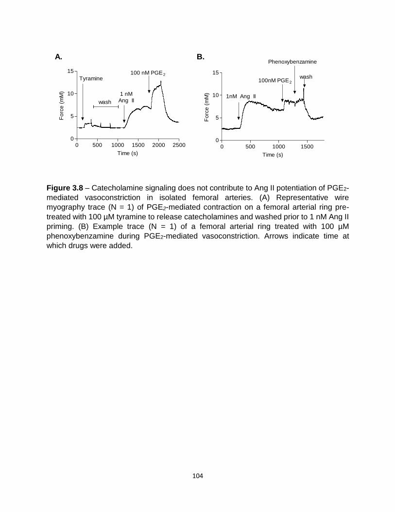
104
0 500 1000 1500 2000 25000
5
10
15
Tyramine
wash
1 nMAng II
100 nM PGE2
Time (s)
Fo
rce
(m
M)
0 500 1000 15000
5
10
15
1nM Ang II
100nM PGE2
Phenoxybenzamine
wash
Time (s)
Fo
rce
(m
M)
A. B.
Figure 3.8 – Catecholamine signaling does not contribute to Ang II potentiation of PGE2-
mediated vasoconstriction in isolated femoral arteries. (A) Representative wire
myography trace (N = 1) of PGE2-mediated contraction on a femoral arterial ring pre-
treated with 100 µM tyramine to release catecholamines and washed prior to 1 nM Ang II
priming. (B) Example trace (N = 1) of a femoral arterial ring treated with 100 µM
phenoxybenzamine during PGE2-mediated vasoconstriction. Arrows indicate time at
which drugs were added.

105
Discussion
While previous investigations have alluded to a balance between EP receptor
signaling in facilitating subsequent hemodynamic physiological responses induced by
PGE2, in general, these conclusions have come from studies using EP receptor knockout
mice. Here we investigated whether Ang II could alter the PGE2 vasodepressor response
to a vasopressor response in wildtype mice. Although in ex vivo femoral arteries Ang II
was able to prime smooth muscle cells to PGE2-induced vasoconstriction, in vivo studies
did not indicate that acute or chronic Ang II caused PGE2 to be a vasopressor in healthy
male mice (Fig. 3.2 & 3.5). Ultimately, these studies were found to be more complex under
in vivo conditions in which additional blood pressure regulatory mechanisms are intact.
Ang II potentiation of PGE2-mediated vasoconstriction may still occur in vivo and may
contribute to blood pressure; however, other inputs such as the sympathetic nervous
system, other circulating hormones, and sodium balance may have affected the overall
blood pressure response observed in the presented studies.
Another complication with the blood pressure readings of the chronic Ang II model
was that even though Ang II osmotic-pump treated mice exhibited hypertension via tail
cuff readings, the baseline blood pressure was the same for mice that were treated with
either an Ang II or saline osmotic pump once they had undergone anesthsia and surgery
for intracartotid blood pressure readings. This loss of elevated blood pressure during/after
surgery could be a result of the blood pressure lowering effects of anesthsia. Additional
studies using mice that are fitted with a telemeter device and indwelling jugular catheters
prior to PGE2 infusions would be nessary to test whether unanesthetized mice would
exhibit altered PGE2 responses in the presence to Ang II.

106
Interestingly, PGE2 did facilitate a robust pressor response in mice that were
exposed to chronic Ang II whose cardiovascular system was compromised after ICBP
surgical procedures (Fig. 3.4). Preliminary studies indicated that this pressor response
was mediated by the EP3 receptor (Fig. 3.6). In initial experiments using protocol 1 mice
were thought to be in a hypovolemic state since these mice exhibited tachycardia,
tachypnea, low baseline MAP, and intense pressor responses to PGE2. Hypovolemia was
thought to develop as a result of blood loss during cannulation of the carotid artery and
dehydration through exposed tissues after surgery. Although this was corrected in
subsequent studies by giving mice intravenous saline supplementation, these earlier
experiments provided a unique physiological state in which PGE2 exhibited pressor
responses. Although further studies would be required to confirm blood volume levels in
these mice, interestingly, these findings suggest that during a state of low blood volume,
PGE2 could exhibit vasopressor qualities.
Hypovolemia is an emergency condition that is often present after trauma, severe
burns, and hemorrhage. Clinical treatment involves stopping the source of blood volume
loss along with transfusion of blood/fluids to regain homeostasis. The body’s initial
physiological response to hypovolemia is enhanced sympathetic activation to increase
cardiac output and vasoconstriction in maintaining core pressure; however, with
continued volume loss this mechanism is not enough to maintain pressure to vital organs
leading to hypotension and cardiac arrest.28 During these late phases of hypovolemic
shock, vasopressor therapy may be beneficial to avert cardiac arrest and avoid
reperfusion injury by lowering the amount of transfusion fluid required to restore blood
pressure homeostasis.310 Although this therapy is heavily debated in the clinic,

107
norepinephrine is commonly used and current investigations into more efficient pressor
agents such as vasopressin are being conducted (NIH funded Clinical Trial
NCT00379522).311, 312 Based on our findings in chronic Ang II mice (Fig. 3.4), exogenous
PGE2 in combination with Ang II may provide beneficial vasopressor effects during life-
threatening hypotension stages of hypovolemia. Certainly, our studies suggest that there
may be physiological differences in hypertensive patients experiencing hypovolemic
shock and that these findings merit further investigation.
Ang II and PGE2 are known to play a role in the physiological response to
hypovolemia. Previous studies investigating the effects of AT1 antagonists in
experimental central hypovolemia have indicated that Ang II via AT1 receptor aides in the
maintenance of MAP and vascular resistance.313-315 In other studies using dogs with
hemorrhagic-induced shock, intravenous infusion of arachidonic acid, the precursor to
prostaglandins, led to attenuated lowering in MAP and increased prostaglandin levels.316
In rats, PGE2 and COX-2 have been identified as essential components to mediating
intestinal ischemia during hemorrhagic shock, a compensatory vasoconstriction of the
mesenteric and surrounding circulation to maintain central blood pressure.317 The present
study provides further evidence that PGE2 may be a vasoconstrictor/pressor during
conditions of shock. Additional investigations into the interaction of Ang II and PGE2 in
hypovolemic shock and into the beneficial versus detrimental effects of this interaction
may unveil novel therapeutic strategies.

108
CHAPTER IV
FUTURE DIRECTIONS AND CONCLUSIONS
Future Directions
Although studies presented in Chapter II have indicated that Ang II and PGE2
synergize to enhance contractile responses of peripheral vasculature, other tissue types
where this synergism may be important are yet to be fully characterized. One region
where such interactions may be particularly important for the regulation of blood pressure
is in the central nervous system. Intracerebroventricular infusion of PGE2 results in a rise
in blood pressure that is exacerbated in spontaneously hypertensive rats.306, 307
Additionally, a combination of Ang II and PGE2 has been shown to reduce cerebral blood
flow.194 The receptors mediating these central cardiovascular effects are incompletely
understood. EP1 receptors in the brain have been identified as crucial for Ang II-
dependent hypertension.192 However, the EP3 receptor is also expressed in the brain,
and further studies are needed to determine its role in regulating central cardiovascular
effects.318
Another current area of research is investigating the contributions of the immune
system to the etiology of hypertension. Studies have indicated that hypertension is
associated with immune cell infiltration into the vasculature, kidney, and heart.319, 320
Additionally, immunosuppressants are protective against salt-sensitive hypertension in
rats acutely pre-treated with Ang II.319 In particular, T cells seem to be key to mediating
Ang II-induced hypertension. RAG-1-/- mice, which lack B and T cells, exhibit blunted

109
hypertension in response to chronic Ang II infusion; furthermore, adoptive transfer of
wildtype T cells into RAG-1-/- mice restores Ang II-sensitivity in these mice.320 While these
studies highlight the contribution of T cells to Ang II-induced hypertension, the signaling
mechanisms which underlie such contributions are incompletely understood. In this
regard, PGE2, which has long been known to regulate inflammatory responses, may be
critical. Preliminary studies done in collaboration with Dr. David Harrison’s lab have
shown that adoptive transfer of EP1/EP3 double knockout T cells into RAG-1-/- mice did
not restore Ang II-sensitivity (data not shown). These results indicate that the EP1 and/or
EP3 receptors may be involved in T cell-driven hypertensive effects in response to Ang II.
Further investigations are needed to understand the role of EP receptor signaling in
regulating T cell function and the effects of these functions on the pathophysiology of
hypertension.
Although our ex vivo analyses indicated that concurrent Ang II and PGE2 signaling
induces peripheral vasoconstriction, simultaneous infusion of PGE2 with Ang II in vivo
resulted in vasodepression. One explanation for the discordance between our ex vivo and
in vivo findings could be that the in vivo experiments were limited to studying the effects
of an intravenous infusion of PGE2. Ang II has long been known to have systemic effects;
however, prostaglandins are primarily local autocrine and paracrine signaling effectors.
Due to its quick first-pass metabolism, PGE2 is not believed to be a physiological effector
in the systemic circulation. Thus, while measuring the ex vivo response of specific tissues
to PGE2 and Ang II may be feasible, determining how such stimulation affects the
contribution of individual tissue types to blood pressure in vivo remains a current
challenge.

110
One approach to specifically study the potential vasopressor effects of PGE2 in
vivo may be to circumvent the PGE2-mediated vasodepressor effects by measuring the
blood pressure responses to infusion of PGE2 with Ang II in EP2-/- mice, a model in which
PGE2 is known to induce vasopressor effects via the EP3 receptor.169 Interestingly, PGE2-
mediated vasoconstriction in the afferent arteriole of EP2-/- mice is prevented by treatment
with an ACE inhibitor.235 This result indicates that Ang II may affect PGE2-induced
vasopressor responses in EP2-/- mice. Future studies using EP2-/- mice may provide an
experimental model to characterize the role of Ang II in modulating PGE2-mediated
vasoconstrictor and vasopressor effects in specific tissue regions.
Despite the complications noted with our in vivo studies, one interesting finding
was that in mice that were likely hypovolemic, PGE2 administration after chronic Ang II
infusion resulted in vasopressor responses (Fig. 3.4). Additional studies would be needed
to further characterize the physiological state of these mice, including confirming the
blood volume levels, blood osmolality state, and plasma levels of norepinephrine to
determine whether these mice had enhanced sympathetic activation. However, these
studies suggest that it would of interest to assess PGE2-responses in an experimental
model of hypovolemia, such as a hemorrhagic shock model, in which blood is withdrawn
until MAP approaches 40 mmHg.317 Since the heart rate data indicated that these mice
may have enhanced sympathetic activation, future studies analyzing the contribution of
sympathetic activity to PGE2-induced vasopressor responses would be of interest.
Studies comparing the effects of PGE2 in hypovolemic mice treated with or without a
ganglionic blocker, alternatively in a mouse model of overactive sympathetic activity, such
as the hypertensive inbred mouse strain BPH/2J, could reveal the contribution of

111
sympathetic activity to PGE2-mediated blood pressure effects.321, 322 Collectively, these
studies could have implications for a variety of disorders and complications, including with
hypovolemia, hyperadrengeric postural orthostatic tachycardia syndrome, and
sympathetically driven hypertension.323-325
One interesting condition that is marked by increased expression of mPGES
resulting in elevated levels of PGE2 and by high levels of renin and Ang II is
hyperprostaglandin E syndrome (Barter syndrome).330 Despite increased levels of both
Ang II and PGE2, patients with Barter syndrome lack a high blood pressure phenotype.330
This lack of increased blood pressure is contrary to what would be predicted from studies
described in Chapter II that indicated Ang II-priming of ex vivo femoral arteries potentiated
PGE2-induced contractions. However, these patients have also been shown to have a
continuous loss of calcium that is partialy independent of elevated PGE2.331 Loss of
calcium may affect the vascular responsiveness to concurrent Ang II and PGE2. Although
the blood pressure phenotype is absent, this rare and unique condition could provide a
model for studying other key physiological functions affected by concurrent Ang II and
PGE2.
In Chapter II my studies indicated that PGE2-induced vasoconstriction potentiated
by Ang II priming occurs through concurrent signaling by the AT1 and EP3 receptors,
resulting in activation of a Ca2+/ROCK/Pyk2 calcium sensitization pathway. However, the
relative contributions of AT1 signaling and EP3 signaling to induction of downstream
mediators in this pathway remain an important topic of investigation. One limitation of our
vascular reactivity studies was that many of the inhibitors (L-type Ca2+ channel blockers,
ROCK inhibitors, and Pyk2 inhibitors) that blocked EP3-mediated contraction also

112
affected Ang II-mediated contractions. In attempts to study each response individually,
experiments were proposed to add inhibitors either before or after Ang II priming but
before PGE2 addition. However, since the time after Ang II that PGE2 exhibits contraction
is short (10-20 min, Fig. 2.3) many of the drugs would not have enough incubation time
on the vessel to confirm appropriate inhibition of specific targets.
It is also unlikely that the signaling mechanism (Ca2+/ROCK/Pyk2) described in
Chapter II is specific to Ang II. Many other vasoconstrictors have been identified by other
investigators, as well as from our studies, to potentiate PGE2-mediated contraction,
including phenylephrine, vasopressin, KCl, and thromboxane receptor agonists (Fig.
2.6).221 Additionally, Pyk2 has been identified to enhance contractile responses induced
by a variety of G-protein coupled receptors, such as adrenergic receptors and
vasopressin receptors.326, 327 The evidence that a variety of contractile agents could affect
PGE2-mediated contraction implies that PGE2 may contribute to vasoconstriction through
this mechanism in numerous different physiological scenarios. More investigations would
be necessary to determine whether Ca2+/ROCK/Pyk2 signaling transduction mediates the
PGE2-induced vasoconstriction when vessels are primed by other contractile agents.
One commonality between all of the contractile agents identified thus far that
potentiate PGE2-mediated contraction, with the exception of KCl, is that they all activate
G-protein coupled receptors known to couple to Gq. Other published work has described
the convergence of the H1 histamine Gq-coupled receptor with the mGlu4 metabotropic
glutamate Gi-coupled receptor through activation of phospholipase C leading to increased
intracellular calcium mobilization.328 A similar signaling mechanism may apply to the
convergence of AT1 (Gq-coupled receptor) with EP3 (Gi-coupled receptor) and would

113
explain why many other Gq-coupled receptors can potentiate EP3-mediated contractions.
Indeed, there is evidence that this mechanism may be involved in AT1/EP3 signaling: in
studies conducted using human erythroleukaemic cells, the EP3-mediated signaling was
shown to converge with thromboxane-induced signaling via this mechanism.329 Further in
vitro investigations into alterations in calcium mobilization occurring by Ang II or PGE2
independently as compared to concurrent stimulation with both ligands, as well as
distinguishing this calcium pathway from EP3-mediated cAMP inhibition and determining
differences in phosphatidylinositol hydrolysis, would provide more information about
whether the H1/mGlu4 mechanism would apply to AT1/EP3 signaling.328
Conclusions
This dissertation project sought to understand the regulation of PGE2-
mediated hemodynamic effects by Ang II. The major findings from this work were that a
sub-contractile dose of Ang II can sensitize peripheral vasculature to PGE2-mediated
vasoconstriction. Potentiation of PGE2-mediated vasoconstriction by Ang II was found to
occur through convergent signaling by the AT1 and EP3 receptors of smooth muscle
cells, resulting in calcium sensitization via a mechanism involving ROCK and Pyk2
kinases. These findings reveal a novel interaction between the EP3 and AT1 receptor in
regulating vascular reactivity. Other contractile agents were also found to facilitate PGE2-
mediated contraction, indicating that this may be a common mechanism through which
PGE2 signaling may be regulated in many different physiological settings. Acute
intravenous or chronic subcutaneous infusion of Ang II in vivo did not substantially alter
PGE2-mediated vasodepressor responses. However, additional preliminary studies

114
indicated that enhanced sympathetic tone in combination with Ang II may switch PGE2
blood pressure responses from vasodepressor to vasopressor effects. Future studies
investigating calcium mobilization and phospholipase C activity in vascular smooth
muscle cells would further elucidate the molecular mechanisms mediating the interaction
of AT1 and EP3 receptors. Additional investigations evaluating AT1 and EP3 synergism
in other tissue types may also clarify the extent by which the interaction of Ang II with
PGE2 can affect overall physiology.

115
REFERENCES
1. Centers for Disease Control and Prevention (CDC) Vital signs: prevalence, treatment, and control of hypertension--United States, 1999-2002 and 2005-2008. MMWR Morb Mortal Wkly Rep 60, 103-108 (2011).
2. Coffman, T.M. Under pressure: the search for the essential mechanisms of hypertension. Nat Med 17, 1402-1409 (2011).
3. Calhoun, D.A. et al. Resistant hypertension: diagnosis, evaluation, and treatment. A scientific statement from the American Heart Association Professional Education Committee of the Council for High Blood Pressure Research. Hypertension 51, 1403-1419 (2008).
4. Hales, S. Statical essays: containing haemastaticks ; or, an account of some hydraulick and hydrostatical experiments made on the blood and blood-vessels of animals ... vol. II. (Printed for W. Innys and R. Manby, at the west-end of St. Paul's ; and T. Woodward, at the Half-Moon between Temple-Gate, Fleet Street, London; 1733).
5. Herringham, W.P. & Womack, F. The Resistance of Arteries to External Pressure: Experiments on the Value of the Sphygmomanometer as a Test of the Blood-pressure. Proc R Soc Med 2, 37-49 (1909).
6. Messerli, F.H. & Panjrath, G.S. The J-curve between blood pressure and coronary artery disease or essential hypertension: exactly how essential? J Am Coll Cardiol 54, 1827-1834 (2009).
7. Cameron, J.S. The description of essential hypertension by Frederick Akhbar Mahomed. Nephrol Dial Transplant 10, 1244-1247 (1995).
8. Osler, W. An Address on High Blood Pressure: Its Associations, Advantages, and Disadvantages: Delivered at the Glasgow Southern Medical Society. Br Med J 2, 1173-1177 (1912).
9. Hay, J. A British Medical Association Lecture on The Significance of a Raised Blood Pressure. Br Med J 2, 43-47 (1931).
10. Veterans Administration Cooperative Study Group on Anti-hypertensive Agents Effects of treatment on morbidity in hypertension. Results in patients with diastolic BP averaging 115 to 129 mmHg. JAMA 202, 1028-1034 (1967).
11. Veterans Administration Cooperative Study Group on Anti-hypertensive Agents Effects of treatment on morbidity and mortality in hypertension: II. Results in patients with diastolic blood pressure averaging 90 through 115 mmHg. JAMA 213, 1143-1151 (1970).
12. Kotchen, T.A. Historical trends and milestones in hypertension research: a model of the process of translational research. Hypertension 58, 522-538 (2011).
13. HAMILTON, M., THOMPSON, E.M. & WISNIEWSKI, T.K. THE ROLE OF BLOOD-PRESSURE CONTROL IN PREVENTING COMPLICATIONS OF HYPERTENSION. Lancet 1, 235-238 (1964).
14. Esunge, P.M. From blood pressure to hypertension: the history of research. J R Soc Med 84, 621 (1991).
15. Ni, M. The Yellow Emperor's Classic of Medicine: A New Translation of the Neijing Suwen with Commentary. (Shambhala, 1995).
16. Hippocrates & Adams, F. The genuine works of Hippocrates. (William Wood, New York; 1847).
17. Celsus, A.C. xxxii,519, 517 p. (printed for D. Wilson and T. Durham,, London; 1756). 18. KEMPNER, W. Treatment of hypertensive vascular disease with rice diet. Am J Med 4,
545-577 (1948).
116
19. Bay, N.S. & Bay, B.H. Greek anatomist herophilus: the father of anatomy. Anat Cell Biol 43, 280-283 (2010).
20. Beamish, R.E. & Adamson, J.D. Treatment of Essential Hypertension with Sodium Thiocyanate. Can Med Assoc J 53, 236-242 (1945).
21. THOMAS, C.B. What is the mode of action of thiocyanate compounds in essential hypertension? Ann Intern Med 37, 106-122 (1952).
22. PEET, M.M. & ISBERG, E.M. The surgical treatment of arterial hypertension. Med Press Egypt 38, 176-185 (1946).
23. SMIRK, F.H. Results of methonium treatment of hypertensive patients, based on 250 cases treated for periods up to 3 1/2 years including 28 with malignant hypertension. Br Med J 1, 717-723 (1954).
24. GERMAN, W.F., MESSINGER, E. & HERMAN, M. Toxicity of thiocyanates used in treatment of hypertension. Ann Intern Med 30, 1054-1059 (1949).
25. Beyer, K.H. Chlorothiazide. How the thiazides evolved as antihypertensive therapy. Hypertension 22, 388-391 (1993).
26. BLACK, J.W. & STEPHENSON, J.S. Pharmacology of a new adrenergic beta-receptor-blocking compound (Nethalide). Lancet 2, 311-314 (1962).
27. Farley, T.A., Dalal, M.A., Mostashari, F. & Frieden, T.R. Deaths preventable in the U.S. by improvements in use of clinical preventive services. Am J Prev Med 38, 600-609 (2010).
28. Guyton, A.C. & Hall, J.E. Textbook of Medical Physiology, Edn. 11th. (Elsevier Inc., Philadelphia, Pennsylvania; 2006).
29. Brozovich, F.V. et al. Mechanisms of Vascular Smooth Muscle Contraction and the Basis for Pharmacologic Treatment of Smooth Muscle Disorders. Pharmacol Rev 68, 476-532 (2016).
30. Amano, M. et al. Phosphorylation and activation of myosin by Rho-associated kinase (Rho-kinase). J Biol Chem 271, 20246-20249 (1996).
31. Jarajapu, Y.P. & Knot, H.J. Relative contribution of Rho kinase and protein kinase C to myogenic tone in rat cerebral arteries in hypertension. Am J Physiol Heart Circ Physiol 289, H1917-1922 (2005).
32. Uehata, M. et al. Calcium sensitization of smooth muscle mediated by a Rho-associated protein kinase in hypertension. Nature 389, 990-994 (1997).
33. Surks, H.K. cGMP-dependent protein kinase I and smooth muscle relaxation: a tale of two isoforms. Circ Res 101, 1078-1080 (2007).
34. Archer, S.L. et al. Nitric oxide and cGMP cause vasorelaxation by activation of a charybdotoxin-sensitive K channel by cGMP-dependent protein kinase. Proc Natl Acad Sci U S A 91, 7583-7587 (1994).
35. Guyton, A.C. & Hall, J.E. Textbook of medical physiology, Edn. 11th. (Elsevier Saunders, Philadelphia; 2006).
36. Crowley, S.D. et al. Distinct roles for the kidney and systemic tissues in blood pressure regulation by the renin-angiotensin system. J Clin Invest 115, 1092-1099 (2005).
37. Schmieder, R.E. End organ damage in hypertension. Dtsch Arztebl Int 107, 866-873 (2010).
38. Paul, M., Poyan Mehr, A. & Kreutz, R. Physiology of local renin-angiotensin systems. Physiol Rev 86, 747-803 (2006).
39. Wu, S.J. et al. The G-217A variant of the angiotensinogen gene affects basal transcription and is associated with hypertension in a Taiwanese population. J Hypertens 21, 2061-2067 (2003).
40. Tsai, C.T. et al. Interaction of gender, hypertension, and the angiotensinogen gene haplotypes on the risk of coronary artery disease in a large angiographic cohort. Atherosclerosis 203, 249-256 (2009).

117
41. Freitas, S.R. et al. Analysis of renin-angiotensin-aldosterone system gene polymorphisms in resistant hypertension. Braz J Med Biol Res 40, 309-316 (2007).
42. Zafarmand, M.H., van der Schouw, Y.T., Grobbee, D.E., de Leeuw, P.W. & Bots, M.L. The M235T polymorphism in the AGT gene and CHD risk: evidence of a Hardy-Weinberg equilibrium violation and publication bias in a meta-analysis. PLoS One 3, e2533 (2008).
43. Su, X. et al. Association between angiotensinogen, angiotensin II receptor genes, and blood pressure response to an angiotensin-converting enzyme inhibitor. Circulation 115, 725-732 (2007).
44. Clauser, E., Gaillard, I., Wei, L. & Corvol, P. Regulation of angiotensinogen gene. Am J Hypertens 2, 403-410 (1989).
45. Jain, S. et al. Upregulation of human angiotensinogen (AGT) gene transcription by interferon-gamma: involvement of the STAT1-binding motif in the AGT promoter. Biochim Biophys Acta 1759, 340-347 (2006).
46. Gowrisankar, Y.V. & Clark, M.A. Regulation of angiotensinogen expression by angiotensin II in spontaneously hypertensive rat primary astrocyte cultures. Brain Res 1643, 51-58 (2016).
47. Printz, M.P., Hawkins, R.L., Wallis, C.J. & Chen, F.M. Steroid hormones as feedback regulators of brain angiotensinogen and catecholamines. Chest 83, 308-311 (1983).
48. Aubert, J., Safonova, I., Negrel, R. & Ailhaud, G. Insulin down-regulates angiotensinogen gene expression and angiotensinogen secretion in cultured adipose cells. Biochem Biophys Res Commun 250, 77-82 (1998).
49. Lai, H.S. et al. Interleukin-6 mediates angiotensinogen gene expression during liver regeneration. PLoS One 8, e67868 (2013).
50. Kumar, A., Sarde, S.J. & Bhandari, A. Revising angiotensinogen from phylogenetic and genetic variants perspectives. Biochem Biophys Res Commun 446, 504-518 (2014).
51. Zhou, A. et al. A redox switch in angiotensinogen modulates angiotensin release. Nature 468, 108-111 (2010).
52. Marks, L.S. & Maxwell, M.H. Tigerstedt and the discovery of renin. An historical note. Hypertension 1, 384-388 (1979).
53. Tigerstedt, R. & Bergman, P. Niere und kreislauf. Scand Arch Physiol 8 (1898). 54. Cohen, S., Taylor, J.M., Murakami, K., Michelakis, A.M. & Inagami, T. Isolation and
characterization of renin-like enzymes from mouse submaxillary glands. Biochemistry 11, 4286-4293 (1972).
55. Murakami, K., Inagami, T., Michelakis, A.M. & Cohen, S. An affinity column for renin. Biochem Biophys Res Commun 54, 482-487 (1973).
56. Yokosawa, H., Inagami, T. & Haas, E. Purification of human renin. Biochem Biophys Res Commun 83, 306-312 (1978).
57. Misono, K.S. & Inagami, T. Structure of mouse submaxillary gland renin. Identification of two disulfide-linked polypeptide chains and the complete amino acid sequence of the light chain. J Biol Chem 257, 7536-7540 (1982).
58. Schweda, F., Friis, U., Wagner, C., Skott, O. & Kurtz, A. Renin release. Physiology (Bethesda) 22, 310-319 (2007).
59. Neves, F.A., Duncan, K.G. & Baxter, J.D. Cathepsin B is a prorenin processing enzyme. Hypertension 27, 514-517 (1996).
60. Persson, P.B. Renin: origin, secretion and synthesis. J Physiol 552, 667-671 (2003). 61. Friis, U.G. et al. Control of renin secretion from rat juxtaglomerular cells by cAMP-specific
phosphodiesterases. Circ Res 90, 996-1003 (2002). 62. Ferguson, S.S. & Feldman, R.D. β-adrenoceptors as molecular targets in the treatment of
hypertension. Can J Cardiol 30, S3-8 (2014). 63. Nguyen, G. et al. Pivotal role of the renin/prorenin receptor in angiotensin II production and
cellular responses to renin. J Clin Invest 109, 1417-1427 (2002).

118
64. Ebihara, A., Nakagawa, T., Nakane, C., Nabi, N.A. & Suzuki, F. Towards three-dimensional structural analysis of (pro)renin receptor. Front Biosci (Elite Ed) 4, 1150-1156 (2012).
65. Cruciat, C.M. et al. Requirement of prorenin receptor and vacuolar H+-ATPase-mediated acidification for Wnt signaling. Science 327, 459-463 (2010).
66. Kinouchi, K. et al. The (pro)renin receptor/ATP6AP2 is essential for vacuolar H+-ATPase assembly in murine cardiomyocytes. Circ Res 107, 30-34 (2010).
67. Ramkumar, N. et al. Renal tubular epithelial cell prorenin receptor regulates blood pressure and sodium transport. Am J Physiol Renal Physiol, ajprenal.00088.02016 (2016).
68. Li, W. et al. Angiotensin II regulates brain (pro)renin receptor expression through activation of cAMP response element-binding protein. Am J Physiol Regul Integr Comp Physiol 309, R138-147 (2015).
69. Germain, S., Konoshita, T., Philippe, J., Corvol, P. & Pinet, F. Transcriptional induction of the human renin gene by cyclic AMP requires cyclic AMP response element-binding protein (CREB) and a factor binding a pituitary-specific trans-acting factor (Pit-1) motif. Biochem J 316 ( Pt 1), 107-113 (1996).
70. Gross, F., Lazar, J. & Orth, H. Inhibition of the renin-angiotensinogen reaction by pepstatin. Science 175, 656 (1972).
71. Wood, J.M., Gulati, N., Forgiarini, P., Fuhrer, W. & Hofbauer, K.G. Effects of a specific and long-acting renin inhibitor in the marmoset. Hypertension 7, 797-803 (1985).
72. Lin, C. & Frishman, W.H. Renin inhibition: a novel therapy for cardiovascular disease. Am Heart J 131, 1024-1034 (1996).
73. Wood, J.M. et al. Structure-based design of aliskiren, a novel orally effective renin inhibitor. Biochem Biophys Res Commun 308, 698-705 (2003).
74. Maibaum, J. et al. Structural modification of the P2' position of 2,7-dialkyl-substituted 5(S)-amino-4(S)-hydroxy-8-phenyl-octanecarboxamides: the discovery of aliskiren, a potent nonpeptide human renin inhibitor active after once daily dosing in marmosets. J Med Chem 50, 4832-4844 (2007).
75. Nussberger, J., Wuerzner, G., Jensen, C. & Brunner, H.R. Angiotensin II suppression in humans by the orally active renin inhibitor Aliskiren (SPP100): comparison with enalapril. Hypertension 39, E1-8 (2002).
76. Esteras, R., Perez-Gomez, M.V., Rodriguez-Osorio, L., Ortiz, A. & Fernandez-Fernandez, B. Combination use of medicines from two classes of renin-angiotensin system blocking agents: risk of hyperkalemia, hypotension, and impaired renal function. Ther Adv Drug Saf 6, 166-176 (2015).
77. Parving, H.H. et al. Cardiorenal end points in a trial of aliskiren for type 2 diabetes. N Engl J Med 367, 2204-2213 (2012).
78. SKEGGS, L.T., KAHN, J.R., LENTZ, K. & SHUMWAY, N.P. The preparation, purification, and amino acid sequence of a polypeptide renin substrate. J Exp Med 106, 439-453 (1957).
79. SKEGGS, L.T., MARSH, W.H., KAHN, J.R. & SHUMWAY, N.P. The purification of hypertensin I. J Exp Med 100, 363-370 (1954).
80. SKEGGS, L.T., KAHN, J.R. & SHUMWAY, N.P. The purification of hypertensin II. J Exp Med 103, 301-307 (1956).
81. Skeggs, L.T., Lentz, K.E., Kahn, J.R. & Hochstrasser, H. Kinetics of the reaction of renin with nine synthetic peptide substrates. J Exp Med 128, 13-34 (1968).
82. Skidgel, R.A., Engelbrecht, S., Johnson, A.R. & Erdös, E.G. Hydrolysis of substance p and neurotensin by converting enzyme and neutral endopeptidase. Peptides 5, 769-776 (1984).
83. Erdos, E.G. & Yang, H.Y. An enzyme in microsomal fraction of kidney that inactivates bradykinin. Life Sci 6, 569-574 (1967).

119
84. Varagic, J., Ahmad, S., Nagata, S. & Ferrario, C.M. ACE2: angiotensin II/angiotensin-(1-7) balance in cardiac and renal injury. Curr Hypertens Rep 16, 420 (2014).
85. Donoghue, M. et al. A novel angiotensin-converting enzyme-related carboxypeptidase (ACE2) converts angiotensin I to angiotensin 1-9. Circ Res 87, E1-9 (2000).
86. Greene, L.J., Camargo, A.C., Krieger, E.M., Stewart, J.M. & Ferreira, S.H. Inhibition of the conversion of angiotensin I to II and potentiation of bradykinin by small peptides present in Bothrops jararaca venom. Circ Res 31, Suppl 2:62-71 (1972).
87. Turini, G.A., Brunner, H.R., Gribic, M., Waeber, B. & Gavras, H. Improvement of chronic congestive heart-failure by oral captopril. Lancet 1, 1213-1215 (1979).
88. Eurich, D.T., Majumdar, S.R., Tsuyuki, R.T. & Johnson, J.A. Reduced mortality associated with the use of ACE inhibitors in patients with type 2 diabetes. Diabetes Care 27, 1330-1334 (2004).
89. Lewis, J.B., Berl, T., Bain, R.P., Rohde, R.D. & Lewis, E.J. Effect of intensive blood pressure control on the course of type 1 diabetic nephropathy. Collaborative Study Group. Am J Kidney Dis 34, 809-817 (1999).
90. Ibrahim, S.L., Jiroutek, M.R., Holland, M.A. & Sutton, B.S. Utilization of angiotensin converting enzyme inhibitors (ACEI) and angiotensin receptor blockers (ARB) in patients diagnosed with diabetes: Analysis from the National Ambulatory Medical Care Survey. Prev Med Rep 3, 166-170 (2016).
91. Heran, B.S., Wong, M.M., Heran, I.K. & Wright, J.M. Blood pressure lowering efficacy of angiotensin converting enzyme (ACE) inhibitors for primary hypertension. Cochrane Database Syst Rev, CD003823 (2008).
92. Bader, M. Tissue renin-angiotensin-aldosterone systems: Targets for pharmacological therapy. Annu Rev Pharmacol Toxicol 50, 439-465 (2010).
93. Kobashi, G. et al. A1166C variant of angiotensin II type 1 receptor gene is associated with severe hypertension in pregnancy independently of T235 variant of angiotensinogen gene. J Hum Genet 49, 182-186 (2004).
94. Wang, W.Y., Zee, R.Y. & Morris, B.J. Association of angiotensin II type 1 receptor gene polymorphism with essential hypertension. Clin Genet 51, 31-34 (1997).
95. Griendling, K.K., Lassègue, B. & Alexander, R.W. Angiotensin receptors and their therapeutic implications. Annu Rev Pharmacol Toxicol 36, 281-306 (1996).
96. de Gasparo, M., Catt, K.J., Inagami, T., Wright, J.W. & Unger, T. International union of pharmacology. XXIII. The angiotensin II receptors. Pharmacol Rev 52, 415-472 (2000).
97. Chen, X. et al. Targeting deletion of angiotensin type 1B receptor gene in the mouse. Am J Physiol 272, F299-304 (1997).
98. Yang, R. et al. Renal vasoconstrictor and pressor responses to angiotensin IV in mice are AT1a-receptor mediated. J Hypertens 28, 487-494 (2010).
99. Oppermann, M., Freedman, N.J., Alexander, R.W. & Lefkowitz, R.J. Phosphorylation of the type 1A angiotensin II receptor by G protein-coupled receptor kinases and protein kinase C. J Biol Chem 271, 13266-13272 (1996).
100. Kim, J. et al. Functional antagonism of different G protein-coupled receptor kinases for beta-arrestin-mediated angiotensin II receptor signaling. Proc Natl Acad Sci U S A 102, 1442-1447 (2005).
101. Thomas, W.G., Motel, T.J., Kule, C.E., Karoor, V. & Baker, K.M. Phosphorylation of the angiotensin II (AT1A) receptor carboxyl terminus: a role in receptor endocytosis. Mol Endocrinol 12, 1513-1524 (1998).
102. Wei, H. et al. Independent beta-arrestin 2 and G protein-mediated pathways for angiotensin II activation of extracellular signal-regulated kinases 1 and 2. Proc Natl Acad Sci U S A 100, 10782-10787 (2003).
103. Revankar, C.M., Vines, C.M., Cimino, D.F. & Prossnitz, E.R. Arrestins block G protein-coupled receptor-mediated apoptosis. J Biol Chem 279, 24578-24584 (2004).
120
104. Aplin, M. et al. Differential extracellular signal-regulated kinases 1 and 2 activation by the angiotensin type 1 receptor supports distinct phenotypes of cardiac myocytes. Basic Clin Pharmacol Toxicol 100, 296-301 (2007).
105. Felker, G.M. et al. Heart failure therapeutics on the basis of a biased ligand of the angiotensin-2 type 1 receptor. Rationale and design of the BLAST-AHF study (Biased Ligand of the Angiotensin Receptor Study in Acute Heart Failure). JACC Heart Fail 3, 193-201 (2015).
106. Macrez, N. et al. A betagamma dimer derived from G13 transduces the angiotensin AT1 receptor signal to stimulation of Ca2+ channels in rat portal vein myocytes. J Biol Chem 272, 23180-23185 (1997).
107. Nishida, M. et al. G alpha 12/13- and reactive oxygen species-dependent activation of c-Jun NH2-terminal kinase and p38 mitogen-activated protein kinase by angiotensin receptor stimulation in rat neonatal cardiomyocytes. J Biol Chem 280, 18434-18441 (2005).
108. Smith, R.D., Baukal, A.J., Dent, P. & Catt, K.J. Raf-1 kinase activation by angiotensin II in adrenal glomerulosa cells: roles of Gi, phosphatidylinositol 3-kinase, and Ca2+ influx. Endocrinology 140, 1385-1391 (1999).
109. Freeman, E.J., Ruehr, M.L. & Dorman, R.V. ANG II-induced translocation of cytosolic PLA2 to the nucleus in vascular smooth muscle cells. Am J Physiol 274, C282-288 (1998).
110. Suzuki, Y. et al. Inflammation and angiotensin II. Int J Biochem Cell Biol 35, 881-900 (2003).
111. Okada, H. A look at transactivation of the EGF receptor by angiotensin II. J Am Soc Nephrol 23, 183-185 (2012).
112. Peng, K. et al. Novel EGFR inhibitors attenuate cardiac hypertrophy induced by angiotensin II. J Cell Mol Med 20, 482-494 (2016).
113. Nguyen Dinh Cat, A., Montezano, A.C., Burger, D. & Touyz, R.M. Angiotensin II, NADPH Oxidase, and Redox Signaling in the Vasculature. Antioxid Redox Signal (2012).
114. Zhao, Y. et al. Elevated pressure causes endothelial dysfunction in mouse carotid arteries by increasing local angiotensin signaling. Am J Physiol Heart Circ Physiol 308, H358-363 (2015).
115. Ying, Z., Giachini, F.R., Tostes, R.C. & Webb, R.C. Salicylates dilate blood vessels through inhibiting PYK2-mediated RhoA/Rho-kinase activation. Cardiovasc Res 83, 155-162 (2009).
116. Eguchi, S., Dempsey, P.J., Frank, G.D., Motley, E.D. & Inagami, T. Activation of MAPKs by angiotensin II in vascular smooth muscle cells. Metalloprotease-dependent EGF receptor activation is required for activation of ERK and p38 MAPK but not for JNK. J Biol Chem 276, 7957-7962 (2001).
117. Booz, G.W., Day, J.N. & Baker, K.M. Interplay between the cardiac renin angiotensin system and JAK-STAT signaling: role in cardiac hypertrophy, ischemia/reperfusion dysfunction, and heart failure. J Mol Cell Cardiol 34, 1443-1453 (2002).
118. Ushio-Fukai, M. et al. Angiotensin II receptor coupling to phospholipase D is mediated by the betagamma subunits of heterotrimeric G proteins in vascular smooth muscle cells. Mol Pharmacol 55, 142-149 (1999).
119. Shanmugam, S., Corvol, P. & Gasc, J.M. Angiotensin II type 2 receptor mRNA expression in the developing cardiopulmonary system of the rat. Hypertension 28, 91-97 (1996).
120. Shanmugam, S., Llorens-Cortes, C., Clauser, E., Corvol, P. & Gasc, J.M. Expression of angiotensin II AT2 receptor mRNA during development of rat kidney and adrenal gland. Am J Physiol 268, F922-930 (1995).
121. Tsutsumi, K. & Saavedra, J.M. Characterization and development of angiotensin II receptor subtypes (AT1 and AT2) in rat brain. Am J Physiol 261, R209-216 (1991).

121
122. Matsumoto, T. et al. Type 2 angiotensin II receptor is expressed in human myometrium and uterine leiomyoma and is down-regulated during pregnancy. J Clin Endocrinol Metab 81, 4366-4372 (1996).
123. Desai, T.J., Brownfield, D.G. & Krasnow, M.A. Alveolar progenitor and stem cells in lung development, renewal and cancer. Nature 507, 190-194 (2014).
124. Miyata, N., Park, F., Li, X.F. & Cowley, A.W. Distribution of angiotensin AT1 and AT2 receptor subtypes in the rat kidney. Am J Physiol 277, F437-446 (1999).
125. Meffert, S., Stoll, M., Steckelings, U.M., Bottari, S.P. & Unger, T. The angiotensin II AT2 receptor inhibits proliferation and promotes differentiation in PC12W cells. Mol Cell Endocrinol 122, 59-67 (1996).
126. Maric, C., Aldred, G.P., Harris, P.J. & Alcorn, D. Angiotensin II inhibits growth of cultured embryonic renomedullary interstitial cells through the AT2 receptor. Kidney Int 53, 92-99 (1998).
127. Yamada, T., Horiuchi, M. & Dzau, V.J. Angiotensin II type 2 receptor mediates programmed cell death. Proc Natl Acad Sci U S A 93, 156-160 (1996).
128. Siragy, H.M. & Carey, R.M. The subtype 2 (AT2) angiotensin receptor mediates renal production of nitric oxide in conscious rats. J Clin Invest 100, 264-269 (1997).
129. Carey, R.M., Wang, Z.Q. & Siragy, H.M. Role of the angiotensin type 2 receptor in the regulation of blood pressure and renal function. Hypertension 35, 155-163 (2000).
130. Matsusaka, T. & Ichikawa, I. Biological functions of angiotensin and its receptors. Annu Rev Physiol 59, 395-412 (1997).
131. Siragy, H.M., Inagami, T., Ichiki, T. & Carey, R.M. Sustained hypersensitivity to angiotensin II and its mechanism in mice lacking the subtype-2 (AT2) angiotensin receptor. Proc Natl Acad Sci U S A 96, 6506-6510 (1999).
132. Ichiki, T. et al. Effects on blood pressure and exploratory behaviour of mice lacking angiotensin II type-2 receptor. Nature 377, 748-750 (1995).
133. Zhang, J. & Pratt, R.E. The AT2 receptor selectively associates with Gialpha2 and Gialpha3 in the rat fetus. J Biol Chem 271, 15026-15033 (1996).
134. Goldenberg, I., Grossman, E., Jacobson, K.A., Shneyvays, V. & Shainberg, A. Angiotensin II-induced apoptosis in rat cardiomyocyte culture: a possible role of AT1 and AT2 receptors. J Hypertens 19, 1681-1689 (2001).
135. WOLF, R.L., MENDLOWITZ, M., GITLOW, S.E. & NAFTCHI, N. Metabolism of angiotensin II-I-131 in normotensive and hypertensive human subjects. Circulation 23, 754-758 (1961).
136. Mendoza-Torres, E. et al. ACE2 and vasoactive peptides: novel players in cardiovascular/renal remodeling and hypertension. Ther Adv Cardiovasc Dis 9, 217-237 (2015).
137. Sukumaran, V. et al. Olmesartan medoxomil treatment potently improves cardiac myosin-induced dilated cardiomyopathy via the modulation of ACE-2 and ANG 1-7 mas receptor. Free Radic Res 46, 850-860 (2012).
138. Kramkowski, K., Mogielnicki, A., Leszczynska, A. & Buczko, W. Angiotensin-(1-9), the product of angiotensin I conversion in platelets, enhances arterial thrombosis in rats. J Physiol Pharmacol 61, 317-324 (2010).
139. Kugler, P. Aminopeptidase A is angiotensinase A. II. Biochemical studies on aminopeptidase A and M in rat kidney homogenate. Histochemistry 74, 247-261 (1982).
140. Albiston, A.L. et al. Evidence that the angiotensin IV (AT(4)) receptor is the enzyme insulin-regulated aminopeptidase. J Biol Chem 276, 48623-48626 (2001).
141. Wright, J.W. & Harding, J.W. Important role for angiotensin III and IV in the brain renin-angiotensin system. Brain Res Brain Res Rev 25, 96-124 (1997).

122
142. Mentlein, R. & Roos, T. Proteases involved in the metabolism of angiotensin II, bradykinin, calcitonin gene-related peptide (CGRP), and neuropeptide Y by vascular smooth muscle cells. Peptides 17, 709-720 (1996).
143. von Euler, U.S. On the specific vaso-dilating and plain muscle stimulating substances from accessory genital glands in man and certain animals (prostaglandin and vesiglandin). J Physiol 88, 213-234 (1936).
144. McCracken, J.A. in Endocrinology. (ed. S.M. P. Michael Conn) 101-114 (Humana Press, 1997).
145. Bergstrom, S. & Sjovall, J. The Isolation of Prostaglandin F from Sheep Prostate Glands. Acta Chemica Scandinavica 14, 1693-1700 (1960).
146. Bergstrom, S. & Sjovall, J. The Isolation of Prostaglandin E from Sheep Prostate Glands. Acta Chemica Scandinavica 14, 1701-1705 (1690).
147. Förstermann, U. & Neufang, B. Elimination from the circulation of cats of 6-keto-prostaglandin E1 compared with prostaglandins E2 and I2. J Pharm Pharmacol 35, 724-728 (1983).
148. Ferreira, S.H. & Vane, J.R. Prostaglandins: their disappearance from and release into the circulation. Nature 216, 868-873 (1967).
149. Wong, P.Y. & McGiff, J.C. Detection of 15-hydroxyprostaglandin dehydrogenase in bovine mesenteric blood vessels. Biochim Biophys Acta 500, 436-439 (1977).
150. Dong, L. et al. Human cyclooxygenase-2 is a sequence homodimer that functions as a conformational heterodimer. J Biol Chem 286, 19035-19046 (2011).
151. Rouzer, C.A. & Marnett, L.J. Cyclooxygenases: structural and functional insights. J Lipid Res 50 Suppl, S29-34 (2009).
152. Cryer, B. & Kimmey, M.B. Gastrointestinal side effects of nonsteroidal anti-inflammatory drugs. Am J Med 105, 20S-30S (1998).
153. Choi, S.H., Aid, S., Choi, U. & Bosetti, F. Cyclooxygenases-1 and -2 differentially modulate leukocyte recruitment into the inflamed brain. Pharmacogenomics J 10, 448-457 (2010).
154. Wallace, J.L., McKnight, W., Reuter, B.K. & Vergnolle, N. NSAID-induced gastric damage in rats: requirement for inhibition of both cyclooxygenase 1 and 2. Gastroenterology 119, 706-714 (2000).
155. Kearney, P.M. et al. Do selective cyclo-oxygenase-2 inhibitors and traditional non-steroidal anti-inflammatory drugs increase the risk of atherothrombosis? Meta-analysis of randomised trials. BMJ 332, 1302-1308 (2006).
156. McGettigan, P. & Henry, D. Cardiovascular risk and inhibition of cyclooxygenase: a systematic review of the observational studies of selective and nonselective inhibitors of cyclooxygenase 2. JAMA 296, 1633-1644 (2006).
157. Gislason, G.H. et al. Risk of death or reinfarction associated with the use of selective cyclooxygenase-2 inhibitors and nonselective nonsteroidal antiinflammatory drugs after acute myocardial infarction. Circulation 113, 2906-2913 (2006).
158. Aljadhey, H. et al. Comparative effects of non-steroidal anti-inflammatory drugs (NSAIDs) on blood pressure in patients with hypertension. BMC Cardiovasc Disord 12, 93 (2012).
159. Krumholz, H.M., Ross, J.S., Presler, A.H. & Egilman, D.S. What have we learnt from Vioxx? BMJ 334, 120-123 (2007).
160. BERGSTROM, S., ELIASSON, R., von EULER, U. & SJOVALL, J. Some biological effects of two crystalline prostaglandin factors. Acta Physiol Scand 45, 133-144 (1959).
161. BERGSTROM, S., DUNER, H., von EULER, U., PERNOW, B. & SJOVALL, J. Observations on the effects of infusion of prostaglandin E in man. Acta Physiol Scand 45, 145-151 (1959).
162. Smith, G.C. The pharmacology of the ductus arteriosus. Pharmacol Rev 50, 35-58 (1998). 163. Dey, I., Lejeune, M. & Chadee, K. Prostaglandin E2 receptor distribution and function in the
gastrointestinal tract. Br J Pharmacol 149, 611-623 (2006).
123
164. Thomas, J., Fairclough, A., Kavanagh, J. & Kelly, A.J. Vaginal prostaglandin (PGE2 and PGF2a) for induction of labour at term. Cochrane Database Syst Rev, CD003101 (2014).
165. Kalinski, P. Regulation of immune responses by prostaglandin E2. J Immunol 188, 21-28 (2012).
166. Yagami, T., Koma, H. & Yamamoto, Y. Pathophysiological Roles of Cyclooxygenases and Prostaglandins in the Central Nervous System. Mol Neurobiol 53, 4754-4771 (2016).
167. Agarwal, K., Batra, A. & Aggarwal, A. Randomized Comparison of Isosorbide Mononitrate and PGE2 Gel for Cervical Ripening at Term including High Risk Pregnancy. Int J Reprod Med 2014, 147274 (2014).
168. Li, M., Thompson, D.D. & Paralkar, V.M. Prostaglandin E(2) receptors in bone formation. Int Orthop 31, 767-772 (2007).
169. Kennedy, C.R. et al. Salt-sensitive hypertension and reduced fertility in mice lacking the prostaglandin EP2 receptor. Nat Med 5, 217-220 (1999).
170. Hockel, G.M. & Cowley, A.W. Prostaglandin E2-induced hypertension in conscious dogs. Am J Physiol 237, H449-454 (1979).
171. Audoly, L.P. et al. Prostaglandin E-prostanoid-3 receptor activation of cyclic AMP response element-mediated gene transcription. J Pharmacol Exp Ther 289, 140-148 (1999).
172. Tilley, S.L. et al. Reproductive failure and reduced blood pressure in mice lacking the EP2 prostaglandin E2 receptor. J Clin Invest 103, 1539-1545 (1999).
173. Audoly, L.P. et al. Role of EP(2) and EP(3) PGE(2) receptors in control of murine renal hemodynamics. Am J Physiol Heart Circ Physiol 280, H327-333 (2001).
174. Guan, Y. et al. Antihypertensive effects of selective prostaglandin E2 receptor subtype 1 targeting. J Clin Invest 117, 2496-2505 (2007).
175. Chen, L. et al. Inactivation of the E-prostanoid 3 receptor attenuates the angiotensin II pressor response via decreasing arterial contractility. Arterioscler Thromb Vasc Biol 32, 3024-3032 (2012).
176. Sugimoto, Y. & Narumiya, S. Prostaglandin E receptors. J Biol Chem 282, 11613-11617 (2007).
177. Abramovitz, M. et al. The utilization of recombinant prostanoid receptors to determine the affinities and selectivities of prostaglandins and related analogs. Biochim Biophys Acta 1483, 285-293 (2000).
178. Kiriyama, M. et al. Ligand binding specificities of the eight types and subtypes of the mouse prostanoid receptors expressed in Chinese hamster ovary cells. Br J Pharmacol 122, 217-224 (1997).
179. Katoh, H., Watabe, A., Sugimoto, Y., Ichikawa, A. & Negishi, M. Characterization of the signal transduction of prostaglandin E receptor EP1 subtype in cDNA-transfected Chinese hamster ovary cells. Biochim Biophys Acta 1244, 41-48 (1995).
180. Jadhav, V., Jabre, A., Lin, S.Z. & Lee, T.J. EP1- and EP3-receptors mediate prostaglandin E2-induced constriction of porcine large cerebral arteries. J Cereb Blood Flow Metab 24, 1305-1316 (2004).
181. Ji, R. et al. EP1 prostanoid receptor coupling to G i/o up-regulates the expression of hypoxia-inducible factor-1 alpha through activation of a phosphoinositide-3 kinase signaling pathway. Mol Pharmacol 77, 1025-1036 (2010).
182. Regard, J.B., Sato, I.T. & Coughlin, S.R. Anatomical profiling of G protein-coupled receptor expression. Cell 135, 561-571 (2008).
183. Zhang, Y. et al. Characterization of murine vasopressor and vasodepressor prostaglandin E(2) receptors. Hypertension 35, 1129-1134 (2000).
184. Hristovska, A.M. et al. Prostaglandin E2 induces vascular relaxation by E-prostanoid 4 receptor-mediated activation of endothelial nitric oxide synthase. Hypertension 50, 525-530 (2007).

124
185. Okuda-Ashitaka, E. et al. Suppression of prostaglandin E receptor signaling by the variant form of EP1 subtype. J Biol Chem 271, 31255-31261 (1996).
186. Suzawa, T. et al. The role of prostaglandin E receptor subtypes (EP1, EP2, EP3, and EP4) in bone resorption: an analysis using specific agonists for the respective EPs. Endocrinology 141, 1554-1559 (2000).
187. Watanabe, K. et al. Role of the prostaglandin E receptor subtype EP1 in colon carcinogenesis. Cancer Res 59, 5093-5096 (1999).
188. Watanabe, K. et al. Inhibitory effect of a prostaglandin E receptor subtype EP(1) selective antagonist, ONO-8713, on development of azoxymethane-induced aberrant crypt foci in mice. Cancer Lett 156, 57-61 (2000).
189. Audoly, L.P. et al. Identification of specific EP receptors responsible for the hemodynamic effects of PGE2. Am J Physiol 277, H924-930 (1999).
190. Stock, J.L. et al. The prostaglandin E2 EP1 receptor mediates pain perception and regulates blood pressure. J Clin Invest 107, 325-331 (2001).
191. Bartlett, C.S., Boyd, K.L., Harris, R.C., Zent, R. & Breyer, R.M. EP1 disruption attenuates end-organ damage in a mouse model of hypertension. Hypertension 60, 1184-1191 (2012).
192. Cao, X. et al. Angiotensin II-dependent hypertension requires cyclooxygenase 1-derived prostaglandin E2 and EP1 receptor signaling in the subfornical organ of the brain. Hypertension 59, 869-876 (2012).
193. Wang, G. et al. COX-1-derived PGE2 and PGE2 type 1 receptors are vital for angiotensin II-induced formation of reactive oxygen species and Ca(2+) influx in the subfornical organ. Am J Physiol Heart Circ Physiol 305, H1451-1461 (2013).
194. Capone, C., Faraco, G., Anrather, J., Zhou, P. & Iadecola, C. Cyclooxygenase 1-derived prostaglandin E2 and EP1 receptors are required for the cerebrovascular dysfunction induced by angiotensin II. Hypertension 55, 911-917 (2010).
195. Neuschäfer-Rube, F., Hänecke, K., Blaschke, V., Jungermann, K. & Püschel, G.P. The C-terminal domain of the Gs-coupled EP4 receptor confers agonist-dependent coupling control to Gi but no coupling to Gs in a receptor hybrid with the Gi-coupled EP3 receptor. FEBS Lett 401, 185-190 (1997).
196. Satoh, S. et al. The key amino acid residue of prostaglandin EP3 receptor for governing G protein association and activation steps. Biochem Biophys Res Commun 255, 164-168 (1999).
197. An, S., Yang, J., So, S.W., Zeng, L. & Goetzl, E.J. Isoforms of the EP3 subtype of human prostaglandin E2 receptor transduce both intracellular calcium and cAMP signals. Biochemistry 33, 14496-14502 (1994).
198. Ceddia, R.P. et al. The PGE2 EP3 receptor regulates diet-induced adiposity in male mice. Endrocrinology (2015).
199. Ushikubi, F. et al. Impaired febrile response in mice lacking the prostaglandin E receptor subtype EP3. Nature 395, 281-284 (1998).
200. Takeuchi, K. et al. Impaired duodenal bicarbonate secretion and mucosal integrity in mice lacking prostaglandin E-receptor subtype EP(3). Gastroenterology 117, 1128-1135 (1999).
201. Breyer, M.D. et al. Regulation of renal function by prostaglandin E receptors. Kidney Int Suppl 67, S88-94 (1998).
202. Breyer, M.D., Jacobson, H.R., Davis, L.S. & Breyer, R.M. In situ hybridization and localization of mRNA for the rabbit prostaglandin EP3 receptor. Kidney Int 44, 1372-1378 (1993).
203. Breyer, R.M. et al. Alternative splicing generates multiple isoforms of a rabbit prostaglandin E2 receptor. J Biol Chem 269, 6163-6169 (1994).

125
204. Namba, T. et al. Alternative splicing of C-terminal tail of prostaglandin E receptor subtype EP3 determines G-protein specificity. Nature 365, 166-170 (1993).
205. Schmid, A., Thierauch, K.H., Schleuning, W.D. & Dinter, H. Splice variants of the human EP3 receptor for prostaglandin E2. Eur J Biochem 228, 23-30 (1995).
206. Oldfield, S., Grubb, B.D. & Donaldson, L.F. Identification of a prostaglandin E2 receptor splice variant and its expression in rat tissues. Prostaglandins Other Lipid Mediat 63, 165-173 (2001).
207. Kotelevets, L. et al. A new mRNA splice variant coding for the human EP3-I receptor isoform. Prostaglandins Leukot Essent Fatty Acids 77, 195-201 (2007).
208. Sugimoto, Y. et al. Two isoforms of the EP3 receptor with different carboxyl-terminal domains. Identical ligand binding properties and different coupling properties with Gi proteins. J Biol Chem 268, 2712-2718 (1993).
209. Irie, A. et al. Third isoform of the prostaglandin-E-receptor EP3 subtype with different C-terminal tail coupling to both stimulation and inhibition of adenylate cyclase. Eur J Biochem 217, 313-318 (1993).
210. Negishi, M., Hasegawa, H. & Ichikawa, A. Prostaglandin E receptor EP3gamma isoform, with mostly full constitutive Gi activity and agonist-dependent Gs activity. FEBS Lett 386, 165-168 (1996).
211. Hasegawa, H., Negishi, M. & Ichikawa, A. Two isoforms of the prostaglandin E receptor EP3 subtype different in agonist-independent constitutive activity. J Biol Chem 271, 1857-1860 (1996).
212. Aoki, J. et al. Signal transduction pathway regulating prostaglandin EP3 receptor-induced neurite retraction: requirement for two different tyrosine kinases. Biochem J 340 ( Pt 2), 365-369 (1999).
213. Katoh, H., Negishi, M. & Ichikawa, A. Prostaglandin E receptor EP3 subtype induces neurite retraction via small GTPase Rho. J Biol Chem 271, 29780-29784 (1996).
214. Irie, A., Segi, E., Sugimoto, Y., Ichikawa, A. & Negishi, M. Mouse prostaglandin E receptor EP3 subtype mediates calcium signals via Gi in cDNA-transfected Chinese hamster ovary cells. Biochem Biophys Res Commun 204, 303-309 (1994).
215. Yamaoka, K. et al. Prostaglandin EP3 receptor superactivates adenylyl cyclase via the Gq/PLC/Ca2+ pathway in a lipid raft-dependent manner. Biochem Biophys Res Commun 389, 678-682 (2009).
216. Cao, J., Shayibuzhati, M., Tajima, T., Kitazawa, T. & Taneike, T. In vitro pharmacological characterization of the prostanoid receptor population in the non-pregnant porcine myometrium. Eur J Pharmacol 442, 115-123 (2002).
217. Singh, J. et al. Structure-activity relationship studies leading to the identification of (2E)-3-[l-[(2,4-dichlorophenyl)methyl]-5-fluoro-3-methyl-lH-indol-7-yl]-N-[(4,5-dichloro-2-thienyl)sulfonyl]-2-propenamide (DG-041), a potent and selective prostanoid EP3 receptor antagonist, as a novel antiplatelet agent that does not prolong bleeding. J Med Chem 53, 18-36 (2010).
218. Clarke, D.L., Giembycz, M.A., Patel, H.J. & Belvisi, M.G. E-ring 8-isoprostanes inhibit ACh release from parasympathetic nerves innervating guinea-pig trachea through agonism of prostanoid receptors of the EP3-subtype. Br J Pharmacol 141, 600-609 (2004).
219. Ariumi, H. et al. Roles of the central prostaglandin EP3 receptors in cardiovascular regulation in rats. Neurosci Lett 324, 61-64 (2002).
220. Zhang, Z.H. et al. EP₃ receptors mediate PGE₂-induced hypothalamic paraventricular nucleus excitation and sympathetic activation. Am J Physiol Heart Circ Physiol 301, H1559-1569 (2011).
221. Hung, G.H. et al. Investigation of the pronounced synergism between prostaglandin E2 and other constrictor agents on rat femoral artery. Prostaglandins Leukot Essent Fatty Acids 74, 401-415 (2006).

126
222. Kobayashi, K., Murata, T., Hori, M. & Ozaki, H. Prostaglandin E2-prostanoid EP3 signal induces vascular contraction via nPKC and ROCK activation in rat mesenteric artery. Eur J Pharmacol 660, 375-380 (2011).
223. Longrois, D. et al. Prostaglandin E₂ induced contraction of human intercostal arteries is mediated by the EP₃ receptor. Eur J Pharmacol 681, 55-59 (2012).
224. Kozłowska, H. et al. EP₃ receptor-mediated contraction of human pulmonary arteries and inhibition of neurogenic tachycardia in pithed rats. Pharmacol Rep 64, 1526-1536 (2012).
225. Ishida, K., Matsumoto, T., Taguchi, K., Kamata, K. & Kobayashi, T. Protein kinase C delta contributes to increase in EP3 agonist-induced contraction in mesenteric arteries from type 2 diabetic Goto-Kakizaki rats. Pflugers Arch 463, 593-602 (2012).
226. Sugimoto, Y. et al. Functional domains essential for Gs activity in prostaglandin EP2 and EP3 receptors. Life Sci 74, 135-141 (2003).
227. Honda, A. et al. Cloning and expression of a cDNA for mouse prostaglandin E receptor EP2 subtype. J Biol Chem 268, 7759-7762 (1993).
228. Regan, J.W. et al. Cloning of a novel human prostaglandin receptor with characteristics of the pharmacologically defined EP2 subtype. Mol Pharmacol 46, 213-220 (1994).
229. Aoyama, T. et al. PGE2 signal through EP2 promotes the growth of articular chondrocytes. J Bone Miner Res 20, 377-389 (2005).
230. Fortner, C.N., Breyer, R.M. & Paul, R.J. EP2 receptors mediate airway relaxation to substance P, ATP, and PGE2. Am J Physiol Lung Cell Mol Physiol 281, L469-474 (2001).
231. Ohno, T. et al. Dilatation and constriction of rat gastric mucosal microvessels through prostaglandin EP2 and EP3 receptors. Aliment Pharmacol Ther 13, 1243-1250 (1999).
232. Jiang, J. & Dingledine, R. Role of prostaglandin receptor EP2 in the regulations of cancer cell proliferation, invasion, and inflammation. J Pharmacol Exp Ther 344, 360-367 (2013).
233. Sato, M. et al. Association between prostaglandin E2 receptor gene and essential hypertension. Prostaglandins Leukot Essent Fatty Acids 77, 15-20 (2007).
234. Chen, J. et al. Increased dietary NaCl induces renal medullary PGE2 production and natriuresis via the EP2 receptor. Am J Physiol Renal Physiol 295, F818-825 (2008).
235. Imig, J.D., Breyer, M.D. & Breyer, R.M. Contribution of prostaglandin EP(2) receptors to renal microvascular reactivity in mice. Am J Physiol Renal Physiol 283, F415-422 (2002).
236. Nguyen, M. et al. The prostaglandin receptor EP4 triggers remodelling of the cardiovascular system at birth. Nature 390, 78-81 (1997).
237. Segi, E. et al. Patent ductus arteriosus and neonatal death in prostaglandin receptor EP4-deficient mice. Biochem Biophys Res Commun 246, 7-12 (1998).
238. Nishigaki, N., Negishi, M. & Ichikawa, A. Two Gs-coupled prostaglandin E receptor subtypes, EP2 and EP4, differ in desensitization and sensitivity to the metabolic inactivation of the agonist. Mol Pharmacol 50, 1031-1037 (1996).
239. Bastepe, M. & Ashby, B. Identification of a region of the C-terminal domain involved in short-term desensitization of the prostaglandin EP4 receptor. Br J Pharmacol 126, 365-371 (1999).
240. Norel, X. et al. in Guide to PHARMACOLOGY, Edn. 09/02/2016 (IUPHAR/BPS, http://www.guidetopharmacology.org/GRAC/ObjectDisplayForward?objectId=343.
241. Schneider, A. et al. Generation of a conditional allele of the mouse prostaglandin EP4 receptor. Genesis 40, 7-14 (2004).
242. Mori, A. et al. Stimulation of prostanoid IP and EP(2) receptors dilates retinal arterioles and increases retinal and choroidal blood flow in rats. Eur J Pharmacol 570, 135-141 (2007).

127
243. Kringelholt, S., Simonsen, U. & Bek, T. Neurogenic contractions in intraocular porcine ciliary arteries are mediated by α₂-adrenoceptors and NPY₁ receptors and are inhibited by prostaglandin E₂ acting on prejunctional EP₄ receptors. Exp Eye Res 107, 32-36 (2013).
244. Nomura, H. et al. Cardiohemodynamic and electrophysiological effects of a selective EP4 receptor agonist ONO--AE1--329 in the halothane-anesthetized dogs. Eur J Pharmacol 761, 217-225 (2015).
245. Pöschke, A. et al. The PGE(2)-EP4 receptor is necessary for stimulation of the renin-angiotensin-aldosterone system in response to low dietary salt intake in vivo. Am J Physiol Renal Physiol 303, F1435-1442 (2012).
246. Friis, U.G. et al. Prostaglandin E2 EP2 and EP4 receptor activation mediates cAMP-dependent hyperpolarization and exocytosis of renin in juxtaglomerular cells. Am J Physiol Renal Physiol 289, F989-997 (2005).
247. McGiff, J.C., Crowshaw, K., Terragno, N.A. & Lonigro, A.J. Release of a prostaglandin-like substance into renal venous blood in response to angiotensin II. Circ Res 27, 121-130 (1970).
248. Aiken, J.W. & Vane, J.R. Intrarenal prostaglandin release attenuates the renal vasoconstrictor activity of angiotensin. J Pharmacol Exp Ther 184, 678-687 (1973).
249. Pallone, T.L. Vasoconstriction of outer medullary vasa recta by angiotensin II is modulated by prostaglandin E2. Am J Physiol 266, F850-857 (1994).
250. Chatziantoniou, C. & Arendshorst, W.J. Prostaglandin interactions with angiotensin, norepinephrine, and thromboxane in rat renal vasculature. Am J Physiol 262, F68-76 (1992).
251. Given, B.D. et al. Prostaglandin E2 analogue elicits renal and hormonal compensatory mechanisms in human hypertension. Hypertension 8, 489-496 (1986).
252. Gerber, J.G., Keller, R.T. & Nies, A.S. Prostaglandins and renin release: the effect of PGI2, PGE2, and 13,14-dihydro PGE2 on the baroreceptor mechanism of renin release in the dog. Circ Res 44, 796-799 (1979).
253. Jensen, B.L., Schmid, C. & Kurtz, A. Prostaglandins stimulate renin secretion and renin mRNA in mouse renal juxtaglomerular cells. Am J Physiol 271, F659-669 (1996).
254. Qi, Z. et al. Opposite effects of cyclooxygenase-1 and -2 activity on the pressor response to angiotensin II. J Clin Invest 110, 61-69 (2002).
255. Jaimes, E.A., Zhou, M.S., Pearse, D.D., Puzis, L. & Raij, L. Upregulation of cortical COX-2 in salt-sensitive hypertension: role of angiotensin II and reactive oxygen species. Am J Physiol Renal Physiol 294, F385-392 (2008).
256. Jia, Z. et al. Microsomal prostaglandin synthase-1-derived prostaglandin E2 protects against angiotensin II-induced hypertension via inhibition of oxidative stress. Hypertension 52, 952-959 (2008).
257. Andresen, B.T. A pharmacological primer of biased agonism. Endocr Metab Immune Disord Drug Targets 11, 92-98 (2011).
258. Stegbauer, J. & Coffman, T.M. New insights into angiotensin receptor actions: from blood pressure to aging. Curr Opin Nephrol Hypertens 20, 84-88 (2011).
259. Paul, M., Poyan Mehr, A. & Kreutz, R. Physiology of local renin-angiotensin systems. Physiol Rev 86, 747-803 (2006).
260. Weir, M.R. & Dzau, V.J. The renin-angiotensin-aldosterone system: a specific target for hypertension management. Am J Hypertens 12, 205S-213S (1999).
261. Inagami, T. Molecular biology and signaling of angiotensin receptors: an overview. J Am Soc Nephrol 10 Suppl 11, S2-7 (1999).
262. Wynne, B.M., Chiao, C.W. & Webb, R.C. Vascular Smooth Muscle Cell Signaling Mechanisms for Contraction to Angiotensin II and Endothelin-1. J Am Soc Hypertens 3, 84-95 (2009).

128
263. Nguyen Dinh Cat, A. & Touyz, R.M. Cell signaling of angiotensin II on vascular tone: novel mechanisms. Curr Hypertens Rep 13, 122-128 (2011).
264. Cat, A. & Touyz, R. (2011). 265. Narumiya, S., Sugimoto, Y. & Ushikubi, F. Prostanoid receptors: structures, properties, and
functions. Physiol Rev 79, 1193-1226 (1999). 266. Yuhki, K. et al. Roles of prostanoids in the pathogenesis of cardiovascular diseases: Novel
insights from knockout mouse studies. Pharmacol Ther 129, 195-205 (2011). 267. Audoly, L.P. et al. Identification of specific EP receptors responsible for the hemodynamic
effects of PGE2. Am J Physiol 277, H924-930 (1999). 268. Ariumi, H. et al. Roles of the central prostaglandin EP3 receptors in cardiovascular
regulation in rats. Neurosci Lett 324, 61-64 (2002). 269. Lawrence, R.A. & Jones, R.L. Investigation of the prostaglandin E (EP-) receptor subtype
mediating relaxation of the rabbit jugular vein. Br J Pharmacol 105, 817-824 (1992). 270. Hata, A.N. & Breyer, R.M. Pharmacology and signaling of prostaglandin receptors: multiple
roles in inflammation and immune modulation. Pharmacol Ther 103, 147-166 (2004). 271. Wright, D.H. et al. Prostanoid receptors: ontogeny and implications in vascular physiology.
Am J Physiol Regul Integr Comp Physiol 281, R1343-1360 (2001). 272. Gao, Q. et al. Effects of global or targeted deletion of the EP4 receptor on the response of
osteoblasts to prostaglandin in vitro and on bone histomorphometry in aged mice. Bone 45, 98-103 (2009).
273. Hata, A.N., Zent, R., Breyer, M.D. & Breyer, R.M. Expression and molecular pharmacology of the mouse CRTH2 receptor. J Pharmacol Exp Ther 306, 463-470 (2003).
274. Natarajan, C., Hata, A.N., Hamm, H.E., Zent, R. & Breyer, R.M. Extracellular loop II modulates GTP sensitivity of the prostaglandin EP3 receptor. Mol Pharmacol 83, 206-216 (2013).
275. Kraemer, M.P., Choi, H., Reese, J., Lamb, F.S. & Breyer, R.M. Regulation of arterial reactivity by concurrent signaling through the E-prostanoid receptor 3 and angiotensin receptor 1. Vascul Pharmacol (2016).
276. Holloway, A.C. et al. Side-chain substitutions within angiotensin II reveal different requirements for signaling, internalization, and phosphorylation of type 1A angiotensin receptors. Mol Pharmacol 61, 768-777 (2002).
277. Kendall, R.T. et al. The beta-arrestin pathway-selective type 1A angiotensin receptor (AT1A) agonist [Sar1,Ile4,Ile8]angiotensin II regulates a robust G protein-independent signaling network. J Biol Chem 286, 19880-19891 (2011).
278. Garrido, A.M. & Griendling, K.K. NADPH oxidases and angiotensin II receptor signaling. Mol Cell Endocrinol 302, 148-158 (2009).
279. Wilcox, C.S. & Pearlman, A. Chemistry and antihypertensive effects of tempol and other nitroxides. Pharmacol Rev 60, 418-469 (2008).
280. Nishibayashi-Yamashita, H. & Sato, R. Vitamin K3-dependent NADPH oxidase of liver microsomes. Purification, properties, and identity with microsomal NADPH-cytochrome c reductase. J Biochem 67, 199-210 (1970).
281. Spitler, K.M., Matsumoto, T. & Webb, R.C. Suppression of endoplasmic reticulum stress improves endothelium-dependent contractile responses in aorta of the spontaneously hypertensive rat. Am J Physiol Heart Circ Physiol 305, H344-353 (2013).
282. Young, C.N. et al. ER stress in the brain subfornical organ mediates angiotensin-dependent hypertension. J Clin Invest 122, 3960-3964 (2012).
283. Chen, J. et al. EGFR signaling promotes TGFβ-dependent renal fibrosis. J Am Soc Nephrol 23, 215-224 (2012).
284. Kagiyama, S. et al. Angiotensin II-induced cardiac hypertrophy and hypertension are attenuated by epidermal growth factor receptor antisense. Circulation 106, 909-912 (2002).

129
285. Chen, J., Chen, J.K., Neilson, E.G. & Harris, R.C. Role of EGF receptor activation in angiotensin II-induced renal epithelial cell hypertrophy. J Am Soc Nephrol 17, 1615-1623 (2006).
286. Kawanabe, Y., Masaki, T. & Hashimoto, N. Involvement of epidermal growth factor receptor-protein tyrosine kinase transactivation in endothelin-1-induced vascular contraction. J Neurosurg 100, 1066-1071 (2004).
287. Berk, B.C. et al. Epidermal growth factor, a vascular smooth muscle mitogen, induces rat aortic contraction. J Clin Invest 75, 1083-1086 (1985).
288. Carmines, P.K., Fallet, R.W., Che, Q. & Fujiwara, K. Tyrosine kinase involvement in renal arteriolar constrictor responses to angiotensin II. Hypertension 37, 569-573 (2001).
289. Hong, Z. et al. Activation of the EGFR/p38/JNK pathway by mitochondrial-derived hydrogen peroxide contributes to oxygen-induced contraction of ductus arteriosus. J Mol Med (Berl) 92, 995-1007 (2014).
290. Chipperfield, A.R. & Harper, A.A. Chloride in smooth muscle. Prog Biophys Mol Biol 74, 175-221 (2000).
291. White, M.M. & Aylwin, M. Niflumic and flufenamic acids are potent reversible blockers of Ca2(+)-activated Cl- channels in Xenopus oocytes. Mol Pharmacol 37, 720-724 (1990).
292. Warner, T.D. et al. Nonsteroid drug selectivities for cyclo-oxygenase-1 rather than cyclo-oxygenase-2 are associated with human gastrointestinal toxicity: a full in vitro analysis. Proc Natl Acad Sci U S A 96, 7563-7568 (1999).
293. Penning, T.D. et al. Synthesis and biological evaluation of the 1,5-diarylpyrazole class of cyclooxygenase-2 inhibitors: identification of 4-[5-(4-methylphenyl)-3-(trifluoromethyl)-1H-pyrazol-1-yl]benze nesulfonamide (SC-58635, celecoxib). J Med Chem 40, 1347-1365 (1997).
294. Zagol-Ikapitte, I.A. et al. Determination of the Pharmacokinetics and Oral Bioavailability of Salicylamine, a Potent γ-Ketoaldehyde Scavenger, by LC/MS/MS. Pharmaceutics 2, 18-29 (2010).
295. Mills, R.D. et al. A role for the tyrosine kinase Pyk2 in depolarization-induced contraction of vascular smooth muscle. J Biol Chem 290, 8677-8692 (2015).
296. Nyberg, M., Gliemann, L. & Hellsten, Y. Vascular function in health, hypertension, and diabetes: effect of physical activity on skeletal muscle microcirculation. Scand J Med Sci Sports 25 Suppl 4, 60-73 (2015).
297. Lydford, S.J., McKechnie, K.C. & Dougall, I.G. Pharmacological studies on prostanoid receptors in the rabbit isolated saphenous vein: a comparison with the rabbit isolated ear artery. Br J Pharmacol 117, 13-20 (1996).
298. Breyer, R.M., Kennedy, C.R., Zhang, Y., Guan, Y. & Breyer, M.D. Targeted gene disruption of the prostaglandin E2 EP2 receptor. Adv Exp Med Biol 507, 321-326 (2002).
299. Eklund, B. & Carlson, L.A. Central and peripheral circulatory effects and metabolic effects of different prostaglandins given I.V. to man. Prostaglandins 20, 333-347 (1980).
300. Armstrong, J.M., Boura, A.L., Hamberg, M. & Samuelsson, B. A comparison of the vasodepressor effects of the cyclic effects of the cyclic endoperoxides PGG, and PGH2 with those of PGD2 and PGE2 in hypertensive and normotensive rats. Eur J Pharmacol 39, 251-258 (1976).
301. Schulz, A., Jankowski, J., Zidek, W. & Jankowski, V. Absolute quantification of endogenous angiotensin II levels in human plasma using ESI-LC-MS/MS. Clin Proteomics 11, 37 (2014).
302. Catt, K.J., Cain, M.D., Zimmet, P.Z. & Cran, E. Blood angiotensin II levels of normal and hypertensive subjects. Br Med J 1, 819-821 (1969).
303. Schuijt, M.P., de Vries, R., Saxena, P.R., Schalekamp, M.A. & Danser, A.H. Vasoconstriction is determined by interstitial rather than circulating angiotensin II. Br J Pharmacol 135, 275-283 (2002).

130
304. Nishiyama, A., Seth, D.M. & Navar, L.G. Angiotensin II type 1 receptor-mediated augmentation of renal interstitial fluid angiotensin II in angiotensin II-induced hypertension. J Hypertens 21, 1897-1903 (2003).
305. Sabri, A. et al. Calcium- and protein kinase C-dependent activation of the tyrosine kinase PYK2 by angiotensin II in vascular smooth muscle. Circ Res 83, 841-851 (1998).
306. Takahashi, H. & Buñag, R.D. Pressor responses to centrally-administered prostaglandin E2 in spontaneously hypertensive rats. Hypertension 3, 426-432 (1981).
307. Campbell, D.J., Duncan, A.M., Kladis, A. & Harrap, S.B. Angiotensin peptides in spontaneously hypertensive and normotensive Donryu rats. Hypertension 25, 928-934 (1995).
308. Simon, G., Abraham, G. & Cserep, G. Pressor and subpressor angiotensin II administration. Two experimental models of hypertension. Am J Hypertens 8, 645-650 (1995).
309. Edgley, A., Kett, M. & Anderson, W. 'Slow pressor' hypertension from low-dose chronic angiotensin II infusion. Clin Exp Pharmacol Physiol 28, 1035-1039 (2001).
310. Beloncle, F., Meziani, F., Lerolle, N., Radermacher, P. & Asfar, P. Does vasopressor therapy have an indication in hemorrhagic shock? Ann Intensive Care 3, 13 (2013).
311. Brown, S.M. et al. Survival after shock requiring high-dose vasopressor therapy. Chest 143, 664-671 (2013).
312. Voelckel, W.G. et al. Vasopressin for hemorrhagic shock management: revisiting the potential value in civilian and combat casualty care. J Trauma 69 Suppl 1, S69-74 (2010).
313. Ryckwaert, F. et al. Haemodynamic effects of an angiotensin-converting enzyme inhibitor and angiotensin receptor antagonist during hypovolaemia in the anaesthetized pig. Br J Anaesth 89, 599-604 (2002).
314. Ryckwaert, F. & Colson, P.H. Hypovolaemia-induced vasodilatation during angiotensin AT1 receptor blockade: role of the AT2 receptor. Pharmacol Res 51, 37-40 (2005).
315. Bedette, D., Santos, R.A. & Fontes, M.A. Cardiovascular reactivity after blockade of angiotensin AT1 receptors in the experimental model of tilting test in conscious rats. Br J Pharmacol 153, 966-971 (2008).
316. Flynn, J.T. & Lefer, A.M. Beneficial effect of arachidonic acid during hemorrhagic shock in the dog. Circ Res 40, 422-428 (1977).
317. Zhang, Y.P., Hao, X.Q., Zhang, L.M. & Tian, Y.T. Enhanced cyclooxygenase-2 activity leads to intestinal dysmotility following hemorrhagic shock. Acta Cir Bras 30, 838-843 (2015).
318. Sugimoto, Y. et al. Distribution of the messenger RNA for the prostaglandin E receptor subtype EP3 in the mouse nervous system. Neuroscience 62, 919-928 (1994).
319. Rodríguez-Iturbe, B. et al. Mycophenolate mofetil prevents salt-sensitive hypertension resulting from angiotensin II exposure. Kidney Int 59, 2222-2232 (2001).
320. Guzik, T.J. et al. Role of the T cell in the genesis of angiotensin II induced hypertension and vascular dysfunction. J Exp Med 204, 2449-2460 (2007).
321. Schlager, G. & Sides, J. Characterization of hypertensive and hypotensive inbred strains of mice. Lab Anim Sci 47, 288-292 (1997).
322. Jackson, K.L. et al. A novel interaction between sympathetic overactivity and aberrant regulation of renin by miR-181a in BPH/2J genetically hypertensive mice. Hypertension 62, 775-781 (2013).
323. Mar, P.L. & Raj, S.R. Neuronal and hormonal perturbations in postural tachycardia syndrome. Front Physiol 5, 220 (2014).
324. Grassi, G. Assessment of sympathetic cardiovascular drive in human hypertension: achievements and perspectives. Hypertension 54, 690-697 (2009).

131
325. Taniguchi, T., Kurita, A., Yamamoto, K. & Inaba, H. Effects of carvedilol on mortality and inflammatory responses to severe hemorrhagic shock in rats. Shock 32, 272-275 (2009).
326. Giachini, F.R. et al. Pyk2 mediates increased adrenergic contractile responses in arteries from DOCA-salt mice - VASOACTIVE PEPTIDE SYMPOSIUM. J Am Soc Hypertens 2, 431-438 (2008).
327. Steppan, J. et al. Vasopressin-mediated enhancement of adrenergic vasoconstriction involves both the tyrosine kinase and the protein kinase C pathways. Anesth Analg 115, 1290-1295 (2012).
328. Yin, S., Zamorano, R., Conn, P.J. & Niswender, C.M. Functional selectivity induced by
mGlu₄ receptor positive allosteric modulation and concomitant activation of Gq coupled receptors. Neuropharmacology 66, 122-132 (2013).
329. Reid, H.M. & Kinsella, B.T. Intermolecular cross-talk between the prostaglandin E2 receptor (EP)3 of subtype and thromboxane A(2) receptor signalling in human erythroleukaemic cells. Br J Pharmacol 158, 830-847 (2009).
330. Amirlak, I. & Dawson, K.P. Bartter syndrome: an overview. QJM 93, 207-215 (2000). 331. Leonhardt, A., Timmermanns, G., Roth, B. & Seyberth, H.W. Calcium homeostasis and
hypercalciuria in hyperprostaglandin E syndrome. J Pediatr 120, 546-554 (1992).