interactions between soil bacteria and arbuscular
TRANSCRIPT
Interactions between Soil Bacteria and Arbuscular Mycorrhizal Fungi
Jonas F. Toljander Faculty of Natural Resources and Agricultural Sciences
Department of Forest Mycology and Pathology Uppsala
Doctoral thesis Swedish University of Agricultural Sciences
Uppsala 2006
Acta Universitatis Agriculturae Sueciae 2006: 39 ISSN 1652-6880 ISBN 91-576-7088-9 © 2006 Jonas Toljander, Uppsala Tryck: SLU Service/Repro, Uppsala 2006
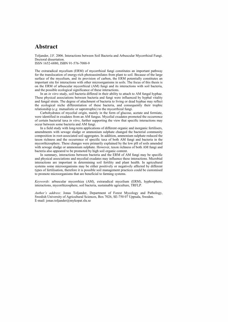
Abstract Toljander, J.F. 2006. Interactions between Soil Bacteria and Arbuscular Mycorrhizal Fungi. Doctoral dissertation. ISSN 1652-6880, ISBN 91-576-7088-9 The extraradical mycelium (ERM) of mycorrhizal fungi constitutes an important pathway for the translocation of energy-rich photoassimilates from plant to soil. Because of the large surface of the mycelium, and its provision of carbon, the ERM potentially constitutes an important site for interactions with other microorganisms in soils. The focus of this thesis is on the ERM of arbuscular mycorrhizal (AM) fungi and its interactions with soil bacteria, and the possible ecological significance of these interactions.
In an in vitro study, soil bacteria differed in their ability to attach to AM fungal hyphae. These physical associations between bacteria and fungi were influenced by hyphal vitality and fungal strain. The degree of attachment of bacteria to living or dead hyphae may reflect the ecological niche differentiation of these bacteria, and consequently their trophic relationship (e.g. mutualistic or saprotrophic) to the mycorrhizal fungi.
Carbohydrates of mycelial origin, mainly in the form of glucose, acetate and formiate, were identified in exudates from an AM fungus. Mycelial exudates promoted the occurrence of certain bacterial taxa in vitro, further supporting the view that specific interactions may occur between some bacteria and AM fungi.
In a field study with long-term applications of different organic and inorganic fertilisers, amendments with sewage sludge or ammonium sulphate changed the bacterial community composition in root-associated soil aggregates. In addition, ammonium sulphate reduced the taxon richness and the occurrence of specific taxa of both AM fungi and bacteria in the mycorrhizosphere. These changes were primarily explained by the low pH of soils amended with sewage sludge or ammonium sulphate. However, taxon richness of both AM fungi and bacteria also appeared to be promoted by high soil organic content.
In summary, interactions between bacteria and the ERM of AM fungi may be specific and physical associations and mycelial exudates may influence these interactions. Microbial interactions are important in determining soil fertility and plant health. In agricultural systems some microorganisms may be either positively or negatively affected by different types of fertilisation, therefore it is possible soil management practices could be customised to promote microorganisms that are beneficial to farming systems. Keywords: arbuscular mycorrhiza (AM), extraradical mycelium (ERM), hyphosphere, interactions, mycorrhizosphere, soil bacteria, sustainable agriculture, TRFLP. Author’s address: Jonas Toljander, Department of Forest Mycology and Pathology, Swedish University of Agricultural Sciences, Box 7026, SE-750 07 Uppsala, Sweden.E-mail: [email protected]


Contents
Introduction, 7 Background, 7 The AM symbiosis and the mycorrhizosphere, 7 Specific associations between AM and bacteria, 9 Identifying possible associations between AM and bacteria, 11 Economical and ecological significance of microbial interactions, 12 Objectives, 14 Methods, 15 Mycorrhizal root organ cultures, 15 Generation of root organ cultures from clover, 15 The fungal strains, 16 Visualisation of microorganisms, 17
Green fluorescent protein tagging of bacteria, 17 Fungal hyphal vitality, 18 Bacterial cell vitality, 18
Characterisation of microbial communities, 18 Terminal restriction fragment length polymorphism of bacterial communities, 18 AM fungal community composition, 20
Analysis of chemical composition of hyphal exudates, 21 Results and discussion, 21 Interactions between extraradical mycelia and soil bacteria, 21 Influence of different nitrogen fertilisers on soil microbial community composition, 24 General conclusions, 28 Outlook, 29 References, 30 List of abbreviations used in this thesis, 37 Acknowledgements, 38
Appendix
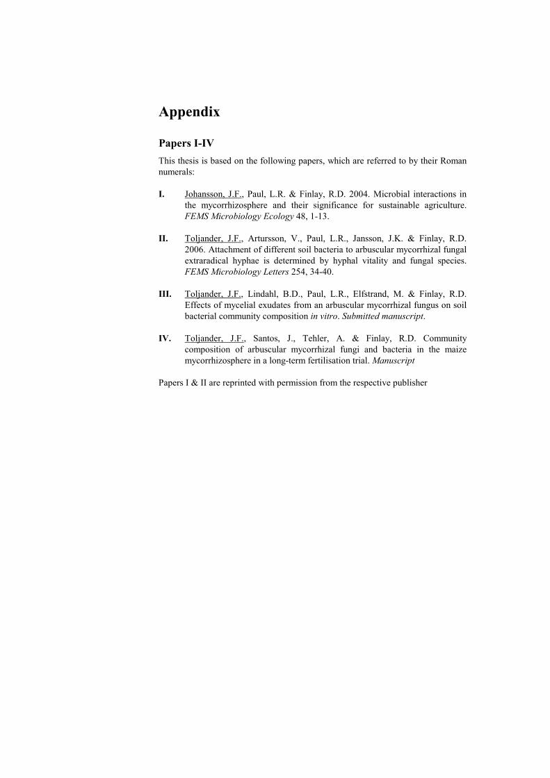
Papers I-IV This thesis is based on the following papers, which are referred to by their Roman numerals: I. Johansson, J.F., Paul, L.R. & Finlay, R.D. 2004. Microbial interactions in
the mycorrhizosphere and their significance for sustainable agriculture. FEMS Microbiology Ecology 48, 1-13.
II. Toljander, J.F., Artursson, V., Paul, L.R., Jansson, J.K. & Finlay, R.D.
2006. Attachment of different soil bacteria to arbuscular mycorrhizal fungal extraradical hyphae is determined by hyphal vitality and fungal species. FEMS Microbiology Letters 254, 34-40.
III. Toljander, J.F., Lindahl, B.D., Paul, L.R., Elfstrand, M. & Finlay, R.D.
Effects of mycelial exudates from an arbuscular mycorrhizal fungus on soil bacterial community composition in vitro. Submitted manuscript.
IV. Toljander, J.F., Santos, J., Tehler, A. & Finlay, R.D. Community
composition of arbuscular mycorrhizal fungi and bacteria in the maize mycorrhizosphere in a long-term fertilisation trial. Manuscript
Papers I & II are reprinted with permission from the respective publisher
7
Introduction
Background Microorganisms are key components of soil biodiversity, determining both soil fertility and plant health. Due to high inputs of pesticides and inorganic fertilisers, the roles of these organisms may have become marginalised in conventional agriculture, leading to a loss of biodiversity as well as function. However, increased environmental awareness in many countries, including Sweden, is progressively leading to a development from conventional intensive agriculture to low-input (sustainable) agriculture. Low-input agriculture refers to farming systems where the use of mineral fertilisers and pesticides is restricted. These agroecosystems are consequently more dependent upon biological control of pests and organic fertilisers to maintain crop health and productivity. It is therefore imperative for us to be able to utilise the potential of, or manipulate, naturally occurring microorganisms. A crucial step towards this is to characterise the organisms involved, their interactions with each other and their abiotic environment. The focus of this thesis is on the extraradical mycelium (ERM) of arbuscular mycorrhizal (AM) fungi, its interactions with soil bacteria, and the possible ecological significance of these interactions.
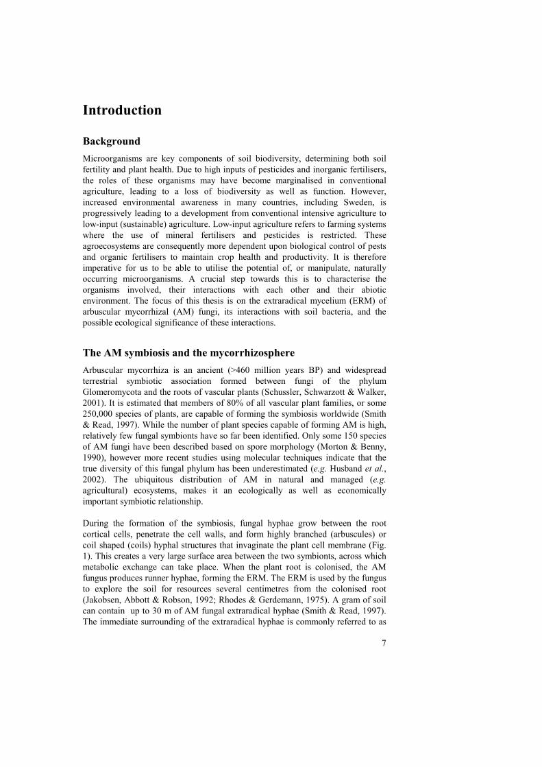
The AM symbiosis and the mycorrhizosphere Arbuscular mycorrhiza is an ancient (>460 million years BP) and widespread terrestrial symbiotic association formed between fungi of the phylum Glomeromycota and the roots of vascular plants (Schussler, Schwarzott & Walker, 2001). It is estimated that members of 80% of all vascular plant families, or some 250,000 species of plants, are capable of forming the symbiosis worldwide (Smith & Read, 1997). While the number of plant species capable of forming AM is high, relatively few fungal symbionts have so far been identified. Only some 150 species of AM fungi have been described based on spore morphology (Morton & Benny, 1990), however more recent studies using molecular techniques indicate that the true diversity of this fungal phylum has been underestimated (e.g. Husband et al., 2002). The ubiquitous distribution of AM in natural and managed (e.g. agricultural) ecosystems, makes it an ecologically as well as economically important symbiotic relationship. During the formation of the symbiosis, fungal hyphae grow between the root cortical cells, penetrate the cell walls, and form highly branched (arbuscules) or coil shaped (coils) hyphal structures that invaginate the plant cell membrane (Fig. 1). This creates a very large surface area between the two symbionts, across which metabolic exchange can take place. When the plant root is colonised, the AM fungus produces runner hyphae, forming the ERM. The ERM is used by the fungus to explore the soil for resources several centimetres from the colonised root (Jakobsen, Abbott & Robson, 1992; Rhodes & Gerdemann, 1975). A gram of soil can contain up to 30 m of AM fungal extraradical hyphae (Smith & Read, 1997). The immediate surrounding of the extraradical hyphae is commonly referred to as
8
the hyphosphere (cf. "rhizosphere", Hiltner, 1904, Fig. 2), and the soil volume influenced by the combined activities of the root and the mycorrhizal fungus is called mycorrhizosphere (Rambelli, 1973, Fig. 2). Because of the small diameter of the individual hyphae and the large numbers of hyphae in the soil, it is possible for the fungus to colonise microsites which the roots cannot access. The large surface area of the AM fungal mycelium may significantly increase the nutrient absorptive surface of the roots (Rambelli, 1973; Rhodes & Gerdemann, 1975).
Fig. 1. Structures of an arbuscular mycorrhizal fungus colonising a clover root. Intraradical hyphae growing between the root cortical cells are indicated by black arrows; arbuscules, here seen as dark structures within root cells, are indicated by white arrows; balloon-shaped objects (v) are vesicles, which are fungal storage organs. Bar = 200 μm. (Photograph by Jonas Toljander) The plant provides the fungus with photosynthetically derived carbohydrates, while the fungus supplies the plant roots with nutrients (Smith & Read, 1997). It is generally believed that the AM fungi primarily are important to their hosts for their superior ability to take up phosphorus which is relatively inaccessible to plants (e.g. McGonigle & Miller, 1996; Miller, 2000; Pearson & Jakobsen, 1993). However, the AM association may also increase the phytoavailability of micronutrients, e.g. copper and zinc (Smith & Read, 1997). In addition, it has even been suggested that some AM fungi are able to mobilise organically bound nitrogen, inaccessible to plants alone, which is subsequently translocated to the plant host (Hodge, Campbell & Fitter, 2001). This will be discussed in more detail below. Apart from influencing the quantity and quality of resources available to the plant host, the ERM also provides an interface for interaction with other soil organisms. A considerable amount (10-20%) of photosynthetically derived carbon may be

9
invested in the mycorrhizal symbiosis by the plant (Johnson et al., 2002). A proportion of the carbon is built into mycorrhizal fungal biomass, and during the fungus’ activities, it could be expected that some of this carbon is actively or passively exuded into the surrounding environment and utilised as a source of energy by other microorganisms in the mycorrhizosphere (Paper I). In addition, live or dead mycorrhizal fungal hyphae themselves, or compounds released by hyphae, may serve as a nutrient source or substratum for other organisms (Papers II and III).
Fig. 2. Schematic view of possible interactions among different components of the mycorrhizosphere. Indirect and direct influence of AM fungi on other components: 1. Supply of energy-rich carbohydrates (C) via extraradical hyphae; 2. pH changes; 3. Competition for nutrients; 4. (a) Stimulatory or (b) inhibitory compounds; 5. Stimulated root growth; 6. Changes in root exudation; 7. Changes in soil structure; 8. Endosymbiotic bacteria in AM fungal structures; 9. Interactions with pathogenic fungi. Effects of bacteria on AM fungi: 10. Effects on receptivity of roots to AM colonisation; Stimulation or inhibition on (11) growth or (12) germination of AM fungal propagules; 13. Modification of soil chemistry. (Figure is modified from Paper I)
Specific associations between AM and bacteria Arbuscular mycorrhizal colonisation may influence the composition of bacterial communities in the mycorrhizosphere, but little is known about the underlying mechanisms. Proposed general mechanisms include (Fig. 2): supply of energy-rich

10
compounds via the ERM (Andrade et al., 1997); changes in soil structure due to AM fungal hyphae and mycelial exudates, which may glue soil particles together (Andrade et al., 1998; Tisdall & Oades, 1979); and changes in root exudation patterns induced by AM colonisation (Marschner & Baumann, 2003; Söderberg, Olsson & Bååth, 2002). Changes in bacterial community structure may also be driven by complex interactions between plant species (or genotype) and fungal species involved (Marschner & Baumann, 2003; Marschner & Timonen, 2005). However, bacterial communities in the mycorrhizosphere may in turn influence the mycorrhizal- or plant development in various ways (Fig. 2), further complicating our understanding of bacterial-mycorrhizal interactions. Because of the complex nature of the mycorrhizosphere it has been difficult to couple effects to specific factors/organisms. For example, the changes in microbial communities can be due to direct (e.g. belowground carbon allocation) or indirect influence of mycorrhizal colonisation (e.g. root exudation) or the combined effects of the mycorrhizal fungi and their biotic and abiotic environment. An example of an indirect interaction between microorganisms in the mycorrhizosphere is the synergistic interaction between nitrogen fixation in the nodules of legumes and mycorrhizal colonisation of the same plants. Both these processes are symbiotic associations involving the host plant, bacteria belonging to Rhizobiaceae and AM fungi. The increased phosphorus supply provided by the fungus supports the rhizobial production of nitrogenase enzymes, which are important for the nitrogen fixation (Puppi, Azcón & Höflich, 1994). The enhanced nitrogen status of the plant promotes further development of the mycorrhizal symbiosis. An important factor in this interaction may be the small spatial scale, since physical proximity between microorganisms is likely to facilitate interactions between them (see discussions of Papers I-III). Evidence is accumulating that some bacteria live within AM fungi, i.e. endosymbiosis. MacDonald et al. (1982) observed bacterium-like organisms (BLOs) in the cytoplasm of some AM fungi. The BLOs, however, remained unidentified largely due to difficulties in culturing them. After the advent of PCR based techniques, Bianciotto et al. (1996a; 2000) used a combined morphological and molecular approach to study BLOs within the tissues of several species of the AM fungal genera Gigaspora and Scutellospora. The BLOs were initially identified as Burkholderia sp., but were later assigned a new taxonomical identity, Candidatus Glomeribacter gigasporarum (Bianciotto et al., 2003). An interesting aspect of these endosymbiotic bacteria is that they are transmitted vertically between fungal generations, suggesting that the bacteria constitute obligate endocellular components of their host fungi (Bianciotto et al., 2004). This deduction is also supported by the fact that total genome size of the endobacterium is estimated to only 1.4 Mb, which is the smallest genome known for a betaproteobacterium (Jargeat et al., 2004). Such small genome sizes are typically found in endocellular bacteria living permanently in their host. The nature of this symbiosis, whether it may be mutualistic, commensalistic or antagonistic, is still unknown, however, the endobacteria cause no apparent harm to the host fungi. Geosiphon pyriforme is another example of a fungus in the phylum Glomeromycota that is a host of endosymbiotic organisms. Ge. pyriforme is a close
11
relative of glomalean fungi and it represents the only known example of endocytobiosis between a fungus (macrosymbiont) and a cyanobacterium (Gehrig, Schüssler & Kluge, 1996). Endosymbiotic intracellular bacteria have been identified in ectomycorrhizal (ECM) fungi (Bertaux et al., 2003). These observations indicate that associations between fungi and endobacteria occur within different groups of mycorrhizal fungi, and that mycorrhiza-associated bacteria may be a widespread and ancient phenomenon. It is highly likely that some bacteria and AM fungi have co-evolved over time, and that specialised bacterial communities commonly associate with AM fungi. Consequently, the traditional perception that the mycorrhizal symbiosis is a bipartite association between a plant and a fungus may need to be revised to also include other associated organisms. A first step to understanding the function of these associations is to characterise the organisms involved.
Identifying possible associations between AM and bacteria Different approaches have been employed in attempts to identify bacteria that are potentially mycorrhiza-associated. Commonly used methods analyse changes in bacterial community composition induced by mycorrhiza. Many earlier studies relied on dilution plate count and various culturing techniques (Ames, Reid & Ingham, 1984; Meyer & Linderman, 1986; Secilia & Bagyaraj, 1987), however more recently, culture independent methods including community fingerprinting methods, such as DGGE or TRFLP have been favoured to characterise bacterial communities in the mycorrhizosphere (Artursson, Finlay & Jansson, 2006a; Marschner & Timonen, 2005; Wamberg et al., 2003). Another approach is to screen bacterial inoculants for possible affinity to mycorrhizal structures. Physical association between microorganisms may be indicative of a close relationship, since it may facilitate cell-to-cell interactions (Papers I & II). Bianciotto et al. (1996b) studied the colonisation of spores and hyphae of the AM fungus Gigaspora margarita by different strains of fluorescent pseudomonads and rhizobia. Bacteria adhering to fungal tissues typically also adhered to non-living substrata (cotton- and quartz fibres). The authors therefore concluded that specific receptors were not involved in the observed attachment. In another study, a different bacterial strain, Pseudomonas fluorescens DF57, did not colonise the hyphae of the AM fungus Glomus intraradices (Ravnskov, Nybroe & Jakobsen, 1999). In addition, the fungus had a negative effect on the population size of the introduced bacteria, since both cell counts and culturability were suppressed in hyphosphere soil. The results from these studies suggest that associations between AM fungi and bacteria may be highly specific, and the outcome of the interactions could vary depending on species or strains involved. A third approach is to screen soil bacteria for possible effects on mycorrhizal fungi. Paenibacillus spp. in particular have received much attention as they have been found to interact with AM fungi (Artursson, Finlay & Jansson, 2006a; Bertaux et al., 2003; Mansfeld-Giese, Larsen & Bodker, 2002) or stimulate AM fungal development (Budi et al., 1999; Hildebrandt, Janetta & Bothe, 2002;
12
Hildebrandt et al., 2006). In addition, further studies have shown that the same Paenibacillus spp. also exhibit antagonism against a range of soil borne pathogens (Budi et al., 1999; von der Weid et al., 2003; von der Weid et al., 2005), and could therefore serve as potential inoculants to improve crop yields in low-input agriculture.
Economical and ecological significance of microbial interactions In low-input cropping systems the natural activities of microorganisms may contribute to the biocontrol of pathogens and improved supply of nutrients, thus maintaining crop health and production. Therefore, the understanding of these interactions and the underlying mechanisms could have implications for the progress of sustainable agriculture. The role of microorganisms in maintaining plant biodiversity and ecosystem functioning was brought into focus by van der Heijden et al. (1998). By using macrocosms to simulate old-fields, they showed that plant biodiversity as well as the fundamental ecosystem functions, nutrient capture and plant productivity, increased significantly with increasing species richness of AM fungi, suggesting that these ecosystem functions can be driven by belowground microbial interactions. The results of van der Heijden et al. emphasise the need to consider AM fungi, and other microorganims, in management practices. However, only a relatively small number of field studies have investigated the long-term effects of conventional versus sustainable agricultural management practices on microbial diversity in relation to crop production. Helgason et al. (1998) showed that the diversity of AM fungal communities was strikingly low in arable sites compared with a woodland. The broad host range of some AM fungi suggests that the change in species composition and low diversity of the fungi in arable fields was probably not the result of plant monoculture, but may reflect other aspects of the agronomic regime such as ploughing, fertilisation or fungicide application. The authors suggested that low AM fungal diversity in arable fields could indicate a smaller functional contribution of AM fungi in these systems. In long term field trials in Switzerland, Mäder et al. (2000) found that root length colonised by mycorrhizal fungi in organic farming systems was 30-60% higher than in conventional systems. In addition, there was an overall higher biomass and diversity of soil animals, and a higher microbial activity in organic systems (Mäder et al., 2002). Crop yields were 20% lower in the organic systems, when input of fertiliser and energy (use of fuel, etc.) was reduced by 34-53% and pesticide input by 97% (Mäder et al., 2002). They concluded that enhanced soil fertility and higher biodiversity found in organic plots may make these systems less dependent on external inputs. Consequently, the monetary loss in production may, at least to some extent, be compensated for by a reduced need for expensive mineral fertilisers and pesticides. The high significance of AM associations in the supply of mineral nutrients, in particular phosphorus, to their plant hosts is generally recognised (Caris et al., 1998; Larsen et al., 1996; Miller, 2000; Nielsen et al., 2002; Pearson & Jakobsen, 1993). In addition to this, the possible role of extracellular phosphatase activity of AM fungi, in relation to mineralisation of organic phosphorus pools in soils has
13
recently attracted attention (Feng et al., 2003; Joner, van Aarle & Vosatka, 2000), although the concept of phosphate solubilising bacteria is probably more well-established. Phosphate solubilising bacteria are widespread in soils and secretion of organic acids and phosphatases are common means for facilitating the conversion of insoluble forms of phosphorus to plant-available forms (Vessey, 2003). The biofertiliser properties of plant growth promoting rhizobacteria are frequently ascribed to their ability to increase the bioavailablity of inorganic and organic phosphorus, and some of these bacteria have documented synergistic effects on e.g. nitrogen fixation and formation of mycorrhizal associations. Toro et al. (1997) demonstrated that both Enterobacter sp. and Bacillus subtilis promoted the establishment of the AM fungus Glomus intraradices and increased plant biomass as well as tissue nitrogen and phosphorus contents. Kim et al. (1998) found that phosphorus content in tomato plants was increased when inoculated with either the AM fungus G. etunicatum, or the phosphate solubilising bacterium Enterobacter agglomerans. Additionally, the highest nitrogen and phosphorus uptake was observed when tomatoes were inoculated with both organisms, suggesting that bacteria and AM fungi may together increase the nutrient uptake in plants. It has also been proposed that AM fungi may contribute to the decomposition of organic matter and the subsequent acquisition of organic nitrogen. Hodge et al. (2001) grew plants of Plantago lanceolata colonised with the AM fungus Glomus hoi in compartmentalised soil microcosms. Decomposition of, and nitrogen capture from, dual-labelled (15N/13C) plant material was increased by the AM fungus, suggesting that some AM fungi can have saprotrophic capabilities. However, since the microcosms were not monoxenic, it remained a possibility that other microorganisms, possibly in association with the AM fungus, were involved in the decomposition. Using monoxenic cultures of another glomalean fungus, G. intraradices, Hawkins et al. (2000) showed that the AM fungus alone was indeed able to acquire nitrogen from labelled amino acids, but only at fairly low rates. The possible significance of synergistic interactions between AM fungi and soil bacteria in relation to general plant nutrition has already been discussed, but in addition to this, microbial interactions may have implications for biological control. A number of studies have indicated that some AM fungi (Caron, 1989; Newsham, Fitter & Watkinson, 1995; Niemira, Hammerschmidt & Safir, 1996), as well as some mycorrhiza associated bacteria (Barea et al., 1998; Budi et al., 1999) exhibit biocontrol properties against root pathogens. Whether AM fungi could be used as biocontrol inoculants practically, or possibly function as co-inoculants with bacteria with biocontrol properties, remains to be explored. A more realistic approach may involve the ability to utilise the potential of indigenous fungi in soils, and promote the activities of beneficial microorganisms already present in the soil.
14
Objectives
The overall aim of the work described in this thesis was to characterise interactions between AM fungi and soil bacteria and their environment using molecular tools. The specific objectives were:
• To review microbial interactions in the mycorrhizosphere and their significance for sustainable agriculture (Paper I). Some aspects of these interactions are investigated in more detail in Papers II-IV.
• To examine the ability of different soil bacteria to attach to vital and
nonvital extraradical hyphae of different AM fungi in vitro (Paper II). Physical associations may facilitate metabolic interaction, and therefore the degree of attachment could reflect the trophic status of the bacteria or the ecological function of the association. An increased understanding of these associations has also a direct application when selecting microbial inoculants for biocontrol or biofertilisation
• To characterise the chemical composition of AM fungal extraradical
mycelial exudates and their possible influence on soil bacterial community structure in vitro (Paper III). Very little is known about the range of compounds released by growing AM fungal mycelia, and their effects on other microorganisms. The exudates may have a general stimulatory effect on bacterial growth, but may also specifically influence certain bacteria. Effect of exudates on bacterial communities may reveal synergistic or antagonistic relationships between AM fungi and bacteria.
• To characterise the influence of different long-term fertilisation regimes
on the community composition of AM fungi and bacteria in the mycorrhizosphere of maize in a field trial (Paper IV). An increasing number of studies report that long term application of mineral fertilisers have detrimental effects on mycorrhizal colonisation, microbial biomass and activity. However, changes in microbial community composition may have consequences for plant productivity. In order to employ management practices that minimise unfavourable effects on the environment and increase the benefits that microorganisms can bring to farming systems (e.g. in terms of plant production), there is a need to describe the possible effects of soil management on the composition of microbial communities.

15
Methods
In the papers of this thesis a number of techniques were employed to characterise the microorganisms and the interactions between AM fungi and bacteria. Since the details of these methods are presented in the respective papers or in articles referred to in the papers, the purpose of this section is only to outline the principal methods.
Mycorrhizal root organ cultures Arbuscular mycorrhizal fungi are obligate biotrophs, meaning that they are completely reliant upon the presence of host plant roots to survive and grow. For the purpose of scientific studies these fungi have traditionally been grown in pot cultures in co-culture with whole plants, however increasing numbers of studies have utilised axenic Ri T-DNA-transformed root cultures (“hairy roots” or root organ cultures) (Bécard & Fortin, 1988). Although root cultures represent artificial and less complex system than would be encountered in natural soils, they offer the advantage of being able to study AM fungi in an axenic, and hence more controlled, environment where only the fungus and its host are present. In particular, studies of the AM fungal ERM and its interactions with its abiotic and biotic environment have gained by utilising different modifications of these systems (e.g. Filion, St-Arnaud & Fortin, 1999; Hawkins, Johansen & George, 2000; Nielsen et al., 2002; Pfeffer et al., 2004; Rufyikiri, Thiry & Declerck, 2003). By using compartmentalised systems, as described in Paper III, the ERM can be studied separately without the direct influence of roots. Only very recently there have been a few reports of certain AM fungal strains being able to complete a life cycle in the absence of plant roots, but in the presence of another, bacterial, symbiont (Hildebrandt, Janetta & Bothe, 2002; Hildebrandt et al., 2006). These studies suggest that the AM association could be more complex than previously held, and that the formation of the symbiosis in soils may involve three symbionts or more. In the study presented in Paper II, carrot (Daucus carota L.) root organ cultures were used, which had been Ri T-DNA transformed according to Becard and Fortin (1988). The roots were inoculated with either of the fungal strains Glomus sp. MUCL 43205 and G. intraradices Schenck & Smith MUCL 43194 and routinely maintained on minimal (M) medium (Bécard & Fortin, 1988) and then transferred to liquid M medium prior to the experiment. For Paper III, clover (Trifolium repens L.) root organ cultures were developed.
Generation of root organ cultures from clover Paper III root organ cultures were generated of clover by Agrobacterium rhizogenes-mediated root transformation essentially as described by Boisson-Dernier et al. (2001). The binary vector p35S GUS INT harboured a gus gene and a kanamycin resistance gene under the control of the CaMV 35S promoter and nos
16
promoter respectively (Lindroth et al., 1999). Transformed roots emerged from the inoculated explants on M medium containing kanamycin (50 mg l-1) as selective agent. To establish root cultures, viable roots emerging from the inoculation site after approximately three weeks were isolated and cultured on selective medium. After three subsequent subcultures on M medium with antibiotics, root cultures apparently free of microbial contamination were transferred to M medium without antibiotics. Absence of A. rhizogenes was verified by PCR with a virD3 specific primer combination (Lindroth et al., 1999). Presence of gus was confirmed by PCR using specific primers. One fast-growing culture “LP1” supporting amplification of the gus insert, without amplifying the ~800 bp virD3 fragment was selected (Fig. 3). To maintain the clover root culture it was subcultured on fresh medium every eight to ten weeks.
Fig. 3. PCR of crude DNA extracts from five different transgenic Trifolium repens L. roots transformed with Agrobacterium rhizogenes. From left: Lanes 2-6 amplified gus, lane 7 gus positive control (p35S GUS INT), lane 8 gus negative control (no DNA template); lanes 10-14 virD3 (absent), lane 15 virD3 positive control, lane 16 virD3 negative control. The virD3 product has a size of 882 bp and the gus product a size of 602 bp. DNA ladder in lanes 1, 9 and 17 represents 1 Kb DNA Ladder (Invitrogen Corporation, Paisley, UK).
The fungal strains The AM fungal strains used in Papers II and III were purchased from GINCO (Louvain-la-Neuve, Belgium), and subcultured in our laboratory on either carrot or clover root organ cultures on M medium. The new clover root organ cultures were successfully colonised with five Glomus strains (all affiliated with Glomus group A): Glomus sp. MUCL 43205, G. proliferum Dalpé & Declerck MUCL 41827, G. intraradices Schenck & Smith MUCL 43194, G. intraradices Schenck & Smith MUCL 43204, and G. clarum Nicolson & Schenck MUCL 46238. However, the inoculation of clover roots with the first three of these strains was most successful, in terms of in vitro production of spores and ERM. Only Glomus sp. MUCL 43205 was used together with clover roots in two-compartment cultures (Fig. 4) in experiments for this thesis (Paper III). For Paper II, Glomus sp. MUCL 43205 and G. intraradices MUCL 43194 colonising carrot root organ cultures were used. Both strains were originally isolated from Fraxinus americana in Québec, Canada. The latter was the first G. intraradices strain to have been propagated on transformed roots (Chabot, Bécard & Piché, 1992), and was selected because it has been used in a range of field- and laboratory studies.
17
Fig. 4. Schematic view of mycorrhizal root organ culture in the split Petri dish system that was used to produce mycelial exudates in Paper III. Arbuscular mycor-rhizal extraradical mycelia, but not roots, were allowed to cross the barrier separating the root compartment from the hyphal compartment thus creating an environment influenced only by the fungal mycelium. Mycelial exudates were collec-ted and incubated with soil bacterial communities, to study the specific effect of exudates on different bacterial taxa. (Figure is reproduced from Paper III).
Visualisation of microorganisms Green fluorescent protein tagging of bacteria The green fluorescent protein (GFP) is a useful reporter molecule for monitoring in vivo gene expression in eukaryotic and prokaryotic cells (Fortineau et al., 2000). The gene encoding the protein, gfp, was originally isolated from the jellyfish Aequorea victoria (Prasher et al., 1992). The protein emits green light when excited with ultraviolet radiation, without the requirement of substrate or cofactor for fluorescence. To visualise and facilitate quantification of bacterial attachment to AM fungal extraradical hyphae in Paper II, five different strains of soil bacteria, each tagged with the gfp gene, were used. Paenibacillus brasilensis PB177 and Paenibacillus peoriae NRRL BD62 were both previously transformed (Artursson & Jansson, 2003; von der Weid et al., 2005) with the pnf8 plasmid containing gfp under the control of the strong constitutive promoter from Listeria (Fortineau et al., 2000). Arthrobacter chlorophenolicus A6G (Elvang et al., 2001) and Pseudomonas fluorescens SBW25 gfp/lux (Unge et al., 1999) were previously chromosomally tagged with gfp or both gfp and luxAB genes (encoding bacterial luciferase), respectively, using mini-transposon vectors. All of the bacterial strains were routinely grown in Luria–Bertani medium. Erythromycin was added to the medium to select for growth of those bacteria that were tagged with the pnf8 plasmid to ensure stability of the plasmid and the GFP phenotype. Following short-term incubation experiments with each of the gfp-tagged bacterial isolates and vital or nonvital hyphae, fluorescing bacterial cells attached to the surface of AM fungal hyphae were quantified using a fluorescence microscope.
18
Fungal hyphal vitality In Paper II vital and nonvital extraradical hyphae of the two AM fungal isolates Glomus sp. MUCL 43205 and G. intraradices MUCL 43194 were used in attachment assays involving single bacterial isolates. To produce nonvital extraradical hyphae, hyphae were excised from axenic mycorrhizal root cultures (Daucus carota L.) grown in liquid media ca. 7 days prior to the experiment. At the time of performing the experiment, excised hyphae, as well as hyphae from intact mycorrhizal cultures, were randomly collected and subjected to staining using fluorescein diacetate (FDA) (Söderström, 1979). Fluorescein diacetate is a non-fluorescent compound which can pass through the cell membrane. When taken up by the hyphae, intracellular esterases cleave off the diacetate upon which a highly fluorescent product (fluorescein) is produced and can be detected using a fluorescence microscope. Because cells with compromised membrane integrity rapidly leak out the product, green-fluorescing hyphae can be assumed to be vital. In addition to the visual vitality assay of hyphae, other observations were made: Nonvital hyphae were generally more brittle than vital hyphae, and vital hyphae tended to be more flexible. However, being more subjective than the FDA staining, these observations were not used to determine hyphal vitality.
Bacterial cell vitality In Paper III bacterial numbers and vitality were quantified to monitor bacterial growth over time in the mycorrhizal and non-mycorrhizal treatment. To rapidly distinguish live bacteria from dead the LIVE/DEAD® BacLight™ Bacterial Vitality kit (Molecular Probes, Leiden, The Netherlands) was used. With this method two nucleic acid stains, SYTO 9 dye (cell-permeant) and propidium iodide (cell-impermeant) are added to a bacterial suspension. The dyes cause live bacteria to fluoresce green and dead bacteria to fluoresce red under a fluorescence microscope. Live and dead cells are quantified using fluorescence microscopy and thereby the proportion of live to dead, or live to total cells, can then be calculated and used to estimate total bacterial vitality or population growth.
Characterisation of microbial communities Terminal restriction fragment length polymorphism of bacterial communities In order to characterise the composition of bacterial communities in samples of Papers III and IV the PCR based method terminal restriction fragment length polymorphism (TRFLP) was employed. With this method DNA template is amplified using fluorescently labelled primers which are homologous to a conserved sequence of the desired gene (usually the 16S rRNA gene for bacteria). In both Papers III and IV, the universal eubacterial forward primer 27f (5'-AGA GTT TGA TCC TGG CTC AG-3') and reverse primer 534r (5'-ATT ACC GCG GCT GCT GG-3') were used in combination for TRFLP PCR. Each primer was labelled with fluorescent labels in the 5’-end. This set of primers amplifies a ~500 base pair (bp) region of the bacterial 16S rDNA sequence. Following PCR, the
19
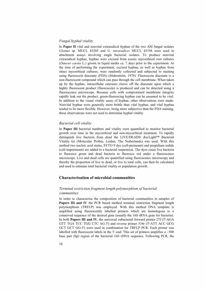
amplified products were digested with restriction endonucleases to produce restriction fragments of varying sizes. For Paper III the restriction endonucleases CfoI, HaeIII and MspI were used, and for Paper IV only CfoI and HaeIII were used. The sizes of the obtained fragments depend on the presence and location of restriction sites recognised by the endonucleases, which is sequence specific and therefore potentially taxon specific. An internal fluorescent DNA size standard is added to each of the samples, which are then analysed using an automated fluorescence-based DNA sequencing instrument. In this process the DNA fragments are separated by capillary electrophoresis. Terminal restriction fragments (TRFs) with the fluorescent 5’-label can thereby be detected and assigned a fragment size measured in base pairs. The method has several advantages compared with its predecessor RFLP. TRFLP is a high-throughput method employing high-resolution separation of fragments by capillary electrophoresis, rather than low-resolution agarose gel separation. In addition, automated fluorescence-based detection enables sensitive detection of fragments at very low concentrations (Felske & Osborn, 2005). Consequently, TRFs that would not be distinguished with RFLP can be detected. Typically, in an environmental sample many fragments of different sizes, each potentially representing a taxon, will be detected. The fragments, or peaks, can be assigned putative identities in several ways: by comparing fragments against fragments produced from clones or isolates with a known identity; by comparison against a public TRFLP database; by comparing the obtained fragment size with the predicted size, by studying the location of specific restriction sites in the DNA sequences of identified clones/isolates. Although TRFLP and other community profiling methods are sensitive methods to detect species in environmental samples, all PCR based methods have inherent problems that need to be considered when interpreting community profiles. PCR primers may have a higher affinity to the DNA belonging to certain taxa, resulting in a skewed representation of species abundance (Allmér, 2005). Furthermore, bias in the PCR reaction caused by impurities in the sample, the relative proportions of reaction constituents (chemicals, and target and non-target DNA) may also be introduced. To test these assumptions, the effect of template dilution prior to PCR and TRFLP on relative abundance of restriction fragments was investigated. Undiluted and diluted samples were all subjected to TRFLP PCR, and amplified products were subsequently digested using CfoI. Following TRFLP the relative abundance of TRFs within each sample was calculated by dividing the area of each peak (TRF) by the total peak area in the sample. This simple test showed that in three out of four dilution series, the ratio between individual peaks in a sample was altered by dilution. In the most extreme case, the relationship between peaks that were high or low in abundance was reversed, even though samples had only been diluted 1:2 (Fig. 5). This indicated that the relative abundance of peaks should be interpreted with great caution when translating the TRFLP profiles into relative abundance of taxa in environmental samples. The occurrence of bacterial taxa identified by TRFLP in Papers III and IV was therefore only recorded in terms of presence, and not their relative abundance abundance, in samples.
20
Fig. 5. Example of the effect of dilution of DNA template prior to PCR and TRFLP on the the relative abundance of terminal restriction fragments (TRFs) in an environmental sample (unpublished data). Relative abundance of TRFs (peaks) has been calculated by dividing the area of each peak by the total peak area in the sample. Each TRF represents a putative bacterial taxon. The figure shows the TRF profile of an undiluted sample, and the same sample diluted two and five times, respectively. For the undiluted sample, the ratio of a:b = 0.25; for the sample diluted two times a:b = 2.67; for the sample diluted five times a:b = 0.36. The test demonstrated that even a small dilution of template before PCR may cause the relative abundance of peaks to be reversed.
AM fungal community composition For Paper IV, AM fungal community composition was analysed by PCR and cloning of fungal 18S rDNA in maize roots, and subsequent sequencing of clone inserts. For PCR the universal eukaryotic primer NS31 (Simon, Lalonde & Bruns, 1992) was used as forward primer. As reverse primers a mixture of three different primers was used: the AM1 primer (Helgason et al., 1998), the AM2 (5’-GTT TCC CGT AAG GTG CCA AA-3’) and AM3 (5’-GTT TCC CGT AAG GTG CCG AA-3’) (Santos, Finlay & Tehler, In Press). The AM1 primer was originally designed as AM fungal specific, but results in important mismatches with sequences of certain glomalean groups. The AM2 and the AM3 are designed to match the same priming site as AM1 in taxa belonging to Glomus group B and group C, respectively, as defined by Schüssler et al. (Schüssler et al., 2001). This primer combination amplifies a 550 bp long sequence in the SSU 18 rRNA gene. Following cloning of PCR products, clones were collected and reamplified using the same primers as above. Purified, reamplified fragments were subsequently sequenced using the NS31 primer as sequencing primer.

21
Analysis of chemical composition of hyphal exudates In Paper III, analysis of the composition of hyphal exudates of Glomus sp. MUCL 43205 was performed by proton nuclear magnetic spectrometry (HNMR). A portion of the liquid from each culture was vacuum-centrifuged at 40ºC until dry. The dry residues were washed with deuterium oxide (D2O) and then vacuum-centrifuged again. The washing procedure was repeated three times. Washed and dried samples were subjected to analysis performed at the Department of Chemistry, SLU, Uppsala, Sweden.
Results and discussion
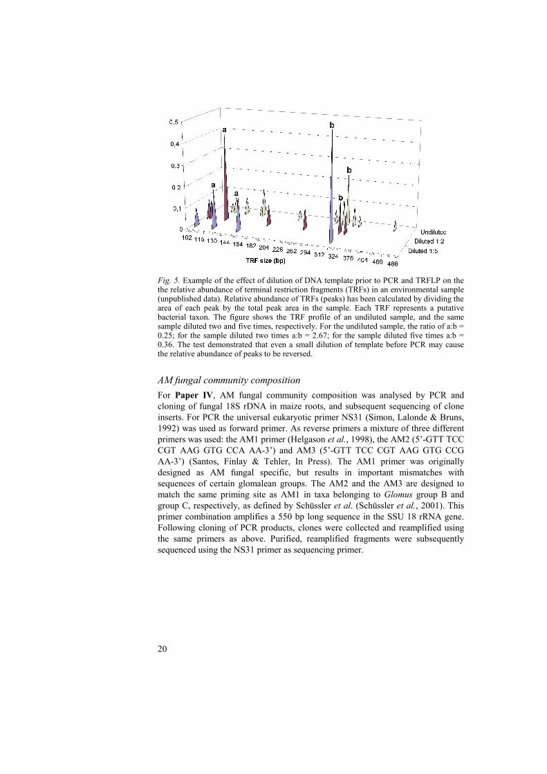
Interactions between extraradical mycelia and soil bacteria The continuous provision of energy-rich compounds, coupled with the large surface area of the mycelium that interacts with the surrounding soil environment, suggests that the AM fungal ERM constitutes an important niche for bacterial colonisation and growth. In Paper II Paenibacillus brasilensis PB177 showed a higher attachment to vital hyphae of both Glomus sp. MUCL 43205 (Fig. 6) and G. intraradices MUCL 43194 in vitro. This bacterial strain, along with several other described Paenibacillus spp., has documented antagonistic effects on plant pathogenic soil fungi (von der Weid et al., 2003; von der Weid et al., 2005). Many Paenibacillus spp. are also nitrogen fixers, exhibiting stimulatory effects on AM development (Budi et al., 1999; Hildebrandt, Janetta & Bothe, 2002; Hildebrandt et al., 2006). Synergistic interactions between microorganisms may have implications for biocontrol or biofertilisation. The degree of dependence on mycorrhizal fungi exhibited by the bacteria may determine the fate of these bacteria in soils, and consequently their usefulness as inoculants. For example, bacterial strains closely associated with AM fungi may utilise fungal hyphae as a substratum, and hyphal exudates for sustenance. Arbuscular mycorrhizal fungi may thereby aid dispersal and persistence of some bacteria in the soil environment. Other bacteria associating with mycorrhizal fungal hyphae may act as saprotrophs and derive carbon and other nutrients through degradation of fungal hyphae. In Paper II, Bacillus cereus VA1 showed higher attachment to nonvital hyphae of both glomalean fungi in the assay (Fig. 6). In another study (Artursson & Jansson, 2003) the same B. cereus strain was frequently associated with damaged parts of hyphae, indicating that these bacteria were utilising the hyphae, or cell contents released from the hyphae, as a substrate. Staddon et al. (2003) demonstrated a rapid turnover of AM fungal hyphae in soil. Their results suggested that AM fungal hyphae in soils live on average only 5–6 days, contributing to a rapid pathway for carbon in soil ecosystems. The results of Paper II suggest that B. cereus may behave as a saprotroph or mycoparasite. Saprotrophic activity contributing to the degradation of hyphae and utilisation of compounds released by damaged hyphae may have contributed to the rapid hyphal turnover observed by Staddon et al.
22
Fig. 6. Examples of attachment of gfp-tagged bacteria to extraradical hyphae of an arbuscular mycorrhizal fungus (Glomus sp. MUCL 43205): Attachment of Paenibacillus brasilensis PB177 to vital hyphae (left); attachment of Bacillus cereus VA1 to nonvital hyphae (right). Bars = 10 μm (Both photographs are reproduced from Paper II) In Paper II, individual bacterial strains exhibited differences in their degree of attachment to vital hyphae of the two tested fungi. Pseudomonas fluorescens SBW25 showed a greater attachment to vital than to nonvital hyphae of Glomus sp. MUCL 43205, whereas this trend was opposite for the attachment to G. intraradices hyphae. Paenibacillus peoriae BD62 did not attach to Glomus sp. MUCL 43205 at all, but showed a high attachment to nonvital hyphae of G. intraradices. These observations indicate that in addition to hyphal vitality, other factors may influence bacterial attachment to hyphae. Differential attachment may also be caused by differences in hyphal surfaces at the cellular level. Another possibility is that the quantity or quality of compounds of mycelial origin may differ between fungal strains. The growth and attachment of certain bacteria may therefore be promoted or inhibited accordingly. It is unclear to which extent mycelia, compared with roots, contribute to the total quantity of carbon available for consumption by soil organisms. There may however be qualitative differences between fungal mycelia and plant roots in the composition of exudates. In addition, root deposition is localised in the vicinity of the root systems, whereas fungal mycelia can create micro-niches or access fractions of the soil which roots alone never access. Consequently, the carbon released by active mycelia, or by hyphae being decomposed, may create hotspots in the soil, and could therefore significantly influence the activity of other organisms where mycelia are present. Fungal metabolites actively or passively exuded by AM fungal hyphae may result in general attraction and growth stimulation of some bacteria that utilise exuded compounds for growth. However, it is also possible that some bacteria are suppressed by inhibitory compounds produced by AM fungi. Antagonism of single bacterial or fungal isolates by AM fungi has previously been reported (Filion, St-
23
Arnaud & Fortin, 1999; Ravnskov, Nybroe & Jakobsen, 1999). It seems likely that such antagonistic interactions are mediated by the AM fungal exudation of compounds with inhibitory or antimicrobial properties. While there is information on the metabolism and translocation of carbon within AM fungal tissues (Bago et al., 2002), the nature of compounds that are actively or passively exuded by mycelia, and their possible effects on their biotic and abiotic environments are not well-known. Some ectomycorrhizal (ECM) fungi, however, have been shown to exude acetic and oxalic acid into their environment (Sun et al., 1999), and ECM fungal production of siderophores and their possible relevance in mineral weathering and nutrient acquisition has also been discussed (Holmström et al., 2004; van Hees et al., 2006). Allen et al. (1996) used scanning electron microscopy to examine production of oxalate crystals on the surfaces of ecto- and arbuscular mycorrhizal fungal hyphae. Only hyphae of some mat-forming ectomycorrhizal fungal species had crystal structures associated with them. Their data suggested that there are fundamental differences in chemical exudation between mycorrhizal fungi, and that this could have implications for uptake and cycling of phosphorus in soils. Organic acids may in addition increase the bioavailability of inorganic phosphorus and metallic ions. Consequently, mycelial exudation and nutrient acquisition by fungi could indirectly influence the composition of bacterial communities in the soil, through competition with other microrganisms for nutrients. Our results (Paper III) show that AM fungal mycelia may exude simple carbohydrates, in the form of glucose, as well as low molecular weight aliphatic acids. Organic acids exuded by roots or mycorrhizal fungal mycelia may be utilised as sources of carbon and nutrients or as chemoattractants for some microorganisms (Grayston, Vaughan & Jones, 1997; Sood, 2003). The presence of mycelial exudates from Glomus sp. MUCL 43205 had a general stimulatory effect on total bacterial growth (Fig. 7) and changed the composition of the bacterial community, with certain bacterial taxa either decreasing or increasing in occurrence. The occurrence of several taxa, in particular three taxa related to Buttiauxella gaviniae (gammaproteobacteria), Legionella wadsworthii (gammaproteobacteria), Flavobacterium sp. (flavobacteria), was significantly increased in response to the mycelial exudates. Several of the bacterial taxa with an increased occurrence in response to mycelial exudates were gammaproteobacteria. In contrast, none of the taxa with a higher frequency of occurrence in samples without AM fungal mycelial exudates was affiliated to this class of bacteria. This is in agreement with many studies reporting synergistic interactions between AM and gammaproteobacteria, leading to increased mycorrhizal colonisation and nutrient uptake in plants (see Artursson, Finlay & Jansson, 2006b and references therein). As indicated in the introduction, bacterial strains belonging to Enterobacteriaceae have previously been shown to promote mycorrhizal establishment (Toro, Azcon & Barea, 1997) and plant nitrogen and phosphorus uptake (Kim, Jordan & McDonald, 1998; Toro, Azcon & Barea, 1997), which may suggest that these strains are interacting synergistically with AM fungi. Indeed, the gammaproteobacterium identified as Buttiauxella gaviniae (Enterobacteriaceae) in Paper III increased in frequency of occurrence in response mycelial exudates from an AM fungus.

24
7
8
9
10
0.75
0.80
0.85
0.90
0.95
0 10 20 30 40 50 60 70 80
Log
no. o
fcel
lsPr
opor
tion
of v
ital c
ells
Time (hours)
a
b
Fig. 7. Bacterial population growth and vitality at different times post incubation in the presence and absence of mycelial exudates from an arbuscular mycorrhizal fungus (Glomus sp. MUCL 43205). (a) Log total number of cells counted at each sampling occasion. (b) Proportion of vital cells, calculated as the number of vital cells divided by the number vital and nonvital cells. Closed and open circles symbolise mean values of samples with or without mycelial exudates, respectively. Error bars represent standard error of the means. Y axes are broken and do not begin at zero. (Figure is reproduced from Paper III)
The results from Papers II and III support the view that interactions between certain bacteria and AM fungi are commonplace, and that there is a degree of specificity in these interactions. These interactions may be facilitated by the physical proximity between the symbiotic organisms. The nature of these interactions, whether mutualistic or parasitic, is highly dependent on which interacting organisms are involved, and may be determined on the species-, or strain level. However, there are also opportunistic bacteria that at times rely on mycelial products released by AM fungi, or bacteria that may utilise the hyphae themselves as a nutrient source or substratum. The understanding of interactions occurring between the AM fungal ERM and its biotic and abiotic environment is still in its infancy. The characterisation of the composition of AM fungal exudates and the effects of these compounds on soil microbial communities may have wide implications: from understanding the specific interactions between the microorganisms in the mycorrhizosphere, to more applied areas of research, such as the study of nutrient cycling and contribution of soil microbial interactions to soil fertility and crop production.
Influence of different nitrogen fertilisers on soil microbial community composition In Paper I the significance of interactions occurring between AM fungi and other microorganisms to sustainable agriculture is outlined. Clearly microorganisms play crucial roles in soil processes of both natural and managed ecosystems, however it is also evident from a large number of studies that intensive management practices (i.e. tillage, mono-cropping, landscape homogeneity, and application of mineral fertilisers) can have detrimental effects on the activity and occurrence of a wide range of organism groups (Eriksson, 1999; Gosling et al., 2006; Mäder et al., 2002; Nyberg, 2006; Salles, 2005; Weibull, 2002). Many studies reporting

25
negative effects of intensive management practices on soil microorganisms have mainly considered total microbial activity or microbial biomass. As indicated by van der Heijden et al. (1998) it may also be important to consider the species composition of these microbial communities, since microbial diversity alone may be an important factor determining ecosystem functions. Removal of crops at harvest, or crop rotation using non-mycorrhizal plants, each represent disturbances, which are also likely to have profound effects on the composition of microbial communities belowground. In order to employ management practices to increase the benefits that microorganisms can bring to sustainable farming systems, we need to describe the possible effects of soil management on the composition of microbial communities. We therefore aimed to describe the changes in community composition of AM fungi and bacteria in the mycorrhizosphere of maize (Zea mays L.) in response to long-term application of different organic and mineral nitrogen fertilisers (Paper IV). An additional aim of this study was to see how removal of crops influenced the community composition of bacteria. At crop harvest the allocation of photosynthates to the mycorrhizal roots is interrupted, resulting in a decline in the mycorrhizal fungal population and, presumably, also in bacteria that associate with the living mycorrhizal roots. A pulsed input of carbon in the form of senescing roots and mycorrhizal fungal mycelia in the soil may favour certain other groups of microorganisms that are saprotrophic or mycotrophic. The main finding presented in Paper IV was that both numbers of taxa and the occurrence of specific AM fungal and bacterial taxa were influenced by the fertilisation treatments. Overall, fewer AM fungal and bacterial taxa were detected in plots amended with (NH4)2SO4, whereas the number of taxa in the different treatments tended to increase with soil pH and organic amendments. Glomus intraradices was detected in all treatments except in the one amended with (NH4)2SO4, and there was a significant reduction in the occurrence of six different bacterial taxa in plots fertilised with (NH4)2SO4. In addition, most of the bacterial taxa that occurred in all treatments were recorded in a smaller number of samples in that treatment. This indicates that there may have been an overall decrease in bacterial and AM fungal population density in plots amended with (NH4)2SO4, causing these taxa to be excluded from detection. This is supported by previous studies at this experimental site, demonstrating that long-term fertilisation with (NH4)2SO4 can result in a reduction in the ratio of microbial biomass-C to soil-C (Witter & Dahlin, 1995; Witter, Mårtensson & Garcia, 1993) and basal respiration (Nyberg, 2006). In Paper IV, the bacterial community composition of the two treatments, (NH4)2SO4 and sewage sludge, were markedly different from the other treatments, and the two treatments were, in addition, different from each other (Fig. 8). The differences in bacterial community composition in plots amended with (NH4)2SO4 and sewage sludge, compared with the other treatments, was primarily related to the lower pH of soils amended with (NH4)2SO4 and sewage sludge. The bacterial community composition was also related to the soil carbon content, which may explain some of the differences in bacterial community composition between

26
(NH4)2SO4 and sewage sludge amended plots. It is also possible that the high metal concentrations of the sewage sludge, or bacterial communities brought with that fertiliser, were influential in determining the bacterial community composition.
Fig. 8. Scatter plot derived from Principal component analysis (PCA) of bacterial TRFLP profiles in root-associated soil aggregates from a Swedish long-term field experiment with amendments of different organic and inorganic nitrogen fertilisers. The cumulative variance in the species data explained by the two first axes in the PCA is 43.8%. The dashed line separates samples from (N) and (S) from the other treatments (W; C; G; F). The shaded area highlights samples taken three weeks after crop harvest. Treatment abbreviations: W without organic material or mineral fertiliser; C Ca(NO3)2; N (NH4)2SO4; G green manure; F farmyard manure; S sewage sludge. Numbers (1-4) after treatment abbreviations in the plot denote time of sampling: 1 July; 2 August; 3 September; 4 October – post harvest. Data for green manure (G) at the October sampling are not available. (Figure is reproduced from Paper IV) Long-term application of mineral nitrogen in the form of (NH4)2SO4 had a negative impact on AM fungal and bacterial diversity. In comparison, more AM fungal species and bacterial taxa were detected in plots amended with Ca(NO3)2, and there was in addition a positive effect on Glomus intraradices. This species has previously been reported as being ‘nitrophilic’ (Jumpponen et al., 2005; Scheublin et al., 2004). The occurrence of this AM fungus in all the fertiliser treatments (except (NH4)2SO4) may reflect its wide ecological adaptation, or possibly its tolerance to fertilisation and other soil management. Helgason et al. (1998) showed that diversity of AM fungal communities of arable fields was lower compared with a woodland. In all the arable fields, regardless of host plant or location, the
27
dominant AM fungal type was a putative G. mosseae not found in the woodland. This species is known to sporulate abundantly and colonises readily from spores, which may be more important in a field that is ploughed annually than in woodland. Several studies have reported decreases in levels of root colonisation (Jensen & Jakobsen, 1980; Mäder et al., 2000), sporulation (Jensen & Jakobsen, 1980) or mycelial development (Gryndler et al., 2001) of AM fungi in response to mineral fertilisers. These studies indicate that nitrogen fertilisation can have a negative impact on the functional structures of AM fungi. The effects of mineral nitrogen on AM fungal community composition are, however, not well-documented. In a previous study a negative correlation was observed between soil mineral nitrogen and the number of AM fungal sequence groups in the roots of Festuca pratensis and Achillea millefolium along a nitrogen gradient of a Swedish pasture (Santos, Finlay & Tehler, In Press), suggesting that N fertilisation may negatively affect species richness of AM fungi colonising plant roots. In Paper IV, the highest number of glomalean species was detected in two treatments (without nitrogen fertilisation; farmyard manure) exhibiting high soil pH, and intermediate- to high levels of total nitrogen. Time of sampling or harvest did not affect taxon richness of bacteria, but did influence the composition of the bacterial community in the mycorrhizosphere, as indicated by the ordination graphs and the record of taxa found in samples at different sampling times (Paper IV). A number of taxa either increased or decreased in frequency of occurrence over time, however only a small number of taxa did so significantly in response to crop harvest (after third sampling), which may be surprising considering the apparently strong correlation between community composition and time of sampling shown in the Redundancy analysis (see Paper IV). This is, however, explained by the stringent analysis of species data employed in Paper IV, which only considered highly significant (P < 0.01) effects of treatment or time of sampling on the bacterial frequency of occurrence. The results from Paper IV suggest that changes of AM fungal and bacterial community composition in the field trial were primarily linked to differences in soil pH induced by fertilisation. Due to the fact that taxon richness of bacteria was also high in plots with sewage sludge amendment, which exhibited a high soil carbon content and low pH, the low pH alone may not explain all changes in microbial community composition. The soil organic matter itself represents a source of carbon and nutrients, which may favour some microorganisms. In addition, stunted plant growth due to long-term (NH4)2SO4 application may have resulted in plants with small root systems, unable to support a large microbial population belowground, e.g. leading to roots with an overall lower level of mycorrhizal colonisation. Low pH in combination with the high phosphorus availability in the sewage sludge treatment may have inhibited AM fungal development and favoured the growth of bacteria. There was also a clear influence of time of sampling, in particular of the last sampling occasion, on the community composition of bacteria. This showed that in addition to the seasonal effect, coupled to climatic conditions and crop growth, there was an effect of crop harvest. This suggests that there was a shift in the bacterial community composition, from bacteria that are dependent on living mycorrhizal roots, to a community dominated
28
by saprotrophic (and mycotrophic) bacteria. However, due to the limited amount of data for the AM fungal community, no relationship between AM fungal and bacterial community composition could be demonstrated. Paper IV, and other studies cited above, show that the structure of microbial communities can be modified according to different agricultural practices. Certain groups of microrganisms may be more sensitive than others in agroecosystems. For example, tillage represents a major disturbance for symbiotic fungi, because the mycelial network in the soil is disrupted (Jansa et al., 2003). In addition, organisms that interact with living roots are dependent on the presence of host plants and, potentially, plant species composition. A relationship between the aboveground plant diversity and belowground microbial diversity has been implied in a number of studies (e.g. Johnson et al., 2004; O'Connor, Smith & Smith, 2002; van der Heijden et al., 1998), however the influence of plant species composition on belowground microbial community composition is poorly understood. Kowalchuk et al. (2002) investigated bacterial community structure in bulk soil and in the rhizosphere of soils with different compositions of plant species. They showed that there was a plant induced-effect on the bacterial community structure. Rhizosphere soil from experimental plots with lower plant diversity showed lower bacterial diversity. In addition, bacterial diversity was generally lower in the rhizosphere than in bulk soil. It was hypothesised that certain microbial functional groups (e.g. mycorrhizal fungi or obligate root pathogens) which are dependent on direct associations with plant roots are likely to be affected more by plant diversity. Therefore, the type and level of interaction between microorganisms and plants, rather than the plant species composition alone, may significantly influence the composition of soil microbial communities. In summary, the combined effects of monoculture, tillage and other management practices have profound effects on soil microbial communities. In a historical perspective it appears possible that millennia of agriculture have favoured certain microbial groups, leading to a general decrease in the diversity of mycorrhizal fungi and bacteria.
General conclusions • The mycorrhizosphere constitutes an important niche for microbial
activity. Microbial interactions in the mycorrhizosphere are determinants of soil fertility and plant health. Our understanding of these interactions therefore has implications for sustainable soil management (Paper I).
• Soil bacteria differ in their ability to attach to AM fungal hyphae. These
physical associations between bacteria and fungi can be influenced by hyphal vitality and the fungal strain concerned. The degree of attachment of bacteria to living or dead hyphae may reflect the ecological niche differentiation of these bacteria, and consequently their trophic relationship (e.g. biotrophic or saprotrophic) to the mycorrhizal fungi (Paper II).
• In addition to the physical proximity of the interacting organisms, low
molecular weight carbohydrates of mycelial origin may also mediate

29
interactions between AM fungi and bacteria. Mycelial exudates can promote the occurrence of certain bacterial taxa, further supporting the view that specific interactions may occur between some bacteria and AM fungi (Paper III).
• Intensive soil management can be detrimental to the diversity and activity
of AM fungi and mycorrhizosphere bacteria (Papers I and IV). In general, taxon richness and the occurrence of specific taxa of both AM fungi and bacteria in the mycorrhizosphere were reduced in soils with the lowest pH. In addition a higher taxon richness of both AM fungi and bacteria tended to be associated with soil with high organic content (Paper IV). Crop harvest changed the soil bacterial community composition, probably leading to an increase of saprotrophic bacteria. Agricultural soil management practices can be customised to promote beneficial microorganisms in order to increase the benefit these organisms could bring to farming systems, e.g. in terms of increased productivity and a reduced need for application of mineral fertilisers or pesticides.
Outlook Many studies of bacterial-AM interactions have only utilised culturable strains of bacteria. However, since most bacteria in soils are unculturable, potentially important interactions between these organisms may have been overlooked. Alternative culturing methods to isolate previously uncultured, slow-growing, bacteria are now available (Zengler et al., 2002). The utilisation of these techniques, possibly in combination with culture independent methods, may further increase our knowledge of microbial communities and the interactions occurring in the mycorrhizosphere. These methods are also likely to improve taxonomical descriptions of bacterial taxa that are currently only described as uncultured or unknown sequences in public databases. This thesis provides a preliminary description of mycelial exudates produced by an AM fungal strain. The continued characterisation of the mycelial exudates of different AM fungi and their involvement in interactions with their biotic and abiotic environment in situ will also improve our understanding of the ecological roles of AM fungi.
30
References
Allen, M. F., Figueroa, C., Weinbaum, B. S., Barlow, S. B. & Allen, E. B. 1996. Differential production of oxalate by mycorrhizal fungi in arid ecosystems. Biology and Fertility of Soils 22, 287-292.
Allmér, J. 2005. Fungal communities in branch litter of Norway spruce: Dead wood dynamics, species detection and substrate preferences. Swedish University of Agricultural Sciences, Department of Forest Mycology and Pathology, Doctoral thesis 2005:125.
Ames, R. N., Reid, C. P. P. & Ingham, E. R. 1984. Rhizosphere bacterial population responses to root colonization by a vesicular-arbuscular mycorrhizal fungus. New Phytologist 96, 555-563.
Andrade, G., Mihara, K. L., Linderman, R. G. & Bethlenfalvay, G. J. 1997. Bacteria from rhizosphere and hyphosphere soils of different arbuscular-mycorrhizal fungi. Plant and Soil 192, 71-79.
Andrade, G., Mihara, K. L., Linderman, R. G. & Bethlenfalvay, G. J. 1998. Soil aggregation status and rhizobacteria in the mycorrhizosphere. Plant and Soil 202, 89-96.
Artursson, V., Finlay, R. D. & Jansson, J. K. 2006a. Combined bromodeoxyuridine immunocapture and terminal-restriction fragment length polymorphism analysis highlights differences in the active soil bacterial metagenome due to Glomus mosseae inoculation or plant species. Environmental Microbiology 7, 1952-1966.
Artursson, V., Finlay, R. D. & Jansson, J. K. 2006b. Interactions between arbuscular mycorrhizal fungi and bacteria and their potential for stimulating plant growth. Environmental Microbiology 8, 1-10.
Artursson, V. & Jansson, J. K. 2003. Use of bromodeoxyuridine immunocapture to identify active bacteria associated with arbuscular mycorrhizal hyphae. Applied and Environmental Microbiology 69, 6208-6215.
Bago, B., Pfeffer, P. E., Zipfel, W., Lammers, P. & Shachar-Hill, Y. 2002. Tracking metabolism and imaging transport in arbuscular mycorrhizal fungi. Metabolism and transport in AM fungi. Plant and Soil 244, 189-197.
Barea, J. M., Andrade, G., Bianciotto, V., Dowling, D., Lohrke, S., Bonfante, P., O'Gara, F. & Azcon-Aguilar, C. 1998. Impact on arbuscular mycorrhiza formation of Pseudomonas strains used as inoculants for biocontrol of soil-borne fungal plant pathogens. Applied and Environmental Microbiology 64, 2304-2307.
Bécard, G. & Fortin, J. A. 1988. Early events of vesicular-arbuscular mycorrhiza formation on Ri T-DNA transformed roots. New Phytologist 198, 211-218.
Bertaux, J., Schmid, M., Prevost-Boure, N. C., Churin, J. L., Hartmann, A., Garbaye, J. & Frey-Mett, P. 2003. In situ identification of intracellular bacteria related to Paenibacillus spp. in the mycelium of the ectomycorrhizal fungus Laccaria bicolor S238N. Applied and Environmental Microbiology 69, 4243-4248.
Bianciotto, V., Bandi, C., Minerdi, D., Sironi, M., Tichy, H. V. & Bonfante, P. 1996a. An obligately endosymbiotic mycorrhizal fungus itself harbors obligately intracellular bacteria. Applied and Environmental Microbiology 62, 3005-3010.
Bianciotto, V., Genre, A., Jargeat, P., Lumini, E., Becard, G. & Bonfante, P. 2004. Vertical transmission of endobacteria in the arbuscular mycorrhizal Fungus gigaspora margarita through generation of vegetative spores. Applied and Environmental Microbiology 70, 3600-3608.

31
Bianciotto, V., Lumini, E., Bonfante, P. & Vandamme, P. 2003. 'Candidatus Glomeribacter gigasporarum' gen. nov., sp nov., an endosymbiont of arbuscular mycorrhizal fungi. International Journal of Systematic and Evolutionary Microbiology 53, 121-124.
Bianciotto, V., Lumini, E., Lanfranco, L., Minerdi, D., Bonfante, P. & Perotto, S. 2000. Detection and identification of bacterial endosymbionts in arbuscular mycorrhizal fungi belonging to the family gigasporaceae. Applied and Environmental Microbiology 66, 4503-4509.
Bianciotto, V., Minerdi, D., Perotto, S. & Bonfante, P. 1996b. Cellular interactions between arbuscular mycorrhizal fungi and rhizosphere bacteria. Protoplasma 193, 123-131.
Boisson-Dernier, A., Chabaud, M., Garcia, F., Bécard, G., Rosenberg, C. & Barker, D. G. 2001. Agrobacterium rhizogenes-transformed roots of Medicago truncatula for the study of nitrogen-fixing and endomycorrhizal symbiotic associations. Molecular Plant-Microbe Interactions 14, 695-700.
Budi, S. W., van Tuinen, D., Martinotti, G. & Gianinazzi, S. 1999. Isolation from the Sorghum bicolor mycorrhizosphere of a bacterium compatible with arbuscular mycorrhiza development and antagonistic towards soilborne fungal pathogens. Applied and Environmental Microbiology 65, 5148-5150.
Caris, C., Hördt, W., Hawkins, H.-J., Römheld, V. & George, E. 1998. Studies of iron transport by arbuscular mycorrhizal hyphae from soil to peanut and sorghum plants. Mycorrhiza 8, 35–39.
Caron, M. 1989. Potential use of mycorrhizae in control of soil-borne diseases. Canadian Journal of Plant Pathology-Revue Canadienne De Phytopathologie 11, 177-179.
Chabot, S., Bécard, G. & Piché, Y. 1992. Life cycle of Glomus intraradix in root organ culture. Mycologia 84, 315-321.
Elvang, A. M., Westerberg, K., Jernberg, C. & Jansson, J. K. 2001. Use of green fluorescent protein and luciferase biomarkers to monitor survival and activity of Arthrobacter chlorophenolicus A6 cells during degradation of 4-chlorophenol in soil. Environmental Microbiology 3, 32-42.
Eriksson, Å. 1999. Recruitment and distribution patterns of plants in Swedish semi-natural grasslands. Stockholm University, Department of Botany, Doctoral thesis.
Felske, A. & Osborn, A. M. 2005. In Molecular Microbial Ecology (Eds, Osborn, A. M. & Smith, C. J.) Taylor & Francis Group, New York, NY, pp. 65-96.
Feng, G., Song, Y. C., Li, X. L. & Christie, P. 2003. Contribution of arbuscular mycorrhizal fungi to utilization of organic sources of phosphorus by red clover in a calcareous soil. Applied Soil Ecology 22, 139-148.
Filion, M., St-Arnaud, M. & Fortin, J. A. 1999. Direct interaction between the arbuscular mycorrhizal fungus Glomus intraradices and different rhizosphere microorganisms. New Phytologist 141, 525-533.
Fortineau, N., Trieu-Cuot, P., Gaillot, O., Pellegrini, E., Berche, P. & Gaillard, J. L. 2000. Optimization of green fluorescent protein expression vectors for in vitro and in vivo detection of Listeria monocytogenes. Research in Microbiology 151, 353-360.
Gehrig, H., Schüssler, A. & Kluge, M. 1996. Geosiphon pyriforme, a fungus forming endocytobiosis with Nostoc (Cyanobacteria), is an ancestral member of the Glomales: Evidence by SSU rRNA analysis. Journal of Molecular Evolution 43, 71-81.
Gosling, P., Hodge, A., Goodlass, G. & Bending, G. D. 2006. Arbuscular mycorrhizal fungi and organic farming. Agriculture Ecosystems & Environment 113, 17-35.

32
Grayston, S. J., Vaughan, D. & Jones, D. 1997. Rhizosphere carbon flow in trees, in comparison with annual plants: The importance of root exudation and its impact on microbial activity and nutrient availability. Applied Soil Ecology 5, 29-56.
Gryndler, M., Hrselova, H., Vosatka, M., Votruba, J. & Klir, J. 2001. Organic fertilization changes the response of mycelium of arbuscular mycorrhizal fungi and their sporulation to mineral NPK supply. Folia Microbiologica 46, 540-542.
Hawkins, H. J., Johansen, A. & George, E. 2000. Uptake and transport of organic and inorganic nitrogen by arbuscular mycorrhizal fungi. Plant and Soil 226, 275-285.
Helgason, T., Daniell, T. J., Husband, R., Fitter, A. H. & Young, J. P. W. 1998. Ploughing up the wood-wide web? Nature 394, 431.
Hildebrandt, U., Janetta, K. & Bothe, H. 2002. Towards growth of arbuscular mycorrhizal fungi independent of a plant host. Applied and Environmental Microbiology 68, 1919-1924.
Hildebrandt, U., Ouziad, F., Marner, F.-J. & Bothe, H. 2006. The bacterium Paenibacillus validus stimulates growth of the arbuscular mycorrhizal fungus Glomus intraradices up to the formation of fertile spores. FEMS Microbiology Letters 254, 258-267.
Hiltner, L. 1904. Über neuere Erfahrungen und Probleme auf dem Gebiet der Bodenbakteriologie unter besonderer Berüksichtigung der Gründüngung und Brache (On recent insights and problems in the area of soil bacteriology under special consideration of the use of green manure and fallowing). Arb. Dtsch. Landwirt. Ges. 98, 59-78.
Hodge, A., Campbell, C. D. & Fitter, A. H. 2001. An arbuscular mycorrhizal fungus accelerates decomposition and acquires nitrogen directly from organic matter. Nature 413, 297-299.
Holmström, S. J. M., Lundström, U. S., Finlay, R. D. & van Hees, P. A. W. 2004. Siderophores in forest soil solution. Biogeochemistry 71, 247-258.
Husband, R., Herre, E. A., Turner, S. L., Gallery, R. & Young, J. P. W. 2002. Molecular diversity of arbuscular mycorrhizal fungi and patterns of host association over time and space in a tropical forest. Molecular Ecology 11, 2669-2678.
Jakobsen, I., Abbott, L. K. & Robson, A. D. 1992. External Hyphae of Vesicular-Arbuscular Mycorrhizal Fungi Associated with Trifolium-Subterraneum L .1. Spread of Hyphae and Phosphorus Inflow into Roots. New Phytologist 120, 371-380.
Jansa, J., Mozafar, A., Kuhn, G., Anken, T., Ruh, R., Sanders, I. R. & Frossard, E. 2003. Soil tillage affects the community structure of mycorrhizal fungi in maize roots. Ecological Applications 13, 1164-1176.
Jargeat, P., Cosseau, C., Ola'h, B., Jauneau, A., Bonfante, P., Batut, J. & Becard, G. 2004. Isolation, free-living capacities, and genome structure of "Candidatus Glomeribacter gigasporarum," the endocellular bacterium of the mycorrhizal fungus Gigaspora margarita. Journal of Bacteriology 186, 6876-6884.
Jensen, A. & Jakobsen, I. 1980. The ocurrence of vesicular-arbuscular mycorrhiza in barley and wheat grown in some Danish soils with different fertilizer treatments. Plant and Soil 55, 403-414.
Johnson, D., Leake, J. R., Ostle, N., Ineson, P. & Read, D. J. 2002. In situ (CO2)-C-13 pulse-labelling of upland grassland demonstrates a rapid pathway of carbon flux from arbuscular mycorrhizal mycelia to the soil. New Phytologist 153, 327-334.
Johnson, D., Vandenkoornhuyse, P. J., Leake, J. R., Gilbert, L., Booth, R. E., Grime, J. P., Young, J. P. W. & Read, D. J. 2004. Plant communities affect arbuscular mycorrhizal fungal diversity and community composition in grassland microcosms. New Phytologist 161, 503-515.
33
Joner, E. J., van Aarle, I. M. & Vosatka, M. 2000. Phosphatase activity of extra-radical arbuscular mycorrhizal hyphae: A review. Plant and Soil 226, 199-210.
Jumpponen, A., Trowbridge, J., Mandyam, K. & Johnson, L. 2005. Nitrogen enrichment causes minimal changes in arbuscular mycorrhizal colonization but shifts community composition-evidence from rDNA data. Biology and Fertility of Soils 41, 217-224.
Kim, K. Y., Jordan, D. & McDonald, G. A. 1998. Effect of phosphate-solubilizing bacteria and vesicular-arbuscular mycorrhizae on tomato growth and soil microbial activity. Biology and Fertility of Soils 26, 79-87.
Kowalchuk, G. A., Buma, D. S., de Boer, W., Klinkhamer, P. G. L. & van Veen, J. A. 2002. Effects of above-ground plant species composition and diversity on the diversity of soil-borne microorganisms. Antonie Van Leeuwenhoek International Journal of General and Molecular Microbiology 81, 509-520.
Larsen, J., Thingstrup, I., Jakobsen, I. & Rosendahl, S. 1996. Benomyl inhibits phosphorus transport but not fungal alkaline phosphatase activity in a Glomus-cucumber symbiosis. New Phytologist 132, 127-133.
Lindroth, A. M., Grönroos, R., Clapham, D., Svensson, J. & von Arnold, S. 1999. Ubiquitous and tissue-specific gus expression in transgenic roots conferred by six different promoters in one coniferous and three angiosperm species. Plant Cell Reports 18, 820-828.
MacDonald, R. M., Chandler, M. R. & Mosse, B. 1982. The ocurrence of bacterium-like organelles in vesicular-arbuscular mycorrhizal fungi. New Phytologist 90, 659-663.
Mansfeld-Giese, K., Larsen, J. & Bodker, L. 2002. Bacterial populations associated with mycelium of the arbuscular mycorrhizal fungus Glomus intraradices. FEMS Microbiology Ecology 41, 133-140.
Marschner, P. & Baumann, K. 2003. Changes in bacterial community structure induced by mycorrhizal colonisation in split-root maize. Plant and Soil 251, 279-289.
Marschner, P. & Timonen, S. 2005. Interactions between plant species and mycorrhizal colonization on the bacterial community composition in the rhizosphere. Applied Soil Ecology 28, 23-36.
McGonigle, T. P. & Miller, M. H. 1996. Mycorrhizae, phosphorus absorption, and yield of maize in response to tillage. Soil Science Society of America Journal 60, 1856-1861.
Meyer, J. R. & Linderman, R. G. 1986. Selective influence on populations of rhizosphere or rhizoplane bacteria and actinomycetes by mycorrhizas formed by Glomus fasciculatum. Soil Biology & Biochemistry 18, 191-196.
Miller, M. H. 2000. Arbuscular mycorrhizae and the phosphorus nutrition of maize: A review of Guelph studies. Canadian Journal of Plant Science 80, 47-52.
Morton, J. B. & Benny, G. L. 1990. Revised Classification of Arbuscular Mycorrhizal Fungi (Zygomycetes) - a New Order, Glomales, 2 New Suborders, Glomineae and Gigasporineae, and 2 New Families, Acaulosporaceae and Gigasporaceae, with an Emendation of Glomaceae. Mycotaxon 37, 471-491.
Mäder, P., Edenhofer, S., Boller, T., Wiemken, A. & Niggli, U. 2000. Arbuscular mycorrhizae in a long-term field trial comparing low-input (organic, biological) and high-input (conventional) farming systems in a crop rotation. Biology and Fertility of Soils 31, 150-156.
Mäder, P., Fliessbach, A., Dubois, D., Gunst, L., Fried, P. & Niggli, U. 2002. Soil fertility and biodiversity in organic farming. Science 296, 1694-1697.
Newsham, K. K., Fitter, A. H. & Watkinson, A. R. 1995. Arbuscular mycorrhiza protect an annual grass from root pathogenic fungi in the field. Journal of Ecology 83, 991-1000.
34
Nielsen, J. S., Joner, E. J., Declerck, S., Olsson, S. & Jakobsen, I. 2002. Phospho-imaging as a tool for visualization and noninvasive measurement of P transport dynamics in arbuscular mycorrhizas. New Phytologist 154, 809-819.
Niemira, B. A., Hammerschmidt, R. & Safir, G. R. 1996. Postharvest suppression of potato dry rot (Fusarium sambucinum) in prenuclear minitubers by arbuscular mycorrhizal fungal inoculum. American Potato Journal 73, 509-515.
Nyberg, K. 2006. Impact of Organic Waste Residues on Structure and Function of Soil Bacterial Communities. Swedish University of Agricultural Sciences, Department of Microbiology, Doctoral Thesis 2006: 20.
O'Connor, P. J., Smith, S. E. & Smith, E. A. 2002. Arbuscular mycorrhizas influence plant diversity and community structure in a semiarid herbland. New Phytologist 154, 209-218.
Pearson, J. N. & Jakobsen, I. 1993. The Relative Contribution of Hyphae and Roots to Phosphorus Uptake by Arbuscular Mycorrhizal Plants, Measured by Dual Labeling with P-32 and P-33. New Phytologist 124, 489-494.
Pfeffer, P. E., Douds, D. D., Bucking, H., Schwartz, D. P. & Shachar-Hill, Y. 2004. The fungus does not transfer carbon to or between roots in an arbuscular mycorrhizal symbiosis. New Phytol 163, 617-627.
Prasher, D. C., Eckenrode, V. K., Ward, W. W., Prendergast, F. G. & Cormier, M. J. 1992. Primary structure of the Aequorea victoria Green-Fluorescent Protein. Gene 111, 229-233.
Puppi, G., Azcón, R. & Höflich, G. 1994. In Impact of arbuscular mycorrhizas on sustainable agriculture and natural ecosystems (Eds, Gianinazzi, S. & Schüepp, H.) Birkhäuser Verlag, Basel, Switzerland, pp. 201-215.
Rambelli, A. 1973. In Ectomycorrhizae (Eds, Marks, G. L. & Koslowski, T. T.) Academic Press, New York, pp. 299-343.
Ravnskov, S., Nybroe, O. & Jakobsen, I. 1999. Influence of an arbuscular mycorrhizal fungus on Pseudomonas fluorescens DF57 in rhizosphere and hyphosphere soil. New Phytologist 142, 113-122.
Rhodes, L. H. & Gerdemann, J. W. 1975. Phosphate uptake zones of mycorrhizal and non-mycorrhizal onions. New Phytologist 75, 555-561.
Rufyikiri, G., Thiry, Y. & Declerck, S. 2003. Contribution of hyphae and roots to uranium uptake and translocation by arbuscular mycorrhizal carrot roots under root-organ culture conditions. New Phytologist 158, 391–399.
Salles, J. F. 2005. Burkholderia community structure in soils under different agricultural management. Leiden University, Netherlands Institute of Ecology, Doctoral thesis.
Santos, J., Finlay, R. D. & Tehler, A. DGGE, 18S rDNA sequence and phylogenetic analysis of arbuscular mycorrhizal fungi colonising Festuca pratensis and Achillea millefolium roots along a fertilisation gradient. New Phytologist In Press.
Scheublin, T., Ridgway, K., Young, J. P. W. & van der Hejden, M. 2004. Nonlegumes, legumes, and root nodules harbour different arbuscular mycorrhizal fungal communities. Applied and Environmental Microbiology 70, 6240-6246.
Schussler, A., Schwarzott, D. & Walker, C. 2001. A new fungal phylum, the Glomeromycota: phylogeny and evolution. Mycological Research 105, 1413-1421.
Schüssler, A., Gehrig, H., Schwarzott, D. & Walker, C. 2001. Analysis of partial Glomales SSU rRNA gene sequences: Implications for primer design and phylogeny. Mycological Research 105, 5-15.
Secilia, J. & Bagyaraj, D. J. 1987. Bacteria and actinomycetes associated with pot cultures of vesicular arbuscular mycorrhizas. Canadian Journal of Microbiology 33, 1069-1073.

35
Simon, L., Lalonde, M. & Bruns, T. D. 1992. Specific amplification of 18S ribosomal genes from VA endomycorrhizal fungi colonizing roots. Applied and Environmental Microbiology 58, 291-295.
Smith, S. E. & Read, D. J. 1997. Mycorrhizal symbiosis. 2nd edition Academic Press, San Diego. pp. 605.
Sood, S. G. 2003. Chemotactic response of plant-growth-promoting bacteria towards roots of vesicular-arbuscular mycorrhizal tomato plants. FEMS Microbiology Ecology 45, 219-227.
Staddon, P. L., Bronk Ramsey, C., Ostle, N., Ineson, P. & Fitter, A. H. 2003. Rapid turnover of hyphae of mycorrhizal fungi determined by AMS microanalysis of 14C. Science 300, 1138-1140.
Sun, Y. P., Unestam, T., Lucas, S. D., Johanson, K. J., Kenne, L. & Finlay, R. 1999. Exudation-reabsorption in a mycorrhizal fungus, the dynamic interface for interaction with soil and soil microorganisms. Mycorrhiza 9, 137-144.
Söderberg, K. H., Olsson, P. A. & Bååth, E. 2002. Structure and activity of the bacterial community in the rhizosphere of different plant species and the effect of arbuscular mycorrhizal colonisation. FEMS Microbiology Ecology 40, 223-231.
Söderström, B. E. 1979. Seasonal fluctuations of active fungal biomass in horizons of a podzolized pine-forest in Central Sweden. Soil Biology & Biochemistry 11, 149-154.
Tisdall, J. M. & Oades, J. M. 1979. Stabilization of soil aggregates by the root systems of rye grass. Australian Journal of Soil Research 17, 429-441.
Toro, M., Azcon, R. & Barea, J. M. 1997. Improvement of arbuscular mycorrhiza development by inoculation of soil with phosphate-solubilizing rhizobacteria to improve rock phosphate bioavailability (P-32) and nutrient cycling. Applied and Environmental Microbiology 63, 4408-4412.
Unge, A., Tombolini, R., Molbak, L. & Jansson, J. K. 1999. Simultaneous monitoring of cell number and metabolic activity of specific bacterial populations with a dual gfp-luxAB marker system. Applied and Environmental Microbiology 65, 813-821.
Wamberg, C., Christensen, S., Jakobsen, I., Muller, A. K. & Sorensen, S. J. 2003. The mycorrhizal fungus (Glomus intraradices) affects microbial activity in the rhizosphere of pea plants (Pisum sativum). Soil Biology & Biochemistry 35, 1349-1357.
van der Heijden, M. G. A., Klironomos, J. N., Ursic, M., Moutoglis, P., Streitwolf-Engel, R., Boller, T., Wiemken, A. & Sanders, I. R. 1998. Mycorrhizal fungal diversity determines plant biodiversity, ecosystem variability and productivity. Nature 396, 69-72.
van Hees, P. A. W., Rosling, A., Essen, S., Godbold, D. L., Jones, D. L. & Finlay, R. D. 2006. Oxalate and ferricrocin exudation by the extramatrical mycelium of an ectomycorrhizal fungus in symbiosis with Pinus sylvestris. New Phytologist 169, 367-377.
Weibull, A.-C. 2002. Diversity in the agricultural landscape: Species richness and composition in relation to farm management, landscape structure and habitat. Swedish University of Agricultural Sciences, Department of Ecology and Crop Production Science, Doctoral thesis.
Vessey, J. K. 2003. Plant growth promoting rhizobacteria as biofertilizers. Plant and Soil 255, 571-586.
Witter, E. & Dahlin, S. 1995. Microbial utilization of [U-14C]-labelled straw and [U-13C]-labelled glucose in soils of contrasting pH and metal status. Soil Biology & Biochemistry 27, 1507-1516.

36
Witter, E., Mårtensson, A. M. & Garcia, F. V. 1993. Size of the soil microbial biomass in a long-term field experiment as affected by different N-fertilizers and organic manures. Soil Biology and Biochemistry 25, 659-669.
von der Weid, I., Alviano, D. S., Santos, A. L. S., Soares, R. M. A., Alviano, C. S. & Seldin, L. 2003. Antimicrobial activity of Paenibacillus peoriae strain NRRL BD-62 against a broad spectrum of phytopathogenic bacteria and fungi. Journal of Applied Microbiology 95, 1143-1151.
von der Weid, I., Artursson, V., Seldin, L. & Jansson, J. K. 2005. Antifungal and root surface colonization properties of GFP-tagged Paenibacillus brasilensis PB177. World Journal of Microbiology & Biotechnology 12, 1589-1595.
Zengler, K., Toledo, G., Rappe, M., Elkins, J., Mathur, E. J., Short, J. M. & Keller, M. 2002. Cultivating the uncultured. Proceedings of the National Academy of Sciences of the United States of America 99, 15681-15686.
37
List of abbreviations used in this thesis
AM Arbuscular mycorrhiza(l) BLO Bacterium-like organism CaMV 35S Cauliflower Mosaic Virus 35S DGGE Denaturing gradient gel electrophoresis ECM Ectomycorrhiza(l) ERM Extraradical mycelium/mycelia FDA Fluorescein diacetate GFP Green fluorescent protein GUS β-glucosidase HNMR Proton nuclear magnetic resonance luxAB Luciferase A and B genes PCA Principal component analysis rDNA Ribosomal DNA RFLP Restriction fragment length polymorphism Ri T-DNA Root inducing transfer-DNA rRNA Ribosomal RNA TRF Terminal restriction fragment TRFLP Terminal restriction fragment length polymorphism virD3 Virulence D3 gene

38
Acknowledgements
I am grateful to all the people – supervisors, co-workers, friends and family – who have supported me in various ways. In particular I would like to thank: My main supervisor Roger Finlay, for accepting me as a PhD student and letting me work within the fascinating research field of mycorrhizal-bacterial interactions. We have had loads of fun and interesting discussions and I have really appreciated your competence, your broad interest in microbial ecology and your open mind to my input in the discussions. Thanks for kind support, patience and for always having a few minutes even though you have tonnes of other things to do. My co-supervisors, Leslie Paul, Björn Lindahl and Malin Elfstrand, for your great support, scientific contributions and advice on different aspects of the projects. My special thanks go to you, Les, for your support as both a supervisor and a good friend. Janet Jansson and Veronica Artursson for our collaboration with Paper III. I hope to be seeing you both around. Juan Santos and Anders Tehler for our collaboration with Paper IV. Juan, special thanks for helping me finish the manuscript in time. Sara Elfstrand for your helpful advice and our interesting discussions regarding the field experiment (Paper IV). Katarina Ihrmark and Ursula Eberhardt, for lots of helpful advice regarding PCR, sequencing and TRFLP. Andy Taylor, for giving me the opportunity to start working with mycorrhizal fungi during my MSc studies, which eventually led to a good paper as well as my staying at the department for further studies. Also thanks for help and advice when writing papers. Karin Backström, for your help with all sorts of administrative and economical issues around the project. Thanks for taking care of me and all the other lost scientists in the department. Everybody who gave me helpful comments on the manuscripts for this thesis. All my colleagues (past and present) and friends at the department and outside work. Special thanks to Henrik & Nannie, Johan, Petra, Cajsa, Rymdhamster, Anna, Micke, Les & Susanne for your support and friendship.
39
Acknowledgements (cont’d)
Special-special thanks to: Min familj: Mamma, Pappa, Kaj, Elina, Farmor, Farfar, Mormor och alla andra släktingar. Jag tycker om er och tänker på er jättemycket, även fast jag inte ringer så ofta. Ylva, för din uppmuntran, ditt stöd, din hjälp och för att du älskar mig. Jag älskar dig massor. Financial support from The Swedish Research Council for Environment, Agricultural Sciences and Spatial Planning (FORMAS) is gratefully acknowledged. Participation in international conferences was kindly supported by travel grants from The Royal Swedish Academy of Agriculture and Forestry (KSLA), Karl-Erik Önnesjös Stiftelse, and Knut & Alice Wallenbergs Stiftelse.
