interactions of pathogen-associated carbohydrates with ... · lena heinbockel of hamburg lübeck,...
TRANSCRIPT

Research Center Borstel
Leibniz-Center for Medicine and Biosciences
Department of Molecular Infection Biology
Director: Professor Dr. U. Schaible
Division of Medical and Biochemical Microbiology PD Dr. Sven Müller-Loennies
Interactions of Pathogen-Associated Carbohydrates with Integral
Components of the Innate and Adaptive Immune System
Dissertation to
Obtain the Doctoral Degree at the University of Lübeck
- from the Department of Natural Sciences -
Submitted by
Lena Heinbockel
of Hamburg
Lübeck, April 2011
ii
Doctoral dissertation approved by the Faculty of Technology and Sciences of the
University of Lübeck Date of doctoral examination: 10.11.2011
Chairman of the examination committee: Prof. Dr. Ulrich Schaible
First reviewer: PD Dr. Sven Müller-Loennies
Second reviewer: Prof. Dr. Thomas Peters

iii

iv
Table of Contents
1.! INTRODUCTION+ 1!
1.1.! General)Overview) 1!
1.2.! Lipopolysaccharide)(LPS)) 2!
1.2.1.! Structure!and!Function! 2!
1.2.2.! The!Role!of!LPS!in!Sepsis! 5!
1.3.! The)Concept)of)AntiAIdiotype)Antibodies) 7!
1.4.! The)Monoclonal)Antibody)WN1)222A5) 9!
1.5.! The)AntiAIdiotype)Antibody)S81A19) 10!
1.6.! Phage)Display) 11!
1.6.1.! The!Principle!of!Phage!Display! 11!
1.6.2.! The!Phagemid!pComb3XSS! 14!
1.6.3.! The!Phagemid!pHen1! 15!
1.7.! Mannose)Binding)Lectin) 16!
1.7.1.! Structure!and!Function! 16!
1.7.2.! Interaction!with!Bacteria! 18!
2.! OBJECTIVES+ 20!
3.! MATERIALS+ 21!
3.1.! Bacteria)Strains)and)LPS)Conjugate) 21!
3.2.! Bacteriophages)and)Phagemids) 22!
3.3.! Oligonucleotide)Primer) 23!
3.4.! Primary)and)Secondary)Antibodies) 25!
3.5.! Mannose)Binding)Lectins) 26!
3.6.! Antibiotics) 26!
3.7.! Enzymes,)Polymerase)Chain)Reaction)and)Reverse)Transcription)Reagents) 26!
3.8.! Kits) 27!

v
3.9.! Buffer,)Staining)and)Developing)Solutions) 27!
3.10.! Culture)Media) 29!
3.11.! Reagents) 30!
3.12.! Laboratory)Equipment) 32!
4.! METHODS+ 34!
4.1.! Phage)Display) 34!
4.1.1.! Construction!of!ScFv!Libraries!from!Immunised!Rabbits! 34!
4.1.2.! Panning!on!Immobilized!Antigens! 42!
4.1.3.! Phage!Reamplification! 43!
4.1.4.! Subcloning!into!the!pHen1!Phagemid! 44!
4.1.5.! Amplification!of!Single!Phages! 46!
4.1.6.! Soluble!Expression!of!ScFvs! 47!
4.1.7.! Preparation!of!Helper!Phages! 47!
4.1.8.! Preparation!of!Electrocompetent!E.#coli! 48!
4.2.! Binding)Assays) 49!
4.2.1.! ELISA! 49!
4.2.2.! Phage!ELISA! 49!
4.2.3.! Inhibition!ELISA! 50!
4.2.4.! Complement!Activation!Assay! 51!
4.3.! General)Methods) 52!
4.3.1.! Storage!of!Bacteria! 52!
4.3.2.! DNA!Staining! 52!
4.3.3.! Sodium!Dodecyl!SulfateSPolyacrylamide!Gel!Electrophoresis!(SDSSPAGE)! 52!
4.3.4.! Western!Blot! 53!
4.3.5.! Polymerase!Chain!Reaction!(PCR)! 54!
4.3.6.! Colony!PCR! 54!
4.3.7.! Agarose!Gel!Electrophoresis! 55!
4.3.8.! Ethanol!Precipitation! 55!
4.3.9.! DNA!Sequencing! 56!
5.! RESULTS+ 57!
5.1.! Construction)of)an)Idiotype)ScFv) 57!

vi
5.2.! AntiAAntiAIdiotype)ScFvs) 58!
5.2.1.! Construction!of!Rabbit!ScFv!Libraries! 58!
5.2.2.! Panning!Procedures! 62!
5.2.3.! Subcloning!into!pHen1! 69!
5.3.! Recognition)of)Bacterial)Carbohydrates)by)MBL) 72!
5.3.1.! MBL!Binding!Studies! 72!
5.3.2.! Complement!Activation!Assay! 83!
6.! DISCUSSION+ 85!
6.1.! Isolation)of)AntiAAntiAIdiotype)ScFvs) 85!
6.2.! Recognition)of)Bacterial)Carbohydrates)by)MBL) 92!
7.! ABSTRACT+ 97!
8.! ZUSAMMENFASSUNG+ 99!
9.! LITERATURE+ 101!
ACKNOWLEDGEMENT+ 108!
CURRICULUM+VITAE+ 109!
ERKLÄRUNG+ 111!

vii
List of Figures and Tables Fig.)1:)Localisation)of)LPS)in)the)outer)leaflet)of)the)gramAnegative)membrane.) 3!
Fig.)2:)The)OApolysaccharide)structures)of)the)E.#coli)serotypes)used)in)this)study.) 4!
Fig.)3:)Chemical)structure)of)E.#coli)LPS)coreAtypes.) 5!
Fig.)4:)The)concept)of)antiAidiotype)antibodies.) 8!
Fig.)5:)Epitope)of)the)antibody)WN1)222A5.) 10!
Fig.)6:)Display)of)antibody)variable)regions)on)the)surface)of)a)filamentous)phage.) 11!
Fig.)7:)Principle)of)phage)display.) 12!
Fig.)8:)The)scFv)valency)is)decisive)influenced)by)the)linker)length.) 13!
Fig.)9:)Phagemid)vector)pComb3XSS.) 15!
Fig.)10:)Phagemid)vector)pHen1) 16!
Fig.)11:)Valency)differences)of)the)carbohydrate)recognition)molecules)from)the)innate)and)
adaptive)immune)system.) 17!
Fig.)12:)Amplification)of)the)antibody)variable)regions.) 36!
Fig.)13:)Overlap)extension)PCR.) 37!
Fig.)14:)Colony)PCR)of)the)scFv)WN1)222A5)after)transformation.) 57!
Fig.)15:)Amino)acid)sequence)of)scFv)WN1)222A5)in)one)letter)code.) 57!
Fig.)16. Total)RNA)and)reverse)transcribed)cDNA.) 58!
Fig.)17:)Amplification)of)the)variable)region)gene)segments)from)rabbit)436.) 59!
Fig.)18:)Overlap)extension)PCR.) 60!
Fig.)19.)Product)increase)of)the)overlap)extension)PCR.) 60!
Fig.)20:)SfiI)restiction)enzyme)digest)of)the)pComb3XSS)phagemid)vector.) 61!
Fig.)21:)Colony)PCR)of)the)rabbit)436)libraries.) 62!
Fig.)22:)Phage)ELISA)against)the)antiAidiotype)S81A19)F(ab)2)as)antigen.) 63!
Fig.)23:)Insert)size)analysis)by)colony)PCR)of)the)scFv)437)library)panning.) 64!
Fig.)24:)Phage)ELISA)with)three)different)antigens.) 65!
Fig.)25:)ScFv)ELISA)with)three)different)antigens)and)two)varied)buffer)conditions.) 66!
Fig.)26:)Insert)size)analysis)by)colony)PCR)of)the)scFv)436)short)linker)library)panning.) 68!
Fig.)27:)Phage)ELISA)against)the)R3)core)type)O111)LPSABSA)conjugate)as)antigen.) 69!
Fig.)28:)Phage)ELISA)against)the)antiAidiotype)S81A19)F(ab)2)as)antigen.) 69!
Fig.)29:)SDSAPAGE)and)western)blot)of)phage)particles.) 70!
Fig.)30:)Western)blot)of)phage)particles.) 71!
Fig.)31:)Phage)ELISA)of)the)scFv)437)library)subcloned)into)pHen1)against)the)antiAidiotype)S81A19)
as)antigen.) 72!
Fig.)32:)Inhibition)ELISA)of)MBL)with)various)HKB.) 74!
Fig.)33:)Inhibition)ELISA)of)MBL)with)heatAkilled)mycobacteria)and)E.#coli.) 75!
Fig.)34:)Inhibition)ELISA)of)MBL)with)E.#coli)heatAkilled)bacteria)and)oligosaccharides.) 77!
Fig.)35:)Inhibition)ELISA)of)rhMBL)with)mannan)and)different)heatAkilled)mycobacteria.) 78!
Fig.)36:)Hydrophob)ELISA)of)various)heatAkilled)mycobacteria.) 79!

viii
Fig.)37:)ELISA)with)rMBLAA)on)a)polysorb)flat)bottom)plate)to)enable)subsequent)fluorescent)
detection)of)bacteria.) 80!
Fig.)38:)Syto)24)staining)of)mycobacteria)coated)to)hydrophobic)wells.) 81!
Fig.)39:)Inhibition)ELISA)of)various)living)mycobacteria.) 82!
Fig.)40:)Complement)activation)assay.) 83!
Fig.)41:)MBL)complement)activation)ELISA.) 83!
Fig.)42:)ELISA)for)the)IgG)and)IgM)detection)in)human)complement)serum.) 84!
Fig.)43:)Different)ELISA)were)compared)for)MBL)binding)studies.) 95!
Tab.)1:)Monosaccharide)specificities)of)different)MBLs.) 18!
Tab.)2:)Immunisation)pattern)of)four)rabbits)immunised)with)S81A19)antiAidiotype.) 34!
Tab.)3:)ScFv)libraries)assembled)in)the)pComb3XSS)vector)and)library)sizes.) 62!
Tab.)4:)Phage)titer)monitoring)of)the)first)R3)conjugate)panning.) 64!
Tab.)5:)Phage)titer)monitoring)of)the)second)R3)conjugate)panning.) 65!
Tab.)6:)Phage)titer)monitoring)of)the)scFv)436)short)linker)library).) 67!
Tab.)7:)Phage)titer)monitoring)of)the)scFv)437)library)during)panning)against)R3)conjugate)and)
antiAidiotype)S81A19)antigens.) 71!

ix
List of Abbreviations
Ab Antibody Ag Antigen ATCC American tissue culture collection BBS Barbital-buffered saline BSA Bovine serum albumin CD Cluster of differentiation cDNA Complementary DNA CFA Complete Freund’s adjuvant cfu Colony forming units Col Colicin CRD Carbohydrate recognition domain ddH2O Double-distilled water dNTP Deoxyribonucleotide E. coli Escherichia coli EDTA Ethylenediaminetetraacetic acid ELISA Enzyme-linked immunosorbent assay F(ab)2 Fragment antigen binding with two binding sites Fab Fragment antigen binding FP Forward primer FucNAc N-acetyl-L-fucosamine GA Glutaraldehyde Gal D-Galactose gIII geneIII of the M13 phage Glc D-Glucose GlcN D-Glucosamine HA Hemagglutinin HC Heavy chain Hep L-glycero-D-manno-Heptose His Histidine HKB Heat-killed bacteria hMBL Human mannose binding lectin HRP Horseradish peroxidase IFA Incomplete Freund’s adjuvant Ig Immunoglobuline Kdo 3-deoxy-D-manno-oct-2-ulosonic acid L-Fuc L-fucose LC Light chain LL Long linker LPS Lipopolysaccharide mAb Monoclonal antibody Man D-Mannose ManN Mannosamine ManNAc N-acetyl-D-mannosamine MASP MBL-associated serine protease MBL-A Mannose binding lectin A (1) from mouse MBL-C Mannose binding lectin C (2) from mouse MHC Major histocompatibility complex NZB New Zealand Black

x
OD Optical density P Phosphate PBS Phosphate buffered saline PCR Polymerase chain reaction pfu Plaque forming units pIII Minor coat protein III of the M13 phage PVDF Polyvinylidene fluoride RC Research center Rha Rhamnose rMBL Recombinant mannose binding lectin RNA Ribonucleic acid RP Reverse primer scFv Single chain variable region fragment SDS-PAGE Sodium dodecyl sulfate polyacrylamide gel electrophoresis SL Short linker TBS Tris buffered saline TLR Toll-like receptor VH Variable heavy chain VL Variable light chain Wt Wild type αMeGlc α-methyl-D-glucose αMeMan α-methyl-D-mannose

Introduction
1
1. Introduction
1.1. General Overview
Carbohydrate-protein interactions play a major role in immunological processes, as in cell-
cell interactions or infection processes. Hence it is of considerable interest to elucidate the
mechanisms of carbohydrate interaction with their binding partners. The generally rather
weak molecular interaction of proteins and carbohydrates is explained in the amphiphilic
character of most carbohydrates. Both the hydrophobic C-H regions and the hydrophilic
hydroxyl groups can be involved in the binding process 1. Regarding the apolar regions of the
carbohydrate, aromatic amino acids are commonly involved in affinity enhancement,
whereas, in the polar regions, hydrogen bonds are the primary binding force.
The valency and display of both binding partners exert an enormous influence on the
interactions of carbohydrates with proteins 2. Weak binding can be compensated through an
increase in interacting molecules, oligomerised molecules thus playing a major role in such
interactions.
Two prominent proteins of the immune system, which recognise surface carbohydrates of
pathogens, the antibodies and mannose-binding lectins (MBLs), will be discussed in greater
detail. The focus of the first part of this study was on the isolation of neutralising antibodies
against the toxic effects of enterobacterial LPS, while the second part was about the
interaction of the C-type lectin mannose binding lectin with different E. coli and
mycobacteria.
Therapeutic antibodies have achieved considerable success in recent decades reviewed in
Beck 2010 3. In the 1880s von Behring detected that the serum of animals immunised with a
toxin can be an effective therapeutic. Since then, the antibody treatment has developed
tremendously, and although polyclonal antibody serum is still applied owing to its ability to
bind various epitopes on antigens, the focus lies increasingly on monoclonal Antibodies
(mAbs). The advantage of mAbs is the increased definability of the application, such as in
terms of allergic reactions, certain dosing and specificity. There are currently 26 therapeutic
mAbs in clinical use approved by the United States Food and Drug Administration (FDA) as
well as thousands undergoing clinical trials (www.clinicaltrials.gov)4-6. Even so, anti-
carbohydrate antibodies are exceedingly rare, explained by their commonly low affinity 7.

Introduction
2
This low affinity is often compensated for through avidity effects, with several anti-
carbohydrate Abs being of the IgM class. An exception here is the mAb WN1 222-5
discussed below.
The MBL, however, makes an important contribution to the innate immune response against
cell-wall-derived carbohydrates 8. It is part of the first line defense against many organisms,
where binding of MBL can lead to activation of complement and phagocytosis. MBL
deficiency is correlated with an increase in susceptibility to various infections 9. On the
contrary intracellular pathogens can benefit from high MBL levels. Furthermore, great
differences are described for the recognition of various bacteria. The discrimination between
bacteria will be discussed more closely.
1.2. Lipopolysaccharide (LPS)
1.2.1. Structure and Function
Lipopolysaccharides (LPSs) are phosphorylated glycolipids localized in the outer membrane
of gram-negative bacteria. They are the major component of the outer leaflet and serve as a
permeability barrier of the bacteria. Furthermore they are playing an important role in the
interaction of commensals and pathogenic bacteria with their host organisms. In the bacterial
kingdom there is an amazing diversity of different LPS structures. Additionally, the
environmental conditions can influence the structure of the molecule 10.

Introduction
3
Fig. 1: Localisation of LPS in the gram-negative cell wall. The gram-negative cell wall consists of two bilayers which form the inner and outer membrane of the bacteria.
The inner membrane is mainly composed of phospholipids, while the outer membrane composition is
asymmetric. The inner layer of the outer membrane, adjacent to the periplasmic space containing the
peptidoglycan, is also composed of phospholipids. The outer layer is constructed mainly of the lipid A part of
LPS molecules. In addition both membranes contain integral membrane proteins. The basic chemical structure of
enterobacterial LPS is shown. Depicted are the membrane-anchoring lipid A region, the subsequent
oligosaccharide core region and the terminal O-polysaccharide, consisting mostly of variable numbers of
oligosaccharide repeating units. For clarity other surface associated structures like capsules have been omitted.
IM: inner membrane, OM: outer membrane, PG: peptidoglycan.
In many bacteria, in particular from Enterobacteriaceae, the LPS molecule is composed of
three structural regions: the hydrophobic lipid A, the hydrophilic core region and the long
hydrophilic O-polysaccharide (Fig.1, reviewed by Rietschel and Brade 11). The lipid A
portion anchors the amphiphilic molecule to the bacterial membrane and is the most
conserved part among different LPS structures. It was identified as the toxic portion of
LPS 12, with enormous differences in activity, depending on the degree of acylation and
phosphorylation influencing the shape of the molecule 13, 14.
A non-conserved, highly variable region of the LPS is, by contrast, the O-polysaccharide,
responsible for the smooth appearance of bacterial colonies. It consists in most cases of a
varying number of repeating units, consisting of two to eight monosaccharides. The good
accessibility of the O-polysaccharide makes it a common epitope (O-antigen) of antibodies.
Different O-antigens are used to differentiate various pathogenic bacteria, termed serotypes.
The compositions of E. coli serotypes have been collected in a database 15, 16. Structural
examples of the three O-types, which have been used in this study, are depicted in Fig. 2.
(NIH)

Introduction
4
Fig. 2: The O-polysaccharide structures of the E. coli serotypes used in this study. The repeating units of the serotype O4 17, O8 18 and O111 19.
The third portion of the LPS, the core region, is also composed of carbohydrates like the
O-polysaccharide, but is far more conserved regarding structural variability. It includes the
LPS characteristic carbohydrate 3-deoxy-D-manno-oct-2-ulosonic acid (KDO) and often
phosphorylated or phosphoethanolamine carrying heptoses 10, 20. In E. coli five different core-
types are distinguished (Fig. 3). A comprehensive overview of the core-structures from E. coli
and other enterobacteria is given by Holst and Müller-Loennies 21.

Introduction
5
Fig. 3: Chemical structure of E. coli LPS core-types 21. Five core-types of E. coli are distinguished due to differences in the outer-core region. For the R2 and R4 one
glycoform is reported. The R1 and R3 core-types are further subdivided into two glycoforms, the OS1 and OS2.
For the K-12 four glycoforms have been described.
LPS molecules can be extremely strong stimulators of the innate immunity in diverse
eukaryotic species ranging from insects to humans (reviewed by Alexander and Rietschel 22).
Since LPS is one of the most potent stimulators of the immune system, it is also known as
endotoxin extensively discussed by Beutler and Rietschel 23.
1.2.2. The Role of LPS in Sepsis
Sepsis is defined as an invasion of microorganisms and/or their toxins into the bloodstream,
accompanied by the organism's reaction to this invasion 24. This definition has been
developed further over the years, but it condenses the two main points of the disease: the
microorganisms as the initiators and the dysregulated immune response as the life-threatening
condition.

Introduction
6
The body reacts to septic infections with a systemic inflammatory response syndrome (SIRS),
an abnormal regulation of cytokines, characterised by an unusual high or low body
temperature, a high heart and respiratory rate and an elevated or depressed number of white
blood cells. These are non-specific criteria, and thus an early diagnosis of sepsis is difficult.
Sepsis can be caused by viral, bacterial, fungal or parasitic pathogens, generally the infection
is due to common bacterial organisms such as Staphylococcus aureus, streptococci,
Enterobacteriaceae and Pseudomonas aeroginosa 25. They can invade through open wounds,
but more frequently the pathogens enter the blood stream as a consequence of diseases such
as pneumonia or urinary-tract infections.
The mortality rate in patients with sepsis lies in a range of 35% and increases to 60% if
patients develop septic shock 25. With that it is a leading cause of death among hospitalised
patients, despite of advances in supportive care and the availability of potent antimicrobials.
The German Sepsis Society estimates that after cardiovascular disease and cancer, it is the
third most common cause of death in Germany 26.
The current treatment of septic patients begins with the rapid administration of appropriate
antibiotics 27, followed by other measures such as source control at the site of infection or a
relatively new medication with recombinant human-activated protein C (rhAPC). Although
antibiotics are the primary aspect of the treatment, they are not sufficient by themselves, and
in some cases they can have adverse effects for the patient. This is mainly because of their
ability to release highly immunologically active bacterial compounds from the cell-wall
should the bacterial cell be destroyed 28. When used in systemic gram-negative infection, e.g.
some classes of ß-lactam antibiotics lead to increased levels of free LPSs 29. No longer
anchored in the bacterial membrane, the lipid A part of the LPS becomes more accessible to
the immune system and thereby more toxic.
Like all mammals, humans have a specific cellular recognition of LPS, initialised by the
combined actions of LPS binding protein (LBP), CD14 and the Toll-like receptor 4 (TLR4)-
myeloid differentiation-2 (MD-2) complex, activating an intracellular signalling network.
Strong activation of this immune cascade can cause severe reactions in the host leading to
sepsis and septic shock 30.
Since LPS is found in the circulation of many patients with septic shock 25, various
approaches have been considered to control the toxic effects of LPS. Examples of such are
intravenous immunoglobulins, endotoxin core-specific antibodies, treatment with cytokines or
cytokine receptor antagonists. On the whole these approaches have yielded disappointing
outcomes for various reasons.

Introduction
7
Nevertheless, there are some promising results regarding the neutralisation of the toxic effects
of LPS by antibodies. One opportunity is the administration of neutralising antibodies to
already infected patients. In animal experiments several of these antibodies have been shown
to protect against harmful effects of LPS 31-34. With the rapidly advancing knowledge about
therapeutic antibodies, the approach of LPS neutralising antibodies is again becoming
attractive.
Another opportunity to obtain protecting antibodies is to actively immunise with a molecule
inducing such antibodies. However, an essential prerequisite for this is the induction of cross-
reactive antibodies. Otherwise the protection comprises only distinct serotypes and would be
insufficient. A promising candidate for vaccination is the J-5 rough type mutant of the E. coli
O111:B4, because the conserved, clearly defined core region in this bacteria is not shielded by
the O-PS and is thus accessible for antibody binding 35. Several groups have used this
approach with varying results, with some promising outcomes recently being determined 36-38.
Nevertheless, the poor quality of the antibody response to carbohydrates remains one of the
main tasks here 39.
The possibility to induce protective immune responses by the use of anti-idiotype antibodies
is discussed in the following.
1.3. The Concept of Anti-Idiotype Antibodies
The vaccination with carbohydrate antigens often leads to poor antibody responses and does
not induce adequate protection. It is mainly attributed to a T-cell-independent immune
response. B-cells are activated by B-cell-receptor crosslinking of carbohydrates,
independently of CD4+ helper T-cells. This response consists primarily of IgM antibodies and
reveals a shorter half-life compared to the typical immune response to protein antigens. The
protein antigens leading to a CD4+ helper T-cell activation allowing the generation of high
affinity antibodies and a subsequent memory response. T-cell activation is enabled through
binding to MHC class II presented molecules, which are almost exclusively proteins. One
exception is zwitterionic capsular polysaccharide from some bacteria, also processed and
presented by MHC II. The majority of carbohydrates are dependent on T-cell antigens like
immunogenic carrier proteins for the activation of the T-cell dependent immune response 39.
Furthermore, proteins can be used to induce carbohydrate-specific antibodies, on the basis of
molecular mimicry. The concept of molecular mimicry of antigens originated in 1974 when

Introduction
8
N. K. Jerne published the network theory of the immune system 40, for which he was awarded
with the Nobel Price in 1984. The immune network theory describes the immune system as a
network of interacting antibodies and lymphocytes, a theory reviewed and extended by others 41-43. One consequence of this theory is that an antibody is not only thought to bind antigens,
but is also an antigen for other antibodies (Fig. 4).
Fig. 4: The concept of anti-idiotype antibodies. The antigen triggers an immune response leading to the production of antibodies specific for the antigen. These
antibodies are called idiotypic antibodies (Ab1), because they carry various epitopes for other antibodies called
idiotopes. Antibodies binding to an idiotype antibody are consequently anti-idiotype antibodies (Ab2).
Dependent on their epitope, different anti-idiotype antibodies can be distinguished. Ab2β bind to the paratope of
the antibody and mimic the internal image of the original antigen. Only Ab2β anti-idiotypes provoke an antibody
reaction leading to anti-anti-idiotype antibodies (Ab3), binding the original antigen similar to the idiotype
antibody. Several anti-idiotype antibodies were characterised as functional mimics of the original antigen, some
being proven also to be structural mimics 44.
Other anti-idiotype antibodies are induced by an idiotype immunisation. Ab2α binds to the variable regions of
the antibody without influencing the antigen binding. Ab2γ binds like the Ab2β to the paratope of the idiotype
without mimicking the antigen, but by steric hindrance inhibits binding to the antigen. Anti-Fc antibodies bind to
the constant region. Figure modified after 6.
Different epitopes on the antibody can be recognised by other antibodies (Ab2). If the
corresponding epitope is located at the antigen-binding site, the Ab2 is potentially an internal
image of the antigen and is termed anti-idiotype antibody (Ab2β). These anti-idiotypes
mimicking the original antigen are potential vaccine candidates able to induce an antibody
response surrogated for carbohydrates or harmful substances. There are auspicious attempts
Introduction
9
for antigen mimicry 45, 46, in particular for carbohydrate mimicking anti-idiotypes 47-54, one of
them is the mAb S81-19 described below.
1.4. The Monoclonal Antibody WN1 222-5
WN1 222-5 is a mouse IgG2aκ with an extraordinary KD value in nanomolar range 56, an
exceptionally high affinity for a carbohydrate-binding antibody, comparable values are
usually observed for antibody-protein interactions.
It was isolated from NZB mice immunised with mixtures of heat-killed rough-form
enterobacteria 31. WN1 222-5 exhibits a remarkable cross-reactivity to different members of
the Enterobacteriaceae. This cross-reactivity is due to a common epitope, the structurally
conserved LPS core region (Fig. 5). This distinguishes WN1 222-5 from antibodies usually
generated against LPS, which likely bind to O-polysaccharides and thus to only one particular
serotype. The E. coli R2 core-type (Fig. 3) was elucidated as the structure with the highest
binding affinity 56. As the main determinants of the core-type epitope the side-chain heptose
and the 4-phosphate of the inner core region could be discovered 56, available for binding both
rough-form and smooth-form LPS. Binding was also shown when LPS was complexed to
high-density lipoprotein either anchored in the bacterial membrane or coated to erythrocytes 57. Furthermore in vitro and in vivo assays showed neutralising activity on the toxic effects of
LPS from WN1 222-5 and its chimerized version SDZ 219-800 31, 58-60.

Introduction
10
Fig. 5: Epitope of the antibody WN1 222-5.
Displayed is the E. coli R3 core-type (chemical structure R3 OS1 in Fig. 3). The antibody WN1 222-5 binds the
structurally conserved inner core region of enterobacterial LPS. As main components of its epitope the side-
chain heptose and the 4-phosphate on the second heptose were determined 31 (highlighted in yellow). Binding
studies with various oligosaccharides also revealed the outer core and the lipid A backbone as relevant for
binding. Although the acyl chains could be determined as irrelevant for WN1 222-5 binding, the binding of the
antibody to the core region can neutralise the toxic effects caused by the lipid A. The image was created by use
of the sweet model construction program 61 and the Yasara simulation program 62.
1.5. The Anti-Idiotype Antibody S81-19
The S81-19 is a mouse IgG isolated subsequently to an immunisation with the mAb
WN1 222-5 by Dr. L. Brade (Research Center Borstel) 63. Anti-idiotype qualities are tested
and the ability to mimic functionally in an immunisation the LPS core region, resulting in the
induction of LPS binding antibodies.
Four chinchilla bastard rabbits were immunised with the mAb S81-19, and the sera were
tested intermittently for their binding activity to defined LPS structures known to be bound by
the idiotype WN1 222-5. The rabbit sera exhibit an increased binding activity to various LPSs
over time, indicating the presence of anti-anti-idiotype antibodies. The antibody response in
the anti-anti-idiotype serum was compared with the binding specificities known for the
Introduction
11
WN1 222-5 idiotype. Some similarities in epitope detection were described, like the binding
to the R3 core-type, while others could not be confirmed.
In order to further characterise the anti-anti-idiotype antibodies, they should be isolated using
the phage display method. A method already proven to be a valid tool for anti-idiotype
isolations, too 64, 65.
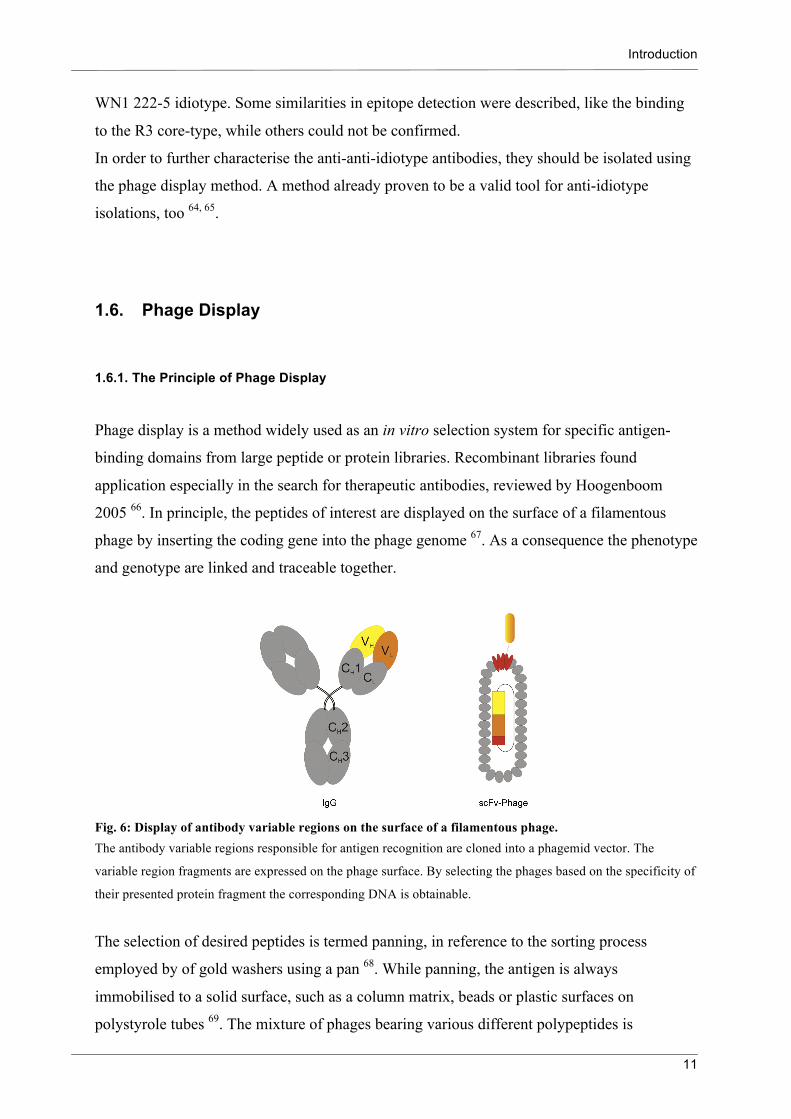
1.6. Phage Display
1.6.1. The Principle of Phage Display
Phage display is a method widely used as an in vitro selection system for specific antigen-
binding domains from large peptide or protein libraries. Recombinant libraries found
application especially in the search for therapeutic antibodies, reviewed by Hoogenboom
2005 66. In principle, the peptides of interest are displayed on the surface of a filamentous
phage by inserting the coding gene into the phage genome 67. As a consequence the phenotype
and genotype are linked and traceable together.
Fig. 6: Display of antibody variable regions on the surface of a filamentous phage. The antibody variable regions responsible for antigen recognition are cloned into a phagemid vector. The
variable region fragments are expressed on the phage surface. By selecting the phages based on the specificity of
their presented protein fragment the corresponding DNA is obtainable.
The selection of desired peptides is termed panning, in reference to the sorting process
employed by of gold washers using a pan 68. While panning, the antigen is always
immobilised to a solid surface, such as a column matrix, beads or plastic surfaces on
polystyrole tubes 69. The mixture of phages bearing various different polypeptides is

Introduction
12
incubated with the immobilised antigen, and non-binders are removed by a number of
washing steps. Subsequently the bound phages are eluated and amplified by infection
of E. coli.
Fig. 7: Principle of phage display. Phage display is used to select scFvs (single chain variable region fragments) with specific binding properties.
Libraries with several different scFv genes were constructed. They were packed into phage particles presenting
the scFvs on their surface. The scFv displaying phages are then applied for the panning procedure. Basically,
four steps were continuously repeated during panning: binding, washing, eluting and amplifying. In this way, the
phages bound to the antigen were multiplied and non-binders removed.
The M13 phage used in this work belongs to the filamentous phages 70. They are viruses
containing a circular single-stranded DNA genome encased in a long protein capsid cylinder.
Eleven genes are encoded in the M13 genome, three being involved in the DNA replication,
three in the assembly of phage particles, and five are part of the phage capsid. The pVIII is the
major coat protein, occasionally used for the fusion and display of relatively small peptides.

Introduction
13
For the display of polypeptides, pIII, in the phage life cycle responsible for infection of E. coli
through the F conjugative pilus, is more suitable. During an infection the capsid proteins are
integrated into the bacterial inner membrane and the phage DNA is translocated into the
cytosol. There it replicates via a double-stranded replicative form, usable for cloning and
sequencing during the phage display. Newly synthesised viral DNA is packed into phage
particles at the bacterial membrane until the end of the around 6700 bp ssDNA is reached.
The M13 phages do not kill their host during this process but the bacteria continue growing
with an approximately 50% slower generation time, observable in plaque formation.
Uncoupling of the gene replication and phage assembly by locating the corresponding genes
on separate plasmids greatly improves the genetic stability and screening of recombinant
polypeptide libraries 71. If the fusion gene is encoded on a separate genome lacking several
genes of the wild type phage, the vector is termed phagemid and a helper phage is necessary
for phage packing in order to complement the genes not encoded on the phagemid genome.
As is typical of plasmids, the phagemid bears an antibiotic resistance. Another plasmid
advantage is the opportunity to isolate abundant double-stranded plasmid DNA for cloning.
The pIII fusion proteins applicable for antigen binding range from small peptides to quite
large antibody F(ab) fragments. In this study single-chain fragments of the variable regions
(scFv) of antibodies are used. During the assembly of the phage display library, the variable
regions of the antibodies’ light and heavy chain are fused together by a linker, leading to a
single-chain fragment. The length of the inserted linker is critical for the display of the
scFv (Fig. 8). Long linkers (18 amino acids) lead to the expression of monomer scFvs on the
phage surface, while short linkers (7 amino acids) can lead to dimer formation, consequently
resulting in further antigen-binding sites 72.
Fig. 8: The scFv valency is decisively influenced by the linker length. The 18 amino acid long linker is pictured on the left. The two variable regions can interact as they prefer through
the flexibility enabled by the long linker. The 7 amino acid short linker is pictured on the right. The linker length
is too short to allow for flexibility and the preferred assembly of the variable regions. This promotes an
attachment of two scFvs together, leading to dimer formation.

Introduction
14
Furthermore, the choice of the vector system influences the quantity of scFv-derived binding
sites and, as a consequence, the valency of the phage considerably 73. Therefore the two
phagemids, used in this study, are described in more detail.
1.6.2. The Phagemid pComb3XSS
The pComb3 phagemid vectors were developed for the monovalent display of large peptides
on the surface of the filamentous phage M13 74. In this study the pComb3XSS vector was
used for phage display 75. This vector was developed for displaying antibody fragments as
fusion proteins to the minor coat protein pIII. The pComb3XSS pIII is amino-terminal
truncated (aa 230-406). This permits fusion protein display without diminished superinfection
by helper phages caused by the pIII amino-terminus.
Since the phagemid has no complete gIII it relies on the incorporation of helper phage pIII
into phage particles for subsequent infections. During the helper phage driven phage
assembly, the scFv-pIII fusion protein and the helper phage wt pIII compete for incorporation
into the phage particle. Thus 0-5 scFv-pIII fusion proteins are presented on the phage.
The expression of soluble scFv is possible due to an amber stop codon upstream to the
5’-terminus of the gIII. In E. coli nonsupressor strains the translation is terminated at the
amber stop codon leading to soluble scFvs.
Soluble scFv can be detected or isolated by the use of two different tags. The 6 x histidine tag
(His-Tag) at the 3’-end of the scFv and the decapeptide hemagglutinine tag (HA-tag) at the
3’-end of the His-tag.
SfiI restriction sites are used for scFv cloning into pComb3XSS, because the SfiI restriction
site is uncommon in genes. It cleaves two copies of its recognition site in one molecule. The
cleavage site is in a degenerated region, GGCCNNNN^NGGCC, enabling the ligation in the
correct orientation by the use of two different cleavage sequences.
5’-upstream to the SfiI cleavage site the sequence of an ompA leader is located (leader of the
E. coli outer membrane protein ompA). ScFvs are expressed as ompA fusion proteins,
transported to the periplasm, where the leader is removed and the scFv-pIII is incorporated
into the phage particle 76. The protein secretion from the cytoplasm into the periplasm
promotes the correct folding of scFvs due to the more oxidising environment and the
availability of proteins that catalyse disulfide bond formation 77.

Introduction
15
Fig. 9: Phagemid vector pComb3XSS. The pComb3XSS vector has phagemid-typical features such as two origins of replications (ori), one for the
plasmid replication (colE1) and one for the phage replication of ssDNA (f1) supplying material for the phage
particles. It carries an ampicillin resistance, used for the selection of infected bacteria. The multiple cloning site
is under the control of the lacZ promoter and provides different restriction sites, enabling cloning of both scFvs
and Fabs. For scFv cloning the double cleavage enzyme SfiI is beneficial. 5’- upstream to the SfiI site an ompA
leader is located. 3’- downstream two tags are placed, followed by an amber stop codon. The minor coat protein
pIII is N-terminal truncated and used for presentation of the scFvs.
1.6.3. The Phagemid pHen1
The pHen1 phagemid is, like the pComb phagemid, a vector for displaying peptides fused to
the minor coat protein pIII 78. The pHen1 has a pelB leader sequence 5’-upstream to a SfiI
cleavage site. For cloning at the 3’-end of the scFvs, a NotI restriction site is integrated,
followed by a c-myc tag and an amber stop codon. In contrast to the pComb vectors, the
pHen1 bears a complete gIII.
The use of the complete pIII, instead of the truncated one, leads to the difference that wt pIII
derived from the helper phage is no longer necessary for subsequent infections of bacteria
with phages. To allow superinfection of already infected bacteria, glucose must be present in
the culture medium.
The independence from helper phage pIII enables the use of hyperphage for phage packaging.
The hyperphage is a helper phage developed for the increase of presented scFv on the phage

Introduction
16
surface 79. It has the pIII phenotype of the helper phage M13K07 but lacks a functional gIII.
Consequently, it infects bacteria cells and packs phage particles like the helper phage but
produces no pIII on its own. Thus, all pIII incorporated into the phage originates from the
vectors scFv-pIII fusion genes. This increases the scFv valency considerably. It is assumed
that a high valency is preferred to monomeric or dimeric display concerning the isolation of
low affinity binders 80.
Fig. 10: Phagemid vector pHen1 The pHen1 vector has the phagemid-typical features like the pComb3XSS. In contrast to the pComb3XSS, it
carries a pelB leader sequence 5’- upstream to the SfiI site. 3’- downstream the scFv can be cloned through a
NotI restriction site, which is followed by a c-myc tag and an amber stop codon. The complete pIII is used for
presentation of the scFvs.
1.7. Mannose Binding Lectin
1.7.1. Structure and Function
Lectins are carbohydrate-binding proteins, excluding enzymes and Igs, which are involved in
the recognition of carbohydrates during innate immune response. They occur in plants,
animals, fungi, microbes and viruses.
In mammalian sera, Mannose-binding lectin (MBL) is found 81 belonging to the family of
collectins, which are calcium-dependent lectins (C-type lectins). MBL opsonises a wide range
of microorganisms and also interacts directly with phagocytes. Furthermore, it is involved in
complement activation via the MBL-associated serin protease (MASP), which shows

Introduction
17
proteolytic activity against the complement component C4, inducing the further complement
cascade.
Fig. 11: Valency differences of the carbohydrate recognition molecules from the innate and adaptive immune system. MBL of the innate immune system binds ligands through their trimeric CRDs (carbohydrate recognition
domains), which are further organised to higher oligomers (A). IgGs of the adaptive immune system bind
through two identical antigen-binding sites to their ligands (B). IgMs form mainly pentameric polymers each
unit composed of two identical binding sites (C). The single binding site of MBLs and IgMs reveals commonly a
low affinity for their ligands, which is compensated though high oligomerisations of the molecules. Thus, the
increase of avidity leads to significant binding and therefore immunologically active compounds.
In general the soluble pattern-recognition receptors of the innate immune system like MBLs
are functionally similar to the antibodies of the adaptive immune system. Collectins are
discussed as broad-spectrum anti-carbohydrate antibodies 82.
MBL especially has a much broader specificity then the name implies. Besides mannose,
other pathogen-associated carbohydrates are recognised, e.g. N-acetylglucosamine, glucose
and fucose. The binding of a single carbohydrate-recognition domain (CRD) to carbohydrates
as mannose and β-D-N-Acetylgalactosamin is determined by the 3-OH and 4-OH of the sugar
ring. Equatorial 3- and 4-OH are bound by the CRDs and the Ca2+ directly participates in the
bond formations 83.
Structurally, MBL is composed of sub-units formed by three identical polypeptide chains.
Each chain contains four regions, the cystein-rich N-terminal region, the collagenous domain,
the short α-helical neck region and the C-terminal CRD. The sub-units of MBL form higher
oligomers, varying in the degree of oligomerisation from dimers to hexamers (reviewed by
Hansen 84).
The oligomerisation is a crucial feature for binding. Concerning the rather weak binding of a
single CRD to a single ligand, it enables an affinity increase due to avidity effects. Thus the

Introduction
18
presentation of the multivalent ligands for the multiple CRDs of an MBL molecule plays an
important role 85.
Two different MBLs are described for mice, MBL-A and –C, whereas only one MBL is found
in humans. Studies of the genes encoding for MBL-A and –C as well as hMBL have revealed
that the hMBL sequence is more closely related to the MBL-C 86, supported by analysis of the
carbohydrate specificity 87. The difference between the two mouse MBLs is also emphasised,
where the MBL-A reveals a higher affinity for monosaccharides than does the MBL-C.
Serum MBL Monosaccharide
hMBL αMeMan, Man > GlcNAc > L-Fuc > ManNAc >> αMeGlc > Glc
MBL-A L-Fuc > αMeMan, Man, GlcNAc > αMeGlc > Glc > ManNAc >>> Fuc > Gal, GlcN > ManN
MBL-C αMeMan > Man > L-Fuc > ManNAc > GlcNAc >>> Glc > αMeGlc >>> ManN > Fuc
Tab. 1: Monosaccharide specificities of different MBLs 87. The 50% inhibition (IC50) values of the monosaccharides specificities are differentiated, whereby > denotes
more than 10%, >> more than 50% and >>> more than 100% higher IC50.
1.7.2. Interaction with Bacteria
The considerable difference in monosaccharide binding introduces the issue of potential
distinctions in pathogen recognition and further effects on the innate immune response.
However, it is necessary to consider the conformational effect and varieties induced by
oligosaccharides. Monosaccharides are not the natural binding partner of the MBL and it is
not possible to predict the binding from the identity of the terminal carbohydrates from
bacterial oligosaccharides alone 88. Therefore an examination of the recognition of pathogenic
and non-pathogenic bacteria is reasonable. The repeating, branched carbohydrates of bacteria
match the regular spacing between the MBL CRDs and are consequently recognised 89.
Furthermore, hMBL occurs in variant allels, which changes both the promotor and structural
region and influences the stability and serum concentration of the protein 90. Different serum
concentrations of hMBL alter the susceptibility to infections 91. MBL deficiencies seem to
increase susceptibility to some infections 92, 93, while tuberculosis yields contrary results 94.
For tuberculosis, a better uptake into macrophages is assumed where the Mycobacteria are

Introduction
19
able to survive. Crucial for the recognition of the bacteria are their cell-wall components
(reviewed by Brennan and Nikaido 95), in particular lipoarabinomannan (LAM) and mannose-
capped lipoarabinomannan (ManLAM) seem to play a major role in phagocytosis 96, 97.
Contrary to assumptions the mannose-cap is not exclusively present in pathogenic
mycobacteria, but also confirmed in M. bovis BCG 98, 99. Nevertheless, different tuberculosis
strains vary in their pathogenicity 100. In particular strains of the M. africanum 2 line are
assumed to differ significant in MBL recognition from M. tuberculosis strains (personal
communication with Dr. K. Walter).
To examine the role of MBL during the recognition of mycobacterial strains, a valid tool for
the MBL mycobacterial interaction was investigated in this study. The interaction of MBL
with the highly mannosylated mycobacterial cell-wall of various strains was studied, as were
the differences between the human and the two murine MBLs.

Objectives
20
2. Objectives
The aim of the first part of the thesis was to isolate anti-anti-idiotype scFvs by using the
phage display method. Therefore RNA material from four different anti-idiotype antibody
S81-19 immunised rabbits was used to construct scFv libraries. The scFv libraries were
screened for scFv binding to the anti-idiotype antibody, R3 core-type rough mutant E. coli
heat-killed bacteria and R3 oligosaccharide conjugated to BSA in order to receive anti-anti-
idiotype scFvs. The anti-anti-idiotype scFv should reveal the binding pattern characteristics of
the idiotype antibody WN1 222-5. The isolation and characterisation contribute to the
understanding of the application from the anti-idiotype approach to induce cross-reactive and
core-specific LPS-neutralising antibodies.
The aim of the second part of the thesis was to introduce a method in order to compare the
interaction of several bacteria with a relevant representative of the innate immune system, the
mannose-binding lectin (MBL). A method should be developed to compare various bacterial
strains as well as different forms of MBL. It ought to be examined whether the rather weak
binding of MBL molecules differ in binding to various mycobacterial strains. The result of
this investigation could provide new insights to the question as to whether cell-wall
composition differences recognised by MBL participate in an increased internalisation of the
bacteria into host cells.
For further MBL-studies an assay should be established to investigate in addition the
capability of MBL for complement activation.

Materials
21
3. Materials
Aqueous solutions were prepared with double deionized (Milli Q) water.
3.1. Bacteria Strains and LPS Conjugate
E. coli ER2537 [F′ lacIq Δ(lacZ)M15 proA+B+/fhuA2 supE thi Δ(lac-
proAB)Δ(hsdMS-mcrB)5], NEB (Frankfurt)
E. coli O111 oligosaccharide Conjugated via glutaraldehyde to BSA, kindly
provided by Prof. Dr. H. Brade (RC Borstel)
E. coli R3 core oligosaccharide Conjugated via glutaraldehyde to BSA (139 nmol
ligand/ml), kindly provided by Prof. Dr. H. Brade
(RC Borstel)
E. coli R3 rough type mutant
bacteria
Heat-killed bacteria, kindly provided by Prof. Dr. H.
Brade (RC Borstel)
E. coli TG1 F'traD36 lacIq ∆(lacZ) M15 proA+B+ /supE ∆(hsdM-
mcrB)5 (rk- mk- McrB-) thi ∆(lac-proAB), kindly
provided by Dr. U. Mamat (RC Borstel)
E. coli TOP10F’ F´lacIq Tn10 (TetR) mcrA Δ(mrr hsdRMS-mcrBC)
Φ80lacZΔM15 ΔlacX74 recA1 araD139 Δ(ara-
leu)7697 galU galK rpsL endA1 nupG. Invitrogen
(Karlsruhe)
M. africanum West African2
10476/01
Heat-killed and living bacteria are kindly provided by
Dr. S. Niemann (RC Borstel)
M. bovis Bovis 4258/00 Heat-killed and living bacteria are kindly provided by
Dr. S. Niemann (RC Borstel)
M. tuberculosis Beijing 1934/03 Heat-killed and living bacteria are kindly provided by
Dr. S. Niemann (RC Borstel)
M. tuberculosis Beijing 17919 Heat-killed bacteria kindly provided by Prof. Dr. U.
Schaible (RC Borstel)
M. tuberculosis Cameroon 1417/02 Heat-killed and living bacteria are kindly provided by
Dr. S. Niemann (RC Borstel)

Materials
22
M. tuberculosis H37Rv ATCC
27294
Heat-killed and living bacteria are kindly provided by
Dr. S. Niemann (RC Borstel)
M. tuberculosis Haarlem 9532/02 Heat-killed and living bacteria are kindly provided by
Dr. S. Niemann (RC Borstel)
M. tuberculosis X-type 4412/04 Heat-killed and living bacteria are kindly provided by
Dr. S. Niemann (RC Borstel)
3.2. Bacteriophages and Phagemids
M13K07 Helper phage M13K07 is a derivative of M13, which
carries the mutation Met40Ile in geneII, leading to a
decreased DNA replication. The p15A origin of
replication and the KanR gene from Tn903 were
inserted within the M13 origin of replication 101.
Invitrogen (Karlsruhe)
M13K07ΔpIII Hyperphage M13K07ΔpIII with a wild-type pIII
phenotype provides helper phage function in
packaging a common phage display phagemid, but
has no functional gIII 79. Progen (Heidelberg)
pComb3XSS Phagemid vector for the presentation of scFv-pIII
(aa230-406) fusion proteins on M13-Phages and the
expression of soluble scFv. Carrier of an AmpR 76.
pHen1 Phagemid vector for the presentation of scFv-pIII
fusion proteins on M13-Phages and the expression of
soluble scFv. Carrier of an AmpR. MRC, Cambridge
University
Materials
23
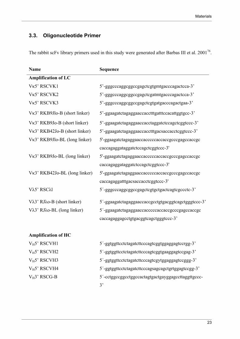
3.3. Oligonucleotide Primer
The rabbit scFv library primers used in this study were generated after Barbas III et al. 200176.
Name Sequence
Amplification of LC
Vκ5’ RSCVK1 5’-gggcccaggcggccgagctcgtgmtgacccagactcca-3’
Vκ5’ RSCVK2 5’-gggcccaggcggccgagctcgatmtgacccagactcca-3’
Vκ5’ RSCVK3 5’-gggcccaggcggccgagctcgtgatgacccagactgaa-3’
Vκ3’ RKB9Jlo-B (short linker) 5’-ggaagatctagaggaaccacctttgatttccacattggtgcc-3’
Vκ3’ RKB9Jo-B (short linker) 5’-ggaagatctagaggaaccacctaggatctccagctcggtccc-3’
Vκ3’ RKB42Jo-B (short linker) 5’-ggaagatctagaggaaccacctttgacsaccacctcggtccc-3’
Vκ3’ RKB9Jlo-BL (long linker) 5'-ggaagatctagaggaaccacccccaccaccgcccgagccaccgc
caccagaggataggatctccagctcggtccc-3'
Vκ3’ RKB9Jo-BL (long linker) 5'-ggaagatctagaggaaccacccccaccaccgcccgagccaccgc
caccagaggataggatctccagctcggtccc-3'
Vκ3’ RKB42Jo-BL (long linker) 5'-ggaagatctagaggaaccacccccaccaccgcccgagccaccgc
caccagaggatttgacsaccacctcggtccc-3'
Vλ5’ RSCλl 5´-gggcccaggcggccgagctcgtgctgactcagtcgccctc-3’
Vλ3’ RJλo-B (short linker) 5´-ggaagatctagaggaaccaccgcctgtgacggtcagctgggtccc-3’
Vλ3’ RJλo-BL (long linker) 5´-ggaagatctagaggaaccacccccaccaccgcccgagccaccgc
caccagaggagcctgtgacggtcagctgggtccc-3’
Amplification of HC
VH5’ RSCVH1 5´-ggtggttcctctagatcttcccagtcggtggaggagtccrgg-3’
VH5’ RSCVH2 5´-ggtggttcctctagatcttcccagtcggtgaaggagtccgag-3’
VH5’ RSCVH3 5´-ggtggttcctctagatcttcccagtcgytggaggagtccggg-3’
VH5’ RSCVH4 5´-ggtggttcctctagatcttcccagsagcagctgrtggagtccgg-3’
VH3’ RSCG-B 5´-cctggccggcctggccactagtgactgayggagccttaggttgccc-
3’

Materials
24
Overlap Extension
RSC-F (sense) 5’-gaggaggaggaggaggaggcggggcccaggcggccgag
ctc-3’
RSC-B (reverse) 5’-gaggaggaggaggaggagcctggccggcctggccactagtg-3’
Further Primers
Sequence
LH301-FP (scFv WN1 222-5
from pSJF8 into pComb3XSS)
5’- gggcccaggcggccgagctcgacatccagatgaaccagtc -3’
LH301-RP (scFv WN1 222-5
from pSJF8 into pComb3XSS)
5’- cctggccggcctggcctgaggagacggtgactgagg -3’
LH383-RP (scFv from
pComb3XSS into pSJF8)
5’- gggggatccagaagcgtagtccggaacgtcgtacgggtatgcacta
gtgactgacggagcctt -3’
NH1704-02 FP (pComb3XSS
primer for colony
PCR/sequencing)
5’- gctatcgcgattgcagtggc -3’
NH1765-02 RP (pComb3XSS
primer for colony
PCR/sequencing)
5’-gcccccttattagcgtttgccatc -3’
LH566-RP_NotI (scFv from
pComb3XSS into pHen1)
5’- gggggcggccgctgagaggacggtgaccag -3’
All oligonucleotide primers were obtained from Eurofins MWG (Ebersberg).

Materials
25
3.4. Primary and Secondary Antibodies
Anti-C4 Rabbit IgG, polyclonal, directed against complement factor
C4, Abcam (Cambridge, UK)
Anti-c-myc Mouse IgG1, monoclonal (9E10), directed against cmyc
(NEQKLISEEDLC), kindly provided by R. MacKenzie
(National Research Council Canada, Ottawa)
Anti-HA Rat IgG1, monoclonal, HRP-conjugated, directed against
hemagglutinin (YPYDVPDYA), Roche Diagnostics
(Mannheim)
Anti-HisG Mouse IgG2a, monoclonal, directed against 6xHis + Gly,
Invitrogen (Karlsruhe)
Anti-Mouse Goat IgG, polyclonal, AP-conjugated, Dianova (Hamburg)
Anti- Mouse Goat IgG, HRP-conjugated, Jackson (West Grove, USA)
Anti- Mouse Goat IgG, polyclonal, Alexa680-conjugated, Invitrogen
(Karlsruhe)
Anti- Mouse Goat IgG, polyclonal, IRDye800-conjugated,
Rockland/Biomol (Hamburg)
Anti-hMBL Mouse IgG1, monoclonal, Cl. D8.18, HRP-conjugated,
Hycult biotech (Uden)
Anti-MBL-A Rat IgG, monoclonal, Cl. 8G6, HRP-conjugated, Hycult
biotech (Uden)
Anti-MBL-C Rat IgG2a, monoclonal, Cl. 16A8, HRP-conjugated,
Hycult biotech (Uden)
Anti-p3 Mouse IgG2a, monoclonal, HRP-conjugated, directed
against M13 p3, NEB (Frankfurt)
Anti-p8 Mouse Ab, monoclonal, HRP-conjugated,Directed against
M13 p8, GE Healthcare (Freiburg)
Anti-rat Goat IgG, HRP-conjugated, Jackson (West Grove, USA)
S81-19 Mouse IgG, monoclonal, directed against WN1 222-5
mAb, kindly provided by L. Brade (RC Borstel)
WN1 222-5 Mouse IgG2a, monoclonal, originated in an immunisation
of NZB-mize with heat-killed S. minnesota and E. coli
strains 31. Kindly provided by L. Brade (RC Borstel).

Materials
26
3.5. Mannose Binding Lectins
rMBL-A 2077-MB R&D systems (Wiesbaden-Nordenstadt)
rMBL-C 2208-MB R&D systems (Wiesbaden-Nordenstadt)
rhMBL 2307-MB R&D systems (Wiesbaden-Nordenstadt)
hMBL Human complement serum, 51764, Sigma-Aldrich (Munich)
3.6. Antibiotics
Ampicillin β-lactam antibiotic, Roth (Karlsruhe)
Carbenicillin β-lactam antibiotic, Roth (Karlsruhe)
Kanamycinsulphate Aminoglycoside, Roth (Karlsruhe)
Polymyxin B sulphate Polypeptide, Sigma-Aldrich (Fluka) (Munich)
Tetracyclin Tetracycline, Serva (Heidelberg)
3.7. Enzymes, Polymerase Chain Reaction and Reverse Transcription Reagents
E. coli RNaseH 2 U/µl Invitrogen (Karlsruhe)
Super Script II 200 U/µl Invitrogen (Karlsruhe)
6 x DNA loading dye Fermentas (St. Leon-Rot)
DEPC-H2O Roth (Karlsruhe)
DNA ladder 1kb and 100 bp Fermentas (St. Leon-Rot)
dNTP mix, 0,1 M Analytic Jena (Jena)
Glycogen 20 mg/ml Roche Diagnostics (Mannheim)
Master Mix hot start Y Peqlab (Erlangen)
NotI Fermentas (St. Leon-Rot)
Oligo(dT) primer Invitrogen (Karlsruhe)
RNA purification system, peqGOLD TriFast Peqlab (Erlangen)
SfiI Fermentas (St. Leon-Rot)
T4 DNA Ligase Fermentas (St. Leon-Rot)

Materials
27
3.8. Kits
AP conjugate substrate kit Biorad (Munich)
Gel extraction, innuPREP Analytic Jena (Jena)
PCR Purification, QIAquick Qiagen (Hilden)
Plasmid Miniprep, QIAprep Spin Qiagen (Hilden)
3.9. Buffer, Staining and Developing Solutions
Agarose gel 1% 1 g agarose/100 ml TAE buffer.
Alkaline phosphatase (AP) buffer 0.1 M NaHCO3,
1 mM MgCl2 x 6 H2O in ddH2O.
pH adjustment with NaOH to 9.8.
BBS/Ca2+ 10 mM Diethylbarbituric acid sodium salt,
145 mM NaCl2,
10 mM CaCl2.
Blocking buffer – blots and ELISA 5% dry milk powder in TBS.
Blocking buffer – ELISA BSA 3% BSA in TBS.
Blocking buffer – ELISA BSA in BBS/Ca2+ 2% BSA in BBS/Ca2+.
Blocking buffer – ELISA casein 2.5% casein, 0.01% thimerosal in PBS.
Blocking buffer – ELISA casein - Tween 2.5% casein,
0.01% thimerosal,
0.05% Tween 20 in PBS
Citrate buffer 0.1 M Na-citrate.
pH adjustment with 2.5 M citric acid to 4.5.
Conservation solution 22% methanol,
5% glycine in ddH2O.
Coomassie Brillant Blue staining solution 0.2% coomassie,
40% methanol,
10% acetic acid (glacial) in ddH2O.
Coomassie destaining solution 20% methanol, 10% acetic acid (glacial) in
ddH2O.

Materials
28
EDTA solution 0.5 M Na2EDTA·2H2O in ddH2O.
pH adjustment to 8.0 with NaOH.
ELISA coating buffer 0.05 M NaHCO3.
pH adjustment with 0.05 M Na2CO3 to 9.2.
0.01% thimerosal.
Sterile through 0.2 µm filter.
Ethanol, 70% (v/v) 700 ml of technical grade ethanol dissolved
in ddH2O to 1 l.
Isopropyl-β,D-thiogalactoside (IPTG) 1 M IPTG in ddH2O. Stored at -20°C.
PBS 0.0027 M KCl,
0.137 M NaCl,
0.0081 M Na2HPO4,
0.0015 M KH2PO4 in ddH2O.
pH adjustment with HCl to 7.2.
Phage precipitation solution 20% PEG 8000,
2.5 M NaCl in ddH2O.
SDS-PAGE running buffer, 5 x 125 mM Tris,
960 mM glycine,
0.5% SDS in ddH2O.
Substrate solution - AP 10 ml AP buffer,
0.1 ml solution A,
0.1 ml solution B (AP conjugate kit).
Substrate solution - HRP 1 mg ABTS/ml 0.1 M citrate buffer pH 4.5,
25 µl 0.1% H2O2.
Syto24 Invitrogen (Karlsruhe)
TAE buffer 40 mM Tris,
0.1% acetic acid (glacial),
1 mM EDTA (pH 8.0) in ddH2O.
TBS (Tris-buffered saline) 50 mM Tris,
150 mM NaCl in ddH2O.
pH adjustment to 7.5 with HCl.
TBST 500 mM NaCl,
20 mM Tris,
0.05% Tween20 in ddH2O.

Materials
29
Transfer buffer 25 mM Tris,
192 mM glycine,
4% methanol in ddH2O.
pH adjustment to 8.2 with HCl.
3.10. Culture Media
All media for bacterial cultivation were sterilised by autoclaving for 20 min. at 121°C and
1,2 bar. The heat-labile components were filtered sterile by passing them through a 0.2 µm
filter.
LB (Luria Bertani) Ready to use, Roth (Karlsruhe)
M9 (minimal) medium 1 l 1 x M9 salts, autoclaved.
Added sterile
0.5 ml 2 M MgCl2,
0.1 ml 1 M CaCl2,
0.5 ml 10 mg/ml vitamin B1,
10 ml 20% glucose,
20 ml 20% peptone 5.
M9 salt solution 10 x 60 g Na2HPO4,
30 g KH2PO4,
10 g NH4Cl,
5 g NaCl, ddH2O to 1 l.
SB (super broth) 10 g MOPS (3(N-
morpholino)propanesulfonic acid),
30 g tryptone,
20 g yeast extract.
pH 7, ddH2O to 1 l.
SOC 20 g tryptone,
5 g yeast extract,
0.5 g NaCl.
pH 7, ddH2O to 1 l, autoclaved.

Materials
30
Added sterile 10 mM MgCl2,
20 mM glucose.
TB (Terrific Broth) 10 x 24 g yeast extract,
12 g tryptone,
4% glycerine in 100 ml ddH2O.
TOP Agar 1 l 1 x LB medium,
6 g agar-agar.
YT 2 x 16 g tryptone,
10 g yeast extract,
5 g NaCl.
Adjusted pH to 7 with NaOH, ddH2O to 1 l.
3.11. Reagents
2-mercaptoethanol Biorad (Munich)
2,2'-azino-bis(3-ethylbenzthiazoline-6-
sulphonic acid) (ABTS)
Sigma-Adrich (Munich)
3(N-morpholino)propanesulfonic acid
(MOPS)
Sigma-Aldrich (Fluka) (Munich)
Acrylamide/Bisacrylamide 30% Biorad (Munich)
Agar-agar Roth (Karlsruhe)
Agarose Eurogentec (Cologne)
Ammonium persulfate (APS), 10% Merck (Darmstadt)
Bovine serum albumin (BSA) PAA (Pasching, AT)
Bromophenol blue Serva (Heidelberg)
Casein Sigma-Adrich (Munich)
Chloroform Merck (Darmstadt)
Disodium hydrogen phosphate Sigma-Aldrich (Fluka) (Munich)
Dithiothreitol (DTT) Invitrogen (Karlsruhe)
Ethanol
Ficoll-Paque GE Healthcare (Amersham) (Munich)
Glycerol Merck (Darmstadt)

Materials
31
Glycine Serva (Heidelberg)
Glycogen Roche (Mannheim)
Hydrochloric acid Merck (Darmstadt)
Hydrogen peroxide Merck (Darmstadt)
Isopropanol
MAPSII elution buffer, pH 6.8 Biorad (Munich)
Methanol Merck (Darmstadt)
N,N,N`,N`-Tetramethylethylendiamine
(TEMED)
Biorad (Munich)
Non fat dry milk Biorad (Munich)
Polyethylene glycol 8000 Roth (Karlsruhe)
Potassium chloride Merck (Darmstadt)
Protein loading dye 6 x Fermentas (St. Leon-Rot)
Protein prestained ladder Fermentas (St. Leon-Rot)
PVDF membrane Millipore (Billerica, USA)
SDS Biorad (Munich)
Sodium azide Merck (Darmstadt)
Sodium chloride Roth (Karlsruhe)
Sodium citrate dihydrate Merck (Darmstadt)
Sodium hydrogen carbonate Merck (Darmstadt)
Sodium hydroxide Merck (Darmstadt)
Stripping solution Thermo Scientific (Rockford, IL, USA)
Sulfuric acid, 95-97% Merck (Darmstadt)
Thimerosal Roth (Karlsruhe)
TMB USB (Staufen)
Tris Roth (Karlsruhe)
Tryptone Roth (Karlsruhe)
Tryptone no. 5 Roth (Karlsruhe)
Tween 20 Biorad (Munich)
Western blot filter paper Schleicher and Schuell (Dassel)
Yeast extract Roth (Karlsruhe)

Materials
32
3.12. Laboratory Equipment
96-well plate NUNC MaxisorpTM, Thermo Scientific (Rockford,
IL, USA)
Blot cell Mini-Trans Biorad (Munich)
Centrifuge bottles, 0.02 l Herolab (Wiesloch)
Centrifuge bottles, 0.25 l Herolab (Wiesloch)
Centrifuge bottles, 0.5 l Herolab (Wiesloch) Centrifuge bottles, 1 l Polypropylene, Beckman Coulter (Krefeld)
Centrifuge Tabletop Rotanta 460R, Hettich (Tuttlingen)
Centrifuge tube, 50 ml CentriStar, Corning (Amsterdam, NL)
Centrifuge, Avanti J-26 XP
Rotor: JLA 8100, JA-10, JA-14 and
JA-20
Beckman Coulter (Krefeld)
Centrifuge, Biofuge pico Heraeus
DNA gel electrophoresis apparatus Biorad (Munich)
Electrophoresis power supply Biorad (Munich)
Electroporation apparatus,
Micro Pulser (0.2 cm cuvettes)
Biorad (Munich)
ELISA reader Tecan
Gel documentation system Biorad transilluminator, software Quantity One 4.6.1
(Munich)
Heat block Duotherm, Biometra (Göttingen)
Micro tubes, 1.5 ml and 2 ml Sarstedt (Nümbrecht)
Microscope Eclipse TS100 Nicon (Melville, USA)
Mini-Trans Blot cell Biorad (Munich)
Needles 0.9 x 40 mm Becton Dickinson (Heidelberg)
Odyssey detection system Li-cor, (Lincoln, NE, USA)
PCR reaction tubes 0.2 ml, lid chain Sarstedt (Nümbrecht)
Pipette aid Pipetboy, Integra Bioscience (Fernwald)
Pippette tips Sarstedt (Nümbrecht)
SDS-PAGE electophoresis chamber Miniprotean II, Biorad (Munich)
Shake incubator Certomat BS-1, B. Braun biotech international
(Sartorius) (Göttingen)
Materials
33
Spectrophotometer NanoDrop, ND-1000, Peqlab (Erlangen)
Syringe Filter, 0.2 µm Thermo Scientific (Nalgene) (Rockford, IL, USA)
Syringe, 2 ml Becton Dickinson (Heidelberg)
Thermocycler Biometra (Göttingen)
Ultrasonic bath Sonorex, Bandelin (Berlin)
Waterbath UC, Julabo (Seelbach)

Methods
34
4. Methods
4.1. Phage Display
The phage display procedures were performed according to Barbas III, Burton et al. 2001.
They comprise as main steps the construction of a scFv library from immunised rabbits and
the selection of distinct binding scFv-phages from this library.
4.1.1. Construction of ScFv Libraries from Immunised Rabbits
The immunisation was performed by Dr. L. Brade. Four chinchilla bastard rabbits (charles
river) were immunised as described in Tab. 2 63.
Rabbit/Material collection Day Amount [µg] Immunogen
436 1 100 S81-19 IgG (+CFA) into lymphnotes
42, 326 150 S81-19 F(ab)2 (+IFA) subcutaneous
Blood, spleen, bone marrow 632
438 1 100 S81-19 IgG (+CFA) into lymph nodes
42, 487 150 S81-19 F(ab)2 (+IFA) subcutaneous
720 130 S81-19 F(ab)2 (+IFA) intravenous
Blood, spleen, bone marrow 728
437 1 100 S81-19 F(ab)2 (+CFA) into lymph nodes
42, 326 150 S81-19 F(ab)2 (+IFA) subcutaneous
Blood, spleen, bone marrow 636
439 1 100 S81-19 F(ab)2 (+CFA) into lymph nodes
42, 487 150 S81-19 F(ab)2 (+IFA) subcutaneous
720 130 S81-19 F(ab)2 (+IFA) intravenous
Blood, spleen, bone marrow 728
Tab. 2: Immunisation pattern of four rabbits immunised with mAb S81-19 anti-idiotype.

Methods
35
4.1.1.1. RNA Isolation
Total RNA of the anit-idiotype immunised rabbits was isolated from spleen, bone marrow and
peripheral blood lymphocytes.
Spleen and bone marrow of the rabbits were homogenised separately in a glass
homogenisator, to lyse the cells, and inactivate RNAses 10 ml peqGOLD TriFast were added.
The mixture was divided in 1.5 ml portions in 2 ml tubes. Afterwards 300 µl chloroform were
added to each resuspension, mixed and incubated for 10 min. at room temperature. It was
centrifuged with 16200 x g for 10 min. at room temperature, that 3 phases were formed, a red
phenol chloroform phase, an interphase with DNA and proteins and a colourless hydrous
phase on the top bearing the RNA. The colourless phase was collected and transferred into a
1.5 ml reaction tube. 750 µl isopropanol were added, mixed and incubated for 15 min. at
room temperature. The tubes were centrifuged with 16200 x g for 10 min. at 4°C and the
isopropanol supernatant was discarded with a pipette. Followed by 2 washing steps, the
pellets were resuspended in 500 µl 70% ethanol in DEPC-ddH2O and centrifuged with 16200
x g for 10 min. at 4°C. The pellets were air dried for 10 min. at room temperature and
afterwards resuspended in 100 µl DEPC-ddH2O in total for each sample.
The isolation of the RNA from rabbit peripheral blood lymphocytes was made using 9 ml
whole blood, collected from the rabbit ear vein. 1 ml 3.8% Na-citrate was provided in the
collection tube. The citrate anticoagulated whole-blood cells were carefully given above 10
ml Ficoll and centrifuged at 1500 x g for 1 h at room temperature. The mononuclear cell layer
was collected from between the Ficoll red blood cells and the plasma. 20 ml PBS was added
to wash the cells and centrifuged with 1500 x g at 4°C for 10 min. The resulting pellet was
resuspended in 5 ml PBS and the cells were counted using CASY cell counter program 1 for
human mononuclear cells.
The cell suspension was divided in 1200 µl portions and centrifuged with 2100 x g for 10
min. at 4°C. To resuspend the pellets 1 ml peqGOLD TriFast was used. Subsequently a
phenol-chloroform extraction was performed as described above.

Methods
36
4.1.1.2. Reverse Transcription
The isolated RNA was reverse transcribed to single- stranded cDNA. 20 µg RNA were
incubated with 4 µg Oligo(dT)12-18 primer and DEPC-ddH2O was added to a total volume of
100 µl. It was incubated for 10 min. at 70°C, cooled down 1 min. on ice and centrifuged to the
bottom of the tube. As a reaction mix 32 µl 5 x first strand buffer, 8 µl 10 mM dNTP mix
(each 10 mM) and 16 µl 100 mM dithiothreitol (DTT) were added and incubated for 5 min. at
42°C. 8 µl of 200 U/µl Super Script II were added and incubated for 1 h at 42°C, then heated
for 15 min. to 70°C, cooled down for 1 min. on ice and centrifuged to the bottom of the tube.
Afterwards 8 µl of 2 U/µl E. coli RNaseH were added and incubated for 20 min. at 37°C. The
library cDNA was stored at -20°C.
4.1.1.3. Construction of ScFvs
In a first round of polymerase chain reaction (PCR) rabbit variable light and heavy chain
genes were amplified from cDNA by using VL and VH primers described by Barbas et al. 76.
The VL primers are provided with a sense primer extension, including a SfiI restriction site
and a reverse primer containing a short or long linker sequence tail. Whereas VH primers have
sense primers containing a linker sequence, corresponding to the VL reverse primer sequence
tail, used for linking both in a second PCR to one fragment together. The VH reverse primers
contain a sequence tail with a SfiI restriction site.
Fig. 12: Amplification of the antibody variable region genes. Vκ, Vλ and VH were amplified using various primer combinations. Simultaneously the linkers were attached to
one site and to the other a SfiI restriction site was introduced.

Methods
37
The following conditions were used for the different primer combinations:
PCR Mixture
~ 0.5 µg cDNA
60 pmol sense primer
60 pmol reverse primer
50 µl master mix hot start Y
100 µl total volume with sterile ddH2O
Cycle Step Temperature Time No. of Cycles
Initial Denaturation 95°C 5 min. 1
Denaturation 94°C 15 sec.
30 Annealing 56°C 30 sec.
Extension 72°C 90 sec.
Final Extension 72°C 10 min. 1
The amplified genes were purified using a PCR purification kit according to the
manufacturers protocol and quantified by measuring the absorbance at a wavelength of 260
nm in the NanoDrop photometer.
In a second round of PCR, called overlap extension PCR, the VL and VH PCR products were
mixed in a 1:1 ratio, to join them randomly via their adhered linkers (Fig. 13). They were
amplified with primers recognising the sequence ends that were generated during the first
PCR round.
Fig. 13: Overlap extension PCR. The sequence tails generated in the first round of PCR were used to amplify the combined variable region gene
fragments.

Methods
38
Cycle Step Temperature Time No. of Cycles
Initial Denaturation 95°C 5 min. 1
Denaturation 94°C 15 sec.
25 Annealing 56°C 30 sec.
Extension 72°C 120 sec.
Final Extension 72°C 10 min. 1
The resulting short and long linker scFv PCR products were combined in 2 separated pools
and ethanol precipitated. Followed by purification of the DNA fragment using a 1% agarose
gel for the separation of different sized fragments. The band with the expected size of 700 bp
was sliced from the gel and regained with the use of a gel extraction kit according to the
manufacturers protocol.
4.1.1.4. Restriction Digest
The purified overlap extension product and the phagemid vector pComb3XSS were prepared
for cloning by a restriction digest with the enzyme SfiI.
Mixture for Insert Digest Mixture for Vector Digest
10 µg PCR product 20 µg pComb3XSS vector
360 U SfiI enzyme 120 U SfiI enzyme
20 µl 10 x NEB2 buffer 20 µl 10 x NEB2 buffer
100 µl total volume with sterile ddH2O 100 µl total volume with sterile ddH2O
PCR Mixture
~ 0.1 µg amplified VL DNA
~ 0.1 µg amplified VH DNA
60 pmol sense primer
60 pmol reverse primer
50 µl master mix hot start Y
100 µl total volume with sterile ddH2O

Methods
39
Both digests were incubated for 16 h at 50°C. The digested DNA fragments were separated
by agarose gel electrophoresis. The PCR product (~700 bp), vector (~3400 bp) and stuffer
(~1600 bp) fragment were retrieved using a gel extraction kit.
The obtained DNA fragments were quantified by measuring the OD at 600 nm in a
spectrophotometer.
4.1.1.5. Ligation
In ligation reactions the T4-DNA-ligase of the E. coli phage T4 was used to combine, under
ATP consumption, covalently the ends of DNA fragments.
Firstly small scale ligations were performed to verify the suitability of the DNA fragments for
the library ligation.
In one reaction the ligation efficacy of the vector was tested by ligating the stuffer fragment as
a control insert back into the sliced vector.
Ligation Mixture
0.14 µg pComb3XSS vector
0.14 µg pComb3XSS stuffer
2 µl 10 x T4 ligase buffer
1 µl T4 ligase
20 µl total volume with sterile ddH2O
In another reaction the vector was tested for self ligation properties.
Ligation Mixture
0.14 µg pComb3XSS vector
2 µl 10 x T4 ligase buffer
1 µl T4 ligase
20 µl total volume with sterile ddH2O

Methods
40
In a third reaction the PCR product was joined to the vector in a small scale test.
Ligation Mixture
0.14 µg pComb3XSS vector
0.07 µg PCR product
2 µl 10 x T4 ligase buffer
1 µl T4 ligase
20 µl total volume with sterile ddH2O
The reactions were incubated for 16 h at room temperature. 1 µl of each reaction was added to
50 µl electrocompetent E. coli ER2537 and transformed by electroporation (program Ec2)
into the cells. The transformed cells were internalised in 1 ml prewarmed (37°C) SOC
medium and incubated for 1 h at 37°C with 220 rpm. Afterwards the culture was diluted 10-
fold and 100-fold and plated on LB plates with carbenicillin (0.1 mg/ml). The plates were
incubated for 16 h at 37°C, the colonies counted, and the number of transformants per µg
vector DNA was calculated.
The large scale library ligation was performed to get a high yield of different rabbit
scFv genes.
Ligation Mixture
1.4 µg pComb3XSS vector
0.7 µg PCR product
20 µl 10 x T4 ligase buffer
10 µl T4 ligase
200 µl total volume with sterile ddH2O
It was incubated for 16 h at room temperature. Afterwards the DNA was ethanol precipitated
and the DNA pellet resuspended in 15 µl sterile ddH2O.

Methods
41
4.1.1.6. Transformation of the Phage Libraries
300 µl electrocompetent E. coli ER2537 were melted on ice, the resuspended 15 µl DNA was
added and the mixture proportioned to 3 cooled 0.2 mm cuvettes. The DNA was transformed
by electroporation (program Ec2), and the cuvette was immediately afterwards flushed with 5
ml prewarmed (37°C) SOC medium. The bacteria culture was incubated for 1 h at 37°C with
220 rpm. Then 10 ml SB medium and 3 µl cabenicillin (100 mg/ml) were added.
2 µl of this culture were diluted in 200 µl SB and 10 µl and 100 µl plated on LB agar
containing 0.1 mg/ml carbenicillin, to determine the titer of phage forming units. The agar
plates were incubated 16 h at 37°C.
The 15 ml culture was incubated for 1 h at 37°C with 220 rpm, then 4.5 µl carbenicillin
(100 mg/ml) were added, and it was incubated for another hour. 2 ml M13K07 helper phages
were added, the culture was transferred to a 500 ml polypropylene centrifuge bottle, 183 ml
SB medium and 92.5 µl carbenicillin (100 mg/ml) were added and the culture was incubated
for 2 h at 37°C with 220 rpm. Then 280 µl of kanamycin were added, and it was further
incubated for 16 h.
The culture was centrifuged with 2830 x g for 15 min. at 4°C and the supernatant transferred
into a 500 ml centrifuge bottle. For phage precipitation 8 g (4%) of PEG-8000 and 6 g (3%)
NaCl were added and dissolved by shaking with 220 rpm for 5 min. at 37°C. For a better
precipitation the culture was then cooled for 30 min. on ice.
The phages were precipitated by centrifuging with 14333 x g for 15 min. at 4°C. The
supernatant was discarded and the pellet dried for 10 min. in the inverted centrifuge bottle, to
remove remaining precipitation liquid. 2 ml of 1% BSA in TBS was used to resuspend the
phage pellet. The 2 ml were transferred to a 2 ml tube and centrifuged with 16200 x g for
5 min. at 4°C. Then the supernatant was passed through a 0.2 µm filter into a new 2 ml tube.
The phage preparation was stored at 4°C.
Methods
42
4.1.2. Panning on Immobilised Antigens
The constructed rabbit scFv libraries were used for the selection of specific binders. Therefore
different variations of the following principle procedure were performed.
Coating: For the first round of panning 2 single wells were coated with 1 of 3 different antigens.
1. 50 pmol/well of E. coli R3 core rough type LPS conjugated via glutaraldehyde to BSA 2. 2 µg/well of E. coli R3 core rough type heat-killed bacteria 3. 1 pmol/well of S81-19 mAb
As coating buffer 0.05 M carbonate buffer was used, and 50 µl/well antigen solution was
incubated 16 h at 4°C. In the first round of panning two wells were used, in the subsequent
rounds one.
Blocking: The well was blocked with 150 µl of one of three different blocking buffers:
3% BSA in TBS, 2.5% casein in PBS or 5% milk powder in TBS and incubated for
1 h at 37°C.
Phage binding: The blocking solution was taped on a paper towel, 50 µl of freshly prepared
phages were added to the well and incubated for 2 h at 37°C.
Washing: The phage solution was discarded from the well, and 150 µl 0.5% Tween20 in TBS
were added. It was resuspended 5 times, then incubated for 5 min., and subsequently the
washing solution was taped out on a paper towel. This washing step was repeated in the first
round of panning 5 times, in the later rounds 8, 10, 12 and 15 times.
Elution: Afterwards the bound phages were acidic eluted with 50 µl 0.1% BSA in 100 mM
glycin-HCl pH 2.2. The acid was incubated for 10 min., then resuspended 10 times, and the
eluate was transferred to a 1.5 ml tube containing as neutralisation solution 3 µl 2 M Tris.
Amplification: The eluted phages were added to 2 ml log-phase E. coli ER2537 culture and
incubated for 15 min. at room temperature. 6 ml SB medium and 1.6 µl carbenicillin
(100 mg/ml) were added to the culture.
To determine the phage output titer after each panning round, 2 µl of the 8 ml culture were
diluted in 200 µl SB medium, 10 µl and 100 µl of this dilution were plated on LB agar
containing 100 µg/ml carbenicillin and incubated overnight at 37°C.

Methods
43
The 8 ml culture was incubated for 1 h at 37°C with 230 rpm, then 2.4 µl carbenicillin
(100 mg/ml) were added and incubated again for 1 h. 1 ml of the helper phages M13K07 was
added for phage particle packaging, together with 91 ml SB medium and 46 µl carbenicillin
(100 mg/ml). It was incubated for 2 h at 37°C with 230 rpm, then 140 µl kanamycin
(50 mg/ml) were added and again incubated overnight.
For the preparation of the phages the culture was centrifuged for 15 min. with 2830 x g at
4°C. To the supernatant 4 g PEG 8000 and 3 g NaCl were added, dissolved for 10 min. with
230 rpm at 37°C and cooled then for 30 min. on ice. It was centrifuged for 15 min. with
14333 x g at 4°C and the precipitation solution carefully removed from the phage pellet. The
pellet was resuspended in 2 ml 1% BSA in TBS and centrifuged again for 5 min. with 16200
x g at 4°C. The phage supernatant was filtered through a 0.2 µm filter and stored at 4°C.
The phage input titer for the next panning round was determined by using 1 µl of a 10-9
dilution of the filtered phage solution in 50 µl log-phase E. coli ER2537 culture. It was
incubated for 15 min. at room temperature, subsequently plated on LB agar containing
100 µg/ml carbenicillin and incubated overnight at 37°C. The colonies were counted and the
phage titer calculated.
4.1.3. Phage Reamplification
To avoid protease cleavage effects of the scFvs on phage selection, it is necessary to use only
freshly prepared phages for panning. On the other hand reamplification can lead to a
decreased complexity of the library and should not be repeated frequently.
10 µl of the phage preparation were used to inoculate a 50 ml E. coli ER2537 culture with an
OD600 of ~1 grown in a 500 ml centrifuge bottle. The culture was incubated for 15 min. at
room temperature, then 10 µl of 100 mg/ml carbenicillin were added and incubated for 1 h at
37°C with 220 rpm. To determine the amount of phage infected bacteria, 10 µl of a 10-4
dilution were plated on LB agar plates, containing 0.1 mg/ml carbenicillin. They were
incubated overnight at 37°C, and the amount of colony forming units were counted.
To the 50 ml culture 15 µl 100 mg/ml carbenicillin were added and incubated for an
additional hour. Then 2 ml M13K07 helper phages, 148 ml SB medium and 75 µl 100 mg/ml
carbenicillin were added and incubated for 2 h at 37°C with 220 rpm. 280 µl 50 mg/ml
kanamycin were added and incubated for additional 16 h.
The phage rescue was performed as described for the library transformation.

Methods
44
4.1.4. Subcloning into the pHen1 Phagemid
The scFv library, derived from S81-19 (anti-idiotype) immunised rabbit 437, was subcloned
into the pHen1 phagemid vector.
4.1.4.1. Plasmid Preparation
Therefore 10 ml E. coli ER2537 log-phase culture in SB medium were infected with 10 µl of
the phage preparation from the 437 library in the pComb3XSS vector. They were allowed to
infect the cells for 15 min. at room temperature and afterwards they were incubated for
30 min. at 37°C with shaking at 230 rpm. Then 5 µl 100 mg/ml carbenicillin were added, and
it was incubated under the same conditions for 1 h. Again 5 µl 100 mg/ml carbenicillin were
added and it was incubated under the same conditions overnight. The plasmid preparation was
performed using a plasmid miniprep kit according to the manufacturers protocol.
4.1.4.2. Appending of a NotI Site
For the appending of a NotI restriction site on the 3’- end of the scFv, a PCR master mix was
prepared.
PCR Mixture
~ 4 µg plasmid DNA
60 pmol NH1704-02_FP primer
60 pmol LH566-RP_NotI primer
800 µl master mix hot start Y
1600 µl total volume with sterile ddH2O
The mixture was given in 0.1 ml portions to 0.2 ml PCR tubes. The thermocycle conditions
were as described for scFv construction.
A PCR purification kit was used, according to the manufacturer protocol, to concentrate the
PCR samples to a volume of 80 µl. Then an agarose gel electrophoresis was performed, and
the expected scFv of around 750 bp was sliced from the gel. For the gel extraction a gel
extraction kit was used according to the manufacturers protocol.
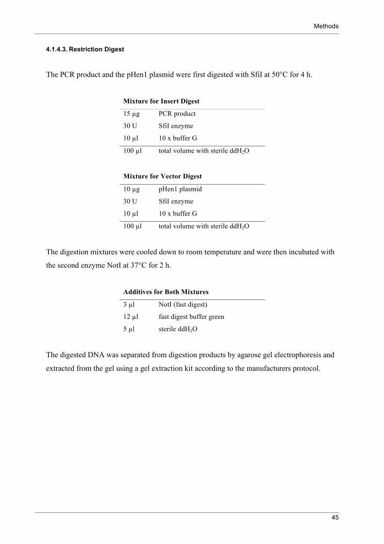
Methods
45
4.1.4.3. Restriction Digest
The PCR product and the pHen1 plasmid were first digested with SfiI at 50°C for 4 h.
Mixture for Insert Digest
15 µg PCR product
30 U SfiI enzyme
10 µl 10 x buffer G
100 µl total volume with sterile ddH2O
Mixture for Vector Digest
10 µg pHen1 plasmid
30 U SfiI enzyme
10 µl 10 x buffer G
100 µl total volume with sterile ddH2O
The digestion mixtures were cooled down to room temperature and were then incubated with
the second enzyme NotI at 37°C for 2 h.
Additives for Both Mixtures
3 µl NotI (fast digest)
12 µl fast digest buffer green
5 µl sterile ddH2O
The digested DNA was separated from digestion products by agarose gel electrophoresis and
extracted from the gel using a gel extraction kit according to the manufacturers protocol.

Methods
46
4.1.4.4. Ligation and Transformation
The ligation mixture was incubated for 4 h at room temperature.
Ligation Mixture
1 µg pHen1 vector
1 µg PCR product
10 µl 10 x T4 ligase buffer
5 µl T4 ligase
100 µl total volume with sterile ddH2O
Afterwards the DNA was precipitated with ethanol and transformed into electrocompetent
E. coli TG1 cells via electroporation, as described in detail for the pComb3XSS library
transformation.
4.1.5. Amplification of Single Phages
Single phages, amplified from infected E. coli colonies from the titer determination plates,
were tested for their antigen binding in ELISA. Therefore single colonies were used to
inoculate 5 ml SB medium containing 50 µg/ml carbenicillin and incubated for 5 h at 37°C
with shaking at 250 rpm. 50 µl M13K07 helper phage was added and incubated for another
2 h. Then 7 µl kanamycin (50 mg/ml) were added and the cultures incubated overnight at
37°C with shaking at 250 rpm.
Phage harvesting was implemented by centrifugation with 10916 x g for 15 min. at 4°C. The
supernatant was mixed in a 5:1 ratio with 20 % PEG and 2.5 M NaCl, and the phages were
precipitated while cooling them for 30 min. on ice. Afterwards it was centrifuged for 10 min.
with 16200 x g at 4°C and the supernatant was carefully removed with a pipette. The pellet
was resuspended in 50 µl TBS.

Methods
47
4.1.6. Soluble Expression of ScFvs
For the infection with single phages 10 ml SB medium, containing 10 µg/ml tetracycline,
were inoculated with 10 µl of a TOP10F’ bacterial stock. It was incubated for 2 h at 37°C
with 230 rpm. Then 10 µl of the single phage preparation were added and allowed to infect
during 20 min. without shaking at room temperature. 10 µl carbenicillin (100 mg/ml) were
added, and it was incubated for 1 h at 37°C with 230 rpm. 100 ml SB medium, 45 µl
carbenicillin (100 mg/ml) and 100 µl 1 M IPTG for the induction of the scFv expression were
added. It was incubated overnight at 37°C with 230 rpm. The culture was centrifuged for
15 min. at 4°C with 2830 x g and the supernatant stored at 4°C.
The cell pellet was weighed and twice the volume MAPSII elution buffer, and 1/100 volume
polymyxin B were added. It was incubated during gently mixing for 4 h at 4°C. Then it was
centrifuged for 15 min. at 4°C with 16200 x g and the scFv containing supernatent stored
at 4°C.
4.1.7. Preparation of Helper Phages
Packaging of pComb3 phages requires the support of helper phages, providing the proteins for
replication and assembly, which are lacking in the pComb3 phagemids. To receive an
appropriate amount of helper phages for the library panning, they were amplified and
concentrated by precipitation.
Single M13K07 helper phages were obtained by inoculating 2 ml of SB medium with 2 µl
electrocompetent E. coli ER2537 (stored at -70°C) and incubated for 90 min. at 37°C with
230 rpm. 1 µl of a 10-6, 10-7 and 10-8 dilution of a 1 x 10¹¹ pfu/ml M13K07 preparation was
used to inoculate 50 µl of E. coli culture. They were incubated for 15 min. at room
temperature, then 3 ml melted top agar (~ 40°C) were added and spread over a LB agar plate.
The plates were incubated overnight at 37°C.
M13K07 infected cells growing slower than non-infected cells lead to a plaque formation.
A single plaque was picked with a pipette tip and used for infection of a 10 ml E. coli ER2537
log-phase culture. It was incubated for 2 h at 37°C with 230 rpm. 5 ml of the culture were
used to inoculate 250 ml SB medium, 350 µl kanamycin (50 mg/ml) were added and it was
incubated for 16 h at 37°C with 230 rpm. Afterwards the culture was centrifuged for 15 min.
Methods
48
with 2830 x g at 4°C and the supernatant was transferred to 50 ml tubes. The tubes were
incubated for 20 min. in a 70°C water bath and then centrifuged like before.
240 ml phage supernatant were precipitated by adding 9.6 g PEG 8000 and 7.2 g NaCl, the
components were solved for 10 min. at 37°C with 220 rpm and cooled for 30 min. on ice. It
was centrifuged for 15 min. with 14333 x g at 4°C and the supernatant carefully removed
from the pellet, to avoid precipitating residues. The pellet was resuspended in 20 ml SB
medium and centrifuged for 10 min. with 16200 x g at 4°C. The phage supernatant was
filtered through a 0.2 µm filter and stored at 4°C.
The phage titer was determined by the same procedure as described to get single phage
plaques.
4.1.8. Preparation of Electrocompetent E. coli
For the generation of electrocompetent E. coli it is necessary to prepare high resistance media
with a low ionic strength. Therefore 5 ml of SB medium were inoculated with a single colony
grown on M9 agar plate for 16 h. This preculture was incubated for 16 h at 37°C with 220
rpm. 2.5 ml of the culture were used to inoculate 500 ml SB in a 2 l flask supplemented with
10 ml 20% glucose and 2.5 ml 2 M MgCl2. It was incubated at 37°C with 200 rpm until an
OD600 of ~0.8 was reached. All following steps were performed on ice.
The bacteria culture was proportioned on 500 ml centrifuge bottles. It was centrifuged for
20 min. with 2830 x g at 4°C, and the supernatant was discarded. Each pellet was resuspended
in 25 ml precooled 10% glycerol combined with another pellet, and 10% glycerol was added
to a total volume of 200 ml. It was centrifuged as before and the pellet was resuspended again
in 200 ml 10% glycerol. After another centrifugation the pellet was resuspended in 13 ml 10%
glycerol and centrifuged for 15 min. with 11000 x g at 4°C. The pellet was resuspended in
2.5 ml 10% glycerol and afterwards in 100 µl volumes proportioned in 1.5 ml screw-cap
tubes. The bacterial suspension was frozen with liquid nitrogen and stored at -70°C.

Methods
49
4.2. Binding Assays
4.2.1. ELISA
Bacteria were immobilised on Nunc PolySorb plates with U bottom shaped wells by
incubation overnight at 4°C with 50 µl/well bacteria solution. The plates were blocked with
200 µl 2% BSA in BBS/Ca2+ for 1 h at 37°C at 110 rpm. The wells were washed 2 times with
BBS/Ca2+.
50 µl rMBL dilution was added and titrated through the following wells. The plate was
incubated for 1 h at 37°C with 110 rpm. The wells were washed 2 times with BBS/Ca2+. 50 µl
of 1 µg/ml anti-hMBL, anti-MBL-A or anti-MBL-C antibodies in blocking buffer were added
and incubated for 1 h at 37°C with 110 rpm. The wells were washed 2 times with BBS/Ca2+.
50 µl of 1 µg/ ml of horseradish peroxidase conjugated anti-mouse antibody or anti-rat
antibody in blocking buffer were added and incubated for 1 h at 37°C with 110 rpm. The
wells were washed 3 times with BBS/Ca2+. 50 µl of ABTS substrate solution were added to
the wells and incubated for 30 min at 37°C with 110 rpm. The optical density was measured
in an ELISA reader at 405 nm.
4.2.2. Phage ELISA
To monitor whether the amount of phages carrying a scFv of interest increases during the
panning rounds, it is possible to perform ELISAs directly with the phages using an anti-M13
antibody. The following ELISA procedure was also used for the detection of amplified single
phages.
In general, the same concentration of antigen in 50 µl/well coating buffer was used to coat a
96-well MaxiSorp plate, as it was used in the previous panning. It was incubated overnight at
4°C.
The antigen solution was discarded by tapping the plate on a paper towel. To block remaining
binding sites on the polystyrene wells, 200 µl/well 2.5% casein in PBS were added and
incubated for 1 h at 37°C. Phages in different concentrations were diluted in blocking buffer,
and 50 µl/well were incubated for at least 1 h at 37°C. The phage solution was removed and
the wells washed with either PBST or PBS between 3 and 10 times, adjusted for the
respective test.
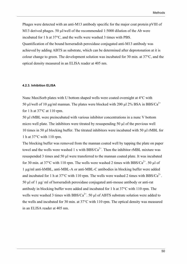
Methods
50
Phages were detected with an anti-M13 antibody specific for the major coat protein pVIII of
M13 derived phages. 50 µl/well of the recommended 1:5000 dilution of the Ab were
incubated for 1 h at 37°C, and the wells were washed 3 times with PBS.
Quantification of the bound horseradish peroxidase conjugated anti-M13 antibody was
achieved by adding ABTS as substrate, which can be determined after deprotonation at it is
colour change to green. The development solution was incubated for 30 min. at 37°C, and the
optical density measured in an ELISA reader at 405 nm.
4.2.3. Inhibition ELISA
Nunc MaxiSorb plates with U bottom shaped wells were coated overnight at 4°C with
50 µl/well of 10 µg/ml mannan. The plates were blocked with 200 µl 2% BSA in BBS/Ca2+
for 1 h at 37°C at 110 rpm.
50 µl rMBL were preincubated with various inhibitor concentrations in a nunc V bottom
micro well plate. The inhibitors were titrated by resuspending 50 µl of the previous well
10 times in 50 µl blocking buffer. The titrated inhibitors were incubated with 50 µl rMBL for
1 h at 37°C with 110 rpm.
The blocking buffer was removed from the mannan coated well by tapping the plate on paper
towel and the wells were washed 1 x with BBS/Ca2+. Then the inhibitor rMBL mixture was
resuspended 3 times and 50 µl were transferred to the mannan coated plate. It was incubated
for 30 min. at 37°C with 110 rpm. The wells were washed 2 times with BBS/Ca2+. 50 µl of
1 µg/ml anti-hMBL, anti-MBL-A or anti-MBL-C antibodies in blocking buffer were added
and incubated for 1 h at 37°C with 110 rpm. The wells were washed 2 times with BBS/Ca2+.
50 µl of 1 µg/ ml of horseradish peroxidase conjugated anti-mouse antibody or anti-rat
antibody in blocking buffer were added and incubated for 1 h at 37°C with 110 rpm. The
wells were washed 3 times with BBS/Ca2+. 50 µl of ABTS substrate solution were added to
the wells and incubated for 30 min. at 37°C with 110 rpm. The optical density was measured
in an ELISA reader at 405 nm.

Methods
51
4.2.4. Complement Activation Assay
Nunc MaxiSorb plates with U bottom shaped wells were coated overnight at 4°C with
50 µl/well of 1 µg/ml mannan or 50 pmol/well Gal-BSA. The plates were blocked with 200 µl
2% BSA in BBS/Ca2+ for 1 h at 37°C at 110 rpm. The blocking buffer was removed from the
mannan coated well by tapping the plate on paper towel and the wells were washed 1 x with
BBS/Ca2+. Human complement serum was diluted 1:40 and 50 µl/well were added to the
wells. It was incubated for 1 h at 37°C with 110 rpm. The wells were washed 2 times with
BBS/Ca2+. 50 µl of 1 µg/ml anti-hMBL antibody in blocking buffer were added and incubated
for 1 h at 37°C with 110 rpm. The wells were washed 2 times with BBS/Ca2+. 50 µl of
1 µg/ml of horseradish peroxidase conjugated anti-mouse antibody or anti-rat antibody in
blocking buffer were added and incubated for 1 h at 37°C with 110 rpm. The wells were
washed 3 times with BBS/Ca2+. 50 µl of ABTS substrate solution were added to the wells and
incubated for 30 min. at 37°C with 110 rpm. The optical density was measured in an ELISA
reader at 405 nm.

Methods
52
4.3. General Methods The methods were modified after Molecular Cloning: A Laboratory Manual 102.
4.3.1. Storage of Bacteria
Individual colonies of all strains were obtained by separating the cells on agar plates. Strains
carrying the F’ factor were plated on M9 agar and other strains were plated on LB, containing
the suitable antibiotic. Incubations were performed at 37°C, and the plates were stored up to
2 weeks at 4°C.
For long time storage 0.5 ml of a fresh 16 h culture was mixed with 0.5 ml sterile glycerol
(87%). The bacterial suspension was frozen with liquid nitrogen and stored at -70°C.
4.3.2. DNA Staining
Bacteria were stained with syto24. 1 µl syto24 was diluted in 100 µl PBS and transferred to
the bacteria-coated polysorb well. It was incubated for 20 min. at 37°C. The bacterial load
was estimated by fluorescence microscopy.
4.3.3. Sodium Dodecyl Sulfate-Polyacrylamide Gel Electrophoresis (SDS-PAGE)
The SDS-PAGE was used for the separation of proteins according to their size.
Firstly the gels were prepared as followed.
12.5% polyacrylamide resolving gel
4.12 ml 30% acrylamide
2.5 ml 1.5 M Tris
100 µl 10% SDS
5 µl TEMED
50 µl 10% APS
3.23 ml ddH2O

Methods
53
The solution was mixed, immediately poured between two glass plates of the SDS-PAGE
apparatus and overlayed with ethanol to prevent bubbles from polymerising. After 45 min. the
ethanol was removed and the gel surface rinsed with ddH2O. A broader linked stacking gel
was provided above the resolving gel to focus the proteins before separation.
The 4% stacking gel was prepared from the following solution:
4% polyacrylamide stacking gel
1 ml 30% acrylamide
1.9 ml 0.5 M Tris
80 µl 10% SDS
8 µl TEMED
40 µl 10% APS
4.5 ml ddH2O
The solution was thoroughly mixed, poured on the top of the resolving gel and a comb was
inserted. After 45 min. the polymerised gel was assembled into the SDS-PAGE apparatus and
running buffer was poured above.
Protein samples were mixed with the appropriate amount of loading dye and, if desired, with
DTT or ß-mercaptoethanol. They were heated for 5 min. to 90°C and loaded on the gel. The
gel was run with 100 V until the dye front reached the end of the gel.
The gel was incubated in coomassie brilliant blue staining solution for 30 min. and afterwards
in coomassie destaing solution until the protein bands were visible. The gel was rinsed with
ddH2O and placed between two in 5% glycerin and 22% ethanol softened cellophane sheets.
4.3.4. Western Blot
The western blot method was used for specific detection of proteins, separated before by
SDS-PAGE. Following SDS-PAGE, the stacking gel was removed and the resolving gel was
used for the transfer of proteins onto a PVDF membrane.
The PVDF membrane was shortly activated in methanol and rinsed afterwards 4 times with
ddH2O. To avoid air bubbles during blotting, 2 foam pads and 4 sheets of filter paper were
wet in transfer buffer before placing them into the gel holder cassette. A foam pad and 2 filter
papers were placed on one side of the cassette, the gel was laid above them and the membrane
Methods
54
was laid thoroughly on the gel, to avoid air inclusions. The other 2 filter papers and the foam
pad were placed on the membrane, the cassette was closed and placed with the membrane in
the direction of the anode into the blotting apparatus. An ice block was placed in the
apparatus, and the rest of the tank was filled with blotting buffer. The proteins were blotted
from the gel to the membrane for 30 min. at 100 V (0.4 A).
Subsequently the membrane was incubated for 1 h in 5% dry milk powder in TBS to block
the free binding sites on the membrane. The primary and, if required, the secondary antibody
were diluted in blocking buffer.
4.3.5. Polymerase Chain Reaction (PCR)
PCR is a method for rapid enzymatic amplification of a specific DNA sequence 103. Therefore
oligonucleotides were used flanking the DNA fragment of interest.
In principle the method is based on 3 reaction steps: denaturation of the DNA strand,
annealing of the specific oligonucleotides to their complementary strand and elongation of the
oligonucleotides by DNA polymerase under usage of free dNTPs. This process is repeated
cyclic, resulting in an exponential amplification of the DNA fragment.
The used DNA polymerase was the Taq polymerase of the thermophilic bacterium
Thermus aquaticus, provided with an inhibitory antibody. The release of the polymerase is
caused by a hot start denaturation step for 5 min. at 95°C, this offers the advantage of a
decreased amount of side products.
The different applications of the PCR required variations in reaction mixtures, temperatures
and times. The tested optimal conditions were described for the respective applications.
As standard 0.1 ml reactions were performed in 0.2 ml tubes in a thermocycler.
PCR products were analysed in agarose gel electrophoresis.
4.3.6. Colony PCR
To verify whether a bacterial colony contains plasmid with an insert of a desired size, single
colonies were picked with an inoculation loop and dipped in a PCR tube containing 20 µl
reaction mixture. Each reaction mixture was composed of 10 µl hot start master mix, 6 pmol
forward primer, 6 pmol reverse primer and water to the total volume of 20 µl.

Methods
55
The PCR conditions were the hot start conditions described for the common PCR.
4.3.7. Agarose Gel Electrophoresis
Plasmid DNA, DNA fragments and PCR products were separated on horizontal agarose gels.
Therefore 1% agarose in TAE buffer was heated in a microwave until the agarose was
dissolved completely, cooled down to approximately 60°C and poured in a gel chamber. The
cooled and hardened gel was used with TAE as running buffer.
Generally 10 µl sample were mixed with 2 µl 6 x loading dye and loaded in the wells, next to
5 µl 1 kb or 100 bp DNA ladder for subsequent analysis of the bands’ sizes. Migration was
performed with supply of 90 V. Afterwards the gel was exposed to a 0.5 µg/ml
ethidiumbromide water bath for 15 min., rinsed once with water and recorded by a UV
transilluminator.
4.3.8. Ethanol Precipitation
To concentrate DNA samples and change buffer solutions, DNA was either ethanol
precipitated or isolated via a PCR purification kit column.
It was precipitated by adding to the DNA sample 1 µl glycogen (20 mg/ml), 1/10 volume of
3 M sodium acetate (pH 4.8) and 2 volumes of 100% ethanol. Then the solution was
extensively mixed and cooled for 1 h at -70°C. The cool sample was centrifuged for 45 min.
with 16200 x g at 4°C, and the supernatant was carefully removed with a pipette.
The DNA pellet was washed by adding carefully 1 ml 70% ethanol, without destroying the
pellet. It was centrifuged again for 5 min., and the supernatant was removed. The pellet was
enabled to dry on air completely and then resuspended in an appropriate volume of the
desired buffer.

Methods
56
4.3.9. DNA Sequencing
The DNA sequences of the scFvs were determined by using a plasmid preparation of a single
colony as template. Therefore 5 ml SB medium, containing 0.1 mg/ml ampicillin, were
inoculated with a single colony from an overnight grown LB + 0.1 mg/ml Ampicillin agar
plate. The liquid culture was incubated overnight at 37°C with 230 rpm.
Subsequently the plasmids were preparated using the plasmid miniprep kit according to the
manufacturer’s protocol. 50-100 ng/µl plasmid DNA and 2 pmol/µl primer, both in a total
volume of 15 µl per sequence analysis were sent to Eurofins MWG operon, Anzinger Str. 7a,
85560 Ebersberg, Germany for sequencing.
The sequencing results were analysed with the either the software 4Peaks (Version 1.7.2) or
DNAStar Lasergene (Version 8.0).

Results
57
5. Results
5.1. Construction of an Idiotype ScFv
For the isolation of antibody fragments with similar binding properties like the idiotype
mAb WN1 222-5 a scFv WN1 222-5 was constructed in order to valuate the usability of the
approach. The scFv and phage WN1 222-5 should be used as controls for the phage panning
procedure and binding assays. Therefore the variable regions of the WN1 222-5 antibody
were cloned into the pComb3XSS vector. Determination of the WN1 222-5 scFv was done by
colony PCR (Fig. 14) and subsequent sequencing (Fig. 15).
Fig. 14: Colony PCR of the scFv WN1 222-5 after transformation. The correct insert size was determined subsequent to transformation by bacterial colony PCR.
Fig. 15: Amino acid sequence of scFv WN1 222-5 in one letter code. The sequence of scFv WN1 222-5 subsequent to cloning into the pComb3XSS vector. Figured with ompA leader
sequence (grey), 18 aa linker (blue), His6 tag (grey), haemagglutinin tag (grey), amber stop codon and the
beginning of the truncated N-terminus of pIII from the M13 phage. Complementarity determining regions
(CDRs) determined after sequence analysis with IMGT database 104.

Results
58
5.2. Anti-Anti-Idiotype ScFvs
5.2.1. Construction of Rabbit ScFv Libraries
ScFv libraries were constructed to obtain the rabbit antibody gene pool for subsequent high
throughput screening for specific binding antibody fragments.
5.2.1.1. Isolation of total RNA
The construction of a scFv library of an immunised rabbit requires the isolation of total
antibody mRNA. Hence, the complete spleen, bone marrow material of the femurs and the
harvested peripheral blood lymphocytes were used from four rabbits immunised with S81-19
mAb as described in methods. The RNA was extracted and reverse transcribed to first-strand
cDNA (Fig. 16).
Fig. 16. Total RNA and reverse transcribed cDNA. RNA and cDNA samples separated on a 1% agarose gel. Isolated total RNA indicates two bands corresponding
to the rRNAs and a broad smear of RNAs of different sizes. Reverse transcribed first-strand cDNA was
constructed using oligo-(dt) primer, excluding non-polyadenylated rRNA from transcription.

Results
59
5.2.1.2. Construction of ScFvs
Different scFv libraries were constructed from the RNA of four different rabbits. The four
rabbits were diverse in serum reaction to various antigens including the LPSs of the E. coli
rough type mutants R1, R2, R3 and R4 (Fig. 3). All sera showed in previous dot blot analysis
a clear binding to the R3 core type oligosaccharide 63.
Furthermore, the scFv libraries were constructed with linkers of two different length for
linking light and heavy chain fragments together to a scFv. A summary of the constructed
scFv libraries is given in Tab. 3.
As demonstrated for the rabbit 436 libraries the PCR reaction with the combined primer pairs
led to amplification of several of the variable region gene segments (Fig. 17). During this first
round of PCR the SfiI restriction site, as well as the linker for the subsequent connection
between the VL and VH chain was attached.
Fig. 17: Amplification of the variable region gene segments from rabbit 436. 1% agarose gel for the size control of the PCR products. Lane and primer combinations: short linker - 1
RSCVK1 and RKB9Jlo-B, 2 RSCVK3 and RKB9Jo-B, 3 RSCVK2 and RKB9Jlo-B, 4 RSCVK1 and RKB42Jo-
B, 5 RSCVK2 and RKB42Jo-B, 6 RSCVK1 and RKB9Jo-B, 7 RSCVK3 and RKB42Jo-B, 8 RSCVK2 and
RKB9Jo-B, 9 RSCλl and RJλo-BL, long linker -10 RSCVK1 and RKB9Jlo-BL, 11 RSCVK3 and RKB9Jo-BL,
12 RSCVK2 and RKB9Jlo-BL, 13 RSCVK1 and RKB42Jo-BL, 14 RSCVK3 and RKB9Jlo-BL, 15 RSCVK2
and RKB42Jo-BL, 16 RSCVK1 and RKB9Jo-BL, 17 RSCVK3 and RKB42Jo-BL, 18 RSCVK2 and RKB9Jo-
BL, 19 RSCλl and RJλo-BL, 20 RSCVH1 and RSCG-B, 21 RSCVH3 and RSCG-B, 22 RSCVH2 and RSCG-B,
23 RSCVH4 and RSCG-B. The desired PCR products were around 400 bp in length.
The first round of PCR was repeated six times and the PCR products with attached VL short
linker, VL long linker or VH were collected in separated pools. Purification on a 1.2% agarose
gel yielded in 1-2 µg PCR product DNA per variable region gene.

Results
60
In the second round of PCR the attached overlapping ends of the variable region light and
heavy chain were joined together and amplified. This overlap extension PCR resulted several
times in low quantities of DNA product, indicated by poorly visible DNA bands in the
agarose gel (Fig. 18).
Fig. 18: Overlap extension PCR. Lane 1: rabbit 436 short linker product, 2: 436 long linker product, 3: 437 short linker product and 4: 437 long
linker product. Overlap extension PCR products with a size of around 750 bp. Unjoined variable region
fragments at 500 bp.
Therefore the reaction conditions where optimised (Fig. 19). The use of hot start Taq
polymerase instead of conventional Taq polymerase led to an increase in scFv PCR product.
Even more crucial for the yield of the desired product was the exact 1:1 molar ratio of the
applied light chain to heavy chain.
The various PCR reactions were collected and concentrated by ethanol precipitation. The
concentrated DNA was separated in an agarose gel, and the desired band at around 750 bp
was extracted. Finally a yield of each scFv PCR product of 10 µg was received.
Fig. 19. Product increase of the overlap extension PCR. Lane 1: rabbit 436 short linker product, 2: 436 long linker product, 3: 437 short linker product and 4: 437 long
linker product. Overlap extension PCR products with a size of around 750 bp. Unjoined variable region
fragments were still detectable at 500 bp.

Results
61
5.2.1.3. Cloning of the ScFvs
10 µg of each scFv PCR product and 20 µg of the pComb3XSS vector per library were
digested with the restriction enzyme SfiI. The digested DNA was controlled and isolated by
agarose gel electrophoresis. In case of the pComb3XSS vector it became apparent that the
cleavage was not complete after one hour as described in the manufacturers protocol
(Fig. 20). The large size of the 1600 bp large stuffer fragment enables the detection of single
cut vector. The enzymatic digestion was considerably enhanced after 16 hours. The digested
DNA was isolated from the agarose gel and directly used for the ligation.
Fig. 20: SfiI restiction enzyme digest of the pComb3XSS phagemid vector. A: 5 µg of pComb3XSS vector after one hour of restriction digest at 50°C with 60 U SfiI. B: Gel extracted
double digested vector and isolated stuffer fragment.
Prior to the library ligation, the quality of the digested vector and inserts were determined by
small scale ligations. The insert small scale ligations resulted, depending on the prepared scFv
insert, in amounts of 4 to 11 x 106 cfu/µg applied vector DNA. Vector plus insert religations
resulted in around 5 x 10⁶ cfu/µg vector DNA. The vector ligations without insert resulted in
amounts between 0.3 to 1.3 x 10⁶ cfu/µg vector DNA.
Ligations of library DNA yielded in library sizes summerised in Tab 3. Afterwards the
bacteria were infected with helper phages for phage packaging.

Results
62
Rabbit 436 Rabbit 437 Rabbit 438 Rabbit 439
Source of RNA Bone
marrow
Bone
marrow
Bone marrow and
spleen
Peripheral blood
lymphocytes
Bone marrow and
spleen
Linker Length 7 aa 18 aa 18 aa 18 aa 18 aa
Total No. of 2 x 10⁵ 2 x 10⁵ 1 x 10⁵ 1 x 10⁵ 1 x 10⁴
Inserts/Independent Transformants
13/15
14/15
8/8
8/10
4/10
Tab. 3: ScFv libraries assembled in the pComb3XSS vector and library sizes.
The quality of the scFv libraries was examined by a size control (Fig. 21) of the inserted scFv
and by sequence analysis of several clones.
Fig. 21: Colony PCR of the rabbit 436 libraries. Insert size analysis by direct use of bacterial colonies for PCR. Lane 1-15 rabbit 436 SL library, DNA band in
lane 9 is oversized for a scFv, and in the colony tested in lane 13 no insert was detected. Lane 16-31 rabbit 436
LL library, solely lane 19 reveals a DNA band of the wrong size. Lane 32-34 rabbit 437 LL library.
5.2.2. Panning Procedures
5.2.2.1. Phage Binding to Anti-Idiotype Antibodies
As a first step the anti-idiotype antibody S81-19 was used as antigen for phage panning.
It was the immunising antigen and should therefore lead to the isolation of phages, which bind
to the protein.
Two different selection procedures were performed. In the first one 100 ng S81-19 mAb per
well were applied for three pannings implemented with the scFv libraries 436 (long linker),
437 and 439.

Results
63
In the second procedure 100 ng S81-19 F(ab)2 per well were used for panning with the
libraries 436 (short linker) and 437, to distinguish binding to the Fc and the variable antibody
domains.
Both procedures were performed under panning conditions like described. The phage titer and
the insert steadiness were monitored and revealed continually stable results. The binding
ability was verified after the panning procedure using the reamplified phage mixtures of the
respective rounds. All five screened scFv libraries showed unambiguous binding after four
rounds of panning against the anti-idiotype S81-19 (Fig. 28). The applied 436 short linker
library revealed clear binding after the third round of panning (Fig. 22).
Ag Anti-Idiotype S81-19 F(ab)2
0.0001 0.001 0.01 0.10
1
2
3
4Phage WN1
436 SL library
after 1. panning
after 2. panning
after 3. panning
after 4. panning
after 5. panning
Phage Dilution
An
tig
en
Bin
din
g (O
D405 n
m)
Fig. 22: Phage ELISA against the anti-idiotype S81-19 F(ab)2 as antigen. The scFv short linker phage library 436 and the phage mixtures subsequent five rounds of panning tested for
binding to the anti-idiotype F(ab)2. The isolated phage WN1 222-5 was used as a binding control. After the
second round of panning a slight binding is detectable, increasing after the third round and reaching the maximal
measured level after the fourth round.
5.2.2.2. Phage Binding to R3-Conjugates
In the next step it was investigated whether a glycoconjugate can be used for phage selection.
Therefore the E. coli R3 core-type oligosaccharide conjugate was applied, a structure shown
to bind to antibodies in the anti-anti-idiotype sera.
The first library used for the selection of antigen binding scFvs against R3-GA-BSA
conjugate was the bone marrow and spleen derived long linker library constructed from donor
material of rabbit 437. The ligand frequency in the conjugate amounted 341 nmol ligand/ml.
The wells for panning were coated with 50 pmol in 50 µl carbonate buffer.

Results
64
Previous to the first, second and fourth panning step the library phages were reamplified. The
panning procedure was performed as described in the method section.
Phage Preparation Phage Titer (cfu/ml) of Panning Round
1 2 3 4 5
Input 3 x 10⁸ 2 x 10¹³ 8 x 10¹² > 4 x 10¹³ 5 x 10¹² Output 1 x 10⁵ 2 x 10⁴ n.d. 3 x 10⁵ 3 x 10⁴
Tab. 4: Phage titer monitoring of the first R3 conjugate panning.
The phage titer was controlled previous and past the binding step of each panning round
(Tab. 4). To estimate the stability of the insert throughout the panning procedure and
amplifications, colonies from titer plates were used straight forward in PCR reactions. The
colony PCR revealed a substantial loss of scFv genes containing phages (Fig. 23).
Fig. 23: Insert size analysis by colony PCR of the scFv 437 library panning. The upper gel shows the colony PCR results subsequent the third panning round. Already indicating a loss of
insert bearing phages during panning. The lower gel shows the results after the fifth panning round. Only 4 of 28
tested colonies had an insert of the correct size.
More important, the amplified phage mixtures subsequent panning rounds as well as
individually amplified phages possessing scFv inserts of the correct size, were screened for
their binding properties in ELISA. No binding phages could be detected.
To investigate whether reduction of the amplifications between the panning rounds would
increase the number of clones with insert, two distinct modifications of the regular panning
procedure have been implemented. The 437 scFv library was reamplified before the initial

Results
65
panning step. Afterwards no further reamplifications of the phages have been included in the
panning procedure.
Additionally the washing steps were decreased. The wash buffer was not resuspended and
subsequent incubated during the washing steps. Instead the wash buffer was given briefly to
the well after phage incubation and immediately removed with care.
Phage Preparation Phage Titer (cfu/ml) of Panning Round
1 2 3 4
Input 1 x 10⁸ 4 x 10¹² 3 x 10¹² 1 x 10¹³ Output 1 x 10⁵ 2 x 10⁴ 6 x 10⁴ 7 x 10³
Tab. 5: Phage titer monitoring of the second R3 conjugate panning.
The phage titer was monitored and revealed no significant loss of phages during the panning
(Tab. 5). Furthermore, under these conditions the colony PCR indicated no loss of scFv
inserts over time.
The phage mixtures of the amplified phages after the panning rounds and several amplified
single phages were analysed in ELISA for their binding activity (Fig. 24). The phage mixtures
revealed a minor signal for binding against the anti-idiotype mAb. Two of the screened single
phages, gained after the fourth panning round, suggested a slight binding. The binding was
repeatedly determined using R3 conjugate as well as the anti-idiotype immunogen and casein
as immobilised antigens. There was a low comparable optical density measurable for the three
antigens, including the casein control antigen.
Ag R3-GA-BSA
0.01 0.1 10.0
0.2
0.4
0.6
0.8
1.0
Phage Dilution
An
tige
n B
ind
ing
(O
D405 n
m)
Ag Anti-Id S81-19
0.01 0.1 10
1
2
3
Phage Dilution
An
tige
n B
ind
ing
(O
D405 n
m)
Ag Casein
0.01 0.1 10.0
0.2
0.4
0.6
0.8
1.0Phage WN1
Phages after 2. panning
Phages after 3. panning
Phages after 4. panning
Single phage 39
Single phage 40
Phage Dilution
An
tige
n B
ind
ing
(O
D405 n
m)
Fig. 24: Phage ELISA with three different antigens. The phage mixtures after the second, third and fourth panning round and two amplified single phages were
determined for their binding activity against the R3 conjugate, the anti-idiotype mAb S81-19 and casein as a
control. Phage WN1 222-5 was applied as a positive control for the phage binding ability.

Results
66
To verify whether there are some specific binding scFvs presented on the phages, the scFvs
were expressed without pIII in an E. coli TOP10F’ nonsuppressor strain (Fig. 25). The scFv
supernatant from the polymyxin B treated bacteria cells was used straight forward for two
ELISA variants in parallel. The first was implemented using 2.5% casein and 0.05% Tween20
in PBS, and in the second 5% skim milk powder in TBS was used as blocking solution and as
a negative binding control. The former reconfirmed the results of the two single phages for
the anti-idiotype and casein binding activity. R3 conjugate binding was not observed. The
second revealed no binding activity of the single phages. Interestingly the control scFv
WN1 222-5 binding was increased using the Tween20 free buffer.
Ag R3-GA-BSA
0.01 0.1 10
1
2
3
ScFv Dilution
An
tige
n B
ind
ing
(O
D405 n
m)
Ag R3-GA-BSA
0.01 0.1 10
1
2
3
ScFv Dilution
An
tige
n B
ind
ing
(O
D405 n
m)
Ag Anti-Id S81-19
0.01 0.1 10
1
2
3
ScFv Dilution
An
tige
n B
ind
ing
(O
D405 n
m)
Ag Anti-Id S81-19
0.01 0.1 10
1
2
3
ScFv Dilution
An
tige
n B
ind
ing
(O
D405 n
m)
Ag Casein
0.01 0.1 10
1
2
3 ScFv WN1
ScFvs after 2. panning
ScFvs after 3. panning
ScFvs after 4. panning
ScFv 39
ScFv 40
ScFv Dilution
An
tige
n B
ind
ing
(O
D405 n
m)
Ag Skim Milk Powder
0.01 0.1 10
1
2
3 ScFv WN1
ScFvs after 2. panning
ScFvs after 3. panning
ScFvs after 4. panning
ScFv 39
ScFv 40
ScFv Dilution
An
tige
n B
ind
ing
(O
D405 n
m)
A
B
Fig. 25: ScFv ELISA with three different antigens and two varied buffer conditions. The first ELISA row (A) shows scFv mixtures and two isolated scFvs determined for their binding to R3
conjugate, anti-idiotype antibody and casein, using casein buffer as blocking and dilution buffer. Only binding of
the scFv WN1 222-5 control was detectable to the R3 conjugate, a minor binding of the other scFvs to the anti-
idiotype and casein is visible.
In the second ELISA row (B) 5% skim milk powder in TBS was used as blocking and diluting buffer. The
overall measured binding of the scFv WN1 222-5 control was slightly higher than for the PBS buffer. No
binding of the isolated scFvs could be detected.

Results
67
5.2.2.3. Phage Binding to Heat-killed Bacteria
One reason for the lack of binding to the R3 conjugate could have been that the antigen
concentration immobilised on the ELISA plate was too low. Therefore the form of antigen
presentation was altered from R3 conjugate to R3 heat-killed bacteria. 2 µg of the bacteria per
well were used to provide a high density of immobilised antigen.
The scFv library 437 was used again for the first panning procedure according to the
described standard protocol. Stable phage titers were observed in the range of 1 x 10¹³ cfu/ml
phages for the input titer and 1 x 10⁵ cfu/ml phages for the output titer. Also the insert control
via colony PCR revealed no loss of scFv inserts. Nevertheless no binding phages to R3
bacteria could be detected in the phage mixtures subsequent the panning rounds.
To investigate whether the source of DNA was responsible for the lack of binding phages, the
scFv library 438, derived from peripheral blood lymphocytes, was applied for an additional
panning. The procedure was performed according to the described standard protocol, except
one further reamplification step subsequent the third panning round. No deviations from the
phage titers compared with the previous panning were observed and no loss of inserts was
detected. No phages binding to R3 bacteria could be detected in the phage mixtures
subsequent the panning rounds.
As a next step an increase in avidity was applied by using the scFv library 436 with a short
linker between the antibodies variable region light and heavy chain for a further panning
against R3 heat-killed bacteria as immobilised antigens. A reamplification step was performed
subsequent the second panning.
The phage titer monitoring during the panning was constant (Tab. 6), with a slight decrease of
phages after the reamplification previous to the third panning round. The insert control of
some individual clones indicated a slight loss of scFv insert carrying phages compared to the
initial library where 87% of the tested colonies had an insert of the correct size (Fig. 26).
Phage Preparation Phage Titer (cfu/ml) of Panning Round
1 2 3 4
Input 4 x 10¹³ 4 x 10¹³ 2 x 10¹¹ 5 x 10¹³ Output n.d. 2 x 10⁵ 2 x 10⁵ 2 x 10⁵
Tab. 6: Phage titer monitoring of the scFv 436 short linker library .
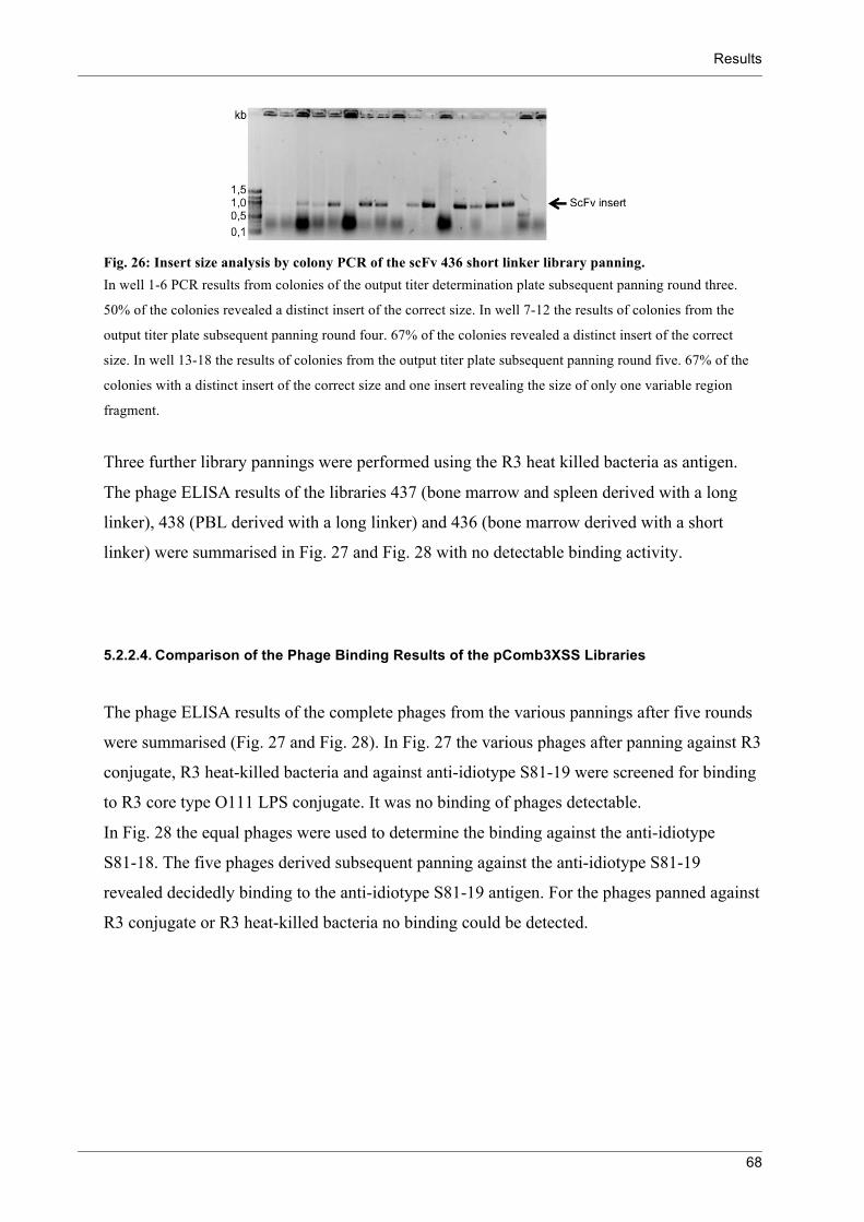
Results
68
Fig. 26: Insert size analysis by colony PCR of the scFv 436 short linker library panning. In well 1-6 PCR results from colonies of the output titer determination plate subsequent panning round three.
50% of the colonies revealed a distinct insert of the correct size. In well 7-12 the results of colonies from the
output titer plate subsequent panning round four. 67% of the colonies revealed a distinct insert of the correct
size. In well 13-18 the results of colonies from the output titer plate subsequent panning round five. 67% of the
colonies with a distinct insert of the correct size and one insert revealing the size of only one variable region
fragment.
Three further library pannings were performed using the R3 heat killed bacteria as antigen.
The phage ELISA results of the libraries 437 (bone marrow and spleen derived with a long
linker), 438 (PBL derived with a long linker) and 436 (bone marrow derived with a short
linker) were summarised in Fig. 27 and Fig. 28 with no detectable binding activity.
5.2.2.4. Comparison of the Phage Binding Results of the pComb3XSS Libraries
The phage ELISA results of the complete phages from the various pannings after five rounds
were summarised (Fig. 27 and Fig. 28). In Fig. 27 the various phages after panning against R3
conjugate, R3 heat-killed bacteria and against anti-idiotype S81-19 were screened for binding
to R3 core type O111 LPS conjugate. It was no binding of phages detectable.
In Fig. 28 the equal phages were used to determine the binding against the anti-idiotype
S81-18. The five phages derived subsequent panning against the anti-idiotype S81-19
revealed decidedly binding to the anti-idiotype S81-19 antigen. For the phages panned against
R3 conjugate or R3 heat-killed bacteria no binding could be detected.

Results
69
Ag O111-GA-BSA
0.01 0.10.0
0.5
1.0
1.5Phage WN1
436 LL after anti-id panning
437 after anti-id panning
436 SL after anti-id panning
438 after anti-id panning
439 after anti-id panning
436 SL after HKB panning
437 after R3-BSA panning
437 after R3 HKB panning
438 after HKB panning
Phage Dilution
An
tig
en
Bin
din
g (O
D405 n
m)
Fig. 27: Phage ELISA against the R3 core type O111 LPS-BSA conjugate as antigen. The phage mixtures derived from the diverse libraries were used in different panning procedures and subsequent
five rounds of panning analysed for binding to the R3 core type O111-GA-BSA conjugate. It was no binding
detectable for the various phages. The isolated phage WN1 222-5 was used as a binding control.
Ag Anti-Idiotype S81-19 F(ab)2
0.001 0.01 0.10
1
2
3
4Phage WN1
436 LL after anti-id panning
437 after anti-id panning
436 SL after anti-id panning
438 after anti-id panning
439 after anti-id panning
436 SL after HKB panning
437 after R3-BSA panning
437 after R3 HKB panning
438 after HKB panning
Phage Dilution
An
tige
n B
ind
ing
(O
D405 n
m)
Fig. 28: Phage ELISA against the anti-idiotype S81-19 F(ab)2 as antigen. The phage mixtures derived from the diverse libraries were used in different panning procedures and subsequent
five rounds of panning analysed for binding to the anti-idiotype S81-19 F(ab)2. It was no binding detectable for
the phages after the panning procedures with R3 conjugate or R3 heat-killed bacteria as antigen (black symbols).
Whereas the phages panned against the S81-19 revealed all a clear binding to it (white symbols). The isolated
phage WN1 222-5 was used as a binding control.
5.2.3. Subcloning into pHen1
To increase the number of scFv displayed by the phages, the scFv library 437 was cloned
from the pComb3XSS into the pHen1 vector (Fig. 10). The pHen1 phagemid includes in
contrast to the pComb3 vectors the complete gIII and enables consequently the infection of
bacteria by these phages. Therefore the application of a hyperphage, lacking the gIII in its

Results
70
genome, for packaging of the phagemid was possible, which should lead to the expression of
up to five copies of the scFv attached to pIII.
The phage library 437 was used for the infection of E. coli ER2537. A plasmid preparation
was used for scFv amplification in a PCR introducing a NotI restriction site at the 3’-end of
the scFv. The scFv genes and the pHen1 vector were digested first with SfiI and subsequent
with NotI. Afterwards the DNA was ligated into the pHen1 vector and amplified using helper
phage M13K07 for phage packaging.
Preliminary to the first panning the 437 pHen1 library was amplified by infecting an E. coli
TG1 culture with the phages and using hyperphage M13K07ΔpIII for phage packaging.
The amplified hyperphage packed phages were controlled and quantitative compared to the
helper phage packed pHen1 phages, the helper phage packed pComb3XSS phages and the
helper phage (Fig. 29). The western blot staining with an anti-cmyc tag antibody reveals the
quantitative production of scFv-pIII fusion proteins by hyperphage packaging (Fig. 30).
Fig. 29: SDS-PAGE and western blot of phage particles. Lane 1: Helper phage M13K07; lane 2: scFv library 437 in pHen1 packed with helper phage; lane 3: scFv library
437 in pHen1 packed with hyperphage and lane 4: scFv library 439 in pComb3XSS. The coomassie stained SDS
gel on the left side revealed the ß-mercaptoethanol reduced and denaturated phage components in similar
quantities. The western blot of the same phages on the right side was stained with the anti-c-myc tag antibody
and developed using alkaline phosphatase. In lane 3 the c-myc tag of the scFv-pIII fusion protein is detectable,
but further molecules are stained unspecifically.
Two further western blots revealed the scFv-pIII fusion protein as abundantly present on
hyperphage packed phage particles (Fig. 30).

Results
71
Fig. 30: Western blot of phage particles. Lane 1: scFv library 437 in pHen1 packed with helper phage; lane 2: scFv library 437 in pHen1 packed with
hyperphage and lane 3: helper phage M13K07. The blot on the left side was stained with an anti-c-myc tag
antibody and further detected by an Alexa680 conjugated anti-mouse antibody. It presents the scFv-pIII fusion
protein in lane 2. In lane 1 the scFv-pIII fusion protein was not detectable. The blot on the left side was stripped
using stripping buffer for 20 min. and stained again with the anti-pIII antibody 105 and subsequent with an IR-dye
conjugated anti-mouse antibody resulting in the blot on the right side. For the phages in lane 1, packed with the
helper phage, the pIII band at around 60 kDa implies high quantities of pIII. In lane 2 the two bands for the scFv-
pIII fusion protein (above) and for the pIII (below) are clearly determinable. In lane 3 the helper phage shows the
band of its pIII.
Two pannings were performed using the R3-GA-BSA conjugate and the anti-idiotype
S81-19 F(Ab)2 as immobilised antigens in parallel. In total four rounds of panning were
implemented, whereas the phages previous to the first and second panning were amplified
with the hyperphage M13K07ΔpIII for phage packaging and the subsequent phages were
packed by the helper phage M13K07.
Minor fluctuations could be recognised in the phage titers, but no substantial loss of phages
(Tab. 7).
Phage Preparation Phage Titer (cfu/ml) of Panning Round
R3-GA-BSA 1 2 3 4
Input 2 x 10¹³ 1 x 10¹³ 2 x 10¹³ 6 x 10¹¹ Output 2 x 10⁴ 2 x 10³ 2 x 10⁶ 3 x 10⁴
S81-19
Input 2 x 10¹³ 2 x 10¹² 5 x 10¹³ 2 x 10¹²
Output 2 x 10⁴ 6 x 10³ 7 x 10⁵ 4 x 10⁶
Tab. 7: Phage titer monitoring of the scFv 437 library during panning against R3 conjugate and anti-idiotype S81-19 antigens.

Results
72
The binding of the phage mixtures subsequent panning rounds was determined by phage
ELISA (Fig. 31).
Ag Anti-Idiotype S81-19
0.01 0.10.0
0.5
1.0
1.5
2.0437 library in pHen1
after 1. panning
after 2. panning
after 3. panning
after 4. panning
after 1. panning
after 2. panning
after 3. panning
after 4. panning
Phage Dilution
An
tige
n B
ind
ing
(O
D405 n
m)
Fig. 31: Phage ELISA of the scFv 437 library subcloned into pHen1 against the anti-idiotype S81-19 as antigen. The amplified 437 library phages and the phages of the panning procedure against the antigen S81-19 are figured
with black symbols. In white symbols the phages of the panning procedure against the antigen R3 conjugate.
In conclusion the phages of the anti-idiotype S81-19 panning showed after three rounds of
panning distinct binding, while the phages panned against the R3 conjugate revealed no
binding to the anti-idiotype. For the phages of both pannings no binding could be detected
against the R3 conjugate.
5.3. Recognition of Bacterial Carbohydrates by MBL
5.3.1. MBL Binding Studies
MBL interacts with various carbohydrates, including different monosaccharides as well as
polysaccharides 87. In humans one form of MBL has been identified, whereas two forms are
found in mice. The three MBLs, human MBL (hMBL) and the murine MBL-A and MBL-C
were investigated for their binding properties to pathogenic and non-pathogenic bacteria and
also to isolated bacterial cell-wall carbohydrates.
At first it was investigated whether the binding to the E. coli R3 rough type mutant, the E. coli
smooth type O8 serotype and M. tuberculosis H37Rv ATCC differs between the three
rMBLs. Therefore an inhibition ELISA was performed detecting rMBL binding to a ligand,
subsequent to preincubation of rMBL with the various heat-killed bacteria. The ELISA
revealed extensive differences between the three rMBLs in recognition of the bacteria

Results
73
(Fig. 32). The rhMBL showed binding to the M. tuberculosis H37, but not to the tested E. coli
preparations. On the contrary the rMBL-A binds to all three heat-killed bacterial preparations
in a comparable manner. For the rMBL-C only a slight binding to the M. tuberculosis H37
was detected.
Another inhibition ELISA was performed with decreased rhMBL and rMBL-C concentrations
and with two different mycobacteria as well as two different E. coli strains (Fig. 33). The
rMBL-A revealed again an obvious binding to all four heat-killed bacterial preparations. But
the inhibition observed was more pronounced for the E. coli O4 and O111 than for the
M. tuberculosis EAI and Beijing. In contrast binding of the rhMBL to the two mycobacterial
strains was observed, while no binding to the E. coli O111 and only a minor binding to the O4
was detected. The rMBL-C showed only limited binding to the M. tuberculosis EAI, but no
inhibition was detectable to the other heat-killed bacteria.
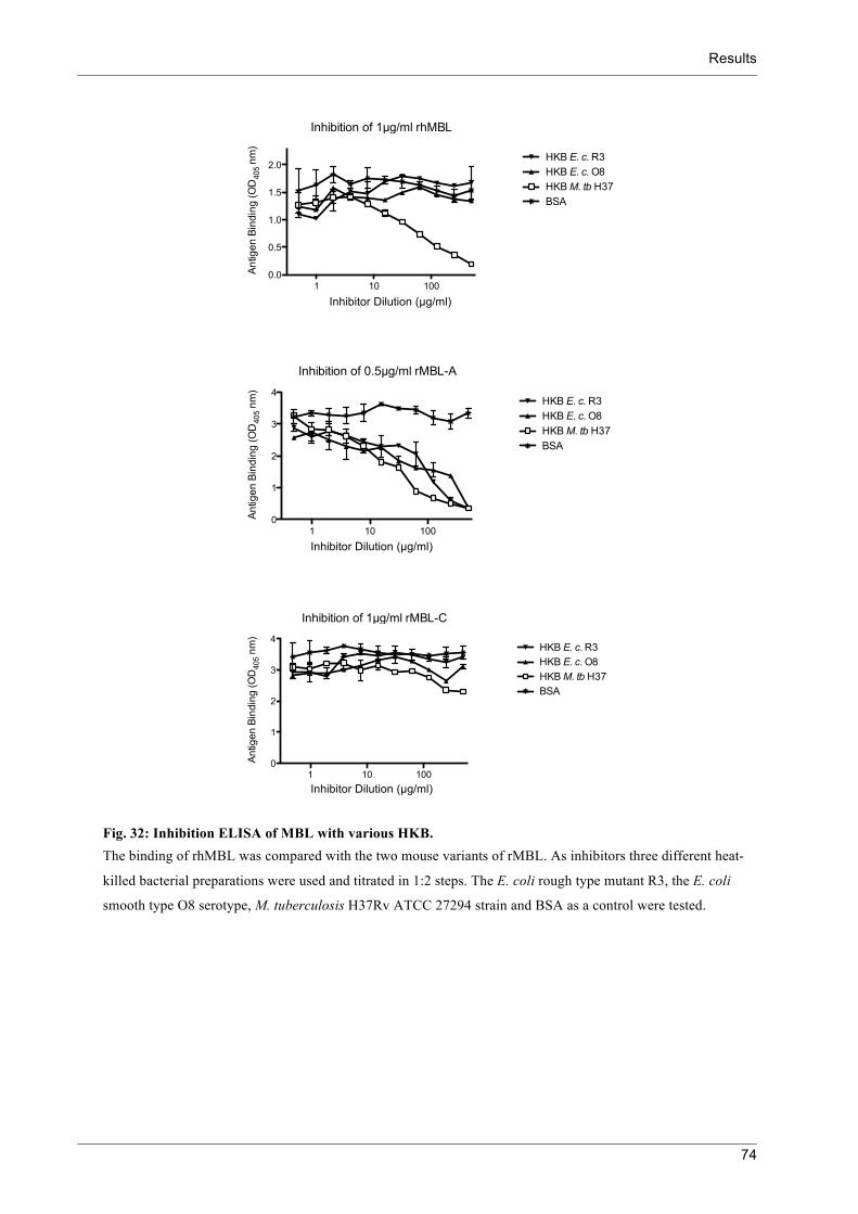
Results
74
Inhibition of 1!g/ml rhMBL
1 10 1000.0
0.5
1.0
1.5
2.0HKB E. c. R3
HKB E. c. O8
HKB M. tb H37
BSA
Inhibitor Dilution !g/ml
An
tige
n B
ind
ing
(O
D405 n
m)
Inhibition of 0.5!g/ml rMBL-A
1 10 1000
1
2
3
4HKB E. c. R3
HKB E. c. O8
HKB M. tb H37
BSA
Inhibitor Dilution !g/ml
An
tige
n B
ind
ing
(O
D405 n
m)
Inhibition of 1!g/ml rMBL-C
1 10 1000
1
2
3
4HKB E. c. R3
HKB E. c. O8
HKB M. tb H37
BSA
Inhibitor Dilution !g/ml
An
tige
n B
ind
ing
(O
D405 n
m)
Fig. 32: Inhibition ELISA of MBL with various HKB. The binding of rhMBL was compared with the two mouse variants of rMBL. As inhibitors three different heat-
killed bacterial preparations were used and titrated in 1:2 steps. The E. coli rough type mutant R3, the E. coli
smooth type O8 serotype, M. tuberculosis H37Rv ATCC 27294 strain and BSA as a control were tested.
Inhibitor Dilution (µg/ml)
Inhibitor Dilution (µg/ml)
Inhibitor Dilution (µg/ml)

Results
75
Inhibition of 0.5 !g/ml rhMBL
0.001 0.01 0.1 10.0
0.5
1.0
1.5HKB EAI 1797/03
HKB Beijing 17919
HKB E. c. O4
HKB E. c. O111
Inhibitor Dilution mg/ml
An
tige
n B
ind
ing
(O
D405 n
m)
Inhibition of 0.5 !g/ml rMBL-A
0.001 0.01 0.1 10
1
2
3
4HKB EAI 1797/03
HKB Beijing 17919
HKB E. c. O4
HKB E. c. O111
Inhibitor Dilution mg/ml
An
tige
n B
ind
ing
(O
D405 n
m)
Inhibition of 0.5 !g/ml rMBL-C
0.001 0.01 0.1 10
1
2
3
4HKB EAI 1797/03
HKB Beijing 17919
HKB E. c. O4
HKB E. c. O111
Inhibitor Dilution mg/ml
An
tige
n B
ind
ing
(O
D405 n
m)
Fig. 33: Inhibition ELISA of MBL with heat-killed mycobacteria and E. coli. Binding of rhMBL, rMBL-A and rMBL-C to M. tuberculosis EAI and Beijing as well as E. coli O4 and O111
was determined by inhibition ELISA. rhMBL binding to mannan was inhibited by preincubation of the rhMBL
with the mycobacteria, but not with E.coli. rMBL-A binds to the four heat killed bacteria, while the best binder
was the E.coli O4 serotype followed by the O111. The mycobacteria revealed as well binding, but to a lower
extent. The rMBL-C reveals slight binding to the M. tuberculosis EAI, not to the other bacteria.
In a next step four different E. coli rough type mutants were tested for their binding potential
to rMBL (Fig. 34). Analyzed were preparations from whole heat-killed bacteria and isolated,
deacylated oligosaccharides derived from all four mutants. Again the three rMBLs differed in
their binding to the bacteria and oligosaccharides. The rhMBL revealed binding to the R2
core type heat-killed bacteria and oligosaccharides, but only moderate binding to the other
Inhibitor Dilution (mg/ml)
Inhibitor Dilution (mg/ml)
Inhibitor Dilution (mg/ml)
Results
76
core types was detected. The rMBL-A showed distinct binding to the R2 and R3 heat-killed
bacteria, moderate binding to the R1 bacteria and no binding to the R4 bacteria. However the
binding to the isolated oligosaccharides varied from the binding to the corresponding heat-
killed bacteria. The R2 oligosaccharide appeared to be the best binding oligosaccharide,
followed by the R1 and further the R4. For the R3 oligosaccharide no binding was detected.
The binding of the rMBL-C to mannan was only slightly inhibited by the R2 heat-killed
bacteria, but not by the other rough mutants. And also for the oligosaccharides no binding to
rMBL-C was detected, except for the R2 oligosaccharide.
In comparison the three tested rMBLs revealed different binding to the bacteria and
oligosaccharides, while the most obvious difference was a generally lower binding of rhMBL
and rMBL-C compared to rMBL-A.

Results
77
Inhibition of 1 !g/ml rhMBL
1 10 1000
1
2
3HKB E. c. R1
HKB E. c. R2
HKB E. c. R3
HKB E. c. R4
Inhibitor Dilution !g/ml
An
tige
n B
ind
ing
(O
D405 n
m)
Inhibition of 0.5 !g/ml rMBL-A
1 10 1000
1
2
3
4HKB E. c. R1
HKB E. c. R2
HKB E. c. R3
HKB E. c. R4
Inhibitor Dilution !g/ml
An
tige
n B
ind
ing
(O
D405 n
m)
Inhibition of 1 !g/ml rhMBL
1 10 1000
1
2
3R1 HyKOH
R2 HyKOH
R3 HyKOH
R4 HyKOH
Inhibitor Dilution !M
An
tige
n B
ind
ing
(O
D405 n
m)
Inhibition of 0.5 !g/ml rMBL-A
1 10 1000
1
2
3
4R1 HyKOH
R2 HyKOH
R3 HyKOH
R4 HyKOH
Inhibitor Dilution !M
An
tige
n B
ind
ing
(O
D405 n
m)
Inhibition of 0,5 !g/ml MBL-C
1 10 1000
1
2
3
4
5HKB E. c. R1
HKB E. c. R2
HKB E. c. R3
HKB E. c. R4
Inhibitor Dilution !g/ml
An
tige
n B
ind
ing
(O
D405 n
m)
Inhibition of 0,5 !g/ml MBL-C
1 10 1000
1
2
3
4
5R1 HyKOH
R2 HyKOH
R3 HyKOH
R4 HyKOH
Inhibitor Dilution !M
An
tige
n B
ind
ing
(O
D405 n
m)
A
B
C
Fig. 34: Inhibition ELISA of MBL with E. coli heat-killed bacteria and oligosaccharides. The binding of rhMBL (A), rMBL-A (B) and rMBL-C (C) to four different E. coli rough type mutants and their
corresponding oligosaccharides was detected by inhibition ELISA. The inhibition of binding from rMBL to
mannan coated wells was determined after preincubation of the rMBL with the bacteria and oligosaccharides.
The R2 core type was the best inhibitor as bacterium and as oligosaccharide for the three rMBL. Broad
differences are shown in the intensity of the inhibition of the three MBL. The rhMBL showed in addition to the
R2 also slight binding to the other rough type bacteria and to the R1 oligosaccharide. The rMBL-A revealed
clear binding to the bacteria, except of the R4 rough type and to the oligosaccharides, except of the R3 core type.
The rMBL-C revealed only minor binding to the R2 bacteria and oligosaccharides, but no further binding was
detected.
The rhMBL revealed in first ELISA studies marginal binding to E. coli heat-killed bacteria
and E. coli core type oligosaccharides. On the contrary there was obvious binding of rhMBL
to M. tuberculosis H37Rv, EAI and Beijing. Further heat-killed mycobacteria were tested for
Inhibitor Dilution (µg/ml)
Inhibitor Dilution (µM)
Inhibitor Dilution (µg/ml)
Inhibitor Dilution (µg/ml)
Inhibitor Dilution (µM)
Inhibitor Dilution (µM)

Results
78
their interaction with rhMBL (Fig. 35). All of the heat-killed bacteria showed similar binding
to rhMBL.
Inhibition of 4 !g/ml rhMBL
0.0001 0.001 0.01 0.1 10
1
2
3
4
5Mannan
H37Rv
X-type 4412/04
Beijing 1934/03
Bovis 4258/00
Cameroon 1417/02
Afri2 10476/01
BSA
Dilution of Bacteria mg/ml
An
tig
en
Bin
din
g (
OD
405 n
m)
Fig. 35: Inhibition ELISA of rhMBL with mannan and different heat-killed mycobacteria. The inhibition of binding from rhMBL to mannan-coated wells was determined after preincubation of the
rhMBL with various heat-killed mycobacteria. All applied mycobacteria revealed binding to rhMBL with only
minor variations in binding strength.
To overcome the potential drawback of the inhibition ELISA, that variations in solubility of
the bacteria could lead to fluctuations in the concentration, a direct binding ELISA was
applied (Fig. 36). Quantification difficulties should be eliminated by covering the bottom of a
96 well plate complete with bacteria. Since mycobacteria have a rather hydrophobic cell-wall
the heat-killed bacteria were coated to a hydrophobic 96 well plate. Subsequently the rMBLs
were titrated 1:2 steps starting from 2 µg/ml. Obvious differences in the recognition of the
different mycobacteria are determined for the rMBL-A. It bound to the M. tuberculosis
Beijing and X-type better then to the H37Rv and to M. bovis Bovis. The binding to the
M. tuberculosis Cameroon and the M. africanum Afri2 was markedly less than to the other
heat-killed bacteria. The rhMBL revealed similar interactions like the rMBL-A, but overall a
decreased binding. The rMBL-C showed also rather minor binding compared to the same
concentration of rMBL-A.
Dilution of Bacteria (mg/ml)

Results
79
rhMBL
0.001 0.01 0.1 10
1
2
3
4Mannan
H37Rv ATCC
X-type 4412/04
Beijing 1934/03
Bovis 4258/00
Cameroon 1417/02
Afri2 10476/01
BSA
Dilution !g/ml
An
tige
n B
ind
ing
(O
D405 n
m)
rMBL-A
0.001 0.01 0.1 10
1
2
3
4Mannan
H37Rv ATCC
X-type 4412/04
Beijing 1934/03
Bovis 4258/00
Cameroon 1417/02
Afri2 10476/01
BSA
Dilution !g/ml
An
tige
n B
ind
ing
(O
D405 n
m)
rMBL-C
0.001 0.01 0.1 10
1
2
3
4
Mannan
H37Rv ATCC
X-type 4412/04
Beijing 1934/03
Bovis 4258/00
Cameroon 1417/02
Afri2 10476/01
BSA
Dilution !g/ml
An
tige
n B
ind
ing
(O
D405 n
m)
Fig. 36: Hydrophob ELISA of various heat-killed mycobacteria. The rhMBL, rMBL-A and rMBL-C were determined for their binding potential to several heat-killed
mycobacteria immobilised on a hydrophobic plastic surface. Again the binding of the rhMBL and the rMBL-C
was in total less then the binding of the rMBL-A. For the rMBL-A obvious differences in the binding to the
different heat killed mycobacteria was observed.
It should be clarified if the heat-killed mycobacteria reveal a similar MBL binding pattern,
like the corresponding living bacteria with an intact cell envelope. Therefore three different
living mycobacteria were titrated and immobilised on a polysorb flat bottom plate and
detected with rMBL-A. The titration was feasible despite the clotting and stacking bacteria.
The rMBL-A varied in binding to the bacteria (Fig. 37). It revealed slight binding to the
Dilution (µg/ml)
Dilution (µg/ml)
Dilution (µg/ml)

Results
80
M. africanum Afri2, an increased binding to the M. tuberculosis H37 and the M. tuberculosis
Haarlem was detected slightly better then the other two.
rMBL-A
0.1 10.0
0.5
1.0
1.5
2.0
2.5
Afri2 10476/01
Haarlem 9532/02
H37Rv ATCC
BSA
Dilution of Bacteria
An
tige
n B
ind
ing
(O
D4
05
nm
)
Fig. 37: ELISA with rMBL-A on a polysorb flat bottom plate to enable subsequent fluorescent detection of bacteria. ELISA to determine the binding of rMBL-A to three different immobilised living mycobacteria. The bacteria
were titrated 1:2 along 7 wells.
It should be clarified if there is a coincidence of differences in binding and the bacterial load
on the hydrophobic plate. Two wells of each mycobacterium were stained and observed in the
fluorescence microscope (Fig. 38). The distribution of the bacteria on the well surface was
irregular and the bacteria did not cover the whole well. Thus, the binding differences of the
rMBL-A in ELISA can not be exclusively attributed to differences in interaction, because
quantities and presentation of the bacteria vary. Therefore an inhibition ELISA with various
living mycobacteria was performed.

Results
81
Fig. 38: Syto 24 staining of mycobacteria coated to hydrophobic wells.
The mycobacteria were stained with DNA dye syto24 subsequent the ELISA in Fig. 37. The two equal wells
with the highest bacterial load of the three different bacteria were stained and monitored in the fluorescence
microscope. The pictures were taken with an exposure time between 0 and 2 seconds. Noticeable is the irregular
distribution of the bacteria at the well. Already the two wells of the same mycobacterium revealed strong
variations in the distribution. A complete coverage of the well with bacteria was not observed.
It was implemented like the inhibition ELISA with the heat-killed mycobacteria (Fig. 35),
except the adjustment of the bacteria proportions. The heat-killed mycobacteria were
lyophilised and to the same concentration dissolved in ELISA BBS/Ca2+ buffer. While the
living bacteria were dissolved in ELISA BBS/Ca2+ buffer and the OD600 was determined. The
inhibition ELISA with the intact mycobacteria revealed for the three rMBLs markedly
different results than the ELISA with the heat-killed bacteria and the ELISA with direct plate
coated bacteria (Fig. 39). Mannan (start 0.25 mg/ml) in solution is bound by rMBL-C far
better than the bacteria, by rhMBL slightly better than the bacteria and by rMBL-A less than
the bacteria, except the M. tuberculosis H37 and Cameroon. The binding of the rhMBL and
the rMBL-A to the M. tuberculosis H37 was reduced compared to the other bacteria, which is
less pronounced for the rMBL-C. In contrast the strongest inhibition by bacteria was
determined for rhMBL and rMBL-C with the M. tuberculosis Beijing and X-type and the
M. africanum Afri2. While the best binding for rMBL-A was detected with the
M. tuberculosis X-type, M. africanum Afri2 and M. bovis Bovis.

Results
82
Inhibition of 1 !g/ml rhMBL
0.001 0.01 0.1 1
0
1
2
3 Mannan
H37Rv
X-type 4412/04
Beijing 1934/03
Bovis 4258/00
Cameroon 1417/02
Afri2 10476/01
BSA
Dilution of Bacteria
An
tig
en
Bin
din
g (
OD
405 n
m)
Inhibition of 0.5 !g/ml rMBL-A
0.001 0.01 0.1 1
0
1
2
3
4Mannan
H37Rv
X-type 4412/04
Beijing 1934/03
Bovis 4258/00
Cameroon 1417/02
Afri2 10476/01
BSA
Dilution of Bacteria
An
tig
en
Bin
din
g (
OD
405 n
m)
Inhibition of 0.5 !g/ml rMBL-C
0.001 0.01 0.1 1
0
1
2
3
4
X-type 4412/04
Mannan
H37Rv
Beijing 1934/03
Bovis 4258/00
Cameroon 1417/02
Afri2 10476/01
BSA
Dilution of Bacteria
An
tig
en
Bin
din
g (
OD
405 n
m)
Fig. 39: Inhibition ELISA of various living mycobacteria. Firstly the optical density of the mycobacteria was determined and the concentration adjusted to an OD600
between 1 and 1.28. The living bacteria were titrated 1:2 through ten wells.
Demonstrated are thus differences in the recognition of the various mycobacteria by a given
rMBL as well as differences in bacterial interaction between rhMBL, rMBL-A and rMBL-C.

Results
83
5.3.2. Complement Activation Assay
MBL is able to activate the complement system through a complexed serine protease. To
determine the activation of complement factor C4 by the MBL-associated serine protease a
complement activation assay was established (Fig. 40).
Fig. 40: Complement activation assay. In the first step the MBL is incubated with the immobilised ligands (1). Secondly the MBL-associated serin
protease cleaves complement factor C4 (2). When C4 is activated a reactive thiol ester is exposed and C4b binds
covalently to closely amino or hydroxyl groups (3) and can be detected by an anti-C4 antibody (4).
rhMBL+α-hMBL
4 2 1 0,5 0,25 0,1250
1
2
3
4
5
Dilution µg/ml
Antig
en B
indi
ng (O
D40
5 nm
)
Serum+α-hMBL
1:40 1:80 1:160 1:320 1:640 1:12800
1
2
3
4
5
Dilution
Antig
en B
indi
ng (O
D40
5 nm
)
Serum+α-C4
1:40 1:80 1:160 1:320 1:640 1:12800
1
2
3
4
5MannanGal-BSABSA
Dilution
Antig
en B
indi
ng (O
D40
5 nm
)
Fig. 41: MBL complement activation ELISA. The binding of rhMBL and serum MBL was compared. The C4 cleavage was determined by detection with an
α-C4 antibody.
After incubation of the serum with the ligands the C4 cleavage was detectable. No binding of
hMBL was determined for the Gal-BSA conjugate and the BSA, but a moderate C4 cleavage.
To investigate, whether antibodies participating in this activation binding of serum IgG and
IgM was determined (Fig. 42).

Results
84
Serum+!-IgG
1:40
1:80
1:16
0
1:32
0
1:64
0
1:12
80
0
1
2
3
4
5
Dilution
An
tig
en
Bin
din
g (O
D405 n
m)
Serum+!-IgM
1:40
1:80
1:16
0
1:32
0
1:64
0
1:12
80
0
1
2
3
4
5Mannan
Gal-BSA
BSA
Dilution
An
tig
en
Bin
din
g (O
D405 n
m)
Fig. 42: ELISA for the IgG and IgM detection in human complement serum. To determine the levels of other complement activating components in the human complement serum, the IgG
and IgM presence was detected.
In conclusion, it was possible to show the complent activation via MBL and the MBL-
associated serine protease. Also other complement activating components, which share
ligands with the MBL, were determined in the serum.

Discussion
85
6. Discussion
Carbohydrates as target molecules of the innate and adaptive immune system are currently
being intensively investigated 39, 82. For the immune system, microorganism-associated
carbohydrates provide a valid target because of their often conserved and unique structures. In
contrast there are the mostly poor immunogenicity of carbohydrates and the low binding
affinities of antibodies and lectins to their carbohydrate-binding partners.
Several strategies are being investigated to overcome this poor immunogenicity and to receive
long-term protection from carbohydrate antigens 106, 107. In this context, peptides that mimic
carbohydrate antigens are being discussed as alternatives to usually on protein carriers
conjugated carbohydrate antigens 48, 108, 109.
In this study, the reaction to a carbohydrate mimicking protein was to be investigated as well
as the binding characteristics to bacterial carbohydrates by the “antibodies of the innate
immune system”, the lectins.
6.1. Isolation of Anti-Anti-Idiotype ScFvs
In initial carbohydrate antigen mimicry experiments mice were immunised with the LPS
neutralising mAb WN1 222-5 by Dr L. Brade 63. From these immunisations the mAb S81-19
was gained and characterised as an anti-idiotype (Ab2β) antibody to the WN1 222-5. The
anti-idiotype S81-19 was further used for an immunisation of four rabbits with the aim of
inducing LPS neutralising antibodies in these rabbits. A binding activity of antibodies in the
rabbit serum against some isolated and well characterised LPS molecules could be shown in
dot blot tests. To further characterise the anti-anti-idiotype antibody response, in particular the
specific binding properties of the raised antibodies, the mimicry derived binding sites were to
be isolated. For this the phage display system was applied, a rapid screening method to
identify binding fragments from a random combinatorial library 110.
A scFv of the WN1 222-5 idiotype antibody was constructed. It was cloned into the
pComb3XSS phagemid vector, like the scFv rabbit libraries. The binding of the idiotype
scFv-phage was tested to verify, whether the binding characteristics of the idiotype antibody

Discussion
86
are transferable to the scFv. The binding to the E. coli heat killed bacteria, the core-
oligosaccharide conjugate and the anti-idiotype antibody S81-19 could be shown. It was used
as a positive control for phage binding.
Initially scFv libraries were constructed from the mRNA of four anti-idiotype immunised
rabbits. To obtain the RNA of plasma cells containing the highest level of specific antibody
mRNA, bone marrow, spleen and peripheral blood lymphocytes were used for the isolation 111. The main deposits for plasma cells are the bone marrow and spleen, which are commonly
used for the isolation of antibody mRNA from immunised individuals. Although the amount
of antibody mRNA in the peripheral blood is also considerable, but it decreases rapidly after
the last antigen boost and, moreover, about 80% of blood B cells are resting or naïve IgM and
IgD cells. The use of peripheral blood lymphocytes is usual for naïve human antibody
libraries rather than for immunised libraries 112. During this study, they were considered as a
potential RNA source because of the possibility that the response of some of the various anti-
anti-idiotype clones covers the response of the paratopic antibodies. The proportion of the B
cells clones to each other could be different and thus is possibly favourable.
As the isolation of RNA from the three different sources was feasible, it was implemented and
stored as cDNA at -80°C until different libraries were constructed. Initial libraries were
constructed using RNA of bone marrow and spleen separate as well as in mixtures of both,
later the RNA repertoire was extended to the peripheral blood lymphocyte RNA. The
isolation of RNA and the reverse transcription were realised fluently.
In the first round of PCR rabbit variable region genes were amplified. In total 24 different
primer combinations were used. As reviewed by Knight 113 this minor number of primers,
compared to mice and humans, is feasible, because rabbit B lymphocytes receive their
antibody diversity rather by gene conversion and hypermutation than by gene rearrangement.
Only one Vλ chain primer combination was used and nine combinations for the Vκ genes. A
few of them resulted in no PCR product at all, whereas others led to abundant amplified
DNA. This implies differing occurrence of the gene RNAs, anyway after immunisation and
thus differences in the antibody abundance.
The second round of PCR, also named overlap extension PCR, was performed to link the light
and heavy chain variable region fragments together to one fragment. Initially the linker

Discussion
87
regions, appended in the first round of PCR, congregate to form one fragment out of the two
variable region fragments. In a second step this single chain fragment was amplified using
one pair of primers, recognising the SfiI restriction site generated during the first round of
PCR.
Initial PCR reactions resulted in low amounts of amplified overlap products, but rather many
starting material and background bands were visible. Several attempts were made to improve
the yield of the overlap product, such as increasing the temperature for primer annealing,
using different polymerases and varying the amount of template material. With the hot start
Taq polymerase, the relation of overlap product to background site products could be
enhanced (Fig. 18, Fig. 19), presumably due to diminished apposition of the primers to rather
unintended template sequences.
Furthermore, the ratio of applied light to heavy chain fragments appeared to be highly critical.
Indeed reasonable, due to the absence of a binding partner, if one fragment quantitatively
prevails over the other and as a consequence is incapable of forming an overlap product.
Therefore the amount of the material was determined in a photometer as described for the
precise quantification of the DNA templates. In subsequent PCR reactions, the DNA
fragments were used in a 1:1 molar ratio, achieving appropriate amounts of overlap extension
products. When 10 µg of isolated and concentrated PCR product were received, it was
digested with the restriction endonuclease SfiI 114. The enzyme requires two copies of its
recognition sequence for cleavage, plenty of single cleaved vector was observed after one-
hour digest at 50°C. Since SfiI has no star activity it was possible to extend the incubation
time to 16 hours. This resulted in considerably higher amounts of double cleaved vector. Thus
also the PCR product was digested for 16 hours. Subsequent digest, the sliced DNA was
straight forward isolated, concentrated, the PCR product was ligated into the pComb3XSS
vector and transformed via electroporation into E. coli ER2537.
Library ligation was performed applying twice the amount vector to insert into the ligation
reaction, to avoid loss of scFvs and consequently library complexity.
An indication of the library complexity is provided subsequent ligation, by determining the
number of transformants after library transformation. Although it is not possible to determine
the absolute complexity of a library, it is not possible that the complexity is higher than the
number of transformants after library ligation. Thus the number of transformants provides a
hint and consequently it is necessary that the number of transformants is not sparse.

Discussion
88
The number of calculated total transformants per library was around 1 x 10⁵. ScFv libraries
where a total number of transformants is in a range of 108 are possible. Still, the versatility of
the library should be sufficient, since it is not a naive library, but from immunised animals.
However, the library quality was high. 80-100% of the tested colonies contained an insert of
the correct size, except for the rabbit 439 library, where in the first colony test only 40% of
the inserts revealed a correct size. Furthermore, in the evaluation of the sequencing results,
correct rabbit variable regions were detectable, as well as the ompA leader sequence, the his-
tag, the haemagglutinin-tag and the introduced linker. There was no indication of general
errors, such as frame shifts or sequence mutations. The sequentially validated scFv libraries
were applied for the selection of specific scFvs.
The selection of antigen-binding scFv-phages was performed using different variations of
panning procedures. As carbohydrate antigen, the E. coli R3 rough mutant oligosaccharide
conjugated through glutaraldehyde to BSA was used. The R3 LPS was determined in dot blot
binding experiments with the rabbit sera after immunisation against various LPS structures as
one of the dominant binding structures. Further binding antibodies to the R3 LPS could be
detected in the sera of all four rabbits.
It has been described that the selection of carbohydrate binding fragments by phage display is
challenging due to the commonly low affinity to the carbohydrate ligands 7, 80. Therefore
firstly the quality of the constructed libraries was examined by panning with the original
protein antigen used for immunisation, the anti-idiotype S81-19, as immobilised ligand.
Pannings against the mAb S81-19 were implemented with the rabbit 436 long linker library,
the 437 library and the 439 library, while the 436 short linker library and the 437 library were
panned against the F(ab)2 S81-19. All phage mixtures revealed clear binding at least after the
fourth round of panning.
The successful panning procedures evidently prove the capability of the scFv libraries.
Therefore the correct expression and folding of the scFvs was validated. Nevertheless non of
the anti-anti-idiotype phages revealed binding to the R3 conjugate or R3 heat killed bacteria,
like the antibodies in the rabbit sera after immunisation. Thus, they show anti-anti-idiotype
reaction, but not a similar carbohydrate-binding pattern like the idiotype antibody WN1 222-5
with the E. coli core-structure epitope.
As a next step the rabbit 437 library constructed from bone marrow and spleen with a long
linker was used in panning direct against the R3 conjugate. The phages yielded after five

Discussion
89
panning rounds were tested in phage ELISA for their binding to the R3 conjugate. No binding
of phages could be detected. The phage titer and the scFv inserts bearing colonies were
monitored during the panning rounds (Tab. 4). Although there was no marked loss of phages
in the titer control determined, a loss of scFv inserts in the bacterial colonies was observable
(Fig. 23).
The loss of functional scFvs could be explained by three phage reamplification steps while
panning. It is necessary to produce fresh phages prior to each panning step, in order to
exclude damaged scFv fusion proteins due to protease cleavage. On the other hand, frequent
amplifications promote the selection of phages with growth, but no affinity advantage 115.
Some of the expressed scFvs even have toxic effects on the bacteria cells and thus cells
without a functionally expressed scFv can have growth advantages.
A second panning was implemented for the same 437 scFv library. The R3 conjugate was
used as immobilised antigen. There was only one phage reamplification step introduced
before the first panning. Subsequent incubation for the phage binding to the conjugate, the
removal of the non-binders by washing was decreased. Shortened washing times were
expected to select for phages with low binding strength, while strong washing conditions lead
to high affinity binders 116.
The phage titer during the panning revealed no significant fluctuations and also the insert
control by colony PCR showed retention of scFv genes in the phagemids (Tab. 5). The phage
mixtures and several single amplified phages were tested for binding in phage ELISA. There
was no binding detectable for the phage mixtures to the R3 conjugate, but a slight binding for
two of the single phages, which emerged as an unspecific binding also to the casein control
(Fig. 24). A minor binding was also detected for the phage mixtures to the S81-19 anti-
idiotype as immobilised antigen. To ensure whether there is a weak binding scFv displayed on
the phage or not, the phage mixtures and the two slight binding phages were used to express
soluble scFvs. The detection of soluble scFvs is less ambiguous than with the often clotting or
in valency varying phages.
With the pComb3XSS phagemid vector it is simply feasible to produce soluble scFvs.
Between the HA-tag 3’-end of the inserted scFv sequence and the 5’-end of the gIII lies an
amber stop codon 117 on the plasmid (Fig. 9). In E. coli suppressor strains like the ER2537 or
TG1 applied for panning phage amplifications, the amber stop codon of the phagemid genome
is suppressed, resulting in a scFv-pIII fusion protein. In the non-suppressor strain E. coli
Top10F’ by contrast the amber stop codon is read as a stop codon and the transcription
terminated at the 3’-end of the scFv gene.

Discussion
90
ELISAs were performed with the soluble scFvs against the R3 conjugate and anti-idiotype
S81-19 as antigens (Fig. 25). In parallel the binding was determined in casein buffer and in
skim-milk-powder buffer. With skim-milk-powder as binding control, no binding of scFvs
was detected. Slight binding against the anti-idiotype of scFvs mixtures was determined, but
no binding of scFvs against the R3 conjugate was determined.
The ligand presentation could play a role for the binding of the scFvs. Since it was not
possible to determine binding scFvs and phages against the R3 conjugate, the presentation of
the R3 core type oligosaccharides was changed. Instead of the isolated and conjugated
oligosaccharides the complete R3 rough-type mutant heat-killed bacteria were applied as
immobilised antigens for panning and subsequent binding detection.
Again the rabbit 437 library was introduced for panning. As with the second panning against
the R3 conjugate, there was no loss of phages or inserts observed, but also no binding phages
were detected subsequent to panning.
With two additional scFv libraries the panning was performed against the R3 heat-killed
bacteria. The rabbit 438 peripheral blood lymphocyte-derived library was used to alter the
variable region mRNA source. And the 436 short linker bone marrow-derived library was
used to increase the valency of the displayed scFv on the phages. Both panning procedures
resulted in no detectable binding against the R3 heat-killed bacteria.
Three reasons were assumed for untraceablity of binding scFv phages.
First, the abundance of antibodies against the R3 oligosaccharide subsequent anti-idiotype
immunisation is outnumbered compared to the high quantity of different anti-anti-idiotype
antibodies. Hence the generation of scFv libraries from immunised rabbits is unsuitable.
Second, the scFvs are not adequately folded or presented on the phage surface.
Third, the affinity of the phage displayed scFvs to the carbohydrate antigen is too low to be
detectable or cannot sustain the panning washing conditions sufficient to exceed unspecific
phages.
The first option to explain the lack of anti-anti-idiotype scFvs which bind to the R3 conjugate
could not be excluded: The minor abundance of R3 LPS binding antibodies in the anti-
idiotype immunised rabbit sera resulted from a low number of R3 LPS binding antibody
mRNA expressing B-cells. If the amount of mRNA of the desired antibody clone is too low
compared to other antibody mRNA the possibility of the isolation of the clone is improbable.
The first risk is the loss of low abundance sequences during library construction and the
second is the loss while panning washing or phage amplification.

Discussion
91
The second reason regarding the correct folding and presentation of scFv could be excluded
due to the practicable use of the libraries for anti-anti-idiotype isolation.
The third argumentation concerning the scFv affinity was revised by an increase in scFv
valency on the phage surface 80. Therefore, the hyperphage M13K07ΔpIII system was
introduced 79. The hyperphage M13K07ΔpIII is a helper phage, with the exception that it
lacks a functional gIII. It is consequently capable of incurring the phage assembly, but does
not express pIII, which competes with the scFv-pIII fusion protein for assembly.
The pComb3XSS phagemid vector lacks the N-terminal domain of the pIII. This leads to a
simplified superinfection with the helper phages, because no immunity to helper phage
infection must be suppressed during phage amplification. The disadvantage, however, is that
the pComb3XSS is not suitable in combination with the hyperphage. The hyperphage is
incapable of supplying functional pIII during phage assembly necessary for subsequent
infection. Hence it is necessary to alter the vector system to a phagemid vector harbouring a
complete gIII.
The pHen1 vector (Fig. 10) is a phagemid vector very similar to the pComb3XSS, whereas
the pHen1 includes the complete gIII 78. Therefore the pHen1 vector is appropriate in
combination with the hyperphage to increase the scFv valency.
The rabbit 437 scFv library was cloned from the pComb3XSS vector to the pHen1 vector.
The amplified pHen1 phage particles were analysed in western blots for their increased scFv
expression compared to the helper phage packed pHen1 phages (Fig. 29). The c-myc tagged
scFvs expressed in the pHen1 vector revealed scFv-pIII fusion proteins, whereas the amount
of determined scFvs in hyperphage packed particles considerably exceeds the helper phage
packed. There was also unspecific detection noticeable in western blot staining also to the
helper phage and pComb3XSS controls without c-myc tag. The alkaline phosphatase
developing was replaced by fluorescent analysis. The c-myc tag detection with the Alexa680
conjugated second antibody revealed a clear band for the hyperphage packed phage particles
without staining of other bands. The blot was destained and detected again with an anti-pIII
antibody. The pIII was determined for the three phage preparations, while for the hyperphage
packed phages also the scFv-pIII fusion protein above the pIII band was clearly visible
(Fig. 29).
Two pannings were performed with the 437 library pHen1 vector. Prior to the first and second
panning step the phages were packed with the hyperphage M13K07ΔpIII, while the phages
used for the third and fourth rounds were packed by the helper phage M13K07. Since it is

Discussion
92
especially suitable to increase the valency during the first panning rounds to minimise the risk
of losing the desired clones when they are in low abundance.
The two pannings were performed in parallel with the R3 conjugate and the anti-idiotype
S81-19 F(ab)2 fragments as antigens. The phage mixtures were tested after the panning rounds
in phage ELISA for binding against the R3 conjugate and the S81-19. The phages panned
against the S81-19 F(ab)2 revealed distinct binding to the S81-19 subsequent to the third
round of panning (Fig. 31). Presumable to clotting phages the ELISA titration curve fluctuates
as often observed for phage ELISAs. Anyhow, the phage binding to the S81-19 is
undoubtedly determined. Phages of both panning procedures were determined for binding to
the R3 conjugate with no indication of binding phages. The valency increase of the scFvs
resulted, like the helper phage packed panning phages, in definite binding of phages
subsequent to the third panning round against the anti-idiotype S81-19. Also like for the
helper phage packed phages, no R3 conjugate binding phages could be isolated. It is
conceivable that the affinity of the scFvs presented on the phage was too low, that it was not
possible to overcome it solely by increasing the valency on the phage. Supplementarily, the
valency of the antigen could be increased to isolate low affinity binders to carbohydrates 118.
Since the low affinity to carbohydrate antigens is a common problem during specific antibody
isolation also with the phage display system 119, it is most likely that it is a major difficulty in
this anti-anti-idiotype isolation approach.
6.2. Recognition of Bacterial Carbohydrates by MBL
MBL is a multi-subunit protein that not only recognises mannan, but a wide variety of
different carbohydrates. The broad-spectrum recognition enables the defence against various
pathogens. Bound to bacteria, MBL activates the complement system or enhances
phagocytosis by opsonisation, reviewed by Ip 2009 8. It thereby makes an important
contribution to first-line immune reactions. However, its role in tuberculosis infection is
controversially discussed 120, 121. The enhanced uptake into the host cells could have adverse
effects on the establishment of the infection. It is discussed whether inhibiting the MBL
activity in mycobacterial infections may even be beneficial for the host 122.
Mycobacteria appear to be the perfectly suitable interaction partner for MBL. With their
highly mannosylated cell-envelope structures like mannose-capped lipoarabinomannan
(ManLAM) or phosphatidyl-myo-inositol mannosides (PIM), they present one of the preferred

Discussion
93
carbohydrate targets of MBL. Furthermore, most of these surface carbohydrates are presented
in branched structures, observed to be preferred by many lectins compared to their linear
counterparts 2, 123.
The molecular composition and organisation of the mycobacterial cell-wall are currently
under investigation 124, while the surface-exposed lipoarabinomannans are discussed as being
the most important structure for immunopathogenesis 123, 125, 126. Systematic studies
concerning the variations in cell-wall composition, comparing the wide variety of different
pathogenic mycobacteria, have not yet been conducted. Strain-specific variations could be a
major cause of increased or modified phagocytosis and consequently altered infectivity.
It was shown that genetic variations among clinical isolates of mycobacteria influence their
pathogenicity 127. Several clinical isolates were applied to investigate differences in the
interaction with MBL.
Since differences in monosaccharide binding specificity were determined for the hMBL and
the two MBL variants existing in mice 87, their preference for pathogenic mycobacteria as
well as for some selected E. coli strains was investigated.
Initially the inhibition of rMBL binding to immobilised ligand by three different bacteria was
determined (Fig. 32). Applied were the E. coli R3 rough-type mutant, the enterotoxigenic
E. coli serotype O8 and the mycobacterial reference strain M. tuberculosis H37Rv ATCC.
The binding of rhMBL, rMBL-A and rMBL-C was markedly different. The rhMBL binding
to the M. tuberculosis H37Rv was considerably high, while no inhibition occurred either with
E. coli rough type or with the smooth type. The rMBL-A, by contrast, binds with no
mentionable differences to the three heat-killed bacteria. While the other murine MBL,
rMBL-C, revealed no clear binding to the bacteria, but a slight binding to the M. tuberculosis
H37Rv.
In a second inhibition ELISA, two more mycobacteria and two more E. coli were determined
for their inhibition potential (Fig. 33). Again, the rhMBL revealed binding to the
mycobacteria, but no clear interaction with E. coli was observed. The rMBL-A binds
noticeably increased to the E. coli strains than to the mycobacteria. The rMBL-C showed only
slight binding to the M. tuberculosis EAI.
The binding of the rMBL-A to the different E. coli serotypes was comprehensible. The O111
with the 3,6-dideoxysugar colitose in its O-polysaccharide repeating unit was likely to bind
decreased to MBL in comparison to the O4 with Glc, Rha, FucNAc and GlcNAc. Still, the
better binding of these two E. coli strains is remarkable compared to the highly mannosylated

Discussion
94
mycobacteria. Expected was rather a picture as for rhMBL with a clear binding to the
mycobacteria, a minor binding to the E. coli O4 and no detectable binding to the O111. The
rMBL-A must therefore have a preference for either the carbohydrate composition of the
E. coli strains or the exposure of them. The rhMBL by contrast seems obviously to prefer the
exposure form of the mycobacterial carbohydrates, as there was no binding to the E. coli O8
detectable, which also bears Man as O-polysaccharide repeating unit components.
Although lectins prefer binding to long and branched structures, they are able to distinguish
between single carbohydrate variations in short oligosaccharide molecules. As depicted in
Fig. 34 they differentiate not only in single carbohydrate variations, but also in their means of
presentation. Only rMBL-A revealed distinct binding to the rough mutant E. coli strains, with
the exception of the binding to the R2 core type of rhMBL. But the binding varied from the
heat-killed bacteria to the isolated oligosaccharides, confirming the presentation form as
crucial.
In Fig. 32 and Fig. 33 mycobacteria turned out to bind, as expected for mannose rich cell
envelopes, to rhMBL. Based on that, several mycobacterial strains were tested for binding.
The inhibition ELISA with heat-killed bacteria was applied. Although quite different strains
were applied, they revealed a similar binding pattern.
Since it came with the heat-killed mycobacteria repeatedly to quantification difficulties due to
poor solubility and stacking of the bacterial particles on tube surfaces, a direct binding ELISA
was introduced. The bottom of a hydrophobic plastic plate was to be completely coated with
hydrophobic cell-wall particles of mycobacteria and the rMBL binding was detected. The
rhMBL and rMBL-C bound to an extremely low extent to the immobilised heat-killed
bacteria, while the rMBL-A revealed increased binding. The rMBL-A clearly distinguishes
between the various heat-killed mycobacteria, whereby the quantification of the indeed
immobilised material could not be clarified.
In order to understand the discrepancy between the outcomes of the inhibition and the direct
ELISA with heat-killed mycobacteria, the coating of the plates should be verified as well as
the display of the cell-wall components. Therefore the direct ELISA was performed with
living mycobacteria. Three different bacteria were immobilised on a hydrophobic flat bottom
plate and detected with rMBL-A. The rMBL-A binding was detected to a different extent for
the bacteria strains. However, in a subsequent syto24 staining of the bacteria, it was shown
that the immobilisation of the bacteria on the surface of the wells was irregular. For this

Discussion
95
reason it cannot be excluded that the detected binding variations are due to differences in
immobilisation.
The inhibition ELISA is therefore still the method of choice for the determination of binding
interactions. Not solely because both binding partners can interact in solution, but another
benefit is also for carbohydrate binding studies, that through the determined binding to
mannan it is confirmed that only the binding of the MBL carbohydrate recognition domain
(CRD) participates in binding.
Fig. 43: Different ELISA were compared for MBL binding studies. The initially introduced direct binding ELISA with heat-killed bacteria as immobilised ligand (1). The ELISA
with immobilised living bacteria as ligand (2). The mannan as immobilised ligand, while MBL-bacterial
interaction was determined by the inhibition of MBL binding (3).
Not been clarified yet was the question why the rhMBL in inhibition ELISA with several
different heat-killed mycobacteria revealed only minor binding differences. It was
investigated whether the cell lysis of the bacteria during heat killing and the subsequent
ultrasonification alter the ligand display in an rMBL recognition relevant manner.
Consequently, the inhibition ELISA was performed using living mycobacteria. The
concentration of the bacterial strains was determined by OD600 measurements of the
resuspensions and subsequent equalisation. Differences in the binding of rhMBL were
detectable, not exhibited with the heat-killed mycobacteria. The reference strain
M. tuberculosis H37 revealed a markedly decreased binding to rhMBL compared to the other
strains, while the M. africanum Afri2 showed the best binding.
Summarising the findings of these studies, the inhibition ELISA with living mycobacteria was
shown to be most promising examination method for the interaction of rMBLs with bacteria.
It has the advantages over the other ELISA variations, that the cell-wall is presented intact

Discussion
96
and the binding interaction takes place in solution. A destroyed cell-wall could support
binding to molecule parts which are otherwise hidden in the intact cell and consequently
would not participate in binding. The avoidance of the immobilisation of the bacteria to solid
surfaces circumvents possible immobilisation differences of the bacteria.
Furthermore, it was shown that rhMBL and the murine rMBL-A and –C differ markedly in
their interaction with different bacteria. The binding to various E. coli strains and
mycobacteria was compared, resulting in the general outcome that rMBL-A binds enhanced
to E. coli, while rhMBL and rMBL-C revealed better binding to mycobacteria. This
observation correlates with the findings that the sequence of hMBL is closer related to the
sequence of MBL-C than of MBL-A 86. All three rMBLs have the ability to discriminate
between different mycobacterial strains. This could be used in further studies regarding the
recognition of various mycobacteria by the immune system.
Furthermore, it could be investigated whether the discrimination in binding plays a role in
subsequent events of the immune system. The complement activation assay was successfully
introduced and could valuably enrich further studies.
Abstract
97
7. Abstract
Carbohydrate-protein interactions play a crucial role in the immunological recognition of
microorganisms. Bacterial carbohydrates as major cell-wall components are therefore of
special interest. In general carbohydrate-protein interactions are described as rather weak
compared to protein-protein interactions. The focus in this thesis was on antibodies and
mannose-binding lectin (MBL) as protein-binding partners. Both take advantage of the same
strategy, an increase in valency, to overcome the weak interaction with carbohydrates and
achieve a significant immune reaction. Antibodies that are often involved in carbohydrate
recognition are the pentameric IgMs, while lectins form oligomers ranging from dimers to
hexamers composed of their trimeric units.
In this study the phage display method was applied to isolate anti-anti-idiotype antibodies.
The peculiarity of these antibodies is that they recognise besides the protein (anti-idiotype),
which was used for immunisation to circumvent carbohydrate immunisation, also the original
antigen of the idiotypic cascade. The original antigen was the E. coli LPS core region, thus a
carbohydrate structure. The induction of an antibody response with similar binding
characteristics as the LPS-neutralising and cross-reactive idiotype antibody WN1 222-5
would be a desirable vaccination strategy.
For the isolation of carbohydrate binding scFvs (single chain variable region fragments), the
valency is contemplated to play an important role in enabling the detection and separation of
scFv-phages from abundant clones. In varying experimental approaches, anti-anti-idiotype
scFvs could be exhibited, but no recognition of the original antigen, the E. coli LPS core-
structures, could be confirmed.
In a second approach the recognition of bacterial carbohydrates was investigated by MBLs,
also referred as the antibodies of the innate immune system. It activates the complement
system independently of antibodies and is involved in enhancing phagocytosis by
opsonisation. Despite the determined monosaccharide preferences of MBLs, it is not possible
to predict the binding to bacterial surface structures. This leads to the assumption that the
exposition of the target structures is crucial, precisely because branched structures seem to be
preferred to linear ones. Various ELISA were introduced to examine the binding of MBLs to
different pathogenic and non-pathogenic E. coli and mycobacteria. Moreover, considerable
differences between human MBL and the two murine MBLs were detected. The rhMBL

Abstract
98
revealed increased binding to mycobacteria compared to E. coli, while the mouse rMBL-A
showed opposite binding preferences.
Furthermore, an MBL complement activation assay was introduced successfully.

Zusammenfassung
99
8. Zusammenfassung
Die Wechselwirkung von Kohlenhydraten mit Proteinen spielt bei der Erkennung von
Mikroorganismen durch das Immunsystem eine entscheidende Rolle. Kohlenhydrate gehören
zu den Hauptbestandteilen der bakteriellen Zellwand und sind daher in diesem
Zusammenhang von besonderem Interesse. Generell werden Kohlenhydrat-Protein-
Wechselwirkungen im Gegensatz zu Protein-Protein-Wechselwirkungen als eher schwach
beschrieben. Der Fokus dieser Arbeit lag auf Antikörpern und Mannose-bindendem Lektin
(MBL) als jenen Proteinen, die an der Kohlenhydrat-Protein Wechselwirkung beteiligt sind.
Beide nutzen die gleiche Strategie, eine hohe Valenz, um trotz der häufig schwachen Affinität
einzelner Bindungsstellen zu Kohlenhydraten, maßgeblich an der Erkennung durch das
Immunsystem beteiligt zu sein. Antikörper, die Kohlenhydrate binden, gehören meist dem
pentameren IgM Isotyp an, und MBLs formen aus ihren Triplett-Untereinheiten Oligomere,
die von Dimeren bis zu Hexameren reichen.
Als ein Ziel dieser Studie sollten Anti-Anti-Idiotyp-Antikörper mit Hilfe der „Phage Display“
Methode isoliert werden. Die Besonderheit dieser Antikörper ist, dass sie neben dem Protein
(Anti-Idiotyp), das für ihre Induzierung verwendet wurde, um eine Kohlenhydrat-
Immunisierung zu umgehen, auch das originale Antigen einer idiotypischen Kaskade
erkennen. Das Antigen war in diesem Fall die E. coli LPS-Kernregion und folglich eine
Kohlenhydratstruktur. Die Induzierung einer Antikörperantwort, welche die
Bindungseigenschaften des Idiotyp Antikörpers WN1 222-5 aufweist, wie Kreuzreaktivität
gegenüber verschiedenen Bakterien und LPS-Neutralisation könnte eine Impfstrategie
darstellen.
Bei der Isolierung eines Kohlenhydrat-bindenden ScFvs (Single chain variable region
fragments) durch „Phage Display“ spielt die Valenz eine wichtige Rolle. Sie ist entscheidend
sowohl bei der Detektion als auch bei der Anreicherung von ScFv-Phagen aus einer großen
Anzahl verschiedener Klone. In unterschiedlichen experimentellen Ansätzen konnten in
dieser Studie Anti-Anti-Idiotypen bestimmt werden, aber es wurde keine Bindung zum
ursprünglichen Kohlenhydrat-Antigen nachgewiesen.
In einem zweiten Ansatz wurde die Erkennung von bakteriellen Kohlenhydraten durch MBL
untersucht, welches auch als Antikörper des angeborenen Immunsystems beschrieben wird.
Es aktiviert das Komplementsystem unabhängig von Antikörpern und ist außerdem als

Zusammenfassung
100
Opsonin an Phagozytoseprozessen beteiligt. Obwohl die Monosaccharidspezifitäten des MBL
beschrieben sind, ist es nicht möglich, aus ihnen Vorhersagen über eine bevorzugte Bindung
an verschiedene bakterielle Oberflächen zu treffen. Dies führt zu der Annahme, dass die
Präsentation der jeweiligen Strukturen entscheidend ist.
Es wurden verschiedene ELISA entwickelt, um die Bindung von MBL an pathogene und
nicht-pathogene E. coli und Mykobakterien Stämme zu untersuchen. Dabei wurden u.a.
erhebliche Unterschiede in der Bindung von humanem und den zwei murinen MBLs
festgestellt. Das rhMBL zeigte eine bessere Bindung an die getesteten Mykobakterien als an
die E. coli, das rMBL-A hingegen bindet bevorzugt an E. coli. Sowohl das hMBL als auch die
beiden murinen Formen unterschieden in ihrer Bindung zwischen verschiedenen
Mykobakterien Stämmen.
Es konnte außerdem ein MBL Komplement-Aktivierungs-Test erfolgreich eingeführt werden.

Literature
101
9. Literature 1. Jimenez-Barbero, J. & al., e. Hevein Domains: An Attractive Model to Study
Carbohydrate-Protein Interactions at Atomic Resolution, in Advances in Carbohydrate Chemistry and Biochemistry. (ed. D. Horton)2006).
2. Wu, A.M., Lisowska, E., Duk, M. & Yang, Z. Lectins as tools in glycoconjugate research. Glycoconj J 26, 899-913 (2009).
3. Beck, A., Wurch, T., Bailly, C. & Corvaia, N. Strategies and challenges for the next generation of therapeutic antibodies. Nat Rev Immunol 10, 345-352 (2010).
4. Reichert, J.M. Antibodies to watch in 2010. MAbs 2, 84-100 (2010). 5. Dimitrov, A.S. Therapeutic Antibodies. (Humana Press, 2009). 6. Dübel, S. Handbook of Therapeutic Antibodies. (Wiley-VCH, 2007). 7. Astrom, E. & Ohlson, S. Detection of weakly interacting anti-carbohydrate scFv
phages using surface plasmon resonance. J Mol Recognit 19, 282-286 (2006). 8. Ip, W.K., Takahashi, K., Ezekowitz, R.A. & Stuart, L.M. Mannose-binding lectin and
innate immunity. Immunol Rev 230, 9-21 (2009). 9. Gupta, K., Gupta, R.K. & Hajela, K. Disease associations of mannose-binding lectin
& potential of replacement therapy. Indian J Med Res 127, 431-440 (2008). 10. Nikaido, H. Molecular basis of bacterial outer membrane permeability revisited.
Microbiol Mol Biol Rev 67, 593-656 (2003). 11. Rietschel, E.T. & Brade, H. Bacterial endotoxins. Sci Am 267, 54-61 (1992). 12. Galanos, C. et al. Synthetic and natural Escherichia coli free lipid A express identical
endotoxic activities. Eur J Biochem 148, 1-5 (1985). 13. Seydel, U., Brandenburg, K. & Rietschel, E.T. A case for an endotoxic conformation.
Prog Clin Biol Res 388, 17-30 (1994). 14. Rietschel, E.T. et al. Bacterial endotoxin: molecular relationships between structure
and activity. Infect Dis Clin North Am 5, 753-779 (1991). 15. www.casper.organ.su.se/ECODAB/list.php (2010/06/04). 16. Stenutz, R., Weintraub, A. & Widmalm, G. The structures of Escherichia coli O-
polysaccharide antigens. FEMS Microbiol Rev 30, 382-403 (2006). 17. Jann, B., Shashkov, A.S., Kochanowski, H. & Jann, K. Structural comparison of the
O4-specific polysaccharides from E. coli O4:K6 and E. coli O4:K52. Carbohydr Res 248, 241-250 (1993).
18. Aucken, H.M. & Pitt, T.L. Serological relationships of the O antigens of Klebsiella pneumoniae O5, Escherichia coli O8 and a new O serotype of Serratia marcescens. FEMS Microbiol Lett 64, 93-97 (1991).
19. Edstrom, R.D. & Heath, E.C. The biosynthesis of cell wall lipopolysaccharide in Escherichia coli. VII. Studies on the structure of the O-antigenic polysaccharide. J Biol Chem 242, 4125-4133 (1967).
20. Vinogradov, E.V. et al. The structures of the carbohydrate backbones of the lipopolysaccharides from Escherichia coli rough mutants F470 (R1 core type) and F576 (R2 core type). Eur J Biochem 261, 629-639 (1999).
21. Holst, O. & Müller-Loennies, S. Microbial Polysaccharide Structures, in Comprehensive Glycoscience: from chemistry to systems biology, Vol. 1. (eds. J.P. Kamerling et al.) 123-179 (Elsevier, Amsterdam; 2007).
22. Alexander, C. & Rietschel, E.T. Bacterial lipopolysaccharides and innate immunity. J Endotoxin Res 7, 167-202 (2001).
23. Beutler, B. & Rietschel, E.T. Innate immune sensing and its roots: the story of endotoxin. Nat Rev Immunol 3, 169-176 (2003).

Literature
102
24. Bone, R.C. et al. Sepsis syndrome: a valid clinical entity. Methylprednisolone Severe Sepsis Study Group. Crit Care Med 17, 389-393 (1989).
25. Cohen, J. Non-antibiotic strategies for sepsis. Clin Microbiol Infect 15, 302-307 (2009).
26. http://www.sepsis-gesellschaft.de/ (2010/12/12). 27. Kumar, A. et al. Duration of hypotension before initiation of effective antimicrobial
therapy is the critical determinant of survival in human septic shock. Crit Care Med 34, 1589-1596 (2006).
28. Jackson, J.J., Kropp, H. & Hurley, J.C. Influence of antibiotic class and concentration on the percentage of release of lipopolysaccharide from Escherichia coli. J Infect Dis 169, 471-473 (1994).
29. Lepper, P.M. et al. Clinical implications of antibiotic-induced endotoxin release in septic shock. Intensive Care Med 28, 824-833 (2002).
30. Cohen, J. The immunopathogenesis of sepsis. Nature 420, 885-891 (2002). 31. Di Padova, F.E. et al. A broadly cross-protective monoclonal antibody binding to
Escherichia coli and Salmonella lipopolysaccharides. Infect Immun 61, 3863-3872 (1993).
32. Fink, M.P. Adoptive immunotherapy of gram-negative sepsis: use of monoclonal antibodies to lipopolysaccharide. Crit Care Med 21, S32-39 (1993).
33. Warren, H.S., Novitsky, T.J., Bucklin, A., Kania, S.A. & Siber, G.R. Endotoxin neutralization with rabbit antisera to Escherichia coli J5 and other gram-negative bacteria. Infect Immun 55, 1668-1673 (1987).
34. Reichert, J.M. & Dewitz, M.C. Anti-infective monoclonal antibodies: perils and promise of development. Nat Rev Drug Discov 5, 191-195 (2006).
35. Müller-Loennies, S., Holst, O. & Brade, H. Chemical structure of the core region of Escherichia coli J-5 lipopolysaccharide. Eur J Biochem 224, 751-760 (1994).
36. Gregory, S.H. et al. Detoxified endotoxin vaccine (J5dLPS/OMP) protects mice against lethal respiratory challenge with Francisella tularensis SchuS4. Vaccine 28, 2908-2915 (2010).
37. Opal, S.M. et al. Active immunization with a detoxified endotoxin vaccine protects against lethal polymicrobial sepsis: its use with CpG adjuvant and potential mechanisms. J Infect Dis 192, 2074-2080 (2005).
38. Cross, A.S., Opal, S., Cook, P., Drabick, J. & Bhattacharjee, A. Development of an anti-core lipopolysaccharide vaccine for the prevention and treatment of sepsis. Vaccine 22, 812-817 (2004).
39. Astronomo, R.D. & Burton, D.R. Carbohydrate vaccines: developing sweet solutions to sticky situations? Nat Rev Drug Discov 9, 308-324 (2010).
40. Jerne, N.K. Towards a network theory of the immune system. Ann Immunol (Paris) 125C, 373-389 (1974).
41. Nisonoff, A. & Lamoyi, E. Implications of the presence of an internal image of the antigen in anti-idiotypic antibodies: possible application to vaccine production. Clin Immunol Immunopathol 21, 397-406 (1981).
42. Fischer, P. & M., U.-F. Emerging Therapeutic Concepts IV: Anti-idiotypic Antibodies, in Handbook of Therapeutic Antibodies. (ed. S. Dübel) (Wiley-vch, 2007).
43. Köhler, H. et al. Idiotypic networks and nature of molecular mimicry: an overview. Methods Enzymol 178, 3-35 (1989).
44. Fields, B.A., Goldbaum, F.A., Ysern, X., Poljak, R.J. & Mariuzza, R.A. Molecular basis of antigen mimicry by an anti-idiotope. Nature 374, 739-742 (1995).
45. McNamara, M.K., Ward, R.E. & Köhler, H. Monoclonal idiotope vaccine against Streptococcus pneumoniae infection. Science 226, 1325-1326 (1984).

Literature
103
46. Fung, M.S. et al. Monoclonal anti-idiotypic antibody mimicking the principal neutralization site in HIV-1 GP120 induces HIV-1 neutralizing antibodies in rabbits. J Immunol 145, 2199-2206 (1990).
47. Westerink, M.A., Giardina, P.C., Apicella, M.A. & Kieber-Emmons, T. Peptide mimicry of the meningococcal group C capsular polysaccharide. Proc Natl Acad Sci U S A 92, 4021-4025 (1995).
48. Lesinski, G.B. & Westerink, M.A. Vaccines against polysaccharide antigens. Curr Drug Targets Infect Disord 1, 325-334 (2001).
49. Westerink, M.A. & Apicella, M.A. Anti-idiotypic antibodies as vaccines against carbohydrate antigens. Springer Semin Immunopathol 15, 227-234 (1993).
50. Cunto-Amesty, G., Luo, P., Monzavi-Karbassi, B. & Kieber-Emmons, T. Exploiting molecular mimicry: defining rules of the game. Int Rev Immunol 20, 157-180 (2001).
51. Behn, U. Idiotypic networks: toward a renaissance? Immunol Rev 216, 142-152 (2007).
52. Grothaus, M.C. et al. Selection of an immunogenic peptide mimic of the capsular polysaccharide of Neisseria meningitidis serogroup A using a peptide display library. Vaccine 18, 1253-1263 (2000).
53. Pashov, A., Perry, M., Dyar, M., Chow, M. & Kieber-Emmons, T. Defining carbohydrate antigens as HIV vaccine candidates. Curr Pharm Des 13, 185-201 (2007).
54. Schreiber, J.R., Nixon, K.L., Tosi, M.F., Pier, G.B. & Patawaran, M.B. Anti-idiotype-induced, lipopolysaccharide-specific antibody response to Pseudomonas aeruginosa. II. Isotype and functional activity of the anti-idiotype-induced antibodies. J Immunol 146, 188-193 (1991).
55. Müller-Loennies, S., Brade, L. & Brade, H. Neutralizing and cross-reactive antibodies against enterobacterial lipopolysaccharide. Int J Med Microbiol 297, 321-340 (2007).
56. Müller-Loennies, S., Brade, L., MacKenzie, C.R., Di Padova, F.E. & Brade, H. Identification of a cross-reactive epitope widely present in lipopolysaccharide from enterobacteria and recognized by the cross-protective monoclonal antibody WN1 222-5. J Biol Chem 278, 25618-25627 (2003).
57. Di Padova, F.E. et al. Anti-lipopolysaccharide core antibodies. Prog Clin Biol Res 388, 85-94 (1994).
58. Iwagaki, A., Porro, M. & Pollack, M. Influence of synthetic antiendotoxin peptides on lipopolysaccharide (LPS) recognition and LPS-induced proinflammatory cytokine responses by cells expressing membrane-bound CD14. Infect Immun 68, 1655-1663 (2000).
59. Bailat, S. et al. Similarities and disparities between core-specific and O-side-chain-specific antilipopolysaccharide monoclonal antibodies in models of endotoxemia and bacteremia in mice. Infect Immun 65, 811-814 (1997).
60. Bahrami, S. et al. Monoclonal antibody to endotoxin attenuates hemorrhage-induced lung injury and mortality in rats. Crit Care Med 25, 1030-1036 (1997).
61. http://www.glycosciences.de/modeling/sweet2/doc/index.php (2008/10/02). 62. http://www.yasara.org/ (2008/10/02). 63. Brade, L. et al. Immunization with an anti-idioypic antibody against the broadly
lipopolysaccharide-reactive antibody WN1 222-5 induces Escherichia coli R3-core-type specific antibodies in rabbits. Innate Immunity, 1-15 (2011).
64. Krishnaswamy, S., Kabir, M.E., Miyamoto, M., Furuichi, Y. & Komiyama, T. Different buffer effects in selecting HM-1 killer toxin single-chain fragment variable anti-idiotypic antibodies. J Biochem 147, 723-733 (2010).
Literature
104
65. Kabir, M.E., Krishnaswamy, S., Miyamoto, M., Furuichi, Y. & Komiyama, T. An improved phage-display panning method to produce an HM-1 killer toxin anti-idiotypic antibody. BMC Biotechnol 9, 99 (2009).
66. Hoogenboom, H.R. Selecting and screening recombinant antibody libraries. Nat Biotechnol 23, 1105-1116 (2005).
67. Smith, G.P. Filamentous fusion phage: novel expression vectors that display cloned antigens on the virion surface. Science 228, 1315-1317 (1985).
68. Parmley, S.F. & Smith, G.P. Antibody-selectable filamentous fd phage vectors: affinity purification of target genes. Gene 73, 305-318 (1988).
69. Dennis, M.S. & Lowman, H.B. Phage Selection Strategies for Improved Affinity and Specificity of Proteins and Peptides, in Phage Display: a practical approach. (eds. T. Clackson & H.B. Lowman) (Oxford University Press, 2004).
70. Model, P. & Russel, M. Filamentous bacteriophage, Vol. 2. (Plenum Press, New York and London, 1988).
71. Hust, M. & Dübel, S. Phage Display Vectors for the In Vitro Generation of Human Antibody Fragments, in Immunochemical Protocols, Vol. 295, Edn. third. (ed. R. Burns) (Human Press Inc., 2005).
72. Dolezal, O. et al. ScFv multimers of the anti-neuraminidase antibody NC10: shortening of the linker in single-chain Fv fragment assembled in V(L) to V(H) orientation drives the formation of dimers, trimers, tetramers and higher molecular mass multimers. Protein Eng 13, 565-574 (2000).
73. Kirsch, M., Zaman, M., Meier, D., Dübel, S. & Hust, M. Parameters affecting the display of antibodies on phage. J Immunol Methods 301, 173-185 (2005).
74. Barbas, C.F., 3rd, Kang, A.S., Lerner, R.A. & Benkovic, S.J. Assembly of combinatorial antibody libraries on phage surfaces: the gene III site. Proc Natl Acad Sci U S A 88, 7978-7982 (1991).
75. http://www.scripps.edu/mb/barbas/ (2011/02/12) 76. Barbas III, C.F., Burton, D.R., Scott, J.K. & Silverman, G.J. Phage Display: A
Laboratory Manual. (Cold Spring Harbor Laboratory Press, 2001). 77. Arbabi-Ghahroudi, M., Tanha, J. & MacKenzie, R. Prokaryotic expression of
antibodies. Cancer Metastasis Rev 24, 501-519 (2005). 78. Hoogenboom, H.R. et al. Multi-subunit proteins on the surface of filamentous phage:
methodologies for displaying antibody (Fab) heavy and light chains. Nucleic Acids Res 19, 4133-4137 (1991).
79. Rondot, S., Koch, J., Breitling, F. & Dübel, S. A helper phage to improve single-chain antibody presentation in phage display. Nat Biotechnol 19, 75-78 (2001).
80. MacKenzie, R. & To, R. The role of valency in the selection of anti-carbohydrate single-chain Fvs from phage display libraries. J Immunol Methods 220, 39-49 (1998).
81. Drickamer, K., Dordal, M.S. & Reynolds, L. Mannose-binding proteins isolated from rat liver contain carbohydrate-recognition domains linked to collagenous tails. Complete primary structures and homology with pulmonary surfactant apoprotein. J Biol Chem 261, 6878-6887 (1986).
82. Palaniyar, N. Antibody equivalent molecules of the innate immune system: parallels between innate and adaptive immune proteins. Innate Immun 16, 131-137 (2010).
83. Weis, W.I., Drickamer, K. & Hendrickson, W.A. Structure of a C-type mannose-binding protein complexed with an oligosaccharide. Nature 360, 127-134 (1992).
84. Hansen, S. & Holmskov, U. Structural aspects of collectins and receptors for collectins. Immunobiology 199, 165-189 (1998).
85. Jayaraman, N. Multivalent ligand presentation as a central concept to study intricate carbohydrate-protein interactions. Chem Soc Rev 38, 3463-3483 (2009).

Literature
105
86. Sastry, R. et al. Characterization of murine mannose-binding protein genes Mbl1 and Mbl2 reveals features common to other collectin genes. Mamm Genome 6, 103-110 (1995).
87. Hansen, S., Thiel, S., Willis, A., Holmskov, U. & Jensenius, J.C. Purification and characterization of two mannan-binding lectins from mouse serum. J Immunol 164, 2610-2618 (2000).
88. Devyatyarova-Johnson, M. et al. The lipopolysaccharide structures of Salmonella enterica serovar Typhimurium and Neisseria gonorrhoeae determine the attachment of human mannose-binding lectin to intact organisms. Infect Immun 68, 3894-3899 (2000).
89. Sheriff, S., Chang, C.Y. & Ezekowitz, R.A. Human mannose-binding protein carbohydrate recognition domain trimerizes through a triple alpha-helical coiled-coil. Nat Struct Biol 1, 789-794 (1994).
90. Möller, M., de Wit, E. & Hoal, E.G. Past, present and future directions in human genetic susceptibility to tuberculosis. FEMS Immunol Med Microbiol 58, 3-26.
91. Varki, A. et al. Essentials of Glycobiology, a genomic view of glycobiology, Edn. 2. (2009).
92. Summerfield, J.A., Sumiya, M., Levin, M. & Turner, M.W. Association of mutations in mannose binding protein gene with childhood infection in consecutive hospital series. BMJ 314, 1229-1232 (1997).
93. Takahashi, K., Ip, W.E., Michelow, I.C. & Ezekowitz, R.A. The mannose-binding lectin: a prototypic pattern recognition molecule. Curr Opin Immunol 18, 16-23 (2006).
94. Selvaraj, P., Narayanan, P.R. & Reetha, A.M. Association of functional mutant homozygotes of the mannose binding protein gene with susceptibility to pulmonary tuberculosis in India. Tuber Lung Dis 79, 221-227 (1999).
95. Brennan, P.J. & Nikaido, H. The envelope of mycobacteria. Annu Rev Biochem 64, 29-63 (1995).
96. Schlesinger, L.S. Entry of Mycobacterium tuberculosis into mononuclear phagocytes. Curr Top Microbiol Immunol 215, 71-96 (1996).
97. Kang, B.K. & Schlesinger, L.S. Characterization of mannose receptor-dependent phagocytosis mediated by Mycobacterium tuberculosis lipoarabinomannan. Infect Immun 66, 2769-2777 (1998).
98. Venisse, A., Berjeaud, J.M., Chaurand, P., Gilleron, M. & Puzo, G. Structural features of lipoarabinomannan from Mycobacterium bovis BCG. Determination of molecular mass by laser desorption mass spectrometry. J Biol Chem 268, 12401-12411 (1993).
99. Chatterjee, D., Lowell, K., Rivoire, B., McNeil, M.R. & Brennan, P.J. Lipoarabinomannan of Mycobacterium tuberculosis. Capping with mannosyl residues in some strains. J Biol Chem 267, 6234-6239 (1992).
100. van der Spuy, G.D. et al. Changing Mycobacterium tuberculosis population highlights clade-specific pathogenic characteristics. Tuberculosis (Edinb) 89, 120-125 (2009).
101. Vieira, J. & Messing, J. Production of single-stranded plasmid DNA. Methods Enzymol 153, 3-11 (1987).
102. Sambrook, J., Fritsch, E.F. & Maniatis, T. Molecular Cloning: A Laboratory Manual. 2nd Edition. Cold Spring Harbor Laboratory Press (1989).
103. Saiki, R.K. et al. Primer-directed enzymatic amplification of DNA with a thermostable DNA polymerase. Science 239, 487-491 (1988).
104. Lefranc, M.P. [Antibody databases: IMGT, a French platform of world-wide interest]. Med Sci (Paris) 25, 1020-1023 (2009).

Literature
106
105. Tesar, M. et al. Monoclonal antibody against pIII of filamentous phage: an immunological tool to study pIII fusion protein expression in phage display systems. Immunotechnology 1, 53-64 (1995).
106. Snapper, C.M. & Mond, J.J. A model for induction of T cell-independent humoral immunity in response to polysaccharide antigens. J Immunol 157, 2229-2233 (1996).
107. Mond, J.J., Lees, A. & Snapper, C.M. T cell-independent antigens type 2. Annu Rev Immunol 13, 655-692 (1995).
108. Clement, M.J. et al. Toward a better understanding of the basis of the molecular mimicry of polysaccharide antigens by peptides: the example of Shigella flexneri 5a. J Biol Chem 281, 2317-2332 (2006).
109. Jiang, X.T., Liu, B.Y., Zhu, P. & Fu, N. Production and characterization of a cross-reactive monoclonal antibody to lipopolysaccharide. Hybridoma (Larchmt) 28, 93-99 (2009).
110. Huse, W.D. et al. Generation of a large combinatorial library of the immunoglobulin repertoire in phage lambda. Science 246, 1275-1281 (1989).
111. Hawlisch, H., Meyer zu Vilsendorf, A., Bautsch, W., Klos, A. & Kohl, J. Guinea pig C3 specific rabbit single chain Fv antibodies from bone marrow, spleen and blood derived phage libraries. J Immunol Methods 236, 117-131 (2000).
112. Burton, D.R. & Barbas, C.F., 3rd Human antibodies from combinatorial libraries. Adv Immunol 57, 191-280 (1994).
113. Knight, K.L. & Winstead, C.R. Generation of antibody diversity in rabbits. Curr Opin Immunol 9, 228-232 (1997).
114. Wentzell, L.M., Nobbs, T.J. & Halford, S.E. The SfiI restriction endonuclease makes a four-strand DNA break at two copies of its recognition sequence. J Mol Biol 248, 581-595 (1995).
115. Hoogenboom, H.R. et al. Antibody phage display technology and its applications. Immunotechnology 4, 1-20 (1998).
116. Guo, X. & Chen, R.R. An improved phage display procedure for identification of lipopolysaccharide-binding peptides. Biotechnol Prog 22, 601-604 (2006).
117. Epstein, R.H. et al. Physiological studies of conditional lethal mutants of bacteriophage T4D. Cold Spring Harbor Symp. Quant. Biol. 28:, 375-394. (1963).
118. Bertozzi, C.R. & Kiessling, L.L. Chemical glycobiology. Science 291, 2357-2364 (2001).
119. Schoonbroodt, S. et al. Engineering antibody heavy chain CDR3 to create a phage display Fab library rich in antibodies that bind charged carbohydrates. J Immunol 181, 6213-6221 (2008).
120. Denholm, J.T., McBryde, E.S. & Eisen, D.P. Mannose-binding lectin and susceptibility to tuberculosis: a meta-analysis. Clin Exp Immunol 162, 84-90 (2010).
121. Cosar, H. et al. Low levels of mannose-binding lectin confers protection against tuberculosis in Turkish children. Eur J Clin Microbiol Infect Dis 27, 1165-1169 (2008).
122. Torrelles, J.B., Azad, A.K., Henning, L.N., Carlson, T.K. & Schlesinger, L.S. Role of C-type lectins in mycobacterial infections. Curr Drug Targets 9, 102-112 (2008).
123. Daffe, M. & Draper, P. The envelope layers of mycobacteria with reference to their pathogenicity. Adv Microb Physiol 39, 131-203 (1998).
124. Pitarque, S. et al. The immunomodulatory lipoglycans, lipoarabinomannan and lipomannan, are exposed at the mycobacterial cell surface. Tuberculosis (Edinb) 88, 560-565 (2008).
125. Khoo, K.H., Tang, J.B. & Chatterjee, D. Variation in mannose-capped terminal arabinan motifs of lipoarabinomannans from clinical isolates of Mycobacterium tuberculosis and Mycobacterium avium complex. J Biol Chem 276, 3863-3871 (2001).

Literature
107
126. Barenholz, A. et al. A peptide mimetic of the mycobacterial mannosylated lipoarabinomannan: characterization and potential applications. J Med Microbiol 56, 579-586 (2007).
127. Homolka, S., Niemann, S., Russell, D.G. & Rohde, K.H. Functional genetic diversity among Mycobacterium tuberculosis complex clinical isolates: delineation of conserved core and lineage-specific transcriptomes during intracellular survival. PLoS Pathog 6, e1000988 (2010).

Acknowledgement
108
Acknowledgement
I would like to express my great gratitude to my supervisor PD Dr. Sven Müller-Loennies for
guiding me through this work, for sharing his wide knowledge with me, for his helpful
scientific instructions, support and fruitful discussions.
I thank Prof. Dr. Helmut Brade for giving me the opportunity to complete this thesis in his
group and for pioneering scientific discussions as well as Dr. Lore Brade for the useful
consulting in all antibody issues.
I am very grateful for PD Dr. Buko Lindner for his kind support and advice from the initial to
the final phase of this thesis.
I would like to thank Kolja Schaale for the help in many situations, constructive discussions
and his every day sunny mood.
Prof. Dr. Satish Raina and Prof. Dr. Graciana Klein I would like to thank for their enthusiasm
in discussing scientific issues and for the great time we had together.
Prof. Dr. Ulrich Schaible I would like to thank for his financial support and his warm way of
leading the department.
For his kind providing of the mycobacteria strains I would like to thank PD Dr. Stefan
Niemann.
I would like to thank Nadine Harmel for introducing me in many methods and being an
enjoyable lab companion.
Max Koistinen and John Constable I would like to thank for helping me with the English
orthography.
I thank Veronika Susott, Irina von Cube and Brigitte Kunz for their excellent technical
assistance.
Christine Steinhäuser and PD Dr. Norbert Reiling I would like to thank for their
mycobacterial support and many constructive conversations.
Yani Kaconis, Nicole Zehethofer, Julius Brandenburg, Susanne Keese and Daniel Kähler
I would like to thank for sharing many moments of frustration and fun with me during this
work.
I would like to thank my parents, grandmothers and friends. Without their encouragement and
understanding it would have been impossible for me to finish this work.
Finally, I offer my regards to all of those who supported me in any respect during the
completion of this thesis.

Erklärung
111
Erklärung
Hiermit versichere ich, dass ich die vorliegende Arbeit selbständig angefertigt habe
und keine weiteren als die angegebenen Quellen und Hilfsmittel verwendet wurden.
Diese Arbeit wurde in der jetzigen oder ähnlichen Form noch bei keiner anderen
Hochschule eingereicht und hat darüber hinaus noch keinen Prüfungszwecken
gedient.
Lena Heinbockel
Hamburg, 2011