intercostal artery laceration following thoracentesis
TRANSCRIPT

Intercostal Artery Laceration Following Thoracentesis
Mary L Yacovone MEd RRT, Ritha Kartan MD, and Manuel Bautista MD
Intercostal artery laceration is an unusual complication following thoracentesis, and has beenreported only in elderly patients. We report a case of a 78-year-old man who developed a massivehemothorax following thoracentesis. Post-thoracentesis radiograph revealed a substantial increasein pleural fluid, and emergency chest tube insertion identified the hemothorax. He underwent rightthoracotomy for repair of the intercostal artery laceration. Key words: intercostal artery laceration;thoracentesis; hemothorax; thoracotomy. [Respir Care 2010;55(11):1495–1498. © 2010 Daedalus En-terprises]
Introduction
Thoracentesis is a common diagnostic and therapeuticprocedure, in which a percutaneously introduced needle isused to remove fluid from the pleural space. Clinically, themost common post-thoracentesis complication is pneumo-thorax, with an incidence of 3–30%.1-10 Table 1 lists post-thoracentesis complications.1-18 There are very few docu-mented cases of intercostal artery laceration duringthoracentesis, and they appear to be most commonly re-ported in the elderly.11 Intercostal artery laceration canalso occur during thoracostomy for chest tube insertion,19
and probably in patients with coarctation of the aorta,which leads to engorgement and tortuosity of the intercos-tal arteries.20 We report a case of an elderly patient whodeveloped a massive hemothorax due to an intercostal ar-tery laceration that occurred despite our following all therecommended thoracentesis procedures.
Case Report
A 78-year-old white man with a history of congestiveheart failure secondary to ischemic cardiomyopathy wasadmitted with generalized weakness and shortness of breaththat had worsened during the week prior to admission. Hecomplained of dyspnea on exertion, and orthopnea, andstated that he used 3 pillows during sleep. Chest radio-graph on admission showed a large right pleural effusion(Fig. 1). Review of the chest radiograph from 3 monthsbefore this presentation showed no pleural effusion. Hismedical history was notable for diabetes mellitus, anemiasecondary to gastrointestinal bleeding, hypothyroidism, hy-perlipidemia, coronary artery disease, coronary artery by-
Mary L Yacovone MEd RRT is affiliated with the Department of HealthProfessions, The Bitonte College of Health and Human Services, Young-stown State University, Youngstown, Ohio. Ritha Kartan MD, and Man-uel Bautista MD are affiliated with the Department of Internal Medicine,Forum Health, and with Northeastern Ohio Universities Colleges of Med-icine and Pharmacy, Youngstown, Ohio.
The authors have disclosed no conflicts of interest.
Correspondence: Mary L Yacovone MEd RRT, Department of HealthProfessions, The Bitonte College of Health and Human Services, Youngs-town State University, Youngstown OH 44515. E-mail: [email protected].
Table 1. Complications Following Thoracentesis
ComplicationOccurrence Rate
(%)
Pneumothorax 3–30Re-expansion pulmonary edema 0.2–14Vasovagal reaction � 3Hemothorax � 1Pneumohemothorax � 1Retained intrapulmonary catheter fragments � 1Splenic laceration � 1Abdominal hemorrhage NDIntercostal artery laceration NDPulmonary hemorrhage NDSubcutaneous implantation of cancer ND
ND � no data available, though case reports of these complications suggest that theiroccurrence rate is � 1%
RESPIRATORY CARE • NOVEMBER 2010 VOL 55 NO 11 1495

pass graft of 4 vessels (in 2005), congestive heart failuresecondary to ischemic cardiomyopathy, an ejection frac-tion of 20%, inducible ventricular tachycardia, placementof an implantable cardioverter defibrillator, hypertension,carotid endarterectomy, and prostate cancer with radiationtreatment. He was an ex-smoker of 30 pack years. Hedenied any fever or chills, chest pain, cough, or hemop-tysis. His medications on presentation included carvedilol,metformin hydrochloride, enalapril, iron, levothyroxine,atorvastatin, tamsulosin hydrochloride, warfarin, aspirin,and oxygen therapy (4 L/min via nasal cannula).
At the time of admission he was afebrile (36.1° C), withblood pressure 135/75 mm Hg, heart rate 78 beats/min,respiratory rate 18 breaths/min, and oxygen saturation 94%while on supplemental oxygen at 4 L/min via nasal can-nula. Auscultation revealed normal heart sounds, a grade 3/6parasternal systolic murmur, bilateral crackles, diminishedbreath sounds and dullness to percussion on the right side,and decreased tactile fremitus. Abdominal examinationfound a fluid wave due to ascites. Physical examinationwas also positive for jugular venous distention and 3�peripheral edema. Both his lower extremities were wrappedfor skin erythema and skin breakdown.
Pertinent laboratory findings included hemoglobin8.1 g/L, CO2 content 30 mmol/L, blood urea nitrogen 45 mg/dL, creatinine 1.5 mg/dL, and INR [prothrombin time in-ternational normalized ratio] 4.5. Chest radiograph on hissecond hospital day revealed an increase in the right ef-fusion. He continued to complain of dyspnea and orthop-nea and had failed diuretic therapy.
The pulmonary service was consulted for a diagnosticand therapeutic thoracentesis. After evaluation, the pulmo-nologist agreed to perform a thoracentesis once his INRwas less than 2.0, and vitamin K and fresh frozen plasmawere administered. On his third hospital day, his INR was
1.6, and, after obtaining informed consent, he underwentright thoracentesis. He was positioned upright and leaningforward throughout the procedure. Once the fluid was lo-cated via ultrasound, the skin was prepped and draped withsterile technique. The skin, the superior aspect of the peri-ostium of the eighth rib at the midscapular line, and theparietal pleura were then anesthetized with 1% lidocaine.An 8 French catheter attached to a 50-mL syringe wasinserted over the same tract of the superior aspect of therib, and approximately 1,200 mL of light amber fluid wasobtained and sent for analysis. He denied any chest dis-comfort or pain during or after the procedure. A chestradiograph was obtained immediately after the thoracen-tesis, and he was closely monitored for changes in vitalsigns.
The pleural fluid analysis revealed pH 7.49, glucose115 mg/dL, albumin 1.7 g/dL, lactate dehydrogenase 88 g/dL, and total protein 3.0 g/dL. Total serum protein was5.7 g/dL, serum albumin was 3.1 g/dL, and serum lactatedehydrogenase was 202 g/dL. The pleural fluid was tran-sudative and secondary to his congestive heart failure. Thechest radiograph was negative for pneumothorax andshowed a reduction in the pleural effusion (Fig. 2).
Approximately 2 hours after the thoracentesis, he com-plained of chest pain and shortness of breath, and his bloodpressure dropped to 80/40 mm Hg. On physical examina-tion he was diaphoretic and his skin was pale. Anotherchest radiograph revealed a substantial increase in the rightpleural opacity (Fig. 3). A hemothorax was immediatelysuspected, and surgery was consulted for chest tube inser-tion. He underwent emergency chest tube insertion, whichimmediately drained a large amount of blood and clots. Hewas transported to the operating room for exploration andcontrol of bleeding in the right chest cavity. Right antero-
Fig. 1. Admission radiograph shows large right pleural effusion. Fig. 2. Radiograph immediately after thoracentesis is negative forpneumothorax and shows reduced pleural effusion.
INTERCOSTAL ARTERY LACERATION FOLLOWING THORACENTESIS
1496 RESPIRATORY CARE • NOVEMBER 2010 VOL 55 NO 11
lateral thoracotomy revealed a right hemothorax full ofclots compressing the right lung and mediastinum, and ableeding intercostal artery. Intraoperatively he received4 units of packed red blood cells, 3 units of fresh frozenplasma, 1 unit of platelets, and 1,300 mL of crystalloid.Estimated blood loss was 2 L. The intercostal artery wasrepaired and a second chest tube was inserted. Postoper-ative hemoglobin was 9.3 g/L and hematocrit was 27.2%.He tolerated the procedure well and was transported instable condition to the intensive care unit. The followingday he was weaned from mechanical ventilation and ex-tubated.
Discussion
Thoracentesis is a diagnostic and therapeutic procedurethat is routinely performed for evaluation of pleural effu-sion. Clinical judgment determines if the information ob-tained from pleural fluid analysis is important for diag-nostic and therapeutic intervention. The most commonlyreported thoracentesis complication is puncture of the vis-ceral pleura, which can cause a pneumothorax. Ultrasoundguidance allows the physician to determine a more accu-rate needle insertion depth into the intercostal space andthus reduces the incidence of pneumothorax.9,21 Evalua-tion of rib-space width or visualization of arterial bloodflow is not determined with the current method of pleuralultrasonography.21,22 Furthermore, ultrasound guidancedoes not completely replace the physical examination andconfirmation of the appropriate site for thoracentesis.
In this case thoracentesis was performed in the recom-mended manner. Ultrasound guidance and chest radiographwere also employed to evaluate the pleural effusion prior
to and immediately following the thoracentesis. In retro-spect, we believe that the choice of the 8 French cathetermay have increased the risk of intercostal artery lacerationin this patient. We hypothesize that this patient’s history ofcoronary artery bypass graft may have produced thoracicanatomical changes. Coronary artery bypass graft can causerib fractures, abnormal rib cage motion, and pleural fibro-sis,23-27 which could increase the risk of intercostal arterylaceration. This case emphasizes the importance of under-standing the anatomy of the rib cage and the anatomicalchanges in the elderly.
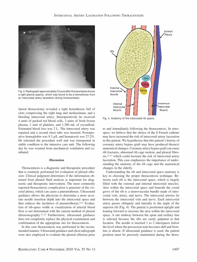
Understanding the rib and intercostal space anatomy iskey to choosing the proper thoracentesis technique. Be-tween each rib is the intercostal space, which is largelyfilled with the external and internal intercostal muscles.Also within the intercostal space and beneath the costalgrove of the rib is a neurovascular bundle made of inter-costal vein, artery, and nerve. The intercostal arteries liebetween the intercostal vein and nerve. Each intercostalartery passes obliquely and laterally to the angle of thesuperior rib (Fig. 4). The patient is positioned upright andleaning forward to increase the area within the intercostalspace. A site midway between the spine and axillary lineis selected because the ribs are easily palpated in thatlocation. The needle is inserted 1 to 2 interspaces belowthe level where the percussion note becomes dull and frem-itus is absent. If ultrasound guidance is used, the patientposition must be precisely maintained during the thora-
Fig. 3. Radiograph approximately 2 hours after thoracentesis showsa right pleural opacity, which was found to be a hemothorax froman intercostal artery laceration during thoracentesis.
Fig. 4. Anatomy of the intercostal rib space.
INTERCOSTAL ARTERY LACERATION FOLLOWING THORACENTESIS
RESPIRATORY CARE • NOVEMBER 2010 VOL 55 NO 11 1497

centesis. The needle should pass over the superior aspectof the rib to decrease the likelihood of injury to the neu-rovascular bundle that traverses the inferior rib margin.
Increase in tortuosity of intercostal arteries and decreasein the “safe area” (the space between the superior aspect ofthe lower rib and the lowest point of the intercostal artery)for thoracentesis appear to occur with aging.11 This in-creases the likelihood of intercostal artery laceration in theelderly, so extreme caution and strict adherence to therecommended thoracentesis techniques are essential.Proper thoracentesis techniques, including physical exam-ination confirmation of appropriate thoracentesis site, cor-rect patient positioning, ultrasound guidance, and closepost-thoracentesis monitoring, did not avoid intercostal ar-tery laceration in our patient, but the close monitoring didallow for early diagnosis and treatment.
The possibility that catheter diameter and distortion ofour patient’s intercostal anatomy may have caused theintercostal artery laceration should also be considered. Thiscase led to a change in catheter selection in our practice.Now we routinely substitute an 18-gauge angiocatheter forthe catheter that is currently part of the kit.
This case report emphasizes a thoracentesis complica-tion that is more likely in an elderly patient. Althoughintercostal artery laceration is rare, it should be considereda potential thoracentesis complication, particularly in theelderly. Every attempt should be made to minimize thisrisk. Close monitoring is necessary, and catheter size shouldbe carefully considered in the planning of thoracentesis inelderly patients.
REFERENCES
1. Collins TR, Sahn SA. Thoracentesis. Clinical value, complications,technical problems, and patient experience. Chest 1987;91(6):817-822.
2. Seneff MG, Corwin RW, Gold LH, Irwin RS. Complications asso-ciated with thoracentesis. Chest 1986;90(1):97-100.
3. Doyle JJ, Hnatiuk OW, Torrington KG, Slade AR, Howard RS.Necessity of routine chest roentgenography after thoracentesis. AnnIntern Med 1996;124(9):816-820.
4. Raptopoulos V, Davis LM, Lee G, Umali C, Lew R, Irwin RS.Factors affecting the development of pneumothorax associated withthoracentesis. AJR Am J Roentgenol 1991;156(5):917-920.
5. Aleman C, Alegre J, Armadans L, Andreu J, Falco V, Recio J, et al.The value of chest roentgenography in the diagnosis of pneumotho-rax after thoracentesis. Am J Med 1999;107(4):340-343.
6. Grogan DR, Irwin RS, Channick R, Raptopoulos V, Curley FJ, Bar-tter T, et al. Complications associated with thoracentesis. Arch InternMed 1990;150(4):873-877.
7. Colt HG, Brewer N, Barbur E. Evaluation of patient-related andprocedure-related factors contributing to pneumothorax followingthoracentesis. Chest 1999;116(1):134-138.
8. Mason RJ, Broaddus VC, Murray JF, Nadel JA (editors). Textbookof respiratory medicine, 4th edition, volume 2. Philadelphia: Mosby/Elsevier/Saunders; 2005:1919.
9. Barnes TW, Morganthaler TI, Olson EJ, Hesley GK, Decker PA.Sonograhically guided thoracentesis and rate of pneumothorax. J ClinUltrasound 2005;33(9):442-446.
10. Despars JA, Sasson CS, Light RW. Significance of iatrogenic pneu-mothoraces. Chest 1994;105(4):1147-1150.
11. Carney M, Ravin CE. Intercostal artery laceration during thoracen-tesis. Chest 1979;75(4):520-522.
12. Feller-Kopman D, Berkowitz D, Boiselle P, Ernst A. Large volumethoracentesis and the risk of reexpansion pulmonary edema. AnnThorac Surg 2007;84(5):1656-1662.
13. Sue DY, Lam K. Retention of catheter fragment after thoracentesis.Postgrad Med 1982;72(1):101-106.
14. Stewart BN, Block AJ. Subcutaneous implantation of cancer follow-ing thoracentesis. Chest 1974;66(4):456-457.
15. Heffner JE, Sahn SA. Abdominal hemmorrhage after perforation ofdiaphragmatic artery during thoracentesis (letter). Arch Intern Med1981;141(9):1238.
16. Feeney CM, Yoshioka H. A fatal case of pulmonary hemorrhagefrom thoracentesis (letter). West J Med 1993;158(6):638-639.
17. Sherman S, Nishio JN, Ravikrishnan KP. Massive pulmonary hem-orrhage complicating thoracentesis in chronic renal failure (letter).Chest 1979;76(1):118.
18. Vardi M, Dori G, Bitterman H. Large-bore thoracentesis: a casereport of a fatal consequence. Eur J Intern Med 2007;18(5):443-444.
19. Urschel JD. Balloon tamponade for hemmorrhage secondary to chesttube insertion. Respir Med 1994;88(7):549-550.
20. Brickner ME, Hillis LD, Lange RA. Congenital heart disease inadults: first of two parts. N Engl J Med 2000;342(4):256-264.
21. Feller-Kopman D. Ultrasound-guided thoracentesis. Chest 2006;129(6):1709-1714.
22. Mayo PH, Doelken P. Pleural ultrasonography. Clin Chest Med 2006;27(2):215-227.
23. Greenwald LV, Baisden CE, Panogiotis SN. Rib fractures in coro-nary bypass patients: radionuclide detection. Radiology 1983;148(2):553-554.
24. Locke TJ, Griffiths TL, Mould H, Gibson GJ. Rib cage mechanicsafter median sternotomy. Thorax 1990;45(6):465-468.
25. Kenyon CM, Pedley TJ, Higenbottam TW. Adaptive modeling of thehuman rib cage in median sternotomy. J Appl Physiol 1991;70(5):2287-2302.
26. Jantz MA, Veena AB. Pleural fibrosis. Clin Chest Med 2006;27(2):181-191.
27. Lee GY, Vaz MA, Ely KA, McDonald EC, Thompson PJ, NesbittJC, et al. Symptomatic persistent post-coronary artery bypass pleuraleffusions requiring operative treatment. Chest 2001;119(3):795-800.
INTERCOSTAL ARTERY LACERATION FOLLOWING THORACENTESIS
1498 RESPIRATORY CARE • NOVEMBER 2010 VOL 55 NO 11