international council for the study of virus and other ... · luis conci, argentina tadeusz...
TRANSCRIPT
1
International Council for the Study of Virus and other Graft
Transmissible Diseases of Fruit Crops (ICVF)
Scientific Committee: Nikolaos Katis, Greece (co-chair ICVF)
Nobuyuki Yoshikawa, Japan (co-chair ICVF)
Marina Barba, Italy Wilhelm Jelkmann, Germany
Assunta Bertaccini, Italy Gerard Jongedijk, The Netherlands
Kadriye Caglayan, Turkey
Thierry Candresse, France
Gabi Krczal, Germany
Stuart MacFarlane, UK
Luis Conci, Argentina Tadeusz Malinowski, Poland
Fiona Constable, Australia Hans Maree, South Africa
Francesco Di Serio, Italy Robert Martin, USA
Nicola Fiore, Chile Hong Ni, P.R. China
Ricardo Flores, Spain Vicente Pallás, Spain
Pascal Gentit, France Svetlana Paunovic, Republic of Servia
Delano James, Canada Josef Spak, Czech Republic
Ioannis Tzanetakis, USA (webmaster)
2
The 24th International Conference on Virus and Other Graft
Transmissible Diseases of Fruit Crops
5-9 June, 2017, Thessaloniki Greece
Organizing Committee
Nikolaos Katis (Aristotle University of Thessaloniki, Greece)
Varvara Maliogka (Aristotle University of Thessaloniki, Greece)
Ioannis Tzanetakis (University of Arkansas, USA)
Christina Varveri (Benaki Phytopathological Institute Athens, Greece)
Elisavet Κ. Chatzivassiliou (Agricultural University of Athens, Greece)
Efthimia Tsagris (University of Crete, Heraklion, Greece)
Kriton Kalantidis (University of Crete, Heraklion, Greece)
Grigoris Amoutzias (University of Thessaly, Larisa, Greece)
Ioannis Livieratos (Mediterranean Agronomic Institute of Chania, Greece)
Evangelos Vellios (University of Thessaly, Volos, Greece)
3
Welcome to the 24th International Conference on Virus and Other
Transmissible Diseases of Fruit Crops (ICVF)
Dear Colleagues,
It’s been almost 30 years since the 14th ICVF Conference was held in Thessaloniki. The
organizing committee is excited and honored to welcome you to the 24th ICVF Conference
from June 5th-9th. In this meeting participants from 28 countries will gather to our
beautiful city to present their intriguing research on viruses and virus diseases of fruit
crops.
In addition to the technical presentations we have included in the program a workshop on
clean plant movement “Transport of plant propagation material through borders and the
production of high quality (virus-tested) propagative material of fruit trees and small
fruits”. During the workshop individuals from seven countries will share their experiences
and discuss their ideas along with the other participants about the future. We will
continue with a round table on the status quo and the role of NGS in certification and
quarantine.
The organizing committee would like to thank all of you for attending the 24th ICVF
Conference. Special thanks go to the invited speakers for accepting our invitation to
present state-of- the art talks on a pressing subject matter.
We hope you will enjoy the meeting, the beauty and the culture of Thessaloniki, the
capital of Greek Macedonia. Keep in mind that we are here to help you enjoy both the
Conference and Greek life.
Nikolaos Katis
Convener of the 24th ICVF and
Members of the Organizing Committee
4
We sincerely thank the sponsors of the 24th International Conference on Virus and other
Graft Transmissible Diseases of Fruit Crops.
Sponsoship
5
Scientific Program of ICVF 2017
Thessaloniki 5-9 June
Sunday, 4 June
17:30-19:30 Registration and poster hanging
19:30-20:00 Invited lecture: A. Molassiotis (Aristotle University of Thessaloniki, School of
Agriculture, Pomology Laboratory, Greece) “Fruit tree cultivation and production in
Greece: present status and future prospects”
20:00 Welcome drink-Greek wine presentation and tasting
Monday, 5 June
08:00-09:00 Registration and poster hanging
09:00-09:20 Meeting opening/ Welcome by the Convener N.I. Katis and the Dean of
Faculty of Agriculture, Forestry and Natural Environment D. Koveos)
09:20-09:50 Invited lecture: V.I. Maliogka (Aristotle University of Thessaloniki, School of
Agriculture, Plant Pathology Laboratory, Greece) “Virus and other graft-transmissible
diseases of fruit trees in Greece-An Overview”
9:50-11:10 Oral Session 1: New and Emerging graft-transmissible pathogens and
related diseases-Εtiology I
Chairs: W. Jelkmann, V. Pallas
09:50-10:10 O-1: Genomic and biological characterization of Apple necrotic mosaic virus
infecting apple trees in China. F. Xing, H.Q. Wang, S.F. Li
10:10-10:30 O-2: The complete sequence of a new Trichovirus isolated from peach trees in
Mexico. R. De la Torre, V. Pallas, J.A. Sánchez-Navarro
10:30-10:50 O-3: Discovery of a novel plant virus with unique genome features from black
currant. T. Thekke-Veetil, T. Ho, J.D. Postman, I.E. Tzanetakis
10:50-11:10 O-4: Molecular characterization of a phytoplasma associated to phyllody and
witches’ broom in strawberry (Fragaria x ananassa Duch.). N. Quiroga, A. Zamorano, W.
Cui, N. Fiore
6
11:10-11:40 Coffee break
11:40-13:00 Oral Session 2- New and Emerging graft-transmissible pathogens and
related diseases-Εtiology II
Chairs: A. Zamorano, N. Hong
11:40-12:00 O-5: A new potexvirus infecting babaco (Carica pentagona) and papaya (C.
papaya) in Ecuador. R.A. Alvarez-Quinto, J.F. Cornejo, D.F. Quito-Avila
12:00-12:20 O-6: A novel emaravirus associated with pear chlorotic leaf spot disease. H.Z.
Liu, G.P. Wang, Z.K. Yang, N. Hong
12:20-12:40 O-7: New cherry-adapted strain of Plum pox virus. S. Chirkov, P. Ivanov, A.
Sheveleva, G. Osipov
12:40-13:00 O-8: Identification and partial characterization of a new ilarvirus infecting
Prunus sp. K. Efthimiou, P.G. Pappi, A. Olmos, T. Candresse, E. Xylogianni, N.I. Katis, V.I.
Maliogka
13:00-14.30 Lunch break
14:30-15:00 Invited lecture: F. Di Serio (Istituto per la protezione sostenibile delle Piante,
CNR, Italy) “Viroids infecting fruit trees: From Bioassays to Next Generation Sequencing”
15:00- 16:20 Oral Session 3- New and Emerging graft-transmissible pathogens
and related diseases-Εtiology III
Chairs: R.R. Martin, A. Marais
15:00-15:20 O-9: Characterization of a new and distinct population of hammerhead
viroid-like RNA detected by next-generation sequencing using total RNA from apple
(Malus domestica) cv. Pacific Gala. A. Messmer, D. Sanderson, G. Braun, P. Serra, R. Flores,
D. James
15:20-15:40 O-10: Occurrence and genetic variability of elderberry carlaviruses infecting
Sambucus nigra in the Czech Republic. D. Šafářová, K. Vavroušková, T. Zemánek, T.
Candresse, M. Navrátil
15:40-16:00 O-11: An un-designed Closterovirus infecting kiwifruit plants grown in China.
S.H. Wen, C.X. Zhu, G.P. Wang, Z.K. Yang, N. Hong
16:00-16:20 O-12: A novel member of the Luteoviridae associated with Raspberry leaf
curl disease. P.L. Di Bello, A. Diaz-Lara, R.R. Martin
16:20-16:50 Coffee break
7
16:50-17:50 Oral Session 4-New and Emerging graft-transmissible pathogens and
related diseases-Εtiology IV
Chair: I. Koloniuk, M. Glasa
16:50-17:10 O-13: Mume virus A: A new capillovirus from Japanese apricot (Prunus
mume). A. Marais, C. Faure, S. Theil, T. Candresse
17:10-17:30 O-14: The Geneva Complex – an interesting disease perspective based on the
results of NGS analysis. D. James, J. Phelan, G. Jesperson
17:30-17:50 O-15: Detection and characterization of fig viruses in Greece. C.L. Sassalou, L.
Lotos, C. Valachas, A. Olmos, A. Chetas, E.K. Chatzivassiliou, N.I. Katis, V.I. Maliogka
17:50-18-10 O-16: Discovery and molecular characterization of a new luteovirus
associated with rapid apple decline in USA. H.W. Liu, L.-P. Wu, Peter, K., D., Liu, Z., Cao,
M.-J., R. Li
18-10-19:40 Poster session I (P1-P19)
Tuesday, 6 June
09:00-09:30 Invited lecture: T. Candresse (INRA, University of Bordeaux, France) “A
perspective on NGS use in plant virology and on implications for the safe movement of
plant materials”
09:30-10:50 Oral Session 5-Detection and Characterization Ι
Chairs: D. James, H. Maree
09:30-09:50 O-17: Evaluation of total RNA high-throughput sequencing for virus
detection. R. Bester, M. Visser, J.T. Burger, H.J. Maree
09:50-10:10 O-18: A high intra-host Cherry virus A (CVA) population heterogeneity
detected in cherry trees in Slovakia. M. Glasa, L. Predajňa, N. Sihelská, Z. Vozárová, K.
Šoltys
10:10-10:30 O-19: NGS studies on fruit tree screenhouses and stock collections in
Hungary. N. Czotter, T. Varga, D. Baráth, J. Molnár, J. Balássy, T. Deák, G.E. Tusnády, L.
Kocsis, É. Preininger, J. Burgyán, É. Várallyay
8
10:30-10:50 O-20: Luteoviruses in Prunus species: how many are there? T. Candresse, A.
Marais, D. Šafářová, M. Navratil, R. Sorrentino, D. Alioto, C. Faure, L. Svanella-Dumas, S.
Theil
10:50-11:20 Coffee break
11:20-12:20 Oral Session 6-Detection and Characterization ΙI
Chair: D. Šafářová, N. Fiore
11:20-11:40 O-21: Study on the occurrence and phylogenetic relationships of pome and
stone-fruit tree phytoplasmas in Greece. M.C. Holeva, E.M. Koutsioumari, P.E. Glynos, C.D.
Karafla, E. Siderea, A. Togias, J. Kagias, S. Ioannidou, Ch. Arampatzis
11:40-12:00 O-22: New developments in the epidemiology of Little cherry virus 1 and its
occurrence in European Plum (Prunus domestica L.) in Belgium. R. Tahzima, S. Massart, G.
Peusens, T. Beliën, K. De Jonghe
12:00-12:20 O-23: Variability of Strawberry crinkle virus. E.T.M. Meekes, M. Hooftman, H.
Konings, M.J.M. Ebskamp
12:20-12:40 O-24: Virus and viroids infecting pear trees (Pyrus communis L.) in Chile. G.
Medina, N. Quiroga, P. Méndez, A. Zamorano, N. Fiore
12:40-14:00 Lunch break
14:00-14:30 Invited lecture: A. Bertaccini (University of Bolognia, Department of
Agricultural Sciences, Italy) “Fruit trees and Phytoplasmas: A well settled association”
14:30-15:50 Oral Session 7- Detection and Characterization ΙΙI
Chairs: D. Skoric, A. Olmos
14:30-14:50 O-25: Germplasm as invaluable source of ribes infecting viruses. J. Přibylová,
J. Špak, I. Koloniuk, K. Petrzik
14:50-15:10 O-26: Pear decline: The fate of Phytoplasmas in an Italian orchard. S.
Paltrinieri, K. Troni, P.P. Bortolotti, R. Nannini, A. Bertaccini
15:10-15:30 O-27: Viruses infecting cherry and sour cherry in the Chech Republic. J. Špak,
J. Přibylová, O. Lenz, J. Fránová, I. Koloniuk
9
15:30-15:50 O-28: Multilocus sequence typing of Croatian ‘Candidatus Phytoplasma mali’
strains. I. Križanac, J. Plavec, Z. Budinšćak, D. Škorić, M. Šeruga Musić
15.50-16.10 Coffee break
16:10-17:30 Oral Session 8-Detection and Characterization ΙV
Chair: J. Špak, C. Varveri
16:10-16:30 O-29: Phytoplasma detection in hazelnut in Chile. S. Perez Fuentealba, S.
Paltrinieri, J. Guerrero, N. Fiore, A. Bertaccini
16:30-16:50 O-30: Detection and characterization of cherry viruses in Chile. A. Zamorano,
N. Quiroga, C. Fernández, L. Rivera, D. Soto, A.M. Pino, N. Fiore
16:50-17:10 O-31: Simultaneous detection of pome fruit tree viruses by triplex
quantitative RT-PCR. I. Malandraki, D. Beri, I. Isaioglou, A. Olmos, C. Varveri, N. Vassilakos
17:10-17:30 O-32: European stone fruit yellows phytoplasmas infecting apricot in Serbia
and in Italy cluster mainly in two main MLT groups. N. Contaldo, J. Stepanović, F. Pallara,
A. Bertaccini, B. Duduk
17:45-20:00 City Tour
21:00 Scientific committee dinner
Wednesday, 7 June
09:00-18:00 Scientific excursion
Visit to Commercial Pome and Stone Fruit orchards in Veroia and Naoussa (District of
Imathia)
- Vergina Archaeological site (Capital of Ancient Macedonia)
- Lunch in Naoussa
- Return to Thessaloniki
Thursday, 8 June
09:00-12:10 Workshop on ‘Transport of plant propagation material through borders and
the production of high quality (virus-tested) propagative material of Fruit trees and small
fruits’. Chair: I.E. Tzanetakis
10
1. 09:00-09:20 Pascal Gentit, France
2. 09:20-09:40 Nicola Fiore, Chile
3. 09:40-10:00 Bob Martin, USA
4. 10:00-10:20 Fiona Constable, Australia
10:20-10:50 Coffee break
5. 10:50-11:10 Hans Maree, South Africa
6. 11:10-11:30 Delano James, Canada
7. 11:30-11:50 Christina Varveri, Greece
11:50-12:10 O-33: The importance and challenges of reference collections. W. Menzel
12:10 -13:40 Lunch break
13:40-15:40 Round table. USDA-NCPN leadership via Skype or zoom. Status quo and role
of NGS in certification and quarantine. How are we approaching sequences vs viruses?
15:40-16:10 Coffee break
16:10-17:40 Poster session II (P20-P38)
20:30 Gala Dinner
Friday, 9 June
09:00-09:30 Invited lecture: K. Kalantidis (University of Crete, Department of Biology,
Greece) “Disarming plant defense by suppressing DICER expression”
9:30-10:30 Oral Session 9- Pathogen-Host Interactions
Chair: S.A. MacFarlane
09:30-09:50 O-34: Viral superinfection in woody and herbaceous hosts. I. Koloniuk, J.
Fránová, J. Přibylová, K. Petrzik, O. Lenz, P. Svoboda, H. Jakešová, J. Špak
09:50-10:10 O-35: UK survey and grafting studies of Raspberry leaf blotch emaravirus.
W.J. McGavin, S. Newman, J. Allen, S.A. MacFarlane
10:10-10:30 O-36: Interaction of the pathogen ‘Candidatus Phytoplasma Mali’ with the
plant host. A. Konnerth, G. Krczal, K. Boonrod
11
10:30-11:00 Closing ceremony
11:00-11:30 Coffee break
11:30 Official Meeting of ICVF
1. Next meeting location with brief introduction of potential hosts
2. Replacement of Scientific Committee members
3. Other Issues
LIST OF POSTERS
P-1. NGS STUDIES OF THE VIROME OF FIG TREES IN GREECE. M. Kaponi, E. Vellios, K.
Filippou, T. Sano
P-2. DIVERSITY OF CHERRY VIRUS A GENOMES. P. Kesanakurti, M. Rott
P-3. SURVEY OF POTENTIAL CICADELLID VECTORS OF PHYTOPLASMAS IN SWEET CHERRY
TREES IN KAHRAMANMARAS-TURKEY: A PRELIMINARY REPORT. M. Gazel, K. Çağlayan, K.
Kaya, H. Başpınar
P-4. IDENTIFICATION OF DIFFERENT PHYTOLASMAS INFECTING SWEET CHERRY TREES IN
TURKEY. M. Gazel, K. Çağlayan
P-5. FIRST DETECTION OF EUROPEAN STONE FRUIT YELLOWS PHYTOPLASMA IN PEACH
TREES ON THE TERRITORY OF CANTON OF GENEVA, SWITZERLAND. Α. Etropolska, F. Lefort
P-6. DETECTION AND CHARACTERIZATION OF RASPBERRY BUSHY DWARF VIRUS IN
DIFFERENT HOSTS IN TURKEY. K. Caglayan, M. Gazel, E. Elçi, M. Viršček Marn, I. Mavric
Pleško
P-7. MOLECULAR CHARACTERIZATION AND GENETIC DIVERSITY OF PLUM POX VIRUS
STRAINS IN THRACE REGION OF TURKEY. K. Gürcan, S. Teber, O. Canbulat, K. Çağlayan
P-8. THE PEAR PSYLLID CACOPSYLLA BIDENS: A PUTATIVE VECTOR OF ‘CANDIDATUS
PHYTOPLASMA PYRI’ IN BULGARIA. A. Etropolska, B. Jarausch, W. Jarausch, D. Burckhardt,
G. Trenchev
P-9. DETECTION AND CHARACTERIZATION OF THE COMPLETE GENOME OF THE FIRST
CHERRY (C) STRAIN ISOLATE OF PLUM POX VIRUS DETECTED IN GERMANY IN SOUR
CHERRY (PRUNUS CERASUS). W. Jelkmann, D. Sanderson, C. Berwarth, D. James
P-10. RUBUS YELLOW NET VIRUS AND BLACK RASBERRY NECROSIS VIRUS, NEWLY
DETECTED VIRUSES IN RASPBERRRY PLANTATIONS IN REPUBLIC OF SERBIA. A.S. Paunovic,
D. Jevremović
P-11. EX-POST ANALYSES OF PUBLIC DATABASES ALLOWED IDENTIFICATION OF A NOVEL
VIRUS INFECTING PEACH. B. Navarro, S. Zicca, F. Palmisano, F. Di Serio
P-12. COLLECTION OF PATHOGENS OF HOPS. P. Svoboda, I. Malirova
12
P-13. HIGH THROUGHPUT SEQUENCING OF siRNAS AND VIRUS DIAGNOSTIC: DO
SEQUENCE ANALYSIS STRATEGIES REALLY MATTER? RESULTS OF AN INTERNATIONAL
PROFICIENCY TESTING. S. Massart, H.J. Maree, I. Adams, M. Chiumenti, K. De Jonghe, I.
Koloniuk, P. Kominek, J. Kreuze, D. Kutnjak, L. Lotos, T. Olivier, M. Pooggin, AB. Ruiz-
García, D. Šafářová, P.H.H. Schneeberger, N. Sela, É. Várallyay, E. Vainio, E. Verdin, M.
Westenberg, Y. Brostaux, T. Candresse
P-14. MONITORING OF VECTORS PHENACOCCUS ACERIS AND PSEUDOCOCCUS
MARITIMUS OF LITTLE CHERRY VIRUS 2 IN SWEET CHERRY ORCHARDS IN BELGIUM. G.
Peusens, R. Tahzima, K. De Jonghe, D. Bylemans, T. Beliën
P-15. INDICATION THAT THE PLUM POX VIRUS P1 PROTEIN IS PREFERENTIALLY
EXPRESSED IN THE ROOTS OF NICOTIANA BENTHAMIANA DURING EARLY STAGE OF
INFECTION. Z. Vozárová, Z. Šubr
P-16. LONG-TERM FOLLOW-UP OF PPV-REC AND PPV-D IN AN EXPERIMENTAL PLUM
ORCHARD. D. Jevremović, A.S. Paunović, S. Dallot
P-17. OCCURRENCE AND DISTRIBUTION OF CITRUS VIRUS AND VIROID DISEASES IN
JORDAN. M.N. Salem, A. Mansour, S. Abu-Romman, A. Abo-Sherbi, A. Abdeen
P-18. IDENTIFICATION OF A NEW VITIVIRUS FROM BLUEBERRY. T. Thekke-Veetil, T. Ho, J.
Polashock, I.E. Tzanetakis
P-19. PARTIAL SEQUENCE OF GOOSEBERRY VEIN BANDING ASSOCIATED VIRUS IN
REDCURRANT IN SLOVENIA. J. Lamovšek, I. Mavrič Pleško, D. Koron, A.B. Ruiz-García, M.
Viršček Marn, A. Olmos
P-20. APRICOT (PRUNUS ARMENIACA L.)–NEW HOST OF LITTLE CHERRY VIRUS 1. D.
Šafářová, C. Faure, T. Candresse, M. Navrátil, T. Nečas, A. Marais
P-21. NOVEL RNA VIRUSES ASSOCIATED WITH APPLE RUBBERY WOOD AND APPLE FLAT
LIMB DISEASES. M. Rott, P. Kesanakurti, I. Boyes, C. Berwarth, H. Rast, W. Jelkmann
P-22. RESULTS OF SURVEILLANCE OF OCCURRENCE OF 'CANDIDATUS PHYTOPLASMA
MALI' AND 'CANDIDATUS PHYTOPLASMA PYRI' IN NURSERIES OF REPUBLIC OF SRPSKA
(BOSNIA AND HERZEGOVINA). D. Delić, M. Radulović, B. Lolić, G. Durić
P-23. IDENTIFICATION OF AN RNA SILENCING SUPPRESSOR ENCODED BY THE GENOME OF
LITTLE CHERRY VIRUS 1. A. Κatsiani, K. Κatsarou, K. Kalantidis, N.I. Κatis, V.Ι. Μaliogka
P-24. SEASONAL FLUCTUATION OF LITTLE CHERRY VIRUS 1 (LChV-1) TITER IN SWEET
CHERRY CULTIVARS. A. Κatsiani, P. Pappi, A. Olmos, K.E. Efthimiou, V.I. Maliogka, N.I.
Katis
P-25. FULL GENOME SEQUENCING OF A DIVERGENT ISOLATE OF LChV-1 AND
MONITORING OF THE VIRUS DERIVED siRNAs IN SWEET CHERRY OVER FOUR SEASONS.
A.T. Katsiani, A. Olmos, T. Candresse, A.B. Ruiz-Garcia, N.I. Katis, V.I. Maliogka
P-26. IDENTIFICATION OF A PRUNUS VIRUS F-LIKE VIRUS IN SWEET CHERRY. L. Lotos, T.
Candresse, A. Olmos, N.I. Katis, V.I. Maliogka
P-27. INCIDENCE AND MOLECULAR VARIABILITY OF APPLE CHLOROTIC LEAF SPOT VIRUS
IN POME FRUITS IN SLOVENIA. M. Viršček Marn, I. Mavrič Pleško
13
P-28. MOLECULAR DETECTION AND CHARACTERIZATION OF PHYTOPLASMAS IN
ORNAMENTAL POMEGRANATES. K. Caglayan, M. Gazel, M. Acıoğlu
P-29. EVALUATION OF THE SUSCEPTIBILITY OF APRICOT CULTIVARS TO PPV IN HIGH-
CONTAINMENT GREENHOUSE. Y. Brans, J. Castaing, K. Bresson
P-30. THE FIRST DETECTION OF PLUM POX VIRUS IN WESTERN SIBERIA. Y. Shneyder, M.
Tikhomirova, O. Morozova, T. Zhivaeva, Y. Prikhodko
P-31. DIAGNOSIS, VIRUS CLEANING AND CRYOPRESERVATION OF RASPBERRY,
BLACKBERRRY AND SHALLOT (RUB&AL). Z. Zhang, C. Spetz, E. Lysøe, D.R. Blystad
P-32. DETECTION AND MOLECULAR CHARACTERIZATION OF AN UNUSUAL VIRUS IN
CHERRY. S. Milusheva, J. Phelan, N. Piperkova, V. Nikolova, D. James
P-33. H2020-MCSA-RISE-2016-VIRUS FREE FRUIT NURSERIES (VirFree). V.I. Maliogka, N.I.
Katis, D. Altenbach, K. Caglayan, C. Chondroudis, F. Codoner, A.T. da Cunha, F. Di Serio, B.
Dichio, L. Formica, H. Ilbagi, K. Kalantidis, S. Li, A. Olmos, G. Pietersen, C. Ritzenthaler, E.
Tanriver
P-34. VIRTOOL: A NEXT GENERATION DATA ANALYSIS TOOL FOR DETECTION OF PLANT
VIRUSES. I. Boyes, M. Rott
P-35. PCR OPTIMISATION IN A PROGRAMME TO MONITOR THE PRESENCE OF
‘CANDIDATUS PHYTOPLASMA MALI’, ‘CA. P. PYRI’ AND ‘CA. P. PRUNORUM’. M. Pastore, E.
Stingo, V. Orologiaio, G. Consalvo, V. Graziano, T. Rosco, A. Salsano, G. Stella, R. Griffo
P-36. EVIDENCE OF CIRCULAR RNA ASSOCIATED WITH BLACKCURRANT LEAF CHLOROSIS
ASSOCIATED VIRUS. D. James, J. Phelan, D. Sanderson
P-37. T-RECS: RAPID AND LARGE-SCALE DETECTION OF RECOMBINATION EVENTS AMONG
DIFFERENT EVOLUTIONARY LINEAGES OF VIRAL GENOMES. M. Tsimpidis, G. Bachoumis, K.
Mimouli, Z. Kyriakopoulou, D.L. Robertson, P. Markoulatos, G.D. Amoutzias
P-38. FIRST INSIGHT INTO STRAWBERRY VIROME USING NEXT GENERATION SEQUENCING
IN THE CZECH REPUBLIC. J. Fránová, I. Koloniuk, J. Přibylová, O. Lenz , J. Kučerová, L.
Valentová

14

15
Invited Lectures
16
FRUIT TREE CULTIVATION AND PRODUCTION IN GREECE: PRESENT STATUS
AND FUTURE PROSPECTS
A. Molassiotis
Laboratoty of Pomology, Faculty of Agriculture, Aristotle University of Thessaloniki, 54124
Thessaloniki, Greece ([email protected])
Greece is characterized by climatic conditions suitable for the cultivation of many kinds of fruit tree crops. In Southern part of Greece, especially Greta and Peloponnisos olives and citrus are grown, whereas the main producing regions for deciduous fruit crops are the Central Macedonia and primarily the area of Pella and Imathia. Peach, kiwifruit, cherry and apple are the four main deciduous fruit species produced in Greece. In the last years, deciduous fruit tree production has moved progressively towards southern regions of Greece looking for extra precocious production (at the end of April and May). Fruits play a very important role for the Greek economy and society as well, since they constitute basic component of Greek nutrition. Notably, the fruits produced in Greece have experienced remarkably high growing rates during the last 7-years. Greece exports a large part of its domestic production (about 80%) mainly to the European countries and to the countries of Eastern Europe. For example, Greece is the largest exporter of canned peaches worldwide, accounting for over the one-third of global canned peach exports. In northern Hemisphere, Greece is the second largest producer of kiwifruit (170,000 tons per year, the production has doubled compared to 7 years ago) following Italy. Meanwhile, the Greek fruit industry has experienced a significant upgrade in terms of production technology and particular what concerns the cultivation of new cultivars, new rootstocks, training systems and development of integrated production and certification systems. Although the high number of cultivars released from worldwide breeding programs increases their availability to growers, however it is difficult to select the most adapted cultivars to specific Greek conditions in terms of their agronomical performance and fruit quality. Additionally, the technological advances in irrigation, fertilization, crop protection, postharvest technology, fruit certification and traceability in the last decades allowed a consistent improvement of Greece competitiveness, in particular to export markets. In the future many opportunities appear for the expansion of Greek fruit and vegetable production. Greece should take the advantage of the ongoing promotion and tendency towards the healthy Mediterranean diet to increase the cultivation and the exports of the high quality fruit-related products.
17
VIRUS AND OTHER GRAFT-TRANSMISSIBLE DISEASES OF FRUIT TREES IN GREECE- AN OVERVIEW
V.I. Maliogka Aristotle University of Thessaloniki, Faculty of Agriculture, Forestry and Natural
Environment, School of Agriculture, Plant Pathology Laboratory, 54 124, Thessaloniki, Greece ([email protected])
Fruit tree cultivation and especially cultivation of Prunus and Malus species is of great importance for the Greek agro-industry. Prunus species constitute the majority of fruit trees plantations and a high percentage of both Prunus and Malus production is exported. Vegetative propagation and grafting of infected plant material have considerably contributed to a wide distribution of several viral and other graft-transmissible agents on these plants species. The main virus in Prunus is Plum pox virus (PPV), which has caused considerable losses in apricot and plum production and is widespread all over the country. Moreover, Ilarviruses including Prunus necrotic ringspot virus (PNRSV) and Prune dwarf virus (PDV) also occur in high proportion in all Prunus species. Recently, Little cherry virus 1 (LChV-1) has been identified in sweet and sour cherries, plum and peach with its incidence in sweet cherries to be up to 94%. However, no association has been observed between the presence of LChV-1 and symptoms of Little Cherry Disease. Peach latent mosaic viroid (PLMVd) and Hop stunt viroid (HSVd) have also been found to be widespread in different Prunus species. Finally, apricot and the Japanese plums are seriously suffering from European Stone Fruit Yellows Phytoplasma (ESFY). The situation in Malus species (apples and pears) is not that serious although a number of viruses such as Apple chlorotic leaf spot virus (ACLSV), Apple stem pitting virus (ASPV), Apple stem grooving virus (ASGV) have been identified in a rather high incidence with the exception of Apple mosaic virus (ApMV). ASPV has been seriously affecting quince, where it causes the Fruit Deformation Disease resulting to unmarketable fruit production. Also, Apple scar skin viroid (ASSVd) and Pear blister canker viroid (PBCVd) have been detected in several Malus plantations especially in Peloponnese. Finally, Apple proliferation phytoplasma has been detected in apples and may be associated with small fruit production in the area of Zagora (Pilion) one of the most important apple producing areas of Greece. Despite our knowledge on these pathogens the etiology of several virus-like diseases is still not clear in Greece while the application of next generation sequencing technologies has revealed the presence of novel viral pathogens with unknown agronomic importance in Prunus or Malus plantations. During the last years, national and European projects are undertaken in order to combat the above graft-transmissible agents through the production of high quality propagative material.

18
19
Oral Session 1-
New and Emerging graft-transmissible
pathogens and related diseases-Etiology I
20
GENOMIC AND BIOLOGICAL CHARACTERIZATION OF APPLE NECROTIC MOSAIC VIRUS INFECTING APPLE TREES IN CHINA
Xing, F.1,2, Wang, H.Q.1 and Li, S.F.2
1 College of Horticulture, China Agricultural University, China 2 Institute of Plant Protection, Chinese Academy of Agricultural Sciences, China
Introduction China is the most extensively apple growing country with about 50% of production worldwide (FAO, 2017). Apple mosaic disease with approximately 62.7% incidence rate in some apple orchards of China, has been a serious threat to the development of apple industry. It is believed that apple necrotic mosaic virus (ApNMV), a recently reported ilarvirus, which was highly associated with mosaic symptom in apple leaves, is the causal agent of apple mosaic disease in China (1). In this paper, sequence-based phylogenetic analyses and biological characterization were conducted to shed light on the genetic differentiation of the Chinese ApNMV isolates with that originating from Japan and the possible herbaceous hosts. Materials and Methods Multiple sequence alignment and phylogenetic analysis were performed using the full genomes and CP genes from 17 Chinese isolates, respectively, as well as ApNMV Japanese-isolate from NCBI. Infectious cDNA clones of ApNMV were constructed using pCASS4-Rz vector and inoculated via agroinfiltration (2). Results and Discussion Total 6 complete nucleotide sequences and other 11 RNA3 partial sequences of ApNMV from different apple tree parts and cultivars were sequenced, whose genomes consist of RNA1 (3378 - 3380 nt), RNA2 (2778 - 2786 nt) and RNA3 (1909 - 1955 nt). Similarities of ApNMV RNA1, RNA2 and RNA3 genome sequences with that originating from Japan were 95.4% - 96.5%, 88.5% - 94.5%, 86.2% - 93.4%, respectively. Phylogenetic analysis suggested that ApNMV was clustered together with Prunus necrotic ringspot virus and Apple mosaic virus, but showing a low sequence similarities (53.3% - 65.5%) with them. Infectious cDNA clones of ApNMV were constructed and inoculated to N. benthamiana via agroinfiltration techniques. A high incidence (96.8%) of ApNMV was recorded in N. benthamiana by RT-PCR consistent with ELISA results after 21 dpi. Sap inoculation by the ApNMV-positive N. benthamiana was infectious to the healthy N. benthamiana seedlings. It showed that among 10 seedlings, only 5 of them were infected with ApNMV after 7 dpi. At the same time, seedlings of Ch. amaranticolor, C. sativus and Ch. quinoa were agroinfiltrated, but no tested plants were infected with ApNMV. Taken together, we concluded that the constructed cDNA clones of ApNMV had infectious bioactivity. Although the genome of ApNMV was recently described (1), this is the first molecular analysis on the intra-strain genetic diversity and biological characterization of ApNMV. Certainly, more isolates from China and abroad need to be obtained to illustrate geographical differentiation and evolutionary history. A wider host range of ApNMV is also need to be determined using the constructed infectious clones.
References
1. Noda, et al., 2017. Journal of General Plant Pathology 83: 83-90. 2. Annamalai, et al., 2005. Virology 338:96-111.
O-1
21
THE COMPLETE SEQUENCE OF A NEW TRICHOVIRUS ISOLATED FROM PEACH
TREES IN MEXICO
De la Torre, R.1, Pallas, V.2 and Sánchez-Navarro, J.A.2 1 FES-Iztacala, UNAM, México
2 Instituto de Biología Molecular y Celular de Plantas, IBMCP (CSIC-UPV),
Valencia, Spain ([email protected])
Introduction
Stone fruit trees are infected by a large number of different and economical important
viruses and viroids. The survey of peach samples from different Mexican regions by
multiplex RT-PCR with the capability to detect and identify eight stone fruit tree viruses (1)
revealed the presence of an unexpected amplicon of 277 nucleotides which electrophoretic
mobility did not match with any of the expected viruses. In the present work, we have
determined the complete sequence of two trichoviruses, one of them not previously
described, including the 5’ and 3’ termini of the viral and subgenomic RNAs.
Materials and Methods
Leaves from symptomatic peach trees showing yellow mottle, chlorotic ringspot, linear
patterns and mosaic were selected to generate enriched dsRNAs preparations (2). RT-PCR
was carried out with the One Step High Fidelity System (Invitrogen, Carlsbad, CA). The
amplified DNA fragments were cloned in bacterial plasmids using the Thermo Scientific ™
InsTAclone ™ PCR Cloning Kit. The 5’ and 3’ termini of viral and subgenomic RNAs were
determined by RACE analysis using specific primers targeting internal regions.
Results and Discussion
The cloning and further analysis of the nucleotide sequence of the unexpected amplicon of
277 nucleotides (nt) revealed a percentage of identity of 83% and 77% with the coat protein
gene of Cherry mottle leaf virus and Peach mosaic virus, respectively. The use of specific
primers targeted to conserved regions of trichoviruses permitted the amplification of the
complete genome of two viruses of 7981nt and 7985nt, respectively. Blast analysis using the
nucleotide sequence of the former virus rendered a percentage of 83% with Peach mosaic
virus (PMV) (DQ117579) meanwhile the second virus showed an identity percentage of
69.3% with the same PMV isolate. Both viruses showed a 69.2% identity between them.
According to Blast analysis, we considered the former virus of 7981nt as a Mexican isolate of
PMV meanwhile the second virus of 7985nt a new Mexican trichovirus that we tentatively
named Peach Mexican virus. RACE analysis revealed the presence of three subgenomic RNAs
for both viruses, starting at nt 5610 (5’GAAATT), 6595 (5’GAACAA) and 7341 (5’GTGATA) for
the Mexican PMV isolate and nt 5630 (5’GAAATT), 6609 (5’GAATAT) and 7342 (5’GTGTGA)
for the new Mexican trichovirus. All together, these results revealed that the new viral
sequences characterized in Mexican peach orchards correspond to a new PMV isolate and a
new trichovirus, respectively.
References 1. Sánchez-Navarro et al., 2005. European Journal of Plant Pathology 111:77-84.
2. Valverde et al., 2017. Mexican Journal of Phytopathology Enero:80-105.
O-2
22
DISCOVERY OF A NOVEL PLANT VIRUS WITH UNIQUE GENOME FEATURES FROM BLACK CURRANT
Thekke-Veetil, T.1, Ho, T,1, Postman, J.D2. and Tzanetakis, I.E.1
1 Department of Plant Pathology, Division of Agriculture, University of Arkansas System, Fayetteville, AR 72701, United States of America ([email protected])
2 National Clonal Germplasm Repository, United States Department of Agriculture, Corvallis, OR 97333, United States of America
Introduction The family Betaflexiviridae (order Tymovirales) is comprised of 11 virus genera (ICTV 2015). Common features shared between betaflexiviruses are: a ‘‘carlavirus-like replicase’’ containing conserved methyltransferase (MTR), helicase (HEL) and RNA-dependent RNA polymerase (RdRp) motifs, a 30 kDa-like or triple gene block (TGB) movement proteins followed by a coat protein (Adams et al., 2004). A study was initiated to identify the causal agent of a symptomatic black currant plant and led to the discovery of a novel virus with unique molecular features that may point to a new genus in the Betaflexiviridae or even a new family in the order Tymovirales.
Materials and Methods Complete genome was obtained by a combination of DOP-RT-PCR followed by large scale and Sanger sequencing. The putative open reading frames (ORFs) were identified through ORF finder and sequences were aligned using Bioedit. Phylogenetic relationships were derived using Mega7.
Results and Discussion The new virus is tentatively named as Black currant virus A (BCVA) and has genome organization and molecular features resembling to members of genera with triple gene block (TGB) movement proteins in the order Tymovirales. The genome consists of five putative open reading frames and terminates in a poly(A) tail. The ORF 1 encodes a viral replicase with the conserved domains of MTR, AlkB, HEL and RdRp domains while the rest of the proteins show no significant homology with any viral proteins. ORFs 2-4 exhibited characteristic features of TGB proteins although their arrangement is unconventional. Phylogenetic analysis based on conserved domain of RdRp placed BCVA in a distinct clade close to members of Betaflexiviridae. Analysis on genome organization and protein characteristics indicates that BCVA is an undescribed virus possibly representing a new genus in the family or a representative of a new family altogether. Identification of this novel virus adds information to the current knowledge on this diverse group of viruses and their evolution. A genome based RT-PCR assay was developed for BCVA which could be used in the black currant certification programs.
References 1. Adams et al., 2004. Archives of Virology 149:1045-1060.
2. ICTV, 2015. Virus Taxonomy: 2015 Release EC 47, London, UK.
O-3
23
MOLECULAR CHARACTERIZATION OF A PHYTOPLASMA ASSOCIATED TO PHYLLODY AND WITCHES’ BROOM IN STRAWBERRY (FRAGARIA X ANANASSA
Duch.) Quiroga, N., Zamorano, A., Cui, W. and Fiore, N.
Departamento de Sanidad Vegetal, Facultad de Ciencias Agronσmicas, University of Chile, Av. Santa Rosa 11315, La Pintana, Santiago, Chile
Introduction The cultivation of strawberries in Chile currently occupies 1.272 ha, the production mainly serving local consumption and frozen export. The export amount reaches 15.333 tons and is expected to be maintained, since the consumption of strawberries in major importing countries (Japan, US, China and Canada) is growing. We reported the first cases of phytoplasma in Chilean strawberries following surveys during the summer of 2015 in the municipality of Santo Domingo, Valparaisoregion, Chile.
Materials and Methods Strawberry variety Camarosa (Fragaria x ananassa Duch.) with symptoms of phyllody and proliferation of buds were collected. For the detection and characterization of the phytoplasmas, four genomic regions corresponding to the 16S rRNA gene, tuf, SSU 12p and LSU 2p were amplified by nested PCR. Each amplification product was cloned using the pGEM-T vector. Colonies were sequenced to discriminate possible multiple infections. Molecular characterization was completed by bioinformatic analyses including alignments, phylogeny and virtual RFLP (in silico).
Results and Discussion Three samples were detected as phytoplasma positive for both 16S rDNA and tuf. Bioinformatic analysis of 16S rDNA established that these isolates belong to ribosomal subgroup 16SrXIII-F, with 99.8% nucleotide identity with an isolate reported in Argentina causing reddening of strawberry leaves (Accession number KJ921643) (1). Due to the lack of reference sequences of 16SrXIII group for tuf, LSU 2p and SSU 12p genes, the phylogenetic analyses showed a clear separation of strawberry isolates with reference strains belonging to other ribosomal groups. This is the first detection of a phytoplasma belonging to the ribosomal group 16SrXIII affecting crops in Chile, as well as the first phytoplasma reported in the cultivation of strawberries in the country. Further studies are ongoing to obtain the complete genome of this strain.
References 1. Fernandez et al., 2015. International Journal of Systematic and Evolutionary Microbiology 65: 2741-2747.
Acknowledgments Study funded in part by National Fund for Scientific and Technological Development (FONDECYT), Postdoc 2017, Chile (No. 3170120).
O-4

24
25
Oral Session 2-
New and Emerging graft-transmissible
pathogens and related diseases-Etiology II
26
A NEW POTEXVIRUS INFECTING BABACO (CARICA PENTAGONA) AND PAPAYA (C. PAPAYA) IN ECUADOR
Alvarez-Quinto, R.A.1, Cornejo, J.F.1 and Quito-Avila, D.F.1,2 1 Centro de Investigaciones Biotecnológicas del Ecuador, Escuela Superior Politécnica del
Litoral, CIBE-ESPOL. Guayaquil-Ecuador ([email protected]) 2 Facultad de Ciencias de la Vida, Escuela Superior Politecnica del Litoral, ESPOL, Km 30.5 Via
Perimetral Campus Gustavo Galindo, Guayaquil-Ecuador
Introduction Papaya (Carica papaya) and babaco (Carica pentagona) are two important fruit crops in Ecuador. While several viruses have been identified in papaya including the potexvirus Papaya mosaic virus (PMoV) and the potyvirus Papaya ringspot virus (PRSV); in babaco, a vegetatively propagated hybrid, little has been done to identify disease-causing viruses. In addition to the genetic relatedness between papaya and babaco, the crops are grown side by side in some subtropical regions, exposing each other to the same viruses and insect vectors. Thus, the identification of babaco viruses is important for monitoring possible new synergistic interactions among papaya and babaco viruses that may lead to new or more severe diseases; but also for management and establishing virus-free certification programs in babaco.
Materials and Methods Leaves from babaco plants showing leaf mottling and mosaic were collected from fields in Santo Domingo province. Twenty-gram batches were used for double-stranded RNA (dsRNA) extraction using a cellulose-based protocol for detection of RNA viruses in plants (1). Bands of approximately 7 kb were observed from dsRNA preparations obtained from symptomatic samples but not from asymptomatic ones. The dsRNA was heat-denatured and used as template for shotgun sequencing by degenerate oligonucleotide-primed RT-PCR as described (2).
Results and Discussion Approximately 80 sequence reads were assembled into five contigs showing sequence homology to several members of the potexvirus genus. Contigs were aligned to the closest hit (Alternathera mosaic virus, AltMV) and used to design primers to fill the gaps. Nucleotide identity between the newly assembled babaco potexvirus (excluding the ends) and AltMV was 65% and 72% for the coat protein and the RdRp, respectively, which according to ICTV guidelines for species demarcation in the genus Potexvirus (3), the babaco potexvirus should be considered a new species. The virus was mechanically inoculated to papaya and induced visible symptoms 12 days after inoculation. Symptoms included vein clearing, which was only present during the young state of the leaves, and water-soaked spots on the lower part of the stem, albeit the virus did not have any effect on plant growth and vigor. Results from virus surveys and its potential effect on symptom severity in PRSV-infected papaya will be presented and discussed in detail.
References 1. Morris TJ, Dodds JA., 1979. Phytopathology 69:854–858 2. Froussard P., 1992. Nucleic Acids Research 20:2900. 3. Adams MJ, et al., 2012. Alphaflexiviridae. In: Virus taxonomy: 9th Report of the International Committee on the Taxonomy of Viruses. Elsevier, San Diego, pp 904–919.
O-5
27
A NOVEL EMARAVIRUS ASSOCIATED WITH PEAR CHLOROTIC LEAF SPOT DISEASE
Liu, H.Z.1, Wang, G.P.1, Yang, Z.K.2 and Hong, N.1 1 State Key Laboratory of Agricultural Microbiology, Huazhong Agricultural University,
Wuhan, Hubei 430070, China ([email protected]) 2The Key Laboratory of Plant Pathology of Hubei Province, Huazhong Agricultural University,
Wuhan, Hubei 430070, China
Introduction The viruses in the genus Emaravirus are usually associated with typical viral diseases of different plants. These viruses are characterized by a multipartite negative-sense RNA genome and spherical virions enveloped by a double membrane (2). In recent field surveys in China, pear plants showing virus disease-like symptoms, including chlorotic spots and ring spots, sometimes accompanied by distortion, were frequently observed. To identify viruses associated with the disease, next-generation sequencing of small RNAs in diseased leaves of a pear plant was carried out, and the incidence of the newly identified virus was investigated in this study.
Materials and Methods The cDNA library of sRNAs was sequenced on Illumina Genome Analyzer. The unique reads was de novo assembled into contigs using the Velvet Software. The viral genome RNAs were sequenced by RT-PCR amplifications using primers designed on sequences of target contigs and conserved termini (3).
Results and Discussion A novel virus was identified and it has a genomic organization similar to that of other emaraviruses (1). Five genome RNAs (RNA1 to RNA 5) were sequenced and they had lengths of 7100, 2045, 1296, 1543 and 1263 nt, of which each contained a single ORF in the negative polarity. The first 13 nucleotides at both 5' and 3' termini of these RNAs were conserved and complementary to each other. ORF1 encoded an RdRp protein, which had five motifs conserved in members of genus Emaravirus and showed 30%-32.8% sequence similarities with the RdRp proteins of other emaraviruses. The proteins encoded by RNA2-RNA4 shared low sequence similarities (about 13-23%) with the corresponding proteins of other emaraviruses. RNA5 encoded a function unknown protein. RT-PCR detections using specific primers designed basing on the obtained sequences confirmed the association of the virus with observed symptoms. These results suggested that the virus was a novel emaravirus, which was the first multipartite and negative-sense RNA virus infecting pear. It is necessary to investigate the worldwide distribution of the disease caused by the virus.
References 1. Mielke-Ehret, et al., 2007. Journal of General Virology 88:1337-1346. 2. Mielke-Ehret, et al., 2012. Viruses 4: 1515-1536. 3. Zheng, et al., 2016. Molecular Plant Pathology DOI: 10.1111/mpp.12421.
O-6
28
NEW CHERRY-ADAPTED STRAIN OF PLUM POX VIRUS Chirkov, S.1, Ivanov, P.1, Sheveleva, A.1 and Osipov, G.2
1Virology Department, Faculty of Biology, Lomonosov Moscow State University, 119234 Moscow, Russia ([email protected])
2Tatar Research Institute of Agriculture, 420059 Kazan, Russia
Introduction Among nine known Plum pox virus (PPV) strains only two, C and CR, were believed able to infect sour and sweet cherries until recently (1). In 2015, unusual PPV isolates (named Tat isolates) were discovered in sour cherry plantings in the middle Volga river region of Russia. They failed to be recognized by RT-PCR using primers specific to the strains C or CR as well as to the strains M and W. Some of the Tat isolates could be detected by RT-PCR using the PPV-D-specific primers P1/PD or by TAS-ELISA with the PPV-C-specific monoclonal antibody AC. The study of their 3'-terminal genomic regions showed that the Tat isolates do not seem to belong to any known strain of PPV (2). This work was aimed at complete genome characterization of distinct but closely related isolates Tat-2 and Tat-4.
Materials and Methods The genomes of the Tat-2 and Tat-4 were determined by the high-throughput sequencing on the Illumina HiSeq 2500 platform.
Results and Discussion Tat-2 and Tat-4 genomes consist of 9792 nucleotides (nt), excluding the poly(A) tail, with the organization typical of PPV. Non-coding regions at the 5'- and 3'-ends are of 146 and 217 nt in length. The large open reading frame of 9426 nt is translated into a polyprotein of 3142 amino acid (aa) residues. KITC, PTK and DAG motifs, responsible for the aphid transmission, are conserved in both isolates. Tat-2 and Tat-4 have the 99.4% and 99.7% identity at the nt and aa levels, respectively. Sequence identity between the Tat-2/Tat-4 and the other cherry-adapted C and CR isolates range from 82.6 to 83.3% for the genomic RNA and from 92.6 to 93.8% for the polyprotein. The average sequence identities between the Tat-2/Tat-4 and isolates of strains D, M, Rec, T, An, EA were 77.6% and 80.0% at the nt and aa levels, respectively. Phylogenetic analysis demonstrated that the isolates Tat-2/Tat-4 formed a separate clade, distinct from C and CR strains and supported by 100% bootstrap value. These isolates originated from root offshoots in abandoned collection plots and also (Tat-2) from the old local sour cherry cultivar Morel Rannyaya, all located approximately 1 km apart. Based on the results of phylogenetic analysis, sequence identities and environmental distribution, the isolates Tat-2 and Tat-4 may represent a new cherry-adapted PPV strain for which the name PPV-CV (Cherry Volga) is proposed.
References 1. Garcia et al., 2014. Molecular Plant Pathology 15: 226-241. 2. Chirkov et al., 2017. Virology 502: 56-62.
O-7
29
IDENTIFICATION AND PARTIAL CHARACTERIZATION OF A NEW ILARVIRUS INFECTING PRUNUS SP.
Efthimiou, K.1, Pappi, P.G.1, Olmos, A.2, Candresse, T.3, Xylogianni, E.1, Katis, N.I.1 and Maliogka, V.I.1
1Aristotle University of Thessaloniki, Faculty of Agriculture, Forestry and Natural Environment, School of Agriculture, Plant Pathology Laboratory, 54 124, Thessaloniki
([email protected]) 2Instituto Valenciano de Investigaciones Agrarias, 46113 Moncada, Valencia, Spain,
3 UMR 1332 BFP, INRA, Univ. Bordeaux, CS20032, 33882 Villenave d’Ornon Cedex, France
Introduction Prunus species are susceptible to a high number of viruses including ilarviruses, which are widespread and have a worldwide distribution. They are transmitted through vegetative propagation, grafting, seed, pollen, whereas thrips are also able to transmit them by transferring infected pollen. So far, Prunus plantations were mainly known to be infected by Prunus necrotic ringspot virus (PNRSV) and Prune dwarf virus (PDV). In this work we report the identification and characterization of a novel ilarvirus species. Materials and Methods In 2012, surveys were conducted in order to study the presence of PNRSV and PDV on Prunus species using the generic primer pairs described by Maliogka et al (1) in a real-time RT-PCR assay. In some occasions the analyzed melting curves differed from those obtained from PNRSV or PDV infected samples. Sequencing of the generic RT-PCR amplicon revealed the presence of a divergent ilarvirus variant. In order to further characterize the genome of this variant new degenerate primers were designed based on the sequence of RNA2 and used in RT-PCR assays. A novel real-time RT-PCR assay was developed and applied in sweet cherry, peach and apricot samples for monitoring the presence of the new ilarvirus.
Results and Discussion An RNA2 fragment of around 2 kbp was efficiently sequenced from two sweet cherry isolates. The two isolates were 99% identical and shared highest homology with Parietaria mottle virus (Acc. No FJ858203 76% nt, 73% aa) and Tobacco streak virus (Acc. No JX463335, JX463338 73-74% nt, 73% aa). More RNA2 partial sequences were obtained from other sweet cherry and peach isolates and were subjected to phylogenetic analysis which revealed their clustering in a distinct monophyletic clade within Subgroup 1 of ilarviruses. The application of the herein developed real-time RT-PCR assay unveiled the frequent occurrence of the new ilarvirus in sweet cherry (19/26), peach (24/61) and apricot (3 /10) trees in Greece. This is the first report of a subgroup 1 ilarvirus infecting Prunus sp. and further studies are currently taking place on its further characterization, spread and agronomic importance.
References 1. Maliogka et al., 2007. Archives of Virology 152:1687‐1698.
O-8

30

31
Invited Lecture

32
33
VIROIDS INFECTING FRUIT TREES: FROM BIOASSAYS TO NEXT GENERATION
SEQUENCING
F. Di Serio
Istituto per la protezione sostenibile delle Piante, Consiglio Nazionale delle Ricerche,
70122 Bari, Italy ([email protected])
With a genome composed of a single-stranded, circular RNA composed of only 250-400
nucleotides, viroids are the smallest infectious agents reported so far. Although viroid
RNAs apparently do not code for any protein, they replicate and accumulate in the
nucleus or in the chloroplast of the infected cells and spread systemically within the host
plant, often causing severe diseases. Therefore, viroids provide a valuable model system
for studying the relationships between structure and function of RNAs and the role of
non-coding RNAs in pathogenesis. Bioassays on indicators, the classical approach for
identification of viroids and for fulfilling Koch’s postulates, can be coupled with advanced
technologies (i.e. next generation sequencing, NGS) for diagnosis, including discovery of
novel viroid-like RNAs, and unveiling the molecular events involved in symptom
elicitation. After summarizing the state of the art about viroids infecting fruit trees,
advances in the dissection of viroid pathogenesis incited by Peach latent mosaic viroid on
its natural host will be discussed.

34

35
Oral Session 3-
New and Emerging graft-transmissible
pathogens and related diseases-Etiology IIΙ
36
CHARACTERIZATION OF A NEW AND DISTINCT POPULATION OF HAMMERHEAD VIROID-LIKE RNA DETECTED BY NEXT-GENERATION
SEQUENCING USING TOTAL RNA FROM APPLE (MALUS DOMESTICA) cv PACIFIC GALA
Messmer, A.1, Sanderson, D.1, Braun, G.2, Serra, P.3, Flores, R.3 and James, D.1
1Sidney Laboratory – Centre for Plant Health, Canadian Food Inspection Agency, North Saanich, British Columbia, Canada ([email protected])
2Agriculture and Agri-Food Canada, 32 Main St, Kentville, Nova Scotia, B4N 1J5, Canada 3Instituto de Biologia Molecular y Celular de Plantas (UPV-CSIC), Valencia, Spain
Introduction Apple trees (Malus domestica) cv. Pacific Gala (PG) were observed showing a range of symptoms including; limb flattening, radial limb cracking, delignification, sparse foliage, a loss of apical dominance and tree decline. It was decided to use a broad range approach that utilised next-generation sequencing (NGS) in attempting to determine the cause of the symptoms observed. Several common apple viruses were detected including Apple chlorotic leaf spot virus (ACLSV), Apple stem grooving virus (ASGV) and Apple stem pitting virus (ASPV). Also a population of hammerhead viroid-like RNA (AHVd RNA-PG), size 433 nts, was detected. Zhang et al. (2) described recently a population of AHVd RNA detected in apple (Malus pumila Mill.) cv. Fuji (1). AHVd RNAFuji (F) are circular, consist of 434 nt and showed no sequence similarity with any sequences deposited in GenBank. They contain strands that could form natural hammerhead structures (2), similar to that associated with viroids belonging to the family Avsunviroidae (1). AHVd RNA-PG isolates were analysed and compared to AHVd RNA-F isolates.
Materials and Methods Total RNA was extracted from both symptomatic and non-symptomatic Pacific Gala (PG) apple trees. The RNA was enriched for poly (A) mRNA using oligo-dT beads. The enriched mRNA was then used as a template for cDNA library production followed by NGS using an Illumina HiSeq 2500 Platform. Reads were assembled and analysed using CLC Genomics Workbench. RT-PCR using 2 different pairs of abutted primers was used; to generate full sequences used to compare and confirm NGS derived sequences and to confirm circularization. Results and Discussion AHVd RNA-PG and AHVd RNA-F are two genetically distinct populations likely representing two distinct species. No evidence was provided of a disease relationship of AHVd RNA-F isolates (2) and as yet neither has any disease(s) been associated conclusively with AHVd RNA-PG isolates. Studies are underway to determine if AHVd RNA-PG isolates are associated with any symptoms in their hosts and to further characterize the RNAs.
References 1. Flores, et al., 2012. Frontiers in Microbiology 3: 1-13. 2. Zhang, et al., 2014. PLOS Pathogens 10: e1004553.
O-9
37
OCCURRENCE AND GENETIC VARIABILITY OF ELDERBERRY CARLAVIRUSES
INFECTING SAMBUCUS NIGRA IN THE CZECH REPUBLIC
Šafářová, D.1, Vavroušková, K.1, Zemánek, T.1, Candresse, T.2 and Navrátil, M.1
1 Department of Cell Biology and Genetics, Faculty of Science, Palacký University, Šlechtitelů 27, 783 71 Olomouc, Czech Republic ([email protected])
2 UMR 1332 BFP, INRA, Univ. Bordeaux, CS20032, 33882 Villenave d’Ornon Cedex, France
Introduction Elderberry (Sambucus nigra L.) is planted widely in central Europe. Due to the presence of antiviral and antiseptic compounds the flowers and fruits are used in traditional medicine and because of its aromatic and colour compounds they are also used for culinary purposes. Consequently, the popularity of elderberry has increased in recent years. Several viruses infecting elderberry have been described, belonging to the Bromoviridae, Secoviridae and Tombusviridae families. New elderberry carlaviruses (Betaflexiviridae) tentatively named Elderberry carlavirus A, B, C, D, and E have been described recently using NGS in an US elderberry germplasm collection (1). This finding opens questions about the spread and genetic variability of elderberry carlaviruses and their importance in the European context.
Materials and Methods Elderberry samples from the Czech Republic were collected in 2015 – 2106 and tested by RT-PCR using universal Elderberry carlavirus primers (1); next generation sequencing was performed on purified elderberry double-stranded (ds) RNAs. The resulting NGS data were analyzed using CLC Genomics Workbench and Geneious; phylogenetic analyses were done using MEGA7.
Results and Discussion RT-PCR detection on total RNA isolated from symptomless or symptomatic trees had a high incidence of these viruses in all sampled localities. Only Elderberry virus A (ElVA) and Elderberry virus B were detected, with a high prevalence of ElVA. The obtained 183bp fragments of the viral replicase showed 81-87% identity with reference isolates of ElVA and ElVB (KJ572560 & KJ572561, respectively). High throughput sequencing allowed the assembly of full-length or almost full-length genomic sequences of 36 isolates belonging to these two species. Analyses of these genomic sequences as well as of coat protein sequences highlight the high variability of the nucleotide sequences of these agents and relative conservation of the encoded amino-acid sequences. A detailed analysis of the diversity and distribution of ElVA and ElVB in the Czech Republic is in progress. References 1. Ho et al., 2016. Virus Research 215:72-75. This work was supported by the MEYS CZ project COST LD15048. The authors would like to acknowledge the contribution of the COST Action FA1407 – DIVAS.
O-10
38
ΑN UN-DESIGNED CLOSTEROVIRUS INFECTING KIWIFRUIT PLANTS GROWN IN
CHINA
Wen, S.H.1, Zhu, C.X.1, Wang, G.P.1, Yang, Z.K.2 and Hong, N.1 1 State Key Laboratory of Agricultural Microbiology, Huazhong Agricultural University, Wuhan, Hubei 430070, China ([email protected]; [email protected])
2 The Key Laboratory of Plant Pathology of Hubei Province, Huazhong Agricultural University, Wuhan, Hubei 430070, China
Introduction The viruses in the family Closteroviridae are usually associated with typical viral diseases of different plants and characterized by possessing a long linear virion containing a positive sense, single-stranded RNA genome (1). In recent field surveys in China, kiwifruit plants showing virus disease-like symptoms, including chlorotic spots and vein yellowing, sometimes accompanied by distortion, were frequently observed. To identify viruses associated with the diseases, RNA-seq was taken for a leaf sample of a diseased kiwifruit plant, and the incidence of a newly identified virus was investigated in this study.
Materials and Methods The cDNA library was prepared using Illumina’s Truseq RNAseq Sample Prep Kit, and RNA- sequencing was performed using Illumina sequencing technology. All low quality raw reads from RNA-seq were filtered, followed by assembly of clean reads into contigs using the Velvet Software. After BLASTX search in the NCBI database, the target viral genome RNA was sequenced by RT-PCR amplifications using primers designed on contig sequences and 5’RACE kit.
Results and Discussion BLASTX search in the NCBI database using assemblied sequences identified a contig of 18,779 bp with amino acid (aa) sequence similarity to several proteins encoded by viruses in the family Closteroviridae. Together with a 206-bp sequence at its 5’terminal obtained by using 5’RACE amplification, the sequence about 19,000 bp covers the whole translated region of viral genomes in the family Closteroviridae and possesses 12 open reading frames (ORFs). ORF1a and ORF1b encode proteins with aa similarities of 30%-50% to replicase and RdRp of viruses in the family Closteroviridae. Proteins encoded by ORF5-ORF8 share low aa similarities (<35%) with HSP70h, HSP90h, CPm and CP of viruses in the family Closteroviridae. Importantly, there are three ORFs between RdRp and HSP70h, which are different from that of known viruses in the family Closteroviridae. Taking together, our results showed a novel un-designed virus infecting kiwifruit plants grown in China and the virus has similar genome structure and proteins with low sequence identities to that of viruses in the family Closteroviridae. RT-PCR detection using primers designed based on the obtained sequences also shows that the virus occurs widely in kiwifruit plants.
References 1. Adams, et al., 2012. Virus taxonomy-9th report ICTV.
O-11
39
A NOVEL MEMBER OF THE LUTEOVIRIDAE ASSOCIATED WITH RASPBERRY LEAF CURL DISEASE
Di Bello, P.L.1, Diaz-Lara, A.2 and Martin, R.R.3
1Department of Botany and Plant Pathology, Oregon State University, Corvallis, Oregon, 97331
2Foundation Plant Services, University of California, Davis, California, 95616 3United States Department of Agriculture, Agriculture Research Service, Horticultural Crops
Research Unit, Corvallis, Oregon 97330 ([email protected])
Introduction Raspberry leaf curl virus (RLCV) was first reported in the 1920s (1), is limited to the genus Rubus and is transmitted in a persistent manner by the small raspberry aphid, Aphis rubicola. It is only reported from North America, principally in the northeastern United States and southeastern Canada, and in the Rocky Mountain regions but not along the west coast. On red raspberry, characteristic severe symptoms usually appear in the growing season following infection where leaves of primocanes as well as floricanes are severely curled, distorted, and chlorotic. Fruit of infected plants is small and crumbly or seedy (2). It is the only virus disease of Rubus spp. that requires bio-assays for plant exports from North America (3). The purpose of this study was to identify virus (es) associated with Raspberry leaf curl disease (RLCD).
Materials and Methods Symptomatic red raspberry plants collected in native habitats in Wisconsin were used in this study. The total nucleic acids were extracted from two red raspberry plants showing typical symptoms of RLCD, digested with DNase, polyribosomal was removed and subjected to next generation sequencing on the Illumina Miseq platform using a 150 bp paired-end kit. The sequence information obtained was analyzed using an in house pipeline. Contigs were validated with RT-PCR and primers were designed to obtain 6x coverage of the genome. Multiple sets of detection primers were developed for initial screening of Rubus spp.
Results and Discussion Two known viruses were found Black raspberry necrosis virus (BRNV) and Rubus yellow net virus (RYNV), and two new viruses one cytorhabdovirus-like and one Luteoviridae-like. The new virus in Luteoviridae is related to viruses in the genera Enamovirus and Polerovirus. The virus was found to be graft transmissible to raspberries and blackberries, and vectored by the large raspberry aphid Amphorophora agathonica. The virus was not detected in commercial Rubus, but wild raspberry and germplasm material from the eastern U.S.A. and Canada tested positive for the virus. The full genome as well as a detection protocol will be presented.
References 1. Bennett, C. W., 1930. Phytopathology 20:787-802. 2. Bolton, A. T., 1970. Canadian Journal of Plant Science 50:667-671. 3. Martin et al., 2013. Plant Disease 97:168-182.
O-12

40
41
Oral Session 4-
New and Emerging graft-transmissible
pathogens and related diseases-Etiology IV
42
MUME VIRUS A: A NEW CAPILLOVIRUS FROM JAPANESE APRICOT (PRUNUS MUME)
Marais, A., Faure, C., Theil, S. and Candresse, T. UMR 1332 BFP, INRA, Univ. Bordeaux, CS20032, 33882 Villenave d’Ornon Cedex, France
Introduction With the widespread use of high-throughput sequencing methods, several new viruses infecting Prunus spp. have been discovered and characterized (1). In particular, a significant number of new viruses belonging to the family Betaflexiviridae have been described in Prunus species, and two new genera proposed, Prunevirus (2) and Robigovirus (3). In contrast, no new Prunus-infecting capillovirus has been identified so far.
Materials and Methods Double stranded RNAs from symptomatic leaves of Prunus mume and flowering cherry trees collected in Japan were purified and analyzed by Illumina sequencing. The data were analyzed using either in house-developed pipelines or CLC Genomics Workbench.
Results and Discussion After demultiplexing and quality trimming, sequencing reads were assembled into contigs. Blast comparisons showed that one of the analyzed P. mume samples was potentially co-infected by two new viruses. Four contigs were identified as belonging to a new luteovirus species (see Candresse et al., this conference). In addition, six contigs, representing about 2% of the total reads, had 71 to 78 % of nucleotide identity to Cherry virus A (CVA) and the newly described Currant virus A (CuVA), members of Capillovirus genus. The contigs were manually assembled into a unique scaffold of 7,552 nt spanning the CuVA genome from position 18 to the 3’ end, with four internal gaps. Completion of the genome by targeted PCR and RACE showed the new agent to have a genomic organization typical of capilloviruses, with two overlapping open reading frames encoding a large replication-associated protein fused to the coat protein (CP), and a putative movement protein (MP). This virus shows only 63.2% and 62.7% of amino acid (aa) identity in the CP with the most closely related viruses, CVA and CuVA, respectively. Considering the species demarcation criteria (less than 80% aa identity in the CP or replicase, and 72% nt identity in the corresponding genes) and phylogenetic analyses, this new virus should clearly be considered as representing a new viral species in the genus Capillovirus. The name Mume virus A is proposed for this new agent. Further studies are needed to evaluate its incidence and pathogenicity in Prunus species as well as its geographical distribution References 1. Roossinck et al. 2015. Phytopathology 105: 716-727. 2. Marais et al. 2015. PLoS One 10(6): e0129469. 3. Villamor et al. 2015. Phytopathology 105: 399-408.
O-13
43
THE GENEVA COMPLEX – AN INTERESTING DISEASE PERSPECTIVE BASED ON THE RESULTS OF NGS ANALYSIS
James, D.1, Phelan, J.1 and Jesperson, G.2
1Sidney Laboratory – Centre for Plant Health, Canadian Food Inspection Agency, North Saanich, British Columbia, Canada ([email protected])
2British Columbia Ministry of Agriculture, Kelowna, British Columbia, Canada Introduction Apples are cultivated globally and are of great economic importance. They are affected by a range of economically important diseases, including a range of virus or virus-like diseases of known and unknown aetiology (2). Apples are vegetatively propagated, usually by grafting. In cases where there are susceptible scion/rootstock combinations, topworking disease may occur when there are infections of Apple chlorotic leaf spot virus (ACLSV), Apple stem grooving virus (ASGV) and/or Apple stem pitting virus (ASPV) (2). Symptoms of stunting, dieback of shoots, leaf twisting, leaf flecking and malformation of leaves were observed on Geneva apple grafted onto M9 rootstock. These symptoms are not typical for topworking disease but the involvement of the associated viruses was considered a possibility.
Materials and Methods Total RNA was extracted from samples of non-symptomatic and from symptomatic Geneva apple trees with QIAGEN’s RNeasy Plant Mini Kit, using a modified protocol as described by Kalinowska et al. (3). The RNA samples were tested initially by PDO RT-PCR (1), and then by NGS analysis.
Results and Discussion The non-symptomatic Geneva apple samples were negative by PDO RT-PCR, while the symptomatic samples were positive, with a PCR product of the expected size (362 bp) observed. PDO RT-PCR will detect members of the genera Capillovirus, Foveavirus and Trichovirus (3). NGS analysis supported these results with the detection of ACLSV and ASPV in the symptomatic plants. The non-symptomatic plants gave negative results. Interestingly NGS analysis detected single plant infections consisting of multiple isolates/variants of ACLSV (5 isolates/variants) and also multiple isolates/variants of ASPV (possibly 14-29). Simultaneous infections with multiple isolates of each of these two different viruses likely contributed to the severe disease symptoms observed in the Geneva apple trees.
References 1. Foissac, et al., 2005. Phytopathology 95:617-625.
2. Jones, JL. Aldwinckle, HS (eds) 1990. Compendium of apple and pear diseases. APS Press, Minnesota, pp. 100.
3. Kalinowska, et al., 2012. Acta Biochimica Polonica 59: 391-393.
O-14
44
DETECTION AND CHARACTERIZATION OF FIG VIRUSES IN GREECE Sassalou, C.L.1, Lotos, L.1, Valachas, C.2, Olmos, A.3, Chetas, A.1, Chatzivassiliou, E.K.2, Katis,
N.I.1 and Maliogka, V.I.1 1Aristotle University of Thessaloniki, Faculty of Agriculture, Forestry and Natural
Environment, School of Agriculture, Plant Pathology Laboratory, 54 124, Thessaloniki ([email protected])
2Agricultural University of Athens, Department of Crop Science, Plant Pathology Laboratory, Iera Odos 75, Votanikos, 11855 Athens, Greece
3Instituto Valenciano de Investigaciones Agrarias, 46113 Moncada, Valencia, Spain
Introduction Fig is infected by a high number of viruses and virus-like agents; however the data on the presence and spread of these pathogens in Greece are rather limited. The aim of this study was to identify and characterize fig viruses occurring in the country using conventional molecular methods along with Next Generation Sequencing (NGS) of small interfering RNAs (siRNAs).
Materials and Methods During 2014-2016, samples were collected from fig trees in gardens, isolated areas or from commercial orchards exhibiting leaf mosaic, chlorotic mottling and deformation. Samples were tested by RT-PCR for the presence of Fig mosaic virus (FMV), Fig badnavirus 1 (FBV-1), Fig cryptic virus (FCV) and Fig fleck associated virus (FFKaV). Moreover, generic nested RT-PCR assays for the detection of Closteroviridae and Betaflexiviridae members were used (1). One sample was also subjected to high-throughput sequencing of siRNAs and the generated data were analysed using Geneious.
Results and Discussion The results revealed a high incidence of FBV-1 and FMV throughout the country, while FCV and FFKaV were identified for the first time in Greece in a limited number of samples. Some trees were tested positive for the presence of a clostero- and/or betaflexivirus and sequencing of the generic RT-PCR products showed the presence of Fig leaf mottle associated virus 1 (FLMaV-1) and a putative new member of the genus Tepovirus. The presence of both viruses was confirmed by species-specific RT-PCR assays developed afterwards. Analysis of the NGS data allowed the reconstruction of almost the complete genomes of FBV-1 and FMV. Also, partial sequences of FLMaV-1, Fig mild mottle virus (FMMV) and the new tepovirus were obtained. Molecular characterization of the new identified viral agents is underway. Further large scale surveys are needed to evaluate their incidence and geographical distribution in different regions in Greece.
References 1. Dovas and Katis, 2003. Journal of Virological Methods 107: 99-106.
O-15
45
DISCOVERY AND MOLECULAR CHARACTERIZATION OF A NEW LUTEOVIRUS
ASSOCIATED WITH RAPID APPLE DECLINE IN USA
Liu, H.W.1, Wu, L.-P.1,2, E.3, Peter, K.4, D.1, Liu, Z.5, Cao, M.-J.6 and Li, R.1
1 USDA-ARS, National Germplasm Resources Laboratory, MD 20705, USA ([email protected]) 2 Nanchang University, School of Life Science, Jiangxi 330031, China
3 Pennsylvania Department of Agriculture, Harrisburg, PA 17110, USA 4 Pennsylvania State University, Fruit Research and Extension Center, Biglerville, PA 17307, USA
5 USDA-ARS, Appalachian Fruit Research Station, WV 25430, USA 6 Southwest University, Citrus Research Institute, Chongqing 400712, China
Introduction
For the last several years, there are many reports in the Northeast states of USA about an
unusual problem of young, dwarf apple trees (1). The problem has been named ‘Rapid Apple
Decline’ (RAD) or “Sudden Apple Decline’ due to the rapid or sudden collapse of apple trees
from the first symptom appearance to tree death. The usual culprits (Tomato ringspot virus,
Phytophthora and first blight infections) that may cause collapse of trees have been ruled
out as causal agents. This study aimed to identify plant pathogens associated with the
affected trees using high throughput sequencing (HTS) technology.
Materials and Methods
Plant tissues were collected from apple trees in orchards. Total RNAs were extracted from
three samples and used for Illumina NextSeq sequencing. Contigs from do novo assembly of
the RNA reads were annotated. The full-length genome of a new luteovirus was obtained by
RT-PCR and 5’/3’-RACE, followed by Sanger sequencing. More samples were collected and
tested by RT-PCR using virus-specific primers.
Results and Discussion
We detected Apple chlorotic leaf spot virus, Apple stem grooving virus, Apple stem pitting
virus and a new virus that shares significant sequence homologies with members of the
family Luteoviridae. The full-length genome of the new virus is of 6.001 nucleotides and
shares identities of 32.7-52.6% with other luteoviruses. Its genome contains a total of seven
open reading frames. Sequence comparisons of the most conserved P2 protein (RdRp) show
that the virus shares the highest amino acid sequence identity (75.9%) with a recently
described Cherry associated luteovirus. The virus is provisionally named Apple rapid decline
associated luteovirus (ARDaV). A virus-specific RT-PCR assay was developed and used to test
apple samples from orchards with RAD in Pennsylvania, New York and West Virginia. ARDaV
was detected from 35 of 151 trees (23%), indicating it is common in the orchards in these
states. Further studies are needed to examine the role of this virus in rapid apple decline.
References
1. Peter, K., 2016. ‘Rapid Apple Decline’ Hits Trees in Northeast. Farm J. AGRPO at
http://www.agprofessional.com/news/industry/rapid-apple-decline-hits-trees-northeast.
2. Domier, L.L., 2012. Luteoviridae. In: King AMQ,Adams MJ, Carstens EB, Lefkowitz EJ (eds) Virus taxonomy,
ninth report of the International Committee on Taxonomy of Viruses. Elsevier Academic Press, pp 1045-1053.
O-16

46

47
Invited Lecture

48
49
A PERSPECTIVE ON NGS USE IN PLANT VIROLOGY AND ON IMPLICATIONS FOR THE SAFE MOVEMENT OF PLANT MATERIALS
T. Candresse UMR 1332 BFP, INRA, Univ. Bordeaux, CS20032, 33882 Villenave d’Ornon Cedex, France
In the past few years, the rapid development of high-throughput sequencing technologies (otherwise known as next generation sequencing, NGS) has impacted many research areas. In virology and, in particular, in plant virology, NGS coupled with developments in bioinformatics have dramatically changed the way virus discovery, etiology efforts or viral population analyses are performed. Among the advantages of such approaches is that they offer for the first time the possibility to perform the complete viral indexing of a plant sample without the need for any prior knowledge (1). Protocols for the efficient analysis of a variety of templates including siRNAs, virion-associated nucleic acids (VANA), double-stranded RNAs (dsRNAs), mRNAs or total RNAs, with or without rRNA depletion are now available, together with efficient pipelines for the bioinformatics analysis of the huge volumes of sequence data involved. As a consequence of this technological progress, a wide range of viruses have been described recently in temperate fruit crops (2) in a rapid and continuous flow of discoveries, some of which will be reported in the present meeting. Still, NGS-based viral indexing has yet to be applied on a large scale in routine diagnostic settings. The price of these technologies, which is still diminishing, is not a limitation for high value plant samples such as mother plants, and already compares favorably with the cost of an extensive indexing performed with classical techniques. But other pitfalls have yet to be overcome, such as validation, comparison of sensitivity with existing techniques or implementation of quality controls (1). These technologies however have a huge potential for the improvement of the safety of the conservation and movement of plant materials, in particular for vegetatively propagated species. Yet, the discovery of novel agents, for which no or very limited biological information is available, could also have for consequence a slowing or even a block in the international movement of plants. The development of guidelines and strategies for the analysis of the risks that may be attached to newly discovered agents is therefore of high priority as is the gathering of biological information on these agents, to inform Pest Risk Analyses carried by authorities (3).
References 1. Massart et al., 2014. Virus Research 188, 90-96. 2. Rubio et al., 2017. Annals of Applied Biology accepted for publication. 3. Massart et al., 2017. Frontiers in Microbiology 8:45. doi: 10.3389/fmicb.2017.00045.

50
51
Oral Session 5-
Detection and Characterization I
52
EVALUATION OF TOTAL RNA HIGH-THROUGHPUT SEQUENCING FOR VIRUS DETECTION
Bester, R.1,2, Visser, M.1,2, Burger, J.T.2 and Maree, H.J.1,2 1 Agricultural Research Council, Infruitec-Nietvoorbij: Institute for Deciduous Fruit, Vines and
Wine, Private Bag X5026, Stellenbosch, 7599, South Africa 2 Department of Genetics, Stellenbosch University, Private Bag X1, Matieland, 7602, South
Africa ([email protected], [email protected])
Introduction The use of high-throughput sequencing (HTS) has become an established method for virus detection (1, 2). We demonstrated in a previous study that sequencing ribo-depleted RNA could be an alternative to sequencing small RNA (3). In this study we extended our investigation to include a grass (Digitaria ciliaris) infected with a DNA virus, with the objective of evaluating the efficiency of detection.
Materials and Methods A field sample displaying typical Maize streak virus (MSV) symptoms was collected in the Western Cape and processed as described in Visser et al. (3). A transcriptome library was prepared from the RNA extract and sequenced on an Illumina HiSeq instrument (2 x 125 bp). Data was quality filtered and trimmed and used to determine the minimum data required to cover the complete genome through rarefication (1000 times subsampling with dataset sizes ranging from 10 thousand to 10 million). The ability to assemble a large portion of the genome de novo was also evaluated using subsampled datasets.
Results and Discussion In this study ribo-depleted RNA was sufficient in obtaining saturated genome coverage for a DNA virus using only one million reads. The ribo-depleted RNA of the MSV-infected grass also performed adequately in terms of the percentage of coverage that could be obtained with the de novo assembled contigs. On average, 70% of the genome could be assembled into contigs using one million reads. Results from a previous study (3) highlighted the effectiveness of ribo-depleted RNA and sRNA in obtaining saturated genome coverage with the least amount of data. The earlier study also identified the de novo assembly of identifiable contigs as a challenge, and demonstrated that ribo-depleted RNA data outperformed sRNA data in terms of the percentage of coverage that could be obtained, particularly with the de novo assembled contigs. Here, we demonstrate that ribo-depleted RNA in a de novo assembly-based approach cannot only be used for the detection of single-stranded RNA viruses, but also circular DNA viruses. Furthermore, as a guideline, we previously suggested that sequencing one million reads would provide sufficient genome coverage for the detection of such viruses.
References 1. Coetzee et al. 2010. Virology 400, 157–163. 2. Wylie et al. 2014. PLoS ONE 9, e105044. 3. Visser et al. 2016. Virology Journal. 13:85.
O-17
53
A HIGH INTRA-HOST CHERRY VIRUS A (CVA) POPULATION HETEROGENEITY DETECTED IN CHERRY TREES IN SLOVAKIA
Glasa, M.1, Predajňa, L.1, Sihelská, N.1, Vozárová, Z.1 and Šoltys, K.2
1Institute of Virology, Biomedical Research Centre, Slovak Academy of Sciences, 84505 Bratislava, Slovakia ([email protected])
2Comenius University Science Park, Ilkovičova 8, 841 04, Bratislava, Slovakia Republic
Introduction Cherry virus A (CVA) is a member of the genus Capillovirus, family Betaflexiviridae. Its single-stranded monopartite RNA genome consists of two overlapping ORFs encoding, respectively, a large polyprotein containing the replication-associated protein translationally fused to the coat protein, and a movement protein (1). Although generally considered as latent in cherries, CVA might still possibly contribute to disorders in various Prunus when in mixed infection with other stone fruit viruses. RNA viruses can evolve as complex viral populations due to the rapid accumulation of mutations (2). This ability may imply a high adaptative potential, allowing for the rapid selection of biologically distinct variants with a higher fitness in new environments. Materials and Methods The CVA isolates analysed were obtained in 2014-2016 from cherry trees growing in various localities in Slovakia. For RT-PCR detection, a newly designed primer pair was used, targeting the 3´part of the CVA genome spanning nts 6084-6695, in the region of overlap between OFR1 and ORF2. The PCR products were either Sanger-sequenced directly or cloned in pGEM-T Easy cloning vector (Promega) prior to individual sequencing of 8 randomly chosen clones from each product. Ribosomal RNA-depleted total RNAs from one sample (1046C) were used for library preparation (Nextera XT, Illumina) subsequently analysed with 200-bp paired-end sequencing on the Illumina MiSeq platform (Illumina). De novo assembly and mapping against reference CVA genomes were performed using CLC Genomics Workbench 7.5 and Geneious 8.1.9. Results and Discussion NGS analysis of the 1046C cherry sample revealed a mixed infection of CVA, Prune dwarf virus and Little cherry virus 2. Interestingly, the complete genomes of two CVA variants could be reconstituted, differing by 16% at the nt level. The presence of these variants in the original cherry tree was further confirmed by variant-specific RT-PCR. Further investigation of the intra tree CVA variability in five additional unrelated cherry samples revealed a homogenous lineage in two trees (average diversity 0.2-0.5%), while the CVA population in the other trees was composed of variants belonging to two or three different evolutionary lineages (average diversity 7.4-8.8%). The presence of highly divergent molecular variants within the CVA population in single trees highlights the complex and heterogeneous nature of viruses, especially those infecting perennial hosts (3). This fact should be considered when designing the effective and polyvalent detection tools needed for studies of the virus aetiology. References 1. Marais et al., 2011. Cherry virus A (Chapter 29, p. 147-150), Virus and Virus-Like Diseases of Pome and Stone Fruits, APS Press, ISBN 978-0-89054-396-2. 2. Predajňa et al., 2012. Virus Research 167: 112-117. 3. Glasa et al., 2017. The Plant Pathology Journal 33: 34-42.
This work was supported by the grants APVV-0174-12 (Slovak Research and Development Agency) and ITMS313021D075 (ERDF).
O-18
54
NGS STUDIES ON FRUIT TREE SCREENHOUSES AND STOCK COLLECTIONS IN HUNGARY
Czotter, N.1, Varga, T.1, Baráth, D.1, Molnár, J.2, Balássy, J.1, Deák, T.3, Tusnády, G.E.2, Kocsis, L.4, Preininger, É.5, Burgyán, J.1 and Várallyay, É.1
1Agricultural Biotechnology Institute, NARIC, Gödöllő, Hungary ([email protected]) 2Institute of Enzymology, Research Center of Natural Sciences, HAS, Budapest, Hungary
3Department of Viticulture, Institute of Viticulture and Oenology, Szent-István University, Budapest, Hungary
4Department of Horticulture, Georgikon Faculty, University of Pannonia, Keszthely, Hungary 5Fruitculture Research Institute, NARIC, Érd, Hungary
Introduction Cultivated plants are constantly exposed to the attack of plant viruses, against which chemical treatment is not effective. Fruit trees are usually propagated by vegetative way, therefore if virus diagnostic methods are not sensitive enough, viruses can easily pass through the propagation material. Regular monitoring of the propagation material is carried out by biotests, ELISA and PCR diagnostic methods in Hungary. ELISA and PCR can only detect known pathogens while biotests demand a lot of time for the appearance of the symptoms. Next generation sequencing (NGS) of the pathogen derived small RNAs is a recent approach, which can identify new viruses or viroids with high sensitivity within a short period of time. This metagenomics technique enables us to find viruses that were unknown in our country.
Materials and Methods Leaf samples from stone fruit and apple samples were collected from isolator houses and stock collections. From the extracted RNA, smallRNA libraries were prepared and sequenced using Illumina platform. The resulted sequences were evaluated by bioinformatics methods using two different approaches: small RNA reads were aligned directly, or contigs were built by Velvet, and aligned to the viral reference genomes of the NCBI database.
Results and Discussion As a result in stone fruit tree samples we identified Cherry virus A (CVA), Little cherry virus 1 (LChV1) and Peach latent mosaic viroid (PLMVd), which were undescribed in Hungary before. Virus-specific primers were designed and fragments of the predicted viruses were amplified in optimised PCR reactions. The cloned PCR products were sequenced and used to generate a virus specific radioactive probe. Using CVA specific probe we could further confirm the presence of this virus by Northern blot analysis. Investigation of viromes of new apple variety candidates provided interesting insights in differences of the size distribution of the small RNA profiles between plants in the isolator house and open air stock collections. Our results demonstrate, that with this new method: small RNA NGS, besides obligatory tested viruses, we could detect CVA, LChV1 and PLMVd. These pathogens haven’t been described in our country before and their role in symptom development and modification during coinfection with other viruses requires further investigations.
O-19
55
LUTEOVIRUSES IN PRUNUS SPECIES: HOW MANY ARE THERE? Candresse, T.1, Marais, A.1, Šafářová, D.2, Navratil, M.2, Sorrentino, R.3, Alioto, D.3, Faure,
C.1, Svanella-Dumas, L.1 and Theil, S.1 1UMR 1332 BFP, INRA, Univ. Bordeaux, CS20032, 33882 Villenave d’Ornon Cedex, France
([email protected]) 2Faculty of Science, Palacký University, Šlechtitelů 27, CZ-783 71 Olomouc, Czech Republic
3Università degli Studi di Napoli “Federico II”, BiPAF - Dipartimento di Agraria, 80055 Portici, Italy
Introduction Luteoviruses have single-stranded, positive sense RNA genomes and icosahedral particles. They are characteristically limited to the phloem of their host plants and are transmitted by aphids on the persistent mode. Until recently, no Prunus-infecting luteovirus had been identified but recent reports, using high-throughput sequencing approaches have described two such agents in nectarine (Nectarine stem pitting associated virus, NSPaV) (1) and in cherry (Cherry-associated luteovirus, ChaLV) (3), raising the question of the prevalence and potential pathogenicity of members of this genus in Prunus species.
Materials and Methods Prunus spp. samples (various species and origins) were analyzed by high-throughput sequencing of purified double stranded (ds) RNAs and the resulting sequencing data analyzed using either in house-developed pipelines or CLC Genomics Workbench.
Results and Discussion Evidence for luteovirus infection, in the form of contigs showing statistically significant Blast scores with members of this genus, was obtained in the past 2-3 years for several Prunus samples, including a nectarine from southern Italy, a cherry from the Czech Republic and several Japanese apricots (Prunus mume) from Japan. In addition, datamining also revealed signs of luteoviral infection in apricot RNASeq data from China (GenBank Bioproject PRJNA237575). Extension or completion of the contigs thus identified and sequence comparisons demonstrate (i) the presence of NSPaV in some of the P. mume Japanese samples (2); (ii) the identification of the virus in Czech cherry as a divergent strain of NSPaV; (iii) the characterization of a novel luteovirus species in some Japanese P. mume samples and in the apricot RNASeq data from China. This novel virus is most closely related to ChaLV but clearly distinct, with an overall 31.2% genome nucleotide divergence (37.2% amino acid divergence in the CP when the species differentiation criterion is of only 10%). While it has not been possible so far to analyze more extensively the tentative virus from southern Italy, sequence comparisons on the short contig available suggest that it might represent yet another species. Taken together these results indicate that a minimum of 3 (possibly 4) luteoviral species have been identified to date in Prunus, infecting different species (nectarine, apricot, cherry, P. mume) in various countries (US, France, Czech Republic, Italy, Japan, China). These findings raise questions about the potential pathogenicity of these aphid-transmitted and therefore potentially epidemic agents.
References 1. Bag et al., 2015. Phytopathology 105:840-846. 2. Candresse et al., 2017. Plant Disease 101: 393. 3. Lenz et al., 2017. Archives of Virology 162:587-590.
O-20

56
57
Oral Session 6-
Detection and Characterization II
58
STUDY ON THE OCCURRENCE AND OF POME- AND STONE-FRUIT TREE PHYTOPLASMAS IN GREECE
Holeva, M.C.1, Koutsioumari, E.M.1, Glynos, P.E.1, Karafla, C.D.1, Siderea, E.1, Togias, A.1, Kagias, J.2, Ioannidou, S.2 and Arampatzis, Ch.2
1Benaki Phytopathological Institute, Department of Phytopathology, Laboratory of Bacteriology, 8 Stefanou Delta, GR-14561 Kifissia, Greece ([email protected])
2Hellenic Ministry of Rural Development and Food, Directorate of Plant Produce Protection, Sygrou Avenue 150, GR-17671 Kallithea, Athens, Greece
Introduction Phytoplasma infections of fruit trees occur worldwide causing important economic losses which extend from reduced production and deterioration of fruit quality to tree decline (2). Moreover, as certain fruit tree phytoplasmas constitute quarantine organisms worldwide, their recording leads to imposition of costly control measures and loss of export markets, which further increases the negative economic impact. In Greece, the presence of pome- and stone-fruit tree phytoplasmas has already been recorded (1, 3); in view of the development of new molecular diagnostic tools of high specificity and sensitivity, a systematic study is being carried out during the last five years to determine the frequency of the occurrence of these pathogens in main fruit production regions of Greece and to genetically characterize them deducing their phylogenetic relationships with those recorded in other countries. The results obtained so far are presented in this communication.
Materials and Methods The samples from stone- and pome-fruit trees were examined in the Laboratory of Bacteriology at Benaki Phytopathological Institute. The samples were sent by Phytosanitary Inspectors or were collected during the research project ‘BIOKARPOS’*. The methodology for detection and identification of the phytoplasmas was based on the EPPO protocols for fruit tree phytoplasmas, employing a nested-PCR targeting the 16SrRNA gene, restriction fragment length polymorphism (RFLP) of PCR products and sequencing. The obtained sequences as well as related sequences of strains identified in other EU and non-EU countries, which were retrieved from NCBI database, were employed to reconstruct the phylogenetic relationships between them. Results and Discussion In a total of 677 samples examined, ca. 11.2% were found infected by the phytoplasmas: ‘Candidatus Phytoplasma pyri’ 8.1%, ‘Ca. Phytoplasma prunorum’ 2.4% and ‘Ca. Phytoplasma solani’ 0.7%. No ‘Ca. Phytoplasma mali’ has been detected. The phylogenetic trees based on the 16SrRNA gene showed that Greek strains of each taxon were grouped together. Further work is underway to obtain new genetic information of more variable genes than the 16SrRNA. The data obtained contribute to a better understanding of epidemiological aspects of these phytoplasma diseases, and promote further studies to relate the genetic variability to the biological properties of the pathogens involved and to draw up control strategies. References 1. Alivizatos and Glynos, 1996. Phytopathologia Mediterranea 35: 221. 2. Heinrich et al., 2001. Plant Molecular Biology Reporter 19: 169–179. 3. Rumbou et al,. 2011. Bulletin of Insectology 64 (Supplement): S259-S260.
*‘BIOKARPOS-09ΣΥΝ-22-638’, funded by the operational program ‘ΕΠΑΝ ΙΙ’ (National Strategic Reference Framework, NSRF 2007-2013) via General Secretariat for Research & Technology (Ministry of Education, Research and Religions).
O-21
59
NEW DEVELOPMENTS IN THE EPIDEMIOLOGY OF LITTLE CHERRY VIRUS 1 AND ITS OCCURRENCE IN EUROPEAN PLUM PRUNUS DOMESTICA L. IN BELGIUM
Tahzima, R.1,3, Massart, S.3, Peusens, G.2, Beliën, T.2 and De Jonghe, K.1
1Flanders Research Institute for Agriculture, Fisheries and Food (ILVO), Plant Sciences Unit, Merelbeke, Belgium ([email protected])
2Proefcentrum Fruitteelt (pcfruit), Department of Zoology, Sint-Truiden, Belgium 3Université de Liège (ULg) Gembloux Agro-Bio Tech, Department of Plant Pathology, Gembloux, Belgium
Introduction Host range is an important aspect of the viral Little cherry disease (LChD). LChD can be caused by distinct viruses (Little cherry virus 1 and 2, Closteroviridae), and is a major constraint to sweet and sour cherry (Prunus avium L. and P. cerasus L.) production worldwide (3). LChV-1 (genus Velarivirus), is known to be graft-transmissible, and is spread with infected propagation plant material, but no vector is known so far. For LChV-2 (genus Ampelovirus) at least two distinct species of mealybugs (Phenacoccus aceris and Pseudococcus maritimus, Hemiptera, Pseudococcidae) have been known to transmit the virus. In this study, we characterize novel LChV-1 plum (P. domestica L. cv. Opal) isolates collected in trees growing at the edge of a plum orchard in the vicinity of a sweet cherry (P. avium L.) orchard known to be infected with LChV-1.
Materials and Methods
Total RNA of leaves and roots collected from plum trees was extracted using the Spectrum Total Plant RNA kit (Sigma-Aldrich, Belgium) and diagnostically tested by RT-PCR with LChV-1 specific primers amplifying major genomic ORFs (1, 2). Subsequently to bidirectional amplicon sequencing, sequences were assembled using BioNumerics V7.6.1 and were compared in GenBank database using BLASTn. The evolutionary relationships of LChV isolates were reconstructed based on partial genomic nucleotide sequences using Maximum likelihood phylogenetic trees inference in MEGA6.
Results and Discussion
From an epidemiological point of view, the presence of LChV-1 on P. domestica L. was confirmed for the first time in Belgium in 12% of the plum samples. BLAST of the assembled amplified sequences revealed a distinct molecular variability between the plum and cherry isolates with significant divergence between the sequences of both adjacent respective orchards suggesting separate viral introductions. Based on RdRp gene sequences, the Belgian plum isolates shared highest identity with the Greek cherry (HG792418) and peach isolates (HG792399) while the Belgian cherry isolate showed homology with the deposited RdRp gene sequences of the Greek cherry (HG792420, HG792398). Partial CP gene sequence of the Belgian plum isolates clustered with the Italian ITMAR (EU715989) and German V2356 (JX669615) cherry isolates sharing 96% and 94% identity, respectively. Further research is needed to provide insight on the importance of LChV natural host shift among Prunus spp., its molecular evolution in association with eventual insect vectors and plant ecological reservoirs.
References 1. Bajet et al., 2008. Plant Disease 92: 2. 234–238. 2. Glasa et al., 2015. Plant Pathology 97: 3. 541-551. 3. Marais et al., 2016. Plant Disease 100: 12. 2544.
O-22
60
VARIABILITY OF STRAWBERRY CRINKLE VIRUS Meekes, E.T.M., Hooftman, M., Konings, H. and Ebskamp, M.J.M.
Naktuinbouw, P.O. Box 40, 2371 AA Roelofarendsveen, The Netherlands ([email protected])
Introduction For the first time in 25 years of strawberry certification, Strawberry crinkle virus (SCV) was able to slip through the cracks of the certification system. The results of 2015 when a cultivar tested positive with (TaqMan) RT-PCR, were compared with the test results of 2010, when it repeatedly tested negative for SCV using both RT-PCR and indicator plant research. Although SCV does not cause any symptoms in this cultivar and does not affect the production of strawberry plants/strawberries, the aim is to avoid a similar situation in the future. For this we looked into the distribution of the virus in the plant (sampling), the genetic variability of the virus (improving PCR) and symptom expression on indicator plants.
Material and Methods Younger and older leaves of SCV infected plants were tested with PCR, to get insight into the distribution within a plant. Phylogenetic analysis was carried for ca. 30 isolates of SCV using a 600 nt amplicon of the RNA-dependent RNA polymerase (RdRp). These isolates are part of the Naktuinbouw collection, from surveys of gardens, allotments and production fields. Based on these data the PCR was optimized. In addition several SCV isolates were grafted on indicators and if no symptoms were observed, the indicators were tested for SCV by PCR.
Results and Discussion In general, SCV is detected in the different leaves of one plant, but variation within a plant can be observed. Sampling a young fully expanded leaf should enable detection. Phylogenetic analysis indicated that SCV is a variable virus for the sequenced amplicon: 13% difference between isolates. Two targets within the RdRp region were chosen to cover the variability present, leading to a triplex PCR, including the internal control. Although this did not lead to a more sensitive test, the test became more robust. Not all isolates of SCV induce symptoms on indicator plants, although the virus was transmitted. Combining all data, the testing system was improved.
O-23
61
VIRUS AND VIROIDS INFECTING PEAR TREES (PYRUS COMMUNIS L.) IN CHILE Medina, G., Quiroga, N., Méndez, P., Zamorano, A. and Fiore, N.
Departamento de Sanidad Vegetal, Facultad de Ciencias Agronómicas, Universidad de Chile. Santa Rosa 11315, La Pintana, Santiago, Chile ([email protected])
Introduction Pear market has increased constantly during the last ten years in Chile and, in consequence, the planted area of pear trees has increased in more than 1500 hectares (near 25% of increment). Due to the lack of a certification program for pear nurseries, the propagation of vegetal material can be accompanied with viral and viroidal infections that can lead to problems in pear production, reaching important loses every year. Thus, it is of high concern the development of surveys that can provide information about the sanitary status of Chilean pear trees regarding viral and viroidal pathogens.
Materials and Methods During spring to summer of 2015 and 2016, 200 samples of pear trees were collected in the main producing areas of Chile: Valparaíso, Metropolitana, O’Higgins, Maule and Bíobio regions. RT-PCR analyses for Apple chlorotic leaf spot virus (ACLSV), Apple mosaic virus (ApMV), Apple stem grooving virus (ASGV), Apple stem pitting virus (ASPV), Tomato ringspot virus (ToRSV), Apple dimple fruit viroid (ADFVd), Apple fruit crinkle viroid (AFCVd), Apple scar skin viroid (ASSVd), Pear blister canker viroid (PBCVd), Hop stunt viroid (HSVd), Peach latent mosaic viroid (PLMVd) detection were performed using previously reported primers. The obtained amplicons were purified and sequenced for phylogenetic analyses using clustalW and MEGA7.
Results and Discussion A total of 49 samples were detected positive to at least one virus or viroid (24.5%). The most prevalent virus was ASPV with a total of 27 positive samples (13.5%), followed by ApMV and ASGV with 12 samples each (6%). PBCVd was found in 5 samples (2.5%), ACLSV and HSVd were found in 2 samples each (1%). Single infections reached 38 samples and double infections were found in 11 samples. With the exception of ApMV and ACLSV, all other pathogens were detected for the first time in pear trees in Chile. In particular, ASPV could be associated to “pear stony pit” and “pear yellow vein”, previously described in Chile in 1970s. Phylogenetic analyses of viruses found in this work showed that there are no differences of Chilean isolates in comparison with isolates found in different regions worldwide.
Acknowledgements This work was partially supported by Project Fondecyt Nº 1140883, CONICYT, Chile
O-24

62

63
Invited Lecture
64
FRUIT TREES AND PHYTOPLASMAS: A WELL SETTLED ASSOCIATION
A. Bertaccini Department of Agricultural Sciences, Alma Mater Studiorum – University of Bologna,
40127 Bologna, Italy ([email protected])
The discovery, just fifty years ago, of a new group of plant pathogens related to bacteria led to the finding of polymorphic prokaryotes, located in the phloem of many plant species affected by yellows-type diseases believed to be caused by viruses, considering their infectious nature, and transmission by insects. The first retrievable records of phytoplasma related symptoms in fruit trees were presented during the first ICVF meeting (1954) when these pathogens were not known. In the last years molecular data have provided considerable insights into phytoplasma molecular diversity, and genetic inter-relationships; significant taxonomic progress has been achieved by the study of the 16S ribosomal gene and other conserved genes allowing designation of 33 ribosomal groups and 41 ‘Candidatus Phytoplasma’ species. However, there is a gap between taxonomy and diseases since it is not uncommon that the same disease is associated with molecularly differentiable phytoplasmas, and with more than just one phytoplasma especially in woody host plants. Full sequencing of genomes of five phytoplasmas and a number of draft sequences provided the knowledge about putative biochemical pathways, confirming that phytoplasmas are very special microorganism lacking relevant bacterial features such as cell wall, mobility, key enzymes and pathways. They appear to have a small efficient chromosome and tricky metabolisms, allowing them to a trans kingdom life of interaction that often increase activity of their hosts such as enhancing insect fitness, plant shoot production, morphology and plant life cycles. They seem to preparing to become permanent cell hosts; however they are still far from loose independence and freedom as can also act as very dangerous pathogens. Epidemiologic studies of phytoplasma-associated diseases allow confirming the possibility to molecularly identify strains that have the most important roles in fruit tree disease outbreaks. These diseases are mainly widespread presence Europe and include apple proliferation, pear decline, and European stone fruit yellows tentatively classified as ‘Ca. P. mali’, ‘Ca. P. pyri’ and ‘Ca. P. prunorum’, all belonging to ribosomal group 16SrX. Apple proliferation, only reported in Europe is one of the most important disease of this species reducing size, weight and quality of fruit. Although it affects most or all varieties of apple, it is associated with relatively genetically homogeneous phytoplasmas and vectored by the psyllids Cacopsylla picta, C. melanoneura and the leafhopper Fieberiella florii. Pear decline, firstly reported in western USA and Canada, is of relevant importance in European pear orchards and was recently identified also in South Amrica. Main symptoms enclose poor shoot and spur growth, dieback, premature reddening and upper rolling of leaves, reduced leaf and fruit size and number. Insect vectors are C. pyricola and C. pyri. European stone fruit yellows is a disease seriously affecting apricot, plum and peach; apricot and Japanese plum. Although symptom severity is fairly variable, infected trees show typical yellows accompanied by leaf roll followed by leaf reddening, and often winter sprouting. ‘Ca. P. prunorum’ vectored by C. pruni is the associated organism, together with some other phytoplasma among which the “stolbur” (‘Ca. P. solani’, 16SrXII-A) is very often present. Severe epidemic of ‘Ca. P.
65
phoenicium’ (16SrIX) were also recently reported in some areas of the Arabian peninsula. The phytoplasma growth in artificial media achieved from periwinkle shoots and from field infected plants should allow the confirmation of the molecular information on phytoplasma biology and possibly pathogenicity gained in the last years. This knowledge will help in defining feasible solutions to reduce the phytoplasma disease impact and in devising the best management strategies, especially in fruit tree crops considering their several years’ permanence in orchards.

66
67
Oral Session 7-
Detection and Characterization III
68
GERMPLASM AS INVALUABLE SOURCE OF RIBES INFECTING VIRUSES Přibylová, J., Špak, J., Koloniuk, I., Petrzik, K.
Biology Centre v.v.i., Institute of Plant Molecular Biology, Czech Academy of Sciences, Branišovská 31, 37005 České Budějovice, Czech Republic ([email protected])
Introduction A research programme on currant-infecting viruses started in the Czech Republic in 2009.
Materials and Methods Leaf samples of black, red and white currants with virus-like symptoms were collected from 21 locations at the Research and Breeding Institute of Pomology, Holovousy (RBIP) and Alenor private germplasm collection. Total nucleic acids isolated from leaf tissue using RNeasy Plant Mini Protocol/DNeasy Plant Mini Kit (Qiagen GmbH, Germany) were used for RT-PCR/PCR with primers for Ribes-infecting viruses. Amplicons were sequenced by Sanger sequencing and viral sequences deposited in GenBank. DsRNAs were isolated from five red currant shrubs according to a liquid chromatography protocol. The library for next generation sequencing (NGS) was prepared using the TruSeq RNA Library Preparation kit (Illumina, USA) and sequenced using the Illumina HiSeq 2500 platform (Seqme Ltd., Czech Republic). Sequences were processed (trimming low quality sequence, duplicate removal, contig assembly) using CLC Genomic WorkBench 7.5 (QIAGEN, Denmark) software.
Results and Discussion Fifteen Czech isolates of Blackcurrant reversion virus (BRV) were sequenced, compared with the GenBank database and phylogenetically analysed. The variability of Gooseberry vein banding associated virus (GVBaV) was analysed on five complete and nine partial sequences and compared with other badnaviruses. A cytorhabdovirus in cv. Titania with BRV infection was observed by transmission electron microscopy and sequence of 338 bases was obtained. On the other hand we did not detect any of the five new Clostero-, Idaeo-, Phytoreo-, Tricho- and Waika- viruses (1) or Actinidia virus X (2), described in Ribes, recently. However, complete nucleotide sequences of two novel viruses, named Currant latent virus (cheravirus), and Currant virus A (capillovirus) were determined by the NGS approach (3, 4) in red currants in cv.Holandský červený in RBIP Ribes germplasm, together with closterovirus-like sequences. Similarly, the Currant latent virus and reads of putative closterovirus- and phytoreovirus-like sequences were identified by the NGS method in red currants collected by J. and P. Kissling from abandoned villages in mountain border areas of the Czech Republic and maintained in their Alenor garden. These sequences are subject of further research and evidence that old, even unnamed accessions in germplasm, which have never been a subject of virus elimination, represent an invaluable source for studies on Ribes-infecting viruses.
References 1. Tzanetakis et al., 2015. Proc. 23rd ICVF meeting Morioka, Japan, p. 45. 2. James D. and Phelan J. 2016. Archives of Virology. 161:507-511. 3. Petrzik, et al., 2016. Archives of Virology 161:491-493. 4. Petrzik, et al., 2016. Archives of Virology 161:1083-1086.
Acknowledgments Authors thank to Mrs. Jitka and Dr. Pascal Kissling, Alenor Conservation garden, Horní Záblatí 31, CZ-384 33 Záblatí, Czech Republic and ing. F. Paprštein, Research and Breeding Institute of Pomology Ltd., 508 01 Hořice, Czech Republic, for kind collaboration and access to Ribes germplasm collections.
O-25
69
PEAR DECLINE: THE FATE OF PHYTOPLASMAS IN AN ITALIAN ORCHARD
Paltrinieri, S.1, Troni, K.1, Bortolotti, P.P.2, Nannini R.2 and Bertaccini, A.1
1Phytobacteriology Laboratory, Department of Agricultural Sciences, Alma Mater Studiorum – University of Bologna, 40127 Bologna, Italy ([email protected])
2Consorzio Fitosanitario Provinciale Modena, Italy
Introduction Pear decline (PD) is a phytoplasma-associated disease that caused severe losses in production since the fifties of the last millennium in the main pear producing areas in both Europe and USA. While in the latter areas the pear production was dramatically reduced, in Italy pear production is still going on although not increasing, however, together with fire blight the PD is still one of the most spread diseases in orchards. Previous reports indicate a ‘Candidatus Phytoplasma pyri’ seasonal movement in the infected trees from roots to upper parts (1). To verify such hypothesis a four-year field trial was set up in a severely PD affected orchard located in Modena province (Italy).
Materials and Methods In a four pear row set up (120 five year-old plants each) of cv Abate Fétel grafted on quince EMH (QR 193-16) plants were selected in September 2013 and tested by nested PCR/RFLP analyses with primers specific for 16S rDNA (2) for PD presence. A total of 8 symptomatic and PD positive plants and 4 asymptomatic and PD negative plants as control was then individually covered by insect-proof nets in February 2014 after a deltamethrin treatment. Symptom monitoring was carried out in the plot in each of the subsequent years together with phytoplasma monitoring by PCR/RFLP analyses in both leaves and roots of the covered selected plants. Insect presence in the plot and inside the single covered plants was monitored by yellow sticky traps.
Results and Discussion Monitoring of the plants under insect-proof nets showed that, while the asymptomatic plants remained negative, those showing symptoms in 2013 had a reduced presence of symptoms over the years and in 2016 proved to be all negative for PD presence, suggesting that no seasonal translocation of ‘Ca. P. pyri’ did occur in the absence of the insect vector. Cacopsylla pyri, C. pyricola and C. pyrisuga proved to be present in the remaining plot where the uncovered plants showed symptoms in a scattered pattern. Those symptoms bearing plants showed infection with ‘Ca. P. pyri’, confirming the need of insect vectors for the spreading of the phytoplasma disease. Since 2015 in this and other orchards of the area there is a strong infestation with Halyomorpha halys that was reported as aster yellows phytoplasma vector in China and recently confirmed under laboratory conditions (3). Work is now in progress to verify if it can also play a role in the spreading of ‘Ca. P. pyri’.
References 1. Seemüller et al., 1984. Journal of Plant Diseases and Protection 91: 371–382. 2. Lee et al., 1995. Phytopathology 85: 728-735. 3. Paltrinieri et al., 2016. L’informatore Agrario 45: 60-61.
O-26
70
VIRUSES INFECTING CHERRY AND SOUR CHERRY IN THE CZECH REPUBLIC Špak, J., Přibylová, J., Lenz, O., Fránová, J. and Koloniuk, I.
Biology Centre v.v.i., Institute of Plant Molecular Biology, Czech Academy of Sciences, Branišovská 31, 37005 České Budějovice, Czech Republic ([email protected])
Introduction Twenty three different viruses are known to infect cherry and sour cherry worldwide (1). The first extensive survey on 19 of these viruses was conducted in the Czech Republic during years 2013-2015.
Materials and Methods
Leaf samples from cherry and sour cherry trees from both, non-symptomatic trees or trees showing symptoms of virus infection were collected in the germplasm collection (Research and Breeding Institute of Pomology Ltd.), and other 26 localities within orchards and wild growing trees (old abandoned orchards and freely growing trees of unknown origin). Samples were tested for 19 cherry infecting viruses by the reverse transcription-polymerase chain reaction (RT-PCR) and Sanger sequencing. Selected trees were analysed using Next generation sequencing (NGS) method. DsRNA or poly(A)-selected total RNA were isolated. Sequencing libraries were prepared using the TruSeq RNA Library Preparation kit (Illumina, USA) and sequenced by Illumina HiSeq 2500 (Seqme Ltd., Czech Republic). Sequences were processed (trimming low quality sequence, duplicate removal, contig assembly) using CLC Genomic WorkBench 7.5 (QIAGEN, Denmark) software.
Results and Discussion Of the 186 tested trees, 10 different viruses were detected by RT-PCR in 107 samples: Apple chlorotic leaf spot virus (ACLSV), Apple mosaic virus (ApMV), Cherry green ring mottle virus (CGRMV), Cherry leaf roll virus (CLRV), Cherry necrotic rusty mottle virus (CNRMV), Cherry virus A (CVA), Little cherry virus 1 (LChV-1), Little cherry virus 2 (LChV-2), Prune dwarf virus (PDV) and Prunus necrotic ring spot virus (PNRSV). Furthermore, reasonable parts of genomic sequences of CGRMV, CLRV, CVA, LChV-2, PDV, PNRSV, CNRMV and novel ChALV virus (2) were obtained from 8 selected samples using NGS. Completion of additional sequences of putative new viruses is in progress. We found substantial differences in the occurrence of viruses in orchards, wild trees and in the germplasm. PNRSV and PDV infections were the most frequent in orchards and wild growing trees, while in the germplasm collection PNRSV, CVA and CLRV were prevalent. Also, the number of virus species was higher in the germplasm collection. For the first time, ApMV, CGRMV, CNRMV, LChV-1 and LChV-2, were detected in cherries in the Czech Republic, however CGRMV and CNRMV only in imported trees within the germplasm collection.
References 1. Hadidi et al., 2011. Virus and Virus-Like Diseases of Pome and Stone Fruits APS Press 429 pp. 2. Lenz et al., 2017. Archives of Virology 162:587-590.
This research was supported by the COST FA 1104 Action and grant LD 14004 of the Czech Ministry of Education Youth and Sports of the Czech Republic.
O-27
71
MULTILOCUS SEQUENCE TYPING OF CROATIAN‘CANDIDATUS PHYTOPLASMA MALI’ STRAINS
Križanac, I.1, Plavec, J.1, Budinšćak, Ž.1, Škorić, D.2 and Šeruga Musić, M.2 1Institute for Plant Protection, Croatian Centre for Agriculture, Food and Rural Affairs, Gorice 68 b,
HR-10000 Zagreb, Croatia 2Department of Biology, Faculty of Science, University of Zagreb, Marulićev trg 9A, HR-10000 Zagreb,
Croatia ([email protected])
Introduction Apple proliferation disease that severely affects apple trees and its associated agent, ‘Candidatus Phytoplasma mali’, are spread in Europe’s apple growing regions. The disease is in nature transmitted by psyllids and can be disseminated further by infected planting material. In 2011, ‘Ca. P. mali’ was detected and molecularly characterized in Croatia for the first time. Until that time, the disease had been regarded as present based on the symptoms and electron microscopy observation of phytoplasma-like cells. The purpose of this study was to assess the presence of variability in ‘Ca. P. mali’ strains from apple trees and in Cacopsylla picta samples collected in Croatian apple orchards from 2011 until 2016 by using a multilocus sequence typing approach.
Materials and Methods Apple orchards in major Croatian fruit growing regions were surveyed and sampled in a four-year period (2011-2014), with additional sampling in 2016. Phytoplasma detection and identification in plant and insect samples was performed by both conventional PCR/RFLP on 16S rRNA gene and quantitative PCR (1). Newly designed primers were used to amplify entire gene sequences (imp, aceF, pnp and secY) in a direct PCR (2). Amplicons were sequenced on both strands, raw nucleotide sequences assembled and edited using the Sequencher™ 4.7 software and aligned with ClustalX 2.0. Phylogenetic analyses were performed with the MEGA 7 software using the maximum parsimony method. Results and Discussion The genetic diversity of European fruit tree phytoplasmas has been previously determined by MLST (3). However, a limited number of ‘Ca. P. mali’ strains were fully genotyped in the scope of that study and the new data are scarce. In this survey, the majority of the strains corresponded to previously described ‘Ca. P. mali’ genotypes with the prevalent sequence type identical to Italian AP15 isolate (comprehensive genotype A13-P10-I23-S12). Nonetheless, new genotypes were detected for each of the four genes studied. In agreement with previous results (3), the most variable gene was imp and the most conserved one was pnp, with seven and two newly discovered genotypes, respectively. Based on the MLST results it is possible to confirm a genetic variability higher than expected and an interesting blend of already described and new genotypes. This could be attributed to Croatia’s geographic position at the intersection of the trade routes, as well as possible psyllid long distance migratory ways between the east and west of the country.
References 1. Baric & Dalla-Via, 2004. Journal of Microbiological Methods 57: 135-145. 2. Križanac et al., 2017. Journal of Plant Pathology 99: 91-97. 3. Danet et al., 2011. Microbiology 157: 438-450.
O-28

72
73
Oral Session 8-
Detection and Characterization IV
74
PHYTOPLASMA DETECTION IN HAZELNUT IN CHILE Perez Fuentealba, S.1, Paltrinieri, S.2, Guerrero, J.3, Fiore, N.4, Bertaccini, A.2
1Lab. Fitopatología, Instituto de Agroindustria, Universidad de La Frontera, Temuco, Chile 2Phytobacteriology Laboratory, Department of Agricultural Sciences, Alma Mater Studiorum –
University of Bologna, 40127 Bologna, Italy ([email protected]) 3Departamento de Producción Agropecuaria, Facultad de Ciencias Agropecuarias y Forestales,
Universidad de la Frontera, Temuco, Chile 4Dpt. Sanidad Vegetal, Facultad de Ciencias Agronómicas, University of Chile, Santiago, Chile
Introduction Decline and yellows are diseases reported since longtime in several European hazelnut (Corylus avellana L.) growing areas and were associated since 20 years to 16SrX group phytoplasma presence (1). More recently in stunted, but also in asymptomatic plants a 16SrIII-B phytoplasma was identified in Oregon (USA) (2). Hazelnut infection by phytoplasmas needs therefore further investigation. During a survey in the Campo Experimental Maquehue, Universidad de La Frontera, Temuco, hazelnut plants cv. Barcelona 12 year-old showing leaf and catkin malformation, and yellowing in some branches were sampled to verify phytoplasma presence. Materials and Methods Leaf and small branch samples from symptomatic and asymptomatic hazelnut plants were collected at the beginning of Spring (December) 2016 and, after a chloroform-based DNA extraction of phloem tissues, they were subjected to nested PCR on the 16S ribosomal gene (3) followed by RFLP analyses and direct amplicon sequencing for phytoplasma identification and phylogenetic clustering, respectively. Results and Discussion Only the symptomatic samples proved to be phytoplasma infected and RFLP analyses on 500 bp amplicons (M1/M2 primers) indicated the presence of phytoplasma of the 16SrI, 16SrIII, and 16SrV groups. In particular, in samples showing yellow leaves phytoplasmas of the 16SrI and 16SV groups were identified in single or mixed infection; moreover RFLP analyses on amplicons obtained with the 16SrI group specific primers showed a Tru1I profile with phytoplasmas of the 16SrI group that was different from all those reported so far. In small branches with yellowing symptoms in the leaflets surrounding the young nuts phytoplasmas of the 16SrIII group were preliminary identified on 500 bp amplicons. Sequence analysis confirmed the phytoplasma clustering and the polymorphism determined by RFLP analyses. The yellowing observed in Chilean hazelnut was believed to be due to the presence of nutritional disorders related to the quite new environment for the crop. The finding of phytoplasmas is now prompting the question if the observed symptoms are induced by phytoplasma infection. The detection of several phytoplasmas in Chilean hazelnuts confirms the species susceptibility to these pathogens (1, 2) and their possible influence on the plant metabolism. Vegetative propagation together with still unknown insect vector(s) could be mainly responsible for the presence of several diverse phytoplasmas in the relatively restricted area where the hazelnut is starting to be grown in Chile.
References 1. Marcone et al., 1996. Plant Pathology 45:857-863. 2. Jomantiene et al., 2000. Plant Disease 84:102. 3. Satta et al., 2016. Phytopathologia Mediterranea 55: 401-409.
O-29
75
DETECTION AND CHARACTERIZATION OF CHERRY VIRUSES IN CHILE Zamorano, A., Quiroga, N., Fernández, C., Rivera, L., Soto, D., Pino, A.M. and Fiore, N.
Departamento de Sanidad Vegetal, Facultad de Ciencias Agronómicas, Universidad de Chile. Santa Rosa 11315, La Pintana, Santiago, Chile ([email protected])
Introduction Sweet cherry production in Chile is one of the most important fruit crop in economical income, placing the country in the first place in cherry production in the southern hemisphere. The propagation of the crop brings a concomitant spread of viruses that commonly affect this species. Several small surveys have been carried out in previous years, detecting Prunus necrotic ringspot virus (PNRSV), Prune dwarf virus (PDV), Cherry green ring mottle virus (CGRMV), Cherry necrotic rusty mottle virus (CNRMV) and Apple chlorotic leaf spot virus (ACLSV), but no additional information is available regarding the other 23 virus and two viroids that can affect cherry trees.
Materials and Methods From October 2015 to March 2016, a survey was performed in the main sweet cherry producing regions of Chile. The sampling method was oriented mainly to symptomatic plants, collecting 45 asymptomatic plants of a total of 223 plants. RT-PCR analysis was performed for 28 viruses and 2 viroids, using previously reported primer pairs. Nineteen samples were randomly selected for small RNA sequencing in Illumina MiSeq platform. Sample reads were trimmed using FASTX and assembled using VELVET.
Results and Discussion PCR analyses showed a high prevalence of PDV, PNRSV, CNRMV and CGRMV (62.8, 55.2, 26.5 and 24.2% of prevalence, respectively). ACLSV was detected in 6.3% of samples. Cherry virus A and Plum bark necrosis stem pitting-associated virus were detected in 48 and 0.9% of samples, respectively, being the first detection of these viruses in Chile. Among 19 samples selected for deep sequencing analyses, four samples, two from San Fernando district and two from Requínoa district, presented contigs that matched with Little cherry virus-1 (LChV-1) references. New specific primer pairs were designed oriented to HSP70 and HSP90 like genes of LChV-1 and the RT-PCR analyses followed by sanger sequencing confirmed the detection. Further analyses are in progress in order to determine the prevalence of LChV-1 in the previously collected samples. At this moment, partial sequences have been used for phylogenetic analyses showing no differences among Chilean isolates and other isolates from the rest of the world.
Acknowledgements This work was supported by project FONDEF Idea ID15I10087, CONICYT, Chile.
O-30
76
SIMULTANEOUS DETECTION OF POME FRUIT TREE VIRUSES BY TRIPLEX QUANTITATIVE RT-PCR
Malandraki, I.1, Beri, D.1, Isaioglou, I.1, Olmos, A.2, Varveri, C.1 and Vassilakos, N.1 1Benaki Phytopathological Institute, Department of Phytopathology, Laboratory of Virology,
St. Delta 8, 14561 Athens, Greece, ([email protected]) 2Plant Protection Centre, Instituto Valenciano de Investigaciones Agrarias (IVIA), 46113
Moncada, Valencia, Spain
Introduction Apple mosaic virus (ApMV), Apple stem pitting virus (ASPV) and Apple stem grooving virus (ASGV) are among the most important pome fruit viruses, freedom from which is a prerequisite in fruit tree certification programs. The development of quantitative real time PCR (qPCR), using novel chemistries and instrumentation platforms, led to improved rapidity, sensitivity, reproducibility and reduced risk of carry-over contaminations. These characteristics often make it the method of choice in routine diagnostics. The possibility of multiplexing, allowing simultaneous detection of different targets in a sample, makes qPCR even more appealing to diagnosticians and epidemiologists (1, 2). In this project, a single tube multiplex RT-qPCR assay was developed to detect simultaneously three pome fruit viruses, so as to minimize the time and labor required for diagnosis while being highly sensitive and specific.
Materials and Methods Total RNA extraction was performed using CTAB (3). Primers and TaqMan MGB probes were designed in this study using appropriate software tools (Primer Express© Software v3.0.1, Multiple Primer Analyzer). Optimization was carried out for MgCl2 concentration, duration of the RT step, as well as nucleic acids, primers and probe concentrations. Nucleic acid standards were prepared by cloning qPCR target regions for each pathogen to plasmid vector pCR®II-TOPO® TA followed by synthesis of RNA transcripts using standard procedures.
Results and Discussion The viruses were simultaneously detected in 10-fold serial dilutions of total RNA from a naturally triple-infected apple tree into RNA of virus-free tested pear, up to dilution 10-4 for ApMV and ASPV, and 10-3 for ASGV. The newly developed RT-qPCR assay was at least a hundred times more sensitive than conventional single RT-PCRs tested in the same transcript and natural infected RNA dilutions. Simultaneous detection of the three targets was achieved in composite samples at least up to the ratio of 1:150 triple-infected to healthy tissue, demonstrating that the developed assay has the potential to be used for rapid and massive virus screening in the frame of certification schemes and surveys.
References 1. Agindotan et al., 2007. Journal of Virological Methods 142: 1-9. 2. Malandraki et al., 2015. Journal of Virological Methods 213: 12-17. 3. Gambino et al., 2008. Phytochemical Analysis 19: 520-525.
O-31
77
EUROPEAN STONE FRUIT YELLOWS PHYTOPLASMAS INFECTING APRICOT IN SERBIA AND IN ITALY CLUSTER MAINLY IN TWO MLT GROUPS Contaldo, N.1, Stepanović, J.2, Pallara, F.1, Bertaccini, A.1 and Duduk, B.2
1Phytobacteriology Laboratory, Department of Agricultural Sciences, Alma Mater Studiorum – University of Bologna, 40127 Bologna, Italy, ([email protected]) 2Institute of Pesticides and Environmental Protection, 11080 Belgrade, Serbia
Introduction European stone fruit yellows (ESFY) is a phytoplasma disease that is quite often associated with death of apricot trees within one or two years. The presence of strains that are differentiable for aggressiveness is reported since very long (1), however there is no clear diversification among the different ESFY strains also because they are infecting other stone fruits such as plum and peach and occasionally also grapevine (2). To verify if the strains infecting apricot in different geographic areas may show differences, strains collected in Serbia and North Italy in 2015 and 2016 were studied. The study was conducted using a multilocus typing approach on three selected genes which were already used for a wide survey of phytoplasma in diverse stone fruit species before (3).
Materials and Methods After their identification at the 16S ribosomal level 38 and 12 ESFY strains from different areas of Serbia and Emilia-Romagna (North Italy), respectively, amplified from symptomatic apricot trees in 2015 and 2016 were selected for further RFLP characterization. Amplicons from ace, imp and secY genes were used as molecular markers (3) by RFLP analyses after nested PCR assays using Tru1I as restriction enzyme.
Results and Discussion While the ace gene was almost always amplified (49 out of 50), secY and imp gene were amplified from a lower number of samples (41 and 31 out of 50 respectively). A RFLP analyses allowed to verify the overall profiles of the genes studied for 31 samples (19 from Serbia and 12 from Italy), and showed that in the profiles of strains from Serbia three lineages could be distinguished and two of these were also detected in the Italian samples. The third profile was only detected in Serbian samples collected in 2016, while the profiles I and II were identified in both samples from 2015 and 2016. SecY gene amplicons showed little or no variation. Only in one case, in a Serbian sample, a diverse profile was identified. Ace gene amplicons showed the highest variability in both countries, however, the Serbian samples showed also a profile that was not detected in the Italian samples. Imp gene amplicons showed no polymorphism at all, however the lack of amplification of a number of samples suggests a greater variability possibly within primer annealing sites, since this gene is coding a membrane protein that is under high environmental pressure.
References 1. Morvan et al., 1991. Acta Horticulturae 293: 555-561. 2. Duduk et al., 2004. Journal of Phytopathology 152: 575-579. 3. Danet et al., 2011. Microbiology 157: 438-450.
O-32
78
THE IMPORTANCE AND CHALLENGES OF REFERENCE COLLECTIONS Menzel, W.
Leibniz-Institute DSMZ- German Collection of Microorganisms and Cell Cultures GmbH, Plant Virus Department, Inhoffenstraße 7 B, 38124 Braunschweig, Germany
The Leibniz-Institute DSMZ- German Collection of Microorganisms and Cell Cultures is one of the largest and most diverse biological resource centers worldwide, comprising certified reference materials of more than 30,000 different types of bacteria and fungi as well as human and animal cell lines, plant viruses and protists. The consequent implementation of international standards like ISO 9001, ISO 17025/ISO 34 and the OECD “Best Practice Guide for Reference Collections” guarantees a maximum quality standard. Annually, more than 40,000 cultures are sent out to nearly 100 countries. Access to comprehensively characterized biological reference materials is a prerequisite for industrial production, research and diagnostic laboratories. The availability of multiple isolates of different geographic and/or host origin of individual taxa provides the opportunity to comprehensively validate molecular and serological diagnostic protocols, fulfilling the requirements of EPPO guidelines PM 7/98 (Specific requirements for laboratories preparing accreditation for a plant pest diagnostic activity) and PM 7/122 (Guidelines for the organization of interlaboratory comparisons by plant pest diagnostic laboratories). DSMZ makes every possible effort to improve and expand the collection whenever possible. However, in contrast to bacteria or fungi, it is not mandatory to deposit type isolates of newly described plant viruses and viroidsin official culture collections. Most are only maintained by individual scientists at research institutes or in local working collections of diagnostic laboratories. In the last decades, such collections came under pressure due to a decrease in financial and human resources. This led to the complete loss of many important plant viruses in the course of time. To ensure the long-term availability of such important virus isolates, there is a need to deposit them in curated culture collections, which have the infrastructure and a sustainable funding to preserve them for future generations. In addition, international collaborations like Q-bank and the Euphresco (European Plant Health Research Coordination) project VirusCollectare are required to establish a platform to link collections. Recently, the ‘Nagoya protocol on access to genetic resources and the fair and equitable sharing of benefits arising from their utilization’ (Convention on Biological Diversity, 2014) was implemented within the EU (EU Commission Notice 1016/C313/01). As a consequence, it already hampers the access to and exchange of reference materials. The Nagoya protocol will be of major concern for new or emerging viruses that were not already present in collections before its ratification.
O-33

79
Invited Lecture

80
81
DISARMING PLANT DEFENCE BY SUPPRESSING DICER EXPRESSION
K. Kalantidis
University of Crete, Department of Biology, Iraklion, Crete, Greece ([email protected])
One of the main mechanisms through which plants respond to plant virus infection is RNA
silencing. In this pathway plants digest viral dsRNA in viral short interfering RNAs
(vsiRNAs) through one or more of the four Dicer proteins they express. vsiRNAs are then
incorporated into Argonaute-containing effector complexes and target cognate
sequences of the viral genome thus suppressing viral infection. In addition, RNA-
dependent-RNA polymerases encoded by plants enhance this process through the
continuous generation of dsRNAs of viral sequence. Antiviral silencing in plants is cell-
autonomous and even a systemic phenomenon, that protects plants from viral infections.
Viruses usually encode proteins that suppress the RNA silencing pathway at various steps.
In order to elucidate plant-virus interactions scientists use amongst other, silencing
deficient mutants, usually in Arabidopsis. However, Arabidopsis is not a good host for
many viruses and also not ideal for studies addressing systemic silencing spread. In order
to overcome these limitations we have used Nicotiana benthamiana, an excellent model
for plant pathology, and generated mutants for each and every Dicer and their
combinations. These plants are valuable tools for plant pathogen interaction and protein
overexpression studies.

82

83
Oral Session 9-
Pathogen-Host interactions

84

85
VIRAL SUPERINFECTIONS IN WOODY AND HERBACEOUS HOSTS Koloniuk, I. 1, Fránová, J.1, Přibylová, J. 1, Petrzik, K. 1, Lenz, O. 1, Svoboda, P. 2, Jakešová, H.3
and Špak, J. 1 1Plant Virology, Biology Centre CAS, Branisovska 31, Ceske Budejovice, Czech Republic
([email protected]) 2Hop Research Institute, Kadaňská 2525, Žatec, Czech Republic
3Dr. Hana Jakešová Clovers and Grass Plant Breeding Institute, Hladké Životice 100, Czech Republic
Introduction The wide introduction of high-throughput methods in plant virology is revealing the complexity of viromes of individual hosts (1). Mixed viral infections are reported for many plant species. Using high throughput sequencing, we explored the viromes of several plant species. The study focused on the superinfection cases – simultaneous infections of a single host by several isolates of the same viral species.
Materials and Methods Samples were kindly provided by the Hop Research Institute (Žatec, CZ), Dr. Hana Jakešová Clovers and Grass Plant Breeding (Hladké Životice, CZ), the Research and Breeding Institute of Pomology (Holovousy, CZ) or collected in South Bohemia. Illumina-ready libraries were sequenced using HiSeq2500/4000 (SEQme s.r.o., Czech Republic). The data analyses were done using CLC Genomic workbench 8.5.1 and Geneious 9.1.7.
Results and Discussion Using NGS and data mining procedures, we explored the viromes of four different plant species, particularly hop, red clover, cherry, and strawberry. Considering the rather small sampling size, the numbers of superinfection cases were unexpectedly high. Intra-sample comparisons revealed strong purifying selection with high rates of synonymous substitutions in all viral genes. Data analyses did not reveal recombination events between different genotypes. Cases of superinfection are of exceptional interest. From the practical point of view, superinfected samples are difficult for detection assays. Interactions of multiple intrahost subpopulations are studied poorly. Diversification of different isolates (i.e. changes in abundance and structure of the subpopulations of one species) implies competition for the host resources and later transmission. The superinfection exclusion (SIE) machinery is known to prevent the coexistence of different viral genotypes via prevention of secondary infections cases at the organism and/or tissue levels (2). Nevertheless, SIE seems to be ineffective for the studied cases. Insight into intrahost interactions between viral genotypes will be invaluable for understanding mechanisms of viral evolution as well as for the prediction of target-effective diagnostic approaches.
References 1. Roossinck, 2015. Frontiers in Microbiology. 5, e368-3. 2. Folimonova et al., 2014. Virology 468-470, 462-471.
This research was supported by the COST Action FA1407 (DIVAS), supported by COST (European Cooperation in Science and Technology), grant LD15035 of the Czech Ministry of Education Youth and Sports of the Czech Republic.
O-34
86
UK SURVEY AND GRAFTING STUDIES OF RASPBERRY LEAF BLOTCH EMARAVIRUS
McGavin, W.J.1, Newman, S.2, Allen, J.2 and MacFarlane, S.A.1 1Cell and Molecular Sciences Group, The James Hutton Institute, Dundee DD2 5DA, UK
([email protected]) 2RSK ADAS Ltd, Cambridge CB23 4NN, UK
Introduction Infestation of raspberry plants by raspberry leaf and bud mite (RLBM; Phylocoptes gracilis) and concurrent infection by a newly discovered emaravirus Raspberry leaf blotch virus (RLBV), which is transmitted by the mite, often leads to the appearance ofa disease previously known as raspberry leaf blotch disorder. Earlier work, before the existence of the virus was known, suggested that the mite was the sole cause of the disease in raspberry and that treatment to remove the mites would prevent the disease from occurring. However, even though mite numbers can be significantly reduced using chemical sprays the leaf blotch disorder has become much more prevalent in UK and European raspberry fields. To understand this disease in better detail a survey of raspberry plantations in England and Scotland was undertaken and the incidence of RLBM and RLBV was recorded. Grafting experiments were done to try and understand whether RLBV is able to move systemically in raspberry plants in the absence of the mite vector, and so to understand whether the severe symptoms of raspberry leaf blotch disorder are caused by the mite, or by the virus or by both.
Materials and Methods For the survey, in 2014 and 2015 more than 150 leaf samples were collected from 28 grower sites in England and Scotland. Details of the raspberry variety, grower crop management practices, presence of wild hosts in the vicinity, disease symptoms, and presence or absence of mites was recorded for each sample. Subsequently, the samples were individually processed for RLBV testing by RT-PCR. Virus transmission tests were done by stapling of infested leaf sections onto expanded leaves of healthy raspberry plants. Disease symptoms, mite presence and virus infection were recorded over several months. To examine whether RLBV was able to infect raspberry plants in the absence of mites, a series of grafting experiments were set-up in which RLBV-infected scions were treated with acaricide and grafted onto virus- and mite-free raspberry rootstock.
Results and Discussion The results of these studies will be presented at the 24th International Conference on virus and other graft transmissible diseases of fruit crops.
O-35
87
INTERACTION OF THE PATHOGEN ‘CANDIDATUS PHYTOPLASMA MALI’ WITH THE PLANT HOST
Konnerth, A., Krczal, G. and Boonrod, K.
RLP Agroscience GmbH, AlPlanta Institute for Plant Research, Breitenweg 71, D-67434 Neustadt an der Weinstraße, Germany ([email protected])
Introduction ‘Candidatus Phytoplasma mali’ is an intracellular plant pathogenic bacterium that resides in the phloem of its plant host and is associated with apple proliferation (AP). AP results in a variety of symptoms such as witches’ broom formation, reduced vigor, and it affects the size and quality of the crop leading to high economical losses in many European countries (1). Since phytoplasmas do not own a cell-wall, their membrane proteins are in direct contact with the host cell. This makes membrane proteins a valuable target for phytoplasma research. In this study three membrane proteins of ‘Ca. P. mali’ were analyzed in regard to protein-protein interaction and induction of symptoms in the plant host.
Materials and Methods Yeast-two-hybrid screens were performed using the Matchmaker Gold Yeast Two-Hybrid System and the universal Arabidopsis Mate & PlateTM Library of Clontech (Takara Bio USA Inc., USA). Agrobacterium-mediated transformation of Arabidopsis thaliana was performed using floral dip.
Results and Discussion The so far unknown hypothetical protein ATP_00036 (Accession number CAP18223) was shown to induce symptoms of small leaves and a bushy appearance when expressed in Arabidopsis thaliana in a stable manner. The hemolysin like-protein (Accession number CAP18463) interacts with atToc33, a GTPase located on the outer envelope of Arabidopsis chloroplasts and is involved in the protein import (2), in yeast two-hybrid. The Sap11-like protein (Accession number CAP18376) of ‘Ca. P. mali’ was shown to induce crinkled leaves and siliques as well as a bushy appearance and small rosettes in Arabidopsis thaliana, similar to Sap11 of ‘Ca. P. asteris’ strain AY-WB (3). Furthermore, Sap11 is transported into the nucleus via passive diffusion. The results from these studies help to understand how the pathogen ‘Ca. P. mali’ interacts with the plant and thus induces symptoms during infection. In a further step disruption of these interactions might be a helpful approach to prevent infection or at least reduce symptoms.
References 1. Seemüller et al., 2011. Apple proliferation phytoplasma. In: Hadidi et al., Virus and Virus-like diseases of Pome and Stone Fruits. St. Paul: Minnesota, 67-74. 2. Aronsson et al., 2010. The Plant Journal 63: 297-311. 3. Sugio et al., 2011. PNAS 108: E1254-E1263.
O-36

88

89
POSTERS

90
91
NGS STUDIES OF THE VIROME OF FIG TREES IN GREECE Kaponi, M.1, Vellios, E.2, Filippou, K.2 and Sano, T.3
1Myrtiotissas 5, Amaroussion, GR15126, Greece ([email protected]), 2Laboratory of Phytopathology, University of Thessaly, Department of Agriculture, Plant
Production and Rural Development, Fytokou str., GR384 46, Volos, Greece 3Plant Pathology Laboratory, Department of Applied Biosciences, Faculty of Agriculture and
Life Sciences, Hirosaki University, 3 Bunkyo-cho, Hirosaki 036-8561, Japan
Introduction Fig mosaic disease (FMD), a commonly observed disorder in fig trees (Ficus carica L.) throughout Greece has a complex aetiology, since more than 10 viruses belonging to 8 genera and three viroids have been reported so far from fig trees cultivated widely in the world (1). Next generation sequencing (NGS) is a bioinformatics-based approach of producing and assembling millions of DNA and cDNA sequence reads. It has facilitated the identification of known and the discovery of more than 100 novel DNA and RNA viruses and 2 viroids, without prior knowledge of their sequence, both in crop plants and in wild plant species (2). Thus, NGS can be an efficient methodology of studying viral pathogens occurring in fig trees.
Materials and Methods Total RNA extracts from symptomatic leaf samples of several FMD-diseased trees collected around Greece were a) tested with in vitro transcribed DIG-labelled riboprobes for FMV, FLMaV-2 and FBV-1 by Northern hybridization in Plant Pathology Laboratory, Hirosaki University; b) sent for RNAseq sequencing in the Bioinformatics Laboratory of FORTH, Crete. NGS data analysis is done using Galaxy platform web tools.
Results and Discussion Up to now, Northern hybridization and/or NGS analysis confirmed the presence of Fig mosaic emaravirus (FMV), Fig badnavirus 1 (FBV-1) and Fig leaf mottle-associated ampelovirus 2 (FLMaV-2), reported in Greek fig previously by sequencing, RT-PCR and PCR (3). This NGS analysis of fig virome in Greece can contribute to estimating the overall sequencing variation and phylogeny of these pathogens.
References 1. Martelli., 2011. p. 281-287. In: “Virus and Virus-like Diseases of Pome and Stone Fruits”. 2. Hadidi et al., 2016. Frontiers in Microbiology 7:1325, 1-12. 3. Kaponi et al., 2015. PSJ Plant Vir. Dis. Work. Rep. (Special Edition) 12, 30-42.
P-1
92
DIVERSITY OF CHERRY VIRUS A GENOMES Kesanakurti, P. and Rott, M.
Canadian Food Inspection Agency, Sidney Laboratory, 8801 East Saanich Rd, North Saanich, British Columbia, Canada, V8L1H3 ([email protected])
Introduction
Cherry virus A (CVA) is a graft-transmissible virus with a worldwide distribution in sweet and
sour cherry. It belongs to the genus Capillovirus, family Betaflexiviridae with no known
vector and is considered as latent virus. Detection relies on dot blot hybridization and RT-
PCR (1, 3) evaluating available CVA assays showed that only 69% of tested isolates were
identified suggesting a large number of divergent genotypes and necessity of additional
sequence information to study their genetic diversity.
Materials and Methods
Thirty-nine fruit accessions from the CFIA, Sidney Laboratory, plant virus repository, infected with CVA, were selected, including Prunus avium, P. cerasus, P. serrulata, P. armeniaca, P. mume, P. persica and P. salicina. Genomes were sequenced from dsRNA extracts (2) on an Illumina HiSeq 2500, assembled using SPAdes, and additional analysis with CLC Genomics Workbench 9.x. and the Recombination Detection Program v.4.46.
Results and Discussion
Seventy five full and 16 partial CVA genomes were assembled. High incidences of multiple CVA infections were observed likely due to breeding techniques and lack of visible symptoms. The long lifespan of prunus species, lack of natural vectors or cultural practices to selectively remove CVA infections, provides an opportunity to observer how the virus behaves with respect to natural evolutionary change and recombination of genotypes. Read coverage varied between CVA genotypes present within an accession suggesting differences in fitness levels. The CVA genomes clustered into six major groups with one group solely containing sequences assembled from non-cherry hosts. CVA genomic sequences derived from P. serrulata appear to have evolved independently from genotypes of other hosts. Three of four recombinant sequences identified also did not cluster with any major groups. Even with low levels of recombination, two recombinants occurred between two parental genotypes present in the same plant. Analysis of SNP frequency in read maps suggested that 80% SNPs were in the range of 2-10% suggesting that large numbers of low frequency variants are present in sequence clouds of CVA genotypes, while 200 SNPs present at a high frequency of 20-48% could be part of mutant networks. The observation that more than 50% of SNPs are identical in read maps and genotypes supports the idea that quasispecies is not a collection of random mutations.
References
1. James, D. and Jelkmann, W., 1998. Acta Horticultura 472: 299-303.
2. Kesanakurti, P et al., 2016. Journal of Virological Methods 236:35-40.
3. Marais, A., et al., 2008. International Society for Horticultural Science (ISHS), Leuven, Belgium.
P-2
93
SURVEY OF POTENTIAL CICADELLID VECTORS OF PHYTOPLASMAS IN SWEET CHERRY TREES IN KAHRAMANMARAŞ-TURKEY: A PRELIMINARY REPORT
Gazel, M.1, Çağlayan, K.1, Kaya K.1 and Başpınar, H.2 1Mustafa Kemal University, Plant Protection Department, Hatay, Turkey
([email protected]) 2Adnan Menderes University, Plant Protection Department, Aydın, Turkey
Introduction Phytoplasmas cause great economic losses in fruit trees production (1) and their spread is due to infected planting material or insect vectors especially cicadellids. The presence and distribution of different phytoplasma diseases has been reported in different Prunus species, but there is limited information on their epidemiological aspect in Turkey. The aim of this study was to gain more information on the occurrence of potential vectors in phytoplasma infected cherry orchards in Kahramanmaraş province of Turkey.
Materials and Methods A survey for cicadellids was conducted in phytoplasma infected sweet cherry orchards during 2 years in Kahramanmaraş province. Specimens were collected in or around phytoplasma-infected sweet cherry orchards by using the beating sheet for trees and by sweep net for weeds in spring (May-June) and autumn (October-November) 2015 and 2016. The collected cicadellids were morphologically identified, extracted by TNES method (2) and analyzed individually by PCR/RFLP assays for phytoplasma presence and by sequencing in both directions of PCR products.
Results and Discussion In total 326 cicadellids were collected from sweet cherry orchards and individually analysed for the phytoplasma presence. Among collected species the most commons were Psammotettix striatus and P. provincialis. Among the different cicadellids, only 77 individuals were found to be infected by phytoplasmas with universal ribosomal primers. The specific nested PCR revealed the presence of ‘Ca. P. asteris’ in 14 individual specimens among phytoplasma positive cicadellids. This is the first result on potential vectors of ‘Ca. P. asteris’ in sweet cherries in Turkey.
References 1. Seemüller and Schneider, 2004. International Journal of Systematic and Evolutionary Microbiology 54: 1217-1226. 2. Peccoud et al., 2013. PLoS ONE 8: e72454.
Acknowledgments This project was supported by MKÜ-BAP with Project Number 15444.
P-3
94
IDENTIFICATION OF DIFFERENT PHYTOPLASMAS INFECTING SWEET CHERRY TREES IN TURKEY
Gazel, M. and Çağlayan, K.
Mustafa Kemal University, Plant Protection Department, Hatay, Turkey ([email protected])
Introduction Among major countries producing cherry in the world, Turkey ranks as first both in the northern hemisphere and in the world. Sweet cherry trees are affected by several phytoplasma diseases (1). The main objective of the present work was to determine phytoplasma presence in sweet cherry trees by PCR/RFLP and sequencing analysis in Turkey.
Materials and Methods Shoot proliferation and abnormal growth were observed in sweet cherry plants located in Eastern Mediterranean region of Turkey. Total DNA was extracted from leaf midribs, scrape of phloem from small branches using a CTAB method (2). DNA was subjected to PCR with P1/P7 primers followed by nested PCR with phytoplasma-universal primers F1/B6 and M1/M2, as well as primer R16(I)F1/R1 specific to 16SrI group. RFLP analyses were performed with Tru1I, Tsp509I enzymes. The partial nucleotide sequences of the 16S rDNA amplified with primers M1/M2 were compared with sequences available in GenBank using the BLAST algorithm.
Results and Discussion Molecular analyses of phytoplasma 16S rDNA PCR products from symptomatic sweet cherry trees revealed the presence of phytoplasmas in 4 out of 96 samples. Restriction digestion patterns of 16Sr gene fragment PCR products with Tru1I, Tsp509I enzymes for all phytoplasma positive samples were identical to each other and to ‘Ca. P. asteris’ (16SrI group). None of the symptomless plants were positive for the presence of phytoplasmas. Analysis with 16SrI group specific R16(I)F1/R1 primers confirmed that all tested symptomatic sweet cherries were infected with aster yellows phytoplasma. Blast analysis showed that the 16S rRNA gene sequences of sweet cherry strains shared a 98-99% of identity with the 16SrI phytoplasmas deposited in GenBank. Due to widespread of aster yellows phytoplasma in solanaceous plants in Turkey, it is of great importance to determine the epidemiological cycle of this bacterium, and subsequently to define proper management practices to control the aster yellows-associated diseases. This study is the first report of ‘Ca. P. asteris’ in sweet cherry trees growing in Eastern Mediterranean Region of Turkey.
References 1. Paltrinieri et al., 2008. Acta Horticulturae 781: 409-416. 2. Doyle et al., 1990. Focus 12: 13-15.
Acknowledgments This work was supported by the TUBİTAK with the project number 213O279.
P-4
95
FIRST DETECTION OF EUROPEAN STONE FRUIT YELLOWS PHYTOPLASMA IN PEACH TREES ON THE TERRITORY OF CANTON OF GENEVA, SWITZERLAND
Etropolska, A. and Lefort, F. Plants and pathogens group, in TNE, hepia, University of Applied Sciences and Arts Western
Switzerland, Route de Presinge 150, 1254 Jussy, Geneva, Switzerland ([email protected])
Introduction In the recent years, ‘Candidatus Phytoplasma prunorum’, the agent of European stone fruit yellows (ESFY) was reported from several apricot orchards of Canton of Valais, the main apricot – growing region in Switzerland (1). The presence of the disease and of its vector Cacopsylla pruni at the eastern part of the Lake Geneva area (2), as well as the existing risk of dissemination of ESFY to other stone fruit orchards along the Lake Geneva are the reasons for which ESFY needs to be studied further. Until now there are no detailed studies about the presence of ESFY and its possible dissemination through C. pruni on the territory of Canton of Geneva, bordering France.
Materials and Methods In 2016, visual observations were conducted in several stone fruit orchards near Geneva. Plant material was collected from symptomatic peach trees in the autumn, when the concentration of the phytoplasma in the upper parts of the tree is the highest. Phloem was prepared from branches and extracted with a CTAB-based adapted protocol (3). PCR amplification of phytoplasma DNA was achieved with the universal primers P1/P7. All positive samples were tested with the ESFY-specific non-ribosomal primers ECA1/ECA2. The ESFY positive controls were kindly provided by W. Jarausch (AIPlanta - Germany).
Results and Discussion Typical symptoms of ESFY, like premature leaf coloration, leaf-roll and partial or complete decline of the trees were found in a peach orchard located in the area of Collonge-Bellerive, close to Geneva city. Out of a total of 10 samples collected from this orchard, ‘Ca. P. prunorum’ was detected in two of them. Trapping confirmed the presence of the vector C. pruni in the infected area. This is a first report of the detection of ‘Ca. P. prunorum’ on the territory of Canton of Geneva. In 2017 additional plant and insect samples will be analyzed in order to understand more about the origin and the diversity of ESFY phytoplasma in this area.
References 1. Genini and Ramel, 2004. Acta Horticulture 657: 455-458. 2. Ackermann et al., 2006. Schweiz. Z. Obst-Weinbau 22/06: 8-10. 3 Lefort and Douglas, 1999. Annals of Forest Science 56: 259–263.
P-5
96
DETECTION AND CHARACTERIZATION OF RASPBERRY BUSHY DWARF VIRUS IN DIFFERENT HOSTS IN TURKEY
Caglayan, K.1, Gazel, M1., Elçi, E.2, Viršček Marn, M. 3 and Mavric Pleško, I.3 1 Mustafa Kemal University, Agriculture Faculty, Plant Protection Department, 31034 Hatay,
Turkey ([email protected]) 2Ömer Halis Demir University, Faculty of Agricultural Sciences and Technologies, Niğde,
Turkey 3Agricultural Institute of Slovenia, Hacquetova ulica 17, 1000 Ljubljana, Slovenia
Introduction Raspberry bushy dwarf virus (RBDV), the only member of genus Idaeovirus, is a seed and pollen-borne virus commonly found in raspberries. RBDV infects wild and cultivated Rubus spp. throughout the world being one of the most important viruses of the crop. RBDV was alsο reported to naturally infect grapevine (Vitis vinifera), the first non-Rubus natural host (1). In this report the results of serological and molecular detection of RBDV in Rubus spp., grapevine and cherry in Turkey are presented.
Materials and Methods This survey was conducted in 2014 and 2015. The presence of RBDV was studied in wild and cultivated Rubus spp. in Bursa and Hatay provinces, and in some grapevine and cherry plants growing in/near Rubus orchards. All together 424 Rubus spp. (329 blackberry, 95 raspberry), 158 grapevine and 50 sweet cherry samples were collected and tested by DAS-ELISA using the reagents from BIOREBA AG, Switzerland according to the manufacturer’s recommendations. All ELISA positive and some suspicious samples were also tested by RT-PCR. Total RNA was extracted from the 100 mg of leaf material using RNeasy Plant Mini Kit (QIAGEN, Germany). Primers amplifying a 280 bp fragment of the virus coat protein gene were used for detection. Selected amplification products were purified and sequenced (IONTEK, Istanbul-Turkey).
Results and Discussion According to DAS-ELISA results 30 out of 424 tested Rubus samples were found positive for RBDV. The infection rate in blackberries was 6.69% whereas in raspberries it was 8.42%. None of the tested grapevine samples was found positive for RBDV both by DAS-ELISA and RT-PCR. However 8 out of 50 sweet cherry samples were found infected by RBDV by both detection techniques. In order to confirm the finding of a new pathogen on cherry in Turkey, the amplified fragments were direcly sequenced. All sequences from Rubus spp and cherry were compared with RBDV sequences deposited in the NCBI. The results show the presence of possible divergent RBDV isolate from raspberries in Turkey and similarities of the cherry isolates with those of grapevine.
References 1. Mavrič et al. 2003. Plant Disease 87: 1148. The research was funded by the bilateral project between TUBITAK-Turkey and Slovenia with the project number 213O042.
P-6
97
MOLECULAR CHARACTERIZATION AND GENETIC DIVERSITY OF PLUM POX VIRUS STRAINS IN THRACE REGION OF TURKEY
Gürcan, K.1,2, Teber, S.1,2, Canbulat, O.1,2 and Çağlayan, K.3
1 Erciyes University, Genome and Stem Cell Research Center, Kayseri, 30038 Turkey, ([email protected])
2 Erciyes University, Department of Agricultural Biotechnology, Kayseri, 30038 Turkey 3 Mustafa Kemal University, Plant Protection Department, 31034, Hatay, Turkey.
Introduction Plum pox virus (PPV) causes sharka disease which was first reported in Bulgaria, border to Thrace region of Turkey. PPV presence in Turkey was first time reported from this region in 1968. Determining strains and genetic diversity of PPV in the Prunus samples collected from this region will help to elucidate molecular epidemiology of PPV in Turkey as well as in Europe.
Materials and Methods Totally 121 samples were collected from apricot, peach and plum trees from 17 locations in Thrace region of Turkey. The P3-6K1 genomic region and the N terminus including the CP and 3’UTR regions of the PPV isolates were sequenced and analyzed along with different PPV isolates deposited in the GenBank database.
Results and Discussion Phylogenetic analysis and nucleotide comparisons revealed that among the tested samples 52, 39, 28, and 2 were belong to PPV-D, PPV-M, PPV-T and PPV-Rec strains, respectively. For the N terminus region (969 nt): Overall diversity in the PPV population was found to be 0.052 and the mean intra-group diversity was 3.35% for W, 1.25% for EA, 1.16% for C, 1.12% for Global-M, 0.99 for Thrace-Rec, 0.90 % for Global-Rec, 0.58 for CR, 0.52 for Thrace-M, 0.50 for Global-D, and 0.40 for Thrace-D. With respect to the P3-6K1 genomic region (664 nt): Overall diversity was 0.039 and the mean intra-group diversity was 3.32, 1.26, 1.04, 1.01, 0.87, 0.64, 0.53, 0.52, 0.35, and 0.33 for W, Global-Rec, C, Global-M, Thrace-T, Thrace-D, Thrace-Rec, Global-D, CR and Thrace-M, respectively. In phylogenetic analysis Thrace-D and -M isolates grouped together adjacent to Global-D and M isolates, respectively. The results showed high diversity and distinct groups of PPV isolates in Thrace region of Turkey. PPV-D, M and Rec isolates were reported in most European countries and in several regions of Turkey such as the central and coastal regions of the country, in addition to PPV-T endemic to Turkey (1, 2). The results showed that Thrace region as a bridge between Europe and Anatolian part of Turkey reflects PPV diversity consistent for both regions.
References 1. Ulubaş Serçe, et al., 2009. Virus Research 142: 121–126. 2. Gürcan and Ceylan, 2016. Turkish Journal of Agriculture and Forestry 40: 746-760.
P-7
98
THE PEAR PSYLLID CACOPSYLLA BIDENS: A PUTATIVE VECTOR OF ‘CANDIDATUS PHYTOPLASMA PYRI’ IN BULGARIA
Etropolska, A.1,2,3, Jarausch, B.1, Jarausch, W.1, Burckhardt, D.4 and Trenchev, G.2 1AlPlanta-IPR, RLP AgroScience, D-67435 Neustadt an der Weinstraße, Germany
2Department of Plant Protection, University of Forestry, 1756 Sofia, Bulgaria 3Plants and pathogens group, in TNE, hepia, University of Applied Sciences and Arts Western
Switzerland, 1254 Jussy, Switzerland ([email protected]) 4Naturhistorisches Museum, CH-4001 Basel, Switzerland
Introduction The southern European, North African and Asian Cacopsylla bidens (Šulc) (Hemiptera: Psyllidae) is one of the pear psyllids which was recorded on European pear (Pyrus communis L.) in several Balkan countries (1, 2). Its general distribution and its presence in Bulgaria are, however, not well known and the importance of this species as a potential vector of ‘Candidatus Phytoplasma pyri’, the agent of pear decline (PD), has never been studied. PD is the most widely distributed fruit tree phytoplasma disease in Bulgaria (3). The aim of this work was to verify the presence of C. bidens in the most important pear-growing regions in Bulgaria and to investigate the possible role of this species in the spread of PD.
Materials and Methods The survey was conducted in 2010–2013 as part of a monitoring program for PD disease and its known and potential vectors in six Bulgarian districts (3). Pear psyllids were collected regularly each year using sweep-netting in several conventional and abandoned pear orchards from the end of February until the end of July. The sampled insects were identified using different identification keys (1). Total DNA was extracted from all insects and the presence of ‘Ca. P. pyri’ was determined by specific PCR detection.
Results and Discussion C. bidens was found in mixed populations with the pear psyllids C. pyri, C. pyricola and C. pyrisuga, which made the morphological identification difficult. Of a total of 650 morphologically identified psyllids, 28 specimens were C. bidens. The species were found in different localities of the districts Sofia, Kjustendil and Dupnica. The presence of ‘Ca. P. pyri’ was confirmed in C. bidens sampled in a PD-infected pear orchard in Dupnica. Specific transmission trials will be necessary to confirm that C. bidens is able to vector ‘Ca. P. pyri’. As PD is known to be transmitted by several species of pear psyllids, the probability is high that C. bidens is also a vector of ‘Ca. P. pyri’. Morphological distinction of C. bidens from other pear psyllid species is difficult for non-specialists. Molecular tools are required as an alternative to help identifying the pear psyllid species which are vectors of PD.
References 1. Burckhardt et al., 2008. www.psyllidkey.info 2. Jerinič-Prodanovič, 2011. Pesticidi i Phytomedicine 26: 147-157. 3. Etropolska et al., 2015. BJAS 21: 1248-1253.
P-8
99
DETECTION AND CHARACTERIZATION OF THE COMPLETE GENOME OF THE FIRST CHERRY (C) STRAIN ISOLATE OF PLUM POX VIRUS DETECTED IN
GERMANY IN SOUR CHERRY (PRUNUS CERASUS) Jelkmann, W.1, Sanderson, D.2, Berwarth, C.1 and James, D.2
1 Julius Kuehn-Institute, Schwabenheimer Strasse 101, D-69221 Dossenheim, Germany ([email protected])
2Canadian Food Inspection Agency, Sidney Laboratory, 8801 East Saanich Rd, North Saanich, British Columbia, Canada, V8L1H3
Introduction Plum pox or Sharka, caused by Plum pox virus (PPV), is considered the most harmful disease affecting stone fruits (Prunus spp.). PPV is spread by aphids. The virus is a member of the genus Potyvirus with a genome of ca. 10 kilobases and is genetically diverse with 9 strains of the virus described (3). The various strains may differ in their geographic distribution, aphid transmissibility, symptom severity and host range. This means therefore that once detection has occurred, strain identification is an important component of any strategy for effective management and control of this virus (3). Cherry was once considered to be immune to PPV infection but we now know that there are at least two strains of the virus, Cherry (C) and Cherry Russian (CR) that infect cherry naturally. In this study a PPV isolate was detected in sour cherry in the Eastern region of Germany and determined to be an isolate of strain C. Isolates of PPV have been detected in Germany before but only D and M isolates were identified previously (1). This is the first detection and complete genome characterization of an isolate of PPV C (GC27) in Germany.
Materials and Methods The PPV isolate characterized in this study was detected in symptomatic sour cherry (Prunus cerasus), variety K 27, originating from Hungary in the 1980s, and grown in Gildow/Plötzin near Berlin, Germany. RNeasy total RNA and dsRNA extractions were used in RT-PCR with validated PPV-specific primers (1). For full genome sequencing oligonucleotide primers were designed from an alignment of known PPV-C sequences. The viral genome was amplified and sequenced.
Results and Discussion The genome of PPV isolate GC27 consists of 9795 nucleotides (nts) and has been deposited in GenBankwith the accession number KY221840. PPV isolate GC27 is most similar to isolates of the C strain of PPV. Full genome nucleotide sequence pairwise comparisons of GC27 with other C isolates revealed ranges of 98 to 99% identity, which agrees well with previous research showing low heterogeneity in this group (2). This is the first confirmed detection of a PPV strain C isolate in Germany. The majority of C isolates were found in Eastern Europe and Russia, with one isolate from Italy (SwC) (2).
References 1. Barba et al., 2011. APS Press, St Paul, Minnesota, USA. pp. 185-197. 2. Glasa et al., 2013. Virology 103: 972-979. 3. James et al., 2013. Canadian Journal of Plant Pathology 35: 431-441.
P-9
100
RUBUS YELLOW NET VIRUS AND BLACK RASPBERRY NECROSIS VIRUS, NEWLY DETECTED VIRUSES IN RASPBERRY PLANTATIONS IN REPUBLIC OF SERBIA
Paunovic, A.S. and Jevremović D. Fruit Research Institute, 32000 Čačak, Kralja Petra I/9, Republic of Serbia
Introduction Republic of Serbia is one of the leading producers in terms of total annual production of raspberry. The cultivar Willamette dominates, but spreading of other cultivars is recorded in recent years. Viral diseases could be significant problem for raspberry growers worldwide, and Serbia is not an exception. Many raspberry plants manifest virus-like symptoms in the field, but only two important viruses of raspberry, Raspberry bushy dwarf virus (RBDV) and Raspberry leaf blotch virus (RLBV) have been detected in the country. Production of healthy raspberry planting material does not meet the annual needs for the establishment of new plantations. As a result a part of growers use either imported reproductive material or even shoots from commercial fields. Both carry the risk of introduction and spread of the viruses. The aim of this study was to analyze presence of Rubus yellow net virus (RYNV) and Black raspberry necrosis virus (BRNV), viruses not previously reported in Serbia.
Materials and Methods A large–scale survey on the presence of RYNV and BRNV was conducted at 28 localities in the main raspberry growing regions of Serbia between 2014 and 2016. A total of 56 samples of six raspberry cultivars (‘Willamette’, ‘Meeker’, ‘Tulameen’, ‘Fertӧdi Zamatos’, ‘Polana’ and ‘Glen Ample’) displaying virus-like symptoms, as well as 91 symptomless samples were collected and analyzed. Samples were also tested for RLBV. Total nucleic acids extracted from fresh raspberry leaves were tested by RT-PCR and PCR for BRNV, RLBV and RYNV respectively, using virus specific primers (1, 2, 3).
Result and Discussion Yellow blotching, twisting of leaves and leaf marginal distortion were observed in all 28 localities. Results confirmed the presence of RYNV, BRNV, and RLBV in single or mixed infections in 53 out of 56 symptomatic raspberry samples. The amplified DNA fragments of expected size were detected in 18 (33.9%), 21 (39.6%) and 36 (66.0%) of infected samples. Mixed infections were found in 16 (30.2%) samples. Every possible virus combination was detected but the most frequent were co-infections of RLBV and RYNV (9 samples), followed by RLBV, RYNV and BRNV triple infections (5 samples) and combination of RLBV and BRNV (2 samples). The viruses were not detected in three symptomatic and in all asymptomatic samples. In the paper, detailed geographical distribution map of detected viruses and their prevalence in cultivated varieties will be presented. The results have confirmed relatively limited occurrence of BRNV and RYNV and wide distribution of RLBV in Serbia.
References 1. Halgren et al., 2007. Phytopathology 97:44.50.
2. Jones et al., 2002. Annals of Applied Biology 141: 1-10. 3. McGavin et al., 2012. Journal of General Virology 93: 430-437.
P-10
101
EX-POST ANALYSES OF PUBLIC DATABASES ALLOWED IDENTIFICATION OF A NOVEL VIRUS INFECTING PEACH
Navarro, B.1, Zicca, S.1, Palmisano, F.2 and Di Serio, F.1
1Istituto per la protezione sostenibile delle Piante, Consiglio Nazionale delle Ricerche, 70122 Bari, Italy ([email protected])
2Centro di Ricerca, Sperimentazione e Formazione “Basile Caramia”, 70010 Locorotondo (BA), Italy
Introduction Many cDNA libraries, generated from plant RNA preparations and sequenced by high-throughput technologies in the last few years, are available in public databases, thus providing useful sources to screen for novel viruses through in silico tools specifically developed to this aim. In this study, we report how a novel putative marafivirus infecting peach was identified by searching peach libraries available in public databases.
Materials and Methods Small RNA libraries from peach were retrieved from Gene Expression Omnibus (GEO) repositories; reads were assembled in de novo contigs by Velvet program (K-mer 15-17); viral sequences sharing significant identity with contigs were searched in NCBI databases by BLASTN and BLASTX; specific primers were designed using selected contigs for detecting viral RNA sequences in peach trees and/or sequencing the virus genome.
Results and Discussion Viral sequences were searched in a GEO cDNA library of small RNAs (21-24 nt) generated from a non-symptomatic peach of the cv. Lovell. Many contigs were found likely deriving from peach latent mosaic viroid, plum bark necrosis stem pitting-associated virus and nectarine stem-pitting associated virus (NSPaV). Also several additional contigs (44) were found ranging in size from 56 to 360 nt and coding for polypeptides with significant similarity (55-95%) with proteins encoded by viruses of the family Tymoviridae. These data suggested that the source plant could be infected by a novel virus, likely a marafivirus. Specific primers were designed using this information and sequences from a putative novel marafivirus were actually detected in symptomless peach trees of several cultivars grown in Italy, suggesting that such a virus is not associated with a disease in peach. This novel virus was frequently found in mixed infections with NSPaV, always in non-symptomatic plants. Molecular features of this novel virus will be presented and compared with those of two recently reported marafiviruses from peach (1, 2).
References 1. Igori et al., 2017. Archives of Virology: doi:10.1007/s00705-017-3255-y. 2. Villamor et al., 2016. Phytopathology: 106: 519-527.
P-11
102
COLLECTION OF PATHOGENS OF HOPS Svoboda, P. and Malirova, I.
Hop Research Institute Co., Ltd., Zatec, Czech Republic
Introduction Dealing with genetic resources in the Czech Republic is governed by Act 148/2003 and Decree 458/2003. This law regulates the conditions and procedures for the protection, conservation and use of genetic resources of plants and microorganisms that are found in the Czech Republic and are important for food and agriculture for the preservation of biological and genetic diversity of the world's natural wealth and to allow their use for the needs of present and future generations. These conditions and procedures are governed by the National Program for the conservation and utilization of plant genetic resources and microorganisms important for Food and Agriculture. Collection of pathogens hops plays an important role in the context of biodiversity conservation of selected pathogens and also serves as a collection of positive controls for diagnostic and research activities.
Materials and Methods New isolates each pathogen hops are obtained in the exploration of a wide range of vegetation hop (old hop garden, wild hops collection etc.). From the results of positive plants are collected vegetative plant parts, transferred to greenhouse conditions, and isolated after a comprehensive health assessment are prepared for inclusion in the collection Individual items of the collection are kept under numerical designation and is led by complete documentation. The data are transmitted to a central database, which is managed by the Crop Research Institute v.v.i. in Praguewww.vurv.cz. Results and Discussion In an isolated greenhouse was in 2016, kept 63 hop plants that contained viruses ApMV, HMV, HLV and viroid HLVd and their mutual mixed infection. Cultivation in a nutrient medium in a Petri dish is maintained 9 isolates of Verticillium alboatrum and 3 isolates of Verticillium dahliae, 81 isolates of pathogens is maintained in culture in vitro in tubes,on calcium chloride is maintained 125 isolates, 240 isolates is retained in the dried form, 84 isolates is stored in a lyophilized state. Acknowledgments This paper was prepared with the support of the National program for conservation and utilization of plant genetic resources, animals and microorganisms important for nutrition and agriculture.
P-12
103
HIGH THROUGHPUT SEQUENCING OF siRNAS AND VIRUS DIAGNOSTIC: DO SEQUENCE ANALYSIS STRATEGIES REALLY MATTER? RESULTS OF AN
INTERNATIONAL PROFICIENCY TESTING Massart, S.1, Maree, H.J.2, Adams, I., Chiumenti, M., De Jonghe, K., Koloniuk, I., Kominek, P., Kreuze, J., Kutnjak, D., Lotos, L., Olivier, T., Pooggin, M., Ruiz-García, A.B., Šafářová, D.,
Schneeberger, P.H.H., Sela, N., Várallyay, É., Vainio, E., Verdin, E., Westenberg, M., Brostaux, Y. and Candresse, T.a
1 Corresponding author: [email protected] - Laboratory of Phytopathology – University of Liège – Gembloux Agro-BioTech – Passage des déportés, 2 – 5030 Gembloux –
Belgium 2 Presenting author: Hans Maree ([email protected], [email protected])
a The affiliations and addresses of all co-authors: http://www.cost.eu/COST_Actions/fa/Actions/FA1407?management
Introduction Recent advances in High Throughput Sequencing (HTS) technologies and bioinformatics changed research approaches for viral pathogens and led to a renewed interest in virus diagnostics. There is however, a need to standardise detection tools before it becomes widely adopted. In this study we compared the performance of existing bioinformatic pipelines through a double-blind large scale proficiency test involving 21 laboratories from 16 countries.
Materials and Methods Ten datasets containing 50.000 (n=3), 250.000 (n=4) and 2.500,. 00 (n=3) 21-24 nt small (s) RNA sequences from three different virus infected plants (apple, grapevine and potato) were used in the study. Laboratories used their own pipelines and had to supply various parameters to answer the main question: Which viruses can you detect in the data?
Results and Discussion The false positive detection rate was very low and mainly related to the identification of host genome-integrated viral sequences or misinterpretation of the results. The sensitivity of virus detection ranged between 35% and 100% for different pipelines, with a marked negative effect of decreasing sequencing depth. The repeatability of analysis, assessed through pseudo-replicate datasets was high (>91%). A principal component analysis completed by a thorough interpretation of the results underlined the most important parameters for diagnostic performance. This work also revealed (i) the non-trivial nature of new virus discovery using sRNAs, (ii) the difficulty to detect viral agents with low sRNA abundance, (iii) the inconsistencies of reference sequence databases for virus diagnostics and, (iv) the importance of scientific expertise for interpretation of diagnostic results. Overall, this work brings key insights into the reliability of bioinformatic pipelines and underlines key parameters for reliable sRNA-based diagnostics of known and unknown viruses.
P-13
104
MONITORING OF VECTORS PHENACOCCUS ACERIS AND PSEUDOCOCCUS MARITIMUS OF LITTLE CHERRY VIRUS 2 IN SWEET CHERRY ORCHARDS IN
BELGIUM Peusens, G.1, Tahzima, R.2, De Jonghe, K.2, Bylemans, D.1 and Beliën, T.1
1Proefcentrum Fruitteelt vzw (pcfruit), Department of Zoology, 3800 Sint-Truiden, Belgium ([email protected])
2Flanders Research Institute for Agriculture, Fisheries and Food (ILVO), Plant Sciences Unit, 9820 Merelbeke, Belgium
Introduction In the early 80’s the presence of Little cherry disease in Belgium was detected in ornamental cherry trees (1) but the presence of both viral agents [Little cherry virus 1 (LChV-1) and Little cherry virus 2 (LChV-2), Closteroviridae] was only recently confirmed by PCR (2). During several years leaf samples of ornamental and sweet cherry trees with suspicious symptoms were sampled and tested. The infection rate of both viruses varied but LChV-2 was the most abundant, single or in combination with LChV-1. Two mealybug species (Pseudococcidae) are known to transmit this virus but as no data were available on their presence in Belgian orchards, the population diversity of this and other phloem sucking insects was monitored in infected cherry trees.
Materials and Methods On a two-weekly interval starting in July until end of October, insects were collected on leaves on yellow sticky traps, by beating into a tray and with a motor-driven aspirator on trees in orchards, cultivated low-stemmed and private high-stemmed, with a single or a mixed infection of LChV-2. All specimens were classified and counted.
Results and Discussion In all orchards scale insects, aphids, leafhoppers and thrips were found to be present in different numbers but mealy bugs, in particular the apple mealybug Phenacoccus aceris, could only be detected on leaves of high-stemmed abandoned cherry trees. As this vector does not occur in cultivated cherry orchards, probably due to the application of insecticides, other insect species in the collections have to be investigated on their ability to transmit LChV-2.
References 1. Legrand et al., 1982. Fruit Belge 50: 125-131. 2. De Jonghe et al., 2016. IPVE Symposium, Avignon, France. Abstract book, 84.
P-14
105
INDICATION THAT THE PLUM POX VIRUS P1 PROTEIN IS PREFERENTIALLY EXPRESSED IN THE ROOTS OF NICOTIANA BENTHAMIANA DURING EARLY
STAGE OF INFECTION Vozárová, Z. and Šubr, Z.
Institute of Virology, Biomedical Research Centre, Slovak Academy of Sciences, 84505 Bratislava, Slovakia ([email protected])
Introduction Genome expression of Plum pox virus (PPV, genus Potyvirus) is based on the “polyprotein strategy”. Polyprotein-derived polypeptides are multifunctional and participate in various processes including viral RNA replication, polyprotein processing, PTGS inhibition, cell-to-cell and systemic movement, encapsidation and assistance at vector-based transmission. The N-proximal P1 protein is believed to be involved in suppression of host defence and/or in virus replication (1), however, its detailed function and mechanism of action during infection cycle remain unknown. Here we tried to trace the viral P1 in the systemic host Nicotiana benthamiana during PPV infection.
Materials and Methods We prepared an infectious cDNA clone of PPV-Rec (isolate BOR-3) where the P1 was N-fused with a hexahistidine tag. N. benthamiana plants were transfected biolistically using an air-gun system (2). Samples from various plant parts including inoculated and non-inoculated leaves, stems, top meristem and roots were collected at various time after infection and analysed by immunoblotting with anti-histidine antibodies. The samples were prepared by grinding the tissues by mortar and pestle and the denaturation step (boiling with SDS-PAGE sample buffer) was performed either before or after centrifugal removal of cell debris.
Results and Discussion To obtain a reliable signal, the samples had to be boiled with 1% SDS before centrifugal clarification, indicating strong affinity of P1his to some plant compound sedimenting with the debris. In the root samples a diffuse band corresponding to molecular weight about 70-80 kDa was detected by Western blot. Larger size than expected (about 36 kDa) might be caused by unknown covalent posttranslational modifications or by atypical release from the polyprotein. The signal strength was at its maximum on the sixth day post inoculation and rapidly disappeared thereafter. Such kinetics of P1 during the early stage of infection is similar to that observed for another potyvirus (3). Interestingly, no (or very weak) reaction with anti-histidine antibody was observed in samples from other plant tissues where PPV capsid protein was well detected. This may be caused e.g. by extremely fast degradation of P1 in the green parts in vivo or during sample preparation. References 1. Rajamäki et al., 2005. Virology 342: 88-101. 2. Predajňa et al., 2010. Acta Virologica 54: 303-306. 3. Martínez et al., 2014. Journal of Virology 88: 10725-10737.
The research was supported by the grant 2/0001/15 from the Scientific Grant Agency of Ministry of Education and Slovak Academy of Sciences (VEGA).
P-15
106
LONG-TERM FOLLOW-UP OF PPV-REC AND PPV-D IN AN EXPERIMENTAL PLUM ORCHARD
Jevremović, D.1, Paunović, A.S.1 and Dallot, S.2
1Fruit Research Institute, Kralja Petra I 9, 32000 Čačak, Serbia ([email protected]) 2 UMR BGPI, INRA, Campus de Baillarguet, 34398, Montpellier cedex 5, France
Introduction Three major strains of Plum pox virus (PPV) (PPV-M, PPV-D and PPV-Rec) out of nine recognized PPV strains are present in Serbian plum orchards (1). PPV-Rec is the most prevalent strain in plum, but information on its competitiveness relative to other strains, particularly to PPV-D, is rare or not available. The aim of our study was to assess the spread and competitiveness of isolates of these two PPV strains in an experimental plum orchard with artificially inoculated trees.
Materials and Methods The experiment was set up in a plum orchard of 400 ‘Čačanska lepotica’ trees planted in 2008. All Prunus trees located in the vicinity of the orchard were precisely mapped, tested for PPV, and partially sequenced when detected. PPV positive trees were considered as external sources of infection. One PPV-D and one PPV-Rec isolate different from the external sources by several mutations in the sequenced genomic fragment were selected from a Serbian collection of PPV isolates. These two isolates were used to inoculate four trees and thus provide a known source of infection in the orchard. From 2008 to 2016 all trees within the orchard were visually inspected for PPV symptoms twice a year and tested by ELISA. All positive samples were strain-typed by IC-RT-PCR and partially sequenced.
Results and Discussion At the beginning of the experiment the relative ratio of PPV-D/PPV-Rec infected trees, including known and external sources, was 1:3. The first cases of infection in the orchard (excluding artificially inoculated trees) were detected in 2009 (1 PPV-D and 2 PPV-Rec). PPV-D appeared to spread more slowly than PPV-Rec, with a total of 13 and 145 infected trees detected up to 2016 for PPV-D and PPV-Rec respectively. Consequently, the area under the disease curve (AUDPC) was 13.87 for PPV-D and 107.62 for PPV-Rec. The sequencing results revealed that external sources constituted the initial sources of infection in the orchard. However, secondary infections related to the known sources of infection were detected as soon as 2011, one year after the detection of PPV in the artificially inoculated trees. With intensive tree growth, infected trees in the orchard became a significant source for secondary infections. Our 9-year study revealed that isolates of the PPV-Rec strain are competitive and are potentially more epidemic than PPV-D isolates in plum orchard in Serbian agro-ecological conditions.
References 1. Jevremović et al., 2014. Pesticides and Phytomedicine 29:97-107.
P-16
107
OCCURRENCE AND DISTRIBUTION OF CITRUS VIRUS AND VIROID DISEASES IN JORDAN
Salem, M.N.1, Mansour, A.1, Abu-Romman, S.2, Abo-Sherbi, A.3 and Abdeen, A.4 1Department of Plant Protection, Faculty of Agriculture, The University of Jordan, Amman 11942,
Jordan ([email protected]) 2Department of Biotechnology, Faculty of Agricultural Technology, Al-Balqa’ Applied University, Al-
Salt 19117, Jordan 3National Center for Agricultural Research and Extension, Baqa'a 19381, Jordan
4Royal Scientific Society, Amman 11941, Jordan
Introduction Citrus is one of the most important fruit crops in Jordan. Approximately 98% of citrus species are planted on sour orange root-stock. Most of the citrus cultivars are grown as grafted plants. Accordingly virus and virus-like pathogens transmitted by grafting as well as insect vectors can cause economic problems. So far, little information is available regarding the phytosanitary status of citrus with respect to virus and viroid diseases. Therefore this research was carried out in order to study the occurrence and distribution of citrus viruses and viroids in the main citrus growing areas in Jordan.
Materials and Methods Field inspections were conducted in the main citrus-fruit growing areas of Jordan Valley and Jarash. Leaf samples were collected through 2014 and 2015 budding periods from citrus orchards and nurseries. Samples were tested by Double antibody sandwich-Enzyme linked immunosorbent assay (DAS-ELISA) for detection of three citrus viruses: Citrus tristeza virus (CTV), Citrus leaf rugose virus (CLRV) and Citrus psorosis virus (CPsV). Furthermore, different sets of published primers specific to citrus viruses and viroids were used for Polymerase chain reaction (PCR) amplification of CLRV, CPsV, Citrus tatter leaf virus (CTLV), CTV, Citrus variegation virus (CVV), Citrus exocortis viroid (CEVd) and Hop stunt viroid (HSVd) (2,3).
Results and Discussion A round 720 citrus samples (Clementine mandarin, Mandalina mandarin, Abu Surra orange, Shamouti orange, Valenci orange, grapefruit and lemon) were collected from 30 fields distributed in the central and northern regions of the Jordan Valley in addition to Jarash. The most common observed symptoms of virus and virus-like diseases on citrus include tree stunting, malformation of branches, trunk and branch bark scaling, trunk and stem pitting of various kinds, gum pockets, leaf deformation and leaf chlorosis. Results of DAS-ELISA and RT-PCR showed the presence of CTV, CPsV, CEVd and HSVd. The identities of the CTV, CPsV, CEVd and HSVd were confirmed by sequencing and sequence analysis. This is the first record for occurrence of CPsV, CEVd and HSVd in Jordan. CTV has been previously reported by Anfoka et al. (1). This research demonstrates clearly that citrus viruses and viroids are widely distributed in Jordan. The presence of citrus viruses and viroids can be one of the reasons behind the low productivity of citrus trees in Jordan which is falling behind that in developed countries.
References 1. Anfoka et al., 2005. Phytopathologia Mediterranea 44: 17-23. 2. Roy et al., 2005. Journal of Virological Methods 129: 47-55.
3. Verhoeven et al., 2004. European Journal of Plant Pathology 110: 823-831.
P-17
108
IDENTIFICATION OF A NEW VITIVIRUS FROM BLUEBERRY Thekke-Veetil, T.1, Ho, T.1, Polashock, J.2. and Tzanetakis, I.E.1
1 Department of Plant Pathology, Division of Agriculture, University of Arkansas System, Fayetteville, AR 72701, United States of America ([email protected])
2 United States Department of Agriculture, United States of America
Introduction Vitiviruses are monopartite single-stranded, positive-sense, RNA viruses belonging to the Betaflexiviridae family (1). The Vitivirus genus currently includes nine members (2). Green mosaic is a disease of unknown etiology occurring in blueberry. Affected plants show green colored mosaic pattern on foliages which is distinguished from blueberry mosaic caused by blueberry mosaic associated virus in the absence of other colored patterns of mosaic. Large scale sequencing of a blueberry plant showing green mosaic disease revealed the presence of an uncharacterized vitivirus, tentatively named blueberry green mosaic associated virus (BGMaV). Here we describe the molecular characterization of BGMaV and its relationshipswith establishedmembers of the genus Vitivirus.
Materials and Methods Genome sequences were obtained using combination of large scale sequencing of degenerate oligonucleotide-primed reverse transcription (RT) PCR products (3) and Sanger sequencing of overlapping RT-PCR products. The ends of termini of the genome were obtained by RLM-5’ RACE PCR and 3’ RACE-PCR with oligo-dT adapter primer (5’ and 3’respectively).The putative open reading frames (ORFs) were determined through ORF finder and sequences were aligned using Bioedit. Phylogenetic analysis was performed using Mega7.
Results and Discussion The BGMaV has a genome organization typical of vitiviruses with five ORFs with a 3’ polyadenylated terminus. The ORFs 1-5 respectively encodes viral replicase, a 144 amino acid (aa) protein of unknown function, 298 aa movement protein, 186 aa coat protein and a 110 aa nucleic acid binding protein. The conserved domains of the polymerase region showed highest identity to its ortholog in Grapevine virus B Phylogenetic analysis based on CP and conserved domains of the polymerase placed BGMaV to a separate clade within the genus. A RT-PCR detection test developed from the genome sequences confirmed the presence of BGMaV in all symptomatic plants. The epidemiology and distribution of BGMaV in the United States is currently being investigated. Characterization of BGMaV calls for its screening in blueberry certification programs toensure reliable supply and propagation of clean plant materials.
References 1. Martelli et al., 1997. Archives of Virology 142:1929-1932.
2. ICTV, 2015. Virus Taxonomy: 2015 ReleaseEC 47, London, UK.
3. Ho et al., 2015.Plant Pathology: Techniques and Protocols (Methods in Molecular Biology), 1302. Springer,
New York, pp. 301-313.
P-18
109
PARTIAL SEQUENCE OF GOOSEBERRY VEIN BANDING ASSOCIATED VIRUS IN REDCURRANT IN SLOVENIA
Lamovšek, J.1, Mavrič Pleško, I.1, Koron, D.1, Ruiz-García, A.B.2, Viršček Marn, M.1, Olmos, A.2
1Agricultural Institute of Slovenia, Hacquetova ulica 17, 1000 Ljubljana, Slovenia ([email protected])
2 Instituto Valenciano de Investigaciones Agrarias, Ctra Moncada Náquera km 4.5, 46113 Moncada, Valencia, Spain
Introduction Gooseberry vein banding disease is economically important disease of gooseberry and other currant species. The causative agent is Gooseberry vein banding associated virus (GVBaV), a member of the genus Badnavirus. The GVBaV has a worldwide distribution, but is present in Europe in countries with high production of currants (Russia, Poland, Germany, France, Belgium, and the Netherlands). The typical disease symptoms include pale-yellow streaks along leaf veins of infected plants which are more pronounced in spring when first leaves expand.
Materials and Methods In summer 2016, leaf samples were taken from a symptomless redcurrant (Ribes rubrum L.) bush from the collection plantation of the Agricultural Institute of Slovenia located in the central region of the country. Next generation sequencing (NGS) of small RNA and ribosomal RNA-depleted total RNA using the Ion Proton system was used to detect possible viruses in the sample. Library preparation and quality control were performed according to manufacturer’s instructions. De novo assembly and mapping to reference by Geneious R.10 software was used for genome sequence reconstruction.
Results and Discussion A total of 9.908.726 reads from 8 to 223 nucleotides in size were obtained. Blastn results of de novo assembled contigs revealed similarities to GVBaV. Mapping of the reads to GVBaV sequences HQ852249 and JQ316114 were performed. The longest contig obtained (6.933 nt) corresponded to the complete sequences of ORF1 and ORF2, and a partial sequence of ORF3. The obtained partial sequence was compared with all available GVBaV sequences from the GenBank. Comparison revealed 97-99% similarity with GVBaV sequences, the highest (99%) with the RC isolate (HQ852249) from the Netherlands (1). To our knowledge this is the first report of GVBaV in Slovenia, and a first report suggesting the presence of GVBaV in non-symptomatic redcurrants.
References: 1. Jones A. T. 2002. Acta Horticulturae 585: 279-285. 2. Xu et al. 2011. Virus Genes 43: 130-137. 3. Jones et al. 2001. Plant Disease 85: 417-422.
Acknowledgements: The research was financially supported by Slovenian Research Agency (L4-5525 and P2-0072). This work was supported by a STSM Grant from the COST Action FA 1407 –DIVAS (Deep Investigation of Virus Associated Sequences).
P-19
110
APRICOT (PRUNUS ARMENIACA L.) – NEW HOST OF LITTLE CHERRY VIRUS 1 Šafářová, D.1, Faure, C.2, Candresse, T.2, Navrátil, M.1, Nečas, T.3 and Marais, A.2
1 Department of Cell Biology and Genetics, Faculty of Science, Palacký University, Šlechtitelů 27, 783 71 Olomouc, Czech Republic ([email protected])
2 UMR 1332 BFP, INRA, Univ. Bordeaux, CS20032, 33882 Villenave d’Ornon Cedex, France 3 Faculty of Horticulture in Lednice, Mendel University in Brno, Valtická 337, 691 44 Lednice,
Czech Republic
Introduction Little cherry virus 1 (LChV-1) is a ssRNA+ virus belonging to the Velarivirus genus (Closteroviridae family) and characterized by a 17kb genome harboring 8 ORFs. LChV-1 is known to be able to infect a range of stone fruit species, with sweet and sour cherries believed to be the main hosts. Susceptible cherry cultivars may show severe damage while ornamental cherries carry latent infections (1); infection in peaches, plums or almonds, although infrequent have also been reported (2).
Materials and Methods Total RNA was isolated using the NucleoSpin RNA isolation kit (Macherey Nagel) from 23 apricot cultivars present in a germplasm collection (Lednice, Czech Republic). cDNA was synthetized using random primers and Bioscript reverse transcriptase. RT-PCR detection of LChV-1 virus was performed using primers targeting ORF8 and the coat protein regions (3). PCR amplicons were sequenced and the obtained sequences were assembled using Seqman (DNASTAR Lasergene) and analysed using MEGA7.
Results and Discussion For the screening of the apricot germplasm collection in Lednice, Czech Republic, leaf samples from nonsymptomatic trees or from trees showing light discolorations were collected. LChV-1-specific PCR product of 419 bp and 449 bp were successfully amplified from 5 (21.7%) of the tested 23 cultivars, namely cvs. Velkopavlovicka VP-LE-12 and VP-LE-118, Magiar Kajszii, Madarska, and Marlen. Blast analysis of amplicon sequences (Acc. No. KX831094-KX831099 and KY368382-KY368387) confirmed the detection of LChV1 in all positive apricots, showing 75-96% identity with ORF8 and CP LChV1 partial sequences in GenBank. Based on the phylogenetic analyses using the neighbor joining algorithm (in agreement for both evaluated fragments), apricot LChV1 isolates form a distinct cluster, together with the closely related European Itmar and UW2 isolates. Czech isolates formed three sub-clusters, one comprising isolates originating from Hungarian host cultivars and two from Czech host cultivars, indicating different sources of infection and/or spread of LChV1 in the apricot germplasm collection. This finding represents the first detection Little cherry virus 1 in apricots, identifying Prunus armeniaca as a host of this virus (3).
References 1. Rott and Jelkmann, 2011. Phytopathology 91:261. 2. Matic et al., 2007. Plant Pathology 89:S75. 3. Šafářová et al., 2017. Plant Disease in press.
This work was supported by the MEYS CZ project COST LD15048.
P-20
111
NOVEL RNA VIRUSES ASSOCIATED WITH APPLE RUBBERY WOOD AND APPLE
FLAT LIMB DISEASES
Rott, M.1, Kesanakurti, P.1, Boyes, I.1, Berwarth, C.2, Rast, H.1 and Jelkmann, W.2
1 Canadian Food Inspection Agency, Sidney Laboratory, 8801 East Saanich Rd, North Saanich, British Columbia, Canada, V8L1H3,
2 Julius- Kuehn Institute, Schwabenheimer Strasse 101, Dossenheim, Germany, 69221.
Introduction
Apple rubbery wood disease (ARWD), first observed in England in 1935, is widely distributed and based on symptoms and graft transmissibility is thought to be caused by a virus (2). The disease is characterised by unusual flexibility of stems and branches. Apple flat limb disease (AFLD), first noticed in 1887 (1), was initially classified as a rough bark disease. The only reliable method of detecting either ARWD or AFLD is by inoculation onto a sensitive host. More recently it has been suggested that the same infectious agent causes ARWD and AFLD (3).
Materials and Methods Twenty-five specimens known to be infected with ARWD and/or AFLD from collections in Germany (Julius Kuehn-Institute), and Canada (Canadian Food Inspection Agency) were studied. Next generation sequencing (NGS) data was obtained for six isolates. All data sets were processed using the custom workflow NuVs in Virtool (www.virtool.ca) for new virus discovery. CLCbio Genomics Workbench version 9.x (Qiagen) was used for genome assembly and additional analysis. Ten primer pairs were designed to individual RNA segments and used in RT-PCR virus detection assays.
Results and Discussion NGS methods were used to identify and characterize several related novel viruses associated with isolates of the ARWD and AFLD, which have been tentatively named Apple rubbery wood associated virus 1 and 2 (ARWaV1 and 2). Additional isolates of ARWD tested positive by RT-PCR with primers designed to either ARWaV1 and/or 2. Neither virus could be found associated with over 100 known ARWD/AFLD free Malus plants or 100 Prunus plants tested by NGS, suggesting association of these viruses is specific to ARWD/AFLD. The two viruses are distantly related to Bunyaviruses, with three RNA segments large (L), medium (M) and small (S) and likely represent a new genus, with the suggested name Rubodvirus. The L RNA codes for the replicase, M for a putative movement protein and S for the nucleocapsid. Unlike definitive bunyaviruses, ARWaV does not code for membrane glycoproteins and is likely not enveloped. This could explain the lack of a natural known vector for ARWD/AFLD. Two distinct M and S components were associated with the ARWaV 2 L RNA, which suggests the reassortment of RNA segments between two ancestral viruses. While two distinct viruses could be identified, it was not possible to specifically associate one with ARWD and the other with AFLD.
References 1. McCrum, R. C. et al.,1960. Maine Agricultural Experiment Station. 2. Wallace, T. et al., 1944. Fruitgrower 98:427. 3. Waterworth, H. E., and Fridlund, P. R.,1989. Virus and viruslike diseases of pome fruits and simulating noninfectious disorders.
P-21
112
RESULTS OF SURVEILLANCE OF OCCURRENCE OF 'CANDIDATUS PHYTOPLASMA MALI' AND 'CANDIDATUS PHYTOPLASMA PYRI' IN NURSERIES
OF REPUBLIC OF SRPSKA (BOSNIA AND HERZEGOVINA) Delić, D., Radulović, M., Lolić, B. and Durić, G.
University of Banjaluka, Faculty of Agriculture, Bulevar vojvode Petra Bojovića 1A, 78000-Banjaluka, Bosnia and Herzegovina ([email protected])
Introduction 'Candidatus Phytoplasma mali', (Apple proliferation -AP) and 'Candidatus Phytoplasma pyri' (PD-pear decline) are harmful organisms regulated by the Council Directive 2000/29/EC/Annex I.A.II list and by EPPO organizations in the A2 list of quarantine pests. Both phytoplasmas belong to the apple proliferation (16SrX) group. The spread of these phytoplasmas occur through propagating and planting material (grafting) and by sap-sucking insects of the Psyllidae family (Homoptera: Psyllidae). In Bosnia and Herzegovina as well as in Republic of Srpska ‘Ca. P. mali’, ‘Ca. P. pyri’ and their potential vectors were identified in 2005 and 2006 (1). Elimination of diseased trees is essential to minimize the starting inoculum of ‘Ca. P. mali’ and ‘Ca. P.pyri’ and production of healthy propagation materials. Therefore in 2013, 2014 and 2016 apple and pear production in nurseries and plantations of mother trees in the Republic of Srpska were under the supervision of phytosanitary inspection services. In this period many samples were taken and subjected to molecular detection to evaluate the presence of phytoplasmas and the phytosanitary status of propagation material.
Materials and Methods Phytosanitary inspectors performed sampling during the summer period in 2013, 2014 and 2016 and provided samples to the laboratory for examination. During the three years 454 samples of apple and pear propagation material (282 apple and 172 pear samples) were analyzed.Presences of ‘Ca. P. mali’ and ‘Ca. P. pyri’ respectively, were done by nested-PCR/RFLP methodology (2, 3). In addition selected PCR products were sent for sequencing.
Results and Discussion The results of the analysis revealed that both phytoplasmas were detected in nurseries and plantations of mother trees. ‘Ca. P. mali’ was identified in 13% of the total number of analyzed apple samples (2013: 0/13; 2014: 7/11; 2016: 29/282). Presence of ‘Ca. P. pyri’ was identified in 22% of analyzed pear samples (2013: 7/12; 2014: 8/31; 2016: 22/172. Using BLAST ‘Ca. P. mali’ and ‘Ca. P. pyri’ 16S rDNA amplified sequences showed 95%-100% identity with the corresponding sequences from the GenBank database. Generally, results show constant presence of both phytoplasmas in propagation material, thus it is very important to produce and to plant healthy stocks to minimize the starting inoculum of phytoplasmas at planting of new orchards, since vectors are present and their control usually is not sufficient to limit the phytoplasma spreading.
References 1. Delić et al., 2007. OEPP/EPPO Bulletin 37: 444–448. 2. Lee et al., 1995. Phytopathology 85: 728-735. 3. Schneideret al., 1995. Molecular and diagnostic procedures in mycoplasmalogy. San Diego, CA: Academic
Press, 369-380.
P-22
113
IDENTIFICATION OF AN RNA SILENCING SUPPRESSOR ENCODED BY THE GENOME OF LITTLE CHERRY VIRUS 1
Κatsiani, A.T.1, Κatsarou, Κ.2, Kalantidis, Κ.2,3, Κatis, Ν.Ι.1 and Μaliogka V.Ι.1
1Aristotle University of Thessaloniki, Faculty of Agriculture, Forestry and Natural Environment, School of Agriculture, Plant Pathology Laboratory, 54124, Thessaloniki, Greece
([email protected]) 2Foundation of Research and Technology, Institute of Molecular Biology and Biotechnology,
70013, Heraklion, Crete, Greece 3 University of Crete, Dept. of Biology, 70013, Heraklion, Crete, Greece
Introduction RNA silencing is a natural defense mechanism of plants against viruses that degrades viral RNA in a sequence-specific manner. As a counter-defense, plant viruses encode one or more suppressor proteins interfering with the silencing pathway with different mechanisms (1). So far, several proteins acting like suppressors have been characterized for viruses belonging to the genera Closterovirus, Ampelovirus and Crinivirus of the family Closteroviridae. However, no similar study has been done yet for the virus species of the newly proposed genus Velarivirus. The purpose of the present work was to screen two proteins (p21, p27) of Little cherry virus-1 (LChV-1) with unknown function encoded from the 3΄ part of the genome for a putative RNA silencing suppressor activity and to study the mechanisms they interfere with this defense pathway.
Materials and Methods An Agrobacterium-mediated transient assay was selected to be used on Nicotiana benthamiana wild type and 16c [genetically modified to express the Green Fluorescent Protein (GFP)] plants. For this purpose, the genes encoding the two proteins (p21, p27) were cloned into a binary plasmid vector (pART27) and each construct was used along with a plasmid vector expressing the GFP (35S- GFP and/or hp-GFP) for the co-agroinfiltration of the plants. Total RNA from infiltrated patches of N. benthamiana plants was extracted and analyzed for the presence of GFP mRNAs and siRNAs in northern blot hybridization assays according to Kalantidis et al. (2).
Results and Discussion The results of this study revealed that p21 is a suppressor of the RNA silencing pathway since it was able to preserve the expression of the GFP mRNA levels in the infiltrated patches of the wild type plants. On the other hand, fluorescence was not preserved in upper systemic leaves of infiltrated 16c plants, indicating that p21 is not able to prevent the long-distance spread of the RNA silencing signal. Further studies are underway in order to unveil the mechanism by which p21 interferes with the silencing pathway.
References 1. Baulcombe, 2004. Nature 431: 356-363.
2. Kalantidis et al., 2002. Molecular Plant Microbe Interactions 15: 826–833.
P-23
114
SEASONAL FLUCTUATION OF LITTLE CHERRY VIRUS 1 (LChV-1) TITER IN SWEET CHERRY CULTIVARS
Κatsiani, A.T.1, Pappi, P.1, Olmos, A.2, Efthimiou, K.E.1, Maliogka, V.I.1 and Katis, N.I.1
1Aristotle University of Thessaloniki, Faculty of Agriculture, Forestry and Natural Environment, School of Agriculture, Plant Pathology Laboratory, 54124, Thessaloniki, Greece,
([email protected]) 2 Instituto Valenciano de Investigaciones Agrarias, 46113 Moncada, Valencia, Spain
Introduction Little cherry virus-1 (LChV-1), a member of the recently proposed genus Velarivirus (Family Closteroviridae), is a graft-transmissible pathogen and its host range includes mainly sweet cherry and other Prunus species (1). It is well known that plant viruses are unevenly distributed in fruit trees and that their titer fluctuates over the year, factors that are affecting the outcome of the diagnostic process. In order to quantify and track changes of virus titers Real Time RT-qPCR assays have been developed and widely used. The objective of this work was to study LChV-1 fluctuation within sweet cherry leaves and phloem tissues throughout the year using a Real Time RT-qPCR assay developed recently by our lab (2) and to determine the most appropriate sampling period and tissue for its reliable detection.
Materials and Methods The viral titer fluctuation was studied in leaves and annual shoots from 8 sweet cherry trees infected with different LChV-1 isolates throughout a year. For that purpose a Real Time RT-qPCR assay has been used (2). The number of the RNA molecules in each sample was calculated by interpolation of the mean Ct value to the standard curve equation. The Pfaffl equation was used to calculate relative levels of the virus quantitation throughout the year for each tissue with the Relative expression software tool (REST) (3).
Results and Discussion The fluctuation of LChV-1 varied among different isolates as well as within each isolate throughout the year. Overall, viral fluctuation in leaves was higher than in shoots. The virus population in shoots increased rapidly early in spring and stayed rather stable to high levels until October. In leaves, the titer was gradually increased during spring and reached its higher values on July while it was lower in all other months. The most appropriate sampling periods for shoots and leaves have also been determined.
References 1. Barba M., Ilardi V, Pasquini G, 2015. Advances in Virus Research 91: 47-83. 2. Katsiani et al., 2017. Plant Disease (Accepted) 3. Pfaffl MW., 2001. Nucleic Acids Research 29: 2002-2007.
P-24
115
FULL GENOME SEQUENCING OF A DIVERGENT ISOLATE OF LChV-1 AND MONITORING OF THE VIRUS DERIVED siRNAs IN SWEET CHERRY
OVER FOUR SEASONS Katsiani, A.T.1, Olmos, A.2, Candresse, T.3, Ruiz-Garcia, A.B.2, Katis, N.I.1
and Maliogka, V.I.1
1Aristotle University of Thessaloniki, Faculty of Agriculture, Forestry and Natural Environment, School of Agriculture, Plant Pathology Laboratory, 54 124, Thessaloniki,
([email protected]), 2 Instituto Valenciano de Investigaciones Agrarias, 46113 Moncada, Valencia, Spain,
3 UMR 1332 BFP, INRA, Univ. Bordeaux, CS20032, 33882 Villenave d’Ornon Cedex, France
Introduction Little cherry virus 1 (LChV-1) is a member of the newly proposed genus Velarivirus in the family Closteroviridae and infects several Prunus species, including sweet cherry in which it seems widespread. The virus exhibits high genetic diversity and so far several isolates belonging to different phylogenetic groups have been characterized (1). The aim of the present work was to obtain the full genome sequence of a divergent variant of LChV-1 using high-throughput sequencing of siRNAs and to monitor the fluctuation of the virus derived siRNAs over the four seasons of the year.
Materials and Methods In winter of 2013, small interfering RNAs (siRNAs) extracted from a sweet cherry tree infected by the G15-3 isolate of LChV-1 were subjected to NGS using the Ion Torrent platform (Ion 318 chip). The generated data were used to reconstruct the virus genome using Geneious. The genome was further confirmed by Sanger sequencing. In addition, siRNAs were isolated (mainly from stems) of the same tree in April, July and October and further sequenced using the same NGS approach.
Results and Discussion Mapping of the siRNA reads to LChV-1 reference genome generated large sequence fragments with several gaps. Primers were designed based on these sequences and used in RT-PCR assays. The obtained amplicons were subjected to Sanger sequencing allowing to close the gaps. The complete genome of the G15-3 isolate has a total length of 16.880 nt and shows 26-27% nucleotide sequence divergence with other fully sequenced LChV-1 isolates (ITMAR, UW2, V2356). The highest levels of variation were observed in the p4, CPm and p27 proteins. The monitoring of virus derived siRNAs throughout the year showed a fluctuation in their levels with the maximum and minimum being observed in spring and summer, respectively. These results correlate with those of RT-qPCR assays conducted on RNA extracted from stems of the same tree during the four seasons.
References 1. Katsiani et al., 2015. Plant Pathology 64: 817-824.
P-25
116
IDENTIFICATION OF A PRUNUS VIRUS F-LIKE VIRUS IN SWEET CHERRY Lotos, L.1, Candresse, T.2, Olmos, A.3, Katis, N.I.1 and Maliogka, V.I.1
1Aristotle University of Thessaloniki, Faculty of Agriculture, Forestry and Natural Environment, School of Agriculture, Plant Pathology Laboratory, 54 124, Thessaloniki,
([email protected]), 2 UMR 1332 BFP, INRA, Univ. Bordeaux, CS20032, 33882 Villenave d’Ornon Cedex, France,
3Instituto Valenciano de Investigaciones Agrarias, 46113 Moncada, Valencia, Spain
Introduction During the last years the broad application of next generation sequencing (NGS) has led to an increase in the number of viruses that are known to infect fruit trees. In the last five years, more than 10 new virus species have thus been identified in stone fruits, belonging in some cases to virus genera previously unknown to infect Prunus species. One of these new viral agents, Prunus virus F (PrVF) is the first fabavirus reported to infect Prunus fruit trees (1). Here, we report the identification of a virus isolate related to PrVF infecting sweet cherry. Materials and Methods In 2014, small interfering RNAs (siRNAs) extracted from a sweet cherry tree known to be infected by Little cherry virus 1 (LChV-1) and Prune dwarf virus (PDV) were subjected to NGS using the Ion Torrent platform with the PGM system (Ion 318 chip). The siRNA sequencing data generated were analyzed using Geneious and CLC Genomics Workbench 8.0.
Results and Discussion De novo assembly of siRNAs generated contigs that matched LChV-1 and PDV sequences, as well as some contigs that were similar to already characterized isolates of PrVF. Large fragments of both RNAs of a PrVF-like virus were obtained by itterative mapping of the siRNA reads to the de novo contigs. RNA1 concatenated genome fragments (4144 nt) showed 67-68% nt (61% aa) sequence identity to PrVF isolates (Acc. No: KX269865-70). In the case of the RNA2 the contigs obtained (1130 nt concatenated length) were 64-66% (55% in aa) identical with PrVF isolates (Acc. No: KX269871-75). Previous work (1) showed high levels of amino acid identities among PrVF isolates, ranging between 94-98% for RNA1 and 90-97% for RNA2 encoded polyproteins. The current species demarcation criteria for the genus Fabavirus include 75% aa identity for CP and 80% for Pro-Pol. Full genome analysis is underway in order to elucidate whether the identified virus is a divergent variant of PrVF or a novel fabavirus species.
References 1. Villamor et al., 2016. Archives of Virology DOI 10.1007/s00705-016-3141-z.
P-26
117
INCIDENCE AND MOLECULAR VARIABILITY OF APPLE CHLOROTIC LEAF SPOT VIRUS IN POME FRUITS IN SLOVENIA
Viršček Marn, M. and Mavrič Pleško, I.
Agricultural Institute of Slovenia, Hacquetova ulica 17, 1000 Ljubljana, Slovenia ([email protected])
Introduction Apple chlorotic leaf spot virus (ACLSV) is one of the most widespread viruses of apple and pear. ACLSV infections of pome fruits are frequently latent, but can cause severe yield losses, especially in mixed infection with other viruses. During the survey of pome fruit viruses the incidence and molecular variability of ACLSV was studied for the first time in Slovenia.
Materials and Methods Apple, pear and medlar (Mespilus germanica) were surveyed in 2014 – 2016. 134 samples of different varieties and age from 16 locations in Slovenia were tested for the presence of ACLSV using DAS-ELISA (Bioreba, Reinach, Switzerland). One to three trees were sampled per variety of the same planting material type, year of planting and location. One step RT-PCR with primer pairs ACLSV sense/ACLSV antisense (1) was used for confirmation. Amplicons of 13 apple, two pear and one medlar sample were directly sequenced. Additionally, 8 PCR products were cloned and 5 to 10 clones per product were sequenced. 459 nucleotides long partial coat protein gene sequences from Slovenia were aligned with selected sequences from NCBI GenBank using BioEdit version 7.2.5. Phylogenetic trees were constructed using MEGA 6 program.
Results and Discussion About one half of the apple varieties, 2 out of 5 pear varieties and the only medlar sampled were infected with ACLSV. Phylogenetic analyses showed clustering of Slovenian sequences in two major groups (P205 and B6) described before (2). High level of nucleotide variability was observed among (up to 18.1%) and within isolates from Slovenia. The highest variability among sequences obtained from the same sample was found in varieties Jonafree (18.6%), Ariwa (17.9%), Goldstar (17.7%) and one of the undetermined varieties (18.1%). Somewhat lower variability was determined among sequences from variety Spartan (11.2%) and from another undetermined variety (7.2%). Our results confirm the high level of ACLSV variability observed before (3) and show that several very distinct sequences can be found in the same tree.
References 1. Menzel et al., 2002. Journal of Virological Methods 99: 81-92. 2. Rana et al., 2014. Journal of Biosciences 35: 605-615. 3. Mathioudakis et al., 2010. Journal of Plant Pathology 92: 139-147.
Acknowledgements The Administration of the Republic of Slovenia for Food Safety, Veterinary and Plant Protection and Slovenian Research Agency (grant No. P4-0072) are greatly acknowledged for financial assistance.
P-27
118
MOLECULAR DETECTION AND CHARACTERIZATION OF PHYTOPLASMAS IN ORNAMENTAL POMEGRANATES
Caglayan, K., Gazel, M. and Acıoğlu, M. Plant Protection Department, Faculty of Agriculture, Mustafa Kemal University, 31000
Antakya-Hatay, Turkey ([email protected])
Introduction Pomegranate (Punica granatum L.) has been cultivated since ancient times through the Mediterranean region, Asia, Africa and some parts of Europe. Certain Punica varieties have ornamental value. There are mainly two ornamental types of pomegranates called as “Dwarf” or “Nana” and “Double Flower” both of which are commonly grown in parks and botanical gardens in Turkey. There are very limited studies on virus and virus-like diseases of pomegranate trees and recently 16SrI-B and 16SrXII-A group phytoplasmas were reported in pomegranates grown in Aydın province of Turkey. This data has been the first phytoplasma report in pomegranates for the world (1). Some phytoplasma-like symptoms have recently been observed in ornamental pomegranate trees in Hatay province. Therefore the aim of this study was the detection and characterization of phytoplasmas in ornamental pomegranate trees type “Double Flower” by PCR/RFLP and DNA sequencing analysis.
Materials and Methods Nucleic acid extraction was performed from symptomati and asymptomatic ornamental pomegranate leaf midribs by Doyle and Doyle method (2). Universal phytoplasma primers P1/P7 followed by R16F2n/R2, F1/B6, 16R758f/16R1232r (M1/M2) and fU3/fU5 primers (3) were used in different nested polymerase chain reaction (PCR) assays. Restriction fragment length polymorphism analysis of nested-PCR products from primers fU3/fU5 also performed. All amplified products were directly sequenced by Iontek (Istanbul, Turkey).
Results and Discussion Samples from symptomatic plants were positive by nested PCR by using M1/M2 and fU3/fU5 primers producing the expected length amplicons of about 500 and 880 bp, respectively. Nested PCR products were digested by different restriction enzymes. All amplicons showed identical restriction profiles, according to the primers and enzyme used and four of them were sequenced in both directions. The obtained amplicons showed 99% identity with ‘Candidatus Phytoplasma solani’ (16SrXII-A subgroup) and ‘Candidatus Phytoplasma pyri’ (16SrX-C subgroup). This results confirm the previous studies on presence of ‘Ca. P. solani’ in cultivated pomegranates (1). This is the first report of 16SrXII-A and 16SrX-C phytoplasmas in ornamental pomegranate trees.
References 1. Gazel et al., 2016. Journal of Phytopathology 164: 136-140. 2. Doyle and Doyle, 1990. Focus 12: 13-15. 3. Duduk et al., 2013. Methods in Molecular Biology 938: 159-171.
P-28
119
EVALUATION OF THE SUSCEPTIBILITY OF APRICOT CULTIVARS TO PPV IN HIGH-CONTAINMENT GREENHOUSE
Brans, Y., Castaing, J. and Bresson, K. Ctifl, Virology and molecular laboratory LVBM, Centre de Lanxade – 28 route des Nébouts -
24130 PRIGONRIEUX – France ([email protected])
Introduction The response of apricot varieties to infection by Plum pox virus (PPV) or Sharka has been evaluated by Ctifl since 2012 with a protocol set up in a high-containment greenhouse. The first assay was set up between 2012 and 2014 with the support of French national producers organizations, apricot breeders and editors. The main goals were to develop and validate a protocol integrating the biological response of apricot varieties to an artificial inoculation, the use of molecular detection tests and to cross-validate the results with the genetic data on the resistance provided by molecular markers.
Material and Methods The plant material was provided by the editors from certified multiplication, in dormant buds grafted on PPV-susceptible rootstock. 20 plants for each cultivar were received at Ctifl in spring 2012 and potted in containers. The plants were then placed in a high-containment module in greenhouse. The plants were monitored during a total of 4 growing seasons from 2012 to 2014 by weekly visual inspection and with sampling to perform PCR tests. The data collected during this assay were correlated with the characterization of the resistant or susceptible alleles of three SSR markers associated with PPV resistance in apricot (QTL locus PPVres) (1).
Results and Discussion The protocol was validated with the confirmation of the inoculation of the rootstocks, with susceptible cultivars showing clear PPV symptoms, confirmed by PCR detection. The resistant cultivars did not show any symptoms nor could the virus be detected. For the newly-tested cultivars, 6 of 8 cultivars showed the same phenotypic behavior as resistant cultivars, with no symptoms and no PCR detection. In a few plants of two other cultivars showing non-permanent symptoms, the virus was detected by molecular tests. Comparison of the phenotypic data observed during the test with the molecular data of the resistance confirmed that resistant-type cultivars have the three alleles of resistance. Two others cultivars with susceptible-like behavior have only one or the three alleles of resistance, confirming previous data that PPVres locus is necessary but not sufficient to induce resistance. The results obtained in this type of evaluation protocol should be confirmed in the orchard and supported with the use of other PPV strains. It is a powerful tool combining phenological response and molecular data of the resistance to PPV.
References 1. Decroocq et al., 2014. Tree Genetics & Genomes 10: 1161-1170.
P-29
120
THE FIRST DETECTION OF PLUM POX VIRUS IN WESTERN SIBERIA Shneyder, Y., Tikhomirova, M., Morozova, O., Zhivaeva, T. and Prikhodko, Y.
All-Russian Plant Quarantine Centre (FGBU VNIIKR), Bykovo, Moscow reg., Russia ([email protected])
Introduction Plum pox virus (PPV) is the most dangerous virus that affects stone fruits around the world. PPV is on the list of quarantine organisms in many countries including Russia. PPV was detected in many regions of the central and southern parts of Russia. Despite annual monitoring of the Russian territories, PPV was not detected farther east of the Ural Mountains. There are six strains of virus that distributed in the Russian Federation: D, W, M, Rec, C and CR. In 2016 during monitoring near the city of Barnaul, the Altai Krai (Western Siberia), for the first time was found a sour cherry plant infected with PPV. The infected plant grew in private garden. The presence of the virus was confirmed by serological and molecular methods. The isolate was named BarS-1 and was add to the collection of isolates of FGBU VNIIKR.
Materials and Methods For detection was used DAS-ELISA kit (Neogen, USA). To identify the virus we performed PCR with universal primers P1/P2 (EPPO, 2004). Based on the results of the sequencing, it was found that the BarS-1 isolate belongs to the PPV-C strain. To determine the genetic relationship to other isolates of this strain PCR was performed with a number of universal and strain-specific primers: Hsoc-2/Csoc-2, M10/M11, s1/as2. We compared the results with isolates of PPV presented in the GenBank NCBI.
Results and Discussion It was found that the genetically closest isolate to BarS-1 is isolate VolK-143 (PPV-C strain previously detected in the Volgograd region of Russia by us), as well as isolates detected in the Republic of Belarus: BY155, BY154, BY181, BY101. The similarity was 98% for the coat protein gene. The route of importation for this isolate is difficult to establish. It was possible to import planting material from the European part of Russia, but the nearest identified isolate of the strain PPV-C located in the Samara region, that is on distance more than 2500 km. In addition, Altai Krai has border with Kazakhstan, there is also a possibility that the infected plant was caught while moving across the territory of the Eurasian Economic Union, however, at the moment there is no official information about the detection of the PPV-C strain in Kazakhstan. The work on obtaining the full sequence of the isolate as well as determining the path of penetration will continue in the future.
References 1. EPPO, 2004. Bulletin OEPP/EPPO Bulletin 21: 267-278. 2. Glasa et al., 2013. Phytopathology 103: 972 – 979. 3. Glasa et al., 2014 Plant Pathology 96: 597- 601.
P-30
121
DIAGNOSIS, VIRUS CLEANING AND CRYOPRESERVATION OF RASPBERRY, BLACKBERRY AND SHALLOT (Rub&Al)
Zhang, Z., Spetz, C., Lysøe, E. and Blystad, D.R.
NIBIO – Norwegian Institute of Bioeconomy Research, Høgskolevegen 7, 1433 Ås, Norway ([email protected])
In Norway, raspberry is an economically, and increasingly important crop; blackberry and shallot are potential crops for increased production and consumption. Raspberry, blackberry and shallot, all are vegetatively propagated. If the mother plants are infected with viruses, then all the next generation plants used for production will also be infected with the same viruses, and the production and quality of the plants will be reduced dramatically. Prerequisites for long-term development and production of these crops are virus diagnosis, virus elimination, and preservation of healthy mother stock and important cultivars.
Work packages:
1. Develop efficient virus detection methods. Newly developed methods based on nucleic acid detection of plant viruses (PCR and Next Generation Sequencing, NGS) will be implemented in the diagnosis of relevant viruses. 2. Eradicate viruses from mother plants. We will eradicate plant viruses by using a therapy (chemical, thermo or cryotreatment) combined with meristem culture. 3. Preserve clean mother plants with cryopreservation technique. Cryopreservation, storage of living biological samples in liquid nitrogen (LN, -196°C). In this condition, all cellular activities cease, and theoretically, plant materials can be stored without any changes for an indefinite period. In addition, we keep the plant materials away from any disease or pest, preserving the clean mother stock for future purpose. 4. Survey of virus occurrence and influence. The establishment of new diagnostic tools will increase our potential for surveying viruses occurring in Norwegian raspberries. This project runs for four years (2016-2019) with a total budget of NOK 4.3 million. It will benefit NIBIO in gaining knowledge and developing diagnostic methods to certify health status of Norwegian production, aid Sagaplant and Gartnerhallen producing certified plant materials and securing nuclear stocks, and ensure Norwegian genetic resource centre acquire a collection for future applications of healthy Norwegian shallots cultivars. This project will establish and preserve healthy nuclear stock plant material as a basis for innovation. Norwegian partners: Sagaplant, Gartnerhallen, Gartnerforbundet, Plants Forever, The Norwegian Genetic Resources Centre and the Norwegian University of Life Sciences (NMBU) International partners: James Hutton Institute (Scotland), Bioversity International (Belgium), The Crop Research Institute (Czech Republic), Northwest Agricultural and Forestry University (China).
P-31
122
DETECTION AND MOLECULAR CHARACTERIZATION OF AN UNUSUAL VIRUS IN CHERRY
Milusheva, S.1, Phelan J.2, Piperkova, N.3, Nikolova, V.1 and James, D.2
1Fruit Growing Institute, 4004, Plovdiv, Bulgaria ([email protected]) 2Sidney Laboratory – Centre for Plant Health, Canadian Food Inspection Agency, North
Saanich, British Columbia, Canada 3Agricultural University, 4000, Plovdiv, Bulgaria
Introduction In the past 10 years re-structuring of the fruit growing industry in Bulgaria resulted in sweet cherry being considered in the first place among the priority fruit tree species for replacing apple. Most of the new cherry orchards are concentrated in the Southern Central Region of Bulgaria. Some serological screenings were carried out during that period to determine the virus status of old and newly established cherry orchards in that region (2). During the surveys some trees manifesting virus-like symptoms for suspect viruses gave negative results after serological testing. It was decided to attempt analysis using other methods to test these trees, serologically negative but symptomatic, for target viruses. By reason of contrasting reports (1) another problem of potential virus infection in cherry was identified. This related to concerns of the occurrence of Plum pox virus (PPV) on this stone fruit species in Bulgaria. The objectives of the current study were; to test selected plants initially by ELISA and IC-RT-PCR, followed by next-generation sequencing (NGS) to conclusively determine the virus status of the cherry plants and develop, if appropriate, a suitable RT-PCR assay for screening.
Materials and Methods The samples for the analysis were collected from orchards located in the Pazardzhik district (Southern Central Region). Samples from sixteen trees showing virus-like symptoms were tested by ELISA for 10 sap-transmissible viruses including PPV. IC-RT-PCR using the primer pair P1/P2 (3) was used also for PPV screening. Selected samples with typical symptoms were analysed by NGS analysis using total RNA with enrichment for poly(A) RNA that was then used for cDNA production. Based on NGS detection of an unusual virus, the associated primers BuC-F/BuC-R were developed and used in RT-PCR to screen symptomatic and non-symptomatic cherry.
Results and Discussion Regardless of the manifested virus-like symptoms, all ELISA tested samples reacted negatively with antibodies against all ten viruses analysed, with the exception of one sample that reacted positively with PPV polyclonal antibodies. The presence of PPV was not confirmed by IC-RT-PCR. NGS analysis resulted in the detection of an unusual virus related to order Picornavirales. Several symptomatic cherry gave positive results when screened using the virus-derived primers. A PCR product of the expected size (438 bp) was observed with the positive samples. This is the first report, as far as we know, of the detection of this virus in sweet cherry but as yet no association with the disease has been determined. Screening and testing are continuing. References 1. Kamenova, et al., 2013. Bulgarian Journal of Agricultural Science 19: 732-736. 2. Milusheva, et al., 2014. Bulgarian Journal of Agricultural Science 20: 1137-1141. 3. Wetzel, et al., 1991. Journal of Virological Methods 33: 355-366.
P-32
123
H2020-MCSA-RISE-2016-VIRUS FREE FRUIT NURSERIES (VirFree) Maliogka, V.I.1, Katis, N.I.1, Altenbach, D.2, Caglayan, K.3, Chondroudis, C.4, Codoner, F.5, da
Cunha, A.T.6, Di Serio, F.7, Dichio, B.8, Formica, L.9, Ilbagi, H.10, Kalantidis, K.11, Li, S.12, Olmos, A.13, Pietersen, G.14, Ritzenthaler, C.15 and Tanrive, E.16
1Plant Pathology Laboratory, School of Agriculture, Aristotle University of Thessaloniki, Greece, 2Bioreba, Switzerland, 3MKU, Turkey, 4Fitotechniki, Greece, 5Lifesequencing, Spain,
6ISPKS, Angola, 7CNR, Italy, 8UBAS, Italy, 9Agritest, Italy, 10NKU, Turkey, 11IMBB-FORTH, Greece, 12IPP-CAAS, China, 13IVIA, Spain, 14UNIP, South Africa, 15CNRS, France, 16Beta, Turkey
Fruit trees and grapevine are propagated vegetatively and are often grafted. As a result they suffer from a high number of pathogens such as viruses and viroids, with some of them causing severe yield losses and reducing the productive life of the affected plants. As these pathogens cannot be controlled by the application of chemicals, the most efficient way to combat them is the production and commercialization of high quality pathogen-tested propagative material. Nevertheless, this procedure is not straightforward and both academia and private sectors are working towards its improvement. In this direction was built the herein interdisciplinary project which brings together participants from both academia and private companies to collaborate through their expertise on the following objectives: 1) to identify new viral and viroid strains or species affecting fruit trees and grapevine, 2) to optimize existing and develop novel detection methods and 3) to improve propagation and sanitation methods for producing high quality (virus-tested) plant material of fruit trees and grapevine. In this project diagnostic tools currently used in certification schemes will be combined with cutting edge technologies such as NGS and Nanobodies. The project has eight academic partners within the EU and associated countries, three within Third Countries and five non-academic partners. The knowledge obtained will be shared among the partners of the project and further disseminated to academic institutions, nurseries and other private sectors. Most importantly training of a new generation of researchers will be done in close contact to the needs of both industrial and academic sectors. In conclusion, the results of this project will enrich the knowledge on viruses/viroids associated with different diseases, improve the sensitivity of their detection methods, lead to new detection products and further contribute to the improvement of the disseminated propagative material of fruit trees and grapevine.
“This project has received funding from the European Union’s Horizon 2020 research and innovation programme under the Marie Skłodowska-Curie grant agreement No 734736”
P-33
124
VIRTOOL: A NEXT GENERATION SEQUENCING DATA ANALYSIS TOOL FOR
DETECTION OF PLANT VIRUSES Boyes, I. and Rott, M.
Canadian Food Inspection Agency, Sidney Laboratory, 8801 East Saanich Road, North Saanich, British Columbia, Canada, V8L 2H3
Introduction
National and international movement of propagative plant material requires reliable testing for the presence of harmful viruses. Next generation sequencing (NGS) is rapidly being developed as a tool for diagnostic testing and can be used for the detection of both known and novel viruses. It provides a major improvement over bioassays by dramatically reducing both the time and cost of testing. Virtool has been developed to make plant virus detection using short reads more accessible.
Results and Discussion
Virtool is a modern web application that manages sample data and allows easy application of multiple analytical workflows. Samples are imported from paired or unpaired FASTQ libraries and undergo automatic trimming and quality assessment. Sample metadata and quality reports are intuitively rendered in the web interface. Samples can be analyzed using one of two workflows. The first is based on Pathoscope (2) and is used to identify known viruses by mapping sample reads against a reference of known viruses and the host genome. The second workflow, NuVs, predicts novel viruses. Known virus- and host-derived reads are removed by alignment to the virus and host references. Unaligned reads are assembled and viral coding regions in the resulting contig are predicted using vFam profile hidden Markov models (3) and HMMER 3.1b2+ (1). Results of both are displayed in a manner that most technicians can understand after a short training session. The virus reference collection is fully editable and all changes are tracked and versioned. Virtool includes a full-fledged user management system that restricts access to sensitive functions and allows assignment of permissions to individual samples. These features have been developed not only to facilitate use of NGS technology by diagnostic technicians, but also to meet the extensive QA requirements of a diagnotics environment.
References
1. Eddy, S.R., 2012. PLoS Computational. Biology.7, e1002195.
2. Hong, C. et al., 2014. Microbiome 2, 33.
3. Skewes-Cox, P. et al., 2014. PLoS One 9, 8.
P-34
125
PCR OPTIMIZATION IN A PROGRAMME TO MONITOR THE PRESENCE OF ‘CANDIDATUS PHYTOPLASMA MALI’, ‘CA. P. PYRI’ AND ‘CA. P. PRUNORUM’ Pastore, M.1, Stingo, E.1, Orologiaio, V.2, Consalvo, G.2, Graziano, V.2, Rosco, T.2, Salsano,
A.2, Stella, G.2 and Griffo, R.2
1Consiglio per la ricerca in agricoltura e l’analisi dell’economia agraria (C.R.E.A.) - Centro per l’Olivicoltura, la Frutticoltura e l’Agrumicoltura (O.F.A.) - Sede di Caserta, via Torrino 3, 81100
Caserta, Italy ([email protected]) 2Dipartimento per la Salute e le Risorse Naturali - Direzione generale per le Politiche Agricole
Alimentari e Forestali, UOD Fitosanitaria della Regione Campania - Centro Direzionale is. A6 – 80143 Napoli, Italy
Introduction The Department for Health and Natural Source in Campania, South Italy, funded a monitoring programme, called URCOFI, to assess the possible presence of ‘Candidatus Phytoplasma mali’, ‘Ca. P. pyri’ and ‘Ca. P. prunorum’ in private farms. In 2015 Caserta and in 2016 Salerno provinces were surveyed and samples were collected and tested. All data were recorded in the web site SIM.FITO.: farmer, fruit species, city, year of implantation, hectare cultivated, number of plants, UTM coordinates. One of the aims was to try to reduce the time to detect phytoplasma presence in infected samples.
Materials and Methods Symptomatic and asymptomatic plants were selected to be analyzed, insects were collected by chromo-tropic traps, and all samples were subjected to nucleic acid extraction. The direct PCR was carried out with the couple of primers P1/P7, the nested PCR with XF01/XR01. The plant samples were collected in double because, if resulted positive to phytoplasma presence, the personnel of laboratory of the Regional Department would test the same samples for confirmation and to give the start at the bureaucratic process of infected plant eradication. Ten farms, per a total of twenty-one plots, were involved in this monitoring plan. The apple cultivars/ecotypes tested were: Annurca, Regina Claudia, Limoncello, Renetta, Golden Delicious; pear: Bella di giugno, Coscia, Spadona; apricot: Farbalì; plum: Angeleno, Friar, Obylnaia, Shiro, Sorriso di primavera, and Black Diamond. The insect species collected and tested were Empoasca decedens and Cacopsylla pyri. To reduce the time of analyses several PCR trials were carried out on positive controls.
Results and Discussion It was possible to reduce the number of cycles for direct PCR from 34 to 26; instead the trials to reduce the PCR nested resulted effective only for two of the positive control samples used. None of the leaf and insect samples resulted positive to molecular tests for detection of the ‘Candidatus Phytoplasma’ searched. This optimization of the times in phytoplasma detection is important for researchers not using kits for extraction of nucleic acids. The evidence of the plants and insects resulted negative to phytoplasma presence is very encouraging and suggests to implement new collaborative programmes between private farmers and public researchers.
Acknowledgments This monitoring programme was funded by Dipartimento Salute e Risorse Naturali- Direzione generale per le Politiche Agricole Alimentari e Forestali, UOD Fitosanitaria della Regione Campania- Napoli inside the Plan URCOFI, years 2015-2016. We thank A. Bertaccini for providing the nucleic acids of phytoplasmas used as positive controls.
P-35
126
EVIDENCE OF CIRCULAR RNA ASSOCIATED WITH BLACKCURRANT LEAF CHLOROSIS ASSCOCIATED VIRUS James, D., Phelan, J. and Sanderson, D.
Sidney Laboratory – Centre for Plant Health, Canadian Food Inspection Agency, North Saanich, British Columbia, Canada ([email protected])
Introduction Blackcurrant leaf chlorosis associated virus (BCLCaV) was detected recently by next-generation sequencing and identified as a new member of the genus Idaeovirus (1). Raspberry bushy dwarf virus (RBDV) is the type member of the genus, with a genome consisting of two RNAs; RNA-1 that encodes the replicase complex and RNA-2 that encodes a movement protein (MP) and the coat protein (CP) of the virus (2). It appears that a subgenomic RNA-2 fragment (RNA-3) is involved in CP expression (2). Detection of a modified RNA-2 (RNA-2m) was detected in an Ecuadorian isolate of RBDV (3). James et al. (1) detected by NGS and confirmed by RT-PCR a similar RNA-2 form associated with BCLCaV. This unusual RNA-2 form consisted of a concatenation of the inverted subgenomic RNA-2 (RNA-3) with the full length RNA-2, with the RNA-2 associated 3’NCR present at both ends. NGS data revealed also evidence of a circular RNA molecule. In this study attempts were made to determine if indeed any circular RNA might be associated with BCLCaV.
Materials and Methods Double-stranded RNA and total RNA were extracted from Ribes nigrum (blackcurrants) plants and from Nicotiana benthamiana plants infected with BCLCaV. A range of approaches were used in attempting to provide further evidence of circularization including; use of two sets of abutted primers for RT-PCR amplification of the molecule and two rounds of digestion of total RNA samples with RNase R enzyme followed by RT-PCR analysis of the digested samples.
Results and Discussion Amplification with abutted primers was observed, also RT-PCR amplification was observed after RNAse R digestion. There is evidence that circular RNA(s) may be associated with BCLCaV. Studies are ongoing.
References 1. James, D., Phelan, F., 2017. Archives of Virology (in press). 2. MacFarlane, S.A., 2011. Ninth Report of ICTV. Elsevier Academic Press, London, pp. 1173-1175. 3. Quito-Avila, et al., 2014. Archives of Virology 159: 2519-2521.
P-36
127
T-RECS: RAPID AND LARGE-SCALE DETECTION OF RECOMBINATION EVENTS AMONG DIFFERENT EVOLUTIONARY LINEAGES OF VIRAL GENOMES
Tsimpidis, M.1, Bachoumis, G.1, Mimouli, K.1, Kyriakopoulou, Z.2, Robertson, D.L.3, Markoulatos, P.2, Amoutzias, G.D.1
1 Bioinformatics Laboratory, Department of Biochemistry and Biotechnology, University of Thessaly, Greece ([email protected])
2 Molecular Virology Laboratory, Department of Biochemistry and Biotechnology, University of Thessaly, Greece
3 Faculty of Life Sciences, University of Manchester, UK
Introduction Current computational tools that detect recombination in viruses are not adapted for the ongoing genomic revolution. A new tool is needed, that will rapidly scan hundreds/thousands of genomes or sequence fragments and detect candidate recombination events that may later be further analyzed with more sensitive and specialized methods.
Materials and Methods A Windows based graphical tool, named T-RECs (1) was developed in Visual Basic, that employs pairwise alignment of sliding windows between sequences of different genotypes to detect recombination events.
Results and Discussion T-RECs can perform (i) genotyping by blast, (ii) clustering of new genomes using the Uclust algorithm, (iii) detect recent recombination events among different evolutionary lineages, (iv) manual inspection of detected recombination events by similarity plots and (v) annotation of genomic regions. This tool is rapid, effective, user-friendly and does not require multiple alignment of sequences under investigation. In a case study, T-RECs successfully detected recombination events in 555 complete genomes and 2500 sequence-fragments from Noroviruses.
References 1. Tsimpidis M, Bachoumis G, Mimouli K, Kuriakopoulou Z, Robertson DL, Markoulatos P, Amoutzias GD.T-RECs, 2017. BMC Bioinformatics. 18:13. PMID:28056784
P-37
128
FIRST INSIGHT INTO STRAWBERRY VIROME USING NEXT GENERATION SEQUENCING IN THE CZECH REPUBLIC
Fránová, J.1, Koloniuk I. 1, Přibylová, J. 1, Lenz, O. 1, Kučerová, J. 2 and Valentová, L. 2
1Biology Centre v.v.i., Institute of Plant Molecular Biology, Czech Academy of Sciences, Branišovská 31, 37005 České Budějovice, Czech Republic ([email protected])
2Research and Breeding Institute of Pomology Ltd., Holovousy, Czech Republic
Introduction A new project aimed at cultivation of healthy strawberry plants has been started in 2016. One of the objectives is the application of next generation sequencing (NGS) for the detection of viral sequences in breeding and propagation material, production fields (Fragaria ananassa) and wild strawberry plants (F. vesca, F. viridis). Implementation of highthrougput sequencing to the traditionally used RT-PCR and biological assays is discussed. Materials and Methods For NGS analyses, different methods of RNA isolation from strawberry leaf tissues were applied: dsRNA, dsRNA or total RNA followed by depletion of ribosomal RNA. Sequencing libraries were prepared using the MuSeek Library Preparation Kit (Illumina, USA) and sequenced by Illumina HiSeq 4000 (Seqme Ltd., Czech Republic). Sequences were processed using CLC Genomic WorkBench 8.5.1 (QIAGEN, Denmark) software. Two step RT-PCR was used for screening of previously described strawberry-associated viruses along with newly identified (from highthrougput sequencing) viral sequences. Identity of PCR amplicons was verified by Sanger sequencing. F. vesca semperflorens cv. Alpine seedlings were graft-inoculated with scions from 11 strawberry plants. Results and Discussion The presence of Strawberry mottle virus (SMoV), Strawberry crinkle virus (SCV), Strawberry mild yellow edge virus (SMYEV), Strawberry vein banding virus, Strawberry latent ringspot virus, Arabis mosaic virus, Tobacco necrosis virus and Tomato bushy stunt virus was previously demonstrated using biological assays, ELISA and multiplex RT-PCR in strawberry plants in our country (1, 2). Totally, we examined 34 strawberry plants by RT-PCR and/or NGS analysis. Sequences of SMoV, SCV, SMYEV were obtained from Czech strawberry samples for the first time; Strawberry polerovirus 1 (SPV1) was for the first time detected in Europe. Sequence fragments with a low identity to viruses from genera Ourmiavirus, Tobravirus, Higrevirus, Nanovirus and to several yet unclassified viruses were also present. Graft-inoculated F. vesca Alpine plants revealed symptoms of SMo and SMYE diseases, and unusual tumor-like formations were observed at the base of stems of three plants. RT-PCR and Sanger sequencing confirmed the graft-transmission of SMYEV, SMoV and also SPV1 to the Alpine plants. NGS revealed no presence of viral sequences in “mother” strawberry plants cv. Andera, Elkat, Emily, Florence and Pegasus cultivated for propagation and breeding purposes and in F. vesca from the Gothard locality. References 1. Fránová et al., 2001. Acta Horticulturae 551:81-86. 2. Thompson et al., 2003. Journal of Virological Methods 111:85-93.
Research was supported by the Ministry of Agriculture of the Czech Republic, Project No. QJ1610365
and with Institutional support RVO60077344
P-38
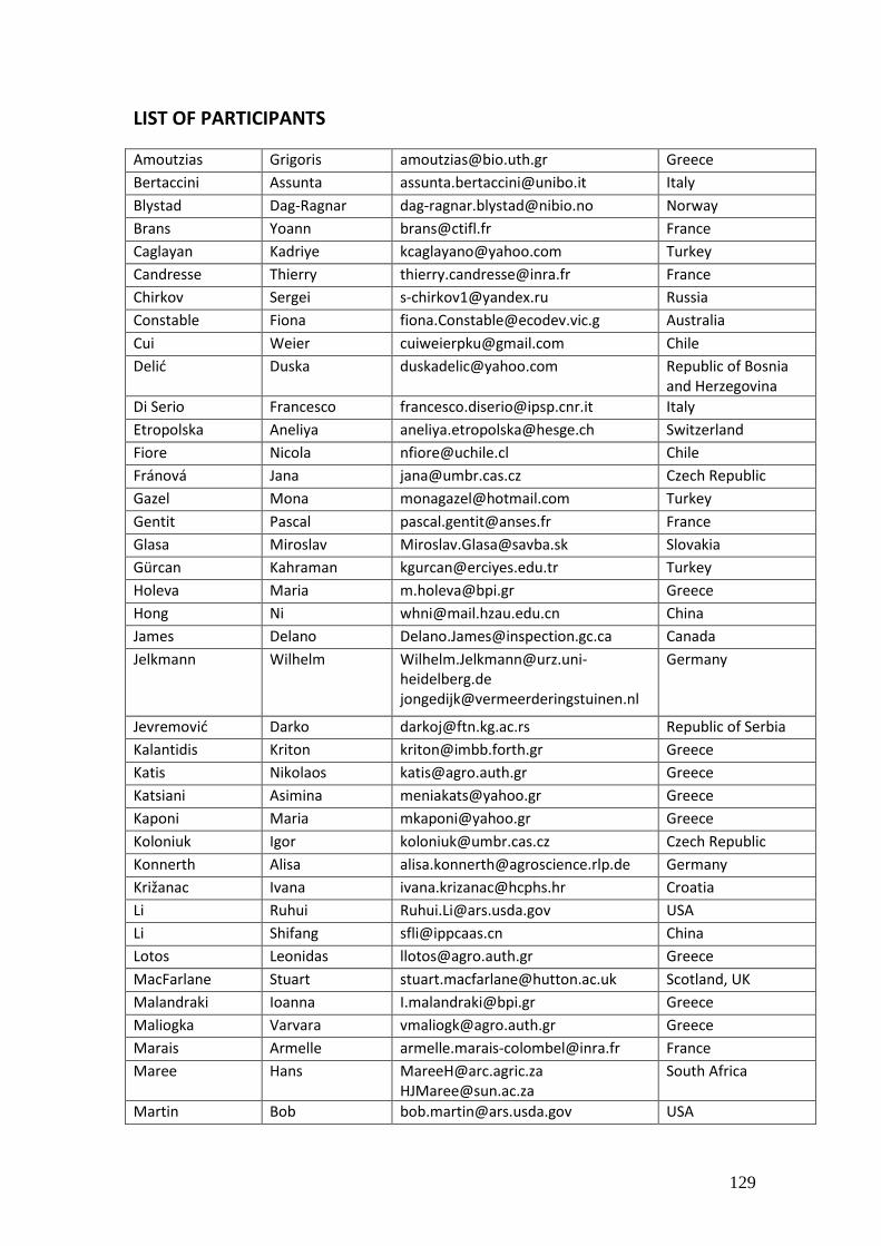
129
LIST OF PARTICIPANTS
Amoutzias Grigoris [email protected] Greece
Bertaccini Assunta [email protected] Italy
Blystad Dag-Ragnar [email protected] Norway
Brans Yoann [email protected] France
Caglayan Kadriye [email protected] Turkey
Candresse Thierry [email protected] France
Chirkov Sergei [email protected] Russia
Constable Fiona [email protected] Australia
Cui Weier [email protected] Chile
Delić Duska [email protected] Republic of Bosnia and Herzegovina
Di Serio Francesco [email protected] Italy
Etropolska Aneliya [email protected] Switzerland
Fiore Nicola [email protected] Chile
Fránová Jana [email protected] Czech Republic
Gazel Mona [email protected] Turkey
Gentit Pascal [email protected] France
Glasa Miroslav [email protected] Slovakia
Gürcan Kahraman [email protected] Turkey
Holeva Maria [email protected] Greece
Hong Ni [email protected] China
James Delano [email protected] Canada
Jelkmann Wilhelm [email protected] [email protected]
Germany
Jevremović Darko [email protected] Republic of Serbia
Kalantidis Kriton [email protected] Greece
Katis Nikolaos [email protected] Greece
Katsiani Asimina [email protected] Greece
Kaponi Maria [email protected] Greece
Koloniuk Igor [email protected] Czech Republic
Konnerth Alisa [email protected] Germany
Križanac Ivana [email protected] Croatia
Li Ruhui [email protected] USA
Li Shifang [email protected] China
Lotos Leonidas [email protected] Greece
MacFarlane Stuart [email protected] Scotland, UK
Malandraki Ioanna [email protected] Greece
Maliogka Varvara [email protected] Greece
Marais Armelle [email protected] France
Maree Hans [email protected] [email protected]
South Africa
Martin Bob [email protected] USA
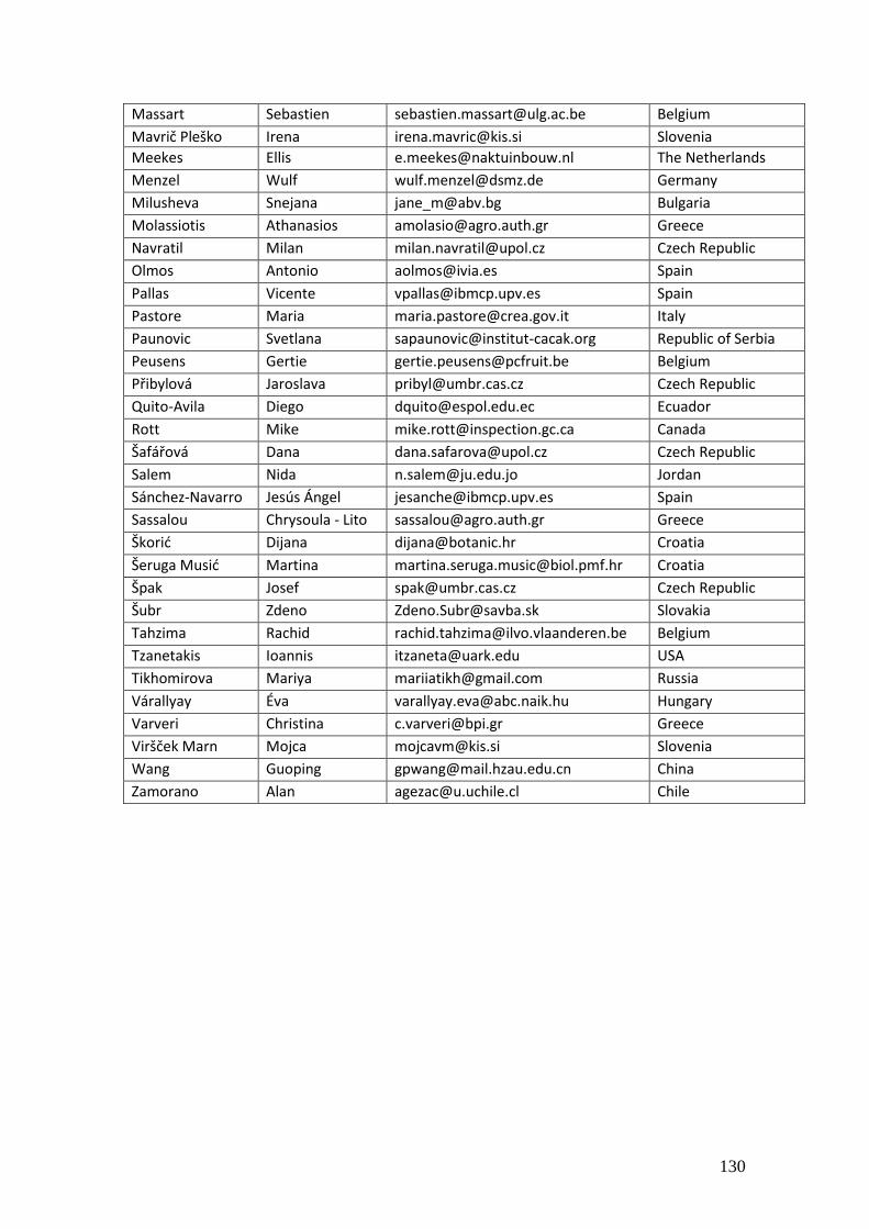
130
Massart Sebastien [email protected] Belgium
Mavrič Pleško Irena [email protected] Slovenia
Meekes Ellis [email protected] The Netherlands
Menzel Wulf [email protected] Germany
Milusheva Snejana [email protected] Bulgaria
Molassiotis Athanasios [email protected] Greece
Navratil Milan [email protected] Czech Republic
Olmos Antonio [email protected] Spain
Pallas Vicente [email protected] Spain
Pastore Maria [email protected] Italy
Paunovic Svetlana [email protected] Republic of Serbia
Peusens Gertie [email protected] Belgium
Přibylová Jaroslava [email protected] Czech Republic
Quito-Avila Diego [email protected] Ecuador
Rott Mike [email protected] Canada
Šafářová Dana [email protected] Czech Republic
Salem Nida [email protected] Jordan
Sánchez-Navarro Jesús Ángel [email protected] Spain
Sassalou Chrysoula - Lito [email protected] Greece
Škorić Dijana [email protected] Croatia
Šeruga Musić Martina [email protected] Croatia
Špak Josef [email protected] Czech Republic
Šubr Zdeno [email protected] Slovakia
Tahzima Rachid [email protected] Belgium
Tzanetakis Ioannis [email protected] USA
Tikhomirova Mariya [email protected] Russia
Várallyay Éva [email protected] Hungary
Varveri Christina [email protected] Greece
Viršček Marn Mojca [email protected] Slovenia
Wang Guoping [email protected] China
Zamorano Alan [email protected] Chile