international day of radiology 2014 interview on brain ... · international day of radiology 2014 ....
TRANSCRIPT

International Day of Radiology 2014 Interview on brain imaging Spain / Prof. Àlex Rovira Cañellas Prof. Àlex Rovira Cañellas, director of the MR Unit at Vall d’Hebron Hospital in Barcelona and president of the Spanish Society of Neuroradiology, spoke to the ESR about the advantages of CT and MRI in brain imaging and the resources available for neuroradiology in Spain. European Society of Radiology: Imaging is known for its ability to detect and diagnose diseases. What kind of brain diseases can imaging help to detect and diagnose? Àlex Rovira Cañellas: Most lesions and diseases that involve the central nervous system (CNS) can be diagnosed with neuroimaging techniques, including congenital and developmental disorders, inflammatory and infectious diseases, traumatic, vascular and tumoural lesions, neurodegenerative disorders, and even neuropsychiatric diseases. This can be achieved thanks to the capacity of these techniques to identify not only structural abnormalities, but also microstructural, metabolic, hemodynamic and functional changes. ESR: How useful is imaging in brain disease management? Does it improve the understanding of disease or improve patient prognosis? ARC: Brain imaging is playing an increasing role not only for establishing an early and accurate diagnosis of many CNS disorders, but also as a biomarker for monitoring and predicting their evolution. This information is becoming essential for monitoring treatment response, particularly in the field of neuro-oncology, cerebrovascular medicine and demyelinating diseases. Techniques such as magnetic resonance imaging (MRI) have significantly improved our understanding of the pathophysiology of different diseases, such as neurodegenerative disorders, multiple sclerosis or hepatic encephalopathy. For example, the identification with MRI of brain white-matter lesions in a young patient presenting with an episode of transient neurological dysfunction indicates that he or she has a high risk of developing multiple sclerosis in the next few years, and, based on the number or volume of these lesions, it can also predict the risk of developing significant clinical disability over the following years. Several studies have demonstrated that MRI is the most valuable diagnostic and prognostic marker in multiple sclerosis, having a major role in identifying patients who require early medical treatment. ESR: What kind of technology and techniques do radiologists use to image the brain? Are there any specific techniques for particular diseases? ARC: There are different neuroradiological techniques such as plain x-ray films, computed tomography (CT), MRI, x-ray angiography and myelography. Traditional x-ray films of the head and spine have lost their importance these days, and are mainly used for assessing degenerative diseases and traumatic lesions of the spine or following surgery. They have largely been replaced by CT and MRI. CT is an x-ray procedure: during the examination, an x-ray tube revolves around the supine patient while at the same time detectors opposite the tube, and rotating with it, measure the radiation. The advantages of CT are the widespread availability of the equipment, making the examination readily available; the short examination time; and the reliability of results in emergency examinations. Moreover, the units are generously constructed so that even people who are afraid of confined spaces can be examined, and unlike MRI, patients with pacemakers and other metal foreign bodies can be examined. The disadvantages of CT are based on the exposure to radiation, although this is slight and further minimised by current technical developments, and the lower tissue contrast compared to MRI. In summary, CT is the first choice in a number of emergency and routine examinations, making MRI unnecessary in these patients.
MRI is a medical imaging procedure to depict organs and tissues using magnetic fields and radio waves. MRI has a number of advantages over other imaging procedures, such as CT. It provides no exposure to radiation, and it has a very high soft tissue contrast, even in the vicinity of bones. This enables it, for example, to image small pathological processes in the spinal cord, which cannot be recorded with CT due to the adjacent spinal column. In addition to images of the structure of the tissues, advanced MRI techniques such as perfusion-weighted imaging, diffusion tensor imaging, proton MR spectroscopy, and functional MR imaging are able to depict microstructural, hemodynamic, metabolic and functional abnormalities within the central nervous system. However, MRI is not generally better than CT. CT can best answer some questions such as bone lesions, for example at the skull base, bone fractures, or fresh blood. Moreover, monitoring and examining is simpler in unconscious patients. Also, patients with pacemakers cannot usually be examined because of possible disruption caused by the magnetic field with MRI. Finally, an MRI examination is considerably more expensive than a CT examination. Angiography is a routine procedure in which vessels supplying the brain or spinal cord can be imaged. Conventional x-ray angiography via catheter has sometimes been replaced in recent years by CT or MR angiography techniques, which are non-invasive. Catheter angiography is used these days in a computer-supported form, the so-called digital subtraction angiography (DSA), in which only arteries and veins are imaged and bone is not visible and does not cover the vessel. Diagnostic angiography is mainly used when one of the following diseases is suspected: Narrowing of the neck arteries which supply the brain (stenosis of the vertebral or carotid artery) or vessels in the brain itself (anterior, middle or posterior cerebral artery, basilar artery); occlusion of the carotid or cerebral arteries, for example in stroke, venous and venous sinus thrombosis, clarification of unexplained cerebral bleeding, if MRI or CT does not definitely reveal aneurysm or vascular malformations (arteriovenous vascular malformation, arteriovenous fistula); inflammatory diseases of the cerebral vessels (so-called vasculitis); and vessel-rich tumours prior to embolisation (obliteration of tumour vessels). Myelography is used to examine the spinal canal. X-ray-dense contrast agent is injected into the cerebrospinal fluid (CSF), which makes the fluid space visible. This is performed through a puncture on the lower spine. Usually, contrast imaging is combined with a CT examination after the x-ray agent has dispersed, since the two methods together provide the best diagnostic results. Usually, spinal column diagnostics with MRI or CT is sufficient, for example in the case of tumour or disc prolapse, so that many myelographies can be avoided these days. If myelography is performed, it is usually in preparation of surgery. Its great advantage is that functional examinations of the spine, such as slipped disc under static stress, and testing the movement that causes pain can be simulated. Therapy with neuroradiological methods, also known as interventional neuroradiology, has become increasingly widespread in the last two decades, mainly with the use of endovascular procedures (using the same technique as for x-ray angiography). Treatment procedures have become standard today, which were hardly available 20 years ago, such as occlusion of aneurysms or vascular malformations, or reopening (recanalisation) of occluded vessels. ESR: What is the difference between a radiologist and a radiographer? Who else is involved in performing brain imaging exams? ARC: Brain imaging procedures are usually performed by qualified technicians, known as radiographers, under the direction of radiologists. Radiographers are also involved in post-processing procedures, as well as other professionals such as computer engineers, and physicians, which are nowadays of great importance to the quantitative assessment of images. Quantitative assessment is especially important in order to use imaging as a real biomarker in the diagnosis and monitoring of CNS diseases, and as a marker of the degree and extension of tissue damage. A radiologist is a medical doctor who is trained in executing, reading and interpreting medical images such as x-ray, CT, ultrasound and MRI scans, and using them to treat health problems in patients. Therefore, they are responsible not only of the proper acquisition of the diagnostic procedure, and of the reading and interpretation of the results obtained, but also in selecting the most appropriate diagnostic plan, and in correlating image findings with other examinations and
tests, recommending further examinations or treatments, and working with the patient’s referring physician to make decisions on the next phase of diagnosis. ESR: How many patients undergo brain imaging exams in your country each year? ARC: Brain imaging represents approximately 25% of all imaging examinations. I do not have precise data on how many patients undergo brain imaging in my country, but at least in our institution we perform around 20,000 brain examinations per year for a reference population of 500,000 inhabitants, so one examination per 25 inhabitants per year. ESR: Access to modern imaging equipment is important for brain imaging. Are hospitals in your country equipped to provide the necessary exams? ARC: Most Spanish hospitals belong directly or indirectly to the public health system, and are usually equipped with adequate imaging technology, allowing for the acquisition of all the necessary exams. However, due to the critical economic situation we are suffering, there are some problems in renewing the technology. We really hope that these restrictions will be lifted during the next few years. ESR: In many countries there are waiting lists for MRI exams. How long can patients typically expect to wait for an exam in Spain? ARC: Timely access to diagnostic imaging services is a priority issue for our public health system. Unfortunately, the resources are limited and waiting lists are in some large hospitals still a big issue. In a large hospital like the one I work in, an ordinary follow-up examination has to wait for around eight months, but an urgent diagnostic imaging procedure is always performed within a few days. ESR: As the global population gets older, the risk of developing neurocognitive and neurodegenerative disorders increases. How can imaging help tackle this issue? ARC: Brain imaging can play a vital role in diagnosing dementia. It can help to rule out alternative and treatable causes for dementia-related symptoms, such as hydrocephalus, or brain tumour or abscess. Imaging can also improve the accuracy of a diagnosis by identifying specific areas and causes of damage linked with particular types of dementia, such as vascular dementia or Alzheimer’s disease. Once the diagnosis has been made, brain imaging can be used to monitor how the condition is developing. Imaging techniques are also of immense help to researchers working to improve understanding of what causes dementia and how it develops, as they can provide information on the structural, functional and metabolic changes. But probably in the near future, brain imaging will also play an essential role in early and accurate diagnosis of different neurodegenerative disorders, particularly when patients are asymptomatic. Early diagnosis could be very important in order to treat patients before the development of irreversible cognitive decline. As soon as clinical trials demonstrate that medical treatments can prevent or reduce the development of neurodegenerative disorders such as Alzheimer’s disease in the ageing population, brain imaging screening programmes will be required, as these techniques seem to be the most appropriate tools for detecting subjects at risk of developing these diseases. ESR: Some imaging techniques, like x-ray and CT, use ionising radiation. What risk does this radiation pose to the patient and what kind of safety measures are in place to protect the patient? ARC: Ever since physicians started regularly ordering CT scans four decades ago, researchers have been worried that the medical imaging procedure could increase a patient’s risk of developing cancer. CT scanners bombard the human body with x-ray beams, which can damage DNA and create mutations that spur cells to grow into tumours. However, x-rays are safe when used with care. Radiologists and x-ray technologists have been trained to use the minimum amount of radiation necessary to obtain the needed results. Properly conducted, imaging carries minimal risks and should be performed when clinically indicated. The amount of radiation used in brain CT is very small and the benefits greatly outweigh the risk of harm. The radiation dose for a brain CT scan is 2-4mSv,
which is comparable to natural background radiation for 8–16 months, and is considered a very low or low additional lifetime risk of fatal cancer. ESR: In general, patients don’t see the radiologist. A patient will discuss the image with the neurologist, neurosurgeon or oncologist. When they ask a question, they’re often told: “I’m not a radiologist”. Why don’t radiologists discuss the image with the patient first? ARC: Radiologists are medical doctors who are responsible not only for reading and interpreting medical images, but also for correlating the image findings with clinical findings and other examinations and tests, recommending further examinations or treatments, and working with the patient’s referring physician to make decisions on the next phase of diagnosis. In some situations, the radiologist should also inform the patient about the results of the examination. Unfortunately, this is not commonly done in clinical practice, due to the limited available time, but at least in many institutions radiologists always inform the patient about the results and interpretation of the examination whenever the patient requests it. ESR: How expensive are radiological examinations to the health service and is there a risk that some of these examinations could be blocked by health technology assessment agencies deeming them to be not cost-effective If so, how can patients help to ensure that these examinations are made available? ARC: In Spain the average cost of a brain examination is relatively low: €90 for a routine brain CT, €160 for a routine brain MRI. There are, in general, no restrictions for using these brain imaging techniques in clinical practice, although the referring physician must justify the need for a specific examination in a patient. However, in the last few years public hospitals have established a maximum number of imaging examinations that can be performed per year, which means that we are now stricter in requiring proper justification of the imaging examination. Despite these restrictions, we always have the possibility to perform any kind of imaging if this is clinically justified. Another issue is using imaging for screening programmes. These types of programmes should be based on positive evidence-based studies otherwise they will be blocked by local health agencies.
Àlex Rovira Cañellas is director of the magnetic resonance unit and head of the department of neuroradiology at University Hospital Vall d’Hebron, Barcelona, Spain. He is also professor of radiology at the Autonomous University of Barcelona. He specialises in diagnostic neuroradiology; head & neck radiology, with a particular interest in demyelinating diseases; stroke; neuro-oncology; hepatic encephalopathy; and head & neck tumours. He is currently president of the Spanish Society of Neuroradiology, co-chairman of the European Multicenter Collaborative Research Network on MRI in Multiple Sclerosis (MAGNIMS), and a member of the Executive Committee of the European Society of Neuroradiology and the Spanish Society of Radiology. He has authored or co-authored more than 220 original papers, 25 review
papers, 25 book chapters or monographs, and has delivered more than 400 conference presentations.

Figure 1 Proton MR spectroscopic imaging of a left temporal lobe glioblastoma multiforme (choline map). The red spot indicates the area of the lesion with higher tumoural activity.
Figure 2 Contrast-enhanced CT and MRI of a right temporal lobe glioblastoma. Both imaging techniques demonstrated the brain mass, but the lesion is better delineated with MRI.
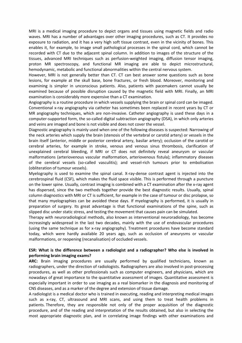
Figure 3 Axial MRI showing a larger vascular malformation in the left brain hemisphere. The red areas indicate increase cerebral blood flow.
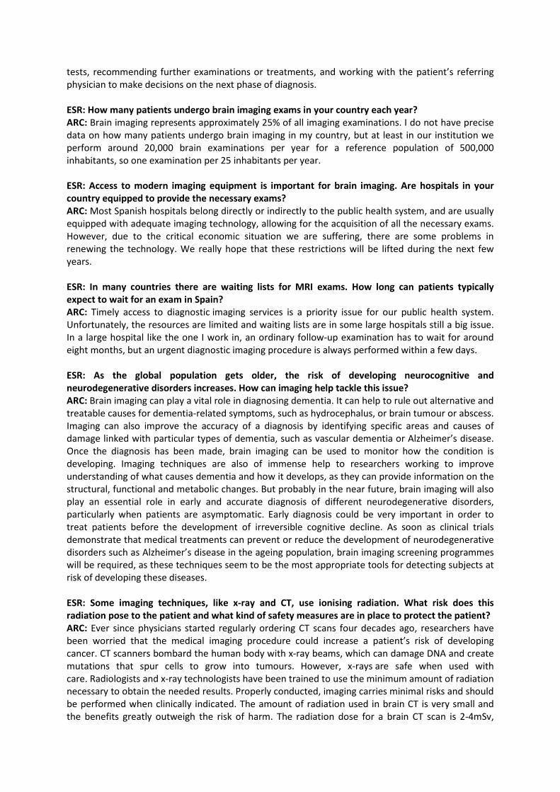
Figure 4 Proton MR spectroscopic imaging of a large inflammatory lesions located in the right periventricular white matter (choline map). The red spot indicates the area of the lesion with higher inflammatory activity.
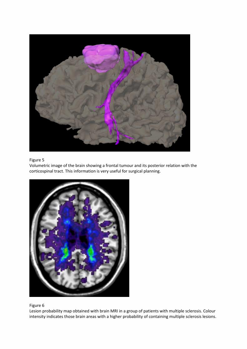
Figure 5 Volumetric image of the brain showing a frontal tumour and its posterior relation with the corticospinal tract. This information is very useful for surgical planning.
Figure 6 Lesion probability map obtained with brain MRI in a group of patients with multiple sclerosis. Colour intensity indicates those brain areas with a higher probability of containing multiple sclerosis lesions.
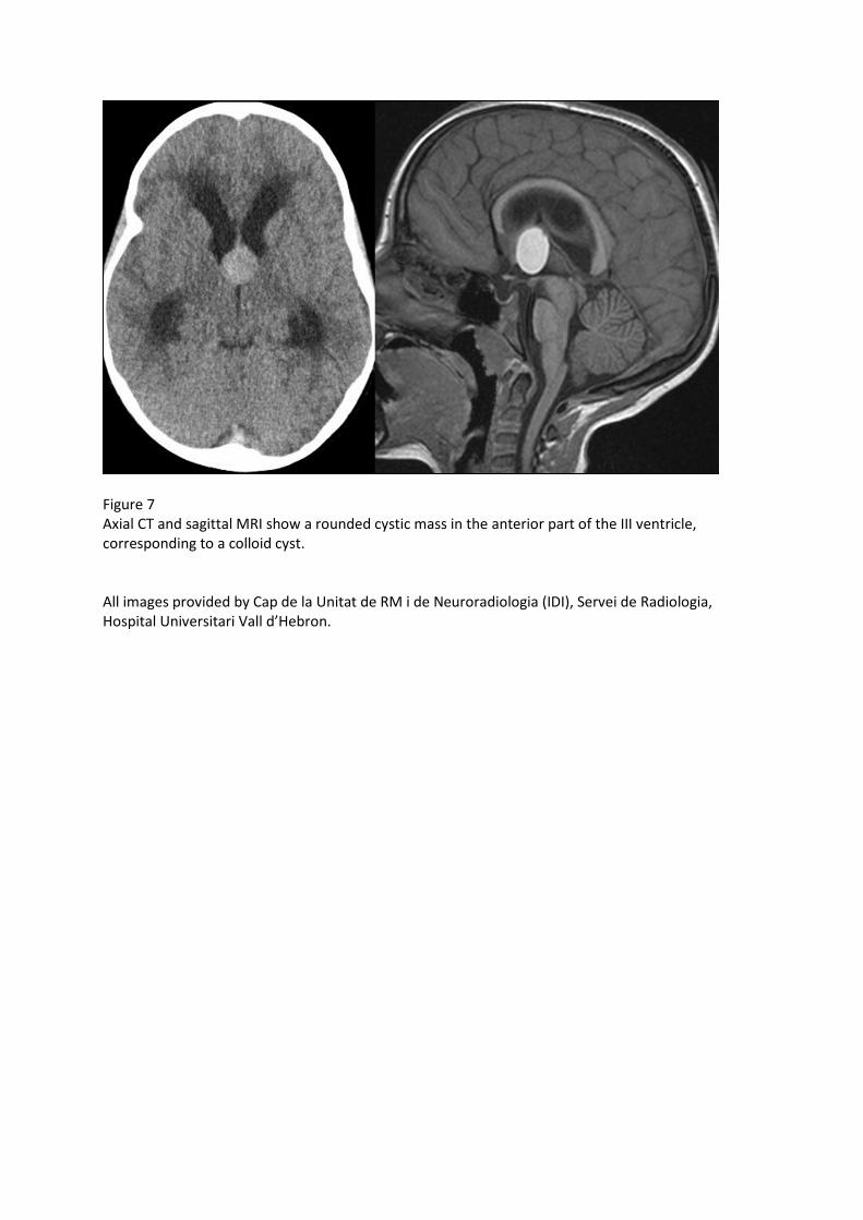
Figure 7 Axial CT and sagittal MRI show a rounded cystic mass in the anterior part of the III ventricle, corresponding to a colloid cyst. All images provided by Cap de la Unitat de RM i de Neuroradiologia (IDI), Servei de Radiologia, Hospital Universitari Vall d’Hebron.