international journal for parasitology - · pdf fileinternational journal for parasitology...
TRANSCRIPT
International Journal for Parasitology 39 (2009) 153–162
Contents lists available at ScienceDirect
International Journal for Parasitology
journal homepage: www.elsevier .com/locate / i jpara
Invited Review
Apicomplexan cytoskeleton and motors: Key regulators in morphogenesis,cell division, transport and motility
Joana M. Santos a, Maryse Lebrun b, Wassim Daher a, Dominique Soldati a, Jean-Francois Dubremetz b,*
a Department of Microbiology and Molecular Medicine, Faculty of Medicine–University of Geneva CMU, 1 rue Michel-Servet, 1211 Geneva 4, Switzerlandb UMR CNRS 5235, Bt 24, CC 107 Université de Montpellier 2, Place Eugène Bataillon, 34095 Montpellier cedex 05, France
a r t i c l e i n f o
Article history:Received 30 July 2008Received in revised form 13 October 2008Accepted 16 October 2008
Keywords:ApicomplexaCytoskeletonActinMyosinMitosisInvasionEgressGlideosome
0020-7519/$34.00 � 2008 Australian Society for Paradoi:10.1016/j.ijpara.2008.10.007
* Corresponding author. Tel.: +33 467143455; fax:E-mail address: [email protected] (J.-F. D
a b s t r a c t
Protozoan parasites of the phylum Apicomplexa undergo a lytic cycle whereby a single zoite produced bythe previous cycle has to encounter a host cell, invade it, multiply to differentiate into a new zoite gen-eration and escape to resume a new cycle. At every step of this lytic cycle, the cytoskeleton and/or thegliding motility apparatus play a crucial role and recent results have elucidated aspects of these pro-cesses, especially in terms of the molecular characterization and interaction of the increasing numberof partners involved, and the signalling mechanisms implicated. The present review aims to summarizethe most recent findings in the field.
� 2008 Australian Society for Parasitology Inc. Published by Elsevier Ltd. All rights reserved.
1. Introduction
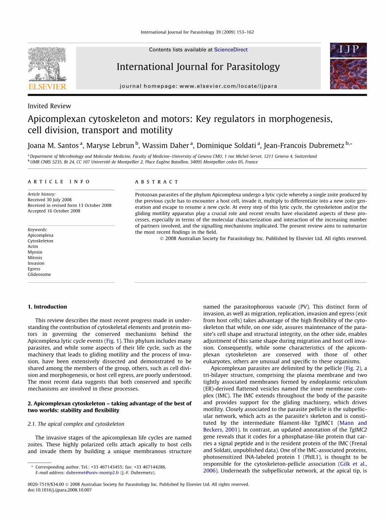
This review describes the most recent progress made in under-standing the contribution of cytoskeletal elements and protein mo-tors in governing the conserved mechanisms behind theApicomplexa lytic cycle events (Fig. 1). This phylum includes manyparasites, and while some aspects of their life cycle, such as themachinery that leads to gliding motility and the process of inva-sion, have been extensively dissected and demonstrated to beshared among the members of the group, others, such as cell divi-sion and morphogenesis, or host cell egress, are poorly understood.The most recent data suggests that both conserved and specificmechanisms are involved in these processes.
2. Apicomplexan cytoskeleton – taking advantage of the best oftwo worlds: stability and flexibility
2.1. The apical complex and cytoskeleton
The invasive stages of the apicomplexan life cycles are namedzoites. These highly polarized cells attach apically to host cellsand invade them by building a unique membranous structure
sitology Inc. Published by Elsevier
+33 467144286.ubremetz).
named the parasitophorous vacuole (PV). This distinct form ofinvasion, as well as migration, replication, invasion and egress (exitfrom host cells) takes advantage of the high flexibility of the cyto-skeleton that while, on one side, assures maintenance of the para-site’s cell shape and structural integrity, on the other side, enablesadjustment of this same shape during migration and host cell inva-sion. Consequently, while some characteristics of the apicom-plexan cytoskeleton are conserved with those of othereukaryotes, others are unusual and specific to these organisms.
Apicomplexan parasites are delimited by the pellicle (Fig. 2), atri-bilayer structure, comprising the plasma membrane and twotightly associated membranes formed by endoplasmic reticulum(ER)-derived flattened vesicles named the inner membrane com-plex (IMC). The IMC extends throughout the body of the parasiteand provides support for the gliding machinery, which drivesmotility. Closely associated to the parasite pellicle is the subpellic-ular network, which acts as the parasite’s skeleton and is consti-tuted by the intermediate filament-like TgIMC1 (Mann andBeckers, 2001). In contrast, an updated annotation of the TgIMC2gene reveals that it codes for a phosphatase-like protein that car-ries a signal peptide and is the resident protein of the IMC (Frenaland Soldati, unpublished data). One of the IMC-associated proteins,photosensitized INA-labeled protein 1 (PhIL1), is thought to beresponsible for the cytoskeleton-pellicle association (Gilk et al.,2006). Underneath the subpellicular network, at the apical tip, is
Ltd. All rights reserved.
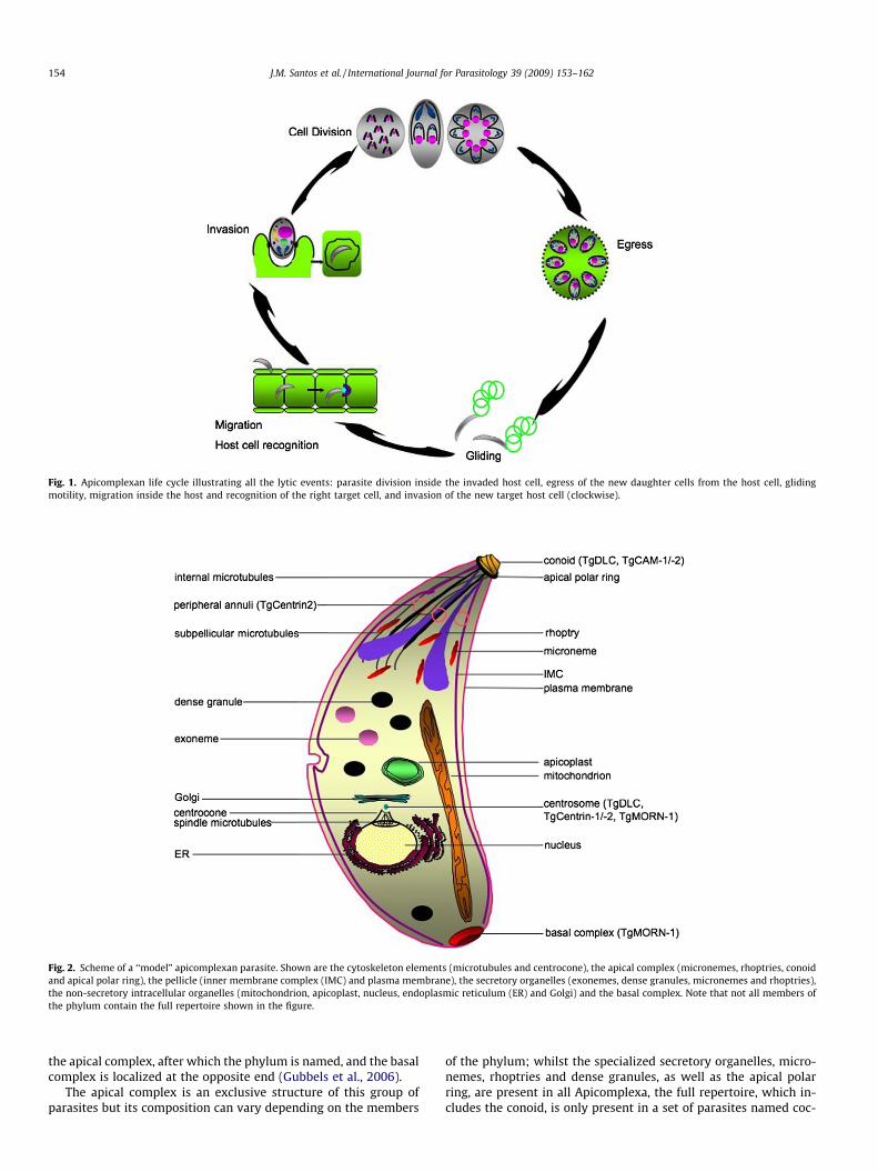
Fig. 1. Apicomplexan life cycle illustrating all the lytic events: parasite division inside the invaded host cell, egress of the new daughter cells from the host cell, glidingmotility, migration inside the host and recognition of the right target cell, and invasion of the new target host cell (clockwise).
Fig. 2. Scheme of a ‘‘model” apicomplexan parasite. Shown are the cytoskeleton elements (microtubules and centrocone), the apical complex (micronemes, rhoptries, conoidand apical polar ring), the pellicle (inner membrane complex (IMC) and plasma membrane), the secretory organelles (exonemes, dense granules, micronemes and rhoptries),the non-secretory intracellular organelles (mitochondrion, apicoplast, nucleus, endoplasmic reticulum (ER) and Golgi) and the basal complex. Note that not all members ofthe phylum contain the full repertoire shown in the figure.
154 J.M. Santos et al. / International Journal for Parasitology 39 (2009) 153–162
the apical complex, after which the phylum is named, and the basalcomplex is localized at the opposite end (Gubbels et al., 2006).
The apical complex is an exclusive structure of this group ofparasites but its composition can vary depending on the members
of the phylum; whilst the specialized secretory organelles, micro-nemes, rhoptries and dense granules, as well as the apical polarring, are present in all Apicomplexa, the full repertoire, which in-cludes the conoid, is only present in a set of parasites named coc-
J.M. Santos et al. / International Journal for Parasitology 39 (2009) 153–162 155
cidians. The apical polar ring and the conoid are both elements ofthe cytoskeleton but while the first one is the microtubule organiz-ing center (MTOC) of the subpellicular microtubules, the conoid isorganized into a hollow cylinder composed of a polymer of alphaand beta tubulins assembled into a new type of protofilamentsheets (Hu et al., 2002) and can move up and down through theapical polar ring and protrude apically at the time of cell invasionin a calcium-dependent fashion (Mondragon and Frixione, 1996;Monteiro et al., 2001). Three proteins likely to be involved in thismotility are dynein light chain (TgDLC), which could be part ofthe motor, and calcium-binding proteins 1 and 2 (TgCAM-1 and -2), which may regulate this kind of motion (Mondragon and Frixi-one, 1996; Hu et al., 2006).
As mentioned above, the subpellicular microtubules, which areimportant for shape, apical polarity and organelle trafficking, areorganized from the apical polar ring, but two other sets of microtu-bules exist in these parasites. One set is found in the mitotic spin-dle, where it coordinates chromosome segregation and originatesfrom MTOCs organized by centrioles in Coccidia (Dubremetz,1973), or by rudimentary spindle pole bodies in other Apicomplexasuch as Plasmodium (Schrevel et al., 1977), and the other set islocalized in the conoid. These different microtubules are uniquelyspecialized, in a phenomenon reflective of the apicomplexan para-sites’ lifestyle. For instance, it was recently found that the subpel-licular microtubules of Plasmodium berghei sporozoites (Cyrklaffet al., 2007) are maintained in a state of ‘‘suspended depolymeriza-tion” by an as yet unidentified molecule that allows them to bendfar beyond what is allowed by regular microtubules undergoingtreadmilling, an ability that is especially important during thetransmigration and invasion processes.
The number and organization of the microtubules can alsodiffer between parasite species and even life cycle stages. Cryp-tosporidium parvum, for instance, was shown not to have subpel-licular microtubules but longitudinal ridges that might perform asimilar function (Matsubayashi et al., 2008), and Besnoitia besno-
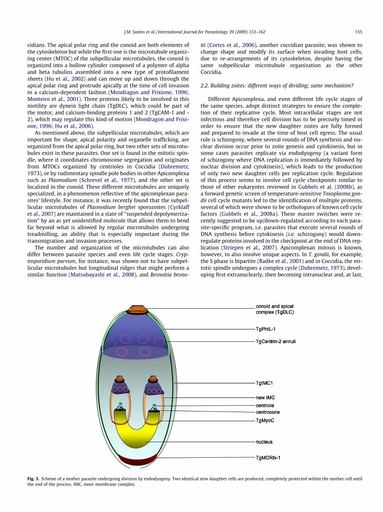
Fig. 3. Scheme of a mother parasite undergoing division by endodyogeny. Two identicalthe end of the process. IMC, inner membrane complex.
iti (Cortes et al., 2006), another coccidian parasite, was shown tochange shape and modify its surface when invading host cells,due to re-arrangements of its cytoskeleton, despite having thesame subpellicular microtubule organization as the otherCoccidia.
2.2. Building zoites: different ways of dividing; same mechanism?
Different Apicomplexa, and even different life cycle stages ofthe same species, adopt distinct strategies to ensure the comple-tion of their replicative cycle. Most intracellular stages are notinfectious and therefore cell division has to be precisely timed inorder to ensure that the new daughter zoites are fully formedand prepared to invade at the time of host cell egress. The usualrule is schizogony, where several rounds of DNA synthesis and nu-clear division occur prior to zoite genesis and cytokinesis, but insome cases parasites replicate via endodyogeny (a variant formof schizogony where DNA replication is immediately followed bynuclear division and cytokinesis), which leads to the productionof only two new daughter cells per replication cycle. Regulationof this process seems to involve cell cycle checkpoints similar tothose of other eukaryotes reviewed in Gubbels et al. (2008b), asa forward genetic screen of temperature-sensitive Toxoplasma gon-dii cell cycle mutants led to the identification of multiple proteins,several of which were shown to be orthologues of known cell cyclefactors (Gubbels et al., 2008a). These master switches were re-cently suggested to be up/down-regulated according to each para-site-specific program, i.e. parasites that execute several rounds ofDNA synthesis before cytokinesis (i.e. schizogony) would down-regulate proteins involved in the checkpoint at the end of DNA rep-lication (Striepen et al., 2007). Apicomplexan mitosis is known,however, to also involve unique aspects. In T. gondii, for example,the S phase is bipartite (Radke et al., 2001) and in Coccidia, the mi-totic spindle undergoes a complex cycle (Dubremetz, 1973), devel-oping first extranuclearly, then becoming intranuclear and, at last,
new daughter cells are produced, completely protected within the mother cell until
156 J.M. Santos et al. / International Journal for Parasitology 39 (2009) 153–162
turning into a pair of centrocones, derived from both the nuclearenvelope and the spindle poles, and characterized by the presenceof the membrane occupation and recognition nexus motif contain-ing protein TgMORN1 (Gubbels et al., 2006).
Multiple studies performed during the past 40 years have sug-gested that there is no fundamental distinction between the vari-ous modes of reproduction, apart from the number of nucleardivisions preceding zoite genesis. In all cases, the morphogenesisof apicomplexan zoites has been described as being coordinatedwith mitosis. The mitotic poles were clearly shown to be the pri-mary organizing centers of both the mitotic spindle and the apicalcytoskeleton of the nascent zoites. The pattern of differentiationhas been described in many Apicomplexa and it was found to beconserved ((Dubremetz, 1975). What has been revealed more re-cently are the molecular features of the structures previously de-scribed. These findings essentially concern the process ofendodyogeny of T. gondii tachyzoites, thus we will only report onthis process (Figs. 3 and 4).
The first sign of cell division is the migration of the centriolesto the basal pole of the nucleus and their replication. These rep-licated centrioles sandwich the spindle and the poles of the spin-dle give rise to the centrocones, on which the kinetochores ofreplicated chromosomes are attached. The primary structureswhich develop next to these centrioles are the two apical polarrings, from which the subpellicular microtubules extend, coveredwith the early IMC. The outer edges of the IMCs appear as ringstructures decorated with TgMORN1 and are the precursors ofthe daughter basal ring complexes, remaining at the basal ends
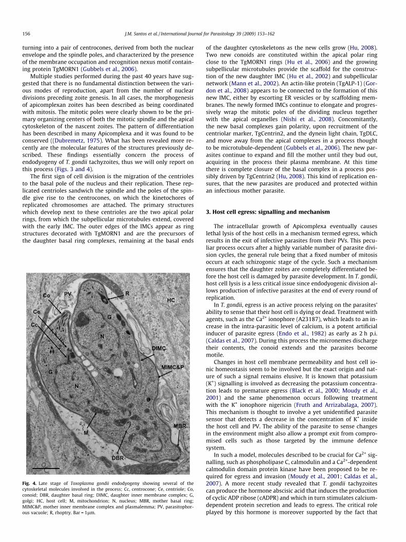
Fig. 4. Late stage of Toxoplasma gondii endodyogeny showing several of thecytoskeletal molecules involved in the process; Cc, centrocone; Ce, centriole; Co,conoid; DBR, daughter basal ring; DIMC, daughter inner membrane complex; G,golgi; HC, host cell; M, mitochondrion; N, nucleus; MBR, mother basal ring;MIMC&P, mother inner membrane complex and plasmalemma; PV, parasitophor-ous vacuole; R, rhoptry. Bar = 1lm.
of the daughter cytoskeletons as the new cells grow (Hu, 2008).Two new conoids are constituted within the apical polar ringclose to the TgMORN1 rings (Hu et al., 2006) and the growingsubpellicular microtubules provide the scaffold for the construc-tion of the new daughter IMC (Hu et al., 2002) and subpellicularnetwork (Mann et al., 2002). An actin-like protein (TgALP-1) (Gor-don et al., 2008) appears to be connected to the formation of thisnew IMC, either by escorting ER vesicles or by scaffolding mem-branes. The newly formed IMCs continue to elongate and progres-sively wrap the mitotic poles of the dividing nucleus togetherwith the apical organelles (Nishi et al., 2008). Concomitantly,the new basal complexes gain polarity, upon recruitment of thecentriolar marker, TgCentrin2, and the dynein light chain, TgDLC,and move away from the apical complexes in a process thoughtto be microtubule-dependent (Gubbels et al., 2006). The new par-asites continue to expand and fill the mother until they bud out,acquiring in the process their plasma membrane. At this timethere is complete closure of the basal complex in a process pos-sibly driven by TgCentrin2 (Hu, 2008). This kind of replication en-sures, that the new parasites are produced and protected withinan infectious mother parasite.
3. Host cell egress: signalling and mechanism
The intracellular growth of Apicomplexa eventually causeslethal lysis of the host cells in a mechanism termed egress, whichresults in the exit of infective parasites from their PVs. This pecu-liar process occurs after a highly variable number of parasite divi-sion cycles, the general rule being that a fixed number of mitosisoccurs at each schizogonic stage of the cycle. Such a mechanismensures that the daughter zoites are completely differentiated be-fore the host cell is damaged by parasite development. In T. gondii,host cell lysis is a less critical issue since endodyogenic division al-lows production of infective parasites at the end of every round ofreplication.
In T. gondii, egress is an active process relying on the parasites’ability to sense that their host cell is dying or dead. Treatment withagents, such as the Ca2+ ionophore (A23187), which leads to an in-crease in the intra-parasitic level of calcium, is a potent artificialinducer of parasite egress (Endo et al., 1982) as early as 2 h p.i.(Caldas et al., 2007). During this process the micronemes dischargetheir contents, the conoid extends and the parasites becomemotile.
Changes in host cell membrane permeability and host cell io-nic homeostasis seem to be involved but the exact origin and nat-ure of such a signal remains elusive. It is known that potassium(K+) signalling is involved as decreasing the potassium concentra-tion leads to premature egress (Black et al., 2000; Moudy et al.,2001) and the same phenomenon occurs following treatmentwith the K+ ionophore nigericin (Fruth and Arrizabalaga, 2007).This mechanism is thought to involve a yet unidentified parasitesensor that detects a decrease in the concentration of K+ insidethe host cell and PV. The ability of the parasite to sense changesin the environment might also allow a prompt exit from compro-mised cells such as those targeted by the immune defencesystem.
In such a model, molecules described to be crucial for Ca2+ sig-nalling, such as phospholipase C, calmodulin and a Ca2+-dependentcalmodulin domain protein kinase have been proposed to be re-quired for egress and invasion (Moudy et al., 2001; Caldas et al.,2007). A more recent study revealed that T. gondii tachyzoitescan produce the hormone abscisic acid that induces the productionof cyclic ADP ribose (cADPR) and which in turn stimulates calcium-dependent protein secretion and leads to egress. The critical roleplayed by this hormone is moreover supported by the fact that

J.M. Santos et al. / International Journal for Parasitology 39 (2009) 153–162 157
the same effect is obtained with the addition of exogenous abscisicacid and that the selective disruption of its synthesis by the inhib-itor fluridone leads to a delay in egress and prompts parasite differ-entiation into bradyzoites (Nagamune et al., 2008). The notion thategress depends on the parasite actin-dependent motility was re-cently challenged by the finding that treatment with actin-disrupt-ing drugs does not delay parasite egress. A new model for egresswas then proposed in which the disruption of host cell actin wouldlead to internal pressure and mechanical rupture of the host cellmembrane, which in turn would activate parasite motion due tothe loss of ions from the host cell (Lavine and Arrizabalaga,2008).
In the case of Plasmodium falciparum, the egress of merozoitesfrom red blood cells (RBCs) is very tightly regulated and involvesthe timely breakdown of the PV membrane followed by vesicula-tion of the RBC membrane (Glushakova et al., 2005). A new classof secretory organelles named exonemes have recently beenidentified and shown to control egress via release of the serineprotease PfSUB1 (Yeoh et al., 2007). Indeed, PfSUB1 is dischargedfrom exonemes into the PV space before host cell rupture and in-duce a proteolytic maturation of the vacuolar marker PfSERA5.This last hypothetical protease is known to be essential for theefficient release of parasites from host RBCs (Delplace et al.,1988; Yeoh et al., 2007; Arastu-Kapur et al., 2008). Similar find-ings have been reported for sporozoite release from mosquitomidgut oocysts, which is completely prevented by the disruptionof another SERA family member, SERA8 (ECP1), (Aly and Matus-chewski, 2005).
4. Gliding machinery in Apicomplexa: the motor that drivesinfection
Migration across biological barriers and active penetration ofhost cells and egress rely on the parasite’s ability to glide. Glidingmotility is critically dependent on actin polymerization and ispowered by a myosin motor (MyoA) ubiquitoulsy conservedacross the phylum (Wetzel et al., 2005; Baum et al., 2006a,b;Jones et al., 2006; Schuler and Matuschewski, 2006). Toxoplasmagondii MyoA (TgMyoA) was originally shown to belong to a motorcomplex including the myosin light chain (TgMLC1) (Herm-Gotzet al., 2002) that is firmly anchored in the plane of the IMC bythe integral membrane glycoprotein GAP50 and the lipid modi-fied GAP45 (Johnson et al., 2007). This organization extends toPlasmodium, where the orthologues of PfMTIP, PfGAP45 andPfGAP50 have been identified in P. falciparum merozoites (Baumet al., 2006b; Jones et al., 2006), and to all other members ofthe phylum.
It has been established that aldolase offers a bridge between theactomyosin system and the host receptor-parasite ligand com-plexes (Jewett and Sibley, 2003; Bosch et al., 2006). This glycoliticenzyme is unexpectedly able to bind to both the C-terminal do-main of an adhesin (TgMIC2 in Toxoplasma and TRAP, MTRAP andTLP in Plasmodium) and the parasite’s actin filaments (Buscagliaet al., 2003; Jewett and Sibley, 2003; Baum et al., 2006b; Heisset al., 2008). This interaction involving aldolase, and potentiallyother proteins, is important for parasite survival (Starnes et al.,2006). In such a model, motility is presumably generated by theposterior translocation of F-actin-aldolase bound to the adhesinproteins driven by the myosin tracks firmly anchored and immobi-lized in the IMC (Johnson et al., 2007).
Despite a clear role of F-actin dynamics in gliding, formal dem-onstration of the presence of F-actin in Apicomplexa has been dif-ficult due to the short size and inherent instability of thesefilaments. Indeed, apicomplexan actin exhibits unusual proper-ties. The majority of actin molecules are maintained in a globular
state (Schmitz et al., 2005; Baum et al., 2006a; Sahoo et al., 2006;Schuler and Matuschewski, 2006) that can be rapidly polymerizedinto microfilaments at a concentration three to fourfold lowerthan mammalian muscle actin, in a process dependent on thepresence of salt, magnesium and ATP (Sahoo et al., 2006). This al-lows a rapid treadmilling process that facilitates directionalmigration, the fast regeneration of new actin subunits for futurerounds of assembly, and avoids unwanted locomotion (Baumet al., 2006a; Sahoo et al., 2006; Schuler and Matuschewski,2006).
Both these actin dynamics and microfilament turnover aresuggested to arise from a sophisticated interaction with a vast ar-ray of actin binding proteins (Schmitz et al., 2005). However, api-complexan genomes contain relatively few conventional actin-binding proteins. Among this limited repertoire are actin depoly-merizing factor (ADF1) (Schuler et al., 2005), capping protein al-pha and beta (Gordon and Sibley, 2005), profilin (Plattner et al.,2008), toxofilin (cofilin) (Poupel et al., 2000) and coronin (Tardi-eux et al., 1998; Figueroa et al., 2004). Unexpectedly, Apicom-plexa lack a canonical actin regulator Arp2/3 complex, which isotherwise widespread among eukaryotes where it drives actinassembly by nucleating filaments from the pointed end. Instead,the Apicomplexa possess formins (Gordon and Sibley, 2005;Baum et al., 2006a) that are known, along with profilin, to driveactin polymerization in a mechanism alternative to that of theArps (Higgs and Peterson, 2005). In T. gondii, profilin was recentlyshown to play a vital role in parasite motility and invasion in aprocess conserved across the phylum, since the Plasmodium profi-lin fully complements a T. gondii profilin knockout strain. Further-more, purified recombinant profilins from three differentApicomplexa are able to control actin polymerization (Plattneret al., 2008). Apicomplexan genomes also encode two or morelarge formins that feature a typical forming homology domain 2(FH2) and a recent study highlighted Plasmodium formin 1(PfFRM1) as a potential effector in actin nucleation during inva-sion, based on its localization at the moving junction and its abil-ity to act as a potent actin nucleator of chicken actin in vitro(Baum et al., 2008).
5. Migration and host cell recognition: how to get there andsense where you are
5.1. Migration: getting there
Plasmodium sporozoites are only able to invade a restricted setof cell-types and have to endure a long journey in order to reachtheir final destination, making migration undeniably fundamentalfor the establishment of a malaria infection. Progress in investi-gating this phenomenon has vastly benefited from the sophisti-cated improvements in bioimaging (Amino et al., 2005, 2007;Frevert et al., 2005; Tarun et al., 2006; Thiberge et al., 2007),and this review will only focus on migration of the Plasmodiumsporozoites from the site of injection to the liver. It is now knownthat once deposited in the skin, sporozoites do not leave immedi-ately but remain at their site of inoculation for 1–3 h after themosquito bite (Yamauchi et al., 2007) before entering blood orlymph vessels (Amino et al., 2006). If invasion of a blood vesseloccurs, the sporozoites are carried in the bloodstream and readilyreach the liver. Once in the liver sinusoids, the next barrier thatsporozoites need to overcome is the endothelial barrier. It hasbeen suggested that to access hepatocytes, sporozoites passthrough the resident liver macrophages (Kupffer cells) (Baeret al., 2007). At this point the parasite circumsporozoite protein(CSP) binds to the liver surface LRP-1 and proteoglycans and pre-vents activation of the respiratory burst, hence contributing to
158 J.M. Santos et al. / International Journal for Parasitology 39 (2009) 153–162
parasite survival (Usynin et al., 2007). As previously shown forEimeria sp. interacting with cells in vitro (Roberts et al., 1971),when encountering the hepatic cells the sporozoites do notimmediately establish infection but first traverse several hepato-cytes (Mota et al., 2001). It was initially hypothesized that thiswould occur so that the host cells would be activated and becomemore receptive to infection (Carrolo et al., 2003), but this theorywas revised when transgenic parasites lacking sporozoite micro-neme proteins essential for cell traversal (SPECT-1 and -2/PPLP1)were shown to be unable to migrate through host cells but none-theless were able to productively invade hepatocytes (Ishinoet al., 2004, 2005b). Given that SPECT mutant parasites are lessinfective than wild type ones in vivo using the rodent malariamodel, it appears that host cell traversal is not essential butmight help the parasite to encounter the optimal host cells (Ami-no et al., 2008). In contrast, crossing hepatocytes also causes therelease of several host cell factors such as NF-jB, which can alertthe immune system and limit the extent of malaria infection inthe liver (Torgler et al., 2008).
In addition to SPECT-1 and SPECT-2 (Ishino et al., 2004, 2005b),two new members of the TRAP family TLP (Moreira et al., 2008)and TRSP (Labaied et al., 2007), CelTOS (Kariu et al., 2006) and aphospholipase PbPL (Bhanot et al., 2005) have been reported tobe involved in cell traversal, however little is known about theirmechanistic contribution to the process.
Several homologues of these proteins have been identified inookinetes. MAOP is a SPECT-2 homologue (Kadota et al., 2004)and CelTOS is expressed at both sporozoite and ookinete stages(Kariu et al., 2006) suggesting that a common mechanism mightexplain membrane rupture of the hepatocyte and of mosquito mid-gut cells.
New steps were also made towards elucidation of the signalsthat induce this type of migration. Calcium-dependent protein ki-nase 3 (PbCDPK-3) was shown to regulate ookinete invasion ofthe midgut wall (Ishino et al., 2006) and PbCDPK-6 was demon-strated to be involved in the switch between migration and inva-sion (Coppi et al., 2007). This calcium signalling is conservedacross the Apicomplexa phylum, as T. gondii CDPK-1 (TgCDPK-1)appears to regulate motility and host cell invasion (Kieschnicket al., 2001). Potassium signalling also contributes to migration gi-ven that exposure of parasites to high concentrations of potassiumleads to a decrease in migration (Kumar et al., 2007), and activationof a potassium channel stimulates apical exocytosis, which causesa decrease in cell traversal (Ono et al., 2008).
5.2. Host cell recognition: how to know it is time to stop migrating andstart invading
Apicomplexa exhibit very diverse preferences in terms of hostand host cell-type specificities with some parasites being able toinvade a wide repertoire of host cells while others are extremelyrestricted.
A generally common feature of host cell recognition seems toinvolve the binding to sialic acids on receptors at the surface ofhost cells. In Plasmodium, the erythrocyte surface protein 175(EBA-175) binds to the heavily sialyated receptor glycophorin Aof RBCs (Tolia et al., 2005). A similar type of interaction might gov-ern Babesia bovis binding to erythrocytes as the presence of a sialy-ated receptor similar to glycophorin A was shown to contribute tohost cell invasion (Takabatake et al., 2007). Recognition of sialicacid was also previously reported to be critical for T. gondii inva-sion (Monteiro et al., 1998). More recently, the adhesive domaincalled microneme adhesive repeat (MAR), present on TgMIC1,was demonstrated to bind selectively to sialic acid (Blumenscheinet al., 2007) however the nature of the receptor(s) on the host cellsurface awaits further investigations.
A set of new data concerning recognition of liver cells by Plas-modium sporozoites revealed that the level of sulfation of surfacehepatocyte glycoproteins named HSPGs serves as a local position-ing system (Coppi et al., 2007). The sporozoites seem to be ‘‘acti-vated” for invasion when they contact the highly sulfated HSPGsof the hepatocytes due the induction of the proteolytic processingof CSP that occurs just prior to invasion (Coppi et al., 2005). Pbs36pand Pbs36 are two members of the 6-cys domain-containing pro-teins family that participate in this process of commitment forinvasion as disruption of these genes leads to continuous traversalof hepatocytes and failure to find suitable host cells (Ishino et al.,2005a).
Despite intense studies on the malaria liver stage, a receptor forsporozoites on hepatocytes has yet to be identified. CD81, a tetra-spanin family member, is involved in the permissiveness of hepa-tocytes to infection but this role seems to be indirect since CD81appears to act as a modulator of an unidentified sporozoite proteinreceptor (Silvie et al., 2003a,b; 2006; Yalaoui et al., 2008). Newclues regarding the answer to this question may be provided bythe analysis of the belr1 locus of chromosome 17, which encodesseveral host cell genes involved in susceptibility of mice to a liverinfection (Goncalves et al., 2008). A productive infection requiresmore, however, than a successful invasion of hepatocytes. It wasrecently shown that parasites lacking the sporozoite low complex-ity asparagine-rich protein (SAP1) were able to migrate and invadebut failed to develop of a productive infection due to the repressionof several genes required for efficient development in the liver (Alyet al., 2008).
6. Invasion: how to go in?
6.1. Moving junction formation
Invasion is a unique process tightly coupled to the sequentialsecretion of two types of apical organelles named micronemesand rhoptries. The micronemes are first discharging proteinsthought to participate in gliding motility and host cell recognition(reviewed in Carruthers and Tomley, 2008) followed by the releasefrom the rhoptries, club-shaped organelles with an anterior partcalled rhoptry ‘neck’ extending in the apical end.
In T. gondii, successful subcellular fractionation resulted in anenrichment in rhoptries, allowing the identification of more than30 rhoptry proteins, some sequestered in the bulb (ROPs) andothers located in the duct part of the rhoptry (RONs) (Bradleyet al., 2005). Lipids are also known to be contents of theseorganelles (Foussard et al., 1991; Besteiro et al., 2008). Sincemany of the identified proteins are conserved across much ofthe phylum and are secreted during invasion, the rhoptries havelong been suspected as playing a key role in the intracellularlifestyle of the Apicomplexa. However, it was only recentlyshown that their contribution to invasion is not restricted toproviding building material for the developing PV; they are alsoinvolved in modifying of the host cell following invasion (Brad-ley et al., 2005; Bradley and Sibley, 2007; Boothroyd and Dubr-emetz, 2008).
Host cell invasion is exceptionally fast, taking about 10s, and itis intimately linked with gliding motility (see above). During thisprocess host cell plasma membrane transmembrane (TM) proteinsbut not glycosylphosphatidylinositol (GPI)–anchored proteins arelargely excluded from the newly formed PV, as shown for invasionof cells by T. gondii (Mordue et al., 1999) and of RBCs by Plasmo-dium merozoites (Aikawa et al., 1978). This remarkable vacuoleremodelling remains a conundrum, but it is known to take placeat the site of close attachment between the parasite and the hostcell membrane, named the moving junction (MJ), suggesting that
J.M. Santos et al. / International Journal for Parasitology 39 (2009) 153–162 159
molecules that build the MJ are involved in this molecular sieving.The term Moving Junction, coined by Aikawa et al. (1978) to de-scribe ‘‘a zone of attachment between the erythrocyte and merozo-ite that moves along the confronted membranes to maintain itsposition at the orifice of the invagination” is a region of tight mem-brane contact (less than 6 nm) between the parasite and the hostcell membranes, with the latter being markedly thickened. It be-gins as a cup covering the parasite apex and rapidly turns into aring encircling the parasite, moving backwards relative to the par-asite, and when entry is completed, fusion occurs at the posteriorend of the parasite. The movement that propels the parasite intothe nascent PV is possible because the proteins forming the MJare probably connected to yet unidentified components of theactomyosin motor.
The molecular components of the MJ remained a mystery for along time but recent reports highlighted an association betweenmicroneme and rhoptry proteins (Alexander et al., 2005). Someof these are hypothetical proteins restricted to Apicomplexa, sug-gesting that these parasites have developed a specific machineryfor host cell invasion that has no counterpart in other cells. Thecooperation between proteins of the micronemes and the rhoptriesat the MJ is supported by several pieces of data published through-out the years. It was first shown that antibodies against the micro-neme protein PfAMA1 inhibit the committed attachment betweenPlasmodium merozoites and RBCs – the initial random surfaceattachment of merozoites to RBCs was not affected but the closejunctional contact was absent (Mitchell et al., 2004). Then Mitalet al. (2005), using an engineered T. gondii strain expressing lessthan 0.5% of TgAMA1, showed that this protein is not involved ingliding motility, or in the initial step of attachment, or in micro-neme release, but it is needed for an intimate attachment to thehost cells. Finally, it was demonstrated that the rhoptry neck pro-tein TgRON4 co-localizes with the MJ (Lebrun et al., 2005) where itassociates with TgAMA1 (Alexander et al., 2005), and that TgAMA1deletion has no effect on RON4 secretion but abolishes its recruit-ment at the MJ and blocks invasion. Moreover the presence ofPfRON4 at the MJ of merozoites invading RBCs (Baum et al.,2008) and the association of PfAMA1 with PfRON4 in P. falciparum,suggest that the collaboration between micronemal AMA1 andrhoptry RON proteins is a conserved feature (Alexander et al.,2006).
The MJ complex is now known to contain other rhoptry proteins(RON2, RON4, RON5). They were identified in T gondii by pull-down experiments with anti-RON4 or anti-AMA1 antibodies, andcross-linking experiments during host cell invasion by this sameparasite have demonstrated that this RON complex is probablypre-formed inside the parasite but only associates with AMA1,which is secreted first, upon discharge of the rhoptries onto theparasite’s surface (Alexander et al., 2005). It is still unclear, how-ever, how the complex is organized at the MJ.
As mentioned above, the MJ complex represents a stableframe at the cell surface onto which the parasite grabs to propelitself inside the cell using its gliding motion, implying that it islinked to the subpellicular motor of the parasite. In such a modelAMA1, which shares homologies with TgMIC2 in its C-terminaldomain, should interact with the glideosome to ensure transloca-tion of the MJ during invasion. However, this protein does notpossess the critical tryptophan in its C-terminus that appearsnecessary for its connection to the sub-membranous motor (Jew-ett and Sibley, 2003) and therefore an indirect interaction ofTgAMA1 with other transmembrane MIC proteins such asTgMIC2 at the MJ seems more likely. This model also implies thatthe MJ is held at the host cell surface by interacting with a stablecytoskeletal structure, indirectly via association with integralhost proteins linked to the cytoskeleton, or directly by interac-tion of the host cell subplasmalemmal cytoskeleton with the
RONs complex inserted into the host plasma membrane. SinceRON4 and RON5 contain no predicted TM domains, RON2, whichis predicted to have two or three TM domains, may be responsi-ble for such a function. This model is supported by electronmicroscopy of the MJ showing a thickening of the host cell mem-brane and a specific substructure (Aikawa et al., 1978), whichsuggests recruitment of proteins at this level. In this exciting sce-nario, the insertion of the parasite’s own invasion apparatus intothe host cell membrane would not only act as a grip and contrib-ute to exclusion of cytoskeletal and TM proteins from the vacu-ole, but would also explain the large diversity of cells invadedby most Apicomplexa.
Interestingly, AMA1 and RONs proteins are present in all thegenomes of Apicomplexa sequenced to date, with the exceptionof the Cryptosporidium genus, which displays a markedly distinctmode of zoite-host cell interaction. In the case of Theileria, no MJis visualized during leucocyte invasion by sporozoites or merozo-ites but this process is considered as a zippering interaction drivenby the host cell (Fawcett et al., 1984), and these sporozoites andmerozoites are known to differ from classical apicomplexan zoitesin many significant aspects (they have no clear apical complex orIMC and are non-motile). Nevertheless, in the tick vector the Thei-leria kinete possesses an apical complex and is motile, suggestingformation of a MJ.
Further studies are obviously necessary to dissect the functionalsignificance of RONs at the MJ, since almost all data reported todate are derived from studies on only one stage of one species, T.gondii.
6.2. Secretion and post-secretory fate of MICs and ROPs: signallingissues
One of the most sophisticated features of invasion by Apicom-plexa is the coordinated secretion of micronemes and rhoptries.As discussed above, microneme secretion is regulated by an in-crease in the cytoplasmic calcium that is released from intracellu-lar stores in the parasite. Host cell calcium is dispensable, asmicroneme discharge can be artificially triggered by ionophores(Carruthers and Sibley, 1999) and occurs before the interactionwith a host cell since it is required for gliding motility (Lovettand Sibley, 2003). On the other hand, secretion of rhoptries cannotbe mimicked in the absence of host cells, suggesting that it isdependent on intimate contact of the parasite with the host cellmembrane.
As the parasite penetrates the host cell, most MIC proteins (ex-cept AMA1) are excluded from entering the vacuole and are pro-gressively capped behind the MJ, remaining confined to theportion of the parasite that still protrudes from the host cell. Incontrast, rhoptry proteins reach at least four destinations: (i) sev-eral RONs remain associated with the MJ; (ii) some ROPs end upin the PV, (iii) others associate with the PVM, and (iv) anothergroup of ROPs are found beyond the PVM, in the host cell nucleus(Boothroyd and Dubremetz, 2008).
How the different proteins reach their final destination is un-known. It may be due to the association with lipids that are some-times visualized using electron microscopy as membrane whorlsinside the rhoptries (Nichols et al., 1983). These lipid vesicles(termed e-vacuoles) are secreted in the host cell cytoplasm andthen fuse with the parasite-containing vacuole, in a mechanismblocked by cytochalasin D (Hakansson et al., 2001). While the sig-nalling leading to microneme exocytosis and its direct conse-quence, i.e. gliding motility, is rather well known (discussedabove), the trigger for rhoptry exocytosis is entirely distinct andhas not been elucidated. It is therefore unlikely that micronemeand rhoptry neck fuse before exocytosis, as is sometimes suggestedin the literature.
160 J.M. Santos et al. / International Journal for Parasitology 39 (2009) 153–162
7. Conclusion
This short review aims to highlight the questions that still re-main concerning every step of the apicomplexan lytic cycle andthe most recent findings regarding the proteins and mechanismsinvolved (check Supplementary Table S1 for a summary). It is stillnot possible to fully understand how these parasites exploit novelprocesses and proteins in order to be efficient pathogens, andtherefore more research is needed. Only in this way will the para-sites’ ‘‘Achilles’ heels” be exposed and the fight against thesepathogens become more efficient.
Appendix A. Supplementary data
Supplementary data associated with this article can be found, inthe online version, at doi:10.1016/j.ijpara.2008.10.007.
References
Aikawa, M., Miller, L.H., Johnson, J., Rabbege, J., 1978. Erythrocyte entry by malarialparasites. A moving junction between erythrocyte and parasite. J. Cell Biol. 77,72–82.
Alexander, D.L., Mital, J., Ward, G.E., Bradley, P., Boothroyd, J.C., 2005. Identificationof the moving junction complex of Toxoplasma gondii: a collaboration betweendistinct secretory organelles. PLoS Pathog. 1, e17.
Alexander, D.L., Arastu-Kapur, S., Dubremetz, J.F., Boothroyd, J.C., 2006. Plasmodiumfalciparum AMA1 binds a rhoptry neck protein homologous to TgRON4, acomponent of the moving junction in Toxoplasma gondii. Eukaryot. Cell 5, 1169–1173.
Aly, A.S., Matuschewski, K., 2005. A malarial cysteine protease is necessary forPlasmodium sporozoite egress from oocysts. J. Exp. Med. 202, 225–230.
Aly, A.S., Mikolajczak, S.A., Rivera, H.S., Camargo, N., Jacobs-Lorena, V., Labaied, M.,Coppens, I., Kappe, S.H., 2008. Targeted deletion of SAP1 abolishes theexpression of infectivity factors necessary for successful malaria parasite liverinfection. Mol. Microbiol. 69, 152–163.
Amino, R., Menard, R., Frischknecht, F., 2005. In vivo imaging of malaria parasites–recent advances and future directions. Curr. Opin. Microbiol. 8, 407–414.
Amino, R., Thiberge, S., Shorte, S., Frischknecht, F., Menard, R., 2006. Quantitativeimaging of Plasmodium sporozoites in the mammalian host. CR Biol. 329, 858–862.
Amino, R., Thiberge, S., Blazquez, S., Baldacci, P., Renaud, O., Shorte, S., Menard, R.,2007. Imaging malaria sporozoites in the dermis of the mammalian host. Nat.Protoc. 2, 1705–1712.
Amino, R., Giovannini, D., Thiberge, S., Gueirard, P., Boisson, B., Dubremetz, J.F.,Prevost, M.C., Ishino, T., Yuda, M., Menard, R., 2008. Host cell traversal isimportant for progression of the malaria parasite through the dermis to theliver. Cell Host Microbe 3, 88–96.
Arastu-Kapur, S., Ponder, E.L., Fonovic, U.P., Yeoh, S., Yuan, F., Fonovic, M., Grainger,M., Phillips, C.I., Powers, J.C., Bogyo, M., 2008. Identification of proteases thatregulate erythrocyte rupture by the malaria parasite Plasmodium falciparum.Nat. Chem. Biol. 4, 203–213.
Baer, K., Roosevelt, M., Clarkson Jr., A.B., van Rooijen, N., Schnieder, T., Frevert, U.,2007. Kupffer cells are obligatory for Plasmodium yoelii sporozoite infection ofthe liver. Cell Microbiol. 9, 397–412.
Baum, J., Papenfuss, A.T., Baum, B., Speed, T.P., Cowman, A.F., 2006a. Regulation ofapicomplexan actin-based motility. Nat. Rev. Microbiol. 4, 621–628.
Baum, J., Richard, D., Healer, J., Rug, M., Krnajski, Z., Gilberger, T.W., Green, J.L.,Holder, A.A., Cowman, A.F., 2006b. A conserved molecular motor drives cellinvasion and gliding motility across malaria life cycle stages and otherapicomplexan parasites. J. Biol. Chem. 281, 5197–5208.
Baum, J., Tonkin, C.J., Paul, A.S., Rug, M., Smith, B.J., Gould, S.B., Richard, D., Pollard,T.D., Cowman, A.F., 2008. A malaria parasite formin regulates actinpolymerization and localizes to the parasite-erythrocyte moving junctionduring invasion. Cell Host Microbe 3, 188–198.
Besteiro, S., Bertrand-Michel, J., Lebrun, M., Vial, H., Dubremetz, J.F., 2008. Lipidomicanalysis of Toxoplasma gondii tachyzoites rhoptries: further insights into therole of cholesterol. Biochem J. 415, 87–96.
Bhanot, P., Schauer, K., Coppens, I., Nussenzweig, V., 2005. A surface phospholipaseis involved in the migration of Plasmodium sporozoites through cells. J. Biol.Chem. 280, 6752–6760.
Black, M.W., Arrizabalaga, G., Boothroyd, J.C., 2000. Ionophore-resistant mutants ofToxoplasma gondii reveal host cell permeabilization as an early event in egress.Mol. Cell Biol. 20, 9399–9408.
Blumenschein, T.M., Friedrich, N., Childs, R.A., Saouros, S., Carpenter, E.P.,Campanero-Rhodes, M.A., Simpson, P., Chai, W., Koutroukides, T., Blackman,M.J., Feizi, T., Soldati-Favre, D., Matthews, S., 2007. Atomic resolution insightinto host cell recognition by Toxoplasma gondii. EMBO J. 26, 2808–2820.
Boothroyd, J.C., Dubremetz, J.F., 2008. Kiss and spit: the dual roles of Toxoplasmarhoptries. Nat. Rev. Microbiol. 6, 79–88.
Bosch, J., Turley, S., Daly, T.M., Bogh, S.M., Villasmil, M.L., Roach, C., Zhou, N.,Morrisey, J.M., Vaidya, A.B., Bergman, L.W., Hol, W.G., 2006. Structure of theMTIP-MyoA complex, a key component of the malaria parasite invasion motor.Proc. Natl. Acad. Sci. USA 103, 4852–4857.
Bradley, P.J., Sibley, L.D., 2007. Rhoptries: an arsenal of secreted virulence factors.Curr. Opin. Microbiol. 10, 582–587.
Bradley, P.J., Ward, C., Cheng, S.J., Alexander, D.L., Coller, S., Coombs, G.H., Dunn, J.D.,Ferguson, D.J., Sanderson, S.J., Wastling, J.M., Boothroyd, J.C., 2005. Proteomicanalysis of rhoptry organelles reveals many novel constituents for host-parasiteinteractions in Toxoplasma gondii. J. Biol. Chem. 280, 34245–34258.
Buscaglia, C.A., Coppens, I., Hol, W.G., Nussenzweig, V., 2003. Sites of interactionbetween aldolase and thrombospondin-related anonymous protein inPlasmodium. Mol. Biol. Cell 14, 4947–4957.
Caldas, L.A., de Souza, W., Attias, M., 2007. Calcium ionophore-induced egress ofToxoplasma gondii shortly after host cell invasion. Vet. Parasitol. 147, 210–220.
Carrolo, M., Giordano, S., Cabrita-Santos, L., Corso, S., Vigario, A.M., Silva, S., Leiriao,P., Carapau, D., Armas-Portela, R., Comoglio, P.M., Rodriguez, A., Mota, M.M.,2003. Hepatocyte growth factor and its receptor are required for malariainfection. Nat. Med. 9, 1363–1369.
Carruthers, V.B., Sibley, L.D., 1999. Mobilization of intracellular calcium stimulatesmicroneme discharge in Toxoplasma gondii. Mol. Microbiol. 31, 421–428.
Carruthers, V.B., Tomley, F.M., 2008. Microneme proteins in apicomplexans. SubcellBiochem. 47, 33–45.
Coppi, A., Pinzon-Ortiz, C., Hutter, C., Sinnis, P., 2005. The Plasmodiumcircumsporozoite protein is proteolytically processed during cell invasion. J.Exp. Med. 201, 27–33.
Coppi, A., Tewari, R., Bishop, J.R., Bennett, B.L., Lawrence, R., Esko, J.D., Billker, O.,Sinnis, P., 2007. Heparan sulfate proteoglycans provide a signal to Plasmodiumsporozoites to stop migrating and productively invade host cells. Cell HostMicrobe 2, 316–327.
Cortes, H.C., Nunes, S., Reis, Y., Staubli, D., Vidal, R., Sager, H., Leitao, A., Gottstein, B.,2006. Immunodiagnosis of Besnoitia besnoiti infection by ELISA and Westernblot. Vet. Parasitol. 141, 216–225.
Cyrklaff, M., Kudryashev, M., Leis, A., Leonard, K., Baumeister, W., Menard, R.,Meissner, M., Frischknecht, F., 2007. Cryoelectron tomography reveals periodicmaterial at the inner side of subpellicular microtubules in apicomplexanparasites. J. Exp. Med. 204, 1281–1287.
Delplace, P., Bhatia, A., Cagnard, M., Camus, D., Colombet, G., Debrabant, A.,Dubremetz, J.F., Dubreuil, N., Prensier, G., Fortier, B., et al., 1988. Protein p126: aparasitophorous vacuole antigen associated with the release of Plasmodiumfalciparum merozoites. Biol. Cell 64, 215–221.
Dubremetz, J.F., 1973. Ultrastructural study of schizogonic mitosis in the coccidian,Eimeria necatrix (Johnson 1930). J. Ultrastruct. Res. 42, 354–376.
Dubremetz, J.F., 1975. Genesis of merozoites in the coccidia, Eimeria necatrix.Ultrastructural study. J. Protozool. 22, 71–84.
Endo, T., Sethi, K.K., Piekarski, G., 1982. Toxoplasma gondii: calcium ionophoreA23187-mediated exit of trophozoites from infected murine macrophages. Exp.Parasitol. 53, 179–188.
Fawcett, D., Musoke, A., Voigt, W., 1984. Interaction of sporozoites of Theileria parvawith bovine lymphocytes in vitro. I. Early events after invasion. Tissue Cell 16,873–884.
Figueroa, J.V., Precigout, E., Carcy, B., Gorenflot, A., 2004. Identification of a coronin-like protein in Babesia species. Ann. NY Acad. Sci. 1026, 125–138.
Foussard, F., Leriche, M.A., Dubremetz, J.F., 1991. Characterization of the lipidcontent of Toxoplasma gondii rhoptries. Parasitology 102, 367–370.
Frevert, U., Engelmann, S., Zougbede, S., Stange, J., Ng, B., Matuschewski, K., Liebes,L., Yee, H., 2005. Intravital observation of Plasmodium berghei sporozoiteinfection of the liver. PLoS Biol. 3, e192.
Fruth, I.A., Arrizabalaga, G., 2007. Toxoplasma gondii: induction of egress by thepotassium ionophore nigericin. Int. J. Parasitol. 37, 1559–1567.
Gilk, S.D., Raviv, Y., Hu, K., Murray, J.M., Beckers, C.J., Ward, G.E., 2006. Identificationof PhIL1, a novel cytoskeletal protein of the Toxoplasma gondii pellicle, throughphotosensitized labeling with 5-[125I] iodonaphthalene-1-azide. Eukaryot. Cell5, 1622–1634.
Glushakova, S., Yin, D., Li, T., Zimmerberg, J., 2005. Membrane transformationduring malaria parasite release from human red blood cells. Curr. Biol. 15,1645–1650.
Goncalves, L.A., Almeida, P., Mota, M.M., Penha-Goncalves, C., 2008. Malaria liverstage susceptibility locus identified on mouse chromosome 17 by congenicmapping. PLoS ONE 3, e1874.
Gordon, J.L., Sibley, L.D., 2005. Comparative genome analysis reveals a conservedfamily of actin-like proteins in apicomplexan parasites. BMC Genomics 6,179.
Gordon, J.L., Beatty, W.L., Sibley, L.D., 2008. A novel actin-related protein isassociated with daughter cell formation in Toxoplasma. Eukaryot. Cell 7, 1500–1512.
Gubbels, M.J., Vaishnava, S., Boot, N., Dubremetz, J.F., Striepen, B., 2006. A MORN-repeat protein is a dynamic component of the Toxoplasma gondii cell divisionapparatus. J. Cell Sci. 119, 2236–2245.
Gubbels, M.J., Lehmann, M., Muthalagi, M., Jerome, M.E., Brooks, C.F., Szatanek, T.,Flynn, J., Parrot, B., Radke, J., Striepen, B., White, M.W., 2008a. Forward geneticanalysis of the apicomplexan cell division cycle in Toxoplasma gondii. PLoSPathog. 4, e36.
Gubbels, M.J., White, M., Szatanek, T., 2008b. The cell cycle and Toxoplasma gondiicell division: tightly knit or loosely stitched? Int. J. Parasitol. 38, 1343–1358.

J.M. Santos et al. / International Journal for Parasitology 39 (2009) 153–162 161
Hakansson, S., Charron, A.J., Sibley, L.D., 2001. Toxoplasma evacuoles: a two-stepprocess of secretion and fusion forms the parasitophorous vacuole. EMBO J. 20,3132–3144.
Heiss, K., Nie, H., Kumar, S., Daly, T.M., Bergman, L.W., Matuschewski, K., 2008.Functional characterization of a redundant Plasmodium TRAP family invasin,TRAP-like protein, by aldolase binding and a genetic complementation test.Eukaryot. Cell 7, 1062–1070.
Herm-Gotz, A., Weiss, S., Stratmann, R., Fujita-Becker, S., Ruff, C., Meyhofer, E.,Soldati, T., Manstein, D.J., Geeves, M.A., Soldati, D., 2002. Toxoplasma gondiimyosin A and its light chain: a fast, single-headed, plus-end-directed motor.EMBO J. 21, 2149–2158.
Higgs, H.N., Peterson, K.J., 2005. Phylogenetic analysis of the formin homology 2domain. Mol. Biol. Cell 16, 1–13.
Hu, K., 2008. Organizational changes of the daughter basal complex during theparasite replication of Toxoplasma gondii. PLoS Pathog. 4, e10.
Hu, K., Roos, D.S., Murray, J.M., 2002. A novel polymer of tubulin forms the conoid ofToxoplasma gondii. J. Cell Biol. 156, 1039–1050.
Hu, K., Johnson, J., Florens, L., Fraunholz, M., Suravajjala, S., DiLullo, C., Yates, J., Roos,D.S., Murray, J.M., 2006. Cytoskeletal components of an invasion machine–theapical complex of Toxoplasma gondii. PLoS Pathog. 2, e13.
Ishino, T., Yano, K., Chinzei, Y., Yuda, M., 2004. Cell-passage activity is required forthe malarial parasite to cross the liver sinusoidal cell layer. PLoS Biol. 2, e4.
Ishino, T., Chinzei, Y., Yuda, M., 2005a. Two proteins with 6-cys motifs are requiredfor malarial parasites to commit to infection of the hepatocyte. Mol. Microbiol.58, 1264–1275.
Ishino, T., Chinzei, Y., Yuda, M., 2005b. A Plasmodium sporozoite protein with amembrane attack complex domain is required for breaching the liver sinusoidalcell layer prior to hepatocyte infection. Cell Microbiol. 7, 199–208.
Ishino, T., Orito, Y., Chinzei, Y., Yuda, M., 2006. A calcium-dependent protein kinaseregulates Plasmodium ookinete access to the midgut epithelial cell. Mol.Microbiol. 59, 1175–1184.
Jewett, T.J., Sibley, L.D., 2003. Aldolase forms a bridge between cell surface adhesinsand the actin cytoskeleton in apicomplexan parasites. Mol. Cell 11, 885–894.
Johnson, T.M., Rajfur, Z., Jacobson, K., Beckers, C.J., 2007. Immobilization of the typeXIV myosin complex in Toxoplasma gondii. Mol. Biol. Cell 18, 3039–3046.
Jones, M.L., Kitson, E.L., Rayner, J.C., 2006. Plasmodium falciparum erythrocyteinvasion: a conserved myosin associated complex. Mol. Biochem. Parasitol. 147,74–84.
Kadota, K., Ishino, T., Matsuyama, T., Chinzei, Y., Yuda, M., 2004. Essential role ofmembrane-attack protein in malarial transmission to mosquito host. Proc. Natl.Acad. Sci. USA 101, 16310–16315.
Kariu, T., Ishino, T., Yano, K., Chinzei, Y., Yuda, M., 2006. CelTOS, a novel malarialprotein that mediates transmission to mosquito and vertebrate hosts. Mol.Microbiol. 59, 1369–1379.
Kieschnick, H., Wakefield, T., Narducci, C.A., Beckers, C., 2001. Toxoplasma gondiiattachment to host cells is regulated by a calmodulin-like domain proteinkinase. J. Biol. Chem. 276, 12369–12377.
Kumar, K.A., Garcia, C.R., Chandran, V.R., Van Rooijen, N., Zhou, Y., Winzeler, E.,Nussenzweig, V., 2007. Exposure of Plasmodium sporozoites to the intracellularconcentration of potassium enhances infectivity and reduces cell passageactivity. Mol. Biochem. Parasitol. 156, 32–40.
Labaied, M., Camargo, N., Kappe, S.H., 2007. Depletion of the Plasmodium bergheithrombospondin-related sporozoite protein reveals a role in host cell entry bysporozoites. Mol. Biochem. Parasitol. 153, 158–166.
Lavine, M.D., Arrizabalaga, G., 2008. Exit from host cells by the pathogenic parasiteToxoplasma gondii does not require motility. Eukaryot. Cell 7, 131–140.
Lebrun, M., Michelin, A., El Hajj, H., Poncet, J., Bradley, P.J., Vial, H., Dubremetz, J.F.,2005. The rhoptry neck protein RON4 re-localizes at the moving junction duringToxoplasma gondii invasion. Cell. Microbiol. 7, 1823–1833.
Lovett, J.L., Sibley, L.D., 2003. Intracellular calcium stores in Toxoplasma gondiigovern invasion of host cells. J. Cell Sci. 116, 3009–3016.
Mann, T., Beckers, C., 2001. Characterization of the subpellicular network, afilamentous membrane skeletal component in the parasite Toxoplasma gondii.Mol. Biochem. Parasitol. 115, 257–268.
Mann, T., Gaskins, E., Beckers, C., 2002. Proteolytic processing of TgIMC1 duringmaturation of the membrane skeleton of Toxoplasma gondii. J. Biol. Chem. 277,41240–41246.
Matsubayashi, M., Takase, H., Kimata, I., Nakagawa, H., Tani, H., Sasai, K., Baba, E.,2008. Electron microscopic observation of cytoskeletal frame structures anddetection of tubulin on the apical region of Cryptosporidium parvum sporozoites.Parasitology 135, 295–301.
Mital, J., Meissner, M., Soldati, D., Ward, G.E., 2005. Conditional expression ofToxoplasma gondii apical membrane antigen-1 (TgAMA1) demonstrates thatTgAMA1 plays a critical role in host cell invasion. Mol. Biol. Cell 16, 4341–4349.
Mitchell, G.H., Thomas, A.W., Margos, G., Dluzewski, A.R., Bannister, L.H., 2004.Apical membrane antigen 1, a major malaria vaccine candidate, mediates theclose attachment of invasive merozoites to host red blood cells. Infect. Immun.72, 154–158.
Mondragon, R., Frixione, E., 1996. Ca(2+)-dependence of conoid extrusion inToxoplasma gondii tachyzoites. J. Eukaryot. Microbiol. 43, 120–127.
Monteiro, V.G., Soares, C.P., de Souza, W., 1998. Host cell surface sialic acid residuesare involved on the process of penetration of Toxoplasma gondii intomammalian cells. FEMS Microbiol. Lett. 164, 323–327.
Monteiro, V.G., de Melo, E.J., Attias, M., de Souza, W., 2001. Morphological changesduring conoid extrusion in Toxoplasma gondii tachyzoites treated with calciumionophore. J. Struct. Biol. 136, 181–189.
Mordue, D.G., Hakansson, S., Niesman, I., Sibley, L.D., 1999. Toxoplasma gondiiresides in a vacuole that avoids fusion with host cell endocytic and exocyticvesicular trafficking pathways. Exp. Parasitol. 92, 87–99.
Moreira, C.K., Templeton, T.J., Lavazec, C., Hayward, R.E., Hobbs, C.V., Kroeze, H.,Janse, C.J., Waters, A.P., Sinnis, P., Coppi, A., 2008. The Plasmodium TRAP/MIC2family member, TRAP-Like Protein (TLP), is involved in tissue traversal bysporozoites. Cell. Microbiol. 10, 1505–1516.
Mota, M.M., Pradel, G., Vanderberg, J.P., Hafalla, J.C., Frevert, U., Nussenzweig, R.S.,Nussenzweig, V., Rodriguez, A., 2001. Migration of Plasmodium sporozoitesthrough cells before infection. Science 291, 141–144.
Moudy, R., Manning, T.J., Beckers, C.J., 2001. The loss of cytoplasmic potassium uponhost cell breakdown triggers egress of Toxoplasma gondii. J. Biol. Chem. 276,41492–41501.
Nagamune, K., Hicks, L.M., Fux, B., Brossier, F., Chini, E.N., Sibley, L.D., 2008. Abscisicacid controls calcium-dependent egress and development in Toxoplasma gondii.Nature 451, 207–210.
Nichols, B.A., Chiappino, M.L., O’Connor, G.R., 1983. Secretion from the rhoptries ofToxoplasma gondii during host-cell invasion. J. Ultrastruct. Res. 83, 85–98.
Nishi, M., Hu, K., Murray, J.M., Roos, D.S., 2008. Organellar dynamics during the cellcycle of Toxoplasma gondii. J. Cell Sci. 121, 1559–1568.
Ono, T., Cabrita-Santos, L., Leitao, R., Bettiol, E., Purcell, L.A., Diaz-Pulido, O.,Andrews, L.B., Tadakuma, T., Bhanot, P., Mota, M.M., Rodriguez, A., 2008.Adenylyl cyclase alpha and cAMP signaling mediate Plasmodium sporozoiteapical regulated exocytosis and hepatocyte infection. PLoS Pathog. 4, e1000008.
Plattner, F., Yarovinsky, F., Romero, S., Didry, D., Carlier, M.F., Sher, A., Soldati-Favre,D., 2008. Toxoplasma profilin is essential for host cell invasion and TLR11-dependent induction of an interleukin-12 response. Cell Host Microbe 3, 77–87.
Poupel, O., Boleti, H., Axisa, S., Couture-Tosi, E., Tardieux, I., 2000. Toxofilin, a novelactin-binding protein from Toxoplasma gondii, sequesters actin monomers andcaps actin filaments. Mol. Biol. Cell 11, 355–368.
Radke, J.R., Striepen, B., Guerini, M.N., Jerome, M.E., Roos, D.S., White, M.W., 2001.Defining the cell cycle for the tachyzoite stage of Toxoplasma gondii. Mol.Biochem. Parasitol. 115, 165–175.
Roberts, W.L., Speer, C.A., Hammond, D.M., 1971. Penetration of Eimeria larimerensissporozoites into cultured cells as observed with the light and electronmicroscopes. J. Parasitol. 57, 615–625.
Sahoo, N., Beatty, W., Heuser, J., Sept, D., Sibley, L.D., 2006. Unusual kinetic andstructural properties control rapid assembly and turnover of actin in theparasite Toxoplasma gondii. Mol. Biol. Cell 17, 895–906.
Schmitz, S., Grainger, M., Howell, S., Calder, L.J., Gaeb, M., Pinder, J.C., Holder, A.A.,Veigel, C., 2005. Malaria parasite actin filaments are very short. J. Mol. Biol. 349,113–125.
Schrevel, J., Asfaux-Foucher, G., Bafort, J.M., 1977. Ultrastructural study of multiplemitoses during sporogony of Plasmodium b. Berghei. J. Ultrastruct. Res. 59, 332–350.
Schuler, H., Matuschewski, K., 2006. Regulation of apicomplexan microfilamentdynamics by a minimal set of actin-binding proteins. Traffic 7, 1433–1439.
Schuler, H., Mueller, A.K., Matuschewski, K., 2005. A Plasmodium actin-depolymerizing factor that binds exclusively to actin monomers. Mol. Biol.Cell 16, 4013–4023.
Silvie, O., Rubinstein, E., Boucheix, C., Mazier, D., 2003a. CD81: a tetraspaninimplicated in Plasmodium infection. Med. Sci. (Paris) 19, 169–171.
Silvie, O., Rubinstein, E., Franetich, J.F., Prenant, M., Belnoue, E., Renia, L., Hannoun,L., Eling, W., Levy, S., Boucheix, C., Mazier, D., 2003b. Hepatocyte CD81 isrequired for Plasmodium falciparum and Plasmodium yoelii sporozoite infectivity.Nat. Med. 9, 93–96.
Silvie, O., Greco, C., Franetich, J.F., Dubart-Kupperschmitt, A., Hannoun, L., vanGemert, G.J., Sauerwein, R.W., Levy, S., Boucheix, C., Rubinstein, E., Mazier, D.,2006. Expression of human CD81 differently affects host cell susceptibility tomalaria sporozoites depending on the Plasmodium species. Cell Microbiol. 8,1134–1146.
Starnes, G.L., Jewett, T.J., Carruthers, V.B., Sibley, L.D., 2006. Two separate, conservedacidic amino acid domains within the Toxoplasma gondii MIC2 cytoplasmic tailare required for parasite survival. J. Biol. Chem. 281, 30745–30754.
Striepen, B., Jordan, C.N., Reiff, S., van Dooren, G.G., 2007. Building the perfectparasite: cell division in Apicomplexa. PLoS Pathog. 3, e78.
Takabatake, N., Okamura, M., Yokoyama, N., Okubo, K., Ikehara, Y., Igarashi, I., 2007.Involvement of a host erythrocyte sialic acid content in Babesia bovis infection. J.Vet. Med. Sci. 69, 999–1004.
Tardieux, I., Liu, X., Poupel, O., Parzy, D., Dehoux, P., Langsley, G., 1998. APlasmodium falciparum novel gene encoding a coronin-like protein whichassociates with actin filaments. FEBS Lett. 441, 251–256.
Tarun, A.S., Baer, K., Dumpit, R.F., Gray, S., Lejarcegui, N., Frevert, U., Kappe, S.H.,2006. Quantitative isolation and in vivo imaging of malaria parasite liver stages.Int. J. Parasitol. 36, 1283–1293.
Thiberge, S., Blazquez, S., Baldacci, P., Renaud, O., Shorte, S., Menard, R., Amino, R.,2007. In vivo imaging of malaria parasites in the murine liver. Nat. Protoc. 2,1811–1818.
Tolia, N.H., Enemark, E.J., Sim, B.K., Joshua-Tor, L., 2005. Structural basis for the EBA-175 erythrocyte invasion pathway of the malaria parasite Plasmodiumfalciparum. Cell 29, 183–193.
Torgler, R., Bongfen, S.E., Romero, J.C., Tardivel, A., Thome, M., Corradin, G., 2008.Sporozoite-mediated hepatocyte wounding limits Plasmodium parasitedevelopment via MyD88-mediated NF-kappaB activation and inducible NOsynthase expression. J. Immunol. 180, 3990–3999.
Usynin, I., Klotz, C., Frevert, U., 2007. Malaria circumsporozoite protein inhibits therespiratory burst in Kupffer cells. Cell Microbiol. 9, 2610–2628.

162 J.M. Santos et al. / International Journal for Parasitology 39 (2009) 153–162
Wetzel, D.M., Schmidt, J., Kuhlenschmidt, M.S., Dubey, J.P., Sibley, L.D., 2005. Glidingmotility leads to active cellular invasion by Cryptosporidium parvumsporozoites. Infect. Immun. 73, 5379–5387.
Yalaoui, S., Zougbede, S., Charrin, S., Silvie, O., Arduise, C., Farhati, K., Boucheix, C.,Mazier, D., Rubinstein, E., Froissard, P., 2008. Hepatocyte permissiveness toPlasmodium infection is conveyed by a short and structurally conserved regionof the CD81 large extracellular domain. PLoS Pathog. 4, e1000010.
Yamauchi, L.M., Coppi, A., Snounou, G., Sinnis, P., 2007. Plasmodium sporozoitestrickle out of the injection site. Cell Microbiol. 9, 1215–1222.
Yeoh, S., O’Donnell, R.A., Koussis, K., Dluzewski, A.R., Ansell, K.H., Osborne, S.A.,Hackett, F., Withers-Martinez, C., Mitchell, G.H., Bannister, L.H., Bryans, J.S.,Kettleborough, C.A., Blackman, M.J., 2007. Subcellular discharge of a serineprotease mediates release of invasive malaria parasites from host erythrocytes.Cell 131, 1072–1083.